Embed Size (px)
Citation preview

Differences in the Organization of Actin in the
Growth Cones Compared with the
Neurites of Cultured Neurons from Chick Embryos
PAUL C. LETOURNEAU, with the technical assistance of ALICE H. RESSLER Department of Anatomy, University of Minnesota, Minneapolis, Minnesota 55455
ABSTRACT Sensory neurons from chick embryos were cultured on substrata that support neurite growth, and were fixed and prepared for both cytochemical localization of actin and electron microscopic observation of actin filaments in whole-mounted specimens. Samples of cells were treated with the detergent Triton X-100 before, during, or after fixation with glutaraldehyde to determine the organization of actin in simpler preparations of extracted cytoskeletons. Antibodies to actin and a fluorescent derivative of phallacidin bound strongly to the leading margins of growth cones, but in neurites the binding of these markers for actin was very weak. This was true in all cases of Triton X-100 treatment, even when cells were extracted for 4 rain before fixation. In whole-mounted cytoskeletons there were bundles and networks of 6-7-nm filaments in leading edges of growth cones but very few 6-7-nm filaments were present among the microtubules and neurofilaments in the cytoskeletons of neurites. These filaments, which are prominent in growth cones, were identified as actin because they were stabilized against detergent extraction by the presence of phallacidin or the heavy meromyosin and $I fragments of myosin. In addition, heavy meromyosin and $I decorated these filaments as expected for binding to F-actin. Microtubules extended into growth cone margins and terminated within the network of actin filaments and bundles. Interactions between microtubule ends and these actin filaments may account for the frequently observed alignment of microtubules with filopodia at the growth cone margins.
Our knowledge of cytoplasmic filaments and their functions has been advanced by recent preparations of whole, unsec- tioned cells or thick sections of cells (14, 39, 53). In these studies, it has been illuminating to examine in three dimen- sions the interactions among filaments and their associations with other cell structures. In addition, detergents have been applied to cells with the effect of producing simplified cyto- skeletons that retain many structural interactions of filaments and also allow access of antibodies and other chemical markers to the cytoskeletal fibers (3, 8, 15, 17, 18, 28, 32, 36, 41, 45, 48). Such studies have revealed new details about the roles of these filaments in cell movements and the determi- nation of cell shape.
In this study similar methods were used to probe the distribution of actin filaments in growing nerve fibers of neurons cultured from chick embryos. This information may help clarify two prominent cell movements during nerve fiber growth: protrusion of filopodia from the nerve tip, and the movement of particulate organelles within neurites. Filamen-
THE JOURNAL OF CELL BIOLOGY • VOLUME 97 October 1983 963-973 © The Rockefeller University Press . 0021-9525/83/10/0963/ I I $1.00
tous material with the approximate diameter of actin fila- ments is present in thin sections of filopodia at the leading edges of growth cones, in the cortex of neurites, and centrally within neurites, interconnecting microtubules, neurofila- ments, and vesicles (9, 54). Actin has been identified in neurons and neuroblastoma cells by immunofluorescence and by visualization of heavy meromyosin (HMM) ~ binding (11, 18, 19, 20, 22, 23, 31, 34, 44, 46). However, the disruptive effects of these various procedures on cell morphology have interfered with precise correlations of identified actin with the filamentous material seen in thin sections of embedded neu- rites. In addition, these reports do not agree on the distribution of actin in growing neurites and the interactions of actin filaments with microtubules.
~Abbreviations used in this paper: HMM, heavy meromyosin; NBD, nitrobenzooxadiazol; PHEM buffer, 60 mM PEPES, 25 mM HEPES, 10 mM EDTA, and 2 mM MgC12, pH 6.9; TX-100, Triton X-100.
963

W e h a v e a t t e m p t e d to e x a m i n e a c t i n in g r o w i n g n e u r i t e s
wh i l e p r e s e r v i n g ove ra l l cy t o ske l e t a l o r g a n i z a t i o n w i t h a c o m -
b i n a t i o n o f s h o r t d e t e r g e n t e x t r a c t i o n s a n d b u f f e r s t h a t s tab i -
l ize m i c r o t u b u l e s , n e u r o f i l a m e n t s , a n d a c t i n f i l a m e n t s . D e t e r -
g e n t - r e s i s t a n t a c t i n f i l a m e n t s f o r m e d a n e x t e n s i v e n e t w o r k
a n d f i l a m e n t b u n d l e s in g r o w t h c o n e m a r g i n s , b u t a c t i n
f i l a m e n t s we re u n c o m m o n in c y t o s k e l e t o n s o f n e u r i t e s . T h i s
r e g i o n a l d i f f e r e n c e in d i s t r i b u t i o n o f a c t i n f i l a m e n t s m a y be
re l a t ed to d i f f e r e n c e s i n t h e o r g a n i z a t i o n a n d ac t iv i ty o f a c t i n
in g r o w t h c o n e s vs. n e u r i t e s . W e a l so o b s e r v e d a s s o c i a t i o n s
a m o n g a c t i n f i l a m e n t s a n d m i c r o t u b u l e s in g r o w t h c o n e s t h a t
m a y l i nk m e c h a n i c a l fo rces g e n e r a t e d a t t h e l e a d i n g edge to
t h e s t r u c t u r a l c o m p o n e n t s o f n e u r i t e s .
MATERIALS AND METHODS
Nerve Cell Culture: Dorsal root ganglia were removed from 9-1 l-d- old chick embryos and dissociated with trypsin (29). The resultant cell suspen- sion was placed into a medium of 40% fresh Ham's Fl2, 10% newborn calf serum (Gibeo Laboratories, Inc., Grand Island, NY), 50% heart conditioned medium (31), and l0 ng/ml nerve growth factor (a gift from Dr. Eric Shooter, Stanford University). 6-16 x 104 cells in 1.5 ml of medium were plated in each 35-mm petri dish (1008, Falcon Labware, Div. of Becton, Dickinson & Co., Oxnard, CA), containing either 75-mesh gold electron microscopic grids (Ted Pella, Inc., Tustin, CA) supporting a polyornithine-treated, carbon-coated 0.5% format film (30), or an 18-ram polyornithine-treated glass coverslip. The dishes were incubated at 37"(2 in a humidified 5% CO2 incubator.
Extraction and Fixation of Cells: A buffer made by Schliwa and van Blerkom (41 ) to stabilize cytoskeletons was used, containing 60 mM PIPES, 25 mM HEPES, l0 mM EGTA, and 2 mM MgCl2, pH 6.9 (PHEM buffer). A culture dish was rinsed with PHEM buffer, and then the cells were either fixed and extracted simultaneously with PHEM buffer containing 0.2% Triton X- 100 (TX-100) and 2% glutaraldehyde for 30 min, or extracted with PHEM buffer containing 0.2% TX-100 for 4 min, and then fixed in PHEM buffer with 2% glutaraldehyde for 30 min. The former treatment seemed to produce a limited extraction before the fixative became effective. In some cases, glutaral- dehyde fixation was followed by 30-min incubation with 0.4% tannic acid in water for electron microscopy of cytoskelctons (2).
Phallacidin Treatment: Nitrobenzooxadiazol (NBD)-phallacidin (Molecular Probes, Inc.) was added to PHEM buffer at 4 x 10 -7 M, and neurons were extracted for 4 min with this solution containing 0.2% TX-100. Fixation with glutaraldehyde and staining with tannic acid followed.
Decoration with HMM: HMM (Sigma Chemical Co., St. Louis, MO) or the SI fragment o fHMM (a gift from Dr. David Thomas, University
of Minnesota) was added at 1 mg/ml to PHEM buffer containing 20% glycerol and 0.2% TX- 100. Cell cultures were rinsed, extracted with this HMM solution for 4 rain, rinsed twice, and fixed with 2% ghitaraldehyde in PHEM buffer. Tannic acid staining followed for electron microscopic studies.
Cytochemical Localization of Actin: Cells on coverslips were extracted and fixed as described above, except that 0.8% glutaraldehyde instead of 2% was used for cytochemistry. After being rinsed, the cells were treated with sodium borohydride and incubated first with rabbit antibodies to actin from chicken gizzard smooth muscle and then with fluorescent goat antisera to rabbit IgG, as described previously (31). Control coverslips were incubated with antibodies preabsorbed with actin. Permeab'dization with ethanol was not needed, because the cells were extracted with TX-100. As an alternative to antibody treatment, fixed cells were incubated in 4 x 10 -7 M NBD-phallacidin for 20 min and rinsed, and slides were mounted in Elvanol.
Electron Microscopy: After extraction, fixation, and tannic acid treatment, the grids with cells were transferred to a grid holder, stained with 2% aqueous uranyl acetate for 30 min, rinsed, dehydrated through ethanol, and critical point dried (30).
For preparation of thin sections, cells were cultured in polyornithine-treated tissue culture dishes (3001, Falcon Labware), fixed, stained with aqueous 2% uranyl acetate, dehydrated through ethanol, and embedded in plastic resin (Polybed; Polyscience~ Inc., Warrington PA). Thin sections were cut parallel to the substratum (54).
Immunofluorescence Photography: Coverslips stained with an- tiactin or NBD-phallacidin were mounted on slides in Elvanol solution and photographed on a Zeiss IM microscope, using Tri-X flm. To allow valid comparisons of fluorescent staining, Figs. 1-6 and the separate group 7-9 were made from cultures fixed and stained on the same day by identical methods, and the photographic exposure was the same for all pictures within a group (e.g., Figs. 1-6 and 7-9).
RESULTS
The cultured neurons were examined by several types of light and electron microscopy to better grasp the relationship of actin organization to overall nerve fiber morphology.
Immunofluorescent Localization of Actin
In neurons extracted with TX-100 after fixation (Figs. 1 and 2), the leading edges of growth cones were brightly fluorescent. In contrast, neurites treated this way were not noticeably brighter than the control neurites that were incu- bated w i t h a n t i b o d y p r e a b s o r b e d w i t h ac t in . T h i s d i f f e r e n c e
in s t a i n i n g o f g r o w t h c o n e s vs. n e u r i t e s m i g h t ref lec t a d i f fer -
FiGures 1-15 Figs. 1 and 2: lmmunocytochemical localization of actin in neurites treated with TX-100 after fixation. Fig. 2 is a control neurite incubated with antibody preabsorbed with actin. Growth cone of experimental neurite (/-) fluoresces brightly, while the control growth cone (t) is not stained. Neurite (N) of experimental neuron is not significantly brighter than control neurite (n). x 800. Figs. 3 and 4: Localization of actin in neurites treated simultaneously with TX-100 glutaraldehyde. Fig. 4 is a neurite of a cell incubated with antibodies preabsorbed with actin. Margins of growth cones (T) of experimental neurite are rich in actin, while the control growth cone (t) is not stained. Neurite (N) of experimental neuron is not brighter than control neurite (n). x 1,150. Figs. 5 and 6: Localization of actin in neurite treated with TX-100 before fixation. Fig. 6 is control neurite incubated with antibody preabsorbed with actin. Bright fluorescence remains at margin (arrows) of extracted growth cone (T), while control growth cone (t) does not stain. Neurite (N) of experimental neuron is very dim. Bright spot (asterisk) is artifact, x 1,250. Fig. 7: NBD-phallacidin staining of neurite treated with TX-100 after fixation. Growth cone margins (arrows) stain brighter than neurite (N). x 725. Fig. 8: NBD-phallacidin staining of neurite treated simultaneously with TX-100 and glutaraldehyde. Growth cone margins (arrow) are brighter than neurite (N). x 725. Fig. 9: NBD-phallacidin staining of neurite treated with TX-100 before fixation. Growth cone (arrow) of a preextracted neuron is stained, but neurite (N) is very dim. x 725. Fig. 10: Portion of a growth cone of a neuron extracted with TX-100 after fixation. Mitochondria (rot) and microtubules (m) enter from distal portion of neurite (N) but growth cone margin contains a network of filamentous material (F). x 9,000. Fig. 11: Enlargement of area A in Fig. 10. A symmetric order of filamentous matrix is visible along concave margins (c) and in fi lopodia (F). Microtubules (m) run along bottom of print, x 25,000. Fig. 12: Filamentous matrix in growth cone margin of a neuron treated with TX-100 after fixation. Variable thickness of matrix segments (arrowheads) is apparent, although more uniform segments of 7 nm diam (arrows) are also present. x 69,000. Fig. 13: Portion of a neurite treated with TX-100 after fixation. Longitudinally oriented microtubules (m) are enmeshed in a filamentous matrix. 10-nm neurofilaments are not easily resolved. Uniform 7-nm thin segments (arrowheads) are apparent in cortex of neurite, x 42,000. Fig. 14: Growth cone of a neurite treated simultaneously with TX- 100 and glutaraldehyde. Microtubules and mitochondria are present in dense material at base of growth cone (B). x 3,400. Fig. 15: Enlarged portion of growth cone margin outlined in Fig. 14. Network of 7-nm filaments (arrowheads) replaced the more variable matrix seen in cells extracted after fixation (Figs. 11 and 12). Bundles of 7-nm filaments (B) pass into filopodia, x 42,000.
964 THE JOURNAL OF CELL BIOLOGY • VOLUME 97, 1983

LETOURNEAU Differences in Actin Organization in Cultured Neurons 965

ence in actin concentration, but might also be due to regional differences in permeability or accessibility of the antibody to actin. The relatively brighter immunofluorescence of growth cones was also present in neurons simultaneously extracted and fixed (Figs. 3 and 4) and in neurons extracted before fixation (Figs. 5 and 6). Neurites were again dim in these more extensively extracted cells, suggesting that accessibility to actin was not a reason for the low fluorescence of neurites. Rather, the data indicated that growth cones contained actin in a form that was stable to detergent extraction, while neurites lacked immunofluorescently detectable actin in a detergent stable form.
Binding of Fluorescent Phallacidin Another marker for actin is phallacidin, a small peptide
that has been used to localize F-actin in other cell types (1, 50, 51). Because phallacidin is much smaller than immuno- globulins, it is unlikely that accessibility to actin is a factor in regional differences in staining with phallacidin. Dorsal root ganglia neurons were extracted and fixed as described above, and then incubated with the fluorescent derivative, NBD- phallacidin. The results were identical to the distribution of antiactin (Figs. 7-9); much phallacidin binded in growth cones, even when cells were extracted before fixation, and the fluorescence of neurites was low, close to the background level of autofluorescence. Thus, binding of phallacidin also sug- gested that a detergent-stable form ofactin was present at high levels in growth cones, but was not detectable in neurites by this method.
Electron Microscopy Neurons, cultured on formvar films over gold grids, were
treated with TX-100 after, during, or before fixation. The grids were examined to determine which structural features are characteristic of each extraction regimen and to identify the ultrastructural components responsible for the great dif- ference in binding of antiactin and phallacidin to growth cones vs. neurites.
Fixation before Extraction Neurites that were extracted with TX-100 after glutaralde-
hyde fixation and not postfixed with osmium tetroxide lacked
electron-dense membrane boundaries. The cytoplasmic spaces of growth cones and neurites were defined by the limits of a matrix or lattice of irregular filamentous segments, within which mierotubules and mitochondria were the major iden- tifiable organelles (Figs. 10-13). This matrix resembles the microtrabecular lattice described in unextracted neurons and other cells by Porter and co-workers (14, 53). The segments of the matrix were irregular in length and width, varying to 10 nm or more wide (Figs. 12, 13). However, among the segments we saw thin, smooth filaments 6-7 nm wide, the size of actin filaments.
This matrix was the only distinguishable feature in the actin-rich growth cone margin (Figs. 10-12). In filopodia, at borders of lamellipodia and along concave borders of the growth cone margin, the filaments of the matrix were densely packed and showed a parallel orientation (Fig. t 1). These were areas of high concentration of actin. The matrix also filled neurites, penetrating from the cortex inward among the longitudinal microtubules (Fig. 13). The 10-nm neurofila- ments were not readily distinguished from the irregular seg- ments of the matrix. Some thin, 6-7-nm filaments were seen in the neurite cortex.
Simultaneous Extraction and Fixation Dorsal root ganglia cells were simultaneously fixed and
extracted by adding TX-100 and glutaraldehyde to the same buffer. Despite having an open, obviously extracted, appear- ance, growth cones treated this way retained their overall morphology, including filopodia (Fig. 14). Instead of an irreg- ular matrix (Fig. 12), the growth cone margin contained a network of longer, more regular 6-7-nm filaments (Fig. 15). These filaments were recognized more clearly as filaments than the irregular lattice in cells extracted after fixation (cf. Figs. 12 and 15). These filaments were grouped into bundles that passed down the length of filopodia (Fig. 15), and a filament network was present throughout the leading edge of growth cones. These findings reproduced the cytochemical localizations of actin.
Microtubules and neurofilaments were readily identifiable in the bases of growth cones and in neurites (Fig. 16). Very few 6-7-nm filaments were present in neurites that were simultaneously fixed and extracted. There was no regular
FIGURES 16-27 Fig. 16: Neurite of a cell treated simultaneously with TX-100 and glutaraldehyde. Microtubules (m) and neurofilaments (n) run down axis of neurite. Thinner filaments are very uncommon, although 7-nm filaments (arrows) are seen oriented orthogonally to microtubules and neurofilaments, x 70,000. Fig. 17: Growth cone of a neurite treated with TX-100 before fixation. Notice the sparse distribution of cytoskeletal material in leading edge (E) compared with growth cones in Figs. 10 and 14. Many microtubules (m) extend from the distal neurite into growth cone margin. Microtubule ends are marked by arrowheads, x 2,000. Fig. 18: Enlarged portion of growth cone margin marked A in Fig. 17. Segments of 7-nm filaments (arrows) are present as are three microtubules (m). Some filaments seem to be connected to microtubules (at arrowheads). Linear segment (S) may be an aggregate of 7-nm filaments (f). x 40,000. Fig. 19: Neurite of a cell treated with TX-100 before fixation. Microtubules (m) and neurofilaments (n) run longitudinally down neurite. A few 7-nm filaments (arrows) are present, possibly connected to microtubules and neurofilaments, x 59,000. Figs. 20-25: Illustrations of the effects of phallacidin and HMM on extraction of neurites by TX-100. Phase-contrast micrographs show neurite and growth cone I -2 min before (Figs. 20, 22, 24) and 1 rain after (Figs. 21, 23, and 25) addition of PHEM buffer with 0.2% TX-100. Figs. 20 and 21 show the total loss of filopodia (arrowheads) and growth cone margins when neurite is treated with PHEM buffer containing 0.2% TX-100. Core of neurite (n) is retained for 20 min after extraction begins, x 600. In Figs. 22 and 23, note the retention of many filopodia (arrowheads) when 5/~M phallacidin is added to extraction buffer. Differences in morphology reflect rapid filopodial changes during the interval between photography and addition of extraction buffer. × 750. Figs. 24 and 25 show retention of many filopodia (arrowheads) when 1 mg/ml HMM is added to extraction buffer. Differences in morphology reflect rapid filopodial changes during the interval between photography and addition of extraction buffer, x 650. Fig. 26: Growth cone of a neurite treated with TX-100 plus 5 #M phallacidin before fixation. Note how much more of the actin network of leading edge of growth cone is retained when phallacidin is present (cf. Fig. 17). x 3,300. Fig. 27: Growth cone of a neurite treated with TX-100 plus 1 mg/ml HMM before fixation. A dense network of decorated filaments fills growth cone margins (E). Compare with growth cone margins in Fig. 17. x 8,200.
966 THE JOURNAL OF CELL BIOLOGY - VOLUME 97, 1983

LETOURNEAU Differences in Actin Organization in Cultured Neurons 967

spacing or orientation of the 6-7-nm filaments in these neurite cytoskeletons, though they often were roughly perpendicular to the longitudinally oriented microtubules and neurofila- ments.
Extraction before Fixation
The effects on living neurites of treatment with 0.2% TX- 100 in PHEM buffer were monitored by light microscopy. Within seconds of adding detergent, the leading edges of growth cones, including all filopodia, disappeared, and the neurites were reduced to a core structure that was stable for many minutes of further incubation (Figs. 20 and 21).
As viewed by electron microscopy, the growth cone margins seemed like a ghost of their former shape, and contained much less cytoskeletal material than those in cells simulta- neously fixed and extracted (cf. Figs. 14 and 17). There was no extensive network or bundles of 6-7-nm filaments, but there were shorter segments and small groups of thin filaments resting on the substratum (Fig. 18). Some of these filaments seemed to contact microtubules that terminated in the leading growth cone margin. It was easy to see microtubules in the margins of these extracted growth cones and follow the course of individual microtubules to their termination in the cell margin (see below). Thus, the areas that stained for actin (Figs. 5 and 9) contained 6-7-nm filaments, but the filaments were reduced in number compared with those in neurons simultaneously fixed and extracted. Perhaps, other filaments were depolymerized or had been washed away by the extrac- tion process. The neurites of cells extracted before fixation closely resembled neurites that were simultaneously fixed and extracted (Fig. 19). Microtubules and neurofilaments were present, but few 7-nm filaments were seen.
Extraction in the Presence of Phallacidin, HMM, or S1
Three molecules that bind to actin filaments, phallacidin or the HMM and S1 fragments of myosin, were added to the buffer for extraction of dorsal root ganglia neurons. Light microscopy of the extraction process revealed that these agents protected filopodia and growth cone margins from disappear- ing when neurons were exposed to TX-100 (Figs. 22-25). This retention of filopodia resulted from stabilization of 7- nm filaments. Figs. 26 and 28 show a network of overlapping filaments and filament bundles in the margins of a phallaci- din-treated growth cone. End-to-side contacts between fila- ments, forming a Y configuration, are also seen. These ilia-
merits were distributed in the regions of intense fluorescent staining of extracted cells with antiactin or NBD-phallacidin. 7-nm filaments were densely packed in the leading margins of growth cones, but the density of filaments decreased in proximal regions of growth cones, and few filaments were preserved in neurites (Fig. 29). Thus, phallacidin, which pre- served thin filaments in growth cones, did not enhance the number of 7-nm filaments in neurites.
When HMM or S 1 was present during extraction, a dense network of decorated filaments occupied the growth cone margin (Fig. 27). These filaments had the arrowhead or braided rope appearance of HMM or S1 bound to actin filaments (Fig. 30), and, as expected, decorated filaments were not present if 10 mM pyrophosphate or 1 mM ATP was also added to the extraction buffer. We could not determine the polarity of HMM or S 1 binding to the majority of these actin filaments, even with tannic acid treatment (2). This may be due to distortion or shrinkage of the decorated filaments during preparation of the grids.
In contrast to the dense HMM and Sl binding in growth cones, very few decorated filaments were seen in neurites (Figs. 31 and 32). Short decorated filaments were scattered at the edges of neurites, and bundles of decorated filaments marked the location of occasional filopodia which protruded along neurites (Fig. 32). The decorated filaments were not associated with the centrally located microtubules and neu- rofilaments of neurites.
Associations of Actin Filaments with Microtubules in Growth C o n e s
An important concern in understanding cytoskeletal func- tions in neurite growth is the association of actin filaments with microtubules. Microtubules enter a growth cone from the distal neurite, and some penetrate far forward to terminate in the leading growth cone margin (Fig. 17). As reported in whole-mounted growth cones (30) and as shown here, micro- tubules that project into growth cone margins are frequently aligned with filopodia and the edges of lamellipodia (Figs. 33 and 34). The two thin sections in Figs. 33 and 34 were taken near the substratum and contain partial en face views of the lower cell surface. Microtubules in these sections run near the lower cell surface for at least 5 um and terminate near microfilament bundles at the bases of filopodia or lamellipo- dia. We examined the more simplified cytoskeletons of deter- gent-treated neurites for evidence of connections between actin filaments and microtubules.
FIGURES 28-33 Fig. 28: Filamentous network in leading edge of growth cone treated with TX-100 and phallacidin before fixation. Filaments of 7 nm diam are noted by arrowheads. Some thicker filaments and linear segments (S) may contain aggregates of the 7-nm filaments, x 70,000. Fig. 29: Neurite of a cell treated with TX-IO0 and phallacidin before fixation. Microtubules (m) and neurofilaments (n) are visible, but the presence of phallacidin has not produced an increase in actin filaments in the neurite, x 70,000. Fig. 30: Network of decorated actin filaments in leading edge of a growth cone treated with TX-100 and I mg/ml $I fragments before fixation. Increased thickness and coiled appearance of single filaments (arrowheads) are obvious, (cf. Fig. 28), but polarity of $I decoration is not easily resolved. Thick segments (S) are bundles of decorated filaments that pass into filopodia. x 70,000. Fig. 31: Neurite of a cell treated with TX-100 and I mg/rnl HMM before fixation. Microtubules (m) and neurofilaments (n) are present, but decorated filaments are not seen in central areas of neurites, x 40,000. Fig. 32: Thickened, decorated filaments in bundles (F) at the edge of a neurite treated with TX-100 and I mg/ml HMM before fixation. Decorated filaments may be retained where filopodia occur at the edge of a neurite, but decorated filaments are absent from adjacent central regions of the neurite, as in Fig. 31. x 40,000. Fig. 33: Association of a microtubule (m) with microfilament bundle (b) in section from the bottom of a growth cone. Alignment of the microtubule and smooth-membraned saccules (S) with the filopodium (F) is obvious, but interaction of microfilaments with the microtubule is not clear. × 41,000.
968 THE JOURNAL OF GILL BIOLOGY • VOLUME 97, 1983

LETOURNEAU Differences in Actin Organization in Cultured Neurons 9 6 9
970 THE JOURNAL OF CELL BIOLOGY • VOLUME 97, 1983

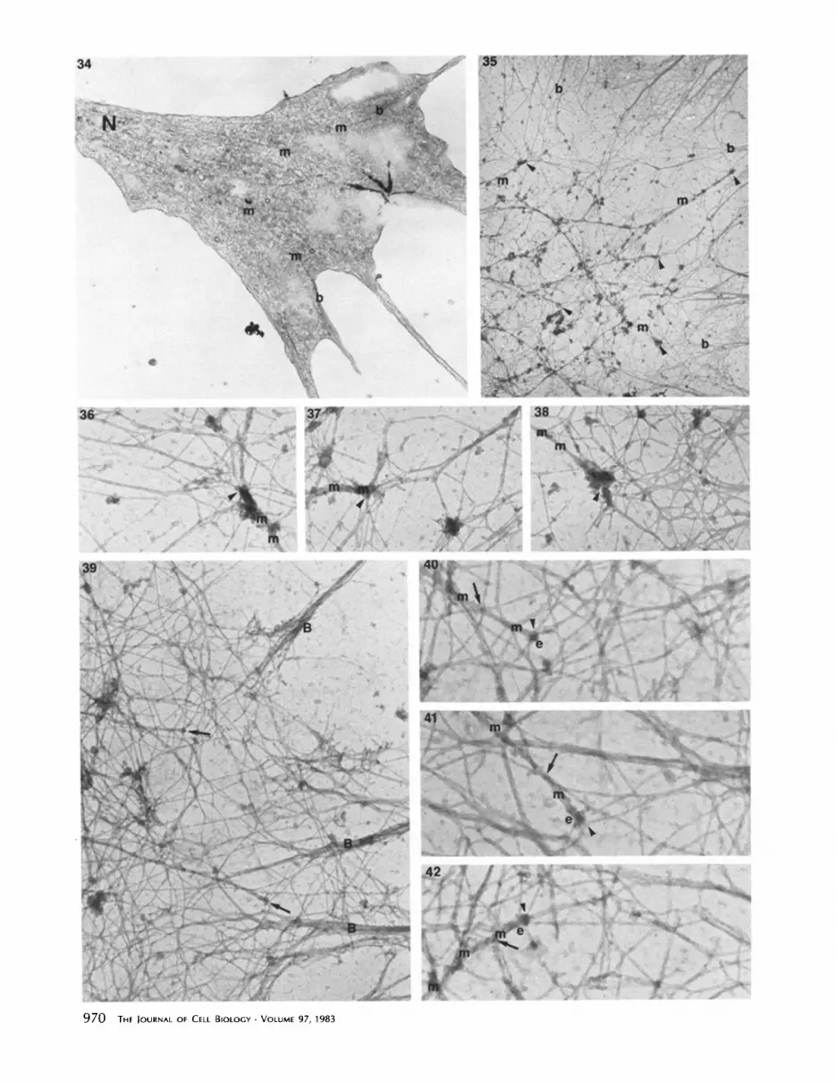
Fig. 35 shows a growth cone extracted in the presence of phallacidin. Several microtubules projected into the leading margin, and each microtubule end may be associated with several actin filaments. Complex associations among actin filaments could be traced across the gaps between the micro- tubule ends and actin filament bundles in the leading margin (Figs. 35-38). We cannot resolve how the actin filaments interacted with these microtubules. Some filaments crossed over the microtubules and may have collapsed onto them during extraction, while other filaments did seem to terminate on individual microtubules.
Fig. 39 shows the margin of a growth cone extracted before fixation in the presence of the S I fragment of myosin. Actin filaments can be recognized by S1 decoration, and several microtubules terminated near the leading margin. Actin fila- ments were seen associated with the microtubules, but the polarity of S 1 binding to these actin filaments could not be determined (Figs. 40-42). However, some of the actin fila- ments that interacted with the microtubule ends also could be seen associated with actin filament bundles projecting inward from filopodia (Fig. 39). As in the phallacidin-treated preparations, some filaments crossed over microtubules, but others may terminate on microtubules. The bulbous ends of microtubules in Figs. 40-42 were a common feature of mi- crotubules in whole-mounted growth cones. They occurred in growth cones extracted without SI (Figs. 17 and 18), and in growth cones fixed and extracted simultaneously. Bulbous ends were not always present, and they may be more difficult to see if present in unextracted specimens or in thin sections.
DISCUSSION
There was a major difference in the presence ofactin filaments between growth cones and neurites in detergent-treated cyto- skeletons of chick sensory neurons. This difference was estab- lished cytochemically with the light microscope and also by electron microscopic demonstrations of actin filaments. Be- fore we relate these data to previous studies of cytoskeletal fibers in growing neurites, we will discuss (a) what detergent treatment does to cells and (b) how actin may be affected by these procedures.
Detergents are used to make ceils permeable to agents that otherwise do not enter cells, and to separate or remove cell contents for morphological and biochemical study. There are several important cautions about extraction procedures.
Extraction before Fixation
The destruction of membrane barriers by TX-100 results in rapid loss of 80% of cell proteins from fibroblasts, but the remaining 20% is stable to further removal (8). However, the cytoskeleton may be altered by loss of accessory molecules,
by binding of extraneous molecules, or by distortions during the extraction period. For example, microtubules remain straight in cytoskeletons of neurites, as they are in thin sec- tions, but neurofilaments are more disordered in cytoskele- tons than they are in thin sections. The effects of phallacidin and myosin fragments show that the buffer can alter preser- vation of cytoskeletal fibers. Because of these cautions, we have examined actin distribution with three extraction meth- ods, thin sections of unextracted cells, and photography of the extraction of living neurites.
Fixation before or during Extraction
It is not surprising that fixation with glutaraldehyde should reduce cell extraction. The extraction of a structure in the presence of glutaraldehyde depends on the chemistry of fixa- tion, time, and temperature and concentration of fixative, and on the protein composition, concentration, and size of the structure. Simultaneous exposure to TX-100 and glutar- aldehyde must involve competition between protein cross- linking and dispersal of components following the removal of membrane barriers.
The most pronounced difference in cytoskeletal morphol- ogy of neurons fixed before vs. during extraction was that the irregular cytoplasmic lattice seen when fixation preceded ex- traction was absent from neurites and growth cones that were simultaneously fixed and extracted (compare Figs. 10-13 with 14-16). The nature of the lattice is unknown. The lattice material may have been naturally or artifactually deposited on the network of actin filaments in growth cones (Figs. 11, 12, and 15), but there was no actin network that could serve as scaffold for the lattice that is seen in neurites (Figs. 13 and 16). A similar absence of a lattice was found in detergent- extracted fibroblasts by Heuser and Kirschner (17).
Extraction of Actin
It is unclear whether all actin is preserved by these proce- dures. In nonmuscle cells, actin exists in several states (G- actin, oligomers of G-actin, and actin filaments), and each form may be fixed and extracted differently (5, 6, 47). G-actin is not cross-linked by the same concentration of glutaralde- hyde that forms very high molecular weight species from F- actin (27). Thus, G-actin or small polymers may have been lost from our neurons, even when fixation preceded extrac- tion. When extraction preceded fixation, some F-actin was lost from the growth cone margins (compare Figs. 14 and 17), but this F-actin and filopodia were preserved by including HMM, S l, or phallacidin in the extraction buffer. Because these agents can also promote actin polymerization (12, 50, 51), F-actin might have been polymerized in growth cones, but not in neurites, during extraction. However, this poly-
FIGURES 34-42 Fig. 34: Nearly complete section of the bottom of a growth cone. Several microtubules (m) can be traced from the distal neurite (N) to terminate near filament bundles (b) within filopodia. × 15,000. Fig. 35: Portion of growth cone margin marked A in Fig. 26. Several ends of microtubules (m) with associated actin filaments are indicated by arrowheads. Some of the filaments associated with microtubules can be traced to filament bundles (b) at the leading edge. x 13,000. Figs. 36-38: Three of the microtubule (m) ends denoted on Fig. 35. The precise contacts of actin filament to the microtubules and their termini (arrowheads) are not clear, but contacts may be made at several points, x 48,000. Fig. 39: Growth cone margin of a neurite treated with TX-100 and $1 fragment before fixation. A network and bundles (B) of decorated actin filaments are present, as are two microtubule termini (arrows). Thickened actin filaments decorated with $1 are associated with the microtubule ends. × 35,000. Fig. 40-42: The two microtubule (m) ends (e) denoted in Fig. 39, and another microtubule end from the same growth cone. Some thickened, decorated filaments appear to cross over the microtubule (arrows) but others may contact the microtubule (arrowheads). x 65,000.
LETOURNEAU Differences in Actin Organization in Cultured Neurons 971

merization would have to occur as G-actin and other soluble components disperse rapidly in the extraction buffer. Whether a form of F-actin is stable during extraction may depend on filament length, the presence of accessory proteins, and fila- ment associations with vesicles, microtubules, or the plasma membrane. For example, short actin filaments attached to the plasma membrane may be dispersed as TX-100 extracts membrane components.
Thus, the actin detected here was not necessarily all cellular actin, and we limit our conclusions to stating that growth cones contained a filamentous actin that could be stabilized by actin-binding agents and that was present in zones of cell surface motility. This form of actin was uncommon in neu- rites, except at points of lateral filopodia.
Relationship to Previous Studies of Actin in Growing Neurites
Actin has previously been reported in growth cones and neurites by immunofluorescence (19, 20, 23, 3 l, 34, 46), and some studies reported that actin is especially concentrated in growth cones (19, 31, 34, 46). We believe that the effects of fixatives and permeabilization on the retention of actin are responsible for some of the differences among reports of actin localization by immunofluorescence. For example, formal- dehyde is inferior to giutaraldehyde in preserving growth cone morphology for immunofluorescent localization of actin (31). In addition, Spooner and Holladay (46) have shown that high dilutions of antibody are critical to demonstrating the relative abundance of actin in growth cone margins.
E L E C T R O N M I C R O S C O P Y OF G R O W T H C O N E S : Anet- work or lattice of microfilaments in growth cone margins has often been described, but the presence of microfilament bun- dles in filopodia and similar areas has not always been dem- onstrated (9, 11, 23, 26, 30, 37, 54). This discrepancy in reporting filament bundles is probably due to cautious inter- pretations of filamentous structures that were altered during preparation (17, 27, 35, 37, 45). Although morphological studies of the binding of $1 or HMM identify the majority of filaments in the network and bundles as actin, the relationship between the network and the bundles is not clear. Actin filaments splay out at the bases of bundles and merge with the overlapping filaments of the network.
A C T I N IN C U L T U R E D N E U R I T E S : Yamada et al. (54) suggested that the lattice of microfilaments extends from the growth cone into the neurite and interconnects microtubules, neurofilaments, and vesicles in a continuous network. How- ever, these projections from microtubules and neurofilaments might contain a number of proteins (10, 13, 33, 38, 40) other than actin. And, as we found, HMM-decorated filaments in the neurite-like processes of neuroblastoma cells are predom- inantly at the cell periphery and in lateral filopodia, with few present centrally in neurites (11, 18).
This finding that few actin filaments are present in growing neurites can be compared with that for mature, nongrowing axons. Biochemical and morphological evidence suggests that the majority of subunits of microtubules and neurofilaments are polymerized into stable structures in axons (4, 2 l, 24, 25, 36). Although actin, too, is a major component of axons (4, 24, 52), the distribution and organization of actin is con- troversial. Ellisman and Porter (14) propose that a micro- trabecular lattice exists throughout the axon and contains actin filaments. However, in two studies of rapidly frozen axons (16, 42), actin filaments were identified only in the
972 THE Journa l OF CELL BIOLOGY • VOLUME 97, 1983
subplasmalemmal cortex, while in the regions containing neurofilaments and microtubules there is a granular and filamentous matrix that may have been preserved as micro- trabeculae by EUisman and Porter. Thus, readily identifiable actin filaments may be concentrated in cortical regions in both embryonic neurites and mature axons, but the relation- ship of these actin filaments to all axonal actin is unclear. Short filaments or polymers of actin may be associated closely with microtubules and neurofilaments. These may be difficult to demonstrate either morphologically or with low concentra- tions of antibodies (46). Thus, one should not assume that there are no actin filaments in the neuritic core, but that there may be a few that are short or difficult to see.
The actin organization in growth cones resembles that of the neurite periphery, though protrusion is suppressed along neurites. Yet, when a neurite is cut, active growth cones form at the cut ends (7, 43, 49). Transection may remove inhibiting factors or concentrate supportive factors that permit poly- merization of long actin filaments and sustained protrusive activity. An obvious similarity between a growth cone and a freshly cut neurite is the presence of many ends of microtu- bules and neurofilaments. Perhaps complex interactions among the monomeric and polymerized forms of all three types of cytoskeletal fibers regulate the expression of protru- sive activity.
Microtubules in Growth Cones and Interactions with Actin Filaments
Previous reports of electron microscopic work have stated that microtubules are present centrally or at the base of the growth cone, but they have rarely reported that microtubules enter growth cone margins or approach filopodia (9, 18, 23, 54). Two studies demonstrate immunofluorescence for tubu- lin in the margins of growth cones (44, 46), though electron microscopy was not done. The use of whole-mounted cells has permitted what might be stained with immunofluores- cence to be compared with ultrastructure. This is especially clear in detergent-extracted neurons, where microtubules are easily traced into the extracted peripheral margins of growth cones (Fig. 17). Our findings agree with immunofluorescence studies indicating that microtubules penetrate far forward into growth cone margins. Because microtubules may depolymer- ize during fixation and extraction, the location of microtubule ends in a growth cone may be influenced by the extraction of tubulin.
Cytoskeletons were examined for associations that mediate the alignment of actin bundles and nearby microtubules, but the resolution was poor. Osmium tetroxide, which disrupts actin, was not used, but distortions may have also been introduced by extraction, fixation, dehydration, and critical- point drying (17, 17, 35, 37, 45). Rapid freezing and drying might reduce distortions (37), but new problems arise, because growth cones are exquisitely sensitive to temperature change or mechanical displacement and shrink within seconds while being mounted on the freezing block. Our observations do suggest that actin filaments may terminate on microtubules at a point of contact (Figs. 40-42), or that actin filaments might contact microtubules elsewhere along the filaments' length. The organization of myosin and actin filaments in a sarcomere illustrates that cytoskeletal fibers can interact lat- erally without end-on contact.
In summary, a detergent-stable, filamentous actin is con- centrated in the growth cone margin, but is infrequent in

neurites. This actin is probably involved in motility of the growth cone margin, but use of this form of actin is highly regulated and suppressed in neurites. The small amount of this form of actin in neurites indicates that axonal transport may use another source of mechanical energy. Besides in- volvement in cell surface motility, this form of actin may interact with microtubules in the growth cone margin, and participate in the transmission of mechanical forces that determine the positions of structural components in the growth cone.
The author thanks Sherry Rogers for a critical reading of this manu- script.
This work was supported by grants from the Graduate School of the University of Minnesota, the Muscular Dystrophy Association, the Minnesota Medical Foundation, and Grant Nos. PCM-7923907 and PCM-8203855 from the National Science Foundation.
Received for publication 14 March 1983, and in revised form 26 May 1983.
REFERENCES
1. Barak, L. S., and R. R. Yocum. 1981. Nitrobenzoxazole-phallacidin: synthesis of a fluorescent actin probe. Anal Biochem 110:31-38.
2. Beg& D. A., R. Rodewald, and L. I. Rebhun. 1979. The visualization ofactin filament polarity in thin section. Evidence for the uniform polarity of membrane-associatad filaments. J. Cell Biol. 79:846-852.
3. Ben-Ze'ev, A., A. Duerr, F. Solomon, and S. Penman. 1979. The outer boundary of the cytoskeleton: a lamina derived from plasma membrane pro~'ins. Cell. 17:859-865.
4. Black, M. M., and R. J. l.a~k. 1980. Slow components of axonal transport: two cytoskeletal networks. J. CellBiol. 86:616-623.
5. Blikstad, I., and L. Carlsson. 1982. On the dynamics of the microfilament system in HeLa cells. J. Cell Biol. 93:122-128.
6. Bray, D., and C. Thomas. 1976. Unpolymerized actin in fibroblasts and brain. J. Mol. Biol. 105:527-544.
7. Bray, D., C. Thomas, and G. Shaw. 1978. Growth cone formation in cultures of sensory neurons. Proc. Natl. Acad. Sci. USA. 75:5226-5229.
8. Brown, S., W. Levinson, and J. A. Spudich. 1976. Cytoskeletal elements of chick embryo fibroblasts revealed by detergent extraction. J. Supramol. Struct. 5:119-130.
9. Bunge, M. B. 1973. Fine structure of nerve fibers and growth cones of isolated sympathetic neurons in culture. J. Cell Biol. 56:713-735.
10. Burton, P. R., and H. Fernandez. 1973. Delineation by lanthanum staining of filamen- tons elements assooatad with the surface of axonal microtubules. J. Celt Sci. 12:567- 583.
II. Chang, C.-M., and R. D. Goodman. 1973. The localization of actin-like fibers in cultured neuroblastoma cells as revealed by heavy meromyosin binding. J. Cell Biol. 57:867-874.
12. Cooke, R., and M. F. Morales. 1971. Interaction of globular actin with myosin subfrag- ments. Z Mot. Biol. 60:249-261.
13. Dentler, W. L., S. Granett, and J. L. Roseobaum. 1975. U l ~ c t a r a l localization of the high molecular weight proteins associated with in vitro-assembled brain microtu- bules. J. Cell Biol. 65:237-241.
14. Ellisman, M. H., and K. R. Porter. 1980. Microtrabocolar structure of the axoplasmic matrix: visualization of cro~-linking structures and their distribution. J. Cell Biol. 87:464-479.
15. Henderson, D., and K. Weber. 1979. Three dimensional organization of microfilaments and microtubules in the cytmkeleton. Exp. Cell Res. 124:301-316.
16. Hirokawa, N. 1982. Cross-linker system between neurofilaments, rnierotubules and membranous organelles in frog axons revealed by the quick-freeze, deep-etching method. Z CellBiol. 94:129-142.
17. Heuser, J. E., and M. W. Kirsehner. 1980. F'dament organization revealed in platinum replicas of freeze-dried cytoskelctnns. Z Cell Biol. 86:212-234.
18. lsenberg, G., and J. V. Small. 1978. Filamentous actin, 100-A filaments and microtu- boles in neuroblastoma cells, their distribution in relation to sites of movement and neuronal transport. Eur. J. Cell Biol. 16:326-344.
19. Isenberg, G., E. Rieske, and G. W. Kreutzberg. 1977. Distribution of actin and tubulin in neuroblastoma cells. Eur. J. Cell Biol. 15:382-389.
20. Joekusch, H., B. M. Jockosch, and M. M. Burger. 1979. Nerve fibers in culture and their interactions with non-neural cells visualized by immunofluorescence. J. Cell BioL 80:629-641.
20. Jockusch, H., B. M. Jockusch, and M. M. Burger. 1979. Nerve fibers in cultu~ and their interactions with non-neural cells visualized by immunofluore~ence. J. Cell Biol.
21. Kirschner, M. W. 1980. Implications of treadmilling for the stability and polarity of actin and tubulin polymers in vivo. J. Cell Biol. 86:330-334.
22. Kueamarski, E. R., and J. L. Rosenbaum. 1979. Chick brain actin and myosin. Isolation and characterization. Z Cell Biol. 80:341-355.
23. Kuczmarsld, E. R., and J. L. Ro~nbaum. 1979. Studies on the organization and localization of actin and myosin in neurons. J. Cell Biol. 80:356-371.
24. Lasek, R. J. 1982 Translooation of the neuronal cytoskeleton and axonal locomotion. Philos. Trans. R. Sac. Lond. B Biol. Sci. 299:313-327.
25. Lasek, R. J., and P. N. Hoffman. 1976. The neuronal cytoskeleton, axonal transport and axonal growth. Cold Spring Harbor Conf Cell Prolif 3(Book A): I02 I-I049.
26. LcBeux, Y. J., and J. Willemot. 1975. An ultrastroctural study of the microfilaments in rat brain by means of heavy meromyosin labehng. I. The perikaryon, the dendrites and theakon. Cell Tissue Res. 160:1-36.
27. Lehrer, S. S. 1981. Damage to actin filaments by glutaraldehyde: protection by tropo- myosin. J. Cell Biol. 90:459--466.
28. Lcnk, R., and S. Penman. 1979. The cytoskeletal framework and poliovirus metabolism. Cell. 16:289-301.
29. Lctourneau, P. C. 1975. Possible roles for cell-to-substratum adhesion in neuronal morphngenesis. Dee. Biol. 44:77-91.
30. Letourneau, P. 1979. Cell-substratum adhesion of neurite growth cones and its role in neurite elongation. Exp. Cell Res. 124:127-138.
31. Letourneau, P. C. 1981. Immunocytochemieal evidence for colocalization in neurite growth cones of actin and myosin and their relationship to cell-substratum adhesions. Dee. Biol. 85:113-122.
32. Letourneau, P. C. 1982. Analysis of microtubule numbor and length in eytoskeletons of cultured chick sensory neurons. J. Neurosci. 2:806-814.
33. Leterrier, J.-F., R. K. H. Liem, and M. L. Shelanski. 1982. Interactions between neurofilaments and microtubule-associatad proteins: a possible mechanism for intraor- ganellar bridging, J. Cell Biol. 95:982-986.
34. Marchisio, P. C., M. Osborn, and K. Weber. 1978. Changes in intracellular organization of mbulin and actin in N-18 neuroblastoma cells during the process of axon extension induced by serum deprivation. Brain Res. 155:229-237.
35. Maupin-Szamicr, P., and T. D. Pollard. 1978. Aetin filament destruction by osmium tetroxide. J. Ceil Biol. 77:837-852.
36. Morris, J. R., and R. J. Lasck. 1982. Stable polymers of the axonal cytnskeleton: the axoplasmic ghost. J. Cell Biol. 92:192-198.
37. Rces, R. P., and T. S. Reese. 1981. New structural features of freeze-substitutad neuritic growth cones. Neuroscience 6:247-254.
38. Rice, R. V., P. F. Roslansky, N. Pascoe, and S. M. Houghton. 1980. Bridges between microtubules and neurofilaments visualized by stereo electron microscopy. J. Ultrastruct. Res. 71:303-310.
39. Ris, H. 1981. Morphology of cyloplasmic filaments in thick sections and critical point dried whole mounts. J. Cell Biol. 91 (2, Pt. 2):305a. (Abstr.)
40. Runge, M. S., T. M. Lang, D. A. Yphantis, M. R. Lifsics, A. Saito, M. Alton, K. Reinke, and R. C. Williams. 1981. ATP-inducad formation of an assocmted complex between microtubules and neurofilaments. Pro¢. NatL Acad. Sci. USA. 78:1431-1435.
41. Schliwa, M., and J. van Blerkom. 1981. Structural interaction of cytoskeletal compo- nents. J. Cell Biol. 90:222-235.
42. Schnapp, B. J., and T. S. Reese. 1982. Cytoplasmic structure in rapid-frozen axons. J. Cell Biol. 94:667-679.
43. Shaw, G., and D. Bray. 1977. Movement and extension of isolated growth cones. Exp. Cell Res. 104:55-62.
44. Shaw, G., M. Osborn, and K. Weber. 1981. Arrangement of neurofilaments, microtu- boles and microfilament-associalcd proteins in cultured dorsal root ganglion ceils. Eur. J. Cell Biol. 24:20-27.
45. Small, J. V. 1981. Organization of actin in the leading edge of cultured cells: influence of osmium tetroxide and dehydratinn on the ultrastrncture of actin meshworks. J. Cell Biol. 91:695-705.
46. Spooner, B. S., and C. R. Hofiaday. 1981. Distribution of tubulin and actin in neurites and growth cones of differentiating nerve cells. Cell Motility 1:167-178.
47. Wang, Yu-Li, F. Lanni, P. L. McNeil, B. R. Ware, and D. L. Taylor. 1982. Mobility of cytoplasmic and membrane-associated actin in living cells. Proc. Natl. Acad. Sci. USA. 79:4660--4664.
48. Webster, R. E., D. Henderson, M. Osborn, and K. Weber. 1978. Three-dimensional electron microscopical visualization of the cyloskeleton of animal cells: immunoferritin identification of actin- and tubulin-containing structures. Proc. Natl. Acad. Sci. USA. 75:5511-5515.
49. Wessells, N. K., S. R. Johnson, and R. P. Nuttall. 1978. Axon initiation and growth cone regeneration in cultured motor neurons. Exp. Cell Res. 117:335-346.
50. Wieland, T. 1977. Modifications of actin by phallotoxios. Naturwissenschafien. 64:303- 309.
51. Wieland, T. H., and H. Fanlstich. 1978. Amatoxins, phallatoxins, phallolysin and antamanide: the biologically active components of poisonoos amanita mushrooms. CRC Crit. Rev. Biochem. 5:185-260.
52. Willard, M., M. Wiseman, J. Levine, and P. Skene. 1979. Axonal transport of actin in rabbit retinal ganglion cells. Z Cell BioL 81:581-591.
53. Wolosewiek, J. J., and K. R. Porter. 1979. Microtrahecular lattice of the cytoplasmic ground substance. J. CellBiol. 82:114--139.
54. Yamada, K. M., B. S. Spooner, and N. K. Wessells. 1971. Ultrastructure and function of growth cones and axons of cultured nerve cells. J. Cell Biol. 49:614-635.
LETOURNEAU Differences in Actin Organization in Cultured Neurons 973


