Embed Size (px)
Citation preview
1
Different evolutionary trajectories of European avian-
like and classical swine H1N1 influenza A viruses
Eleca J. Dunham1, Vivien G. Dugan1, Emilee K. Kaser1, Sarah E. Perkins2,
Ian H. Brown3, Edward C. Holmes2,4, and Jeffery K. Taubenberger1,*
1Laboratory of Infectious Diseases, National Institute of Allergy and Infectious Diseases,
National Institutes of Health, Bethesda, MD, USA
2Center for Infectious Disease Dynamics, Department of Biology, The Pennsylvania State
University, University Park, PA, USA
3Virology Department, Veterinary Laboratories Agency - Weybridge, Addlestone, Surrey, UK
4Fogarty International Center, National Institutes of Health, Bethesda, MD, USA
*Address correspondence to:
Jeffery K. Taubenberger, M.D., Ph.D.,
Laboratory of Infectious Diseases, National Institute of Allergy and Infectious Diseases
National Institutes of Health, 33 North Drive, Room 3E19A.2, MSC 3203
Bethesda, MD 20892-3203 USA
Tel. 1-301-443-5960; Fax. 1-301-480-5722
email: [email protected]
Copyright © 2009, American Society for Microbiology and/or the Listed Authors/Institutions. All Rights Reserved.J. Virol. doi:10.1128/JVI.02565-08 JVI Accepts, published online ahead of print on 18 March 2009
at UNIV OF CALIF SAN DIEG
O on April 28, 2009
jvi.asm.org
Downloaded from
2
Abstract. In 1979 a lineage of avian-like H1N1 influenza A virus emerged in European swine
populations independently from the ‘classical’ swine H1N1 virus lineage that had circulated in
pigs since the ‘Spanish’ influenza pandemic of 1918. To determine whether these two distinct
lineages of swine-adapted A/H1N1 viruses have evolved in similar ways, as might be expected
given their common host species and origin from avian-like A/H1N1 ancestors, we compared
patterns of nucleotide and amino acid change in whole genome sequences of both groups. An
analysis of nucleotide compositional bias across all 8 genomic segments for the two swine
lineages showed a clear lineage-specific bias, although a segment-specific effect was also
apparent. As such, there only appears to be a relatively weak host-specific selection pressure.
Strikingly, despite each lineage evolving in the same species host for decades, amino acid
analysis revealed little evidence for either parallel or convergent changes. These findings
suggest that although adaptation due to evolutionary lineages can be distinguished, there are
functional and structural constraints on all gene segments, and that the evolutionary trajectory of
each lineage of swine A/H1N1 virus has a strong historical contingency. Thus, in the context of
emergence of an influenza A virus strain via a host-switch event, it is difficult to predict what
specific polygenic changes are needed for mammalian adaptation.
at UNIV OF CALIF SAN DIEG
O on April 28, 2009
jvi.asm.org
Downloaded from
3
Introduction
Swine influenza A viruses (IAV) of the H1N1 subtype currently circulate as two distinct
lineages within North American and European swine populations [1,2]. While the first clinical
observations of swine IAV infection coincided with the 1918 human influenza pandemic [3-4,
reviewed in 5], the first North American swine IAV isolates were not obtained until 1930 [6].
Termed ‘classical swine’ A/H1N1, this genetically and antigenically stable viral lineage
presumably emerged by stable transfer of the human 1918 pandemic virus to swine [5,7], and
subsequently spread to swine in other parts of the world including Europe in 1976 [1,8].
Independently, a novel lineage of ‘avian-like’ H1N1 swine IAV emerged in Europe in 1979 that
essentially replaced classical swine IAV [1,9,10]. To date, this second lineage of swine IAV is
enzootic throughout swine-producing regions of Western Europe where it co-circulates with
swine IAVs of H3N2 and H1N2 subtypes [11]. All eight gene segments of the prototype H1N1
viruses of this lineage are thought to be derived from a closely related Eurasian avian influenza
A viruses by a stable host switch without reassortment, and this lineage is phylogenetically and
antigenically distinct from the classical swine H1N1 lineage [9,12-14].
The processes by which avian influenza A viruses (IAV) stably switch hosts and acquire
mutations that facilitate replication and efficient transmission in a new host species are
fundamental to understanding the ecology of these viruses, but also are of critical importance to
public health and veterinary preparedness. IAV from the genetically and antigenically divergent
avian reservoir pool have been associated with stable host switch events to novel host species
including humans, swine, domestic poultry, and horses [15-17]. The last three human influenza
pandemic viruses all contained two or more novel genes that were very similar to those found in
IAV of wild birds, derived either by reassortment with circulating human strains in formation of
the 1957 and 1968 pandemic viruses [18,19], or possibly by whole genome adaptation in the
case of the 1918 pandemic virus [20-23]. Other novel influenza viruses derived by stable host
switch from avian influenza viruses have also been recently isolated from pigs, including
at UNIV OF CALIF SAN DIEG
O on April 28, 2009
jvi.asm.org
Downloaded from
4
another independent introduction of A/H1N1 influenza viruses in China [24], A/H4N6 influenza
viruses in Canada [25], and most recently A/H2N3 influenza viruses in the United States [26].
Similarly, a stable lineage of A/H3N8 influenza virus emerged in dogs in the United States
following a host switch event without reassortment from the equine A/H3N8 lineage [27]. Given
the present concern that an avian influenza virus, especially the currently circulating lineages of
highly pathogenic avian H5N1, could initiate a new pandemic if the virus stably adapts to
humans is also a question of considerable biomedical importance [28]. Together, these
examples demonstrate that reassortment is not a pre-requisite for IAV emergence in novel
hosts.
Swine have been hypothesized to be the mixing vessel in which avian and human
influenza A viruses reassort, resulting in the emergence of novel human pandemic influenza
strains [1,29]. However, direct or experimental data linking swine as intermediaries in the
emergence of past pandemics is lacking. Swine are the only animals documented to be
susceptible to infection with avian, swine, and human origin IAV [1] and co-infections with both
avian and human IAV have been reported [13,30-33]. This has been attributed to the fact that
swine tracheal epithelium expresses both 2,3- (avian IAV preferred) and 2,6-(mammalian IAV
preferred) N-acetylneuraminic acid-galactose linked receptors [34], and it is believed that avian
IAV adapted to swine undergo a shift from 2-3 to 2-6 binding, a critical step required in the
adaptation of an avian virus to a human host [35]. A subset of amino acids that are invariant in
all avian hemagglutinin (HA) subtypes but vary in mammalian-adapted HAs have been identified
[36]. It is possible that this set of mutations (or a subset thereof) play important roles in the
adaptation of avian IAV to swine.
Whether common genetic changes are associated with the adaptation to specific host
species, such that they are predictive for future events, or if genetic changes are made up of
unique constellations of mutations that occur independently in each host switch event, is an
at UNIV OF CALIF SAN DIEG
O on April 28, 2009
jvi.asm.org
Downloaded from
5
important question. The process by which the 1918 A/H1N1 influenza pandemic virus emerged
and adapted both to humans and swine is not yet fully elucidated, although the virus is avian-
like in both its coding sequences [21,37] and nucleotide composition [22]. The European avian-
like swine A/H1N1 viruses emerged independently of the 1918 pandemic from an avian-like
source [9,10,13]. We therefore sought to compare changes that might be associated with
mammalian adaptation between these two swine H1N1 lineages.
To address whether the two swine H1N1 lineages were evolving in parallel, as might be
expected given their common host species, we examined patterns of base composition variation
to determine relative nucleotide usages. We examined in detail the amino acid sites that had
previously been reported as important for mammalian adaptation to determine whether these
mutations appeared as parallel genetic changes, and therefore always required for avian H1N1
IAV to adapt to pigs, or whether there is more flexibility in the adaptive process.
at UNIV OF CALIF SAN DIEG
O on April 28, 2009
jvi.asm.org
Downloaded from
6
Materials and Methods
Viral culture, viral cDNA amplification and sequencing
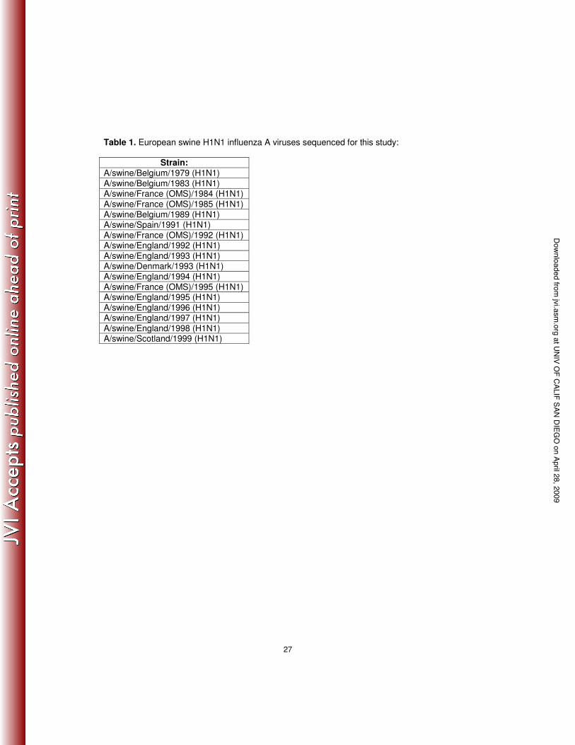
Seventeen viral isolates of European avian-like swine H1N1 IAV’s were selected for genomic
sequencing, as shown in Table 1, from the International Reference Laboratory at VLA,
Weybridge, UK. Viruses were propagated by inoculation into the allantoic cavities of 9–11-day-
old embryonated chicken eggs originating from a commercial specific-pathogen-free flock. Total
RNA was extracted from infected allantoic fluid using the QIAamp viral RNA kit (Qiagen,
Valencia CA) and first strand cDNA was reverse transcribed from viral RNA with the universal
influenza primer [38]. Subsequent PCR amplification of all influenza A virus segments was
performed using overlapping primer sets for each of the 8 segments using standard methods
(Phipps et al. 2004; Dugan et al. 2008). Sequences generated for this analysis have been
deposited in GenBank (Accession numbers XXXXXX-ZZZZZZZ).
Sequence analysis
In addition to the 17 European swine IAV genomes sequenced for this study, 3 European swine
avian-like H1N1 genomes, 38 classical swine H1N1 IAV genomes, other available swine H1N1
full-length gene sequences, and 81 human A/H1N1 genomes were downloaded from the
Influenza Virus Resource (http://www.ncbi.nlm.nih.gov/genomes/FLU/FLU.html) and/or
GenBank. The genome sequence of the 1918 pandemic IAV and representative Eurasian avian
sequences were used to infer the ancestry of the two swine lineages. Whole genome
sequences were not available for the Eurasian avian viral sequences analyzed. Therefore, the
NCBI BLAST database was used to find highly similar avian sequences for each European
swine influenza gene segment. Consensus sequences were derived from the Eurasian avian
IAV sequences for each segment. Within these data, we compiled separate manually aligned
gene segments (coding regions) using Se-Al {Rambaut, 1996 #2967). Sequence alignments
at UNIV OF CALIF SAN DIEG
O on April 28, 2009
jvi.asm.org
Downloaded from
7
consisted of the following coding regions for each segment: 197 PB2 (2,277 nt) sequences, 203
PB1 (2271 nt) sequences, 198 PA (2148 nt) sequences, 214 HA (1218 nt) sequences, 234 NP
(1494 nt) sequences, 231 NA (1410 nt) sequences, 193 M1/2 (1002 nt) sequences, and 224
NS1/2 (831 nt) sequences. Sequence alignments are available upon request.
Phylogenetic analysis
The best-fit GTR+I+4 model of nucleotide substitution was determined using ModelTest 3.7
{Posada, 1998 #2787}, and resulting parameter estimates were imported into PAUP* [39] to
create maximum likelihood (ML) trees through TBR branch-swapping (parameter values
available upon request). Whole genome sequences and a consensus Eurasian avian sequence
were concatenated (in the absence of reassortment – see below) to infer the evolutionary
relationship of swine H1N1 IAV. Individual gene segment phylogenetic trees are available in the
Supplementary Material.
To estimate the rates of evolutionary change and the Time to the Most Recent Common
Ancestor (TMRCA), we applied a Bayesian Markov Chain Monte Carlo (MCMC) approach
available in the BEAST package [40], employing a relaxed (uncorrelated lognormal) molecular
clock in all cases [41]. For each data set we utilized the Bayesian skyline coalescent prior (as
demographic history was a nuisance parameter in our analysis), with a 10% burn-in, and
assuming a GTR+I+4 model of nucleotide substitution. Uncertainty in parameter estimates is
reflected in the 95% highest probability density (HPD) values, and all chains were run for
sufficient length to ensure convergence as assessed using the TRACER program
(http://tree.bio.ed.ac.uk/software/tracer/). For the estimates of TMRCA, the most recent
sequence used for classical swine H1N1 was 1991 (A/swine/Maryland/23239) and 2004
(A/swine/Spain/53207/2004) for European swine H1N1. Selective pressures on codon sites
were estimated along the branches of the swine H1N1 phylogenetic trees for all 8 genes using
at UNIV OF CALIF SAN DIEG
O on April 28, 2009
jvi.asm.org
Downloaded from
8
Datamonkey (http://www.datamonkey.org/) [42]. The best-fit codon model is fitted to the data
using parameters obtained from the best-fit nucleotide substitution model. A one degree of
freedom likelihood ratio test is applied to the data to determine whether the instantaneous rate
of synonymous () and nonsynonymous () substitution differ, and whether this difference is
based on > (negative selection) or < (positive selection) and is significant.
Analysis of base composition
With the exception of the Eurasian avian sequences for which insufficient whole H1N1 genome
sequences were available, genome sequences were used to calculate base composition in all
lineages. A method similar to Shultes, et al. [43] was used to calculate the frequency of GU
(G+U), GA (G+A) and GC (G+C) across all eight gene segments for the European swine,
classical swine, human H1N1, Eurasian avian and the 1918 H1N1 IAV lineages. Unambiguous
calculations of the base compositional space of these IAV genes were defined by the following
three parameters: GU (frequency of G+ frequency of U), GA (frequency of G+ frequency of A)
and GC (frequency of G+ frequency of C) for each gene segment. Base composition
frequencies of complete and 1st and 2nd codons were calculated using the PAUP* package [39].
Base compositional data were then graphically plotted using the R version 2.7.0 statistical
program (2008). Third position codon GC content for each gene segment of swine IAV was
measured using the GCUA (General Codon Usage Analysis) package [44].
Amino acid analysis
Amino acid differences among the lineages were recorded as a change at an amino acid site
compared to the putative ancestral sequence. Given its location at the root of the human and
classical swine H1N1 clades, we used the 1918 Brevig Mission sequence as the ancestral
sequence to infer changes for classical swine and human H1N1 viruses. In the case of the
at UNIV OF CALIF SAN DIEG
O on April 28, 2009
jvi.asm.org
Downloaded from
9
European swine H1N1 viruses, we collected highly similar Eurasian avian sequences from
1977-1998 for each gene segment to infer amino acid changes in European swine H1N1 IAV.
at UNIV OF CALIF SAN DIEG
O on April 28, 2009
jvi.asm.org
Downloaded from
10
Results
Phylogenetic analysis of classical and European swine A/H1N1 sequences
Maximum likelihood trees resulted in the same phylogenetic relationship across all eight gene
segments between the two swine H1N1 IAV lineages (Supplementary Material), strongly
suggesting that they are each monophyletic, and with no major reassortment events. We
therefore concatenated the major open reading frames of all eight gene segments and inferred
an overall genomic ML tree (Figure 1). As expected, this revealed that the two swine lineages
formed distinct clades with the classical swine H1N1 IAV lineage derived from the 1918
pandemic influenza sequence, and the European swine H1N1 IAV lineage derived from the
Eurasian avian consensus sequence.
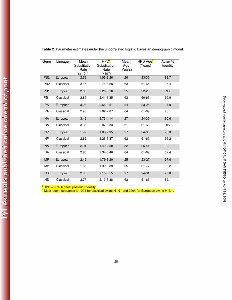
Our estimate of rates of nucleotide substitution gave similar values for each gene
segment in both swine lineages, ranging from 1.86 to 3.45 x 10⁻³ substitutions/site/year (Table
2), and broadly similar to those seen in other RNA viruses [45]. Under these substitution rates,
the TMRCA of the European swine A/H1N1 lineage was 24-32 years before the most recent
sample (95% HPD of 22-41 years), which indicates that the sampled isolates of European swine
A/H1N1 arose between the years 1963 and 1982 (Table 2). This is in strong accordance with
previous studies that first detected this lineage of H1N1 IAV in pigs in Northern Europe in 1979
[9,10]. The TMRCA of classical swine was estimated at 66-76 years before the most recent
sample (95% HPD of 64-76 years), which is also historically consistent with the first isolate of
H1N1 IAV in pigs in North America in 1930 [6].
Analysis of amino acid changes across lineages
Amino acid site differences were used to determine how frequently amino acid changes
occurred in parallel or convergently in the two swine lineages compared to the number that
diverged across these lineages (Supplementary Table 1). Overall, 23.5% of the changes were
at UNIV OF CALIF SAN DIEG
O on April 28, 2009
jvi.asm.org
Downloaded from
11
parallel genetic changes between the two swine lineages (Supplementary Table 1). Similarly,
there were only 6 (2.9%) convergent genetic changes (i.e., starting from a different ancestral
amino acid site) noted across all gene segments. Thus, the largest class of changes – 73.5% –
were those that experienced divergent evolution across all genes, indicating that the swine
A/H1N1 viral lineages have experienced strikingly dissimilar evolutionary trajectories
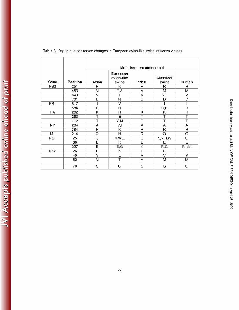
(Supplementary Table 1). Key amino acid changes in the internal gene segments that are
unique to European swine H1N1 influenza A viruses are listed in Table 3.
‘Internal’ gene changes: A series of 32 changes consistently associated with human influenza
viruses compared to avian IAV were identified by Finkelstein et al. [46]. Of the 10 changes
identified in PB2 by Finklestein et al., the European avian-like swine H1N1 IAV lineage only
shows a single parallel change, at residue 271 (Supplementary Table 1), in which the most
recent isolates differ from the avian IAV with a T271I (most human IAV have T271A but the
1918 virus has the avian 271T). In contrast, classical swine viruses maintain the human-
associated changes at residues 64, 199, 475, 627, and 702 (this residue reverted back to the
avian 702K after 1975). Of the 10 changes identified in PA, the European avian-like swine
H1N1 IAV lineage only shows a single parallel change, S409N in some isolates, which is also
observed in most classical swine isolates (Supplementary Table 1). In contrast, the classical
swine isolates also maintain the D55N change seen in 1918 and subsequent human IAV
strains. A total of nine changes have been proposed to be important in mammalian adaptation
in the NP protein [46,47]. The European avian-like swine H1N1 IAV lineage contains none of
these changes; however, a single strain, A/swine/England/1993 (H1N1), possesses the V33I
change observed in both human and classical swine IAV (Supplementary Table 1). Three
changes have been proposed for M1 [46], but the European avian-like swine IAV maintain the
avian consensus at all three. In contrast, the classical swine IAV lineage shares the T121A
change with human M1 (Supplementary Table 1). Four adaptive changes have been proposed
at UNIV OF CALIF SAN DIEG
O on April 28, 2009
jvi.asm.org
Downloaded from
12
for the M2 protein in the extracellular domain at residues 14, 16, 18, and 20 [48]. Most of the
European avian-like swine H1N1 strains share two of these changes, E16G and K18R, with
human and classical swine H1N1 IAV lineages. Classical swine strains also share the G14E
and S20N changes with human strains (Supplementary Table 1). It is also of note that a
number of the European avian-like swine strains contain mutations associated with resistance to
adamantane drugs [49], often with more than one resistance mutation at M2 residues 26, 27,
30, 31, and 34 within the ion channel domain. Three changes have been proposed for NS1
[46], and individual European avian-like swine strains bearing single changes at each of these
three residues are observed (residues 81, 215, and 227), but they are not the same mutations
seen in human strains and were not fixed in the swine lineage. Classical swine strains have the
avian 227E, but most European avian-like swine strains possess an E227G change
(Supplementary Table 1).
Changes in HA and NA: Classical swine strains maintain the critical HA receptor binding
domain mutation E190D (H3 numbering) as do the majority of European avian-like swine strains
(A/swine/Netherlands/3/80 retains the avian-consensus glutamic acid). Classical swine strains
possess the avian glycine at 225, whereas this receptor-binding domain residue is variable in
European avian-like swine strains, and includes the avian 225G, but also G225E and G225K
(Supplementary Table 1). Interestingly, European avian-like swine strains also show variability
in receptor binding residues 135, 137, and 138 unlike classical swine strains that maintain the
avian consensus at these sites. Thus, some European avian-like swine strains show
V135I,A,S,T; A137I,V; and A138S. We also found evidence of positive selection at residue 145
(Supplementary Table 1).
European avian-like swine strains also show changes in or near mapped antigenic site
regions in human H1 as previously reported [14]. Most of these changes are in or near the
mapped Ca and Sb antigenic regions [50,51]. These viruses also lose two potential N-linked
at UNIV OF CALIF SAN DIEG
O on April 28, 2009
jvi.asm.org
Downloaded from
13
glycosylation sites conserved in avian H1 sequences and the 1918 virus [52]. In more recent
strains, residues 104-106 NGT become NGA and in most European avian-like swine strains
residues 304-306 NSS become NSN. However, these strains gain two potential glycosylation
sites at residues 212-214 where ADA becomes NHT (in antigenic Sb region) and at residues
291-293 where NCD becomes NCT in most strains.
The neuraminidase (NA) of the European avian-like swine strains maintains the 15
conserved amino acids making up the active site of the enzyme [53] and no mutations
associated with neuraminidase inhibitor resistance are observed. The NA also maintains the
full-length stalk and the 7 potential N-linked glycosylation sites predicted for the 1918 influenza
virus [54]. Some European avian-like swine strains gain an additional potential glycosylation
site at residues 386-388, where SFS becomes NFS or NYS.
Analysis of Nucleotide Compositional Space of Individual Gene Segments
To investigate changes in base composition through time, an indicator of the evolutionary
processes that shape genetic diversity in influenza virus, the percentage GC content at the third
position codon of each gene segment was plotted over time (Figure 2). The directionality of
third position GC content change is measured as the percentage change over time from the
ancestral sequence.
The GC content of each of the eight gene segments for the classical swine and
European swine lineages tended to decrease over time from their ancestral sequences (1918
for classical swine; Eurasian avian for the European swine). As compared with the 1918
sequence, the change of third position base composition over time for classical swine and
human H1N1 in the NA gene is less marked than the other segments and the trajectory of the
HA and NP genes in the human H1N1 lineages shows an unexpected increase in GC content at
the third position over time (Figure 2). The GC content for the PB2 gene is similar across
lineages, suggesting functional and structural constraints on the gene segment.
at UNIV OF CALIF SAN DIEG
O on April 28, 2009
jvi.asm.org
Downloaded from
14
Some variation in nucleotide base compositional bias is expected for the eight gene
segments based on the different molecular functions of the gene products (which in turn effects
amino acid usage). If base compositional bias for the eight gene segments is universal, so that
all segments evolve in the same way irrespective of host, it is expected that no difference in the
clustering of these genes in compositional space across the different A/H1N1 lineages over time
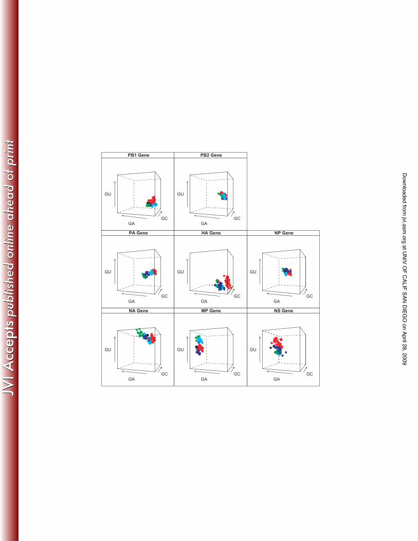
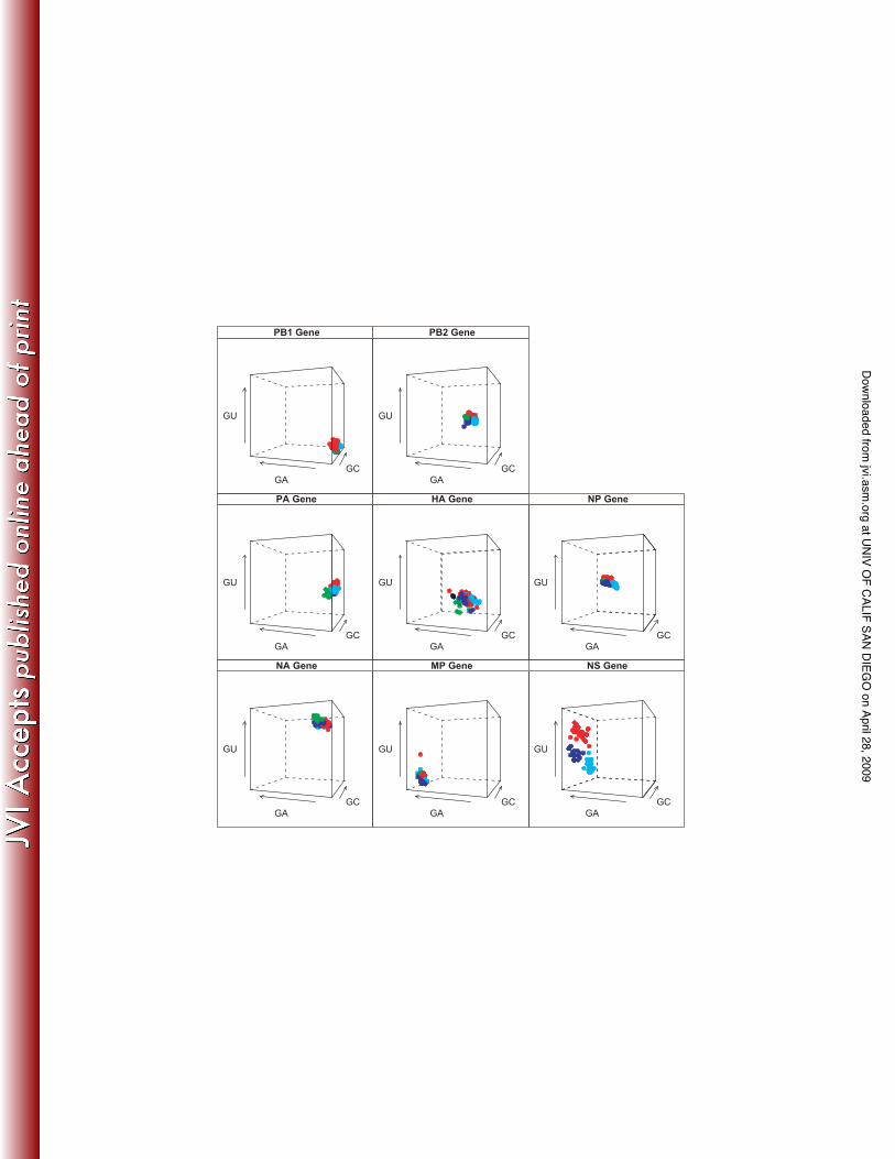
would be observed. Nucleotide compositional analysis revealed that each gene segment has a
unique clustering profile, revealing a powerful segment specific bias (Figure 3a). However, each
A/H1N1 lineage within this overall bias subdivides the clustering profile in space, indicating that
there is also a lineage-specific effect on gene composition. Furthermore, the 1918 sequence in
general occupies contiguous compositional space with classical swine and human A/H1N1
lineages, as would be expected since classical swine and human A/H1N1 viruses are direct
descendants of the 1918 virus. Similarly, the Eurasian avian and European swine A/H1N1
lineages occupy contiguous compositional space in this analysis. There is an overall lack of
overlap in compositional space clustering between the two swine A/H1N1 lineages across most
of the gene segments. The HA and the MP genes show a greater spread in compositional
space compared to the other gene segments. Analysis for the 1st and 2nd codons also revealed
a considerable difference in nucleotide composition between the two swine H1N1 lineages
(Figure 3b). The compositional space is partitioned by a unique compositional profile with very
little overlap. The Eurasian avian and European swine showed more overlap for the polymerase
genes. The NP and NS genes showed the most specific pattern for all lineages with little or no
overlap in compositional space. The HA gene segment profile shows the greatest spread, most
likely indicative of antigenic drift. The base compositional results for M2 and NS2 (NEP) are
shown in the Supplementary Material.
at UNIV OF CALIF SAN DIEG
O on April 28, 2009
jvi.asm.org
Downloaded from
15
Discussion
Our phylogenetic analysis supports the independent emergence of classical and European
swine H1N1 IAV, and the estimates for the TMRCA gave ranges of times of origin for both
swine A/H1N1 swine lineages similar to those previously reported [1,2,6]. Yet, within this
phylogenetic history, our analysis of whole genomes of swine H1N1 IAV revealed that the
lineages are experiencing largely divergent, rather than convergent or parallel, evolution; there
were approximately 3 times as many mutations producing divergent evolution than resulting in
homoplasy.
The third position base composition analysis revealed that each swine lineage is
diverging from its putative ancestor by generally decreasing in GC content at the third codon
position over time. The movement of human H1N1 to a higher GC content for the HA gene
implies that selection for antigenic differences may affect the trajectory of third position GC
content over time, although this will need to be explored in more detail. This trend is also
reflected in the NP gene, which shows an overlap in both of the swine lineages, suggesting that
both the HA and NP genes are highly host-specific. Although synonymous changes at the third
codon position can be partially attributed to neutral evolution [55-57], the similar patterns of
decreasing GC content over time for all of the gene segments again argue for host-specificity.
The nucleotide compositional analysis revealed several evolutionary patterns (Figures
3a-b). First, each gene segment has a distinctive ‘signature’ nucleotide compositional space
profile. This trend strongly suggests that there are functional and structural constraints acting
on each segment individually, which will clearly need to be explored further. However, within
these segment-specific profiles, the compositional space is also partitioned by a lineage effect
(Figure 3). This strongly signifies that natural selection has played a major role in shaping
nucleotide composition in IAV, reflective of the past history of each virus. Second, the wide
distribution of points in the HA gene shows a strong host-effect in compositional space, again
likely driven by antigenic drift (such that selection on amino acid changes has a secondary
at UNIV OF CALIF SAN DIEG
O on April 28, 2009
jvi.asm.org
Downloaded from
16
effect on nucleotide composition). Third, the swine lineages share the same compositional
space away from the human A/H1N1 and 1918 sequences, again supporting the idea that there
is strong selection for host-specific antigenic change. The 1st and 2nd codon compositional
analysis revealed a more distinct pattern by clade. The classical swine and human H1N1 show
very little overlap, indicating host-specific compositional bias (Figure 3b). The Eurasian avian
and European swine clades overlap for the polymerase genes. This suggests that although
European swine H1N1 emerged almost 30 years ago, the polymerase genes are still very
‘avian-like’. Interestingly, the NP gene shows the most host-specific pattern with no overlap
among the groups. However, the lack of overlap between the two swine H1N1 lineages
suggests that host-specificity is contingent upon the history of the IAV. Similar to Rabadan et al.
[22], we found that the 1918 virus is avian-like in the nucleotide composition of the gene
segments.
The biased nucleotide composition at the 1st and 2nd codon positions is reflective of
nonsynonymous changes in amino acid usage. In the context of a single host switch event, the
mutations identified in the 1918 influenza virus and subsequently maintained in human influenza
viruses and in classical swine strains may represent a set of crucial functional changes from an
ancestral avian IAV [46]. However, the lack of parallel evolution in the independent emergence
of the European avian-like swine strains suggests that the acquisition of a polygenic set of
functional changes may be different between independent host switch events. The utility of
identifying these mutations as proxies to define whether a future IAV is acquiring changes
important in mammalian adaptation might be limited. For example, of the 10 amino acid
changes identified in PB2 [21,46], only more recent European avian-like swine strains share a
single change from the avian consensus at one of these sites, residue 271, encoding a T271I.
Crucially, they lack the PB2 E627K change [58], even in those strains isolated after 20 years of
circulation in swine. Thus, this particular mutation may not be necessary for mammalian
adaptation in general, or at least swine adaptation in particular. However, European avian-like
at UNIV OF CALIF SAN DIEG
O on April 28, 2009
jvi.asm.org
Downloaded from
17
swine strains do possess a D701N change from the avian consensus that may also play a role
in mammalian adaptation [59], but lack the K702R associated with human PB2 genes and early
classical swine H1N1 strains [21]. Classical swine viruses after the mid 1970’s reverted to the
avian lysine at 702, but continued to possess the E627K. The D701N change was observed as
one of six changes after mouse adaptation of an avian H7N7 virus [59]. None of the other five
changes is observed in human, classical swine, or European avian-like swine lineages and may
be specific to this mouse adaptation experiment. The D701N mutation has also been observed
in a small minority of human H5N1 isolates, but has been linked to increased pathogenicity in
experimental mouse H5N1 infection model [60]. Recently the structure of the C-terminal end of
PB2 was resolved and structural analysis suggests that this region of PB2 contains a nuclear
localization signal and complexes with the importin 5 [61]. The structure suggests that the
changes observed at residues 701 and 702 may be important in the interaction with importin 5,
suggesting a biological explanation for changes at these sites associated with host switch
events.
In the H1 subtype, only a single amino acid change is required to alter receptor
specificity from 2-3 to gain the ability to bind 2-6, E190D (using the H3 numbering) [62]. The
1918 pandemic viruses possessed HA with two receptor-binding variants – either with a single
E190D change from the avian consensus, or two changes – E190D plus G225D [63]. The form
with two changes is highly specific for 2-6 [62,64]. Both the 1918 pandemic virus and its
derivatives and the European avian-like swine virus HAs have the E190D mutation crucial for
2-6 binding [62]. This indicates that H1 subtype HAs involved in switching from an avian host
to a mammalian host may need to acquire this particular mutation for stable host adaptation.
Other changes observed in the receptor binding domain of the European avian-like swine
viruses (at residues 135, 137,138 and 145) may also play a role in altering receptor specificity,
but this has not been evaluated experimentally.
at UNIV OF CALIF SAN DIEG
O on April 28, 2009
jvi.asm.org
Downloaded from
18
In summary, our study demonstrates that we should consider the role of historical
contingency, reflected in a strong lineage-specific effect, in the emergence of influenza A
viruses from an avian reservoir into a new mammalian host, and that mutations identified as
important in prior host switch events may or may not be observed in future such events. The
host switch events leading to the emergence of the European avian-like swine lineage from
birds, and the recent emergence of a canine H3N8 IAV lineage derived from equine H3N8
viruses [27] demonstrates that even in the absence of reassortment, stable host adaptation can
occur in influenza A viruses by acquisition of crucial mutations.
at UNIV OF CALIF SAN DIEG
O on April 28, 2009
jvi.asm.org
Downloaded from
19
Acknowledgments
This work was supported in part by the intramural program of the NIAID and the NIH, and by the
Alfred P. Sloan Foundation Graduate Scholarship and the NIH/INRO fellowship program.
at UNIV OF CALIF SAN DIEG
O on April 28, 2009
jvi.asm.org
Downloaded from
20
References
1. Brown IH (2000) The epidemiology and evolution of influenza viruses in pigs. Vet Microbiol
74: 29-46.
2. Olsen CW, Brown IH, Easterday BC, Van Reeth K (2006) Swine Influenza. In: Straw BE,
Zimmerman JJ, D'Allaire S, Taylor DJ, editors. Diseases of Swine. 9th ed. Aimes, Iowa:
Blackwell Publishing Professional. pp. 469-482.
3. Koen J (1919) A practical method for field diagnosis of swine diseases. . Am J Vet Med 14:
468-470.
4. Chun J (1919-1920) Influenza including its infection among pigs. . Nat Med J 5: 34-44.
5. Taubenberger JK, Reid AH, Janczewski TA, Fanning TG (2001) Integrating historical, clinical
and molecular genetic data in order to explain the origin and virulence of the 1918
Spanish influenza virus. Philos Trans R Soc Lond B Biol Sci 356: 1829-1839.
6. Shope RE (1931) Swine influenza. I. Experimental transmission and pathology. J Exp Med
54: 349-359.
7. Dorset M, McBryde CN, Niles WB (1922) Remarks on hog flu. Journal of the American
Veterinary Medical Association 62: 162-171.
8. Olsen CW (2002) The emergence of novel swine influenza viruses in North America. Virus
Res 85: 199-210.
9. Pensaert M, Ottis K, Vandeputte J, Kaplan MM, Bachmann PA (1981) Evidence for the
natural transmission of influenza A virus from wild ducts to swine and its potential
importance for man. Bull World Health Organ 59: 75-78.
10. Scholtissek C, Burger H, Bachmann PA, Hannoun C (1983) Genetic relatedness of
hemagglutinins of the H1 subtype of influenza A viruses isolated from swine and birds.
Virology 129: 521-523.
at UNIV OF CALIF SAN DIEG
O on April 28, 2009
jvi.asm.org
Downloaded from
21
11. Maldonado J, Van Reeth K, Riera P, Sitja M, Saubi N, et al. (2006) Evidence of the
concurrent circulation of H1N2, H1N1 and H3N2 influenza A viruses in densely
populated pig areas in Spain. Vet J 172: 377-381.
12. Donatelli I, Campitelli L, Castrucci MR, Ruggieri A, Sidoli L, et al. (1991) Detection of two
antigenic subpopulations of A(H1N1) influenza viruses from pigs: antigenic drift or
interspecies transmission? J Med Virol 34: 248-257.
13. Schultz U, Fitch WM, Ludwig S, Mandler J, Scholtissek C (1991) Evolution of pig influenza
viruses. Virology 183: 61-73.
14. Brown IH, Ludwig S, Olsen CW, Hannoun C, Scholtissek C, et al. (1997) Antigenic and
genetic analyses of H1N1 influenza A viruses from European pigs. J Gen Virol 78 ( Pt 3):
553-562.
15. Webster RG, Bean WJ, Gorman OT, Chambers TM, Kawaoka Y (1992) Evolution and
ecology of influenza A viruses. Microbiol Rev 56: 152-179.
16. Alexander DJ, Brown IH (2000) Recent zoonoses caused by influenza A viruses. Rev Sci
Tech 19: 197-225.
17. Swayne DE (2007) Understanding the complex pathobiology of high pathogenicity avian
influenza viruses in birds. Avian Dis 51: 242-249.
18. Scholtissek C, von Hoyningen V, Rott R (1978) Genetic relatedness between the new 1977
epidemic strains (H1N1) of influenza and human influenza strains isolated between 1947
and 1957 (H1N1). Virology 89: 613-617.
19. Kawaoka Y, Krauss S, Webster RG (1989) Avian-to-human transmission of the PB1 gene of
influenza A viruses in the 1957 and 1968 pandemics. J Virol 63: 4603-4608.
20. Reid AH, Taubenberger JK, Fanning TG (2004) Evidence of an absence: the genetic origins
of the 1918 pandemic influenza virus. Nat Rev Microbiol 2: 909-914.
21. Taubenberger JK, Reid AH, Lourens RM, Wang R, Jin G, et al. (2005) Characterization of
the 1918 influenza virus polymerase genes. Nature 437: 889-893.
at UNIV OF CALIF SAN DIEG
O on April 28, 2009
jvi.asm.org
Downloaded from
22
22. Rabadan R, Levine AJ, Robins H (2006) Comparison of avian and human influenza A
viruses reveals a mutational bias on the viral genomes. J Virol 80: 11887-11891.
23. Greenbaum BD, Levine AJ, Bhanot G, Rabadan R (2008) Patterns of evolution and host
gene mimicry in influenza and other RNA viruses. PLoS Pathog 4: e1000079.
24. Guan Y, Shortridge KF, Krauss S, Li PH, Kawaoka Y, et al. (1996) Emergence of avian
H1N1 influenza viruses in pigs in China. J Virol 70: 8041-8046.
25. Karasin AI, Schutten MM, Cooper LA, Smith CB, Subbarao K, et al. (2000) Genetic
characterization of H3N2 influenza viruses isolated from pigs in North America, 1977-
1999: evidence for wholly human and reassortant virus genotypes. Virus Res 68: 71-85.
26. Ma W, Vincent AL, Gramer MR, Brockwell CB, Lager KM, et al. (2007) Identification of
H2N3 influenza A viruses from swine in the United States. Proc Natl Acad Sci U S A
104: 20949-20954.
27. Crawford PC, Dubovi EJ, Castleman WL, Stephenson I, Gibbs EP, et al. (2005)
Transmission of equine influenza virus to dogs. Science 310: 482-485.
28. Webster RG, Govorkova EA (2006) H5N1 influenza--continuing evolution and spread. N
Engl J Med 355: 2174-2177.
29. Scholtissek C, Burger H, Kistner O, Shortridge KF (1985) The nucleoprotein as a possible
major factor in determining host specificity of influenza H3N2 viruses. Virology 147: 287-
294.
30. Hinshaw VS, Webster RG, Easterday BC, Bean WJ, Jr. (1981) Replication of avian
influenza A viruses in mammals. Infect Immun 34: 354-361.
31. Castrucci MR, Donatelli I, Sidoli L, Barigazzi G, Kawaoka Y, et al. (1993) Genetic
reassortment between avian and human influenza A viruses in Italian pigs. Virology 193:
503-506.
at UNIV OF CALIF SAN DIEG
O on April 28, 2009
jvi.asm.org
Downloaded from
23
32. Brown IH, Harris PA, McCauley JW, Alexander DJ (1998) Multiple genetic reassortment of
avian and human influenza A viruses in European pigs, resulting in the emergence of an
H1N2 virus of novel genotype. J Gen Virol 79 ( Pt 12): 2947-2955.
33. Zell R, Motzke S, Krumbholz A, Wutzler P, Herwig V, et al. (2008) Novel reassortant of
swine influenza H1N2 virus in Germany. J Gen Virol 89: 271-276.
34. Gambaryan AS, Karasin AI, Tuzikov AB, Chinarev AA, Pazynina GV, et al. (2005) Receptor-
binding properties of swine influenza viruses isolated and propagated in MDCK cells.
Virus Res 114: 15-22.
35. Stevens J, Blixt O, Glaser L, Taubenberger JK, Palese P, et al. (2006) Glycan microarray
analysis of the hemagglutinins from modern and pandemic influenza viruses reveals
different receptor specificities. J Mol Biol 355: 1143-1155.
36. Matrosovich MN, Gambaryan AS, Teneberg S, Piskarev VE, Yamnikova SS, et al. (1997)
Avian influenza A viruses differ from human viruses by recognition of
sialyloligosaccharides and gangliosides and by a higher conservation of the HA
receptor-binding site. Virology 233: 224-234.
37. Taubenberger JK, Morens DM (2006) 1918 Influenza: the mother of all pandemics. Emerg
Infect Dis 12: 15-22.
38. Hoffmann E, Stech J, Guan Y, Webster RG, Perez DR (2001) Universal primer set for the
full-length amplification of all influenza A viruses. Arch Virol 146: 2275-2289.
39. Swofford DL (2003) PAUP*. Phylogenetic Analysis Using Parsimony (*and other methods).
Version 4. ed. Sunderland, MA: Sinauer Associates.
40. Drummond AJ, Rambaut A (2003) BEAST. 1.3 ed.
41. Drummond AJ, Ho SY, Phillips MJ, Rambaut A (2006) Relaxed phylogenetics and dating
with confidence. PLoS Biol 4: e88.
42. Pond SL, Frost SD (2005) Datamonkey: rapid detection of selective pressure on individual
sites of codon alignments. Bioinformatics 21: 2531-2533.
at UNIV OF CALIF SAN DIEG
O on April 28, 2009
jvi.asm.org
Downloaded from
24
43. Schultes E, Hraber PT, LaBean TH (1997) Global similarities in nucleotide base composition
among disparate functional classes of single-stranded RNA imply adaptive evolutionary
convergence. Rna 3: 792-806.
44. McInerney JO (1998) GCUA: general codon usage analysis. Bioinformatics 14: 372-373.
45. Duffy S, Shackelton LA, Holmes EC (2008) Rates of evolutionary change in viruses:
patterns and determinants. Nat Rev Genet 9: 267-276.
46. Finkelstein DB, Mukatira S, Mehta PK, Obenauer JC, Su X, et al. (2007) Persistent host
markers in pandemic and H5N1 influenza viruses. J Virol 81: 10292-10299.
47. Reid AH, Fanning TG, Janczewski TA, Lourens RM, Taubenberger JK (2004) Novel origin
of the 1918 pandemic influenza virus nucleoprotein gene. J Virol 78: 12462-12470.
48. Reid AH, Fanning TG, Janczewski TA, McCall S, Taubenberger JK (2002) Characterization
of the 1918 "Spanish" influenza virus matrix gene segment. J Virol 76: 10717-10723.
49. Schmidtke M, Zell R, Bauer K, Krumbholz A, Schrader C, et al. (2006) Amantadine
resistance among porcine H1N1, H1N2, and H3N2 influenza A viruses isolated in
Germany between 1981 and 2001. Intervirology 49: 286-293.
50. Caton AJ, Brownlee GG, Yewdell JW, Gerhard W (1982) The antigenic structure of the
influenza virus A/PR/8/34 hemagglutinin (H1 subtype). Cell 31: 417-427.
51. Raymond FL, Caton AJ, Cox NJ, Kendal AP, Brownlee GG (1983) Antigenicity and evolution
amongst recent influenza viruses of H1N1 subtype. Nucleic Acids Res 11: 7191-7203.
52. Reid AH, Fanning TG, Hultin JV, Taubenberger JK (1999) Origin and evolution of the 1918
"Spanish" influenza virus hemagglutinin gene. Proc Natl Acad Sci U S A 96: 1651-1656.
53. Colman PM, Varghese JN, Laver WG (1983) Structure of the catalytic and antigenic sites in
influenza virus neuraminidase. Nature 303: 41-44.
54. Reid AH, Fanning TG, Janczewski TA, Taubenberger JK (2000) Characterization of the
1918 "Spanish" influenza virus neuraminidase gene. Proc Natl Acad Sci U S A 97: 6785-
6790.
at UNIV OF CALIF SAN DIEG
O on April 28, 2009
jvi.asm.org
Downloaded from
25
55. Yang Z, Nielsen R (2008) Mutation-selection models of codon substitution and their use to
estimate selective strengths on codon usage. Mol Biol Evol 25: 568-579.
56. McVean GAT, Charlesworth B (1999) A population genetic model for the evolution of
synonymous codon usage: patterns and predictions. Genetics Research 74: 145-158.
57. Kimura M (1983) The neutral theory of molecular evolution. Cambridge: Cambridge
University Press.
58. Subbarao EK, London W, Murphy BR (1993) A single amino acid in the PB2 gene of
influenza A virus is a determinant of host range. J Virol 67: 1761-1764.
59. Gabriel G, Dauber B, Wolff T, Planz O, Klenk HD, et al. (2005) The viral polymerase
mediates adaptation of an avian influenza virus to a mammalian host. Proc Natl Acad
Sci U S A 102: 18590-18595.
60. Li Z, Chen H, Jiao P, Deng G, Tian G, et al. (2005) Molecular basis of replication of duck
H5N1 influenza viruses in a mammalian mouse model. J Virol 79: 12058-12064.
61. Tarendeau F, Boudet J, Guilligay D, Mas PJ, Bougault CM, et al. (2007) Structure and
nuclear import function of the C-terminal domain of influenza virus polymerase PB2
subunit. Nat Struct Mol Biol 14: 229-233.
62. Stevens J, Blixt O, Tumpey TM, Taubenberger JK, Paulson JC, et al. (2006) Structure and
receptor specificity of the hemagglutinin from an H5N1 influenza virus. Science 312:
404-410.
63. Reid AH, Janczewski TA, Lourens RM, Elliot AJ, Daniels RS, et al. (2003) 1918 influenza
pandemic caused by highly conserved viruses with two receptor-binding variants. Emerg
Infect Dis 9: 1249-1253.
64. Srinivasan A, Viswanathan K, Raman R, Chandrasekaran A, Raguram S, et al. (2008)
Quantitative biochemical rationale for differences in transmissibility of 1918 pandemic
influenza A viruses. Proc Natl Acad Sci U S A 105: 2800-2805.
at UNIV OF CALIF SAN DIEG
O on April 28, 2009
jvi.asm.org
Downloaded from
27
Table 1. European swine H1N1 influenza A viruses sequenced for this study:
Strain: A/swine/Belgium/1979 (H1N1) A/swine/Belgium/1983 (H1N1) A/swine/France (OMS)/1984 (H1N1) A/swine/France (OMS)/1985 (H1N1) A/swine/Belgium/1989 (H1N1) A/swine/Spain/1991 (H1N1) A/swine/France (OMS)/1992 (H1N1) A/swine/England/1992 (H1N1) A/swine/England/1993 (H1N1) A/swine/Denmark/1993 (H1N1) A/swine/England/1994 (H1N1) A/swine/France (OMS)/1995 (H1N1) A/swine/England/1995 (H1N1) A/swine/England/1996 (H1N1) A/swine/England/1997 (H1N1) A/swine/England/1998 (H1N1) A/swine/Scotland/1999 (H1N1)
at UNIV OF CALIF SAN DIEG
O on April 28, 2009
jvi.asm.org
Downloaded from
28
Table 2. Parameter estimates under the uncorrelated logistic Bayesian demographic model.
Gene Lineage Mean
Substitution Rate
(x 10-3)
HPDa Substitution
Rate (x10-3)
Mean Age
(Years)
HPD Ageb (Years)
Avian % Identity
PB2 European 2.56 1.95-3.26 26 23-30 98.7
PB2 Classical 3.15 2.71-3.58
63
61-65
96.4
PB1 European 2.66 2.20-3.10 25 22-28 98
PB1 Classical 2.89
2.41-3.35
62
60-68
95.9
PA European 3.08 2.66-3.51 24 23-25 97.9
PA Classical 2.45
2.02-2.87
64
61-69
95.1
HA European 3.45 2.73-4.14 27 24-30 90.6
HA Classical 3.33
2.87-3.83
61
61-63
86
NP European 1.99 1.63-2.35 27 24-30 96.8
NP Classical 2.82
2.28-3.37
62
61-66
96.2
NA European 2.01 1.49-2.50 32 25-41 92.1
NA Classical 2.90
2.34-3.46
64
61-69
87.4
MP European 2.49 1.79-3.20 25 23-27 97.6
MP Classical 1.86
1.30-2.39
65
61-77
99.2
NS European 2.80 2.10-3.55 27 24-31 93.8
NS Classical 2.77
2.13-3.38
63
61-66
89.1
a HPD = 95% highest posterior density. b Most recent sequence is 1991 for classical swine H1N1 and 2004 for European swine H1N1.
at UNIV OF CALIF SAN DIEG
O on April 28, 2009
jvi.asm.org
Downloaded from
29
Table 3. Key unique conserved changes in European avian-like swine influenza viruses.
Most frequent amino acid
Gene Position Avian
European avian-like
swine
1918
Classical
swine
Human PB2 251 R K R R R
483 M T,A M M M 649 V I V V,I V 701 D N D D D
PB1 517 I V I I I 584 R H R R,H R
PA 262 K R K K K 263 T E T T T 712 T V,M T T T
NP 284 A V,I A A A 384 R K R R R
M1 214 Q H Q Q Q NS1 25 Q R,W,L Q K,N,R,W Q
66 E K E E E 227 E E,G K R,G R, del
NS2 26 E K E E E 49 V L V V V 52 M T M M M
70 S G S G G
at UNIV OF CALIF SAN DIEG
O on April 28, 2009
jvi.asm.org
Downloaded from
30
Figure Legends
Figure 1. Maximum likelihood tree of concatenated genome sequences (54 whole genomes) of
European and classical swine H1N1 influenza A virus. Horizontal branch lengths are drawn to a
scale of nucleotide substitutions per site. Bootstrap values (>75%) are shown next to the
relevant nodes. The tree is mid-point rooted for clarity only. Classical swine H1N1 is in blue and
European swine H1N1 is in red.
Figure 2. Synonymous third codon position G+C content over time for all 8 genes across
European and classical swine, Eurasian avian, 1918, and human H1N1 influenza A viruses.
Classical swine H1N1 is in red, European swine H1N1 in blue, Eurasian avian sequences in
green, human H1N1 in light blue, and 1918 H1N1 in black.
Figure 3a. Overall Nucleotide composition of 8 gene segments by H1N1 influenza A lineages.
Axes correspond to the frequency of G+U, G+A and G+C for each gene segment. Classical
swine H1N1 is in red, European swine H1N1 in blue, Eurasian avian sequences in green,
human H1N1 in light blue, and 1918 H1N1 in black.
Figure 3b. Nucleotide composition of 8 gene segments at the 1st and 2nd codon positions. Axes
correspond to the frequency of G+U, G+A and G+C for each gene segment. Classical swine
H1N1 is in red, European swine H1N1 in blue, Eurasian avian sequences in green, human
H1N1 in light blue, and 1918 H1N1 in black.
at UNIV OF CALIF SAN DIEG
O on April 28, 2009
jvi.asm.org
Downloaded from
100
100
100
100
100
100
100
98
97
98
at UNIV OF CALIF SAN DIEG
O on April 28, 2009
jvi.asm.org
Downloaded from
Year
3rd
po
sit
ion
GC
co
nte
nt
0.30
0.35
0.40
0.45
0.50
1920 1940 1960 1980 2000
HA Gene
MP Gene
1920 1940 1960 1980 2000
NA Gene
NP Gene
NS Gene
0.30
0.35
0.40
0.45
0.50
PA Gene
0.30
0.35
0.40
0.45
0.50
PB1 Gene
1920 1940 1960 1980 2000
PB2 Gene
at UNIV OF CALIF SAN DIEG
O on April 28, 2009
jvi.asm.org
Downloaded from
GC
GA
GU
NA Gene
GC
GA
GU
MP Gene
GC
GA
GU
NS Gene
GC
GA
GU
PA Gene
GC
GA
GU
HA Gene
GC
GA
GU
NP Gene
GC
GA
GU
PB1 Gene
GC
GA
GU
PB2 Gene
at UNIV OF CALIF SAN DIEG
O on April 28, 2009
jvi.asm.org
Downloaded from
GC
GA
GU
NA Gene
GC
GA
GU
MP Gene
GC
GA
GU
NS Gene
GC
GA
GU
PA Gene
GC
GA
GU
HA Gene
GC
GA
GU
NP Gene
GC
GA
GU
PB1 Gene
GC
GA
GU
PB2 Gene
at UNIV OF CALIF SAN DIEG
O on April 28, 2009
jvi.asm.org
Downloaded from


