Embed Size (px)
Citation preview

Differential distribution of the KCl cotransporter KCC2 inthalamic relay and reticular nuclei
P. Barthó1, J. A. Payne2, T. F. Freund1, and L. Acsády1
1Institute of Experimental Medicine, Hungarian Academy of Sciences, 1083 Budapest, Szigony u.43, Hungary2Department of Physiology and Membrane Biology, School of Medicine, University of California,Davis, CA, USA
AbstractIn the thalamus of the rat the reversal potential of GABA-induced anion currents is more negativein relay cells than in neurones of the reticular nucleus (nRt) due to different chloride extrusionmechanisms operating in these cells. The distribution of KCl cotransporter type 2 (KCC2), themajor neuronal chloride transporter that may underlie this effect, is unknown in the thalamus. Inthis study the precise regional and ultrastructural localization of KCC2 was examined in thethalamus using immunocytochemical methods. The neuropil of all relay nuclei was found todisplay intense KCC2 immunostaining to varying degrees. In sharp contrast, the majority of thenRt was negative for KCC2. In the anterior and dorsal part of the nRt, however, KCC2immunostaining was similar to relay nuclei and parvalbumin and calretinin were found tocolocalize with KCC2. At the ultrastructural level, KCC2 immunoreactivity was mainly located inthe extrasynaptic membranes of thick and thin dendrites and the somata of relay cells but was alsofound in close association with asymmetrical synapses formed by cortical afferents. Quantitativeevaluation of KCC2 distribution at the electron microscopic level demonstrated that the density ofKCC2 did not correlate with dendritic diameter or synaptic coverage but is 1.7 times higher onperisynaptic membrane surfaces than on extrasynaptic membranes. Our data demonstrate that theregional distribution of KCC2 is compatible with the difference in GABA-A reversal potentialbetween relay and reticular nuclei. At the ultrastructural level, abundant extrasynaptic KCC2expression will probably play a role in the regulation of extrasynaptic GABA-A receptor-mediatedinhibition.
KeywordsGABA; KCl cotransporter; rat; reticular nucleus; thalamus
IntroductionThe polarity of the response to the major inhibitory transmitter GABA in the central nervoussystem critically depends on the intracellular chloride concentration (Sivilotti & Nistri,1991). Ligand-gated GABA-A receptors are chloride channels whose activation induceschloride flux through the plasma membrane. The direction of the chloride movement,however, depends on the chloride equilibrium potential (ECl). In most cell types this is keptslightly below the resting membrane potential, thus activation of GABA-A receptors results
© 2004 Federation of European Neuroscience Societies
Correspondence: Dr László Acsády, as above. E-mail: [email protected].
Europe PMC Funders GroupAuthor ManuscriptEur J Neurosci. Author manuscript; available in PMC 2009 January 26.
Published in final edited form as:Eur J Neurosci. 2004 August ; 20(4): 965–975. doi:10.1111/j.1460-9568.2004.03562.x.
Europe PM
C Funders A
uthor Manuscripts
Europe PM
C Funders A
uthor Manuscripts

in a chloride influx from the extracellular space into the neurone inducing membranehyperpolarization (Kaila, 1994). However, in a number of cell types or during ontogenesisECl equals or is more depolarized than the resting membrane potential which results inshunting or depolarizing GABA-A responses (Rivera et al., 1999; Vardi et al., 2000; Gulacsiet al., 2003).
Inhibitory synaptic activity plays a pivotal role in shaping normal and pathophysiologicalrhythms in the thalamus. The firing mode of the two main cell types, excitatorythalamocortical relay cells and inhibitory reticular cells, is greatly influenced by the polarityof GABA-A responses (Bazhenov et al., 1999). Interestingly, different chloride transportmechanisms seem to operate in neurones of the relay and the reticular nuclei. Activechloride extrusion in relay cells sets the reversal potential of the GABA-A current 10 mVmore hyperpolarized than in reticular cells, which results in hyperpolarizing and shuntingGABA-A responses, respectively (Ulrich & Huguenard, 1997). Cation-chloridecotransporters are good candidates to account for the differential chloride distribution inrelay and reticular cells as they participate in the active transport of chloride ions into or outof the cell (Payne et al., 2003). At present, there are seven types of cation-chloridecotransporters in mammals (Delpire, 2000), four of which are KCl cotransporters (KCCs)(Gillen et al., 1996). Of the four KCC isoforms, the neurone-specific KCC2 (Payne et al.,1996; Lu et al., 1999) seems to be essential for maintaining the transmembrane chloridegradient that underlies hyperpolarizing inhibitory postsynaptic potentials (Rivera et al.,1999). KCC2 expression has been shown to correlate with neuronal maturation and changein polarity of the GABA-A reversal potential (Rivera et al., 1999; Stein et al., 2004). Indeed,in early postnatal life, when the KCC2 expression level in the hippocampus is low, GABA-A responses are depolarizing (Ben-Ari et al., 1989) and, in KCC2 knockout mice, GABA isexcitatory in spinal cord motoneurones (Hubner et al., 2001). Ion transporters also regulatethe concentration of osmolytes in the intra- and extracellular compartments, implying a rolein the control of cellular volume in the normal condition and during pathological states ofextreme synaptic activity (Reid et al., 2001; Woo et al., 2002; Payne et al., 2003). In thehippocampus, KCC2 has been shown to be located preferentially in pyramidal cell spines inthe proximity of excitatory synapses (Gulyas et al., 2001), suggesting a role in buffering theosmotic load accompanying excitatory synaptic activity. However, it was not clear whetherincreased KCC2 density in the spines was due to the small size of the compartment (i.e.relatively large osmolytic load) or to the proximity of the synapse. The distribution of KCC2in the thalamus is unknown at present. In this study, using highly selectiveimmunocytochemical methods, we first examined whether the distribution of KCC2 in thethalamus can explain the observed difference in chloride gradients in reticular and relaycells. Next, using quantitative immunogold methods, we also examined the preciselocalization of KCC2 in various dendritic compartments and in relation to excitatorysynapses.
Materials and methodsPerfusion and preparation of tissue section
Eighteen male Wistar rats (Charles River, Hungary) were used for KCC2immunocytochemistry. All experimental procedures were performed according to the ethicalguidelines of the Institute of Experimental Medicine, Hungarian Academy of Sciences andapproved by the Ethical Committee. Rats were deeply anaesthetized by Equithesin(chlornembutal, 0.3 mL/100 g), then perfused through the heart first with physiologicalsaline for 2 min and then with either of the following two types of fixative. Fixative A (n =10) contained 4% paraformaldehyde (TAAB, UK), 15% picric acid and 0.05%glutaraldehyde (TAAB) in phosphate buffer. In the case of fixative B (n = 8), the saline wasfollowed first by 100 mL 2% paraformaldehyde and 3.6% acrolein (Sigma, St Louis, MO,
Barthó et al. Page 2
Eur J Neurosci. Author manuscript; available in PMC 2009 January 26.
Europe PM
C Funders A
uthor Manuscripts
Europe PM
C Funders A
uthor Manuscripts

USA) in phosphate buffer and then by 300 mL 2% paraformaldehyde. Following perfusions,coronal, horizontal or parasagittal sections (60 μm thick) were cut from the thalamus on aVibratome, washed, cryoprotected in 30% sucrose in 0.1 M phosphate buffer overnight andfreeze-thawed in an aluminium foil boat over liquid nitrogen.
ImmunocytochemistryAfter extensive washes in Tris-buffered saline (TBS, pH 7.4), sections were incubated in 3%bovine serum albumin (Sigma) for 45 min and then with rabbit anti-KCC2 antibody (1 :300-500; Williams et al., 1999) for 2 days. For 3,3′-diaminobenzidine (DAB)immunostaining, sections were incubated in goat anti-rabbit antiserum (1 : 300; VectorLaboratories, Burlingame, CA, USA) for 2 h followed by avidin-biotinylated-horseradishperoxidase complex (ABC, 1 : 300; Vector Laboratories) for 2 h. Between each step sectionswere washed in TBS three times for 10 min. The specificity of the antibodies was studiedextensively by the laboratories of origin. The immunoperoxidase reaction was developedwith DAB as a chromogen. For pre-embedding immunogold staining following the primaryantibody, the sections were incubated in 1 nm gold-conjugated goat anti-rabbit antiserum(1 : 60; Amersham, UK) dissolved in TBS containing 0.8% bovine serum albumin and 0.1%gelatin overnight, postfixed in 1% glutaraldehyde in TBS and then silver intensified with anR-Gent intensification kit (Aurion, Wageningen, the Netherlands). All sections were treatedwith OsO4 (1% for 45 min for DAB staining and 0.5% for 30 min at 4 °C for immunogoldstaining in 0.1 M phosphate buffer), dehydrated in ethanol and propylene oxide andembedded in Durcupan (ACM, Fluka, Buchs, Switzerland). During dehydration, the sectionswere treated with 1% uranyl acetate in 70% ethanol for 40 min. Selected blocks containingidentified thalamic nuclei were re-embedded and 65-nm-thick ultrathin sections were cutwith an Ultramicrotome (Reichert), mounted on grids and examined with an electronmicroscope (7100; Hitachi). Digital images were collected using a GATAN camera. Forfluorescent double immunostainings, the combinations of rabbit anti-KCC2 antiserum andmouse anti-parvalbumin (1 : 500; Swant, Bellinzona, Switzerland) or mouse anti-calretinin(1 : 1500; Swant) were used. The second layer was Alexa 594-conjugated donkey anti-rabbit(1 : 200; Molecular Probes) and fluoroscein isothiocyanate-conjugated donkey anti-mouse(1 : 50; Jackson). Following washes in TBS the sections were mounted and covered byVectashield. The sections were evaluated using a fluorescent microscope (Axioscope; Zeiss)using the following filter sets: fluoroscein isothiocyanate, 450-490/512-542 and Alexa 594,546 ± 12/590 LP (absorption and emission in nm). Light microscopic images were scannedwith a digital camera (DP 70; Olympus). Brightness and contrast were adjusted, ifnecessary, using Adobe Photoshop 7.0.
Quantitative electron microscopic analysisThe relative KCC2 density across different subcellular compartments was estimated in 151dendrites and 12 somata randomly selected from the electron micrographs. The dendriteswere subdivided into five categories according to their minor diameter (< 0.4, 0.4-0.6,0.6-0.8, 0.8-1.0 and > 1.0 μm smaller diameter). The perimeter and area were measured withan NIH Image Analyzer and the number of gold particles was counted for each profile. Forthe calculation of density, the perimeter and surface area data were pooled within givendendritic categories and were divided by the total number of gold particles belonging to thiscategory (for this reason we have not put error bars on the figures). Pooling was preferred totaking the gold particle: perimeter ratio for each profile and averaging because the numberof profiles containing zero gold particles was higher in the thin than in the thick dendritesand this would have biased the grand average.
Barthó et al. Page 3
Eur J Neurosci. Author manuscript; available in PMC 2009 January 26.
Europe PM
C Funders A
uthor Manuscripts
Europe PM
C Funders A
uthor Manuscripts

In the electron micrographs, the synaptic area was calculated using a polygon made up of aseries of trapezoids, each 0.065 μm high (the thickness of the electron microscopic section),using the following formula
where lfirst and lsecond ... llast refer to the length of the synapse in the first and second ... lastsection of the series containing the given synapse.
Perisynaptic areas were defined as an annulus surrounding a synapse by 200 nm, composedof two rectangles on the top and bottom of the synapse, four parallelograms on the sides ofthe synapse and four quarters of a cycle connecting the rectangles and the parallelograms.For qualitative purposes this area was approximated using the following equation
The first two parts of the equation refer to the area of the rectangle, the second part refers tothe parallelograms, where n is the number of sections containing the synapse, and the lastpart is the sum of the four quarters of the circle. If the synapses continued outside or endedcloser than 200 nm to the end of the reconstructed sample, the outlying area was notconsidered. No overlapping of perisynaptic areas occurred in our sample and thus no goldparticle was assigned to two synapses. The parameters of the dendrites were measured usingNIH Image Analyzer. After calculation of peri- and extrasynaptic areas, the number of goldparticles in each area was calculated, the data pooled over all dendrites and the ratio taken.
ResultsRegional distribution
At the light microscopic level dense to moderate KCC2 immunostaining was seen in allthalamic nuclei except the reticular nucleus (nRt) (Fig. 1). No qualitative difference wasobserved between the different fixatives but, in general, animals perfused with fixative B(containing acrolein, see Materials and methods) showed stronger immunostaining.
The KCC2 immunoreactivity was prominent in the primary sensory relay nuclei (first ordernuclei), being strongest in the ventral posterolateral and ventral posteromedial nuclei (Fig.1A). Higher order (associational) nuclei (Guillery & Sherman, 2002) of the same sensorymodalities always expressed slightly less dense immunoreactivity than their first ordercounterpart, i.e. immunostaining in the somatosensory ventral posterolateral and ventralposteromedial nuclei was more dense than in the nucleus posterior (Fig. 1A). The visual,dorsal lateral geniculate nucleus displayed stronger KCC2 immunoreactivity than the higherorder lateral posterior nucleus (Fig. 1B) and KCC2 expression in the ventral part of theauditory medial geniculate nucleus was stronger than in the dorsal part of the medialgeniculate. KCC2 immunoreactivity in the ventral motor nuclei (including ventromedial,ventrolateral and ventral anterior) was similar to first order sensory nuclei. The intralaminarand the midline nuclear group showed stronger immunostaining than the adjacentmediodorsal nucleus and posterior thalamic nucleus. The most prominent expression ofKCC2 among the midline nuclei was found in the reuniens and rhomboid nuclei (Fig. 1A).The anterior nuclear group showed weak immunostaining.
Other diencephalic sites, including zona incerta, ventral lateral geniculate nucleus andanterior pretectal nucleus, expressed prominent KCC2 immunostaining that was similar or
Barthó et al. Page 4
Eur J Neurosci. Author manuscript; available in PMC 2009 January 26.
Europe PM
C Funders A
uthor Manuscripts
Europe PM
C Funders A
uthor Manuscripts

even stronger than the densest thalamic immunoreactivity observed in the ventralposterolateral nucleus and ventral posteromedial nucleus (Fig. 1B).
At higher power KCC2 immunoreactivity showed homogeneous neuropil staining in allnuclei. The staining had a punctate appearance in both immunogold- and DAB-reactedmaterial. In good quality DAB immunostainings individual somata and proximal dendritescould be distinguished. In these cases the labelling was clearly localized to the plasmamembranes.
Reticular nucleusThe most conspicuous aspect of KCC2 immunostaining in the thalamus was the lack ofimmunoreactivity in the majority of the nRt. In contrast, the dorsal ‘head’ region and theanterior part of the nRt displayed KCC2 immunoreactivity similar to the adjacent relaynuclei (Figs 1 and 3).
Examination of thalamic sections cut in coronal (Fig. 1A), horizontal (Fig. 1C) orparasagittal planes revealed that the KCC2-positive part of the nRt comprised the dorsal partof the nucleus throughout its anteroposterior extent. The anterior nRt, which envelops theanterior surface of the dorsal thalamus, was positive for KCC2 throughout its dorsoventralextent. In addition, weak KCC2 staining was also present in the caudal-most part of the nRt.The rest of the nucleus was free of labelling.
To verify the presence of KCC2 within the dorsal and anterior part of the nucleus, adjacentsections were immunoreacted for parvalbumin (Fig. 1D-G), a well-known marker of nRtneurones, and fluorescent double immunostainings were performed for KCC2 andparvalbumin (Fig. 2). Low power images of the double-immunostained material clearlydemonstrated KCC2-immunoreactive structures in the dorsal part of the nRt, whereas theventral part was free of labelling. Examination of individual dendrites in the head of the nRtwith a high power fluorescent microscope demonstrated the colocalization of parvalbuminand KCC2 in the same dendritic profiles (Fig. 2). It should be noted that the somaticmembranes of nRt cells were either not or only weakly labelled for KCC2.
Calretinin-immunoreactive cells have been reported to be present selectively in the dorsaland anterior part of the nRt (Lizier et al., 1997). The similar distribution of calretinin andKCC2 prompted us to examine the codistribution of the two markers. In our fluorescentdouble-immunostained material, calretinin-positive cells formed a rim at the periphery of thenRt head as described previously (Lizier et al., 1997). In this part of the nRt, calretinin andKCC2 showed an overlapping distribution but KCC2 immunostaining was not confined tothe periphery of the nucleus and was present in the entire width of the nRt (Fig. 3A-C). Atthe caudal part of the nRt, where calretinin-positive cells are scattered throughout thenucleus, the nRt also showed weak KCC2 immunostaining. Examination of individualdendrites at high power demonstrated the colocalization of KCC2 and calretinin in nRtdendrites at both the rostral and caudal levels (Fig. 3D-G). However, not all KCC2-positivenRt dendrites expressed calretinin.
Ultrastructural distributionThe KCC2 immunoreactivity was examined at the ultrastructural level in the ventralposteromedial nucleus and in the nRt using two chromogens. DAB was used to examine thegeneral distribution of KCC2 and silver-intensified immunogold labelling was applied toexamine the precise ultrastructural localization and the quantitative distribution of KCC2protein.
Barthó et al. Page 5
Eur J Neurosci. Author manuscript; available in PMC 2009 January 26.
Europe PM
C Funders A
uthor Manuscripts
Europe PM
C Funders A
uthor Manuscripts

For the identification of axon terminals in the ventral posteromedial nucleus, Guillery’sclassification was used (Guillery, 1969a,b). Terminals establishing excitatory synapsesbelonged to either small (RS type) terminals, which are known to be mainly of corticalorigin (Jones & Powell, 1969; Wong-Riley, 1972), or large (RL type) terminals. RLterminals may have a peripheral origin (Szentagothai, 1963; Colonnier & Guillery, 1964;(Ralston & Herman, 1969) or, in the case of higher order nuclei, may also originate incortical layer V (Mathers, 1972; Hoogland et al., 1991). It should be noted that axons ofbrainstem cholinergic neurones are also known to have RS morphology in the thalamus(Erisir et al., 1997).
In the DAB-stained material of the ventral posterior nucleus KCC2 immunoreactivity waspresent exclusively postsynaptically (data not shown). Nearly all dendritic profiles displayedKCC2 immunoreactivity. Thin dendrites were entirely filled by the DAB precipitate whereasin the somata and thick dendrites of relay cells the immunoprecipitate was localized alongthe plasma membrane. In our material no KCC2 immunostaining was seen in either RL, RSor reticular (F) axon terminals.
The KCC2 immunogold staining demonstrated a highly specific staining pattern andreplicated the results of the DAB material. Most gold particles were associated with thecytoplasmic side of plasma membranes of dendrites and somata, with the exception of a fewwhich could be localized to inner membrane structures and may indicate proteinstransported in the dendrites (Figs 4 and 5). Gold particles were often seen adjacent toasymmetrical synapses (Fig. 4C-D). Accumulation of gold particles near symmetricalsynapses or synapses formed by primary sensory afferent terminals was not readily observedin the cases examined (Fig. 4A and B).
In the KCC2-positive dorsal sector of the nRt the staining pattern was similar to that of theventral posteromedial nucleus (Fig. 5). Labelling was seen exclusively on postsynapticstructures and never on axon terminals. Other parts of the nRt, in contrast, were almostentirely devoid of immunostained profiles.
Distribution of KCC2 in different soma-dendritic compartmentsRelay cells are highly polarized. Somata, proximal and distal dendrites receive distinctafferents and have different surface : volume ratios (Steriade et al., 1997). In addition,proteins responsible for ion transport can be differentially distributed along the plasmamembranes (Budde et al., 1998; Destexhe et al., 1998; Lorincz et al., 2002). This promptedus to examine the density of gold particles indicating KCC2 molecules in the somata and infive dendritic categories of relay cells: dendrites with minor diameter above 1 μm receivingmainly large excitatory terminals of primary sensory afferents and dendrites with minordiameter between 1 and 0.8, 0.8 and 0.6, 0.6 and 0.4 and below 0.4 μm receiving smallexcitatory terminals of RS type. In total 157 profiles were measured in the ventral posteriornucleus.
The density of KCC2 immunoreactivity was relatively even in the various soma-dendriticcompartments. No clear preference or gradient of KCC2 molecules to any of thesesubcellular compartments was revealed (Fig. 6A). KCC2 density, as measured by goldparticle/membrane length (see Materials and methods), was similar for the somata and thefive dendritic categories. However, due to the different surface : volume ratio of thindendrites, this results in a higher density of KCC2 in thinner dendrites if the same data areillustrated as gold particles/cross-sectional area, which reflects the volume of thepostsynaptic elements (Fig. 6B).
Barthó et al. Page 6
Eur J Neurosci. Author manuscript; available in PMC 2009 January 26.
Europe PM
C Funders A
uthor Manuscripts
Europe PM
C Funders A
uthor Manuscripts

Distribution of KCC2 in perisynaptic membrane surfacesA recent study described strong KCC2 immunoreactivity in the spines and thornyexcrescences of hippocampal pyramidal cells adjacent to asymmetrical synapses (Gulyas etal., 2001). In order to determine whether KCC2 has a preferential association to excitatorysynapses in the thalamus, the gold particle density was measured in peri- and extrasynapticareas of 1-μm-long distal dendritic segments of relay cells (n = 22) reconstructed from serialultrathin sections in the ventral posterior nucleus. The perisynaptic area was defined as a200-nm annulus around the synapse. The perisynaptic area defined in this way accounted for15% of the total dendritic surface. Fifty-five synapses were encountered in our sample.
We included only RS type terminals in our analysis. Synapses formed by primary afferentterminals (RL) were not analysed because of the convoluted shape of the postsynaptic target,their low density and the apparent lack of accumulation of gold particles around thesesynapses. The number of RS type synapses ranged from one to six per dendritic segment inour sample. The summed gold particle density was 4.34 gold particles/μm2 in perisynapticareas, whereas it was only 2.49 gold particles/μm2 in extrasynaptic membranes (Fig. 7). Thedistribution of gold particles in peri- and extrasynaptic areas differed significantly from ahomogeneous distribution (Chi-square probe P < 0.01). Using our definition of perisynapticarea (see Materials and methods) the ratio of the total number of extrasynaptically vs.perisynaptically located gold particles was 3 : 1. Our conclusion is that the KCC2 proteinhas a certain preference for perisynaptic localization but a significant proportion is located inthe extrasynaptic position.
KCC2 density does not correlate with synaptic coverageIn order to determine whether the KCC2 density on a given dendrite is correlated with theamount of synaptic input of the same dendrite, we reconstructed 47 dendritic segments from15 ultrathin sections and examined the correlation of the synaptic coverage (summedsynaptic area of each dendrite divided by the total dendritic surface) and gold particledensity of the total dendritic surface. Synapses covered 1-8% of the total dendritic surface.Again our sample included small calibre dendrites where RS type terminals predominate.We found an eightfold difference in synaptic coverage and a fourfold difference in goldparticle density among individual dendrites but no significant correlation was found betweenthe two variables (Pearson R2 = 0.0095) (Fig. 8).
DiscussionIn the present study we found that all relay nuclei of the thalamus expressed KCC2immunoreactivity, whereas the majority of the nRt lacked this chloride extrusion protein.However, unlike the rest of the nucleus, the rostral and dorsal parts of the nRt were positivefor KCC2. Immunogold staining demonstrated that KCC2 is homogeneously distributed indifferent calibre somato-dendritic domains. KCC2 was more concentrated in the vicinity ofexcitatory synapses established by RS terminals, which are mainly of cortical layer VIorigin. No correlation was found between the synaptic coverage and the density of KCC2staining on individual dendrites.
Differential expression of KCC2 in relay and reticular neuronesOur results show that, while all relay nuclei express KCC2, the nRt is entirely devoid of thistransporter, except for the rostral and dorsal part of the nucleus. Earlier work suggested thatthe expression of KCC2 protein has a strong influence on the GABA-A reversal potential.During development, an increasing KCC2 level has an important role in changing theoriginally depolarizing GABA-A response to a hyperpolarizing response (Rivera et al.,1999). Our anatomical data imply different responses to GABA in relay and reticular
Barthó et al. Page 7
Eur J Neurosci. Author manuscript; available in PMC 2009 January 26.
Europe PM
C Funders A
uthor Manuscripts
Europe PM
C Funders A
uthor Manuscripts

neurones. Indeed, it has been shown that the EGABA in relay cells is set more negative (−81mV) than in reticular neurones (−71 mV) (Ulrich & Huguenard, 1997) via a furosemide-sensitive active chloride extrusion mechanism. Our data showing a lack of KCC2 in themajority of the nRt and strong expression of the cotransporter in relay cells are entirelyconsistent with these results and suggest that KCC2 is one of the major factors that establishthe observed difference in GABA-A reversal potential between relay and reticular nuclei.This conclusion is also supported by the fact that KCC2 has been shown to be furosemidesensitive (Payne, 1997). A similar situation was observed in the retina where KCC2 isexpressed in cells with EGABA-A more negative than the resting membrane potential,whereas another cationchloride cotransporter NKCC is expressed in those cells whereEGABA-A is more positive than the resting membrane potential (Vardi et al., 2000). Morerecently, in the substantia nigra, a similar difference was observed between neurones of thepars reticulata and pars compacta (Gulacsi et al., 2003). KCC2-negative dopaminergicneurones in the pars compacta received significantly less negative GABA-A inhibitorypostsynaptic potential than the KCC2-positive GABAergic cells in the pars reticulata. Otherchloride transport mechanisms present in the dopaminergic neurones were not sufficient toreduce this difference, which further suggests that, regardless of the neuronal systemstudied, KCC2 has a major role in setting the GABA-A reversal potential.
The expression of KCC2 not only differed between reticular and relay nuclei but alsovarious relay nuclei expressed different levels of KCC2. ECl measurements, however, havebeen performed only in first order somatosensory nuclei and thus the reason for thisdifference and its functional consequence are less clear. The more weakly stained higherorder sensory nuclei receive different types of excitatory and inhibitory input from their firstorder counterparts (Sherman & Guillery, 1996; Bartho et al., 2002) and the ratio of variousexcitatory inputs is also dissimilar (Wang et al., 2003). This may result in different synapticcoverage and a difference in synapse-associated KCC2 expression.
Recently, besides the well-known nRt afferents, higher order thalamic nuclei have beenshown to receive selective GABAergic input from the zona incerta (Bartho et al., 2002).Here we show that, unlike the nRt, the zona incerta shows very strong KCC2immunostaining. These data suggest that a major difference is expected in local GABA-A-mediated events between the two nuclei supplying higher order nuclei with GABAergicinputs.
Heterogeneous KCC2 expression in the reticular nucleusThe rostral and dorsal parts of the nRt expressed KCC2 at a similar level to the relay nucleibut in sharp contrast to the rest of the nucleus. Based on these data we predict that theKCC2-positive rostrodorsal nRt will have an EGABA-A similar to relay cells and thereforeGABA might have a direct hyperpolarizing effect instead of shunting. The morphologicalbasis of inhibition within the nRt has been described previously (Yen et al., 1985; Pare &Steriade, 1993; Pinault et al., 1997; Landisman et al., 2002) and, in many experimentalconditions, it seems to play a major role in shaping the synchrony of nRt activity. Reducingintra-nRt inhibition leads to prolonged nRt bursts (Bal & McCormick, 1996; von Krosigk etal., 1993; Huntsman et al., 1999) or to the increase of nRt synchrony (Sohal & Huguenard,2003) which consequently elicits larger rebound bursts in relay cells. These events causespike and wave discharges similar to absence epilepsy (DeLorey et al., 1998).
Unfortunately, no systematic studies have been performed within the nRt to discriminate therole of different nRt sectors in these events. Our data, however, strongly suggest that thepolarity and magnitude of GABA-A responses can be significantly different in the KCC2-positive and -negative parts of the nucleus and will probably result in differences in burstpattern and synchrony. Indeed, recent data indicate that the polarity of evoked inhibitory
Barthó et al. Page 8
Eur J Neurosci. Author manuscript; available in PMC 2009 January 26.
Europe PM
C Funders A
uthor Manuscripts
Europe PM
C Funders A
uthor Manuscripts

postsynaptic potentials in nRt cells can change during different phases of cortical oscillation,demonstrating the importance of the GABA-A reversal potential in the nRt in vivo.Hyperpolarizing inhibitory postsynaptic potentials evoked during the depolarizing phase ofthe cortical slow oscillations are reversed during the opposite EEG phase and can triggerfull-blown high frequency bursts in nRt cells (Bazhenov et al., 1999).
The KCC2-positive part of the nRt comprises the ‘limbic’ and ‘visual’ (Coleman &Mitrofanis, 1996) sectors of the nucleus. Many of the cells in this region are positive for thecalcium-binding protein calretinin. In the dorsal part of the nRt calretinin-positive and -negative cells have been separated according to their projection pattern (Lizier et al., 1997).The colocalization of KCC2 and calretinin suggests that nRt neurones projecting toipsilateral medial thalamic nuclei are among those in which the intracellular chlorideconcentration is lower than in the rest of the nucleus. The limbic sector of the nRt has beenshown to project to anterior, intralaminar, midline, mediodorsal and certain higher ordersensory nuclei (Steriade et al., 1984; Gonzalo-Ruiz & Lieberman, 1995; Lozsadi, 1995;Lizier et al., 1997) and also to contralateral relay and reticular nuclei (Raos & Bentivoglio,1993). These nuclei are not directly involved in the classical relay of peripheral informationbut support cognitive thalamocortical functions including selective attention, memoryformation or consciousness. Thus, if the presence of KCC2 induces different firing patternsin the rostral and dorsal nRt, it is expected to control relay cell activity involved in higherorder cortical function.
KCC2 is distributed evenly in different subcellular compartmentsIf KCC2 only has a role in setting the chloride equilibrium, it is expected to concentrate insubcellular compartments where there is a strong chloride load, i.e. GABA-A receptor-mediated inhibition. This is not the case in the hippocampus, where KCC2 was found toaccumulate near glutamatergic synapses on the dendritic spines of CA1 pyramidal cells andon the thorny excrescences of CA3 pyramidal cells (Gulyas et al., 2001). These sites are theloci of intense excitation. In addition, KCC2 was more strongly expressed in interneuronetypes receiving stronger excitatory input (Gulyas et al., 2001). These data were implicated tosupport the role of KCC2 in controlling the volume changes caused by the osmolyte loadaccompanying excitatory synaptic transmission. We reasoned that, if KCC2 has a role involume regulation, it would display a higher density in small calibre dendrites where similarosmolyte entry would cause a proportionally larger effect. Our data, however, did notsupport this hypothesis. There was no correlation between synaptic coverage and KCC2density and KCC2 was evenly distributed along the membranes of somatic and dendriticcompartments with widely different diameters. Although, because of the different surface :volume ratio of small and large diameter compartments, this causes a larger KCC2/volumedensity in thin dendrites, we conclude that relay cells do not have specific mechanisms toaccumulate KCC2 in small diameter compartments. We have also observed a higher KCC2density in perisynaptic areas near excitatory synapses but this was not sufficient to produce asignificant correlation between synaptic coverage and immunogold particle density,probably due to the abundant extrasynaptic KCC2 expression.
In summary, the accumulation of gold particles near excitatory synapses is less pronouncedin the thalamus than in the hippocampus. Unlike in the hippocampus, where excitatorysynapses are established exclusively on spines with restricted volumes, in the thalamusdendritic shafts are the prime target of excitatory corticothalamic terminals. Thus, our datado not directly rule out the role of KCC2 in osmolyte homeostasis coupled to excitatorysynaptic transmission but suggest that the osmotic load may be better buffered in the largercompartments of dendritic shafts than in spines.
Barthó et al. Page 9
Eur J Neurosci. Author manuscript; available in PMC 2009 January 26.
Europe PM
C Funders A
uthor Manuscripts
Europe PM
C Funders A
uthor Manuscripts

The only synapse type in relay cells that appears to be homogeneously distributed alongproximal and distal dendrites is the GABAergic input from the nRt (Liu et al., 1995).Furthermore, the thalamus is enriched in GABA-A receptor subunits (delta and alpha4)which are characterized by high GABA affinity, slow desensitization and extrasynapticlocalization (Pirker et al., 2000). In the cerebellum and the hippocampus these subunits havebeen shown to be activated by spillover mechanisms and apparently induce a significanttonic background GABA-A conductance in principal cells (Brickley et al., 1996; Nusser etal., 1998; Wei et al., 2003). Based on the receptor distribution, similar spillover mechanismsmay operate in the thalamus, as also suggested by the abundant extrasynaptic expression ofhigh affinity GABA-B receptors in relay cells (Kulik et al., 2002). Thus, the homogeneousdistribution and abundant extrasynaptic KCC2 expression suggests that one of the mainroles of KCC2 may be to lower chloride concentration along the entire somato-dendriticdomain of relay cells to increase the driving force of hyperpolarizing GABA-A currentsthrough both extrasynaptically and synaptically located GABA-A receptors.
AcknowledgmentsWe thank Katalin Lengyel, Erzsebet Oszwald and Gyõzõ Goda for technical assistance. This work was supportedby the Hungarian Scientific Research Fund (OTKA F32327), Wellcome Trust and NIH (NS36296).
Abbreviations
DAB 3,3′-diaminobenzidine
KCC2 KCl cotransporter type 2
nRt thalamic reticular nucleus
TBS Tris-buffered saline
ReferencesBal T, McCormick DA. What stops synchronized thalamocortical oscillations? Neuron. 1996; 17:297–
308. [PubMed: 8780653]
Bartho P, Freund TF, Acsady L. Selective GABAergic innervation of thalamic nuclei from zonaincerta. Eur. J. Neurosci. 2002; 16:999–1014. [PubMed: 12383229]
Bazhenov M, Timofeev I, Steriade M, Sejnowski TJ. Selfsustained rhythmic activity in the thalamicreticular nucleus mediated by depolarizing GABAA receptor potentials. Nat. Neurosci. 1999;2:168–174. [PubMed: 10195202]
Ben-Ari Y, Cherubini E, Corradetti R, Gaiarsa JL. Giant synaptic potentials in immature rat CA3hippocampal neurones. J. Physiol. 1989; 416:303–325. [PubMed: 2575165]
Brickley SG, Cull-Candy SG, Farrant M. Development of a tonic form of synaptic inhibition in ratcerebellar granule cells resulting from persistent activation of GABAA receptors. J. Physiol. 1996;497:753–759. [PubMed: 9003560]
Budde T, Munsch T, Pape HC. Distribution of L-type calcium channels in rat thalamic neurones. Eur.J. Neurosci. 1998; 10:586–597. [PubMed: 9749721]
Coleman KA, Mitrofanis J. Organization of the visual reticular thalamic nucleus of the rat. Eur. J.Neurosci. 1996; 8:388–404. [PubMed: 8714709]
Colonnier M, Guillery RW. Synaptic organization in the lateral geniculate nucleus of the monkey. Z.Zellforsch. Mikrosk. Anat. 1964; 62:333–355. [PubMed: 14218147]
DeLorey TM, Handforth A, Anagnostaras SG, Homanics GE, Minassian BA, Asatourian A, FanselowMS, Delgado-Escueta A, Ellison GD, Olsen RW. Mice lacking the beta3 subunit of the GABAAreceptor have the epilepsy phenotype and many of the behavioral characteristics of Angelmansyndrome. J. Neurosci. 1998; 18:8505–8514. [PubMed: 9763493]
Barthó et al. Page 10
Eur J Neurosci. Author manuscript; available in PMC 2009 January 26.
Europe PM
C Funders A
uthor Manuscripts
Europe PM
C Funders A
uthor Manuscripts

Delpire E. Cation-chloride cotransporters in neuronal communication. News Physiol. Sci. 2000;15:309–312. [PubMed: 11390932]
Destexhe A, Neubig M, Ulrich D, Huguenard J. Dendritic lowthreshold calcium currents in thalamicrelay cells. J. Neurosci. 1998; 18:3574–3588. [PubMed: 9570789]
Erisir A, Van Horn SC, Sherman SM. Relative numbers of cortical and brainstem inputs to the lateralgeniculate nucleus. Proc. Natl Acad. Sci. U.S.A. 1997; 94:1517–1520. [PubMed: 9037085]
Gillen CM, Brill S, Payne JA, Forbush B 3rd. Molecular cloning and functional expression of the K-Clcotransporter from rabbit, rat, and human. A new member of the cation-chloride cotransporterfamily. J. Biol. Chem. 1996; 271:16 237–16 244.
Gonzalo-Ruiz A, Lieberman AR. GABAergic projections from the thalamic reticular nucleus to theanteroventral and anterodorsal thalamic nuclei of the rat. J. Chem. Neuroanat. 1995; 9:165–174.[PubMed: 8588832]
Guillery RW. An abnormal retinogeniculate projection in Siamese cats. Brain Res. 1969a; 14:739–741.[PubMed: 5822442]
Guillery RW. The organization of synaptic interconnections in the laminae of the dorsal lateralgeniculate nucleus of the cat. Z. Zellforsch. Mikrosk. Anat. 1969b; 96:1–38. [PubMed: 5772028]
Guillery RW, Sherman SM. Thalamic relay functions and their role in corticocortical communication:generalizations from the visual system. Neuron. 2002; 33:163–175. [PubMed: 11804565]
Gulacsi A, Lee CR, Sik A, Viitanen T, Kaila K, Tepper JM, Freund TF. Cell type-specific differencesin chloride-regulatory mechanisms and GABA(A) receptor-mediated inhibition in rat substantianigra. J. Neurosci. 2003; 23:8237–8246. [PubMed: 12967985]
Gulyas AI, Sik A, Payne JA, Kaila K, Freund TF. The KCl cotransporter, KCC2, is highly expressedin the vicinity of excitatory synapses in the rat hippocampus. Eur. J. Neurosci. 2001; 13:2205–2217. [PubMed: 11454023]
Hoogland PV, Wouterlood FG, Welker E, Van der Loos H. Ultrastructure of giant and small thalamicterminals of cortical origin: a study of the projections from the barrel cortex in mice usingPhaseolus vulgaris leuco-agglutinin (PHA-L). Exp. Brain Res. 1991; 87:159–172. [PubMed:1721878]
Hubner CA, Stein V, Hermans-Borgmeyer I, Meyer T, Ballanyi K, Jentsch TJ. Disruption of KCC2reveals an essential role of K-Cl cotransport already in early synaptic inhibition. Neuron. 2001;30:515–524. [PubMed: 11395011]
Huntsman MM, Porcello DM, Homanics GE, DeLorey TM, Huguenard JR. Reciprocal inhibitoryconnections and network synchrony in the mammalian thalamus. Science. 1999; 283:541–543.[PubMed: 9915702]
Jones EG, Powell TP. An electron microscopic study of the mode of termination of cortico-thalamicfibres within the sensory relay nuclei of the thalamus. Proc. R. Soc. Lond. B Biol. Sci. 1969;172:173–185. [PubMed: 4388108]
Kaila K. Ionic basis of GABAA receptor channel function in the nervous system. Prog. Neurobiol.1994; 42:489–537. [PubMed: 7522334]
von Krosigk M, Bal T, McCormick DA. Cellular mechanisms of a synchronized oscillation in thethalamus. Science. 1993; 261:361–364. [PubMed: 8392750]
Kulik A, Nakadate K, Nyiri G, Notomi T, Malitschek B, Bettler B, Shigemoto R. Distinct localizationof GABA(B) receptors relative to synaptic sites in the rat cerebellum and ventrobasal thalamus.Eur. J. Neurosci. 2002; 15:291–307. [PubMed: 11849296]
Landisman CE, Long MA, Beierlein M, Deans MR, Paul DL, Connors BW. Electrical synapses in thethalamic reticular nucleus. J. Neurosci. 2002; 22:1002–1009. [PubMed: 11826128]
Liu XB, Honda CN, Jones EG. Distribution of four types of synapse on physiologically identified relayneurons in the ventral posterior thalamic nucleus of the cat. J. Comp. Neurol. 1995; 352:69–91.[PubMed: 7714240]
Lizier C, Spreafico R, Battaglia G. Calretinin in the thalamic reticular nucleus of the rat: distributionand relationship with ipsilateral and contralateral efferents. J. Comp. Neurol. 1997; 377:217–233.[PubMed: 8986882]
Barthó et al. Page 11
Eur J Neurosci. Author manuscript; available in PMC 2009 January 26.
Europe PM
C Funders A
uthor Manuscripts
Europe PM
C Funders A
uthor Manuscripts

Lorincz A, Notomi T, Tamas G, Shigemoto R, Nusser Z. Polarized and compartment-dependentdistribution of HCN1 in pyramidal cell dendrites. Nat. Neurosci. 2002; 5:1185–1193. [PubMed:12389030]
Lozsadi DA. Organization of connections between the thalamic reticular and the anterior thalamicnuclei in the rat. J. Comp. Neurol. 1995; 358:233–246. [PubMed: 7560284]
Lu J, Karadsheh M, Delpire E. Developmental regulation of the neuronal-specific isoform of K-Clcotransporter KCC2 in postnatal rat brains. J. Neurobiol. 1999; 39:558–568. [PubMed: 10380077]
Mathers LH. The synaptic organization of the cortical projection to the pulvinar of the squirrelmonkey. J. Comp. Neurol. 1972; 146:43–60. [PubMed: 4627260]
Nusser Z, Sieghart W, Somogyi P. Segregation of different GABAA receptors to synaptic andextrasynaptic membranes of cerebellar granule cells. J. Neurosci. 1998; 18:1693–1703. [PubMed:9464994]
Pare D, Steriade M. The reticular thalamic nucleus projects to the contralateral dorsal thalamus inmacaque monkey. Neurosci. Lett. 1993; 154:96–100. [PubMed: 7689715]
Payne JA. Functional characterization of the neuronal-specific K-Cl cotransporter: implications for [K+]o regulation. Am. J. Physiol. 1997; 273:C1516–C1525. [PubMed: 9374636]
Payne JA, Stevenson TJ, Donaldson LF. Molecular characterization of a putative K-Cl cotransporter inrat brain. A neuronal-specific isoform. J. Biol. Chem. 1996; 271:16 245–16 252.
Payne JA, Rivera C, Voipio J, Kaila K. Cation-chloride co-transporters in neuronal communication,development and trauma. Trends Neurosci. 2003; 26:199–206. [PubMed: 12689771]
Pinault D, Smith Y, Deschenes M. Dendrodendritic and axoaxonic synapses in the thalamic reticularnucleus of the adult rat. J. Neurosci. 1997; 17:3215–3233. [PubMed: 9096155]
Pirker S, Schwarzer C, Wieselthaler A, Sieghart W, Sperk G. GABA(A) receptors:immunocytochemical distribution of 13 subunits in the adult rat brain. Neuroscience. 2000;101:815–850. [PubMed: 11113332]
Ralston HJ 3rd, Herman MM. The fine structure of neurons and synapses in ventrobasal thalamus ofthe cat. Brain Res. 1969; 14:77–97. [PubMed: 5783117]
Raos V, Bentivoglio M. Crosstalk between the two sides of the thalamus through the reticular nucleus:a retrograde and anterograde tracing study in the rat. J. Comp. Neurol. 1993; 332:145–154.[PubMed: 8331209]
Reid KH, Li GY, Payne RS, Schurr A, Cooper NG. The mRNA level of the potassium-chloridecotransporter KCC2 covaries with seizure susceptibility in inferior colliculus of the post-ischemicaudiogenic seizure-prone rat. Neurosci. Lett. 2001; 308:29–32. [PubMed: 11445278]
Rivera C, Voipio J, Payne JA, Ruusuvuori E, Lahtinen H, Lamsa K, Pirvola U, Saarma M, Kaila K.The K+/Cl- co-transporter KCC2 renders GABA hyperpolarizing during neuronal maturation.Nature. 1999; 397:251–255. [PubMed: 9930699]
Sherman SM, Guillery RW. Functional organization of thalamocortical relays. J. Neurophysiol. 1996;76:1367–1395. [PubMed: 8890259]
Sivilotti L, Nistri A. GABA receptor mechanisms in the central nervous system. Prog. Neurobiol.1991; 36:35–92. [PubMed: 1847747]
Sohal VS, Huguenard JR. Inhibitory interconnections control burst pattern and emergent networksynchrony in reticular thalamus. J. Neurosci. 2003; 23:8978–8988. [PubMed: 14523100]
Stein V, Hermans-Borgmeyer I, Jentsch TJ, Hubner CA. Expression of the KCl cotransporter KCC2parallels neuronal maturation and the emergence of low intracellular chloride. J. Comp. Neurol.2004; 468:57–64. [PubMed: 14648690]
Steriade M, Parent A, Hada J. Thalamic projections of nucleus reticularis thalami of cat: a study usingretrograde transport of horseradish peroxidase and fluorescent tracers. J. Comp. Neurol. 1984;229:531–547. [PubMed: 6209310]
Steriade, M.; Jones, E.; McCormick, D. Thalamus. Vol. 1. Elsevier Science; Oxford: 1997.
Szentagothai J. The structure of the synapse in the lateral geniculate body. Acta Anat. (Basel). 1963;55:166–185. [PubMed: 14101379]
Ulrich D, Huguenard JR. Nucleus-specific chloride homeostasis in rat thalamus. J. Neurosci. 1997;17:2348–2354. [PubMed: 9065495]
Barthó et al. Page 12
Eur J Neurosci. Author manuscript; available in PMC 2009 January 26.
Europe PM
C Funders A
uthor Manuscripts
Europe PM
C Funders A
uthor Manuscripts

Vardi N, Zhang LL, Payne JA, Sterling P. Evidence that different cation chloride cotransporters inretinal neurons allow opposite responses to GABA. J. Neurosci. 2000; 20:7657–7663. [PubMed:11027226]
Wang S, Van Horn SC, Sherman SM. Synaptic distribution in the somatosensory thalamic nuclei ofthe cat. Soc. Neurosci. Abstr. 2003:699.20. Abstract viewer/Itinerary Planner. Washington, DC:Society for Neuroscience.
Wei W, Zhang N, Peng Z, Houser CR, Mody I. Perisynaptic localization of delta subunit-containingGABA(A) receptors and their activation by GABA spillover in the mouse dentate gyrus. J.Neurosci. 2003; 23:10 650–10 661.
Williams JR, Sharp JW, Kumari VG, Wilson M, Payne JA. The neuron-specific K-Cl cotransporter,KCC2. Antibody development and initial characterization of the protein. J. Biol. Chem. 1999;274:12 656–12 664.
Wong-Riley MT. Neuronal and synaptic organization of the normal dorsal lateral geniculate nucleus ofthe squirrel monkey, Saimiri sciureus. J. Comp. Neurol. 1972; 144:25–59. [PubMed: 4623848]
Woo NS, Lu J, England R, McClellan R, Dufour S, Mount DB, Deutch AY, Lovinger DM, Delpire E.Hyperexcitability and epilepsy associated with disruption of the mouse neuronal-specific K-Clcotransporter gene. Hippocampus. 2002; 12:258–268. [PubMed: 12000122]
Yen CT, Conley M, Hendry SH, Jones EG. The morphology of physiologically identified GABAergicneurons in the somatic sensory part of the thalamic reticular nucleus in the cat. J. Neurosci. 1985;5:2254–2268. [PubMed: 4020436]
Barthó et al. Page 13
Eur J Neurosci. Author manuscript; available in PMC 2009 January 26.
Europe PM
C Funders A
uthor Manuscripts
Europe PM
C Funders A
uthor Manuscripts

Fig. 1.Differential distribution of KCl cotransporter type 2 (KCC2) in relay and reticular thalamicnuclei. Heterogeneity of staining pattern within the thalamic reticular nucleus (nRt). (A andB) Low power light micrographs showing the distribution of KCC2 on coronal thalamicsections at two antero-posterior levels. Note in A that relay nuclei are immunopositive butthe majority of the nRt lacks the protein. In the dorsal portion of the nRt (arrows), however,KCC2-immunoreactivity is similar to the adjacent relay nuclei. D, dorsal; L, lateral. (C) In ahorizontal thalamic section only the rostral portion of the nRt is positive for KCC2. A,anterior. Boxed areas are enlarged in D and F. The corresponding parts of the adjacentsections are shown (E-G) immunostained for parvalbumin (PV) that precisely outlines thenRt. Note that the sharp boundary of KCC2 immunostaining (arrowheads in D) present atthe border of the nRt and ventral posterolateral nucleus (VPL) in the posterior part of thethalamus is hardly visible (arrowheads in F) between the anterior nRt and ventrolateralnucleus (VL). Small arrows label landmark capillaries. Scale bars, 500 μm (A-C); 100 μm
Barthó et al. Page 14
Eur J Neurosci. Author manuscript; available in PMC 2009 January 26.
Europe PM
C Funders A
uthor Manuscripts
Europe PM
C Funders A
uthor Manuscripts

(D-G). APT, anterior pretectal nucleus; CL, centrolateral nucleus; DLG; dorsal lateralgeniculate nucleus; fr, fasciculus retroflexus; LD, laterodorsal nucleus; LP, lateral posteriornucleus; MD, mediodorsal nucleus; ml, medial lemniscus; Po, posterior nucleus; PC,paracentral nucleus; Pf, parafascicular nucleus; Re, reuniens nucleus; Rh, rhomboid nucleus;VLG, ventral lateral geniculate nucleus; VM, ventromedial nucleus; VPM, ventralposteromedial nucleus; ZI, zona incerta.
Barthó et al. Page 15
Eur J Neurosci. Author manuscript; available in PMC 2009 January 26.
Europe PM
C Funders A
uthor Manuscripts
Europe PM
C Funders A
uthor Manuscripts

Fig. 2.Colocalization of KCl cotransporter type 2 (KCC2) and parvalbumin (PV) in the dorsalthalamic reticular nucleus (nRt). (A and B) High power fluorescent images of nRt neuronesdouble immunostained for KCC2 and PV. Note the colocalization of the two markers in thedendrites (arrowheads) but the scarcity of the KCC2 signal in the PV-positive soma (s).Scale bars, 10 μm.
Barthó et al. Page 16
Eur J Neurosci. Author manuscript; available in PMC 2009 January 26.
Europe PM
C Funders A
uthor Manuscripts
Europe PM
C Funders A
uthor Manuscripts
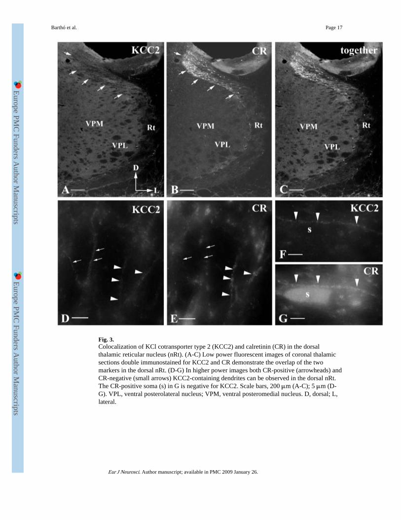
Fig. 3.Colocalization of KCl cotransporter type 2 (KCC2) and calretinin (CR) in the dorsalthalamic reticular nucleus (nRt). (A-C) Low power fluorescent images of coronal thalamicsections double immunostained for KCC2 and CR demonstrate the overlap of the twomarkers in the dorsal nRt. (D-G) In higher power images both CR-positive (arrowheads) andCR-negative (small arrows) KCC2-containing dendrites can be observed in the dorsal nRt.The CR-positive soma (s) in G is negative for KCC2. Scale bars, 200 μm (A-C); 5 μm (D-G). VPL, ventral posterolateral nucleus; VPM, ventral posteromedial nucleus. D, dorsal; L,lateral.
Barthó et al. Page 17
Eur J Neurosci. Author manuscript; available in PMC 2009 January 26.
Europe PM
C Funders A
uthor Manuscripts
Europe PM
C Funders A
uthor Manuscripts

Fig. 4.Ultrastructural localization of KCl cotransporter type 2 (KCC2) molecules in the ventrobasalcomplex. (A and B) High power electron micrographs showing thick proximal dendrites (d1)of a relay cell receiving inputs from primary afferents (RL) and from terminals showing thecharacteristics of the thalamic reticular nucleus input (F). Small calibre distal dendrites(d2-4) are contacted by corticothalamic terminals (asterisks). Silver-intensified gold particles(some of them labelled by arrowheads) on the intracellular surface of the membranes alsoindicate the location of KCC2 molecules in thick and thin dendrites. In these cases thedistribution of the silver signal is rather homogeneous along the membranes. There is noapparent accumulation of gold particles in the vicinity of excitatory or inhibitory synapses.(C and D) In these examples of thin relay cell dendrites (d1-4) almost all asymmetricalsynapses established by RS-type terminals (asterisks) are accompanied by silver deposits
Barthó et al. Page 18
Eur J Neurosci. Author manuscript; available in PMC 2009 January 26.
Europe PM
C Funders A
uthor Manuscripts
Europe PM
C Funders A
uthor Manuscripts
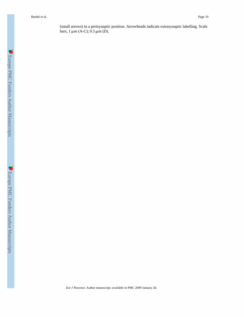
(small arrows) in a perisynaptic position. Arrowheads indicate extrasynaptic labelling. Scalebars, 1 μm (A-C); 0.5 μm (D).
Barthó et al. Page 19
Eur J Neurosci. Author manuscript; available in PMC 2009 January 26.
Europe PM
C Funders A
uthor Manuscripts
Europe PM
C Funders A
uthor Manuscripts

Fig. 5.High power electron micrographs showing KCl cotransporter type 2 (KCC2)immunoreactivity in the dorsal thalamic reticular nucleus (nRt). (A-D) Silver-intensifiedgold particles demonstrate the membrane localization of the transporter on nRt dendrites(d1-d4). Some of the particles (small arrows in A and B) are localized near the asymmetricalsynapses established by terminals of presumably cortical (asterisk in A, C and D) orthalamic (asterisk in B) origin. Arrowheads in C and D indicate KCC2 protein associatedwith intracellular membrane organelles. Scale bars, 0.5 μm (A-D).
Barthó et al. Page 20
Eur J Neurosci. Author manuscript; available in PMC 2009 January 26.
Europe PM
C Funders A
uthor Manuscripts
Europe PM
C Funders A
uthor Manuscripts

Fig. 6.Relationship between gold particle density indicating KCl cotransporter type 2immunoreactivity and dendritic diameter. Bar graphs demonstrate the density of goldparticles on different dendritic categories and on the somata of relay cells measured in singleultrathin sections in the ventrobasal complex. The density of gold particles relative tomembrane area (A) is similar across all compartments. The density of gold particles relativeto volume (B), however, shows a marked decrease from thin dendrites to soma. For the lackof error bars see Materials and methods.
Barthó et al. Page 21
Eur J Neurosci. Author manuscript; available in PMC 2009 January 26.
Europe PM
C Funders A
uthor Manuscripts
Europe PM
C Funders A
uthor Manuscripts

Fig. 7.Average gold particle density in perisynaptic areas (a 200-nm annulus around synapticspecializations) and extrasynaptic areas on the surface of reconstructed dendritic segments.Synapses in this analysis included asymmetrical membrane specializations established byRS terminals on distal relay cell dendrites. Perisynaptic membranes contain significantlymore KCl cotransporter type 2 than extrasynaptic membranes. For the lack of error bars seeMaterials and methods.
Barthó et al. Page 22
Eur J Neurosci. Author manuscript; available in PMC 2009 January 26.
Europe PM
C Funders A
uthor Manuscripts
Europe PM
C Funders A
uthor Manuscripts

Fig. 8.Scatter plots demonstrating the gold particle density as a function of synaptic coverage(synaptic areas/dendritic surface). Our sample included excitatory terminals established bysmall terminals. Each individual plot represents a relay cell dendritic segment reconstructedfrom 15 65-nm-thick ultrathin section in the ventrobasal complex. Both the synapticcoverage and the density of gold particles show high variability but there is no correlationbetween the two parameters.
Barthó et al. Page 23
Eur J Neurosci. Author manuscript; available in PMC 2009 January 26.
Europe PM
C Funders A
uthor Manuscripts
Europe PM
C Funders A
uthor Manuscripts


![[환자안전 주의경보] KCl 정맥 내 단독 주입 - 한국병원약사회](https://img.pdfslide.net/doc/110x75/6311083bd43a2591a904e3f7/-kcl-.jpg)


