Embed Size (px)
Citation preview
Induction of NMDA and GABAA Receptor-Mediated Ca2�
Oscillations With KCC2 mRNA Downregulation in InjuredFacial Motoneurons
HIROKI TOYODA,1,3 KOJI OHNO,2 JUNKO YAMADA,4 MASAHIKO IKEDA,1 AKIHITO OKABE,1
KOHJI SATO,2 KENJI HASHIMOTO,3 AND ATSUO FUKUDA1,4
Departments of 1Physiology, 2Anatomy, and 3Oral and Maxillofacial Surgery, Hamamatsu University School of Medicine,Hamamatsu, Shizuoka 431-3192; and 4Department of Biological Information Processing, Graduate School of ElectronicScience and Technology, Shizuoka University, Hamamatsu, Shizuoka 432-8011, Japan
Submitted 22 August 2002; accepted in final form 31 October 2002
Toyoda, Hiroki, Koji Ohno, Junko Yamada, Masahiko Ikeda,Akihito Okabe, Kohji Sato, Kenji Hashimoto, and Atsuo Fukuda.Induction of NMDA and GABAA receptor-mediated Ca2� oscilla-tions with KCC2 mRNA downregulation in injured facial motoneu-rons. J Neurophysiol 89: 1353–1362, 2003. First published November13, 2002; 10.1152/jn.00721.2002. To clarify the changes that occur in�-aminobutyric acid type A (GABAA) receptor-mediated effects andcontribute to alterations in the network activities after neuronal injury,we studied intracellular Ca2� concentration ([Ca2�]i) dynamics in arat facial-nerve-transection model. In facial motoneurons, an elevationof the resting [Ca2�]i, GABA-mediated [Ca2�]i transients, enhance-ment of the glutamate-evoked [Ca2�]i increases, and spontaneous[Ca2�]i oscillations were induced by axotomy. All these axotomy-induced modifications were abolished by the GABAA-receptor antag-onist bicuculline and N-methyl-D-aspartate (NMDA)-receptor antag-onist D(�)-2-amino-5-phosphonopentanoic acid. A downregulation ofK�-Cl� cotransporter (KCC2) mRNA, an increase in intracellularCl� concentration ([Cl�]i), and transformation of GABAergic hyper-polarization to depolarization were also induced by axotomy. Wesuggest that in axotomized neurons KCC2 downregulation impairsCl� homeostasis and makes GABA act depolarizing, resulting inendogenous GABA inducing [Ca2�]i oscillations via facilitation ofNMDA-receptor activation. Such GABAA-receptor-mediated [Ca2�]i
oscillations may play a role in neural survival and regeneration.
I N T R O D U C T I O N
�-Aminobutyric acid (GABA), the principal inhibitory neu-rotransmitter in the brain, decreases neural activity by hyper-polarizing the membrane potential via Cl� influx throughGABAA receptor channels. However, early in development,GABA seems to depolarize and excite the neuronal membranepotential and increase the [Ca2�]i (Ben-Ari 2002; Ben-Ari etal. 1989, 1997; Chen et al. 1996; Cherubini et al. 1990;Khazipov et al. 1997; Leinekugel et al. 1995; LoTurco et al.1995; Owens et al. 1996; Yuste and Katz 1991). The moredepolarized equilibrium potential for Cl�, effected by theintracellular Cl� increase, may be involved in this immature
action of GABA (Chen et al. 1996; Cherubini et al. 1990;Owens et al. 1996; Serafini et al. 1995).
Cation-chloride cotransporters are considered to play a crit-ical role in intracellular Cl� homeostasis (Kaila 1994). Underphysiological conditions, K�-Cl� cotransporter (KCC2) ap-pears to extrude Cl� from the neuron (Jarolimek et al. 1999;Kakazu et al. 1999; Rivera et al. 1999), while NKCC1, aNa�,K�-2Cl� cotransporter, is a candidate for the promotionof Cl� accumulation within the cell (Plotkin et al. 1997; Sunand Murali 1999; but see DeFazio et al. 2000). In immatureneurons, downregulated KCC2 and upregulated NKCC1 ex-pression may thus be responsible for depolarizing and excita-tory action of GABA (Kakazu et al. 1999; Yamada et al. 2002).
Network-driven membrane potential and Ca2� oscillationshave been reported in immature hippocampus (Ben-Ari 2002;Ben-Ari et al. 1989, 1997; Cherubini et al. 1990; Khazipov etal. 1997; Leinekugel et al. 1995) and neocortex (Garaschuk etal. 2000; Yuste and Katz 1991). GABAergic excitation andsecondary activation of N-methyl-D-aspartate (NMDA) recep-tors have been postulated as an underlying mechanism of theseCa2� oscillations (Ben-Ari 2002; Ben-Ari et al. 1997; Khazi-pov et al. 1997; Leinekugel et al. 1997). It has been reportedthat axotomy of vagal motoneurons can transform theGABAergic effect from inhibitory to excitatory by increasing[Cl�]i via downregulation of KCC2 so that exogenously ap-plied GABA then evokes a transient [Ca2�]i increase (Na-bekura et al. 2002). However, because this study has been doneby using acutely isolated neurons, it is yet to be studiedwhether this [Ca2�]i increase is related to induction of net-work-driven Ca2� oscillations accompanied by an alteration inendogenous GABAergic and NMDA receptor-mediated func-tions.
To clarify the changes in endogenous GABAA receptor-mediated effects resulting from in vivo neural injury and theunderlying mechanism, we used a facial-nerve-transectionmodel because GABA-containing vesicles in presynaptic ter-minals on facial motoneurons are enlarged after axotomy(Vaughan 1994) and axotomized motoneurons are considered a
Address for reprint requests: A. Fukuda, Dept. of Physiology, HamamatsuUniversity School of Medicine, Hamamatsu, Shizuoka 431-3192, Japan (E-mail: [email protected]).
The costs of publication of this article were defrayed in part by the paymentof page charges. The article must therefore be hereby marked ‘‘advertisement’’in accordance with 18 U.S.C. Section 1734 solely to indicate this fact.
J Neurophysiol 89: 1353–1362, 2003.First published November 13, 2002; 10.1152/jn.00721.2002.
13530022-3077/03 $5.00 Copyright © 2003 The American Physiological Societywww.jn.org

model of regeneration (Streit and Graeber 1993). We studiedchanges in the [Ca2�]i responses induced by either endogenousor exogenous GABA and glutamate; this enabled us to evaluatefunctional alterations in local neural circuits following axo-tomy. We also evaluated the molecular and physiological basisfor those changes.
M E T H O D S
All experiments conformed to the guidelines for animal experimen-tation at Hamamatsu University School of Medicine on the ethical useof animals, and all efforts were made to minimize the number ofanimals used and their suffering.
In situ hybridization histochemistry
We used adult male Wistar rats weighing about 150 g as well asyoung ones of either sex at postnatal day (P) 10 (Japan SLC, Shi-zuoka, Japan). Under pentobarbital anesthesia (50 mg/kg ip), wetransected the right facial nerve just distal to the posterior auricularbranch and removed about 5 mm of the distal nerve. At 1, 3, 7, 14, 21,28, 42, 56, and 112 days after this operation, they (n � 5 at each timepoint) were deeply anesthetized with ether and killed. Brains werequickly removed and frozen on powdered dry ice. Frozen sections (16�m thick) were cut on a cryostat, thaw-mounted onto silane-coatedslides, then stored at �80°C.
The in situ hybridization histochemical technique used for KCC2and NKCC1 is described in detail elsewhere (Kanaka et al. 2001).Briefly, hybridization was performed by incubating paraformalde-hyde-fixed sections for 24 h at 42°C in a buffer of the followingcomposition: 0.6 M NaCl and 0.06 M sodium citrate, 50% deionizedformamide, 0.12 M phosphate buffer, 2.5% tRNA, 10% dextransulfate in Denhardt’s solution, containing [35S]dATP (37–55.5 TBq/mmol; New England Nuclear, Boston, MA)-labeled probes (1–2 �107 dpm/ml, 0.2 ml/slide). The sections were coated with KodakNBT-2 emulsion, kept at 4°C for 2–3 wk, then developed in D-19developer. Because the expression levels of KCC2 and NKCC1mRNAs were different, we used the exposure times of 2 wk for KCC2and 3 wk for NKCC1 for emulsion autoradiography. The sections
were counterstained with thionin solution to allow morphologicalidentification.
The probes for KCC2 and NKCC1 mRNAs (Kanaka et al. 2001)were complementary to the bases 2,981–3,016 and 2,914–2,949,respectively, of these mRNAs (Moore-Hoon and Turner 1998; Payneet al. 1996). The specificity of the probes has been already confirmed(Kanaka et al. 2001). For semi-quantative analysis of labeled neurons,four sections were randomly chosen from three animals killed at eachof the nine postsurgical time points. Neurons with three times moregrains than the background level were considered to be positivelylabeled. Motoneurons were counted on thionin-stained sections.
Preparation of brain slices
The young rats were subjected to right-facial-nerve transection;after 3 days (P10–12), they were deeply anesthetized and killed. Ablock of brain including the facial nucleus was quickly removed andplaced in cold (4°C), oxygenated, modified artificial cerebrospinalfluid (ACSF). The solution contained the following (in mM): 170sucrose, 126 NaCl, 2.5 KCl, 1.25 NaH2PO4, 12.0 MgSO4, 26.0NaHCO3, and 30.0 glucose. Coronal slices (400 �m) through thefacial nucleus were cut in the modified ACSF using a vibratome(Leica VT-100, Germany). Slices were allowed to recover for 60 minon nylon meshes (with 1-mm pores) that were submerged in dishescontaining standard ACSF consisting of (in mM) 126 NaCl, 2.5 KCl,1.25 NaH2PO4, 2.0 MgSO4, 2,0 CaCl2, 26.0 NaHCO3, and 20.0glucose. The dishes were then placed in a tightly sealed box filled with95% O2-5% CO2 at a pressure of 50 kPa at room temperature.
Gramicidin-perforated patch-clamp recordings
Gramicidin-perforated patch-clamp recording was carried out aspreviously described (Ebihara et al. 1995). Patch electrodes weremade from borosilicate capillary tubing (diameter, 1.5 mm; GarnerGlass) using a Narishige PP-83 vertical puller (Narishige). The elec-trode resistance was within the range 3–4.5 M�. The pipette solutioncontained (in mM) 130 KCl, 5 NaCl, 0.4 CaCl2, 1.0 MgCl2, 1.1EGTA, and 10 HEPES (pH 7.3 with KOH). Gramicidin (50 mg/ml)was dissolved in the pipette solution just before the experiment. Facialmotoneurons in slices were viewed on a monitor via a �40 water-
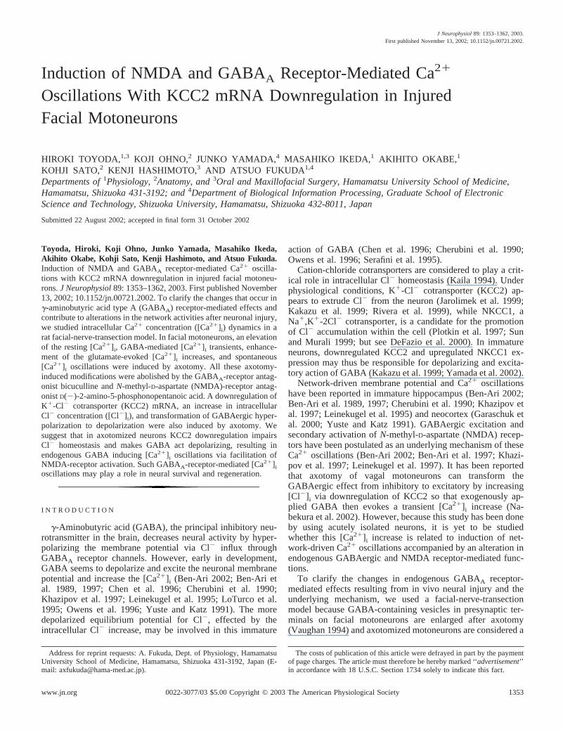
FIG. 1. Facial-nerve transection downregulates K�-Cl� cotransporter (KCC2) mRNA expression in the facialnucleus. A: dark-field photomicrographs showing rat facialnucleus expressing KCC2 mRNA hybridization signals 1day, 3 days, 4 wk, 8 wk, and 16 wk after facial-nervetransection. Note marked reduction in KCC2 mRNA in theaxotomized facial nucleus in the period from 3 days to 4wk with recovery by 16 wk. - - -, the regions of facialnuclei. Bar � 1 mm. B: bright-field photomicrographs ofcounter-stained section shows that KCC2 mRNA hybrid-ization signals localized in facial motoneurons (M) wereseverely downregulated in axotomized neurons 3 daysafter transection. Note recovery of signals at 16 wk afterfacial-nerve transection. Signal-positive cells were large insize and considered to be motoneurons (M), whereassmall-sized presumed glial cells (G) were lacking in sig-nals. Bar � 50 �m. C: time course of recovery in thepercentage of facial motoneurons expressing KCC2mRNA on the axotomized side. Error bars � SD (n � 5).
1354 TOYODA ET AL.
J Neurophysiol • VOL 89 • MARCH 2003 • www.jn.org

immersion objective lens with the aid of an infrared differentialinterference-contrast (IR-DIC) filter and a CCD-camera (C2400-79;Hamamatsu Photonics). Real-time video images were contrast-en-hanced by a video processor (Argus-20; Hamamatsu Photonics).Membrane currents and potentials were recorded using an Axopatch1D amplifier (Axon Instruments). Data were digitized using an A/Dconverter (Digidata 1200, Axon Instruments) and analyzed by meansof pCLAMP8 software. To measure EGABA, voltage steps were ap-plied and GABA (10 �M) was pressure-applied through the patchpipette to the soma of the neurons at each membrane potential. Allexperiments were carried out at 30°C.
Ca2� imaging using fura-2
The methods used for Ca2� imaging were similar to those describedpreviously (Fukuda et al. 1998a). Neurons were loaded with the Ca2�
indicator, fura-2, by incubating slices for 60 min with fura-2 acetoxylmethyl (10 �M) in ACSF containing 0.01% pluronic F127. Sliceswere then laid on the glass bottom of a submerged-type chamber, andthis was placed on a microscope stage and continuously perfused withstandard ACSF gassed with 95% O2-5% CO2 at a rate of 2–3 ml/min.In some experiments, the concentration of Mg2� in the standardACSF was reduced to 1 or 0.5 mM (low-Mg2�). The bathing solutionwas maintained at 30°C and had a pH of 7.4.
Fura-2 fluorescence was excited using a multi-wavelength mono-chrometer (C6789; Hamamatsu Photonics) and the emitted light wasfiltered using a band-pass filter (510 nm). Fluorescence images wereobtained using a �40 objective lens (Plan Fluor, N.A. 0.75; Nikon)via a cooled-CCD camera (C6790–81; Hamamatsu Photonics) fittedto an up-light microscope (E600-FN; Nikon). Data were stored foroff-line analysis by means of image-processing software (Aqua Cos-mos; Hamamatsu Photonics). [Ca2�]i was expressed as the ratio of thefura-2 fluorescence intensities excited at 340 and 380 nm (RF340/F380).Changes with time in RF340/F380 were monitored in facial motoneu-rons by taking measurements every 10 s. These were converted into[Ca2�]i using the following equation: [Ca2�]i � Kd [(R � Rmin)/(Rmax �R)]� (Grynkiewicz et al. 1985) in which Kd is the effective dissociationconstant of fura-2 and � is the ratio of fluorescence intensities at 380 nmexcitation for fura-2/(fura-2 � Ca2�). Rmin � 0.2, Rmax � 7.2, � � 3.3,and Kd � 146 nM were obtained using the calibration method (Williamset al. 1985). All drugs were applied by bath perfusion.
Cl� imaging using 6-methoxy-N-ethylquinolinium iodide
The devices and materials used for Cl� imaging were similar tothose described in the preceding text for Ca2� imaging, and thetechniques used for Cl� imaging were as described previously(Fukuda et al. 1998b; Schwartz and Yu 1995). Briefly, prior tobath-loading of the slices, 6-methoxy-N-ethylquinolinium iodide(MEQ) was reduced to a cell-permeable form, diH-MEQ (Biwersi andVerkman 1991). This reduction of MEQ (2 mg/100 �l) was achievedby addition of 15 �l of 12% NaBH4 solution and bubbling with N2 for30 min. DiH-MEQ was extracted from the reaction mixture as ayellow organic layer, a portion of which was added to ACSF to yielda final concentration of 360 �M.
Neurons were loaded with MEQ by incubating slices with diH-MEQ for 60 min. MEQ was excited at 355 nm to emit fluorescenceand this was filtered at 460 nm. The perfusion medium was changedfrom standard ACSF to a calibration solution containing 0 mM Cl�,in which NaCl was substituted by equimolar methylsulfuric acidpotassium salt and to which tributyltin, a Cl�-OH� antiporter (20�M), and nigericin, a K�-H� antiporter (14 �M), were added (Sim-chowitz et al. 1991). When in the presence of these reagents for �20min, [Cl�]i can be assumed to have equilibrated across the plasmamembrane of the neurons in the slice. At the end of the procedure,total quenchable intracellular-MEQ fluorescence was measured fol-lowing the addition of 150 mM KSCN. The resting [Cl�]i was
obtained by calculating the ratio of the fluorescence measured in theabsence of Cl� (F0: FCl�0 � FSCN) to that measured at the resting[Cl�]i (FRest: FCl � rest � FSCN) and fitting the values to the calibra-tion curve, with a Kq of 30.6 M�1.
Drugs
The following drugs were used: tRNA from Roche (Mannheim,Germany), D(�)-2-amino-5-phosphonopentanoic acid (D-AP5) and6-cyano-7-nitroquinoxaline-2,3-dione (CNQX) from Tocris Cookson(Ballwin, MO), Denhardt’s solution from Nacalai tesque (Kyoto,Japan), NBT-2 emulsion and D-19 developer from Kodak (Rochester,NY), fura-2 acetoxyl methyl and pluronic F127 from Dojindo (Ku-mamoto, Japan), dextran sulfate, tetrodotoxin (TTX), (�)-bicucullinemethiodide, GABA, L-glutamate, nifedipine, methylsulfuric acid po-tassium salt, and gramicidin from Sigma (St. Louis, MO), MEQ fromMolecular Probes (Eugene, OR).
FIG. 2. Expressions of KCC2 mRNA and Na�,K�-2Cl� cotransporter(NKCC1) mRNA in 10-day-old rat facial nucleus after axotomy. A: ex-pression of KCC2 mRNA in 10-day-old rat facial nucleus: on the intactside, it was comparable to that of the adult, but it was clearly reduced onthe axotomized side at 3 days after axotomy. Bright-field photomicrographsshow that KCC2 mRNA hybridization signals in facial motoneurons (M)were clearly reduced on the axotomized side. Bars � 1 mm (dark-fieldphotomicrograph) and 50 �m (bright-field photomicrograph). B: dark- andbright-field photomicrographs show lack of difference in expression ofNKCC1 mRNA between intact and axotomized facial nucleus at 3 daysafter axotomy. The areas surrounded by dashed lines indicate the regions offacial nuclei. Note that NKCC1 mRNA signals were detected both inmotoneurons (M) and in presumed glial cells (G).
1355AXOTOMY INDUCES GABA-MEDIATED CA2� OSCILLATION
J Neurophysiol • VOL 89 • MARCH 2003 • www.jn.org

Through this report, “intact” is used to refer to neurons on the sidecontralateral to the facial-nerve section.
R E S U L T S
Facial-nerve transection downregulates expression of KCC2mRNA
We performed unilateral transection of the facial nerve inadult rats. The number of axotomized neurons stained bythionin was comparable to those of intact neurons throughoutthe observation period. To assess the general expression pat-terns and the time course of the changes in the expression ofKCC2 and NKCC1 mRNAs in the facial nucleus after axo-tomy, we evaluated in situ hybridization signals using filmautoradiography.
Dark-field photomicrography revealed that the very intenseKCC2 mRNA level was in decline as early as the first post-operative day and that it was almost abolished 3 days after theaxotomy (Fig. 1A). This severe downregulation of KCC2mRNA expression was sustained for 3 wk before a gradualrecovery. By 16 wk after the axotomy, the KCC2 mRNA levelhad recovered to the control level (Fig. 1C). Bright-field pho-tomicrography revealed that the KCC2 mRNA hybridizationsignals were positive in 95.1 � 2.3% (mean � SD throughouttext) of large-sized (50 �m) cells (considered to be facialmotoneurons) but not in the small-sized presumed glial cells(Fig. 1B). After the precipitate fall that immediately followedaxotomy, recovery was evinced by 43.5 � 8.5% of neuronsbeing positive at 8 wk and 81.8 � 9.3% at 16 wk (Fig. 1, B andC). There are reportedly no interneurons in the facial nucleus,and only a minority of cells project via an extra-facial-nerveroute from this nucleus (Røste 1989), so most neurons in theipsilateral facial nucleus (i.e., facial motoneurons) were, notsurprisingly, affected by the axotomy.
We used young rats, aged 10–12 days, for all the opticalimaging and patch-clamp recordings. To compare the charac-teristics of facial motoneurons at this age with those of the
adult, we studied changes in the expressions of KCC2 mRNAin the facial nucleus on P10 after unilateral facial-nerve tran-section 3 days earlier (n � 5). The expression of KCC2 mRNAwas comparable to that of the adult on intact side, and it wasclearly reduced on the axotomized side as in adult (Fig. 2A).There were no apparent signs of cell loss or severe deteriora-tion such as swelling (Fig. 2A). At 8 wk after transection, theKCC2 mRNA level showed a substantial recovery, the timecourse being comparable to that seen in the adult (not shown).
In P10 rats (n � 5), NKCC1 mRNA was expressed at levelscomparable to those seen in the adult (n � 5). Bright-fieldobservation showed that the hybridization signals for NKCC1were localized both in neurons and in glial cells (Fig. 2B).Facial-motoneuron axotomy did not change the expression ofNKCC1 mRNA either in motoneurons or in glial cells at anyday after operation either in adult or young rats. Figure 2Bshows that there was no apparent difference in the expressionof NKCC1 mRNA between intact and axotomized facial nucleion P10 after transection 3 days earlier.
Facial-nerve transection increased [Cl�]i in motoneurons
To study the functional consequences of facial nerve tran-section on Cl� homeostasis, we compared the resting [Cl�]i ofaxotomized neurons with that of intact neurons. First, weexamined the EGABA in axotomized neurons by means ofgramicidin-perforated patch-clamp recording. This techniqueallows measurement of EGABA with the intracellular Cl� intact(Ebihara et al. 1995). In the current-clamp mode, GABA (100�M) induced a hyperpolarization in intact neurons (n � 3) anda depolarization in axotomized neurons (n � 6; Fig. 3A).Axotomized neurons had a resting membrane potential of–65.8 � 5.2 mV (n � 7) and an EGABA of –52.1 � 5.7 mV,while the corresponding values in intact neurons were –65.6 �1.5 mV (n � 5) and –70.9 � 5.3 mV (Fig. 3A). We calculated[Cl�]i for each neuron from the Nernst equation, using mea-sured EGABA and a [Cl�]o of 132.5 mM. The [Cl�]i in axoto-
FIG. 3. Increases in resting [Cl�]i after axotomy. A: bygramicidin-perforated patch-clamp recordings in the current-clamp mode, GABA (100 �M) hyperpolarized intact neuronsand depolarized axotomized neurons. Reversal potential forGABA-evoked currents in voltaged-clamp mode shifted to thepositive direction after axotomy. GABA was applied usingpressure-pulses of 35–100 KPa for 10–50 ms. Right: I-V curvesfor GABA-evoked currents in intact and axotomizd neurons.B: estimation of resting [Cl�]i by means of Cl� imaging.Values for maximum and total quenchable MEQ fluorescencewere obtained in 0 mM Cl� and 150 mM SCN�, respectively.Resting [Cl�]i in individual neurons was estimated from theStern-Volmer relationship. Resting [Cl�]i was significantlyhigher in axotomized neurons than in intact neurons (right).
1356 TOYODA ET AL.
J Neurophysiol • VOL 89 • MARCH 2003 • www.jn.org

mized neurons was 17.8 � 4.6 mM (n � 7), a value signifi-cantly higher than that obtained for intact neurons 8.5 � 4.6mM (n � 5; Mann-Whitney U test, P � 0.001).
We also compared the resting [Cl�]i by optical imagingusing MEQ. To this end, we changed the perfusion mediumfrom standard ACSF to calibration solution containing 0 mMCl� together with tributyltin and nigericin. This allowed us toobtain a ratio of MEQ fluorescence values (resting [Cl�]i over0 mM [Cl�]i). We estimated the resting [Cl�]i from Stern-Volmer plots with Kq � 30.6 M�1. The resting [Cl�]i ofaxotomized neurons was 24.4 � 17.3 mM (n � 10; 3 slices),significantly higher than that in intact neurons (11.1 � 8.5 mM,n � 9; 3 slices; Mann-Whitney U test, P � 0.05; Fig. 3B).Although absolute [Cl�]i values measured by this method maynot be strictly reliable (Fukuda et al. 1998b; Woll et al. 1996),collectively, the preceding results suggest, in terms of compar-ison, that the depolarizing action of GABA in axtomizedneurons is due to an elevated [Cl�]i and a depolarized Cl�
equilibrium potential.
GABA-mediated rise in intracellular Ca2� induced by facial-nerve transection
We loaded facial motoneurons with fura-2, measured theratio of the fluorescence intensities excited at 340 nm and 380nm (RF340/F380) and used it to calculate [Ca2�]i (Fig. 4A). Intactneurons had a resting [Ca2�]i of 43.5 � 7.3 nM (n � 15; 4slices), and this was altered neither by 100 �M GABA nor 20�M bicuculline (n � 15; 4 slices; Fig. 4B). In axotomizedfacial motoneurons, resting [Ca2�]i was 57.6 � 8.6 nM (n �24; 5 slices), significantly higher than in intact cells (P �0.005, Mann-Whitney U test). In two-thirds of cells tested afteraxotomy (16/24; 5 slices), GABA (100 �M) evoked marked[Ca2�]i increases (by 46.5 � 8.1 nM). Bicuculline not onlyblocked this increase but also decreased the baseline Ca2�
level, by 12.1 � 2.6 nM (n � 8, P � 0.005; 3 slices) to a levelcomparable to that seen in intact cells (Fig. 4C). The increasesin [Ca2�]i evoked by GABA were completely blocked by 50�M nifedipine, an L-type Ca2�-channel blocker (n � 10; 3slices; Fig. 4D) and by 1 �M tetrodotoxin (TTX), a sodium-channel blocker (n � 12; 3 slices: not shown), while the resting[Ca2�]i was altered by neither of these agents. In contrast,D-AP5 did not block GABA-evoked [Ca2�]i transient but re-duce the baseline Ca2� level by 7.5 � 3.1 nM (n � 14, P �0.05; 3 slices; Fig. 4E).
[Ca2�]i changes in response to glutamate in facialmotoneurons
To help us evaluate functional alterations in local neuralcircuits, we studied changes in the [Ca2�]i responses to high-dose (100 �M) and low-dose (5 and 10 �M) glutamate. Inintact neurons, bath-application of glutamate (100 �M) for 8min increasd the peak [Ca2�]i by 131.9 � 11.2 nM (n � 18; 4slices) with a return to baseline 15.0 � 3.6 min after the end ofthe glutamate application (Fig. 5A). In axotomized neurons,glutamate evoked larger and more prolonged (�40 min, notreturned to baseline during observation period) increases in the[Ca2�]i (by 216.3 � 12.6 nM, n � 16; 4 slices; Fig. 5C). Inintact neurons, addition of bicuculline prolonged (27.8 � 5.4min) and enlarged the glutamate-evoked [Ca2�]i increases
(after bicuculline, by 169.1 � 12.4 nM, n � 9; 3 slices; Fig.5A). By contrast, in axotomized neurons, the glutamate-evoked[Ca2�]i increases were reduced by bicuculline in both ampli-tude (after bicuculline, by 181.4 � 15.6 nM, n � 19; 5 slices)and duration (35.7 � 6.4 min; Fig. 5C).
We also studied changes in the [Ca2�]i responses to high-dose glutamate in the presence of TTX (1 �M) to clarify thealternative network-based effects of GABA and glutamate. Inintact neurons, addition of TTX prolonged the glutamate-evoked [Ca2�]i increases both duration (29.3 � 5.0 min) and
FIG. 4. [Ca2�]i transients elicited by GABA application in facial motoneu-rons. A: facial motoneurons loaded with fura-2 were clearly identified byfluorescence excited at 380 nm. Note that there are no apparent differences insize or shape between axotomized and intact facial motoneurons. Circles in theloaded neurons correspond the area of the ratio measurements. Bar � 50 �m.B: intact neurons responded neither to GABA (100 �M) nor to bicuculline (20�M). C: axotomized facial motoneurons showed an [Ca2�]i rise in response toGABA (100 �M) that was completely blocked by bicuculline (20 �M). Notethat bicuculline also decreased the baseline Ca2� level, suggesting that endog-enous GABAA-receptor activation might be partly responsible for the basal[Ca2�]i. D: [Ca2�]i increases evoked by GABA were also completely blockedby nifedipine (50 �M), but this agent did not alter the baseline [Ca2�]i level.E: D(�)-2-amino-5-phosphonopentanoic acid (D-AP5; 50 �M) did not blockGABA-evoked [Ca2�]i transient but reduced the baseline [Ca2�]i level. Notethat in axotomized neurons (C–E), basal [Ca2�]i is higher than that in intactneurons (B).
1357AXOTOMY INDUCES GABA-MEDIATED CA2� OSCILLATION
J Neurophysiol • VOL 89 • MARCH 2003 • www.jn.org

amplitude (by 159.6 � 25.7 nM, n � 19; 5 slices; Fig. 5B),whereas in axotomized neurons the glutamate-evoked [Ca2�]i
increases were reduced by TTX in duration (32.2 � 4.7 min)and amplitude (by 182.4 � 33.1 nM, n � 12; 3 slices; Fig. 5D).In the presence of TTX, additions of bicuculline affectedneither duration nor amplitude of glutamate-evoked [Ca2�]i
increases in intact (30.4 � 5.7 min, by 171.1 � 14.0 nM, n �8; 3 slices; Fig. 5B) and in axotomized (31.4 � 5.9 min, by176.4 � 18.3 nM, n � 11; 3 slices; Fig. 5D) neurons.
Bath applications of low-dose glutamate (5 and 10 �M)for 2-min failed to evoke an [Ca2�]i response in intactneurons (n � 9; 3 slices; Fig. 6A) even in the presence ofbicuculline (n � 10; 3 slices; Fig. 6B), indicating thesedoses were not sufficient to evoke any Ca2� transients.However, in axotomized neurons, low-dose glutamate raisedthe [Ca2�]i level (by 25.6 � 8.1 nM in response to 5 �Mglutamate; by 49.9 � 8.7 nM in response to 10 �M) (n � 9;3 slices; Fig. 6C). The �-amino-3-hydroxy-5-methylsox-azole-4-propionate (AMPA)-receptor antagonist 6-cyano-7-nitroquinoxalene-2,3-dione (CNQX, 10 �M) caused onlymarginal reduction (n � 12; 3 slices; Fig. 6D), whereas theNMDA-receptor antagonist D-AP5 (50 �M) blocked thiseffect of low-dose glutamate (n � 10; 3 slices; Fig. 6E).Addition of TTX (n � 13; 4 slices; Fig. 6F) and bicuculline(n � 9; 3 slices; Fig. 6G) abolished these low-dose gluta-mate-induced [Ca2�]i increases. These results indicate thatendogenous GABA exerts on excitatory action in co-oper-ation with glutamate in axotomized facial motoneurons tofacilitate Ca2� influx through NMDA receptor.
FIG. 5. [Ca2�]i changes in response to high-dose (100 �M) glutamate. A: inintact neurons, glutamate (100 �M, ■ ) evoked [Ca2�]i increases that hadreturned to baseline 15 min after termination of glutamate application (left). Inthe presence of bicuculline, glutamate-evoked [Ca2�]i increases were largerand more prolonged, the level not returning to baseline until 28 min afterglutamate application (right). B: in the presence of TTX, the glutamate-evoked[Ca2�]i increases were enhanced in both duration and amplitude (left). Addi-tion of bicuculline did not change the responses apparently (right). C: inaxotomized facial motoneurons, glutamate evoked increases in [Ca2�]i thatwere larger and more prolonged than those seen in intact neurons. Note that[Ca2�]i did not return to baseline level within the time frame of the experiment,and that the baseline level was higher than in intact neurons (left). Addition ofbicuculline to axotomized facial motoneurons reduced baseline [Ca2�]i andshortened the duration of the glutamate-evoked [Ca2�]i increases. Note that inthe presence of bicuculline, both the size and duration of glutamate-induced[Ca2�]i increases, as well as the baseline [Ca2�]i level, were comparablebetween intact and axotomized neurons (right). D: the glutamate-evoked[Ca2�]i increases were reduced by TTX in duration and amplitude in axoto-mized neurons (left). Further addition of bicuculline did not affect the re-sponses significantly (right). Bicuculline and TTX were continuously perfusedbefore, during, and after bath application of glutamate.
FIG. 6. [Ca2�]i changes elicited by low-dose (5 and 10 �M) glutamate.Intact neurons showed no response to low-dose glutamate (5 �M, ■ ; 10 �M,�) in the absence (A) or presence (B) of bicuculline. In axotomized neurons,application of low-dose glutamate evoked transient [Ca2�]i increases (C).6-cyano-7-nitroquinoxalene-2,3-dione (CNQX) did not (D) but D-AP5 (E),TTX (F), and bicuculline (G) abolished these responses. CNQX, D-AP5, TTX,and bicuculline were continuously perfused before, during, and after bathapplication of glutamate.
1358 TOYODA ET AL.
J Neurophysiol • VOL 89 • MARCH 2003 • www.jn.org

Spontaneous [Ca2�]i oscillations induced in axotomizedfacial motoneurons
We used low-Mg2� (1 or 0.5 mM) ACSF to reduce Mg2�
block of NMDA receptors and/or enhance presynaptic releaseof transmitters. Oscillation-like spontaneous Ca2� transientshardly occurred at all in intact neurons even in 0.5 mM extracel-lular Mg2� concentration ([Mg2�]o; Fig. 7A). In contrast, inaxotomized neurons, spontaneous [Ca2�]i oscillations occurredeven in normal ACSF (20/28; 3 slices), and their amplitude andfrequency, as well as the resting [Ca2�]i, increased as the [Mg2�]owas reduced (Fig. 7B). These effects of low-Mg2� ACSF, onresting [Ca2�]i and [Ca2�]i oscillations, were reduced by theaddition of D-AP5 (50 �M; Fig. 7C), whereas CNQX (10 �M)was ineffective (Fig. 7D). The [Ca2�]i oscillations were alsoreversibly diminished by TTX (n � 10; 3 slices; Fig. 7E). In someaxotomized neurons, there were synchronous spontaneous [Ca2�]ioscillations during perfusion with low-Mg2� ACSF (3 of 10slices; Fig. 7F). The synchronous or nonsynchronous spontaneous[Ca2�]i oscillations and the increase in resting [Ca2�]i were re-versibly blocked by 20 �M bicuculline (Fig. 7F). This suggeststhat they are mediated by endogenous GABAA-receptor activa-tion.
D I S C U S S I O N
Depolarizing action of GABA following change in Cl�
homeostasis
Of [Cl�]i regulators, KCC2, which normally carries Cl� outof the cell along with K�, is largely responsible for keeping
[Cl�]i low in mature neurons (Jarolimek et al. 1999; Kakazu etal. 1999; Rivera et al. 1999), endowing them with hyperpolar-izing responses to GABA (Ganguly et al. 2001; Lu et al. 1999;Rivera et al. 1999). NKCC1, which carries Cl� into the cellusing Na�-driving forces, helps to maintain a high [Cl�]i inimmature neurons, with the result that GABA acts in an exci-tatory manner (Kakazu et al. 1999; Plotkin et al. 1997; but seeDeFazio et al. 2000). In our study, intact facial motoneuronsdisplayed hyperpolarizing responses to GABA, however, afteraxotomy GABA caused depolarization (Fig. 3A), and the rest-ing [Cl�]i of axotomized neurons was significantly higher thanthat of intact neurons as demonstrated both by gramicidin-perforated patch-clamp recordings and by optical imaging us-ing MEQ. The observed downregulation of KCC2 mRNAwithout changes in NKCC1 is consistent with an increase in[Cl�]i. Thus the depolarizing action of GABA in axotomizedneurons may be caused by a positive shift in the equilibriumpotential for Cl� consequent on the [Cl�]i increase that followsKCC2 downregulation as reported previously (Nabekura et al.2002).
Interaction of GABAergic excitation and Ca2� signaling
In the present study, bicuculline blocked the Ca2� increasesevoked by GABA in axotomized neurons, suggesting that thisresponse is mediated by the GABAA-receptor Cl� channel.Nifedipine also blocked the [Ca2�]i increases, indicating thatthe Ca2� influx occurred through L-type voltage-dependentCa2� channels (VDCa2�s) (Ganguly et al. 2001; Nabekura etal. 2002; Obrietan and van den Pol 1995; van den Pol et al.
FIG. 7. Spontaneous [Ca2�]i oscillations oc-curred in axotomized facial motoneurons. A: spon-taneous [Ca2�]i oscillations hardly occur at all inintact neurons even in low (1 or 0.5 mM) [Mg2�]o.
B: in axotomized neurons, spontaneous [Ca2�]i
oscillations occur, their amplitude and frequencyincreasing as [Mg2�]o is progressively reduced.C: these effects of low-Mg2� ACSF, on resting[Ca2�]i and [Ca2�]i oscillations, were blocked bythe addition of D-AP5. D: application of CNQX didnot block these [Ca2�]i oscillations. E: addition ofTTX blocked these [Ca2�]i oscillations reversibly.F: among axotomized neurons, synchronous spon-taneous [Ca2�]i oscillations were occasionally seenduring superfusion with medium containing 0.5mM [Mg2�]o. These [Ca2�]i oscillations were re-versibly abolished by bicuculline. Results illus-trated in A–F were from 6 different slices. Note thatbaseline [Ca2�]i rose progressively as [Mg2�]o waslowered in axotomized neurons but not in intactneurons.
1359AXOTOMY INDUCES GABA-MEDIATED CA2� OSCILLATION
J Neurophysiol • VOL 89 • MARCH 2003 • www.jn.org

1996; Yuste and Katz 1991). Because TTX also blocked thisCa2� influx, our data suggest that the GABA-evoked [Ca2�]irise in axotomized neurons is primarily mediated by an initialdepolarization due to a Cl� efflux, with the ensuing actionpotential causing opening of VDCa2�s. These results are com-parable to previous reports (Nabekura et al. 2002; van den Polet al. 1996).
We evaluated alterations in the local neural circuitry bystudying changes in the [Ca2�]i responses to glutamate. Boththe amplitude and duration of glutamate-evoked [Ca2�]i in-creases were greater in axotomized neurons than in intactneurons. Bicuculline decreased the glutamate response in axo-tomized neurons but enhanced it in intact neurons. In axoto-mized neurons, detectable [Ca2�]i increases could be evokedby glutamate concentrations too low to induce a response inintact neurons. The addition of bicuculline abolished theseresponses. Because the addition of bicuculline had no furthereffects in the presence of TTX, the endogenous GABAA re-ceptor-mediated actions induced by axotomy might be net-work-mediated. These results suggest that feed-forwardGABAergic inhibition in intact neurons is changed to feed-forward GABAergic excitation in axotomized neurons, withthe consequence that the threshold for glutamate-induced[Ca2�]i increases was reduced (see Fig. 8).
In immature hippocampal neurons (Ben-Ari et al. 1988) andimmature vagal motoneurons (Furukawa et al. 2000), the volt-age-dependent Mg2� block of NMDA channels are reduced. In
addition to this, the depolarizing action of GABA, achieved viaGABAA-receptors in immature neurons also tends to removeMg2� block (Ben-Ari 2002; Ben-Ari et al. 1997; Khazipov etal. 1997; Leinekugel et al. 1997). In adult vagal motoneurons,axonal injury leads to the reaquisition of the immature char-acteristics of NMDA receptor (Furukawa et al. 2000) andGABAergic action (Nabekura et al. 2002). If this also occurredin the axotomized facial motoneurons, GABAergic depolariza-tion might furthur reduce the voltage-dependent Mg2� block ofNMDA channels, thus facilitating Ca2� influx through them.This hypothesis is compatible with the present results thatbicuculline and D-AP5 each but not CNQX blocked the [Ca2�]i
increase induced by low-dose glutamate. This could providefor a synergy between GABA and glutamate, thus makingGABA an excitatory transmitter as shown previously in im-mature hippocampal neurons (Ben-Ari 2002; Ben-Ari et al.1989, 1997; Khazipov et al. 1997; Leinekugel et al. 1997). Theimplication is that neural injury may cause neurons to reacquiregreater plasticity, with some immature characteristics. Indeed,a GABA-induced Ca2� increase after an injury may allow theneuron to modulate gene expression (Bading et al. 1993;Berninger et al. 1995), influence growth-cone guidance (Obri-etan and van den Pol 1996) and possibly reduce cell deathresulting from the presence of a suboptimal cytosolic Ca2�
(Franklin and Johnson 1992). Thus a GABA-induced elevationin [Ca2�]i is likely to promote neuronal recovery.
FIG. 8. Schematic illustration of possible mechanisms underlying the elevation in resting [Ca2�]i and generation of [Ca2�]i
oscillations in axotomized facial motoneurons. Left: under normal conditions, GABA hyperpolarizes facial motoneurons by causingan influx of Cl� through GABAA receptor-channels. Such GABAergic hyperpolarization may inhibit opening of voltage-dependentNa� channels (VDNa�s), voltage-dependent Ca2� channels (VDCa2�s), and NMDA receptor-channels; hence, high-dose gluta-mate-induced increases in [Ca2�]i are attenuated by a presumed feed-forward GABAegic inhibition. Right: in axotomized neurons,in which Cl� extrusion is inhibited by KCC2 downregulation, an accumulation of Cl� occurs via unaltered NKCC1. GABA thusdepolarizes axotomized facial motoneurons by causing an efflux of Cl� through GABAA receptor-channels, and hence opening theVDNa�, with the ensuring action potential causing opening of VDCa2� and inducing a transient Ca2� influx. The endogenousGABAA-receptor-mediated depolarization may contribute to removal of the Mg2�-block, decreasing threshold for glutamate(NMDA)-induced [Ca2�]i increases and enhancing even a weak effect of glutamate. When glutamate is released from presynapticneurons within the axotomized facial nucleus, the existing collapse of the Cl� gradient could thus convert feed-forward GABAergicinhibition to excitation, provoking GABAA receptor- and NMDA receptor-mediated spontaneous [Ca2�]i oscillations.
1360 TOYODA ET AL.
J Neurophysiol • VOL 89 • MARCH 2003 • www.jn.org

Mechanisms underlying GABAA-receptor-mediated increasein resting [Ca2�]i and Ca2� oscillations
In axotomized facial motoneurons, in which the resting[Ca2�]i was significantly higher than in intact cells, bicucullinenot only blocked the GABA-evoked [Ca2�]i increase but alsodecreased the baseline Ca2� level (Fig. 4). In whole cellpatch-clamp recording, spontaneous postsynaptic currents(sPSCs) in axotomized neurons were blocked by bicuculline,whereas not in intact neurons (not shown). These results sug-gest that endogenous GABAA-receptor activation (Flint et al.1998; LoTurco et al. 1995; Owens et al. 1996) may help toraise basal Ca2� levels in such damaged neurons. BecauseD-AP5 had comparable effect with bicuculline on resting[Ca2�]i, activation of a NMDA receptor may also be involvedin increases in resting [Ca2�]i. Although effects of NMDA andGABAA receptors on resting [Ca2�]i may imply depolarizationof axotomized neurons by tonic activation of these receptors,resting membrane potential were comparable between intactand axotomized neurons in the present and in the previous(Nabekura et al. 2002) studies. A TTX-insensitive backgroundactivation of these receptors could increase resting [Ca2�]ibecause [Ca2�]i transients last longer than the accompanyingmembrane potential transients.
We demonstrated here that spontaneous [Ca2�]i oscillationswere present in axotomized neurons, a phenomenon reversiblyblocked by bicuculline, suggesting GABAA receptor involve-ment, though a possibility of another mediator than GABAA toparticipate in the axotomy-induced [Ca2�]i oscillations cannotbe ruled out. In these neurons, the amplitude and frequency ofthe spontaneous [Ca2�]i oscillations were increased as[Mg2�]o was lowered and were completely abolished by D-AP5 but not by CNQX. Thus reduced Mg2�-dependent blockof NMDA receptor-channels may be further facilitated byendogenous GABAA-receptor-mediated depolarization in axo-tomized neurons, so that this could induce spontaneous [Ca2�]ioscillations by elevating [Ca2�]i set-point via NMDA-receptoractivation (Leinekugel et al. 1997). These phenomena might benetwork-driven because [Ca2�]i oscillation was TTX sensitive(see Fig. 8).
Functional significance of the GABAA-mediated [Ca2�]ioscillations
GABAA-mediated spontaneous synaptic potentials can oc-cur early in postnatal development, which can precede evokedGABAergic synaptic potentials in the neocortex (Luhmann andPrince 1991). The early maturation of GABA-release mecha-nisms and the early development of GABAA-mediated spon-taneous synaptic events suggest that GABA has trophic effectson developing neurons and a functional role in synaptogenesis(Barbin et al. 1993; Behar et al. 1996; Spoerri 1988). Becausedeafferentation, excepting GABAergic terminal (Vaughan1994), occurs in the axotomized facial motor nucleus (Blinz-inger and Kreutzberg 1968; Søreide 1981), GABAA-receptor-mediated depolarization might be related to regeneration ofthese synapses. Previous morphorogical and physiologicalstudies indicate the regeneration of neural circuitry in the facialnucleus with transient upregulation of GABAergic afferent tomotoneurons (Vaughan 1994) and increases in neural excit-ability (Nishimura et al. 1992). Therefore the presence of the
TTX-sensitive Ca2� oscillations in axotomized neurons wouldbe the result of an increase in the amount of activity within thefacial nucleus caused by alterations in the intrinsic propertiessuch as Cl� homeostasis.
In conclusion, Ca2� oscillation induced by the switch toGABAergic excitation that occurs in axotomized neurons andthat is induced by a change in the Cl� homeostasis after KCC2downregulation, could play an important role in neural survivaland regeneration in the facial nucleus. Although the presentfindings were obtained in young animals, based on the simi-larities in the KCC2 mRNA level and its reduction by axo-tomy, a potential for induction of a GABA-mediated excitatoryevents might be maintained in adulthood.
We thank Drs. H. J. Luhmann and W. Kilb for critically reading thismanuscript and Dr. R. Timms for language editing.
This work was supported by Grants-in-Aid for Scientific Research,13210065 and 14017041 [on Priority Areas (C)-Advanced Brain Projec] andGrant 12557077 from the Ministry of Education, Science, Sports, Culture andTechnology, Japan, a grant from the Ministry of Health, Welfare and Labor,Japan, and by a grant provided by the Ichiro Kanehara Foundation to A.Fukuda.
REFERENCES
Bading H, Ginty DD, and Greenberg ME. Regulation of gene expression inhippocampal neurons by distinct calcium signaling pathways. Science 260:181–186, 1993.
Barbin G, Pollard H, Gaiarsa JL, and Ben-Ari Y. Involvement of GABAA
receptors in the outgrowth of cultured hippocampal neurons. Neurosci Lett152: 150–154, 1993.
Behar TN, Li YX, Tran HT, Ma W, Dunlap V, Scott C, and Barker JL.GABA stimulates chemotaxis and chemokinesis of embryonic cortical neu-rons via calcium-dependent mechanisms. J Neurosci 16: 1808–1818, 1996.
Ben-Ari Y. Excitatory actions of gaba during development: the nature of thenurture. Nat Rev Neurosci 3: 728–739, 2002.
Ben-Ari Y, Cherubini E, Corradetti R, and Gaiarsa JL. Giant synapticpotentials in immature rat CA3 hippocampal neurones. J Physiol 416:303–325, 1989.
Ben-Ari Y, Cherubini E, and Krnjevic K. Changes in voltage dependence ofNMDA currents during development. Neurosci Lett 94: 88–92, 1988.
Ben-Ari Y, Khazipov R, Leinekugel X, Caillard O, and Gaiarsa JL.GABAA, NMDA and AMPA receptors: a developmentally regulated “me-nage a trois.” Trends Neurosci 20: 523–529, 1997.
Berninger B, Marty S, Zafra F, da Penha Berzaghi M, Thoenen H, andLindholm D. GABAergic stimulation switches from enhancing to repress-ing BDNF expression in rat hippocampal neurons during maturation in vitro.Development 121: 2327–2335, 1995.
Biwersi J and Verkman AS. Cell-permeable fluorescent indicator for cyto-solic chloride. Biochemistry 30: 7879–7883, 1991.
Blinzinger K and Kreutzberg G. Displacement of synaptic terminals fromregenerating motoneurons by microglial cells. Z Zellforsch Mikrosk Anat 85:145–157, 1968.
Chen G, Trombley PQ, and van den Pol AN.Excitatory actions of GABAin developing rat hypothalamic neurons. J Physiol 494: 451–464, 1996.
Cherubini E, Rovira C, Gaiarsa JL, Corradetti R, and Ben Ari Y. GABAmediated excitation in immature rat CA3 hippocampal neurons. Int J DevNeurosci 8: 481–490, 1990.
DeFazio RA, Keros S, Quick MW, and Hablitz JJ. Potassium-coupledchloride cotransport controls intracellular chloride in rat neocortical pyra-midal neurons. J Neurosci 20: 8069–8076, 2000.
Ebihara S, Shirato K, Harata N, and Akaike N. Gramicidin-perforatedpatch recording: GABA response in mammalian neurones with intact intra-cellular chloride. J Physiol 484: 77–86, 1995.
Flint AC, Liu X, and Kriegstein AR. Nonsynaptic glycine receptor activationduring early neocortical development. Neuron 20: 43–53, 1998.
Franklin JL and Johnson EM Jr. Suppression of programmed neuronaldeath by sustained elevation of cytoplasmic calcium. Trends Neurosci 15:501–508, 1992.
1361AXOTOMY INDUCES GABA-MEDIATED CA2� OSCILLATION
J Neurophysiol • VOL 89 • MARCH 2003 • www.jn.org

Fukuda A, Muramatsu K, Okabe A, Shimano Y, Hida H, Fujimoto I, andNishino H. Changes in intracellular Ca2� induced by GABAA receptoractivation and reduction in Cl� gradient in neonatal rat neocortex. J Neu-rophysiol 79: 439–446, 1998a.
Fukuda A, Tanaka M, Yamada Y, Muramatsu K, Shimano Y, and NishinoH. Simultaneous optical imaging of intracellular Cl� in neurons in differentlayers of rat neocortical slices: advantages and limitations. Neurosci Res 32:363–371, 1998b.
Furukawa Y, Okada M, Akaike N, Hayashi T, and Nabekura J. Reductionof voltage-dependent magnesium block of N-methyl-D-aspartate receptor-mediated current by in vivo axonal injury. Neuroscience 96: 385–392, 2000.
Ganguly K, Schinder AF, Wong ST, and Poo M.GABA itself promotes thedevelopmental switch of neuronal GABAergic responses from excitation toinhibition. Cell 105: 521–532, 2001.
Garaschuk O, Linn J, Eilers J, and Konnerth A. Large-scale oscillatorycalcium waves in the immature cortex. Nat Neurosci 3: 452–459, 2000.
Grynkiewicz G, Poenie M, and Tsien RY. A new generation of Ca2�
indicators with greatly improved fluorescence properties. J Biol Chem 260:3440–3450, 1985.
Jarolimek W, Lewen A, and Misgeld U. A furosemide-sensitive K�-Cl�
cotransporter counteracts intracellular Cl� accumulation and depletion incultured rat midbrain neurons. J Neurosci 19: 4695–4704, 1999.
Kaila K. Ionic basis of GABAA receptor channel function in the nervoussystem. Prog Neurobiol 42: 489–537, 1994.
Kakazu Y, Akaike N, Komiyama S, and Nabekura J.Regulation of intra-cellular chloride by cotransporters in developing lateral superior olive neu-rons. J Neurosci 19: 2843–2851, 1999.
Kanaka C, Ohno K, Okabe A, Kuriyama K, Itoh T, Fukuda A, and SatoK. The differential expression patterns of messenger RNAs encoding K-Clcotransporters (KCC1, 2) and Na-K-2Cl cotransporter (NKCC1) in the ratnervous system. Neuroscience 104: 933–946, 2001.
Khazipov R, Leinekugel X, Khalilov I, Gaiarsa JL, and Ben-Ari Y.Synchronization of GABAergic interneuronal network in CA3 subfield ofneonatal rat hippocampal slices. J Physiol 498: 763–772, 1997.
Leinekugel X, Medina I, Khalilov I, Ben-Ari Y, and Khazipov R. Ca2�
oscillations mediated by the synergistic excitatory actions of GABA(A) andNMDA receptors in the neonatal hippocampus. Neuron 18: 243–255, 1997.
Leinekugel X, Tseeb V, Ben-Ari Y, and Bregestovski P.Synaptic GABAA
activation induces Ca2� rise in pyramidal cells and interneurons from ratneonatal hippocampal slices. J Physiol 487: 319–329, 1995.
LoTurco JJ, Owens DF, Heath MJ, Davis MB, and Kriegstein AR.GABAand glutamate depolarize cortical progenitor cells and inhibit DNA synthe-sis. Neuron 15: 1287–1298, 1995.
Lu J, Karadsheh M, and Delpire E. Developmental regulation of the neu-ronal-specific isoform of K-Cl cotransporter KCC2 in postnatal rat brains.J Neurobiol 39: 558–568, 1999.
Luhmann HJ and Prince DA. Postnatal maturation of the GABAergic systemin rat neocortex. J Neurophysiol 65: 247–263, 1991.
Moore-Hoon ML and Turner RJ. Molecular and topological characterizationof the rat parotid Na�-K�-2Cl� cotransporter1. Biochim Biophys Acta 1373:261–269, 1998.
Nabekura J, Ueno T, Okabe A, Furuta A, Iwaki T, Shimizu-Okabe C,Fukuda A, and Akaike N. Reduction of KCC2 expression and GABAA
receptor-mediated excitation after in vivo axonal injury. J Neurosci 22:4412–4417, 2002.
Nishimura Y, Asahara T, Yamamoto T, and Tanaka T. Observations onmorphology and electrophysiological properties of the normal and axoto-mized facial motoneurons in the cat. Brain Res 596: 305–310, 1992.
Obrietan K and van den Pol AN. GABA neurotransmission in the hypothal-amus: developmental reversal from Ca2� elevating to depressing. J Neurosci15: 5065–5077, 1995.
Obrietan K and van den Pol AN. Growth cone calcium elevation by GABA.J Comp Neurol 372: 167–175, 1996.
Owens DF, Boyce LH, Davis MB, and Kriegstein AR.Excitatory GABAresponses in embryonic and neonatal cortical slices demonstrated by gram-icidin perforated-patch recordings and calcium imaging. J Neurosci 16:6414–6423, 1996.
Payne JA, Stevenson TJ, and Donaldson LF.Molecular characterization ofa putative K-Cl cotransporter in rat brain. A neuronal-specific isoform.J Biol Chem 271: 16245–16252, 1996.
Plotkin MD, Snyder EY, Hebert SC, and Delpire E. Expression of theNa-K-2Cl cotransporter is developmentally regulated in postnatal rat brains:a possible mechanism underlying GABA’s excitatory role in immaturebrain. J Neurobiol 33: 781–795, 1997.
Rivera C, Voipio J, Payne JA, Ruusuvuori E, Lahtinen H, Lamsa K,Pirvola U, Saarma M, and Kaila K. The K�/Cl� co-transporter KCC2renders GABA hyperpolarizing during neuronal maturation. Nature 397:251–255, 1999.
Røste GK. Non-motoneurons in the facial and motor trigeminal nuclei pro-jecting to the cerebellar flocculus in the cat. A fluorescent double-labellingand WGA-HRP study. Exp Brain Res 75: 295–305, 1989.
Schwartz RD and Yu X. Optical imaging of intracellular chloride in livingbrain slices. J Neurosci Methods 62: 185–192, 1995.
Serafini R, Valeyev AY, Barker JL, and Poulter MO. Depolarizing GABA-activated Cl� channels in embryonic rat spinal and olfactory bulb cells.J Physiol 488: 371–386, 1995.
Simchowitz L, Textor JA, and Vogt SK. Use of tributyltin to probe contri-bution of Cl(�)-HCO3- exchange to regulation of steady-state pHi in humanneutrophils. Am J Physiol Cell Physiol 261: C906–C915, 1991.
Søreide AJ.Variations in the perineuronal glial changes after different typesof nerve lesion: light and electron microscopic investigations on the facialnucleus of the rat. Neuropathol Appl Neurobiol 7: 195–204, 1981.
Spoerri PE. Neurotrophic effects of GABA in cultures of embryonic chickbrain and retina. Synapse 2: 11–22, 1988.
Streit WJ and Graeber MB. Heterogeneity of microglial and perivascularcell populations: insights gained from the facial nucleus paradigm. Glia 7:68–74, 1993.
Sun D and Murali SG. Na�-K�-2Cl� cotransporter in immature corticalneurons: a role in intracellular Cl� regulation. J Neurophysiol 81: 1939–1948, 1999.
van den Pol AN, Obrietan K, and Chen G.Excitatory actions of GABA afterneuronal trauma. J Neurosci 16: 4283–4292, 1996.
Vaughan DW. Effects of peripheral axotomy on presynaptic axon terminalswith GABA-like immunoreactivity. Anat Rec 238: 248–262, 1994.
Williams DA, Fogarty KE, Tsien RY, and Fay FS. Calcium gradients insingle smooth muscle cells revealed by the digital imaging microscope usingFura-2. Nature 318: 558–561, 1985.
Woll E, Gschwentner M, Furst J, Hofer S, Buemberger G, Jungwirth A,Frick J, Deetjen P, and Paulmichl M. Fluorescence-optical measurementsof chloride movements in cells using the membrane-permeable dye diH-MEQ. Pfluegers 432: 486–493, 1996.
Yamada J, Okabe A, Toyoda H, and Fukuda A.Development of GABAer-gic responses and Cl� homeostasis are regulated by differential expressionof cation-Cl� cotransporters: gramicidin-perforated patch-clamp and single-cell multiplex RT-PCR study. Soc Neurosci Abstr 28: 148.4, 2002.
Yuste R and Katz LC. Control of postsynaptic Ca2� influx in developingneocortex by excitatory and inhibitory neurotransmitters. Neuron 6: 333–344, 1991.
1362 TOYODA ET AL.
J Neurophysiol • VOL 89 • MARCH 2003 • www.jn.org


