Embed Size (px)
Citation preview
BioMed CentralBMC Cancer
ss
Open AcceResearch articleDifferential expression of 12 histone deacetylase (HDAC) genes in astrocytomas and normal brain tissue: class II and IV are hypoexpressed in glioblastomasAgda KB Lucio-Eterovic*1,4, Maria AA Cortez2, Elvis T Valera1, Fabio JN Motta2, Rosane GP Queiroz1, Helio R Machado1, Carlos G Carlotti Jr1, Luciano Neder3, Carlos A Scrideli1 and Luiz G Tone1Address: 1Department of Pediatrics, Faculty of Medicine of Ribeirao Preto, University of Sao Paulo, Brazil, 2Department of Genetics, Faculty of Medicine of Ribeirao Preto, University of Sao Paulo, Brazil, 3Department of Pathology, Faculty of Medicine of Ribeirao Preto, University of Sao Paulo, Brazil and 4The University of Texas MD Anderson Cancer Center, Department of Neuro-Oncology 6767 Bertner Avenue, BSRB (5th floor), 77030, Houston, Texas, USA
Email: Agda KB Lucio-Eterovic* - [email protected]; Maria AA Cortez - [email protected]; Elvis T Valera - [email protected]; Fabio JN Motta - [email protected]; Rosane GP Queiroz - [email protected]; Helio R Machado - [email protected]; Carlos G Carlotti - [email protected]; Luciano Neder - [email protected]; Carlos A Scrideli - [email protected]; Luiz G Tone - [email protected]
* Corresponding author
AbstractBackground: Glioblastoma is the most lethal primary malignant brain tumor. Although considerable progress has beenmade in the treatment of this aggressive tumor, the clinical outcome for patients remains poor. Histone deacetylases(HDACs) are recognized as promising targets for cancer treatment. In the past several years, HDAC inhibitors (HDACis)have been used as radiosensitizers in glioblastoma treatment. However, no study has demonstrated the status of globalHDAC expression in gliomas and its possible correlation to the use of HDACis. The purpose of this study was to evaluateand compare mRNA and protein levels of class I, II and IV of HDACs in low grade and high grade astrocytomas andnormal brain tissue and to correlate the findings with the malignancy in astrocytomas.
Methods: Forty-three microdissected patient tumor samples were evaluated. The histopathologic diagnoses were 20low-grade gliomas (13 grade I and 7 grade II) and 23 high-grade gliomas (5 grade III and 18 glioblastomas). Eleven normalcerebral tissue samples were also analyzed (54 total samples analyzed). mRNA expression of class I, II, and IV HDACs wasstudied by quantitative real-time polymerase chain reaction and normalized to the housekeeping gene β-glucuronidase.Protein levels were evaluated by western blotting.
Results: We found that mRNA levels of class II and IV HDACs were downregulated in glioblastomas compared to low-grade astrocytomas and normal brain tissue (7 in 8 genes, p < 0.05). The protein levels of class II HDAC9 were also lowerin high-grade astrocytomas than in low-grade astrocytomas and normal brain tissue. Additionally, we found that histoneH3 (but not histone H4) was more acetylated in glioblastomas than normal brain tissue.
Conclusion: Our study establishes a negative correlation between HDAC gene expression and the glioma gradesuggesting that class II and IV HDACs might play an important role in glioma malignancy. Evaluation of histone acetylationlevels showed that histone H3 is more acetylated in glioblastomas than normal brain tissue confirming thedownregulation of HDAC mRNA in glioblastomas.
Published: 19 August 2008
BMC Cancer 2008, 8:243 doi:10.1186/1471-2407-8-243
Received: 11 April 2008Accepted: 19 August 2008
This article is available from: http://www.biomedcentral.com/1471-2407/8/243
© 2008 Lucio-Eterovic et al; licensee BioMed Central Ltd. This is an Open Access article distributed under the terms of the Creative Commons Attribution License (http://creativecommons.org/licenses/by/2.0), which permits unrestricted use, distribution, and reproduction in any medium, provided the original work is properly cited.
Page 1 of 10(page number not for citation purposes)
BMC Cancer 2008, 8:243 http://www.biomedcentral.com/1471-2407/8/243
BackgroundGliomas, the most common brain tumor, are currentlyclassified as astrocytic, ependymal, oligodendroglial andchoroid plexus tumors. Among astrocytic tumors, gliob-lastoma (World Health Organization grade IV [1]) is themost lethal primary malignant brain tumor. Althoughconsiderable progress has been made in its treatment, theclinical prognosis associated with this tumor remainspoor.
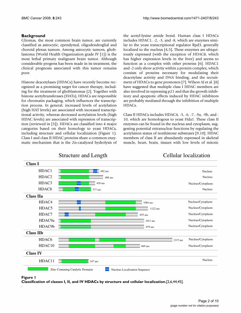
Histone deacetylases (HDACs) have recently become rec-ognized as a promising target for cancer therapy, includ-ing for the treatment of glioblastomas [2]. Together withhistone acetyltransferases (HATs), HDACs are responsiblefor chromatin packaging, which influences the transcrip-tion process. In general, increased levels of acetylation(high HAT levels) are associated with increased transcrip-tional activity, whereas decreased acetylation levels (highHDAC levels) are associated with repression of transcrip-tion (reviewed in [3]). HDACs are classified into 4 majorcategories based on their homology to yeast HDACs,including structure and cellular localization (Figure 1).Class I and class II HDAC proteins share a common enzy-matic mechanism that is the Zn-catalyzed hydrolysis of
the acetyl-lysine amide bond. Human class I HDACsincludes HDAC1, -2, -3, and -8, which are enzymes simi-lar to the yeast transcriptional regulator Rpd3, generallylocalized to the nucleus [4,5]. These enzymes are ubiqui-tously expressed (with the exception of HDAC8, whichhas higher expression levels in the liver) and seems tofunction as a complex with other proteins [6]. HDAC1and -2 only show activity within a protein complex, whichconsists of proteins necessary for modulating theirdeacetylase activity and DNA binding, and the recruit-ment of HDACs to gene promoters [7]. Wilson AJ et al. [8]have suggested that multiple class I HDAC members arealso involved in repressing p21 and that the growth inhib-itory and apoptotic effects induced by HDAC inhibitorsare probably mediated through the inhibition of multipleHDACs.
Class II HDACs includes HDAC4, -5, -6, -7, -9a, -9b, and -10, which are homologous to yeast Hda1. These class IIenzymes can be found in the nucleus and cytoplasm, sug-gesting potential extranuclear functions by regulating theacetylation status of nonhistone substrates [9,10]. HDACmembers of class II are abundantly expressed in skeletalmuscle, heart, brain, tissues with low levels of mitotic
Classification of classes I, II, and IV HDACs by structure and cellular localizationFigure 1Classification of classes I, II, and IV HDACs by structure and cellular localization.[2,6,44,45].
Zinc Contaning Catalytic Domaim Nucleus Localization Sequence
HDAC1
HDAC2
HDAC3
HDAC8
HDAC4
HDAC7
HDAC5
HDAC9a
HDAC6
HDAC10
HDAC11
HDAC9b
Class I
Class IIa
Class IIb
Class IV
482 aas
Structure and Length Cellular localization
Nucleus
Nucleus/Cytoplasm
Nucleus
Nucleus
Nucleus/Cytoplasm
Nucleus/Cytoplasm
Nucleus/Cytoplasm
Nucleus/Cytoplasm
Nucleus/Cytoplasm
Nucleus/Cytoplasm
Nucleus/Cytoplasm
Nucleus
488 aas
428 aas
377 aas
1084 aas
1122 aas
855 aas
1011 aas
879 aas
1215 aas
669 aas
347 aas
Page 2 of 10(page number not for citation purposes)
BMC Cancer 2008, 8:243 http://www.biomedcentral.com/1471-2407/8/243
activity [11,12]. Functionally, Class II HDACs is thoughtto act as transcriptional corepressors by deacetylatingnucleosomal histones. These enzymes do not binddirectly to DNA; they are thought to be recruited to dis-tinct regions of the genome by sequence specific DNAbinding proteins [13-15].
Class III HDACs is composed of the Sirtuins (SIRT) pro-teins 1–7, which are homologous to the yeast Sir2 proteinand require NAD+ for deacetylase activity in contrast to thezinc-catalyzed mechanism used by class I and II HDACs[16-18].
An additional HDAC expressed by higher eukaryotes is aZn-dependent HDAC (HDAC11 in mammals). Thisenzyme is phylogenetically different from both class I andclass II enzymes and is therefore classified separately asclass IV [19] reviewed in [5].
The use of HDAC inhibitors (HDACis) for the treatmentof cancer is an area of active investigation. In gliomas,HDACis have been used for the treatment of glioblastomain combination with radiation therapy and chemother-apy. Some authors have demonstrated that HDACis havea radiosensitizing effect on glioblastoma cells in vitro andin vivo [20-23] and also seem to be associated with inhi-bition of glioma cell growth by both cell-cycle arrest andapoptosis [24-26]. Despite the widespread use of HDACis,the mechanistic implications remain to be elucidated.
To this date, there are no studies that demonstrate the sta-tus of global HDAC gene expression and protein levels inastrocytomas. The purpose of this study was to evaluateand compare mRNA and protein levels of class I, II and IVof HDACs in low grade and high grade astrocytomas andnormal brain tissue and to correlate the findings with themalignancy in astrocytomas.
MethodsPatients SamplesFor this study, tumor samples of 43 patients (19 men and24 women) ranging in age from 1.3 to 79 years (mean age24.6 years, with a median and a standard deviation of12.8 ± 22.6 years) were evaluated. The histopathologicdiagnoses were 20 low-grade gliomas (13 grade I and 7grade II) and 23 high-grade gliomas (5 grade III and 18glioblastomas). In addition, 11 samples of normal cere-bral tissue were analyzed. Frozen tumor and normal spec-imens were microdissected. Diagnoses were based on2007 World Health Organization criteria [1].
For tumor microdissection, tumor samples were placedon a cooled platform and immediately positioned on thecutting base of the cryostat under Tissue Tek (Fisher Scien-tific, Pittsburg, PA). After rapid freezing in liquid nitrogen,
the sample was cut and immediately captured on a cover-slip, stained with hematoxylin and eosin, and evaluatedby image apposition. The area of interest in the originalcryopreserved tumor block was then trimmed, and themicrodissected sample was transferred to a previouslyidentified tube, which was immediately placed under dryice.
Prior to initiation, the research here presented wasapproved by the Research Ethics Committee of the Uni-versity Hospital of the Faculty of Medicine of University ofSao Paulo, processes number 9375/2003 and 7645/99.The mentioned Committee is in agreement with the Hel-sinki Declaration requirements for research carried out onhumans. Informed consent was also taken from eachpatient (or their legal representative) involved in thisproject, also in accordance to the Helsinki Declaration.
RNA extraction and cDNA synthesisTotal cellular RNA was extracted using Trizol® Reagent(Invitrogen, Carlsbad, CA, USA) and RNA was reversetranscribed to single-stranded cDNA using a High Capac-ity Kit (Applied Biossystems, Foster City, CA, USA)according to the manufacturer's protocol.
Quantitative real-time polymerase chain reaction (qRT-PCR)Messenger RNA expression level for each HDAC was eval-uated using an ABI 7500 machine (Applied Biosystems,Foster City, CA, USA). Amplifications were obtained usingon demand TaqMan® probes (Applied Biosystems, FosterCity, CA, USA) for each HDAC. For relative quantificationof gene expression, standard curves were constructed foreach gene by considering at least 3 points in triplicate of10-fold serial dilution of cDNA in water, starting from1:10 of a volume of undiluted cDNA transcribed from 1.0μg of total RNA. The slopes of standard curves rangedfrom -3.17 to -3.87. Blank and standard controls (calibra-tors) were run in parallel to verify amplification efficiencywithin each experiment. To normalize differences in theamount of total cDNA added to each reaction, β-glucuro-nidase (GUS β) gene expression was used as an endog-enous control. As a calibrator sample (reference samplefor relative quantification), the U343 cell line was used.To obtain the Ct (cycle) values, we established a thresholdof 0.1. All reactions were made in duplicate, and all pro-cedures were carried out at 4°C.
Western BlottingFor protein analysis, 30 μg of each sample was loaded andseparated by sodium dodecyl sulphate-polyacrylamide gelelectrophoresis [27]. Proteins were transferred to nitrocel-lulose membranes, and the membranes were then incu-bated in 1% Tris-buffered saline Tween-20 (TBST)containing 5% (w/v) dried non-fat milk for 1 hour at
Page 3 of 10(page number not for citation purposes)
BMC Cancer 2008, 8:243 http://www.biomedcentral.com/1471-2407/8/243
room temperature. The primary antibodies were diluted at1:3000 in TBST containing 5% (w/v) milk: HDAC9(Abcam, Cambridge, MA), Acetyl-Lys H3 (Abcam, Cam-bridge, MA), and Acetyl-Lys H4 (Upstate BiotechnologyLake Placid, NY), and the membranes were incubated for1 hour at room temperature, The membranes were thenwashed 3 times with TBST, incubated with the secondaryantibody (1:5000 in TBST) labeled by horseradish perox-idase (Abcam, Cambridge, MA) for 1 hour at room tem-perature, and washed 3 times with TBST. The secondaryantibody was visualized using electron chemiluminescentreagent (Pierce, Rockford, IL). Films were exposed from10 to 60 seconds and developed.
Statistical analysisComparison of gene expression between groups of tumorwas performed by nonparametric testes Mann-Whitneyand Kruskall-Wallis. The level of significance was set at p< 0.05 in all analyses.
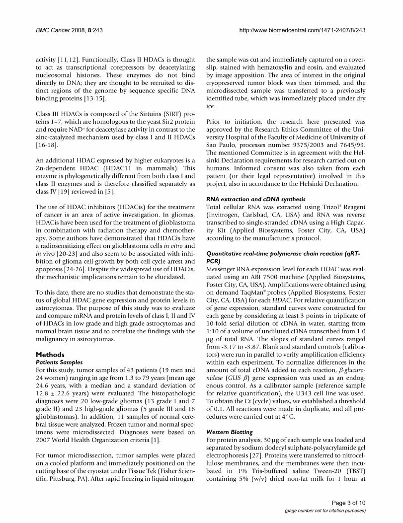
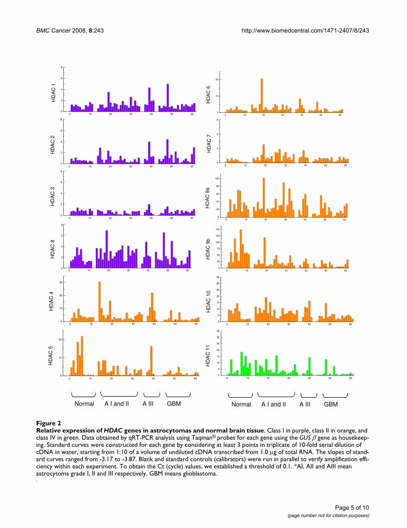
ResultsGlobal expression of HDAC genes in gliomas and normal brain tissueThe expression of 12 HDAC genes was analyzed using rel-ative quantification of mRNA levels in normal brain,astrocytomas grades I, II and III, and glioblastomas (Fig-ure 2). Class I HDAC genes (HDAC1, -2, -3, and -8)showed lower levels of expression (relative expression of0.5 to 6.0 approximately) compared to the other classesstudied. The highest values of expression for class I wereseen for HDAC8 (2.0 to 6.0). Expression of HDAC class IIand class IV were higher, with values of approximately 1.0to 150.0. The highest level of mRNA was observed forHDAC9a and HDAC9b, with relative expression reachingvalues above 100 for normal brain tissue and grade Iastrocytomas; however, HDAC6 and HDAC7 showed lev-els of expression comparable with those of class I (approx-imately 1.0 and 3.0).
Table 1 shows the median values for each HDAC gene inall groups analyzed. For class I, the highest median valueobserved was 2.87 (HDAC8 in low-grade astrocytoma)and the lowest median value was 0.62 (HDAC3 in astro-cytoma grade III). For class II, the highest median was61.51 (HDAC9b in normal brain) and the lowest was 0.56(HDAC7 in glioblastoma). HDAC11, the only member ofclass IV, had the lowest median value for normal brain tis-sue (3.67) and the highest for glioblastoma (1.02).
Comparison of the HDAC mRNA levels in low- and high-grade gliomas and normal brain tissueWe compared mRNA levels of HDAC genes (the mediansof relative expression) in low- and high-grade gliomas andalso in normal brain. Among class I HDACs, significant
differences in gene expression between tumor groups werenot observed (Table 2).
Seven of 8 class II HDAC genes (exception for HDAC4)were expressed at lower levels in high-grade astrocytomascompared to low-grade astrocytomas (p < 0.05; Table 2).The same significant difference for low-grade and high-grade gliomas was observed for HDAC11 (class IV).
We compared the most malignant form of astrocytoma(glioblastoma) with the other 3 tumors (astrocytomasgrades I, II and III) and normal brain in order to establisha correlation between HDAC expression and tumor grade(Table 3). As mentioned above, no significant differencein HDAC expression was observed for class I; however, forclasses II and IV, there was a decrease in expression ofthese genes in glioblastoma compared to that in othergroups. Moreover, this downregulation appeared to fol-low a pattern in which lower-grade tumors had a largernumber of HDAC genes at lower expression levels. Com-parison between glioblastoma and grade III astrocytomashowed that 4 of 8 genes were expressed at lower levels inglioblastoma samples (HDAC4, -6, -7, and -11). Compar-ison with low-grade astrocytoma (grades I and II) showedthat the expression of 6 of 8 genes was lower in glioblast-omas (except for HDAC5 and -7). Finally, when we com-pared glioblastoma with normal brain, 7 of 8 genesstudied, with the exception of HDAC7, were expressed atlower levels in glioblastoma.
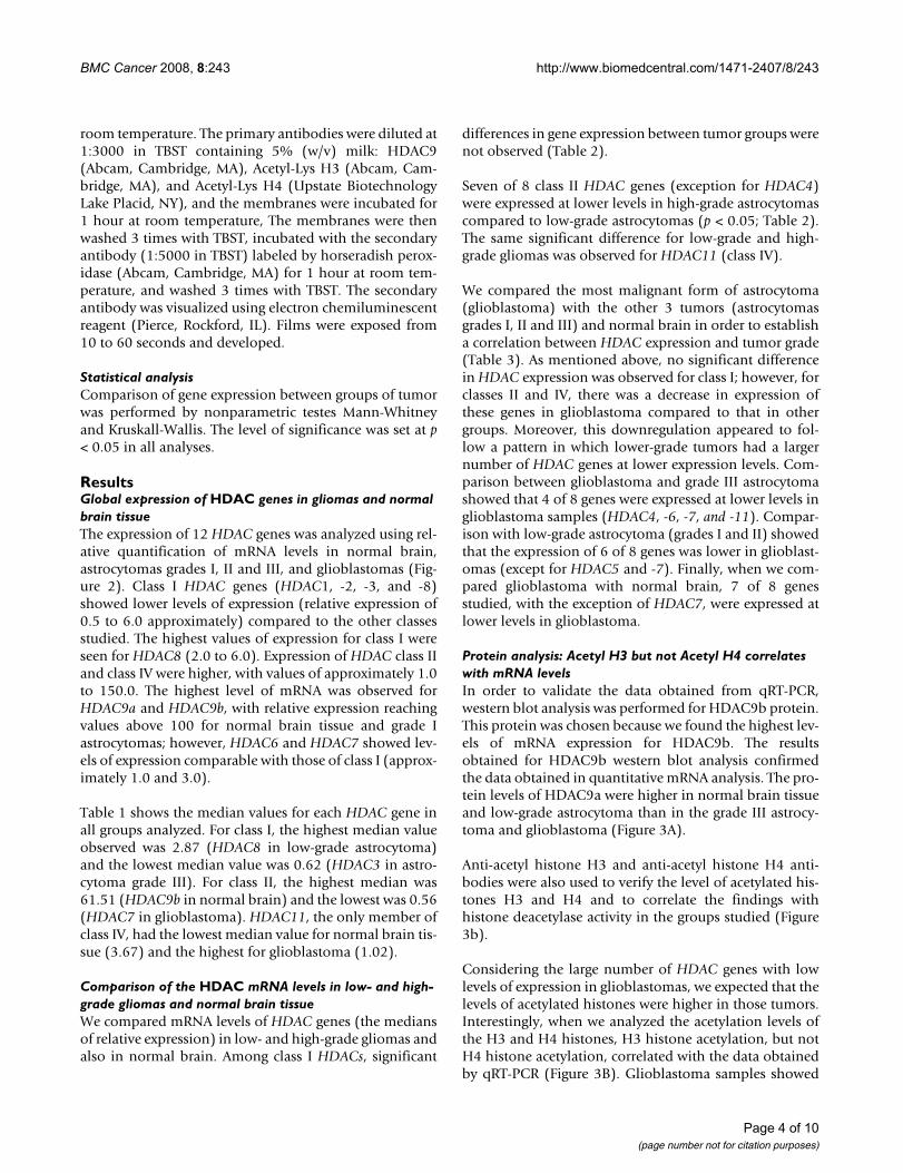
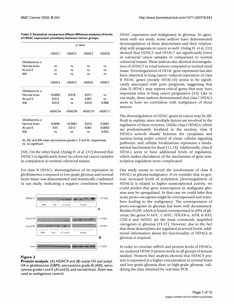
Protein analysis: Acetyl H3 but not Acetyl H4 correlates with mRNA levelsIn order to validate the data obtained from qRT-PCR,western blot analysis was performed for HDAC9b protein.This protein was chosen because we found the highest lev-els of mRNA expression for HDAC9b. The resultsobtained for HDAC9b western blot analysis confirmedthe data obtained in quantitative mRNA analysis. The pro-tein levels of HDAC9a were higher in normal brain tissueand low-grade astrocytoma than in the grade III astrocy-toma and glioblastoma (Figure 3A).
Anti-acetyl histone H3 and anti-acetyl histone H4 anti-bodies were also used to verify the level of acetylated his-tones H3 and H4 and to correlate the findings withhistone deacetylase activity in the groups studied (Figure3b).
Considering the large number of HDAC genes with lowlevels of expression in glioblastomas, we expected that thelevels of acetylated histones were higher in those tumors.Interestingly, when we analyzed the acetylation levels ofthe H3 and H4 histones, H3 histone acetylation, but notH4 histone acetylation, correlated with the data obtainedby qRT-PCR (Figure 3B). Glioblastoma samples showed
Page 4 of 10(page number not for citation purposes)
BMC Cancer 2008, 8:243 http://www.biomedcentral.com/1471-2407/8/243
Page 5 of 10(page number not for citation purposes)
Relative expression of HDAC genes in astrocytomas and normal brain tissueFigure 2Relative expression of HDAC genes in astrocytomas and normal brain tissue. Class I in purple, class II in orange, and class IV in green. Data obtained by qRT-PCR analysis using Taqman® probes for each gene using the GUS β gene as housekeep-ing. Standard curves were constructed for each gene by considering at least 3 points in triplicate of 10-fold serial dilution of cDNA in water, starting from 1:10 of a volume of undiluted cDNA transcribed from 1.0 μg of total RNA. The slopes of stand-ard curves ranged from -3.17 to -3.87. Blank and standard controls (calibrators) were run in parallel to verify amplification effi-ciency within each experiment. To obtain the Ct (cycle) values, we established a threshold of 0.1. *AI, AII and AIII mean astrocytoma grade I, II and III respectively. GBM means glioblastoma.
0 10 20 30 40 50 600
10
20
30
HD
AC
4
0 10 20 30 40 50 600
10
20
HD
AC
5
0 10 20 30 40 50 600
10
20
HD
AC
6
0 10 20 30 40 50 600
2
4
6
HD
AC
7
0 10 20 30 40 50 600
20
40
60
80
100
HD
AC
9a
0 10 20 30 40 50 600
25
50
75
100
125
150
HD
AC
9b
0 10 20 30 40 50 600
5
10
15
20
25
30
35
HD
AC
10
0 10 20 30 40 50 600
5
10
15
20
25
30
35
HD
AC
11
0 10 20 30 40 50 600
2
4
6
8
HD
AC
1
0 10 20 30 40 50 600
2
4
6
8
HD
AC
2
0 10 20 30 40 50 600
2
4
6
8
HD
AC
3
0 10 20 30 40 50 600
2
4
6
8
HD
AC
8
Normal A I and II A III GBM Normal A I and II A III GBM
BMC Cancer 2008, 8:243 http://www.biomedcentral.com/1471-2407/8/243
higher levels of acetylated histone H3 than normal brainand low-grade gliomas.
DiscussionIn this study we evaluated and compared mRNA levels of12 HDAC genes in astrocytomas and normal brain tissue.As mentioned before, the acetylation levels of histones isa process regulated by two groups of important enzymes:HATs and HDACs, and the balance of the activities of
these two enzymes is tightly related to the gene expressionstatus in the cell. The regulation of HDACs has been stud-ied, but is not yet well established. The reason for that ismaybe because HDACs seem to be regulated at multiplelevels including cellular compartmentalization, associa-tion with other factors, abundance, activity states andgenome-wide distribution.
Considering that altered gene expression is frequentlyobserved in cancer, a relationship between HDACs andcancer progression has been postulated. Wade P. [3] sug-gested that loss of targeting of class I HDACs through dis-ruption of a transcriptional corepressor and theinappropriate redistribution of class II resulted in the mis-regulated gene expression in cancer.
Although a crucial role for HDACs in gene transcriptionand their possible involvement in cancer has been pro-posed, no studies have demonstrated the expression pro-file of 3 classes of HDACs simultaneously in brain tumor.
Our study did not find differential gene expression of classI HDACs in high-grade and low-grade gliomas which mayindicate that class of deacetylases seems to not be directlyinvolved with malignancy of gliomas. Only a few studieshave evaluated the level of class I HDAC expression incancer: Huang BH et al. [28] demonstrated that HDAC1and HDAC2 seem to be upregulated in colon cancer; ChoiJH et al. [29] demonstrated an overexpression of HDAC1mRNA 68% of gastric cancer tissues studied by them (17of 25) and elevated expression of HDAC1 protein was alsodetected in 61% of the gastric cancer samples (11 of 18).Expression of class I HDAC3 was also shown elevated inastrocytic glial tumors compared to nonmalignant gliosis
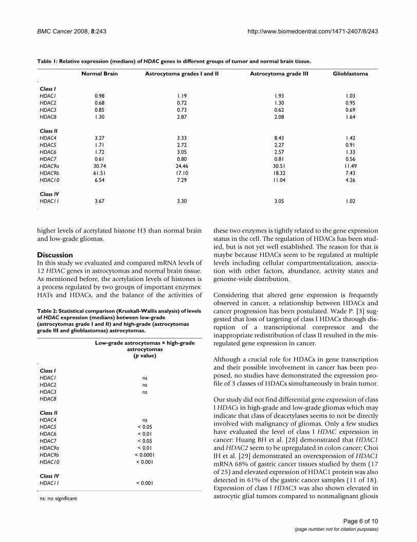
Table 1: Relative expression (medians) of HDAC genes in different groups of tumor and normal brain tissue.
Normal Brain Astrocytoma grades I and II Astrocytoma grade III Glioblastoma
Class IHDAC1 0.98 1.19 1.93 1.03HDAC2 0.68 0.72 1.30 0.95HDAC3 0.85 0.73 0.62 0.69HDAC8 1.30 2.87 2.08 1.64
Class IIHDAC4 3.27 3.33 8.43 1.42HDAC5 1.71 2.72 2.27 0.91HDAC6 1.72 3.05 2.57 1.33HDAC7 0.61 0.80 0.81 0.56HDAC9a 30.74 24.46 30.51 11.49HDAC9b 61.51 17.10 18.32 7.43HDAC10 6.54 7.29 11.04 4.26
Class IVHDAC11 3.67 3.30 3.05 1.02
Table 2: Statistical comparison (Kruskall-Wallis analysis) of levels of HDAC expression (medians) between low-grade (astrocytomas grade I and II) and high-grade (astrocytomas grade III and glioblastomas) astrocytomas.
Low-grade astrocytomas × high-grade astrocytomas
(p value)
Class IHDAC1 nsHDAC2 nsHDAC3 nsHDAC8
Class IIHDAC4 nsHDAC5 < 0.05HDAC6 < 0.01HDAC7 < 0.05HDAC9a < 0.01HDAC9b < 0.0001HDAC10 < 0.001
Class IVHDAC11 < 0.001
ns: no significant
Page 6 of 10(page number not for citation purposes)
BMC Cancer 2008, 8:243 http://www.biomedcentral.com/1471-2407/8/243
[30]. On the other hand, Ozdag H. et al. [31] showed thatHDAC1 is significantly lower in colorectal cancer samplesin comparison to normal colorectal tissues.
For class II HDACs, downregulation of its expression inglioblastoma compared to low-grade gliomas and normalbrain tissue was demonstrated and statistically confirmedin our study, indicating a negative correlation between
HDAC expression and malignancy in gliomas. In agree-ment with our study, some authors have demonstrateddownregulation of these deacetylases and their relation-ship with prognosis in cancer as well. Ozdag H. et al. [31]showed that HDAC5 and HDAC7 are significantly lowerin colorectal cancer samples in comparison to normalcolorectal tissues. These authors also showed downregula-tion of HDAC5 in renal tumors compared to normal renaltissue. Downregulation of HDAC gene expression has alsobeen observed in lung cancer: reduced expression of classII HDAC genes (mostly HDAC10) seems to be signifi-cantly associated with poor prognosis, suggesting thatclass II HDACs may repress critical genes that may haveimportant roles in lung cancer progression [32]. Like inour study, these authors demonstrated that class I HDACsseem to have no correlation with malignancy of thosetumors.
The downregulation of HDAC genes in cancer may be dif-ficult to explain, since multiple factors are involved in theregulation of these enzymes. Unlike class I HDACs, whichare predominantly localized in the nucleus, class IIHDACs actively shuttle between the cytoplasm andnucleus being under control of classic cellular signalingpathways, and cellular localizations represents a funda-mental mechanism for them [12,14]. Additionally, class IIHDACs seem to have additional levels of regulation,which makes elucidation of the mechanism of gene tran-scription regulation more complicated.
Our study seems to reveal the involvement of class IIHDACs in glioma malignancy. If we consider that, in gen-eral, increased levels of acetylation (downregulation ofHDACs) is related to higher transcriptional activity, wecould predict that gene transcription in malignant glio-mas may be upregulated. In that case we could infer thatsome proto-oncogenes might be overexpressed and some-how leading to the malignancy. The overexpression ofproto-oncogenes in gliomas has been well documented.Besides EGFR, which is found overexpressed in 40% of gli-omas, the genes N-MYC, C-MYC, PDGFR-α, MYB, K-RAS,CDK-4 and MDM2 are the most commonly amplifiedoncogenes in gliomas [33-37]. However, due to the factthat these deacetylases are regulated at several levels, addi-tional information about the functionality of HDACs ingliomas is required.
In order to correlate mRNA and protein levels of HDACs,we analyzed HDAC9 protein levels in all groups of tumorsstudied. Western blot analysis showed that HDAC9 pro-tein is expressed at a higher concentration in normal brainand low-grade gliomas than in high-grade gliomas, vali-dating the data obtained by real-time PCR.
Table 3: Statistical comparison (Mann-Whitney analysis) of levels of HDAC expression (medians) between tumor groups.
p value
HDAC1 HDAC2 HDAC3 HDAC8
Glioblastoma xNormal brain ns ns ns nsAI and II ns ns ns nsAIII ns ns ns ns
HDAC4 HDAC5 HDAC6 HDAC7
Glioblastoma xNormal brain <0.0001 0.018 0.011 nsAI and II 0.012 ns 0.001 nsAIII 0.010 ns 0.010 0.008
HDAC9a HDAC9b HDAC10 HDAC11
Glioblastoma xNormal brain 0.0096 <0.0001 0.015 0.0001AI and II 0.01 0.013 0.001 0.0003AIII ns ns ns 0.023
AI, AII, and AIII mean astrocytoma grades I, II and III, respectively.ns: no significant
Protein analysisFigure 3Protein analysis. (A) HDAC9 and (B) acetyl H3 and acetyl H4 in glioblastoma (GBM), astrocytoma grade III (AIII), astro-cytoma grades I and II (AI and II), and normal brain. Actin was used as endogenous control.
17 kDa
10 kDa
Acetyl H4
Acetyl H3
37 kDaActin
HDAC 9
GBM A I and IIA III Normal
135 kDa
A
B
GBM A I and IIA III Normal
Page 7 of 10(page number not for citation purposes)
BMC Cancer 2008, 8:243 http://www.biomedcentral.com/1471-2407/8/243
We also evaluated HDAC activity by analyzing histoneacetylation levels in tumor samples and normal brain.When we analyzed the levels of H3 and H4 acetylated his-tones, we observed an increased level in acetylation of H3but not of H4 histone in glioblastomas compared to low-grade astrocytomas and normal brain tissue. Consideringthe low levels of HDAC expression in glioblastomas, itwas expected that the levels of acetylated histones shouldbe more elevated in those tumors, as demonstrated herefor histone H3. On the other hand, the lack of correlationbetween low HDAC expression and high histone H4deacetylation levels in glioblastomas could be explainedby the existence of cofactors or unidentified regulators.Some few authors have already demonstrated that the spe-cificity of HDACs depends on cofactors which makesHDAC specificity a complicated process [38-41]. It istempting to speculate that class II of HDACs could beresponsible for deacetylation of histone H3 more thanhistone H4. However, the data here presented are notenough to infer about the specificity of HDACs in astrocy-tomas. Moreover, it has been observed that differencesbetween HDAC subtype specificity do not coincide withthe division into class I and class II enzymes. HDAC1,HDAC3 (class I), and HDAC6 (class II) seem to be verysimilar in substrate specificity and mainly differ in thedegree of specificity [39]. Until now, a lot of data has beengenerated about HDAC research, but the natural sub-strates of different HDACs and their substrate specificitiesis still not well understood.
The class IV HDAC11 enzyme seems to be an unusualmember of the HDAC family. Its sequence is not homol-ogous to any other HDAC, and it may have distinct phys-iological roles. This enzyme, like members of the class IIHDACs is expressed more in brain, heart, skeletal muscle,and kidney [19]. In our study, we also found high levelsof HDAC11 in normal brain tissue and significant differ-ential expression was also observed for this enzyme inlow-grade and high-grade gliomas. Little is known aboutthis unique HDAC, and its relation to malignancy of glio-mas is yet to be elucidated.
HDACisEven considering that to evaluate the effect of HDACinhibitors in gliomas was not the aim of the present study,the finding that most of the HDAC genes are downregu-lated in glioblastomas could lead us to consider thatHDACis may not be effective in the treatment of high-grade gliomas. Some studies have demonstrated radiosen-sitization of gliomas after HDACi treatment [21,22].Therefore, despite the low levels of HDAC gene expressionin glioblastomas, HDACis seem to be potential therapeu-tical targets for glioma treatment. The explanation to thismay point to the existence of nonhistone substrates forHDACs. Although histones are the most thoroughly stud-
ied as HDAC substrates, several reports have shown thatHDACs are also responsible for the deacetylation ofdiverse types of nonhistone proteins, including transcrip-tional factors, signal transduction mediators, and molecu-lar chaperones (a summarized table of nonhistonesubstrates for HDACs can be found in [2]). Additionally,recent evidences suggest that modulation of gene expres-sion through histone remodeling might not be the onlyprocess responsible for the antiproliferative action ofHDACis [42,43]. Although the present study has demon-strated that most of HDACs genes are downregulated inglioblastomas, no experiment was performed in order toanalyze the effect of HDAC inhibitors on glioma treat-ment, therefore the effect of these inhibitors on gliomatreatment should be addressed in a separate manuscript.
ConclusionOur study has established a negative correlation betweenHDAC gene expression and glioma grade, suggesting thatclass II and class IV HDACs might play an important rolein glioma malignancy. This differential HDAC expressionmay provide insight into development of novel treatmentapproaches for this devastating disease. However, a morecomplete understanding of the biological function andspecificity of the diverse HDAC isoforms and theirinvolvement in the cancer process is necessary.
Competing interestsThe authors declare that they have no competing interest.
Authors' contributionsExperiments and collection of data were performed byAKBL-E, MAAC, ETV, FJNM and RGPQ. Collection oftumor samples was performed by HRM and CGCJr.Microdissection of the tumor samples was performed byLN. AKBL-E was responsible for data analysis and inter-pretation and also wrote the manuscript. Manuscriptreviewing was made by AKBL-E, MAAC, CAS and LGT.
AcknowledgementsWe thank Dr. John F. de Groot and Dr. Vinay Puduvalli from MD Anderson Cancer Center (Houston, Tx, USA) for providing Actin and Acetyl-H4 anti-bodies, respectively. We also thank Priscila C. Leite for the technical assist-ance in this work.
References1. Louis DN, Ohgaki H, Wiestler OD, Cavenee WK, Burger PC, Jouvet
A, Scheithauer BW, Kleihues P: The 2007 WHO classification oftumours of the central nervous system. Acta neuropathologica2007, 114(2):97-109.
2. Lin HY, Chen CS, Lin SP, Weng JR, Chen CS: Targeting histonedeacetylase in cancer therapy. Medicinal research reviews 2006,26(4):397-413.
3. Wade PA: Transcriptional control at regulatory checkpointsby histone deacetylases: molecular connections betweencancer and chromatin. Human molecular genetics 2001,10(7):693-698.
4. Gray SG, Ekstrom TJ: The human histone deacetylase family.Experimental cell research 2001, 262(2):75-83.
Page 8 of 10(page number not for citation purposes)
BMC Cancer 2008, 8:243 http://www.biomedcentral.com/1471-2407/8/243
5. Gallinari P, Di Marco S, Jones P, Pallaoro M, Steinkuhler C: HDACs,histone deacetylation and gene transcription: from molecu-lar biology to cancer therapeutics. Cell research 2007,17(3):195-211.
6. de Ruijter AJ, van Gennip AH, Caron HN, Kemp S, van KuilenburgAB: Histone deacetylases (HDACs): characterization of theclassical HDAC family. The Biochemical journal 2003, 370(Pt3):737-749.
7. Zhang Y, Ng HH, Erdjument-Bromage H, Tempst P, Bird A, ReinbergD: Analysis of the NuRD subunits reveals a histone deacety-lase core complex and a connection with DNA methylation.Genes & development 1999, 13(15):1924-1935.
8. Wilson AJ, Byun DS, Popova N, Murray LB, L'Italien K, Sowa Y,Arango D, Velcich A, Augenlicht LH, Mariadason JM: Histonedeacetylase 3 (HDAC3) and other class I HDACs regulatecolon cell maturation and p21 expression and are deregu-lated in human colon cancer. The Journal of biological chemistry2006, 281(19):13548-13558.
9. Grozinger CM, Hassig CA, Schreiber SL: Three proteins define aclass of human histone deacetylases related to yeast Hda1p.Proceedings of the National Academy of Sciences of the United States ofAmerica 1999, 96(9):4868-4873.
10. Verdin E, Dequiedt F, Kasler HG: Class II histone deacetylases:versatile regulators. Trends Genet 2003, 19(5):286-293.
11. Kao HY, Downes M, Ordentlich P, Evans RM: Isolation of a novelhistone deacetylase reveals that class I and class II deacety-lases promote SMRT-mediated repression. Genes & develop-ment 2000, 14(1):55-66.
12. Verdel A, Khochbin S: Identification of a new family of highereukaryotic histone deacetylases. Coordinate expression ofdifferentiation-dependent chromatin modifiers. The Journal ofbiological chemistry 1999, 274(4):2440-2445.
13. Lemercier C, Verdel A, Galloo B, Curtet S, Brocard MP, Khochbin S:mHDA1/HDAC5 histone deacetylase interacts with andrepresses MEF2A transcriptional activity. The Journal of biologi-cal chemistry 2000, 275(20):15594-15599.
14. Miska EA, Karlsson C, Langley E, Nielsen SJ, Pines J, Kouzarides T:HDAC4 deacetylase associates with and represses the MEF2transcription factor. The EMBO journal 1999, 18(18):5099-5107.
15. Wang AH, Bertos NR, Vezmar M, Pelletier N, Crosato M, Heng HH,Th'ng J, Han J, Yang XJ: HDAC4, a human histone deacetylaserelated to yeast HDA1, is a transcriptional corepressor. MolCell Biol 1999, 19(11):7816-7827.
16. Blander G, Guarente L: The Sir2 family of protein deacetylases.Annual review of biochemistry 2004, 73:417-435.
17. Imai S, Armstrong CM, Kaeberlein M, Guarente L: Transcriptionalsilencing and longevity protein Sir2 is an NAD-dependenthistone deacetylase. Nature 2000, 403(6771):795-800.
18. Marmorstein R: Structure and chemistry of the Sir2 family ofNAD+-dependent histone/protein deactylases. BiochemicalSociety transactions 2004, 32(Pt 6):904-909.
19. Gao L, Cueto MA, Asselbergs F, Atadja P: Cloning and functionalcharacterization of HDAC11, a novel member of the humanhistone deacetylase family. The Journal of biological chemistry 2002,277(28):25748-25755.
20. Lopez CA, Feng FY, Herman JM, Nyati MK, Lawrence TS, LjungmanM: Phenylbutyrate sensitizes human glioblastoma cells lack-ing wild-type p53 function to ionizing radiation. Internationaljournal of radiation oncology, biology, physics 2007, 69(1):214-220.
21. Camphausen K, Cerna D, Scott T, Sproull M, Burgan WE, Cerra MA,Fine H, Tofilon PJ: Enhancement of in vitro and in vivo tumorcell radiosensitivity by valproic acid. International journal of can-cer 2005, 114(3):380-386.
22. Entin-Meer M, Yang X, VandenBerg SR, Lamborn KR, Nudelman A,Rephaeli A, Haas-Kogan DA: In vivo efficacy of a novel histonedeacetylase inhibitor in combination with radiation for thetreatment of gliomas. Neuro-oncology 2007, 9(2):82-88.
23. Kim JH, Shin JH, Kim IH: Susceptibility and radiosensitization ofhuman glioblastoma cells to trichostatin A, a histonedeacetylase inhibitor. International journal of radiation oncology, biol-ogy, physics 2004, 59(4):1174-1180.
24. Sawa H, Murakami H, Kumagai M, Nakasato M, Yamauchi S, Mat-suyama N, Tamura Y, Satone A, Ide W, Hashimoto I, Kamada H: His-tone deacetylase inhibitor, FK228, induces apoptosis andsuppresses cell proliferation of human glioblastoma cells invitro and in vivo. Acta neuropathologica 2004, 107(6):523-531.
25. Ugur HC, Ramakrishna N, Bello L, Menon LG, Kim SK, Black PM, Car-roll RS: Continuous intracranial administration of suberoy-lanilide hydroxamic acid (SAHA) inhibits tumor growth inan orthotopic glioma model. Journal of neuro-oncology 2007,83(3):267-275.
26. Wetzel M, Premkumar DR, Arnold B, Pollack IF: Effect of trichos-tatin A, a histone deacetylase inhibitor, on glioma prolifera-tion in vitro by inducing cell cycle arrest and apoptosis.Journal of neurosurgery 2005, 103(6 Suppl):549-556.
27. Laemmli UK: Cleavage of structural proteins during theassembly of the head of bacteriophage T4. Nature 1970,227(5259):680-685.
28. Huang BH, Laban M, Leung CH, Lee L, Lee CK, Salto-Tellez M, RajuGC, Hooi SC: Inhibition of histone deacetylase 2 increasesapoptosis and p21Cip1/WAF1 expression, independent ofhistone deacetylase 1. Cell death and differentiation 2005,12(4):395-404.
29. Choi JH, Kwon HJ, Yoon BI, Kim JH, Han SU, Joo HJ, Kim DY:Expression profile of histone deacetylase 1 in gastric cancertissues. Jpn J Cancer Res 2001, 92(12):1300-1304.
30. Liby P, Kostrouchova M, Pohludka M, Yilma P, Hrabal P, Sikora J, Bro-zova E, Kostrouchova M, Rall JE, Kostrouch Z: Elevated and dereg-ulated expression of HDAC3 in human astrocytic glialtumours. Folia biologica 2006, 52(1-2):21-33.
31. Ozdag H, Teschendorff AE, Ahmed AA, Hyland SJ, Blenkiron C,Bobrow L, Veerakumarasivam A, Burtt G, Subkhankulova T, ArendsMJ, Collins VP, Bowtell D, Kouzarides T, Brenton JD, Caldas C: Dif-ferential expression of selected histone modifier genes inhuman solid cancers. BMC genomics 2006, 7:90.
32. Osada H, Tatematsu Y, Saito H, Yatabe Y, Mitsudomi T, Takahashi T:Reduced expression of class II histone deacetylase genes isassociated with poor prognosis in lung cancer patients. Inter-national journal of cancer 2004, 112(1):26-32.
33. Goussia AC, Agnantis NJ, Rao JS, Kyritsis AP: Cytogenetic andmolecular abnormalities in astrocytic gliomas (Review).Oncology reports 2000, 7(2):401-412.
34. Jiang R, Mircean C, Shmulevich I, Cogdell D, Jia Y, Tabus I, Aldape K,Sawaya R, Bruner JM, Fuller GN, Zhang W: Pathway alterationsduring glioma progression revealed by reverse phase proteinlysate arrays. Proteomics 2006, 6(10):2964-2971.
35. Kleihues P, Ohgaki H: Primary and secondary glioblastomas:from concept to clinical diagnosis. Neuro-oncology 1999,1(1):44-51.
36. Michotte A, Neyns B, Chaskis C, Sadones J, In 't Veld P: Neu-ropathological and molecular aspects of low-grade and high-grade gliomas. Acta neurologica Belgica 2004, 104(4):148-153.
37. Ng HK, Lam PY: The molecular genetics of central nervoussystem tumors. Pathology 1998, 30(2):196-202.
38. Kolle D, Brosch G, Lechner T, Pipal A, Helliger W, Taplick J, Loidl P:Different types of maize histone deacetylases are distin-guished by a highly complex substrate and site specificity.Biochemistry 1999, 38(21):6769-6773.
39. Riester D, Hildmann C, Grunewald S, Beckers T, Schwienhorst A:Factors affecting the substrate specificity of histone deacety-lases. Biochemical and biophysical research communications 2007,357(2):439-445.
40. Wegener D, Hildmann C, Riester D, Schwienhorst A: Improvedfluorogenic histone deacetylase assay for high-throughput-screening applications. Analytical biochemistry 2003,321(2):202-208.
41. Wiren M, Silverstein RA, Sinha I, Walfridsson J, Lee HM, Laurenson P,Pillus L, Robyr D, Grunstein M, Ekwall K: Genomewide analysis ofnucleosome density histone acetylation and HDAC functionin fission yeast. The EMBO journal 2005, 24(16):2906-2918.
42. Brinkmann H, Dahler AL, Popa C, Serewko MM, Parsons PG, GabrielliBG, Burgess AJ, Saunders NA: Histone hyperacetylation inducedby histone deacetylase inhibitors is not sufficient to causegrowth inhibition in human dermal fibroblasts. The Journal ofbiological chemistry 2001, 276(25):22491-22499.
43. Johnstone RW, Licht JD: Histone deacetylase inhibitors in can-cer therapy: is transcription the primary target? Cancer cell2003, 4(1):13-18.
44. Acharya MR, Sparreboom A, Venitz J, Figg WD: Rational develop-ment of histone deacetylase inhibitors as anticancer agents:a review. Molecular pharmacology 2005, 68(4):917-932.
Page 9 of 10(page number not for citation purposes)
BMC Cancer 2008, 8:243 http://www.biomedcentral.com/1471-2407/8/243
Publish with BioMed Central and every scientist can read your work free of charge
"BioMed Central will be the most significant development for disseminating the results of biomedical research in our lifetime."
Sir Paul Nurse, Cancer Research UK
Your research papers will be:
available free of charge to the entire biomedical community
peer reviewed and published immediately upon acceptance
cited in PubMed and archived on PubMed Central
yours — you keep the copyright
Submit your manuscript here:http://www.biomedcentral.com/info/publishing_adv.asp
BioMedcentral
45. Drummond DC, Noble CO, Kirpotin DB, Guo Z, Scott GK, BenzCC: Clinical development of histone deacetylase inhibitors asanticancer agents. Annual review of pharmacology and toxicology2005, 45:495-528.
Pre-publication historyThe pre-publication history for this paper can be accessedhere:
http://www.biomedcentral.com/1471-2407/8/243/prepub
Page 10 of 10(page number not for citation purposes)


