Embed Size (px)
Citation preview

Differential requirements for retinal degenerationslow intermolecular disulfide-linked oligomerizationin rods versus cones
Dibyendu Chakraborty1, Xi-Qin Ding1, Shannon M. Conley1, Steven J. Fliesler2,{
and Muna I. Naash1,�
1Department of Cell Biology, University of Oklahoma Health Sciences Center, Oklahoma City, OK 73104, USA and2Departments of Ophthalmology and Pharmacological & Physiological Science, Saint Louis University School of
Medicine, St. Louis, MO 63104, USA
Received October 6, 2008; Revised November 13, 2008; Accepted November 27, 2008
It is commonly assumed that the ultrastructural organization of the rim region of outer segment (OS) discs inrods and lamellae in cones requires functional retinal degeneration slow/rod outer segment membraneprotein 1 (Rds/Rom-1) complexes. Cysteine-150 (C150) in Rds has been implicated in intermolecular disulfidebonding essential for functional Rds complexes. Transgenic mice containing the Rds C150S mutation(C150S-Rds) failed to form higher-order Rds oligomers, although interactions between C150S-Rds andRom-1 occurred in rods, but not in cones. C150S-Rds mice exhibited marked early-onset reductions incone function and abnormal OS structure. In contrast, C150S-Rds expression in rods partly rescued therds1/2 phenotype. Although C150S-Rds was detected in the OSs in rods and cones, a substantial percentageof C150S-Rds and cone opsins were mislocalized to different cellular compartments in cones. The results ofthis study provide novel insights into the importance of C150 in Rds oligomerization and the differences inRds requirements in rods versus cones. The apparent OS structural differences between rods and cones maycause cones to be more susceptible to the elimination of higher-order Rds/Rom-1 oligomers (e.g. as mediatedby mutation of the Rds C150 residue).
INTRODUCTION
Highly evolved mammalian vision is made possible by thespecialized organelles known as outer segments (OSs) foundon both rod and cone photoreceptor cells. The microanatomyand structure of these modified cilia have been eloquentlydescribed (1–3). In rods, it was historically thought that theplasma membrane successively evaginates and invaginateswith subsequent membrane fusion to form stacks of sealedorganized discs encased on all sides by the plasma membrane.Although an alternative theory to evagination/invagination hasrecently been proposed (4), it remains quite clear that rod OSscontain stacks of discs completely distinct from the plasmamembrane. On the contrary, in cones, membrane fusion does
not take place; instead, stacks of lamellae are formed, whichremain contiguous with the plasma membrane (2). As rodsand cones mediate different types of vision, the structuraland biochemical features of their OSs are necessarily distinct.Although the biochemistry of rod- and cone-mediated visualsignaling is well understood, it remains a persistent questionas to how cones and rods achieve their distinct differencesin OS structure. This puzzle is made more complex by theobservation that a key structural protein of the OS, calledretinal degeneration slow (Rds), is expressed in both rodsand cones.
Rds is a tetraspanin protein and is localized to the disc rimof the rod and cone OSs (5,6). Rds is necessary for the for-mation of OSs: retinal degeneration slow (rds2/2) mice
†Present address: Department of Ophthalmology (Ross Eye Institute), University at Buffalo, SUNY, Buffalo, NY, USA and Research Service, VeteransAdministration, Western New York Healthcare System, Buffalo, NY 14215, USA.
�To whom correspondence should be addressed at: Department of Cell Biology, University of Oklahoma Health Sciences Center, 940 StantonL. Young Blvd., BMSB 781, Oklahoma City, OK 73126-0901, USA. Tel: þ1 4052718001 ext 47969; Fax: þ1 4052713548; Email: [email protected].
# The Author 2008. Published by Oxford University Press. All rights reserved.For Permissions, please email: [email protected]
Human Molecular Genetics, 2009, Vol. 18, No. 5 797–808doi:10.1093/hmg/ddn406Advance Access published on December 2, 2008

lacking Rds completely fail to develop OS structures, whereasthe heterozygous (rdsþ/2) mice exhibit disorganized OSs withimproperly formed discs (5–9). In humans, over 80 mutationsin the RDS gene have been found to associate with a variety ofinherited retinal degenerative diseases, including autosomal-dominant retinitis pigmentosa, cone–rod dystrophy and mul-tiple forms of macular dystrophy (10,11) (http://www.retina-international.org/sci-news/rdsmut.htm). Consistentwith its role in the maintenance of rods and cones, Rdsmutations manifest as rod and/or cone dystrophies withvarying levels of severity, suggesting that the protein has dis-tinct functions in rod and cone photoreceptors (12). Our recentwork (13) using the cone-dominant mouse model lacking thetranscription factor neural retina leucine zipper (nrl2/2) (14)supports this hypothesis. In the absence of Rds, cones in thenrl2/2 retina retain the capacity for phototransduction andare able to elaborate some OS structure (albeit aberrant, dys-morphic structures that lack disc rims), in stark contrast torods of the rds2/2 retinas, which do not form OSs and haveno detectable electroretinographic (ERG) responses (13).Clearly, rod and cone OS formation and maintenance bothrequire Rds, but not in the same way.
Further understanding of the role of Rds in rods versus conesrequires analysis of the function of Rds on the biochemicallevel. It is known that Rds forms non-covalent homo- andhetero-tetramers with its non-glycosylated homologue, rodouter segment membrane protein 1 (Rom-1) (15), anothermember of the tetraspanin family also localized to the discrim region. These tetrameric complexes are assembled in theinner segment before trafficking to the rim region of the OS.Once in the OS, these tetramers can further interact with oneanother through intermolecular disulfide bonds to form octa-mers and higher-order oligomers (i.e. complexes larger than tet-ramers) (16). Rom-1 does not participate in these higher-orderoligomers in rods or cones (17) and elimination of Rom-1(rom-12/2) does not lead to significant structural abnormalitiesin the photoreceptor OSs (18), leading us to hypothesize thathigher-order Rds oligomers are the functional unit of Rdsinvolved in the maintenance of rim structures. Early mutagen-esis studies (19) suggest that the seven conserved Cys residuesin the D2 loop of Rds play a critical role in maintaining theinteractions between Rds and other proteins which are necess-ary for OS structural integrity. The role of Cys-mediated disul-fide bonds in Rds/Rds interactions during complex formation isvalidated by the observation that a significant fraction of Rdsmigrates as a dimer on non-reducing sodium dodecyl sulfate–polyacrylamide gel electrophoresis (SDS–PAGE), but as amonomer in the presence of reducing agents (16). One of theD2 loop cysteines, in particular, C150 is thought to be function-ally important for Rds–Rds disulfide bonding, based on in vitrostudies with C150S-mutant Rds. In COS cells, C150S-Rds wasunable to form disulfide bond-mediated dimers (19), butretained the ability to form heterotetramers with Rom-1. Thissuggests that C150 is not involved in tetrameric subunit assem-bly, but is required for intermolecular disulfide bonding and,thus, higher-order oligomer formation and, by extension, OSmorphogenesis and maintenance.
In this study, we investigated the role of C150 using an invivo model system in order to understand the role of intermo-lecular disulfide bonding in the function of Rds, particularly
with regard to the formation and maintenance of rod versuscone OSs. As COS cells do not form OSs and, hence, areinadequate to model the structural complexity of rod andcone OSs, the findings presented here using transgenic miceare of particular importance. Our experiments provide criticalnew insights into how rods and cones differentially utilize Rdsand shed some light on the question of how a single proteincan be directly and critically involved in the formation of cel-lular structures as different as those of rod and cone OSs.
RESULTS
Expression of C150S-Rds in transgenic mice
To understand the differential role of C150-mediated Rds oli-gomerization in rod versus cone OS morphogenesis, we gener-ated two transgenic models expressing C150S-Rds. Transgeneexpression was driven in rods by the mouse opsin promoter(named MOP-T) and in cones by the human red/green coneopsin promoter (named COP-T) (Fig. 1A). Both these promo-ters have been used in transgenic mice to drive cell-specificexpression (20,21), with the COP-T promoter directing robustexpression in both blue and red/green cones (21,22). TwentyMOP-T and 15 COP-T lines were generated and evaluated forsite of integration and levels of transgene expression. Generalexamination of these mice revealed no side effects of the trans-gene on animal weight or behavior. After eliminating the rdmutation (23), all founders were moved into the C57BL/6 back-ground for several generations and then crossed with rds2/2
mice on C57BL/6 background to obtain transgenic mice onall rds genetic backgrounds [wild-type (WT), rdsþ/2 andrds2/2]. In this study, we present data from the highest expres-ser lines for the MOP and COP transgenes.
To evaluate levels and integrity of the transgene transcript,northern blot and quantitative RT–PCR (qRT–PCR) analysiswere performed on total retinal RNA isolated from transgenicretinas on different rds backgrounds (Fig. 1B and C). Northernblot hybridized with RDS cDNA probe detected two tran-scripts (1.6 and 2.7 kb) from the WT allele (Fig. 1B, left)and a large band (.9 kb) from the RDS allele in the rdsþ/2
sample (Fig. 1B, left) (8,24,25). When the blot was hybridizedto an SV40 poly-A probe, a 2.4 kb transgene transcript wasonly detected in MOP-T/WT (Fig. 1B, right). qRT–PCRanalysis on MOP-T and COP-T retinas on rds2/2 backgroundrevealed transgene transcript levels of 50 and 25% of the WT,respectively. This suggests that the amount of MOP-Tmessage is equivalent to that of one WT allele, whereas theCOP-T message is close to half of one WT allele (Fig. 1C).
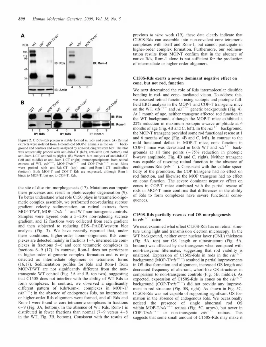
C150S-Rds protein is stably expressed in rods and cones
To determine the levels of C150S-Rds protein in the twoselected lines, western blot analysis and immunoprecipitation(IP) were performed on retinal extracts from MOP-T andCOP-T mice in the rds2/2 background (Fig. 2). Figure 2Ashows non-reducing western blots probed with anti-Rds-CT(left) and anti-Rom-1-CT (right) antibody. MOP-TC150S-Rds is expressed in the absence of endogenous Rds(lane 3, left) and stabilizes Rom-1 which is not usually detectedin the rds2/2 (lanes 5 and 6, right). In WT retinal extract,
798 Human Molecular Genetics, 2009, Vol. 18, No. 5

monomers, dimers and higher-order Rds complexes are readilydetected, whereas the MOP-T extract in the rds2/2 backgroundshows monomers. Occasionally, a very faint dimer-sized bandof unknown origin is observed in MOP-T/rds2/2. It is possiblethat alterations in the structure of the Rds-D2 loop arising fromthe C to S substitution have prompted a small fraction ofC150S-Rds protein to form dimers mediated by one of theother D2 loop cysteines; however, very little dimer is everobserved, and higher-order complexes are never detected.These results confirm earlier in vitro observations thatsuggested that C150-mediated intermolecular disulfidebonding is necessary for normal Rds dimerization (19).Because the photoreceptor population of WT retinas is com-prised of .95% rods and only �3–5% cones, we were notable to calculate COP-T protein levels by western blot analysis.As indicated below, we used IP in conjunction with the westernblot analysis to examine C150S-Rds in COP-T retinas.
C150S-Rds associates with Rom-1 in rods, but not in cones
Rds and Rom-1 are known to form hetero- and homomericcomplexes of different sizes (16,17). To evaluate the role ofRds C150 in these associations in rods versus cones,
co-immunoprecipitation (co-IP) with Rds-CT and Rom-1-CTantibodies was performed on retinal extracts from MOP-Tand COP-T mice on the rds2/2 background and from WTand rds2/2 extracts as controls (Fig. 2B). The blots wereprobed with anti-Rds-CT (Fig. 2B, top) and withanti-Rom-1-CT (Fig. 2B, bottom). As in the WT, IP withRds-CT antibody from MOP-T/rds2/2 extract brought downRom-1 (lane 3). On the contrary, IP from COP-T/rds2/2
failed to bring down Rom-1 (lane 6). Reciprocal IP withRom-1 antibody confirmed that Rom-1 is present in COP-T/rds2/2, but does not associate with C150S-Rds in cones(lane 9). These results clearly demonstrate that C150S-Rdscan bind to Rom-1 in rods, but not in cones, and serve asfurther confirmation that Rds has different roles in the twophotoreceptor cell types. In addition, these results suggestthat different conditions are necessary for Rds complex for-mation in rods versus cones.
C150S-Rds is not capable of forming higher-ordercomplexes in rods
Rds molecules are normally assembled into tetramers beforetrafficking to the OS and into oligomers in the OS, likely at
Figure 1. Generation and expression of the C150S-Rds transgene. (A) Schematic diagram of C150S transgene constructs. The transgene is composed of thefull-length mouse RDS cDNA with the C150S mutation followed by an SV40 poly-A tail and directed either to rods by the MOP or to cones by the humanred/green opsin promoter (COP). (B) Northern blot analysis of whole retinal RNA extracts (10 mg/sample) taken from 1-month-old MOP-T transgenic, non-transgenic WT littermates and rdsþ/2 mice. Two different probes were hybridized to the membrane: one from exon 1 of the RDS cDNA that recognizesboth endogenous and transgene transcripts (left) and an SV40 poly-A probe that recognizes the transgenic message only (right). (C) qRT–PCR was used tomeasure levels of total RDS message from WT, rds2/2, MOP-T/rds2/2 and COP-T/rds2/2 retinas. Analysis was performed on retinas from 3- to4-week-old mice using primer specific to the endogenous RDS gene.
Human Molecular Genetics, 2009, Vol. 18, No. 5 799
the site of disc rim morphogenesis (17). Mutations can impairthese processes and result in photoreceptor degeneration (9).To better understand what role C150 plays in tetrameric/oligo-meric complex assembly, we performed non-reducing sucrosegradient velocity sedimentation on retinal extracts fromMOP-T/WT, MOP-T/rds2/2 and WT non-transgenic controls.Samples were layered onto a 5–20% non-reducing sucrosegradient, and 12 fractions were collected from each gradientand then subjected to reducing SDS–PAGE/western blotanalysis (Fig. 3). We have recently reported that, underthese conditions, higher-order homo–oligomeric Rds com-plexes are detected mainly in fractions 1–4, intermediate com-plexes in fractions 5–6 and core tetrameric complexes infractions 6–9 (17). In contrast, Rom-1 does not participatein higher-order oligomeric complex formation and is onlydetected as intermediate oligomers or tetrameric forms(16,17). Sedimentation profiles for Rds and Rom-1 fromMOP-T/WT are not significantly different from the non-transgenic WT control (Fig. 3A and B, top two), suggestingthat C150S does not interfere with the ability of WT Rds toform complexes. In contrast, we observed a significantlydifferent pattern of Rds/Rom-1 complexes in MOP-T/rds2/2; in the absence of endogenous Rds, no intermediateor higher-order Rds oligomers were formed, and all Rds andRom-1 were found as core tetrameric complexes in fractions6–9 (Fig. 3A, bottom). In the absence of WT Rds, Rom-1 isdistributed in fewer fractions than normal (7–9 versus 4–8in the WT, Fig. 3B, bottom). Consistent with the results of
previous in vitro work (19), these data clearly indicate thatC150S-Rds can assemble into non-covalent core tetramericcomplexes with itself and Rom-1, but cannot participate inhigher-order complex formation. Furthermore, our sedimen-tation results from MOP-T confirm that in the absence ofnative Rds, Rom-1 alone is not sufficient for the productionof intermediate or higher-order oligomers.
C150S-Rds exerts a severe dominant negative effect oncone, but not rod, function
We next determined the role of Rds intermolecular disulfidebonding in rod- and cone- mediated vision. To address this,we assessed retinal function using scotopic and photopic full-field ERG analysis in the MOP-T and COP-T transgenic miceon the WT, rdsþ/2 and rds2/2 genetic backgrounds (Fig. 4).At 1 month of age, neither transgene affected rod function inthe WT background, although the MOP-T mice exhibited a22% reduction in maximum scotopic a-wave amplitude at 6months of age (Fig. 4B and C, left). In the rdsþ/2 background,the MOP-T transgene provided some rod functional rescue at 1and 6 months of age (Fig. 4B and C, left). In contrast to themild functional defect in MOP-T mice, cone function inCOP-T mice was devastated in both WT and rdsþ/2 back-grounds at all time points (�75% reduction in photopicb-wave amplitude, Fig. 4B and C, right). Neither transgenewas capable of rescuing retinal function in the absence ofendogenous Rds (rds2/2). Consistent with the cellular speci-ficity of the promoters, the COP transgene had no effect onrod function, and likewise the MOP transgene had no effecton cone function. The severe dominant negative effect oncones in COP-T mice combined with the partial rescue ofrods in MOP-T mice confirms that differences in the abilityof Rds to form complexes have severe functional conse-quences.
C150S-Rds partially rescues rod OS morphogenesisin rds1/2 mice
We next examined what effect C150S-Rds has on retinal struc-ture using light and transmission electron microscopy. In theWT background, neither outer nuclear layer (ONL) thickness(Fig. 5A, top) nor OS length or ultrastructure (Fig. 5A,bottom) was affected by the transgenes when compared withnon-transgenic littermates, suggesting that rod viability isunaltered. Expression of C150S-Rds in rods in the rdsþ/2
background (MOP-T/rdsþ/2) resulted in partial improvementsin OS disc formation and alignment, increased OS length anddecreased frequency of aberrant, whorl-like OS structures incomparison to non-transgenic controls (Fig. 5B, middle). Asexpected, expression of C150S-Rds in cones on the rdsþ/2
background (COP-T/rdsþ/2) did not provide any improve-ment in rod structure (Fig. 5B, right). As shown in Fig. 5C,C150S-Rds was not capable of supporting significant OS for-mation in the absence of endogenous Rds. We occasionallynoticed the presence of single abnormal rod OSwithin MOP-T/rds2/2 retinas (Fig. 5C, arrow), but never inCOP-T/rds2/2 or non-transgenic rds2/2 retinas. Thissuggests that some small amount of C150S-Rds may make it
Figure 2. C150S-Rds protein is stably formed in rods and cones. (A) Retinalextracts were isolated from 1-month-old MOP-T animals in the rds2/2 back-ground and controls and were analyzed by non-reducing western blot. The blotwas sequentially probed with anti-Rds-CT (left), anti-actin (left bottom) andanti-Rom-1-CT antibodies (right). (B) Western blot analysis of anti-Rds-CT(left and middle) or anti-Rom-1-CT (right) immunoprecipitants from retinalextracts of WT, rds2/2, MOP-T/rds2/2 and COP-T/rds2/2 mice. Blotswere probed with anti-Rds-CT (top) and anti-Rom-1-CT antibodies(bottom). Both MOP-T and COP-T Rds are expressed, although Rom-1binds to MOP-T, but not to COP-T, Rds.
800 Human Molecular Genetics, 2009, Vol. 18, No. 5

to the OS of rods, but not cones and that only in rare instancesis this protein sufficient to support the formation of OSs.
COP-T causes dominant negative cone degenerationand protein mislocalization
Since cones are so scarce in the COP-T retinas, we usedimmunohistochemistry and EM immunogold labeling withS- or M- cone opsin antibodies to study the effects ofC150S-Rds on cone structure and to evaluate the number ofcone cells. Although both S- and M- cones were detected in
COP-T/WT retinas (Fig. 6A, right), there was a significantdecrease in cell number for both S (54% reduction) and M(71%) cones as early as P30 (Fig. 6B). In addition to thedecrease in cone number in COP-T/WT retinas, we alsonoticed considerable structural defects in the OSs of theremaining cones. EM immunogold labeling of both S and Mcones (with the opsin antibodies mentioned above) showedthat the OSs of both cone cell types have elongated, slightlyswirly lamellae and are significantly shorter and morerounded in COP-T/WT compared with the normal conesOSs seen in the MOP-T/WT (Fig. 6C).
Figure 3. The effect of C150S-Rds on the pattern of complex assembly in rods. Non-reducing velocity sedimentation was performed on WT, MOP-T/WT andMOP-T/rds2/2 retinal extracts in the presence of NEM and Triton X-100. Fractionated gradients (1–12) were collected and evaluated by western blot usinganti-Rds-CT (A) and anti-Rom-1-CT (B) antibodies. Image analysis was performed on blots from three independent experiments for each genotype and corre-sponding densitometry plots (mean+S.E.M.) are presented to show the distribution patterns of Rds and Rom-1 complexes. The distribution of these complexes issimilar in MOP-T/WT and WT retinal extracts. However, in the absence of native Rds, C150S Rds was unable to make higher-order or intermediate complexesbut retained the ability to make tetramers (bottom).
Human Molecular Genetics, 2009, Vol. 18, No. 5 801

To enable us to determine the localization of the C150S-Rds, it was necessary to distinguish between native Rds andtransgenic Rds protein. We therefore introduced a P341Qmodification at the C-terminus of both transgenes to enablethe use of the monoclonal antibody 3B6 (mAb 3B6). Gluta-mine at position 341 is the only residue of the 3B6 antigenicsite shared by human, bovine and rat Rds, but is substitutedby a proline in mouse. In a previously published study, wedemonstrated that the P341Q modification does not have anynegative effect on the structure, function or localization ofRds, but does render it detectable by mAb 3B6 (26). Wehave also introduced this modification into our other Rdstransgenic lines (C214S and R172W) and further documented
the specificity of the mAB 3B6 to Rds with the P341Q modi-fication (25,27).
Although both transgenes were expressed in the OS, weobserved that C150S-Rds was also mislocalized to the innersegment, outer nuclear layer and outer plexiform layer inCOP-T/WT retinas (Fig. 6D, top). When we used an anti-Rdspolyclonal antibody that recognizes endogenous Rds and, to alesser extent, transgenic Rds (Fig. 6D, bottom), we observedrobust immunolabeling throughout the OS layer of MOP-T,COP-T transgenic and non-transgenic retinas, with only asmall amount of mislocalization in the COP-T retina. Thissuggests that a substantial portion of the transgenic proteinis mislocalized when expressed in cones but that the
Figure 4. Functional defects associated with the C150S mutation. (A) Scotopic (left) and photopic (right) ERG wave forms from 1-month-old MOP-T, COP-Tand non-transgenic animals in the WT, rdsþ/2 and rds2/2 backgrounds. (B) Quantitation of scotopic (left) and photopic (right) ERG amplitudes of 1-month-oldC150S mice and controls in the WT, rdsþ/2 and rds2/2 backgrounds. At P30 a significant cone defect is associated with the expression of C150S-Rds in WT andrdsþ/2 backgrounds and no rod or cone function was seen when C150S is expressed in the rds2/2 background. (C) ERG analysis of 6-month-old C150S mice inall genetic backgrounds. Expression of C150S (MOP-T) exerted a late-onset functional defect in rods. However, expression of C150S-Rds in cones (COP-T)continued to dramatically affect cone function. Shown are means+S.E.M. from 5–7 mice. Significant differences as measured by one-way ANOVA (P ,
0.001) between transgenic and non-transgenic littermates are marked with asterisks (�).
802 Human Molecular Genetics, 2009, Vol. 18, No. 5

overwhelming majority (if not all) of the native Rds is properlytrafficked to the OSs. As the transgenic C150S protein was par-tially mislocalized when expressed in cones, we decided to seewhether the cone opsins were also mislocalized. Sections fromCOP-T/WT and WT non-transgenic controls were stained withshort wavelength (S-) opsin or medium wavelength (M-) opsin.In Figure 7, we show that both S-opsin and M-opsin areexpressed not only in the OS, but are mislocalized throughoutthe photoreceptor and are particularly noticeable in thesynapses in the outer plexiform layer.
DISCUSSION
Unlike other tetraspanin proteins, Rds and Rom-1 have onlyone free Cys that can participate in intermolecular disulfidebond formation (19,28). In the present study, we used micethat express a transgene lacking this free Cys residue(C150S) to study the role of intermolecular disulfidebonding separately in rods (MOP-T) and cones (COP-T).Our initial characterization shows that the C150S-Rdsmutant protein is properly folded, stable and capable offorming tetramers, but cannot mediate higher-order oligomer(i.e. larger than tetramer) formation. The lack of C150 leadsto a severe dominant negative functional and structuraldefect in cones, including accelerated cone degeneration andcone opsin mislocalization. In contrast, rods are much moreresistant to the lack of C150 in Rds. These phenotypicchanges are supported by distinct differences in the biochemi-cal role of C150 in rods and cones. Our data show that in coneslacking C150, Rds loses the ability to interact with Rom-1,whereas in rods this interaction is maintained. Consistentwith the absolute requirement for higher-order Rds oligomersto support the formation of OSs, photoreceptors expressingonly C150S mutant form of Rds (MOP-T/rds2/2 andCOP-T/rds2/2) do not support OS formation.
Comparisons between this study and our previous work high-light the phenotypic differences between mutations whichinterrupt intramolecular disulfide bonding compared withthose which interrupt intermolecular disulfide bonding. Wehave previously studied an intramolecular disulfide bondingRds mutant (C214S) and reported that the mutant protein isnot stable and has a simple loss-of-function phenotype (25),which is in stark contrast to the C150S phenotype reportedhere, which manifests as neither a simple loss-of-function nora simple gain-of-function phenotype. The fact that other tetra-spanin proteins do not have an additional unpaired cysteine (aC150 equivalent) (29) suggests that tetraspanin proteins inretinal photoreceptors have evolved to fulfill a different rolethan the traditional tetraspanin proteins.
The hypothesis that Rds intermolecular disulfide bondingdepends on C150 was first formulated based on the resultsof several lines of experimental evidence. It has been shown
Figure 5. C150S improves rod photoreceptor OS structure in rdsþ/2 retina.Shown are representative light (top) and electron (bottom) microscopy fromretinal sections of MOP-T, COP-T and non-transgenic controls in the WT(A), rdsþ/2 (B) and rds2/2 (C) backgrounds. Eyes presented in A and Bwere taken from mice at P180 and in C from mice at P30. Expression ofC150S-Rds had no effect on rod OS structure in the WT background and
led to partial improvement in OS structure in the rdsþ/2 background. Asexpected, no change in rod OS structure was noticed in COP-T/rdsþ/2 orCOP-T/WT retinas compared with non-transgenic controls. (C) Except inrare instances (arrow) C150S-Rds was not sufficient to support OS formationin the absence of endogenous Rds. RPE, retinal pigment epithelium; OS, outersegment; IS, inner segment; ONL, outer nuclear layer; INL, inner nuclearlayer. Scale bar, 4 mm (EM) and 20 mm (light).
Human Molecular Genetics, 2009, Vol. 18, No. 5 803

that WT Rds is capable of flattening isolated microsomal ves-icles (mimicking an OS disc rim), but when C150S-Rds isexpressed, this flattened morphology is lost (30). This tran-sition was also induced by the addition of reducing agents tothe preparation, supporting the idea that it is the ability ofC150 to form disulfide bonds that results in the formation ofthe rim-like structure (30). These data correlate nicely withreports showing that, after transfection of COS cells,C150S-Rds protein does not form disulfide bonds. On theother hand, the exogenous protein was stable, properlyfolded and could form heterotetramers with Rom-1 (19).While these studies provide a fairly clear picture of the roleof C150 in Rds complex formation, they provide very littleinsight into how that role is relevant to different OS structuresof rods and cones or to the electrophysiological competence ofthese cells required for normal vision.
Previous work has shown that Rds has a different function inrods versus cones (13,31); however, no cellular or biochemicaldata has been heretofore available to explain why this should bethe case. Here we report three findings that contribute to ourunderstanding of the differential requirement of rods and
cones for Rds. First, we show a difference in the pattern ofRds/Rom-1 binding in rods and cones. Our biochemicalstudies show that in rods, C150S-Rds can interact withRom-1 to form tetramers, whereas in cones, C150S-Rds doesnot associate with Rom-1. Second, we show a difference inthe phenotypic consequences of expressing an oligomeriza-tion-incompetent form of Rds (i.e. C150S-Rds) in rods versuscones. In good agreement with the Xenopus laevis study citedabove (9), we show that in rods, the presence of the C150SRds mutation does not cause a dominant degeneration (i.e. inthe presence of native Rds), whereas in cones, C150S-Rds isassociated with a striking, dominant negative structural andfunctional defect. Third, we show evidence suggesting thatRds traffics differently in rods versus cones. Consistent withstudies in amphibian rods, C150S-Rds in murine rods trafficsproperly to the OS, but in cones is often mislocalized through-out the photoreceptor and is associated with mislocalization ofcone opsins.
Studies have shown that in some cases protein mislocaliza-tion and dominant degenerations (32,33) have been associatedwith overexpression. It is important to note here that overex-
Figure 6. (A) Paraffin-embedded retinal sections from WT and COP-T/WT mice were labeled with anti-S-opsin (red) or anti-M-opsin (green) and counterstainedwith DAPI (blue). RPE, retinal pigment epithelium; OS, outer segment; IS, inner segment; ONL, outer nuclear layer. Scale bar, 50 mm. (B) The number of conesin retinas of COP-T/WT mice at P30 was significantly decreased compared with non-transgenic controls (mean+S.E.M. n ¼ 3, �¼P , 0.001 by Student’st-test). (C) Immunogold labeling of M- and S-cones (with the corresponding opsin antibodies) in COP-T/WT (top) and MOP-T/WT (bottom) retinas at P30.Expression of C150S Rds in cones (COP-T/WT) results in shorter, abnormal cone lamellae compared with the normal cones seen in the MOP-T/WT. Scalebars: EM, 4 mm; IM, 2 mm. (D) Paraffin-embedded retinal sections were labeled with mAb 3B6 to visualize C150S Rds or with anti-Rds-CT polyclonal antibodyto visualize both endogenous and transgenic Rds. C150S-Rds is properly localized to the OS of MOP-T/WT retinas, whereas mislocalized to the inner segments,outer nuclear layer and the outer plexiform layer of COP-T/WT retinas. Scale bar, 20 mm.
804 Human Molecular Genetics, 2009, Vol. 18, No. 5

pression of C150S-Rds in cones is not the cause of theobserved phenotype. Although we and others have shownthat opsin overexpression causes degeneration (33,34), wehave clearly demonstrated that Rds overexpression does notcause any toxic or degenerative response (26). We havepreviously shown in several other transgenic lines that trans-gene message levels are not correlated with the proteinlevels (35), possibly due to lower translation efficiency ofthe transgene message. Although we report that COP-Tmessage levels are �25% of WT levels, this does not likely
mean that C150S protein levels in COP-T are 25% of WT.This is supported by the lack of detection of the COP-Tprotein on western blots of retinal homogenates. We haveshown that Rds levels much less than 25% of WT are detect-able by western blot analysis (26,27). Furthermore, our pre-liminary characterization (biochemical and functional) ofmid-expresser COP-T lines showed the same phenotype asthe COP-T line presented here (data not shown).
How is it that identical molecules of Rds have a differentability to bind Rom-1 in different cell types (i.e. C150S-Rdscan bind Rom-1 in rods, but not in cones)? To answer thisquestion, we turn to studies from non-ocular tetraspanin pro-teins (e.g. CD151 and CD9), which demonstrate that such pro-teins function as central anchoring points, binding a variety ofother proteins to form functional membrane microdomains(36–38). It has been shown that Rds interacts with several pro-teins in addition to Rom-1 (39,40). We hypothesize thatadditional interacting proteins bind with Rds/Rom-1 tetramers(in the inner segment during trafficking) or higher-order com-plexes (in the OS). It is likely that these binding partners aredifferent in rods versus cones, thus allowing Rds to behavedifferently in these two cell types.
More importantly, what is the biological purpose of differen-tial Rds/Rom-1 binding in rods versus cones? We showed pre-viously that, in the WT retina, Rom-1 is found as part of theheterotetramers and intermediate oligomeric complexes butnot higher-order oligomers. However, in the cone-dominantnrl2/2 retina, Rom-1 is only found as part of heterotetramers(17). It is possible that cones rely on these Rds/Rom-1 tetramersto perform a yet to be elucidated role in maintaining the openpart of cone rims (a function which would not be necessary inrods). In this case, inhibiting the interaction between Rds andRom-1 in cones (e.g. in the case of the C150S mutation)would lead to a distinct dominant cone degenerative phenotype.It has been well established that Rom-1 is less important in themaintenance of rod OS structure than is Rds, but the role ofRom-1 specifically in cones has not been well studied.
In response to the second point raised above, the vast differ-ences in the consequences of ablating Rds intermolecular dis-ulfide bonding in rods versus cones relate to the core of whatmakes rods different from cones. Differences in rod versuscone OS biogenesis are poorly understood, due in part to thelack of good cone-dominant animal models; but structurallythe two types of OSs are quite distinct. While we (andothers) have thoroughly characterized the composition ofRds complexes in rods (16,17), little is known about the com-position of Rds complexes in cones. This is particularlyimportant, as cones do not form closed OS discs; rather,they maintain open membranous lamellae. The rim regionsof these lamellae have two distinct environments: aroundone portion of the OS the lamellae are covered by plasmamembrane, thus forming a rim-membrane microstructureindistinguishable from that found in rods; around the rest ofthe cone, the lamellae are open to the extracellular space,which represents an entirely different rim environment fromthat found in rods. Although we have recently shown (17)that the distribution of Rds complexes (oligomers, intermedi-ate complexes and tetramers) is similar in both WT (rod-dominant) and nrl2/2 (cone-dominant) mouse retinas, it isimpossible to tell whether Rds complexes on the open side
Figure 7. Paraffin-embedded sections from COP-T mice and non-transgeniclittermates were collected at P30 and stained with anti-S-opsin or anti-M-opsinto label the cone opsin proteins (left and right). In the presence of theC150S-Rds protein in the COP-T retina (top), both S- and M-opsin are distrib-uted throughout the photoreceptor in contrast to the WT (bottom) in which theprotein is restricted to the OS. Scale bar 10 mm.
Human Molecular Genetics, 2009, Vol. 18, No. 5 805

of cones are the same as Rds complexes on the closedside of cones or whether the structural demands on Rdscomplexes are the same in rods (closed discs inside the cell)versus cones (lamellae in open contact with the extracellularspace).
Indeed, evidence from our current study suggests that thestructural demands on Rds complexes in rods and cones arequite different. We show that the C150S-Rds is capable of par-tially alleviating the Rds haploinsufficiency phenotype in therdsþ/2 mouse. From our previous work, we know that rodstructure improves as the quantity of Rds (normal or mutant)present increases. In this case, we propose that C150S can par-ticipate in higher-order oligomers (or else why would itimprove rod structure?), but that as these oligomers would beheld together by fewer disulfide bonds than normal (due tothe presence of the C150S), they are less structurally soundthan the WT oligomers. In rods, this alteration in structural stab-ility would logically be less important than in cones. First, rodOSs are entirely contained by the plasma membrane and are of acylindrical shape and, therefore, less likely to be adverselyaffected by any mechanical forces in the retina. Secondly, inrod OSs, it has been shown that Rds binds to the GARPsubunit of the cyclic nucleotide-gated channel, which isexpressed on the plasma membrane, thus providing anadditional layer of structural stability for the OS. On the con-trary, cone lamellar rims are exposed to the extracellularspace, and the conical shape of cone OSs may make their lamel-lae more subject to perturbation. Additionally, we have shown(unpublished data) that Rds does not bind to the cone cyclicnucleotide-gated channel, thus eliminating a secondarysupport mechanism for cone lamellae. These observationssuggest that proper formation of cone OSs relies on thepresence of Rds that can make perfect, structurally resilienthigher-order oligomers, whereas in rods the total amount ofRds oligomers is the primary determinant of structure. Thishypothesis is supported by our past work on the R172Wmutation. R172W-Rds causes a dominant cone degeneration(similar to C150S), but the only biochemical differencebetween R172W-Rds and WT-Rds is that R172W-Rds contain-ing complexes are slightly more subject to tryptic digestion thanWT, again suggesting that although R172W-Rds can form oli-gomers, those complexes are slightly less stable than normaland, therefore, are not capable of supporting cone OS structure.
Finally, the observation that some C150S-Rds is misloca-lized in cones, but not rods, provides a further explanationfor the severity of the C150S cone defect. Studies in amphi-bian rods have shown that Rds contains an OS traffickingsignal in its C-terminus; so there is no immediately apparentreason why cone trafficking would be affected by C150S,unless rods and cones traffic Rds differently. This hypothesisis supported by the observation that C150S-Rds mislocaliza-tion in cones is accompanied by cone opsin mislocalization,whereas in rods substantial evidence has shown that Rds traf-ficking occurs by a separate pathway from rhodopsin traffick-ing. Certainly, mislocalization of cone opsin would contributeto the cone-dominant visual defect we observe in COP-T mice.
The data shown here represent a significant step forward inour understanding rod and cone OS biogenesis and the role ofRds in that process. We present evidence to suggest that Rdsintermolecular disulfide bonding is differentially important
in rods versus cones, likely due to the differential structuralrequirements of the closed rod discs compared with the opencone lamellae and that Rom-1 may be differentially requiredby rods versus cones. Finally, we show evidence suggestingthat OS trafficking may be quite different in rods versuscones. These data together provide several new avenues forfurther investigations into the critical cellular processesrequired for the formation of rod and cone OSs and, thus,the mechanisms underlying vision. Furthermore, these resultsdramatically enhance our understanding of the role of Rds inrods versus cones and will further efforts to understand andtreat the many rod- and cone-dominant blinding diseasescaused by mutations in Rds.
MATERIALS AND METHODS
Generation and characterization of C150S transgenic mice
The transgene consists of the 1.6 kb full-length mouse RDScDNA carrying the C150S mutation and the P341Q modifi-cation to enable specific detection with the mAb 3B6, andwas based on our previously published normal mouse periph-erin/Rds (NMP) transgene (26). The P341Q modification wasincluded to facilitate the detection of the transgene product inthe presence of WT Rds, using the mAB 3B6 (a generous giftfrom Dr Robert S. Molday, University of British Columbia).The transgene was expressed in rods by a 221 bp fragmentof the MOP (20) and in cones by a 6.5 kb fragment of thehuman red/green-opsin promoter (COP) (generously sharedby Dr Jeremy Nathans, John Hopkins University, MD)which has been shown to direct robust expression in allmouse cone types (21,22). Pigmented transgenic animalswere generated, bred and screened as described previously(22,23,25). Twenty potential founders for the MOP transgene(MOP-T) and 15 potential founders for COP transgene(COP-T) underwent initial characterization and the highestexpressing lines from each group were used to generate thedata presented in this study. Real-time PCR was performedin triplicate on each cDNA sample with at least three indepen-dent animals from each group (iCycler; Bio-Rad Labora-tories), and cT values were calculated against the neuronalhouse keeping gene hypoxanthine phosphoribosyltransferase(HPRT). Results are presented as mean+standard error ofthe mean (S.E.M.). All experiments and animal maintenancewere approved by the local Institutional Animal Care andUse Committee (IACUC; University of Oklahoma HealthSciences Center, Oklahoma City, OK, USA) and conformedto the guidelines on the care and use of animals adopted bythe Society for Neuroscience and the Association for Researchin Vision and Ophthalmology (Rockville, MD, USA).
Western blot analysis and IP
Western blot analysis and IP were performed using polyclonalantibodies specific to the C-terminal region of Rds (Rds-CT)and Rom-1 (Rom-1-CT) (17,27). Frozen retinas were hom-ogenized on ice in solubilization buffer containing 50 mM
Tris–HCl, pH 7.5, 100 mM NaCl, 5 mM EDTA, 1% TritonX-100, 0.05% SDS, 2.5% glycerol and 1.0 mM phenylmethyl-sulfonyl fluoride. Western blot (20 mg protein) and IP (100 mg
806 Human Molecular Genetics, 2009, Vol. 18, No. 5

protein from MOP-T and 300 mg protein from COP-T) wereundertaken as previously described (17). For analysis ofdisulfide-linked dimers, gel electrophoresis under non-reducing condition was conducted by omitting dithiothreitolfrom the sample buffer.
Velocity sedimentation analysis
Non-reducing velocity sedimentation was performed on200 mg whole retinal extract as described previously (17,27).Continuous density gradients of 5–20% sucrose were preparedby sequentially layering 0.5 ml each of 20, 15, 10 and 5% (w/v) sucrose solutions in phosphate-buffered saline containing0.1% Triton X-100 and 10 mM N-ethylmaleimide (NEM). Gra-dients were allowed to become continuous by diffusion atroom temperature for 1 h and then chilled on ice for at least30 min prior to sample loading. On average, 12 fractionswere collected for each sample and labeled 1–12, where #1is the heaviest fraction (from the bottom of the tube) and#12 to the lightest fraction (from the top of the tube). Thesamples were then analyzed by SDS–PAGE and western blot-ting.
Electroretinography
Rod and cone full-field electroretinographies (ERGs) wereperformed as previously described (13,24,27). Measurementof scotopic a-wave amplitude was made from the pre-stimulusbaseline to the a-wave trough, and the b-wave amplitude wasmade from the trough of the a-wave to the crest of b-wave. Toevaluate photopic response, animals were light adapted for5 min prior to recording and measurement of photopicb-wave amplitude was made from the trough of the a-waveto the crest of the b-wave.
Immunofluorescence labeling
Tissue fixation and sectioning were performed as previouslydescribed (17). Primary antibodies were anti-M-opsin(1:1000), anti-S-opsin (1:200), Rds-CT (1:1000) and mAb3B6 (1:100) (26). Anti-S-opsin and anti-Rds-CT were generatedin house (31) and anti-M-opsin was generously provided byDr Cheryl Craft (University of Southern California) (41).Antigens were visualized after incubation with Alexa Fluorw
488 or 555-conjugated goat IgGs (Molecular Probes). Conenumber was quantified by counting the number of cone OSs(stained with either M- or S-opsin) in the 400 mm immediatelysuperior to (S-cones) or inferior to (M-cones) the optic nerve(N ¼ 3–5). Fluorescent images were captured using either a20� (air, 0.75 NA) or 60� (oil, 1.42 NA) objective with anOlympus BX-62 microscope equipped with a spinning disc con-focal unit. Images were stored and deconvolved (no neighborsparadigm) using Slidebookw version 4.2 and are either epifluor-escent (Fig. 6) or single slices of a confocal stack (Fig. 7).Figure assembly was done in Adobe Photoshop CS.
Light/electron microscopy and immunogold labeling
The methods employed for tissue collection and processing forplastic-embedment light and electron microscopy and immu-
nogold labeling were as previously described (13,25). Tissuesections were obtained with a Reichert–Jung Ultracut Emicrotome using glass or diamond knives. Thin (600–800 A) sections were collected on copper 75/300 mesh gridsfor EM analysis and stained with 2% (w/v) uranyl acetateand Reynolds’ lead citrate. Sections for immunogold were col-lected on nickel 75/300 mesh grids; primary antibodies(anti-M-opsin and anti-S-opsin) were used at 1:10 dilution;secondary antibodies (AuroProbew 10 nm gold-conjugatedgoat anti-rabbit IgG; Amersham) were used at 1:50 dilution.Sections were viewed with a JEOL 100CX electron micro-scope at an accelerating voltage of 60 keV.
ACKNOWLEDGEMENTS
We are grateful to Drs Molday and Craft for sharing their anti-bodies and to Dr Nathans for sharing his promoter. We thankAlexander Quiambao, Mark Ballard, Jeff S. Skaggs, BarbaraA. Nagel and Carla Hansens for providing excellent technicalassistance.
Conflict of Interest statement. None of the authors has anyconflict of interest.
FUNDING
This study was supported by grants from the National Insti-tutes of Health [EY10609 (M.I.N.), EY018656 (M.I.N.) andEY007361 (S.J.F.)], Core Grant for Vision ResearchEY12190 (M.I.N.), the Foundation Fighting Blindness(M.I.N.) and the Knights Templar Eye Research Foundation(D.C.). Dr Naash is the recipient of a Research to PreventBlindness James S. Adams Scholar Award. Dr Fliesler is therecipient of a Research to Prevent Blindness Senior ScientificInvestigator Award.
REFERENCES
1. Corless, J.M., Fetter, R.D. and Costello, M.J. (1987) Structural features ofthe terminal loop region of frog retinal rod outer segment diskmembranes. I. Organization of lipid components. J. Comp. Neurol., 257,1–8.
2. Eckmiller, M.S. (1987) Cone outer segment morphogenesis: taper changeand distal invaginations. J. Cell Biol., 105, 2267–2277.
3. Roof, D.J. and Heuser, J.E. (1982) Surfaces of rod photoreceptor diskmembranes: integral membrane components. J. Cell Biol., 95, 487–500.
4. Chuang, J.Z., Zhao, Y. and Sung, C.H. (2007) SARA-regulated vesiculartargeting underlies formation of the light-sensing organelle in mammalianrods. Cell, 130, 535–547.
5. Arikawa, K., Molday, L.L., Molday, R.S. and Williams, D.S. (1992)Localization of peripherin/Rds in the disk membranes of cone and rodphotoreceptors: relationship to disk membrane morphogenesis and retinaldegeneration. J. Cell Biol., 116, 659–667.
6. Molday, R.S., Hicks, D. and Molday, L. (1987) Peripherin: a rim-specificmembrane protein of rod outer segment discs. Invest. Ophthalmol. Vis.Sci., 28, 50–61.
7. Connell, G., Bascom, R., Molday, L., Reid, D., McInnes, R.R. andMolday, R.S. (1991) Photoreceptor peripherin is the normal product of thegene responsible for retinal degeneration in the Rds mouse. Proc. NatlAcad. Sci. USA, 88, 723–726.
8. Ma, J., Norton, J.C., Allen, A.C., Burns, J.B., Hasel, K.W., Burns, J.L.,Sutcliffe, J.G. and Travis, G.H. (1995) Retinal degeneration slow (Rds) inmouse results from simple insertion of a t-haplotype-specific element intoprotein-coding exon II. Genomics, 28, 212–219.
Human Molecular Genetics, 2009, Vol. 18, No. 5 807
9. Loewen, C.J., Moritz, O.L., Tam, B.M., Papermaster, D.S. and Molday,R.S. (2003) The role of subunit assembly in peripherin-2 targeting to rodphotoreceptor disk membranes and retinitis pigmentosa. Mol. Biol. Cell,14, 3400–3413.
10. Kajiwara, K., Hahn, L.B., Mukai, S., Travis, G.H., Berson, E.L. andDryja, T.P. (1991) Mutations in the human retinal degeneration slow genein autosomal dominant retinitis pigmentosa. Nature, 354, 480–483.
11. Wells, J., Wroblewski, J., Keen, J., Inglehearn, C., Jubb, C., Eckstein, A.,Jay, M., Arden, G., Bhattacharya, S., Fitzke, F. et al. (1993) Mutations inthe human retinal degeneration slow (RDS) gene can cause either retinitispigmentosa or macular dystrophy. Nat. Genet., 3, 213–218.
12. van Soest, S., Westerveld, A., de Jong, P.T., Bleeker-Wagemakers, E.M.and Bergen, A.A. (1999) Retinitis pigmentosa: defined from a molecularpoint of view. Surv. Ophthalmol., 43, 321–334.
13. Farjo, R., Skaggs, J.S., Nagel, B.A., Quiambao, A.B., Nash, Z.A., Fliesler,S.J. and Naash, M.I. (2006) Retention of function without normal discmorphogenesis occurs in cone but not rod photoreceptors. J. Cell Biol.,173, 59–68.
14. Mears, A.J., Kondo, M., Swain, P.K., Takada, Y., Bush, R.A., Saunders,T.L., Sieving, P.A. and Swaroop, A. (2001) Nrl is required for rodphotoreceptor development. Nat. Genet., 29, 447–452.
15. Goldberg, A.F., Moritz, O.L. and Molday, R.S. (1995) Heterologousexpression of photoreceptor peripherin/Rds and Rom-1 in COS-1 cells:assembly, interactions, and localization of multisubunit complexes.Biochemistry, 34, 14213–14219.
16. Loewen, C.J. and Molday, R.S. (2000) Disulfide-mediatedoligomerization of peripherin/Rds and Rom-1 in photoreceptor diskmembranes: implications for photoreceptor outer segment morphogenesisand degeneration. J. Biol. Chem., 275, 5370–5378.
17. Chakraborty, D., Ding, X.Q., Fliesler, S.J. and Naash, M.I. (2008) Outersegment oligomerization of Rds: evidence from mouse models andsubcellular fractionation. Biochemistry, 47, 1144–1156.
18. Clarke, G., Goldberg, A.F., Vidgen, D., Collins, L., Ploder, L., Schwarz,L., Molday, L.L., Rossant, J., Szel, A., Molday, R.S. et al. (2000) Rom-1is required for rod photoreceptor viability and the regulation of diskmorphogenesis. Nat. Genet., 25, 67–73.
19. Goldberg, A.F., Loewen, C.J. and Molday, R.S. (1998) Cysteine residuesof photoreceptor peripherin/Rds: role in subunit assembly and autosomaldominant retinitis pigmentosa. Biochemistry, 37, 680–685.
20. Quiambao, A.B., Peachey, N.S., Mangini, N.J., Rohlich, P., Hollyfield,J.G. and al-Ubaidi, M.R. (1997) A 221-bp fragment of the mouse opsinpromoter directs expression specifically to the rod photoreceptors oftransgenic mice. Vis. Neurosci., 14, 617–625.
21. Wang, Y., Smallwood, P.M., Cowan, M., Blesh, D., Lawler, A. andNathans, J. (1999) Mutually exclusive expression of human red and greenvisual pigment-reporter transgenes occurs at high frequency in murinecone photoreceptors. Proc. Natl Acad. Sci. USA, 96, 5251–5256.
22. Fei, Y. and Hughes, T.E. (2001) Transgenic expression of the jellyfishgreen fluorescent protein in the cone photoreceptors of the mouse. Vis.
Neurosci., 18, 615–623.23. Naash, M.I., Hollyfield, J.G., al-Ubaidi, M.R. and Baehr, W. (1993)
Simulation of human autosomal dominant retinitis pigmentosa intransgenic mice expressing a mutated murine opsin gene. Proc. Natl Acad.
Sci. USA, 90, 5499–5503.24. Cheng, T., Peachey, N.S., Li, S., Goto, Y., Cao, Y. and Naash, M.I. (1997)
The effect of peripherin/Rds haploinsufficiency on rod and conephotoreceptors. J. Neurosci., 17, 8118–8128.
25. Stricker, H.M., Ding, X.Q., Quiambao, A., Fliesler, S.J. and Naash, M.I.(2005) The Cys214!Ser mutation in peripherin/Rds causes aloss-of-function phenotype in transgenic mice. Biochem. J., 388,605–613.
26. Nour, M., Ding, X.Q., Stricker, H., Fliesler, S.J. and Naash, M.I. (2004)Modulating expression of peripherin/Rds in transgenic mice: criticallevels and the effect of overexpression. Invest. Ophthalmol. Vis. Sci., 45,2514–2521.
27. Ding, X.Q., Nour, M., Ritter, L.M., Goldberg, A.F., Fliesler, S.J. andNaash, M.I. (2004) The R172W mutation in peripherin/Rds causes acone–rod dystrophy in transgenic mice. Hum. Mol. Genet., 13,2075–2087.
28. Hemler, M.E. (2001) Specific tetraspanin functions. J. Cell Biol., 155,1103–1107.
29. Hemler, M.E. (2003) Tetraspanin proteins mediate cellular penetration,invasion, and fusion events and define a novel type of membranemicrodomain. Annu. Rev. Cell Dev. Biol., 19, 397–422.
30. Wrigley, J.D., Ahmed, T., Nevett, C.L. and Findlay, J.B. (2000)Peripherin/Rds influences membrane vesicle morphology: implications forretinopathies. J. Biol. Chem., 275, 13191–13194.
31. Farjo, R., Fliesler, S.J. and Naash, M.I. (2007) Effect of Rds abundance oncone outer segment morphogenesis, photoreceptor gene expression, andouter limiting membrane integrity. J. Comp. Neurol., 504, 619–630.
32. Stieger, K., Mendes-Madeira, A., Meur, G.L., Weber, M., Deschamps,J.Y., Nivard, D., Provost, N., Moullier, P. and Rolling, F. (2007) Oraladministration of doxycycline allows tight control of transgeneexpression: a key step towards gene therapy of retinal diseases. GeneTher, 14, 1668–1673.
33. Olsson, J.E., Gordon, J.W., Pawlyk, B.S., Roof, D., Hayes, A., Molday,R.S., Mukai, S., Cowley, G.S., Berson, E.L. and Dryja, T.P. (1992)Transgenic mice with a rhodopsin mutation (Pro23His): a mouse model ofautosomal dominant retinitis pigmentosa. Neuron, 9, 815–830.
34. Tan, E., Wang, Q., Quiambao, A.B., Xu, X., Qtaishat, N.M., Peachey,N.S., Lem, J., Fliesler, S.J., Pepperberg, D.R., Naash, M.I. et al. (2001)The relationship between opsin overexpression and photoreceptordegeneration. Invest. Ophthalmol. Vis. Sci., 42, 589–600.
35. Naash, M.I., Wu, T.H., Chakraborty, D., Fliesler, S.J., Ding, X.Q., Nour,M., Peachey, N.S., Lem, J., Qtaishat, N., al-Ubaidi, M.R. et al. (2004)Retinal abnormalities associated with the G90D mutation in opsin.J. Comp. Neurol., 478, 149–163.
36. Serru, V., Le Naour, F., Billard, M., Azorsa, D.O., Lanza, F., Boucheix, C.and Rubinstein, E. (1999) Selective tetraspan–integrin complexes (CD81/alpha4beta1, CD151/alpha3beta1, CD151/alpha6beta1) under conditionsdisrupting tetraspan interactions. Biochem. J., 340, 103–111.
37. Wu, X.R., Medina, J.J. and Sun, T.T. (1995) Selective interactions ofUPIa and UPIb, two members of the transmembrane 4 superfamily, withdistinct single transmembrane-domained proteins in differentiatedurothelial cells. J. Biol. Chem., 270, 29752–29759.
38. Yauch, R.L., Berditchevski, F., Harler, M.B., Reichner, J. and Hemler,M.E. (1998) Highly stoichiometric, stable, and specific association ofintegrin alpha3beta1 with CD151 provides a major link tophosphatidylinositol 4-kinase, and may regulate cell migration. Mol. Biol.Cell, 9, 2751–2765.
39. Boesze-Battaglia, K., Song, H., Sokolov, M., Lillo, C., Pankoski-Walker,L., Gretzula, C., Gallagher, B., Rachel, R.A., Jenkins, N.A., Copeland,N.G. et al. (2007) The tetraspanin protein peripherin-2 forms a complexwith melanoregulin, a putative membrane fusion regulator. Biochemistry,46, 1256–1272.
40. Poetsch, A., Molday, L.L. and Molday, R.S. (2001) The cGMP-gatedchannel and related glutamic acid-rich proteins interact with peripherin-2at the rim region of rod photoreceptor disc membranes. J. Biol. Chem.,276, 48009–48016.
41. Zhu, X., Brown, B., Li, A., Mears, A.J., Swaroop, A. and Craft, C.M.(2003) GRK1-dependent phosphorylation of S and M opsins and theirbinding to cone arrestin during cone phototransduction in the mouseretina. J. Neurosci., 23, 6152–6160.
808 Human Molecular Genetics, 2009, Vol. 18, No. 5


