Embed Size (px)
Citation preview

Differential uPAR recruitment in caveolar-lipid rafts by
GM1 and GM3 gangliosides regulates
endothelial progenitor cells angiogenesis
Francesca Margheri a, #, Laura Papucci a, #, Nicola Schiavone a, #, Riccardo D’Agostino a, b,Silvana Trigari b, Simona Serrat�ı a, Anna Laurenzana a, Alessio Biagioni a, Cristina Luciani a,
Anastasia Chill�a a, Elena Andreucci a, Tommaso Del Rosso c, Giancarlo Margheri b,Mario Del Rosso a, d, *, Gabriella Fibbi a
a Department of Experimental and Clinical Biomedical Sciences, Section of Experimental Pathology and Oncology,University of Florence, Florence, Italy
b Institute of Complex Systems (ISC), Consiglio Nazionale delle Ricerche (CNR), Florence, Italyc Department of Physics, Pontificia Universidade Catolica do Rio de Janeiro, Rio de Janeiro, Brazil
d Istituto Toscano Tumori, Florence, Italy
Received: February 25, 2014; Accepted: July 24, 2014
Abstract
Gangliosides and the urokinase plasminogen activator receptor (uPAR) tipically partition in specialized membrane microdomains called lipid-rafts.uPAR becomes functionally important in fostering angiogenesis in endothelial progenitor cells (EPCs) upon recruitment in caveolar-lipid rafts.Moreover, cell membrane enrichment with exogenous GM1 ganglioside is pro-angiogenic and opposite to the activity of GM3 ganglioside. On thesebasis, we first checked the interaction of uPAR with membrane models enriched with GM1 or GM3, relying on the adoption of solid-supportedmobile bilayer lipid membranes with raft-like composition formed onto solid hydrophilic surfaces, and evaluated by surface plasmon resonance(SPR) the extent of uPAR recruitment. We estimated the apparent dissociation constants of uPAR-GM1/GM3 complexes. These preliminary obser-vations, indicating that uPAR binds preferentially to GM1-enriched biomimetic membranes, were validated by identifying a pro-angiogenic activityof GM1-enriched EPCs, based on GM1-dependent uPAR recruitment in caveolar rafts. We have observed that addition of GM1 to EPCs culture med-ium promotes matrigel invasion and capillary morphogenesis, as opposed to the anti-angiogenesis activity of GM3. Moreover, GM1 also stimulatesMAPKinases signalling pathways, typically associated with an angiogenesis program. Caveolar-raft isolation and Western blotting of uPAR showedthat GM1 promotes caveolar-raft partitioning of uPAR, as opposed to control and GM3-challenged EPCs. By confocal microscopy, we have shownthat in EPCs uPAR is present on the surface in at least three compartments, respectively, associated to GM1, GM3 and caveolar rafts. FollowingGM1 exogenous addition, the GM3 compartment is depleted of uPAR which is recruited within caveolar rafts thereby triggering angiogenesis.
Keywords: angiogenesis� uPAR� GM1� GM3� lipid rafts� caveolar-lipid rafts� endothelial progenitor cells�endothelial colony-forming cells�MAPKinases
Introduction
Gangliosides are neuraminic acid-containing glycosphingolipids andare characteristic components of the plasma membrane of eukary-otic cells, where they typically partition within specialized microdo-mains called lipid-rafts (LRs), composed by tightly packedsphingomyelin (SM) and cholesterol (chol), as opposed to the other
parts of the membrane that are mainly constituted by phospholipids.Such membrane microdomains serve as organizers for the assemblyof signalling molecules [1]. Gangliosides are shed from the cellmembrane and accumulate in the microenvironment and in plasma,maintaining their property to be efficiently incorporated into the cell
#These authors contributed equally to this work.
*Correspondence to: Prof. Mario Del Rosso,Department of Experimental and Clinical Biomedical Sciences,
University of Florence
Florence, Italy.
Tel.: +39-055-4598205
Fax: +39-055-4598900E-mail: [email protected]
ª 2014 The Authors.
Journal of Cellular and Molecular Medicine published by John Wiley & Sons Ltd and Foundation for Cellular and Molecular Medicine.
This is an open access article under the terms of the Creative Commons Attribution License, which permits use,
distribution and reproduction in any medium, provided the original work is properly cited.
doi: 10.1111/jcmm.12410
J. Cell. Mol. Med. Vol XX, No X, 2014 pp. 1-11

membrane [2]. Since tumours shed gangliosides into the microenvi-ronment in greater quantities than do healthy tissues, the potentialimportance of gangliosides in tumour cell growth and tumour angio-genesis has been thoroughly investigated [3]. Experimental evidenceindicates that gangliosides are not angiogenic by themselves, but actsynergistically with the main angiogenesis inducers [4, 5], even ifcontradictory data have been reported for both GM1 and GM3 [6,7]. All the experimental approaches included exogenous gangliosidesenrichment, but the diversified conditions under which the ganglio-sides are added to cell cultures may cause different incorporationsinto the cell membrane, making comparison of results difficult.Therefore, the results from these studies could only be consideredas indirect evidence that gangliosides modulate tumour angiogenesisby modulating growth factor signalling. The presence of the uroki-nase-type plasminogen activator receptor (uPAR) in LRs has beenpreviously reported in human embryonic kidney (HEK)-293 cells [8].We have shown that endothelial colony-forming cells (ECFCs), asubset of endothelial progenitor cells (EPCs), require uPAR in caveo-lar-LRs to perform an efficient angiogenic program [9]. Given thereported observation that uPAR is recruited in LRs in HUVEC and itscolocalization with GM1 [10], here we have studied the interaction ofGM1 and GM3 with uPAR to investigate the possibility of a ganglio-side-dependent uPAR recruitment within caveolar-LRs in ECFCs, aswell as its functional import in terms of angiogenesis. For these pur-poses, we first checked the interaction of uPAR with membranemodels enriched with GM1 or GM3. In particular, we relied on theadoption of solid-supported mobile bilayer lipid membranes withLR-like composition (solid-supported raft-like membranes, ssRLM)formed onto solid hydrophilic surfaces and evaluated with non-inva-sive optical tools (surface plasmon resonance, SPR) the amount ofuPAR recruited on both enriched ssRLM and, for the first time atour knowledge, we estimated the apparent dissociation constants ofuPAR-GM1/GM3 complexes. These observations were validated byidentifying a pro-angiogenic activity of GM1-enriched ECFCs, basedon GM1-dependent uPAR recruitment in caveolar-LRs. We havefound that uPAR is present on the ECFC surface in at least threecompartments, one associated to GM1, another associated to GM3and a third one associated to caveolar-LRs. Following GM1 exoge-nous addition the GM3 compartment is depleted of uPAR which isrecruited within caveolar-LRs thereby triggering angiogenesis-relatedtransduction pathways that eventuate in enhanced ECFC invasionand capillary morphogenesis.
Materials and methods
Fabrication of GM1 and GM3-enriched ssRLMand utilization of plasmonic transducers foruPAR adsorption studies
The ssRLMs were assembled by exploiting the lipid vesicles fusion on
plasmonic transducers (PTs) that occurs when liposomes are in contact
with the hydrophilic interfaces of the 40-nm thick SiO2 layers of PTs [11],
using the same procedure previously described [12, 13]. The resulting
ssRLMs had the molar composition GM10.1(GM30.1)SM0.5Chol0.4 wheresuffixes 0.1, 0.5, 0.4 refer to 10%, 50%, 40% molar concentration,
respectively, in agreement with the known lipid composition of LRs of the
eukaryotic cells membranes [14] and of their average GM1/GM3 content
[15]. The uPAR (R&D Systems, Minneapolis, MN, USA) solution used forthe binding tests had a concentration of 5 lg/ml (8.5 9 10�8 M) in HBS.
Such a concentration was selected after preliminary experiments aimed at
determining the lowest uPAR concentration giving the maximal reflectivityin SPR uPAR-gangliosides association kinetics.
The monitoring of the binding reactions between uPAR and the gan-
gliosides inglobated into the LR-like membranes was performed by
using the home made SPR spectrometer which was already describedin [13]. The processing of the kinetic data was performed with ORIGIN
8.6 softaware. For the convenience of the reader, we describe the mea-
surement method in Figure S1.
Endothelial progenitor cells isolation andtreatment with gangliosides
Endothelial progenitor cells were isolated from >50 ml human umbilical
cord blood of health newborns, essentially as described in [9], upon
selection of cord blood units with a number of total nucleated cells<1.3 9 109 (threshold of suitability for the banking established by the
Umbilical Cord Bank of Careggi, Firenze, Italy) after maternal informed
consent and in compliance with Italian legislation. These cells were
referred to as ECFCs previously described [9]. The colonies were mechan-ically picked from the original plate and seeded onto another gelatin
coated-well with EGM-2 (Endothelial Growth Medium, Microtech, Napoli,
Italy) 10% FBS for expansion. To enrich the cell membrane with specificgangliosides, semi-confluent (80%) ECFC cultures were washed with
HBSS and starved by overnight incubation in EGM-2 2% FBS. ECFCs were
washed again and treated for 2.5 hrs with EGM-2 2% FBS containing
5.0 lM GM1 or GM3. Such a concentration was selected on the basis ofprevious observations [7] and of preliminary data (data not shown) indi-
cating that GMs concentrations between 5 and 10 lM were the most effi-
cient for stimulation (GM1) or inhibition (GM3) of ECFCs invasion. After
incubation ECFCs were washed again and utilized for experiments.
Isolation, characterization and solubilization ofECFC caveolar-LRs
Isolation of caveolar-LR fractions from ECFC lysates was performed
with the Caveolae/Rafts isolation kit of Sigma-Aldrich (Milano, Italy), asdescribed [9]. Here, we used ECFC preparations from three different
newborns. Lyophilized microdomains proteins were solubilized as previ-
ously described [16] and quantified by Bradford method.
Cell viability assay and in vitro parameters ofangiogenesis
The viability of ECFCs under various conditions was determined by a cell
proliferation assay using the Water-Soluble-Tetrazolium-salt (WST-1)
reagent (Roche Italia, Milano, Italy), as previously reported [17]. Invasionwas studied in Boyden chambers in which the upper and lower wells were
separated by porous polycarbonate filters coated with Matrigel (50 lg/fil-ter) [9]. ECFCs (20 9 103) were placed in the upper compartment of the
2 ª 2014 The Authors.
Journal of Cellular and Molecular Medicine published by John Wiley & Sons Ltd and Foundation for Cellular and Molecular Medicine.

chamber and migration was evaluated after 6 hrs. Migration was expressedas the absolute number of migrated cells � SD. In vitro capillary morpho-
genesis was performed in 13-mm tissue culture wells coated with Matrigel,
as described [9]. The experimental conditions were the same used for inva-
sion assay. ECFCs were plated (60 9 103/well) in complete EGM-2 med-ium, supplemented with 2% FCS and incubated at 37°C, 5% CO2. Plates
were photographed at 6 hrs and at 24 hrs. Six to nine photographic fields
from three plates were scanned for each point. Results were quantified tak-ing as 100% the number of alveolar-like structures of the control � SD.
Immunofluorescence confocal microscopy
Control and treated ECFCs were grown on coverslips in EGM-2, fixed
and permeabilized according to routine immunocytochemistry methods
[18]. The anti-human primary antibodies used were: anti-uPAR R3
(1:40, rabbit polyclonal, Santa Cruz, CA, USA), anti-caveolin-1 (1:400;Sigma-Aldrich), anti-GM3 (1:100, mouse monoclonal Ab, Cosmo Bio,
DBA Italia, Milano, Italy) Cholera toxin-beta subunit (CTB; 10 lg/ml;
Sigma-Aldrich) was used to study GM1 distribution. The secondaryantibodies used for single and double immunostainings were: CY3-
conjugated antimouse IgG (1:800; C2181; Sigma-Aldrich) and FITC-
conjugated anti-rabbit IgG (1:800; F-4151; Sigma-Aldrich). For nuclear
staining, samples were incubated with DAPI (2 lg/ml), for 15 min. Themounting procedure and the confocal analysis were run as previously
described [8]. Colocalization was determined by ‘Just Another Colocali-
sation Plugin’ of ImageJ software, as described [9, 19].
Western blot analysis
Western blots were performed on solubilised caveolar-LR and non-LRfractions or on total cell lysates to study the involved transduction path-
ways. Sample preparation and processing, as well as the WB conditions
and gel detection were as previously described [16]. The primary anti-
bodies were: anti-uPAR (1:500, rabbit polyclonal, Santa Cruz), anti-cave-olin-1 (1:1000, rabbit polyclonal, Sigma-Aldrich); anti-phospho-ERK
(p42/p44) (200 lg/ml, 1:500; rabbit polyclonal, Santa Cruz); anti-ERK-2
(200 lg/ml, 1:500; rabbit polyclonal, Santa Cruz); anti-phospho-p38
(1:500, rabbit polyclonal, Cell Signaling, Danvers, MA, USA); anti-p38(1:500, rabbit polyclonal, Biosource, Life Technologies Europe, Monza,
Italy); anti AKT (200 lg/ml, 1:500; rabbit polyclonal Santa Cruz); anti
pAKT (200 lg/ml, 1:500; rabbit polyclonal, Santa Cruz); anti ß1 integrin(200 lg/ml; 1:500, rabbit polyclonal, Santa Cruz).
Statistical analysis
Results are expressed as mean � SD. Comparisons were performed by
the Student’s test, differences were considered statistically significant at
P < 0.05.
Results
uPAR recruitment on GM1 and GM3-enrichedssRLMs
Fabrication of GM1 and GM3-enriched ssRLMs was first checked byexploiting their affinity with the beta subunit of the Cholera toxin
(CTB) [11] and Wheat Germ Agglutinin (WGA) [12], respectively.Once settled the reproducible generation of gangliosides-enrichedSM/Chol ssRLM, we examined their affinity for recombinant humanuPAR with SPR tests. On the basis of the results obtained in five dif-ferent uPAR binding experiments, a real affinity between uPAR andssRLMs:GM1/ssRLMs:GM3 has been revealed, as exemplified by theassociation sensorgram shown in Figure 1A. Although the reflectivityvariation is limited to 0.02, the resolution is good enough to evidencethe receptor recruitment. In particular, the amount of uPAR adsorbedon ssRLM:GM1 (dark arrow, Fig. 1A) was twice as much than that onGM1-free ssRLM (the control membranes, blue arrow), while theincrease was only 30% for ssRLM:GM3 (red arrow), so that the over-all amount of uPAR adsorbed on GM1-rich ssRLM is 2.6 times that inpresence of GM3. It is worth noticing that the adsorption of uPAR onthe ssRLM occurs even in the absence of its GPI anchor, which is thephysiological linker of uPAR to the cell membrane.
The association rate constants kon were calculated from a mono-phasic exponential best fit of the traces reported in Figure 1Bobtained after the subtraction of the control kinetics from those ofFigure 1A. Because of the small response of the sensor, the differ-ence traces are very noisy, and allow only a rough estimate of kon val-ues, that were calculated to be (4.7 � 0.9) 9 10�3/sec. and(1.1 � 0.2) 9 10�3/sec. for uPAR-GM1 and uPAR-GM3 respec-tively. The association constants kass = (5.5 � 1.0) 9 104/M/sec.and kass = (1.3 � 0.3) 9 104/M/sec. are readily found. After tworinsing cycles, in the presence of the sole HBSS, we tried to measurethe dissociation constants of the complexes, starting from GM1-uPAR. Rather than the expected decline of the reflectivity, we noticedfurther very slow increases of the signal, likely because of reassem-bling of uPAR on the ssRLM [20]. Nevertheless, for at least one sam-ple of ssRLM:GM1, the rearrangement occurred in a shorter time,that allowed us to record the subsequent uPAR dissociation, whosetrack is reported in Figure 1C. Its best fit permits to calculate kdiss,that results (9.0 � 1.9) 9 10�6/sec. Considering the correspondingassociation constant, the value of KD = (1.6 � 0.6) 9 10�10 M isfound for the apparent dissociation constant [12]. Even if a similarevaluation for ssRLM:GM3 could not be pursued, nevertheless thesteady behaviour of the SPR traces (not reported) evidence thatuPAR-GM3 complexes are quite stable. Aiming to obtain also thessRLM:GM3-uPAR dissociation constant and a more precise kdiss forthe ssRLM:GM1-uPAR system, we switched to a different kind ofoptical transducers, namely a plasmon waveguide resonator (PWR),widely discussed for the good of the interested reader in the Supple-mentary Material. The KD value found for uPAR-GM1 complex(5.3 � 0.4) 9 10�10 M/sec., is approximately fourfold that one(1.30 � 0.26) 9 10�10 M, found for uPAR-GM3.
Exogenous addition of GM1 and GM3 modifiesthe phenotypic behaviour of ECFCs
Given the observed high affinity of gangliosides-uPAR interaction andthe known importance of uPAR and gangliosides in angiogenesis, westudied the pro-angiogenic effects of exogenous gangliosides addition
ª 2014 The Authors.
Journal of Cellular and Molecular Medicine published by John Wiley & Sons Ltd and Foundation for Cellular and Molecular Medicine.
3
J. Cell. Mol. Med. Vol XX, No X, 2014

to ECFCs, as related to uPAR function and distribution. Angiogenesisin vitro is usually studied by evaluating endothelial cell invasion of areconstituted physiological matrix (Matrigel invasion), which is repre-sentative of the very first step of endothelial cells recruitment withinangiogenesis matrices, and the formation of tubular-like structureswithin the same matrix (capillary morphogenesis). Upon verificationthat GM1/3 treatment did not affect ECFC viability and proliferation,as shown by WST-1 assay and counting of viable cells (Fig. 2A), westudied their angiogenesis properties. While GM3 treatment produceda decrease in ECFC Matrigel invasion, GM1 enrichment resulted into anincrease in the same parameter (Fig. 2B). Similar results were obtainedin capillary morphogenesis induction experiments, where GM1 additionproduced an increase in capillary-like tubules, while GM3 induced adecrease with respect to the spontaneous activity of control untreatedECFCs (Fig. 2C). The function of uPAR in GM1/3-dependent capillarymorphogenesis was evaluated in the presence of an uPAR-blockingantibody, that produced inhibition of tubular-like structures formationin both control and GM1-treated ECFCs, while it did not affect thealready reduced morphogenetic properties of GM3-treated ECFCs(Fig. 2C). These data, while indicating the relevance of uPAR in GM1-dependent angiogenesis, also suggest that the negative effect of GM3could possibly depend on the scarce availability of an uPAR pool func-tionally suitable to perform a proper angiogenesis program.
GM1 and GM3 enrichment of ECFCs differentiallyregulates uPAR recruitment in caveolar-LRs anduPAR-dependent signalling pathways in ECFCs
Extracts of ECFCs under control conditions and after gangliosidestreatment were subjected to density gradient separation, as described[9, 16]. Fractions were assayed by Western blotting for the presenceof caveolin-1 as caveolar-LR marker (fractions 2–6) and of ß1-inte-grin as non-caveolar-LR marker (fractions 7–9). The distribution ofcaveolin-rich fractions did not differ between control and ganglio-sides-treated ECFCs (Fig. 3A). Therefore, we collected separately thecaveolar-LR and non-caveolar-LR fractions from three differentexperiments with three different ECFC lines and performed an electro-phoretic separation of caveolar-LR and non-caveolar-LR proteins fol-lowed by blotting with an anti-uPAR antibody. In control ECFCs uPARwas detectable almost exclusively in the non-caveolar-LR fraction,while it shifted to the caveolar-LR fraction following treatment withboth GM1 and GM3 (Fig. 3B). However, as evident in the shown blot-ting and in the histogram, the caveolar-LR uPAR enrichment resultedmuch higher following GM1 treatment than upon GM3 addition.
Several uPAR signalling pathways are activated in different celltypes, leading to the description of an ‘uPAR signalosome’ which maydiffer among various cells, depending on the uPAR partner moleculecapable of signalling [21]. However, available evidence indicate thatuPAR-dependent angiogenesis pathways mainly involves MAP-kinaseactivation to produce p-ERK and p-p38 [9, 22]. Our Western blottinganalysis, performed on ECFCs lysates after 20 min. of GM1/GM3
A
B
C
Fig. 1 uPAR-gangliosides association/dissociation kinetics evaluated by
SPR. (A) Association kinetics. Association kinetics of uPAR withssRLM:GM1 (▬), with ssRLM:GM3 (▬) and with the control GM1,
GM3-free ssRLM (▬). (B) The association kinetics after the control
subtraction. Dashed lines: best fit exponential curves. The values of theassociation rate constant 4.7 9 10�3/sec. and = 1.1 9 10�3/sec. for
uPAR-GM1 and uPAR-GM3 respectively. (C) Dissociation kinetics of
uPAR from ssRLM:GM1 as recorded by SPR. Dissociation constant:
9 9 10�6/sec. (Error: �15%).
4 ª 2014 The Authors.
Journal of Cellular and Molecular Medicine published by John Wiley & Sons Ltd and Foundation for Cellular and Molecular Medicine.

A
B
C
Fig. 2 Phenotypic effects of ganglioside
enrichment of ECFCs. (A) WST-1 cell via-bility assay (left) and proliferation of via-
ble cells (right) (C = control). (B) Matrigel
invasion under control conditions and in
the presence of gangliosides � anti- uPARantibody. (C) Capillary morphogenesis
under control conditions and in the pres-
ence of gangliosides � anti- uPAR anti-
body. Capillary tube formation (shown athigher magnification in the inset) was
quantified, taking as 100% the number of
alveolar-like structures of the control (val-ues are shown within pictures � SD).
Magnification 100X. Each point of each
experiment was performed in triplicate
with three different ECFC lines and valuesare expressed �SD. *P < 0.05, signifi-
cantly different from control ECFCs.
ª 2014 The Authors.
Journal of Cellular and Molecular Medicine published by John Wiley & Sons Ltd and Foundation for Cellular and Molecular Medicine.
5
J. Cell. Mol. Med. Vol XX, No X, 2014

addition, revealed that p-ERK and p-p38 were up-regulated only byGM1, while p-AKT production (another typical phosphorylated mole-cule of the uPAR signalosome) did not show any appreciable variation
(Fig. 3C). These data are in agreement with the GM1-enhanced locali-zation of uPAR with caveolar-LRs that are the most important signal-ling platforms in uPAR angiogenesis in ECFCs [9, 16].
A
B
C
Fig. 3 Ganglioside treatment of ECFCs:
uPAR distribution in lipid-raft and non-raftfractions and MAPKinases phosphoryla-
tion. (A) Western blot of caveolin-positive
(caveolar-LRs) and b1-integrin-positivefractions (no-caveolar-LRs) obtained bydensity gradient centrifugation under con-
trol conditions and ganglioside treatment.
(B) Western blot with uPAR antibody of
collected caveolar-LR fractions (identifiedby the positive blotting for caveolin-1) and
no-caveolar-LR fractions (identified by the
positive blotting for b1-integrin). The blot
shown is representative of three differentexperiments performed in three different
ECFC preparations. The right side of panel
B show the densitometry quantification ofuPAR, normalized for the caveolar-LR and
no-caveolar-LR markers. (C) Western blot
of total and phosphorylated forms of the
MAPKinases ERK, p38 and AKT, repre-sentative of three different experiments
performed in three different ECFC prepara-
tions in confluent ECFC in the presence of
2% FCS (the standard culture conditionsof ECFCs) and following 20 min. of 5 lMgangliosides addition. In each blot MW
are reported on the left.
6 ª 2014 The Authors.
Journal of Cellular and Molecular Medicine published by John Wiley & Sons Ltd and Foundation for Cellular and Molecular Medicine.
Confocal analysis of uPAR, GM1, GM3 andcaveolin after GM1 and GM3 enrichment ofECFCs
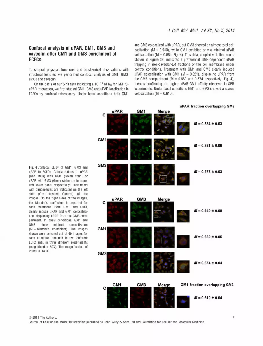
To support physical, functional and biochemical observations withstructural features, we performed confocal analysis of GM1, GM3,uPAR and caveolin.
On the basis of our SPR data indicating a 10�10 M KD for GM1/3-uPAR interaction, we first studied GM1, GM3 and uPAR localization inECFCs by confocal microscopy. Under basal conditions both GM1
and GM3 colocalized with uPAR, but GM3 showed an almost total col-ocalization (M = 0.940), while GM1 exhibited only a minimal uPARcolocalization (M = 0.584; Fig. 4). This data, coupled with the resultsshown in Figure 3B, indicates a preferential GM3-dependent uPARtrapping in non-caveolar-LR fractions of the cell membrane undercontrol conditions. Treatment with GM1 and GM3 clearly induceduPAR colocalization with GM1 (M = 0.821), displacing uPAR fromthe GM3 compartment (M = 0.680 and 0.674 respectively; Fig. 4),thereby confirming the higher uPAR-GM1 affinity observed in SPRexperiments. Under basal conditions GM1 and GM3 showed a scarcecolocalization (M = 0.610).
Fig. 4 Confocal study of GM1, GM3 anduPAR in ECFCs. Colocalizations of uPAR
(Red stain) with GM1 (Green stain) or
uPAR with GM3 (Green stain) are in upper
and lower panel respectively. Treatmentswith gangliosides are indicated on the left
side (C = Untreated Control) of the
images. On the right sides of the images,
the Mander’s coefficient is reported foreach treatment. Both GM1 and GM3,
clearly induce uPAR and GM1 colocaliza-
tion, displacing uPAR from the GM3 com-partment. In basal conditions, GM1 and
GM3 show minimal colocalization
(M = Mander’s coefficient). The images
shown were selected out of 60 images foreach condition obtained in two different
ECFC lines in three different experiments
(magnification 60X). The magnification of
insets is 140X.
ª 2014 The Authors.
Journal of Cellular and Molecular Medicine published by John Wiley & Sons Ltd and Foundation for Cellular and Molecular Medicine.
7
J. Cell. Mol. Med. Vol XX, No X, 2014

Since the analysis of uPAR distribution in caveolar-LR and non-caveolar-LR fractions following GM1 and GM3 ECFC enrichmentshowed a preferential caveolar-LR uPAR homing after GM1 stimula-tion, we also studied caveolin-1-uPAR colocalization by confocalmicroscopy upon GM1 and GM3 ECFC enrichment. These experi-ments clearly indicated that GM1 but not GM3 recruits uPAR to
caveolae, as indicated by merged images and by Mander’s coeffi-cients reported on the right of each lane (Fig. 5). The Image J analy-sis shown in Table 1 not only indicated a strong GM1-dependentincrease in uPAR-caveolin colocalization with respect to both controland GM3-treated ECFCs but also showed that GM1 treatment inducedhigher numbers and larger areas of caveolar-LR clusters.
Fig. 5 Confocal study of Caveolin anduPAR in ECFCs. The pictures show colo-
calization of uPAR (Red stain) with Caveo-
lin-1 (Green stain). Treatments with
gangliosides are indicated on the left side(C = Untreated Control) of the images. On
the right sides of the images, the cyto-
fluorogram is reported for each treatment.GM1, but not GM3, clearly recruits uPAR
to caveolae (magnification 60X).
Table 1 Confocal analysis of uPAR and caveolin 1
Confocalanalysis EPC
Maximum caveolar raft,cluster area, lm2
(based on Cav-1 signal)
Caveolar raft,cluster number(based on Cav-1 signal)
Colocalization(uPAR fraction overlapping Cav-1)
CTRL 3.259 � 0.12 17.00 � 0.10 0.504 � 0.02*
GM1 7.00 � 0.49 (P < 0.05†) 144.9 � 11.91 (P < 0.05†) 0.780 � 0.04* (P < 0.05†)
GM3 4.98 � 0.34 (P < 0.05†) 78.9 � 6.23 (P < 0.05†) 0.534 � 0.01*
*Mander’s coefficients (0 = absence of colocalization; 1 = total colocalization).†P indicates significance of differences between control and the relevant parameters.Analysis performed by Image J software of image in Figure 5: caveolar-raft clustering, number of clusters and uPAR-CAV-1 colocalization arereported. GM1 treatment induces caveolar-cluster formation as deduced by increased cluster area and cluster number. GM3 has a lower powerto induce caveolar clustering and does not induce Cav-1 and uPAR colocalization.
8 ª 2014 The Authors.
Journal of Cellular and Molecular Medicine published by John Wiley & Sons Ltd and Foundation for Cellular and Molecular Medicine.

It is noteworthy that GM1 and GM3 do not colocalize with eachother or with caveolin (Figs 4 and 5), as already reported in breastcancer cells and in canine kidney epithelial cells [23, 24].
Overall, these data indicate that membrane uPAR is distributed inat least three compartments in ECFCs: the first one associated to ca-veolar-LRs, the second to GM1 and the third to GM3. This is deduc-ible by the evidence that (i) caveolin does not colocalize with GM1and GM3, (ii) GM1 and GM3 show minimal colocalization, while (iii)uPAR colocalizes with all of them. Most importantly, only GM1 treat-ment stimulates a relevant recruitment of uPAR in caveolar LRs,MAPKinases signalling, invasion and capillary morphogenesis ofECFCs.
Discussion
In this study, we have shown by SPR that GM1 and GM3 embeddedin artificial ssRLM efficiently bind soluble uPAR, exhibiting very highaffinity (KD ~10�10 M). Moreover, SPR and PWR measurements haveshown that GM1 binds to uPAR more effectively than GM3, so thatGM3-uPAR complexes are likely to detach in presence of a properenrichment of competing GM1. On these basis, we planned to recon-cile our data with the data of literature showing that GM1 promotesangiogenesis [4–6] while GM3 does not, exhibiting an anti-angiogen-esis activity under some experimental settings [5], and that uPARlocalization in caveolar-LRs is indispensable for an efficient angiogen-esis program in EPCs/ECFCs [9, 16], commonly considered the mainsource of vasculogenesis in human tumours [25]. We have shownthat uPAR present on ECFC surface is distributed in at least threecompartments, respectively associated to caveolae, GM1 and GM3.The three compartments are not static rather they can modify theiruPAR content to satisfy angiogenesis needs. In fact, we have shownthat under resting control conditions uPAR is mainly localized in GM3non-caveolar-LR compartment of the cell membrane (Figs 3B, 4 and5); GM1 enrichment increases the uPAR-GM1 compartment by pro-ducing a detachment of uPAR from the G3 compartment (Fig. 4), aresult that is in agreement with the higher affinity of GM1 measuredwith the optical tests on ssRLMs; GM1 treatment stimulates uPARlocalization in caveolar-LRs (Figs 3B and 5), promotes ECFC invasionand capillary morphogenesis that are inhibited by uPAR-blocking anti-bodies, and stimulates the pro-angiogenesis-related ERK and p38phosphorylation. These data account for the already reported yin-yang activities of gangliosides in angiogenesis, with GM1-dependentangiogenesis counterbalanced by GM3 anti-angiogenesis [4–6], anddefine a so far unknown property of GM1 to promote a caveolar-LRenrichment of uPAR. We have previously reported that uPAR localiza-tion in caveolae is indispensable for ECFC angiogenesis both in vitroand in vivo [9]. As shown also in the present study, some authorsdemonstrated that uPAR can be found both in caveolar-LR and non-caveolar-LR fractions and that uPAR distribution in different fractionswas associated with different signalling pathways and, consequently,biological processes [8, 26, 27]. Caveolae are functionally and mor-phologically distinct forms of LRs characterized by the presence ofthe protein caveolin-1 and are particularly abundant in endothelialcells, playing a fundamental role in their function [28]. VEGF localizes
in caveolae upon interaction with its type-2 receptor (VEGFR2), form-ing a ‘functional platform’, together with other signalling molecules,such as uPAR, involved in angiogenesis [16, 29].
On the basis of these data and of our observations, we suggestthat GM1-dependent localization of uPAR in caveolar-LRs accountsfor GM1-dependent angiogenesis.
GM1 activity promoting caveolar-LRs localization of uPAR alsoaccounts for the reported synergy between GM1 and classical angio-genesis inducers, such as basic FGF2 and VEGF [4, 7], since bothpromote uPAR recruitment in caveolar-LRs [this study and nine].Several data show that uPAR represents a central mediator of growthfactor-induced endothelial cell migration and undergoes up-regulationand caveolar-LR localization following pro-angiogenesis factors chal-lenge of endothelial cells and ECFCs [9, 22, 30]. Therefore, it is likelythat GM1 and angiogenesis factors cooperate in caveolar-LRs uPARpartitioning, thus lowering the amount of pro-angiogenetic moleculesrequired to trigger an efficient angiogenesis program.
In tumour cell lines, the tumourigenic potential correlates with thecellular levels of gangliosides [31] and the ability to form experimen-tal tumours can be affected by the artificial manipulation of tumourganglioside levels [32], indicating tumour GM3 overexpression as adeterminant of non-aggressive tumour behaviour. The anti-angiogen-esis properties of GM3 and its inability to stimulate a caveolar-LRlocalization of uPAR strongly support these observations. Moreover,many tumours release high amounts of complex gangliosides, suchas GM1 [33]. In this types of tumours GM1 may exert its pro-angio-genic activity either alone, as shown by our data, or in synergism withpro-angiogenesis factors. An interesting study has recently shownthat GM1 gangliosides recruit tumour-produced soluble uPAR (su-PAR) on HUVEC LRs and that such a recruitment triggers an angio-genesis program, thus enlightening a so far unknown mechanism oftumour angiogenesis [10].
Although many studies show that cholera toxin, and thereforeGM1, is associated with caveolae and LR domains in various celltypes and that cholera-toxin can be internalized by caveolar-LR-dependent endocytosis, we did not find any colocalization betweencaveolar-LRs and GM1 in ECFCs. The lack of association betweenGM1 and caveolar-LRs has been reported previously in other celltypes, such as canine kidney epithelial cells and mammary epithe-lial tumour cells [23, 24], but has never been described before inendothelial cells or in EPCs. It is evident that these featuresinvolve an indirect mechanism of GM1-dependent caveolar-LR dis-tribution of uPAR. A possible explanation for GM1-dependentuPAR localization in caveolar-LR in the absence of a caveolar-LRlocalization of GM1 in ECFCs may be found in the reported prop-erty of non-caveolar-LR GM1 to mediate endocytosis of choleratoxin-B in a dynamin-dependent but caveolar-independent fashion[23]. On this basis, one may hypothesize that, once internalized,uPAR and GM1 follow different pathways within the extremelycomplex intracellular trafficking network, leading uPAR to caveo-lar-LR localization, as previously described for constitutive endocy-tosis and recycling of uPAR [34].
Finally, the occurrence of an agreement between the activities ofthe gangliosides versus uPAR when embedded in biomimetic mem-branes and on living cells, recently documented for the GM1 affinity
ª 2014 The Authors.
Journal of Cellular and Molecular Medicine published by John Wiley & Sons Ltd and Foundation for Cellular and Molecular Medicine.
9
J. Cell. Mol. Med. Vol XX, No X, 2014

with the b-subunit of the Cholera toxin [12], is an important confirma-tion of the predictive properties of the novel mobile raft-like macro-membranes, proposed in [13] and here fruitfully exploited asguideline for more targeted biological investigation on living cells.
Acknowledgements
This study was supported by grants of the Ente Cassa di Risparmio di
Firenze to MDR, GF and GM; Istituto Toscano Tumori to MDR; Associazi-
one Italiana Ricerca sul Cancro to MDR (AIRC grant no. IG 2013, 14266);Brazilian FAPERJ to TDR. FM was supported by a post-doctoral joint fel-
lowship of the European Union and Regione Toscana within the project
UNIFI-4 MELOTAC. MDR, GM, GF conceived the study and wrote thepaper. ST, GM, RDA and TDR prepared the SPR program and performed
SPR experiments. FM, LP, NS, EA performed confocal microscopy and iso-
lated ECFCs. SS, AL, AB, CL, AC performed caveolar-LRs isolation, angio-genesis in vitro and signalling experiments.
Conflicts of interest
The authors confirm that there are no conflicts of interest.
Supporting information
Additional Supporting Information may be found in the onlineversion of this article:
Data S1 Plasma waveguide resonator (PWR) technology.
References
1. Pike LJ. The challenge of lipid rafts. J Lipid
Res. 2008; 50: S323.
2. Sonderfeld S, Conzelman E, SchwarzmannG, et al. Incorporation and metabolism ofganglioside GM2 in skin fibroblasts from
normal and GM2 gangliosidosis subjects.
Eur J Biochem. 1985; 149: 247–55.3. Birkl�e S, Zebg G, Gao L, et al. Role of
tumor-associated gangliosides in cancer
progression. Biochimie. 2003; 85: 455–63.4. De Cristian G, Morbidelli L, Alessandri G,
et al. Synergism between, gangliosides andbasic fibroblast growth factor in favouring
survival, growth, and motility of capillary
endothelium. J Cell Physiol. 1990; 144: 505–10.
5. Koochekpour S, Merzak A, Pilkington GJ.Vascular endothelial growth factor produc-
tion is stimulated by gangliosides and tgf-beta isoforms in human glioma cells in vitro.
Cancer Lett. 1996; 102: 209–15.6. Ziche M, Morbidelli L, Alessandri G, et al.
Angiogenesis can be stimulated or repressedin vivo by a change in GM3:GD3 ganglioside
ratio. Lab Invest. 1992; 67: 711–5.7. Slevin M, Kumar S, He X, et al. Physiologi-
cal concentrations of gangliosides GM1,
GM2 and GM3 differentially modify basic-
fibroblast-growth-factor-induced mitogene-
sis and the associated signalling pathway inendothelial cells. Int J Cancer. 1999; 82:
412–23.8. Cunningham O, Andolfo A, Santovito ML,
et al. Dimerization controls the lipid raftpartitioning of uPAR/CD87 and regulates its
biological functions. EMBO J. 2003; 22:
5994–6003.9. Margheri F, Chill�a A, Laurenzana A, et al.
Endothelial progenitor cell-dependent angio-
genesis requires localization of the full-
length form of uPAR in caveolae. Blood.
2011; 118: 3743–55.10. Rao JS, Gujrati M, Chetty C. Tumor-associ-
ated soluble uPAR-directed endothelial cell
motility and tumor angiogenesis. Oncogene-
sis. 2013; 2: e53.
11. Mao Y, Tero R, Imai Y, et al. The morphol-ogy of GM1x/SM0.6-x/Chol0.4 planar bilay-
ers supported on SiO2 surfaces. Chem Phys
Lett. 2008; 460: 289–94.12. Margheri G, D’Agostino R, Trigari S,
et al. The b-Subunit of Cholera Toxin has
a high affinity for ganglioside GM1 in solid
supported lipid membranes with a lipidraft-like composition. Lipids. 2014; 49:
203–6.13. Margheri G, D’Agostino R, Del Rosso M,
et al. Fabrication of GM3-enriched Sphyng-omyelin/Cholesterol solid-supported lipid
membranes on Au/SiO2 plasmonic sub-
strates. Lipids. 2013; 48: 739–47.14. Simons K, Toomre D. Lipid rafts and signal
transduction. Nat Rev Mol Cell Biol. 2000; 1:
31–9. Review. Erratum in: Nat Rev Mol Cell
Biol 2001; 2:216.15. Binder VH, Barragan V, Menger FM.
Domains and rafts in lipid membranes.
Angew Chem Int Ed Engl. 2003; 42:
5802–27.16. Chill�a A, Magherini F, Margheri F, et al.
Proteomic identification of VEGF-dependent
protein enrichment to membrane caveolar-
raft microdomains in endothelial progenitorcells. Mol Cell Proteomics. 2013; 12: 1926–38.
17. Margheri F, Schiavone N, Papucci L, et al.GDF5 regulates TGFß-dependent angiogene-
sis in breast carcinoma MCF-7 cells: in vitro
and in vivo control by anti-TGFß peptides.
PLoS ONE. 2012; 7: e50342.
18. Margheri F, Manetti M, Serrati S, et al.Domain 1 of the urokinase-type plasminogenactivator receptor is required for its morpho-
logic and functional, beta2 integrin-mediated
connection with actin cytoskeleton in human
microvascular endothelial cells: failure ofassociation in systemic sclerosis endothelial
cells. Arthritis Rheum. 2006; 54: 3926–38.19. Bolte S, Cordelieres FP. A guided tour into
subcellular colocalization analysis in lightmicroscopy. J Microsc. 2006; 224: 213–32.
20. Edwards PR, Lowe PA, Leatherbarrow RJ.Ligand loading at the surface of an opticalbiosensor and its effect upon the kinetics of
protein-protein interactions. J Mol Recognit.
1997; 10: 128–34.21. Eden G, Archinti M, Furlan F, et al. The uro-
kinase receptor interactome. Curr Pharm
Des. 2011; 17: 1874–89.22. Brunner PM, Heier PC, Mihaly-Bison J,
et al. Density enhanced phosphatase-1down-regulates urokinase receptor surface
expression in confluent endothelial cells.
Blood. 2011; 117: 4154–61.23. Lajoie P, Kojic LD, Nim S, et al. Caveolin-1
regulation of dynamin-dependent, raft-medi-
ated endocytosis of cholera toxin-B sub-unit
occurs independently of caveolae. J Cell MolMed. 2009; 13: 3218–25.
24. Chen Y, Qin J, Chen ZW. Fluorescence-topo-graphic NSOM directly visualizes peak-valley
polarities of GM1/GM3 rafts in cell mem-brane fluctuations. J Lipid Res. 2008; 49:
2268–75.25. Li Calzi S, NeuMB, Shaw LC, et al. EPCs and
pathological angiogenesis: when good cells
go bad.Microvasc Res. 2010; 79: 207–16.
10 ª 2014 The Authors.
Journal of Cellular and Molecular Medicine published by John Wiley & Sons Ltd and Foundation for Cellular and Molecular Medicine.

26. Sahores M, Prinetti A, Chiabrando G, et al.uPA binding increases UPAR localization to
lipid rafts and modifies the receptor microd-
omain composition. Biochim Biophys Acta.
2008; 1778: 250–9.27. Kiyan J, Smith G, Haller H, et al. Uroki-
nase-receptor-mediated phenotypic changes
in vascular smooth muscle cells require theinvolvement of membrane rafts. Biochem J.
2009; 423: 343–51.28. Frank PG, Woodman SE, Park DS, et al.
Caveolin, caveolae, and endothelial cell func-tion. Arterioscler Thromb Vasc Biol. 2003;
23: 1161–8.
29. Liao WX, Feng L, Zhang H, et al. Compart-mentalizing VEGF-induced ERK2/1 signaling
in placental artery endothelial cell caveolae:
a paradoxical role of caveolin-1 in placental
angiogenesis in vitro. Mol Endocrinol. 2009;23: 1428–44.
30. Poettler M, Unseld M, Mihaly-Bison J,et al. The urokinase receptor (CD87)represents a central mediator of growth
factor-induced endothelial cell migra-
tion. Thromb Haemost. 2012; 108: 357–66.
31. Deng W, Li R, Ladisch S. Influence of
cellular ganglioside depletion on tumor
formation. J Natl Cancer Inst. 2000; 92:912–7.
32. Prinetti A, Aureli M, Illuzzi G, et al. GM3
synthase overexpression results in reduced
cell motility and in caveolin-1 upregulation inhuman ovarian carcinoma cells. Glycobiolo-
gy. 2010; 20: 62–77.33. Fuentes R, Allman R, Mason MD. Ganglio-
side expression in lung cancer cell lines.
Lung Cancer. 1997; 18: 21–33.34. Cortese K, Sahores M, Madsen CD, et al.
Clathrin and LRP-1-independent constitutiveendocytosis and recycling of uPAR. PLoS
ONE. 2008; 3: e3730.
ª 2014 The Authors.
Journal of Cellular and Molecular Medicine published by John Wiley & Sons Ltd and Foundation for Cellular and Molecular Medicine.
11
J. Cell. Mol. Med. Vol XX, No X, 2014


