Embed Size (px)
Citation preview
21Shalaby and Hussieny
Egypt J. Forensic Sci. Appli. Toxicol Vol 17 (1) June 2017
DIMETHOATE HEPATOTOXICITY IN MICE EXPOSED TO CHRONIC INTOXICATION
Nashwa Mohamad Mohamad Shalaby and Abeer Ramzy Hussieny
Departments of Forensic Medicine and Clinical Toxicology; Faculty of Medicine; Zagazig
University.
ABSTRACT Background: Because pesticides play an important role in agriculture field, and
their use has increased, the evaluation of their toxic effects is of major concern to
public health. Aim: The aim of this work was to study the propensity of dimethoate
(DM) to cause hepatic function disturbance in mice. Material and Methods:
Dimethoate was administered orally at doses of (20mg/kg body weight) dissolved in 1
ml corn oil [1/20 of the LD50 (380mg/kg)] once daily for 14 weeks. Biochemical
parameters in serum were studied: aminotransferases (ALT and AST), alkaline
phosphatase (ALP), total proteins and albumin. Liver will be examined by light and
electron microscope to evaluate histopathological changes. Results: Our results
indicated that: the levels of the ALT and AST, ALP as well as bilirubin, in the serum
of treated rabbits showed highly significant (P<0.0001) increase compared to control
animals, whereas either, total protein, and albumin were highly significantly decreased
(P<0.0001).Light microscopic examination of the hepatic tissue in dimethoate treated
group showed congested portal vein, dilated bile duct , hepatocytes with vacuolated
cytoplasm and deeply stained shrunken nuclei and hemorrhage. While electron
microscope of the same group showed areas of hemorrhage, apoptotic nucleus and
necrotic areas. In conclusion: Our study demonstrated that administration of DM
orally at doses of (20mg/kg body weight) [1/20 of the LD50] for 14 weeks induced
disturbance in the liver function .It is recommended to do Further investigations to
prove the implication of oxidative stress in liver function disturbance and study the
role of antioxidant in its limitation.
Key words: Dimethoate, liver, organophosphates.
INTRODUCTION Dimethoate (DM) is an
organophosphates insecticide which acts
by interfering with the activities of
cholinesterase (Gore, 2001).
Dimethoate is used to kill both mites
and insects. It is also used as a residual
wall spray in farm buildings for house
flies and has been administered to
livestock for control of botflies. It is
available in aerosol spray; dust, and
emulsifiable concentrate (Mirajkar and
Pope, 2005).
The oral LD50 for DM in rats is 60
to 387 mg/kg (Dreher, 2001a), 60
mg/kg in mice, 400 mg/kg in dogs, 200
mg/kg in hamsters, 300 mg/kg in
rabbits, 350 mg/kg in guinea pigs, and
100 mg/kg in cats (Meister, 1992). The
dermal LD50 in rabbits is 1,000 mg/kg,
and 353 mg/kg in rats. A dermal LD50
of greater than 2,000 mg/kg in rats has
also been reported (Dreher, 2001b &
Dreher, 2001c).
The extensive use of dimethoate
carries a health hazard to animals and
humans due to its persistence in soil and
crops (WHO/IPCS, 1996). Majority of
population is exposed to lower doses of
dimethoate via food, contaminated
22Shalaby and Hussieny
Egypt J. Forensic Sci. Appli. Toxicol Vol 17 (1) June 2017
drinking water, or by application of
household insecticides containing
dimethoate (Sharma et al., 2005).
Repeated or prolonged exposure to
organophosphates may result in many
effects as acute exposure, including the
delayed symptoms. Workers repeatedly
exposed to DM reported impaired
memory, disorientation, depressions,
irritability, confusion, headache, speech
difficulties, delayed reaction times,
nightmares, sleepwalking and
drowsiness or insomnia. Also, influenza-
like condition with headache, nausea,
weakness, loss of appetite, and malaise
has also been reported (Ogutcu et al.,
2008). Dimethoate affects the functions of
multiple organs including liver.
Dimethoate was reported to alter the
level of the marker parameters related to
the liver in rats and mice (Chatterjea
and Shinde, 2005; Attia and Nasr,
2009; Saafi et al., 2011 and Khan et
al., 2013). Organophosphorus insecticides
affect other organs (Betrosianet al.,
1995; and Senanayke1998), specially
CNS, (Desi et al., 1998; and Lengylet
al., 2005), kidney (Kossmannet al.,
1997), and pancreas (Hagar and
Fahmy 2002; Kamath and Rajini
2007; Kamath et al. 2008). Also immune-toxic effect were
reported (Institóris et al. 1995, 1999;
Undeger et al. 2000) with several
adverse effects in the reproductive
system of male and female mice
(Mahadevaswami and Kaliwal 2002,
2004; Farag et al. 2007; Astiz et al.
2009). Dimethoate was reported to cause
both benign and malignant neoplasms of
the liver, endocrine organs, and
lymphatic system (Reuber 1984),
besides being considered as human
teratogen and mutagen (Hallenbeck and
Cunningham-Burns, 1985).
The main toxic effect of OP
pesticides is the inhibition of
acetylcholinesterase (De-Bleecker et al.,
1993; and Dongren et al., 1999). Also
OP compounds induce oxidative stress
in humans (Ranjbar et al. 2002) and
animals (Debnath and Mandal 2000) ,
leading to different types of DNA
lesions including single- and double -
DNA strand breaks, cross links,
Chromosomal aberrations and DNA
base oxidation in toadfish lymphocytes
(Lopes et al. 1998; Twigg et al. 1998;
Ellingham et al., 1986). And it was also
reported to increase the incidence of
numerical but not structural
chromosomal aberration in male Wistar
rats (Undeger et al. 2000; Nehéz and
Dési 1996).Other studies done by
Gillot-Delhalle et al. (1983) founded
that DM was non-mutagenic.
So the aim of the present study is to
investigate the hepatotoxic effect of
diemethoate either by biochemical
changes or by histopathological lesions.
MATERIAL& METHODS Material
[A] Chemicals
Dimethoate: It was obtained from
Sigma, Aldrich in Germany imported by
Cairo Chemical Company.
Corn oil was used for preparing
suspensions of DM.
[B] Animals
This study was carried out on 30
adult male albino rats, their weights
ranged from 120-150 gms, they were
obtained from animal’s house, Faculty
of Veterinary Medicine, Zagazig
University. Before commencing the
experimentation, all animals were
subjected to 14 days period of passive
preliminaries in order to be adapting to
23Shalaby and Hussieny
Egypt J. Forensic Sci. Appli. Toxicol Vol 17 (1) June 2017
the new environment, to ascertain their
physical wellbeing and to exclude any
diseased animals. The experimental
procedures were carried out according to
the National Institute of Health
Guidelines for Animal Care.
[C]Experimental Design:
Animals were divided into 3 groups
each of 10 rats as follow:
Group I (negative control group):- These rats will receive only regular
diet and tap water for 14 weeks to
measure the basic parameters.
Group II (positive control
group):-
Each rat will be gavaged orally with
1mL corn oil once daily for 14 weeks.
Group III (dimethoate treated
group):-
These rats will be gavaged orally
with dimethoate (20mg/kg body weight)
dissolved in 1ml corn oil [1/20 of the
LD50 (380mg/kg)] (Kamath et al.,
2008) once daily for 14 weeks.
Rats were weighed every week and
the doses were adjusted according to the
changes in the body weight.
Methods:
At the end of the experimental
periods, rats of all groups were used to
measure the following parameters.
[A] Biochemical parameters
After the experimental periods the
animals were sacrificed, the blood was
immediately collected and centrifuged to
obtain serum which discarded and kept
at - 21 º C for the biochemical testes.
(1) Alanine- aminotransferase
(ALT) and Aspartate-
aminotransferase (AST) Assay:
The estimation was carried out
according to the method originally
developed by (Reitman and Frankel
1957).
(2) Alkaline phosphatase Assay:
ALP was determined using a
colorimetric method as described by
(Kind and King 1954).
(3) Total Protein Assay:
The total protein was determined by
Biuret method explained by (Tietz
1976)
(4) Albumin Assay:
Serum albumin was determined
according to the method of (Doumaset
al., 1971).
(5) Bilirubin assay: The estimation was carried out
according to the method originally
developed by (Pearlman and lee,
1974).
[B] Tissue parameters:
Liver was immediately dissected out
and grossly inspected to assess any gross
abnormalities then washed with cold
normal saline and used for
histopathological study.
(1) Light microscope examination: The liver was fixed in 10% formalin
saline. After fixation, tissues were
embedded in paraffin blocks and
processed for the preparation of 5 u.
thickness sections. These sections were
subjected for Hematoxylin and Eosin
stains (Horobin and Bancroft, 1998)
and then examined by light microscope.
(2) Electron microscope
examination:
Immediately after dissection, minute
specimens will be rinsed in 0.1M
phosphate buffer pH 7.2 (PB) to remove
blood from the surface. Liver tissues
greater than 2 cm long were minced into
smaller pieces of approximately3 x 3
mm and were fixed in 3 percent
glutaraldehyde, buffered with phosphate
buffer for 3 hours. It was rinsed twice
with phosphate buffer for 10 minutes per
rinse. The tissues were then fixed in 2
percent aqueous osmium tetroxide for 2
hrs and rinsed in 3 changes of distilled
24Shalaby and Hussieny
Egypt J. Forensic Sci. Appli. Toxicol Vol 17 (1) June 2017
water for 10 minutes. Each dehydration
was accomplished by immersion in a
graded series of ethanol solutions of 25,
50, 75, 95and 100 percent. Infiltration
with propylene oxide and embedding
with increasing concentrations of
propylene oxide followed by
dehydration were carried. Thin sections
(600nm) were obtained by use of Ultra
microtome and were placed on a
copper200 – mesh grid. They were
stained with uranyl acetate and lead
citrate and examined with GEOL-
TEM1010electron microscope
(Goodhew et al., 2003).
Statistical analysis:
Data were analyzed by Statistical
Package of Social Science (SPSS),
software version 22.0 (SPSS Inc.,
2013).
RESULTS I)-Biochemical parameters results:
As regard the control groups
(negative and positive control group):
There was a non-significant difference
between them as regard biochemical
parameters (Tables 1).
So the negative control group was
chosen to compare with the treated
group (Dimethoate treated group). LD50
of Dimethoate resulted in a statistically
high significant increase in the level of
alanine - aminotransferase (ALT)
aspartate - aminotransferase (AST) and
alkaline phosphatase (ALP) in the serum
of treated group, as compared to the
control.
Total protein and albumin levels
showed highly significant decrease in
the serum of treated with Dimethoate as
compared to control (Tables 2).
II)-Histopathological changes:
Macroscopic appearance showed
non-significant changes in size or
abnormal masses compared with the
control groups. Cut sections were
apparently normal.
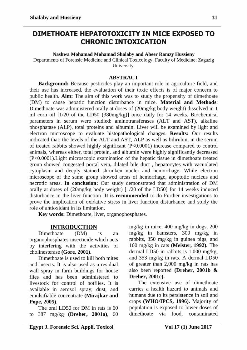
Light Microscopic examination of
the liver specimens in the control
untreated group showed a normal
histological picture. The central vein lies
at the center of the lobule surrounded by
the hepatocytes with strongly
eosinophilic granulated cytoplasm, and
distinct nuclei. In addition, between the
strands of hepatocytes the hepatic
sinusoids are exhibited as shown in
(Fig.1(.
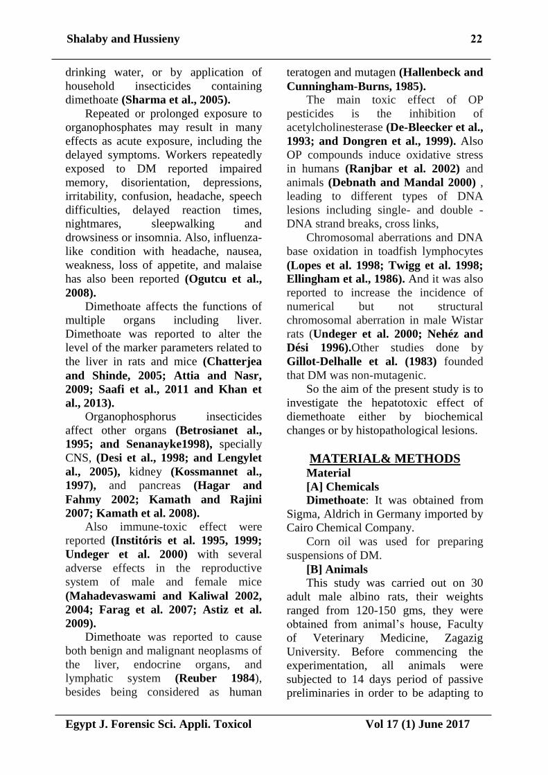
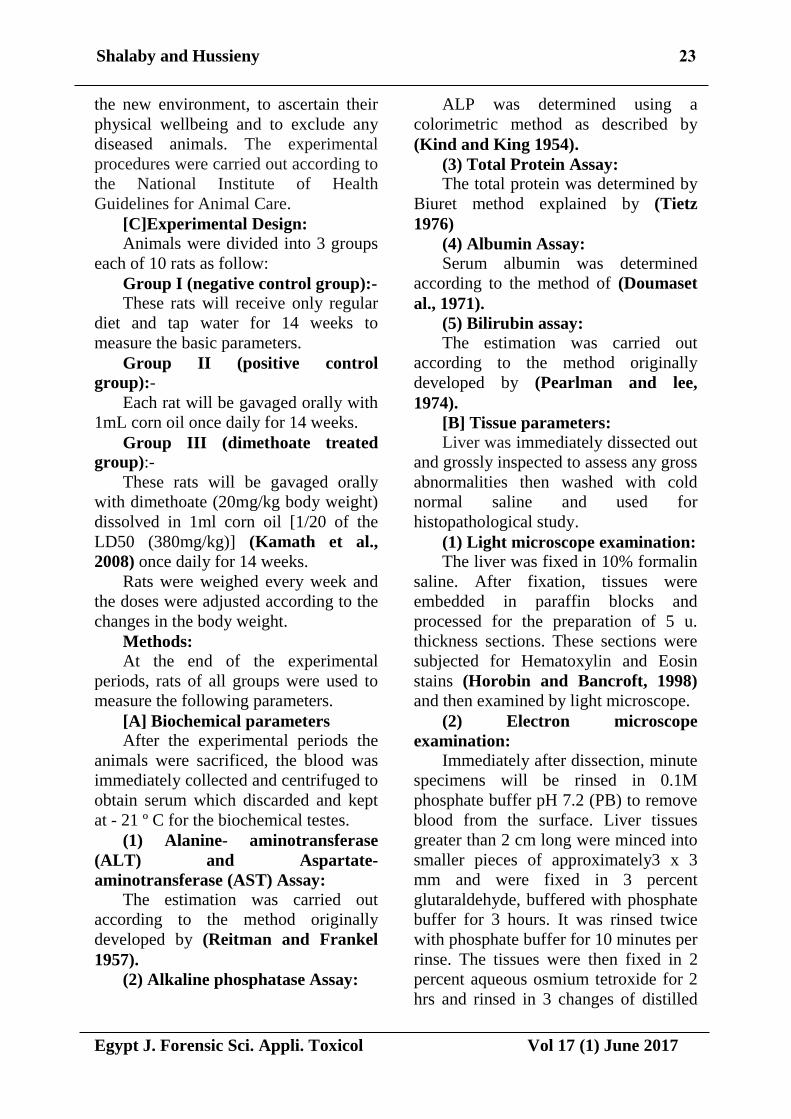
While the liver of mice treated with
dimethoate showed congestion portal
vein(Fig.2), dilated bile duct ,
Hepatocytes with vacuolated cytoplasm
and deeply stained shrunken nuclei
(Fig.3) and liver hemorrhage (Fig.5 ),
Electron microscopic examination
of the control group showed hepatocytes
appeared with eu-chromatic nuclei
containing prominent nucleoli. The
cytoplasm contained numerous
mitochondria, rough endoplasmic
reticulum (Figs.5).While dimethoate
treated group showed areas of
hemorrhage, apoptotic nucleus and
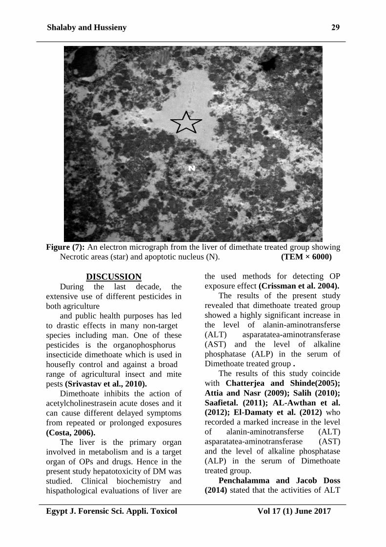
necrotic areas (Figs.6, 7).
25Shalaby and Hussieny
Egypt J. Forensic Sci. Appli. Toxicol Vol 17 (1) June 2017
Table (1): the liver function tests for the negative control group (I) and corn oil group
(II) group, unpaired t test (mean±SD).
Group
Parameter
Negative control
group(I)
Corn oil treated
Group(II) T P
Mean±SD Mean ±SD
AST 14.11±3.49 14.99±3.47 0.5654 >0.05
ALT 25.2±3.40 25.8±3.41 0.3940 >0.05
ALK 95.6±2.6 97.4±2.7 1.5186 >0.05
Total protien 6.11±0.25 6.15±0.23 0.3724 >0.05
Albumin 3.72±0.16 3.73±0.18 0.1313 >0.05
Globlin 1.82±0.09 1.83±0.11 0.2225 >0.05
Bilrubin 1.60±0.05 1.59±0.02 0.5872 >0.05
Number of sacrificed rats for each group was 10 rats.
SD : Standard Deviation. . p>0.05 : non-significant
Table (2): the liver function tests for the negative control group (I) and Dimethoate
treated group, unpaired t test (mean ±SD).
Group
Parameter
Negative control
group(I)
Dimethoatetreated
group(III)
T
P
Mean±SD Mean ±SD
AST 14.11±3.49 118.24±1.49 86.7744 <0.0001***
ALT 25.2±3.40 167.3±12.11 35.7252 <0.0001***
ALK 95.6±2.6 119.7±4.22 15.3755 <0.0001***
Total protien 6.11±0.25 4.2±0.19 19.2351 <0.0001***
Albumin 3.72±0.16 2.33±0.07 25.1689 <0.0001***
Globlin 1.82±0.09 1.64±0.03 6.0000 <0.0001***
Bilrubin 1.60±0.05 1.85±0.06 10.1222 <0.0001***
Number of sacrificed rats for each group was 10 rats.
SD : Standard Deviation. **: Highly–significant (P<0.001)
26Shalaby and Hussieny
Egypt J. Forensic Sci. Appli. Toxicol Vol 17 (1) June 2017
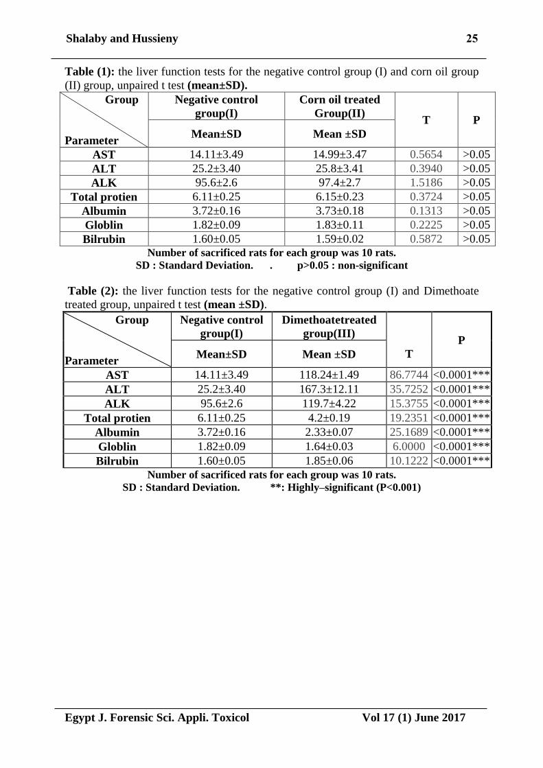
Figure (1): A photomicrograph of a section from the liver of a control group showing
hepatocytes (h) arranged in plates radiating from the central vein (cv) and
separated by blood sinusoids (s); hepatocytes are polygonal in shape, with central
rounded vesicular nuclei and acidophilic cytoplasm. (H&E X 400)
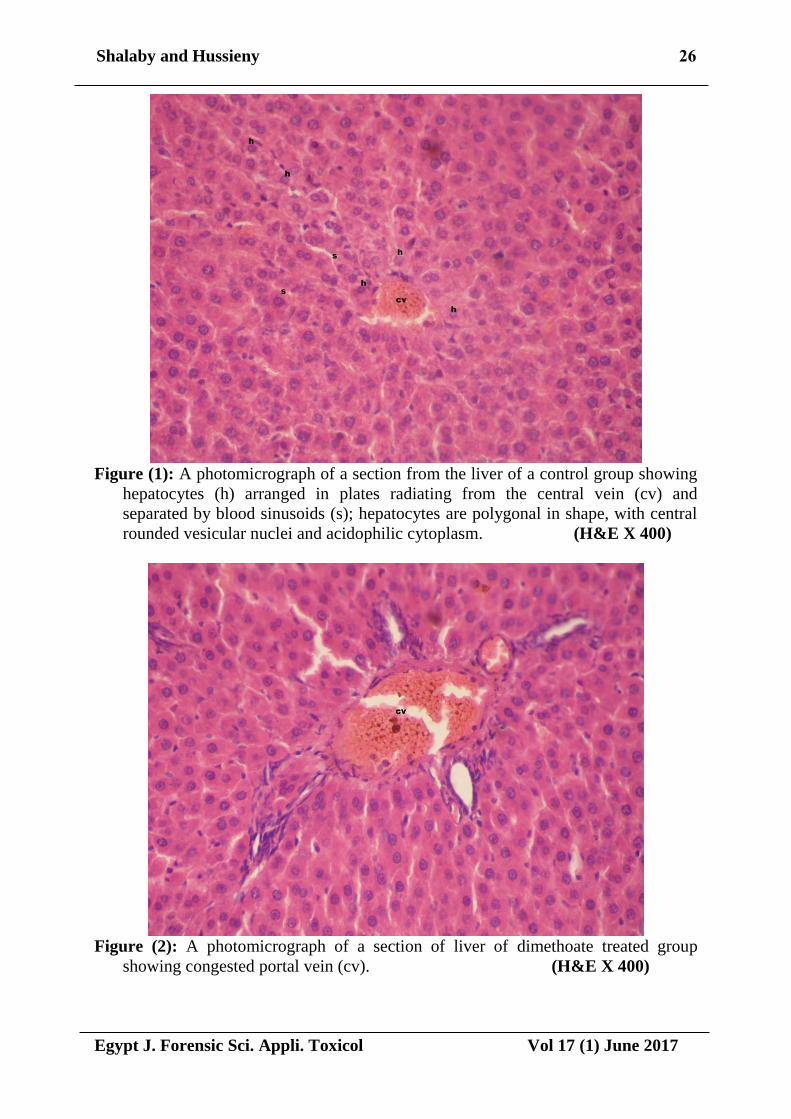
Figure (2): A photomicrograph of a section of liver of dimethoate treated group
showing congested portal vein (cv). (H&E X 400)
27Shalaby and Hussieny
Egypt J. Forensic Sci. Appli. Toxicol Vol 17 (1) June 2017
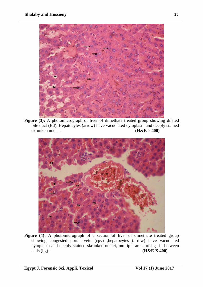
Figure (3): A photomicrograph of liver of dimethate treated group showing dilated
bile duct (Bd). Hepatocytes (arrow) have vacuolated cytoplasm and deeply stained
skrunken nuclei. (H&E × 400)
Figure (4): A photomicrograph of a section of liver of dimethate treated group
showing congested portal vein (cpv) ,hepatocytes (arrow) have vacuolated
cytoplasm and deeply stained skrunken nuclei, multiple areas of hgs in between
cells (hg) . (H&E X 400)
28Shalaby and Hussieny
Egypt J. Forensic Sci. Appli. Toxicol Vol 17 (1) June 2017
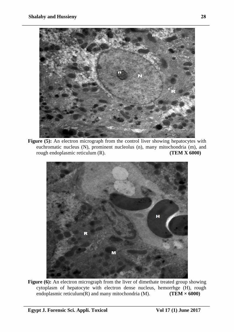
Figure (5): An electron micrograph from the control liver showing hepatocytes with
euchromatic nucleus (N), prominent nucleolus (n), many mitochondria (m), and
rough endoplasmic reticulum (R). (TEM X 6000)
Figure (6): An electron micrograph from the liver of dimethate treated group showing
cytoplasm of hepatocyte with electron dense nucleus, hemorrhge (H), rough
endoplasmic reticulum(R) and many mitochondria (M). (TEM × 6000)
29Shalaby and Hussieny
Egypt J. Forensic Sci. Appli. Toxicol Vol 17 (1) June 2017
Figure (7): An electron micrograph from the liver of dimethate treated group showing
Necrotic areas (star) and apoptotic nucleus (N). (TEM × 6000)
DISCUSSION During the last decade, the
extensive use of different pesticides in
both agriculture
and public health purposes has led
to drastic effects in many non-target
species including man. One of these
pesticides is the organophosphorus
insecticide dimethoate which is used in
housefly control and against a broad
range of agricultural insect and mite
pests (Srivastav et al., 2010).
Dimethoate inhibits the action of
acetylcholinestrasein acute doses and it
can cause different delayed symptoms
from repeated or prolonged exposures
(Costa, 2006). The liver is the primary organ
involved in metabolism and is a target
organ of OPs and drugs. Hence in the
present study hepatotoxicity of DM was
studied. Clinical biochemistry and
hispathological evaluations of liver are
the used methods for detecting OP
exposure effect (Crissman et al. 2004).
The results of the present study
revealed that dimethoate treated group
showed a highly significant increase in
the level of alanin-aminotransferse
(ALT) asparatatea-aminotransferase
(AST) and the level of alkaline
phosphatase (ALP) in the serum of
Dimethoate treated group .
The results of this study coincide
with Chatterjea and Shinde(2005);
Attia and Nasr (2009); Salih (2010);
Saafietal. (2011); AL-Awthan et al.
(2012); El-Damaty et al. (2012) who
recorded a marked increase in the level
of alanin-aminotransferse (ALT)
asparatatea-aminotransferase (AST)
and the level of alkaline phosphatase
(ALP) in the serum of Dimethoate
treated group.
Penchalamma and Jacob Doss
(2014) stated that the activities of ALT
30Shalaby and Hussieny
Egypt J. Forensic Sci. Appli. Toxicol Vol 17 (1) June 2017
and AST in DM exposed rats showed
statistically significant increased.
Alteration in protein metabolic profiles
was dose – and time- dependent.
Serum ALT and AST are
considered to be among the
mostsensitive markers for the diagnosis
of hepatotoxicity (Kutlu et al., 2007
and Saafi et al., 2011).
According to Begum et al. (2007);
Nagarjuna et al. (2008) ALT and AST
are synthesis from amino acids.
Repeated dose of DM hasa damaging
effect on the cell metabolism leading to
impaired protein synthesis.
Dewan et al. (2004); Ncibi et al.
(2008) concluded that pesticide causes
liver damage and leakage of cytosolic
enzymes from hepatocytes and other
body organs into the blood.
According to Friedman et al.
(2003) elevation of liver enzymes may
occur due to increased gene expression
due to long term requirement of
detoxification of pesticides.
The results of the present study
revealed that dimethoate treated group
showed that total protein and albumin
levels were highly significant decreased
in the serum of treated with Dimethoate
as compared to control.
According to Attia and Nasr,
(2009) there was decrease in serum
total protien, albumin and globulin and
increase in bilirubin level in dimethoate
treated rats.
Dimethoate administration resulted
in a significant decrease in serum total
protien, albumin and globulin. The
reduction in serum protein, particularly
albumin, could be due tothe changes in
protein and free amino acid metabolism
and their synthesis in the liver. Also,
the protein level suppression may be
due to loss of protein either by reduce
in protein synthesis or increased
proteolytic activity or degradation. In
addition, the observed decrease in
serum proteins may be due to the
damaging effect of dimethoate on liver
cells, as confirmed by the increase in
activities of serum AST and ALT
(Yeragi et al., 2003). A decrease in globulin is expected
as globulin (mostly ã-globulins) could
be consumed in the production of
antibodies in response to dimethoate
exposure (Institoris, et al., 1999).
The change in serum bilirubin
which is considered as indicator of liver
function may provide further evidence
on dimethoate induced hepatotoxicity
(Saafi et al., 2011 and Khan et al.,
2013)
The results of the study revealed
that Macroscopic examination of the
liver of all the studied groups revealed
normal appearance with no significant
changes in size or abnormal masses
compared with the control groups. Cut
sections were apparently normal. While
light Microscopic examination of the
liver specimens of the rats revealed the
following:
The light microscopic examination
of the liver sections in the control
untreated group showed a normal
histological picture. The central vein
lies at the centre of the lobule
surrounded by the hepatocytes with
strongly eosinophilic granulated
cytoplasm, and distinct nuclei. While
the liver of mice treated with
dimethoate showed congestion portal
vien,dilated bile duct , Hepatocytes
with vacuolated cytoplasm and deeply
stained skrunken nucleiandhemorrhage.
Electron microscopic examination
of the control group showed
hepatocytes appeared with euchromatic
nuclei containing prominent nucleoli.
The cytoplasm contained numerous
31Shalaby and Hussieny
Egypt J. Forensic Sci. Appli. Toxicol Vol 17 (1) June 2017
mitochondria, rough endoplasmic
reticulum. While dimethoate treated
group showed areas of hemorrhage,
apoptotic nucleus and necrotic areas.
These results are agreement with
many authors; Selmanoglu and Akay
(2000) who reported similar
histopathological changes including
mononuclear cell infiltration,
congestion, hydropic degeneration and
hepatocellular damage in the liver of
male rats treated with dimethoate,
endosulfan and carbaryl.
Also, Sharma et al. (2005)
whofound that a 30-day exposure of
male rats to technical grade dimethoate
at doses of 6 and30 mg/kg caused portal
inflammation, centrizonal congestion
and focal hepatocyte necrosis in the
liver of rats .
Sayim, (2007); Gokcimenet al.,
(2007) and Elhalwagyet al.,(2008) Suggested that may occur hemorrhage,
inflammatory cell infiltration.
In conclusion, in the present
experiment sub-chronic dimethoate
exposure in adult rats induced
hepatotoxicity proved by both
biochemical and histopathological
changes.
It is recommended, to decrease
use of pesticide in our environment and
replace most of them by more safe
natural material. Also continuous
studies needed for evaluation the
toxicity of pesticide and role of
antioxidant in protection from its toxic
effect.
REFERENCES
Al-Awthan, Y.S.; Al-Douis, MA.; El-
Sokkary, G.H. and Aqlan, E.M.
(2012): Dimethoate-induced
Oxidative Stress and
Morphological Changes in the
Liver of Guinea Pig and the
Protective Effect of Vitamin C and
E. Asian Journal of Biological
Sciences, 5(1):9-19.
Attia, A.M. and Nasr, H.M. (2009): Dimethoate-induced changes in
biochemical parameters of
experimental rat serum and its
neutralization by black seed
(Nigella sativa L.) oil. Slovak
Journal of Animal Science, 42(2):
87-94.
Begum, G. (2007): Cypermethrin –
induced biochemical perturbations
in freshwater fish Clariasbatrachus
at sublethal exposure and after
released into freshwater. Drug and
Chemical Toxicology, 30:55-65.
Betrosian, A.; Balla, M.; Kafiri,
G.;Kofinas, G.; Makri, R. and
Kakouri A. (1995): Multiple
system organ failure from
organophosphate poisoning. J. Clin.
Toxicol., 33 (3):257-260.
Chatterjea, M.N. and Shinde, R.
(2005): Text Book of Medical
Biochemistry.6th ed. Jaypee Broth.
New-Delhi, P: 644.
Costa, L.G. (2006): Current issues in
organophosphate toxicology. Clin.
Chim . Acta., 366 (1-2):1-13.
Crissman, J.W.; Goodman, D.G.;
Hildebrandt, P.K.; Maronpot,
R.R.; Prater, D.A., and Riley J.H.
(2004): Best practice guideline:
toxicologic histopathology.
Toxicol. Pathol., 32: 126-131.
Desi, I.; Nagymajteny, L.; Papp,
A.and Schulz, H. (1998): Experimental model studies of
pesticide exposure.
Neurotoxicology, 19 (4–5), 611–
616.
Dreher, D.M. (2001a): Iso-dimethoate:
acute oral toxicity in the rat: acute
toxic class method. Safepharm
Laboratories Limited. DTF Doc
32Shalaby and Hussieny
Egypt J. Forensic Sci. Appli. Toxicol Vol 17 (1) June 2017
No: ‘463-004. [CHA; sub: 12564,
Ref: 3-36/Vol 3-21] .
Dreher, D.M. (2001b): Dimethoate
400 g/L EC, stabilized. Acute
Dermal Toxicity (Limit Test) in the
rat. [CHA; sub: 12564, Ref: 3-
86/Vol 3-41].
Dreher, D.M. (2001c): Dimethoate 400
g/L EC, stabilized. Acute dermal
irritation in the rabbit. [CHA; sub:
12564, Ref: 3-87/Vol 3-41].
El-Damaty, E.M.A.; Farrag, A.H.;
Rowayshed, G. and Fahmy, H.M.
(2012): Biochemical and
Histopathological Effects of
Systemic Pesticides on Some
Functional Organs of Male Albino
Rats. Journal of Applied Sciences
Research, 8(11):5459-5469.
Elhalwagy, M.E.A.; Darwish,N.S.
and Zaher,
E.M.(2008):Prophylactic effect of
green tea polyphenols against liver
and kidney injury induced by
Fenitrothion
insecticide.pestic.Biochem.Phys.,91
: 81-89.
Farag, AT.; Karkour, TA.and El-
Okazy, A. (2006): Developmental
toxicity of orally administered
technical dimethoate in rats. Birth
Defects Res B Dev Reprod
Toxicol., 77(1):40-46.
Gokcimen, A.; Gulle, K.; Demirin,
H.; Bayram, D.; Kocak, A. and
Altuntas,I. (2007):Effect of
diazinon at different doses on rat
liver and pancreas tissues. Pesticide
Biochem.Physiol., 87:103-108.
Gomes, J.; Dawodu, A.; H.Lioyd, O.;
Revitt, D.M. and Anilal S.V.
(1999): Hepatic injury and
disturbed amino acids metabolism
in mice following to prolonged
exposure to organophosphorus
pesticides.Hum. Exp. Toxicol., 18
(1):33-37.
Goodhew, P.J; Humphreys, J. and
Beanland, R. (2003): Electron
microscopey and analysis .In:
Taylor and Francis. 4thed.
London,New York.
Gore, A.C., (2001): Environmental
toxicant effects on neuroendocrine
function. Endocrine, 14: 235-246.
Hagar, H. and Fahmy A. (2002): A
biochemical, histological, and
ultrastructural evaluation of the
effect of Dimethoate intoxication
on rats pancreas. Toxicol.Lett.,
133:161-170.
Hayes, W.J. and Laws, E.R. (1990): Handbook of Pesticide Toxicology,
heavy metals in rats. Hum. Exp.
Toxicol., 18(2):88-94.
Horobin, R.W. and Bancroft, J.D.
(1998): Hematoxylin and eosin as
an oversight stain. In:
Trubleshooting Histology stains, 1st
ed., Churchill Livingstone Press,
San Francisco, pp. 88- 93.
Institoris, l.; Siroki, O.; Desi, l. and
Undeger, u. (1999): Examination
of repeated dose combined
exposure by dimethoate and
twoheavy metals in rats. Hum. Exp.
Toxicol., 18(2):88-94
Kamath, V. and Rajini, P.S. (2007): Altered glucose homeostasis and
oxidative impairment in pancreas of
rats subjected to dimethoate
intoxication. Toxicology, 231(2-
3):137-146.
Kamath, V.; Joshi, A.K.R. and
Rajini, P.S. (2008): Dimethoate
induced biochemical perturbations
in rat pancreas and its attenuation
by cashew nut skin extract.
Pesticide Biochemistry and
Physiology, 90(1):58-65.
33Shalaby and Hussieny
Egypt J. Forensic Sci. Appli. Toxicol Vol 17 (1) June 2017
Khan, A.A.; Shah, M.A. and
Rahman, S.U. (2013): Occupational Exposure to
Pesticides and Its Effects on Health
Status of Workers in Swat. Journal
of Biology and Life Science, 4(2).
Kossmann, S.; Magner-Krezel. Z.;
Sobieraj, R.andSzwed Z. (1997): The assessment of nephrotoxic
effect based on the determination of
the activity of some selected
enzymes in urine. Przegel. Lek., 54
(10):707-711.
Lengyl, Z.; Fazakas, Z. and
Nagymajteny L. (2005): Change
in the central nervous activity of
rats treated with Dimethoate in
combination with other
neurotoxicants in different phases
of ontogenesis.m Arh. Hig. Rada.
Toxicol., 56: 257-264.
Meister, R.T. (1992): Farm chemicals
handbook. Willoughby, OH:
Meister Publishing Company.
Willoghby, OH.
Mirajkar N. and Pope C.N. (2005):
Dimethoate. Encyclopedia of
Toxiocology, pp. 47-49
Nagarjuna, (2008): Effect of
cypermethrin on hematological,
protein metabolism and histological
studies in albino rats. Ph.D. Thesis,
Sri Venkateswara University,
Tirupati, India.
Ogutcu, A.; Uzunhisarcikli, M.;
Kalender, S.; Durak, D.;
Bayrakdaa, F.and Kalender, Y.
(2008): The effects of
organophosphate insecticide
diazinon on malondialdehyde levels
and myocardial cells in rat heart
tissue and protective role of vitamin
E, Pest. Biochem. Physiol., 86(2):
93–98.
Pearlman, F.C. And Lee, R.T. (1974): Detecction and measurement of
total bilirubin in serum, with use of
surfactant as solubilizing agent.
ClinChem., 20(4):447-453.
Penchalamma, R. and Doss,J.(2015): Alteration in Protein Metabolic
Profiles in Liver Tissue of Rats
during Dimethoate Toxicosis.
I.O.S.R., 9: 30-33
Saafi, E.B.; Louedi, M.; Elfeki, A.;
Zakhama, A.; Najjar, M.F.;
Hammamia, M. and Achour, L.
(2011): Protective effect of date
palm fruit extract (Phoenix
dactylifera L.) on dimethoate
induced-oxidative stress in rat liver.
Experimental and Toxicologic
Pathology, 63(5):433.441.
Salih, E.M.A. (2010): Toxic Effect of
Dimethoate and Diazinon on the
Biochemical and Hematological
Parameters in Male Rabbits. Jordan
Journal of Biological Sciences,
3(2):77-82.
Sayim,F. ( 2007):Dimethoate induced
biochemical and histopathology
changes in the liver of
rats.Exp.Toxicol.Pathol.,59:237-
243.
Senanayake, N. (1998): Organophosphorus insecticides
poisoning.Ceylon.Med.J.43:22-29.
Sharma, Y.; Bashir, S.; Irshad, M.;
Nagc, T.C. and Dogra, T. (2005):
Dimethoate-induced effects on
antioxidant status of liver and brain
of rats following subchronic
exposure. Toxicology, 215(2):173
181.
SPSS Inc (2013): SPSS for windows,
version 22.0. Chicago, SPSS Inc.
http://www.unimunester.de/imperia
/md/content/ziv/service/softwar
e/spss/handbuecher/englisch/spss_b
rief_guide_22.0.pdf.
Srivastav, A.K.; Mirshra, D.;
Shrivastava, S.; Srivastav, S.K.
34Shalaby and Hussieny
Egypt J. Forensic Sci. Appli. Toxicol Vol 17 (1) June 2017
and Srivastav A.K .(2010): Acute
toxicity and behavioural responses
of Heteropneustes fossilis to an
organophosphate insecticide,
dimethoate. Int. J. Pharma Bio Sci.,
1: 359-363.
WHO/IPCS, (1996): Principles and
methods for assessing direct
immunotoxicity associated with
exposure to chemicals.
Environmental Health Criteria vol.
180. WHO, Geneva, 110– 112.
35Shalaby and Hussieny
Egypt J. Forensic Sci. Appli. Toxicol Vol 17 (1) June 2017
التسمم الكبدى بالدايمثيوات فى الجرذان المتعرضة للتسمم المزمن نشوى محمد محمد شلبى ,عبير رمزى حسينى
جامعة الزقازيق. –كلية الطب –قسم الطب الشرعى والسموم اإلكلينيكية
الملخص العربى
الزراعة المعاصرة مع زيادة استخداماتها لذا فقد وجب الن المبيدات الحشرية تلعب دور هام فى المقدمة:
دراسة االثار السمية للدايميثوات على الكبد الهدف من البحث:علينا دراسة اثارها السمية التى تهم المجتمع.
: مجموعة 3من ذكور الجرذان البيضاء البالغة مقسمة بالتساوى الى 30 استخدام خطة البحث:تم
14سيتم اعطاء الجرذان الطعام والشراب بدون أى عالج لمدة -)مجموعة ضابطة سالبة(:المجموعة األولى
مل( من 1سيتم اعطاء كل جرذ )-المجموعه الثانيه )مجموعه ضابطه موجبه(:أسبوع لقياس المعايير األساسية.
لثالثه )مجموعة ا المجموعهبوع أس 14زيت الذره )كمذيب لدايميثوات( عن طريق الفم مره واحده يوميا لمدة
( عن طريق الفم مره مجم/كجم 20) زيت الذره ا مل سيتم اعطاء الجرذان الدايميثوات مذاب فى-الدايميثوات(:
لقياس وطيفة الكبد التى أسبوع. تم استخدام فئران كل المجموعات في نهاية مدة البحث 14واحده يوميا لمدة
انسجةو فحص . تتضمن االمينوترسفيراز وااللكلين فوسفاتيز ونسبة البروتين وااللبومين ومعدالت البلوروبين
زيادة نسبة داللة احصايية ل أظهرت نتائج.الكبد عن طريق الميكرسكوب الضوئى وااللكترونى
ومين والبروتين فى المجموعة المعالجة بالدايميثوات .و االمينوترنسفيراز،االلكلين فوسفاتيز مع نقص فى االلب
أوضح الفحص المجهري الضوءى باستخدام صبغة الهيماتوكسلين واأليوسين لشرائح الكبد لذكور الجرذان
احتقان فى البيضاء في مجموعة الدايميثوات وجود تغيرات هستوباثولوجية واضحة أشتملت تلك التغيرات علي
ع اتساع فى االوردة وارتشاح مع وجود نزيف فى انسجة الكبد وتراكم للدهون مع تضخم ، كما الخاليا الكبدية م
مجم/كجم ( 20بجرعة ) لدايميثواتا اعطاءأن نستنتجاوضح الفحص االلكترونى وجود نزيف وتلف فى الخاليا.
دور ل مستقبيلية بدراسة ونوصى له آثار سمية على خاليا الكبد أسبوع 14عن طريق الفم مره واحده يوميا لمدة
ستخداماوالحد من االكسدة فى االثار السمية لمادة الدايميثوات ودور مضادات االكسدة فى الحد من هذه االثار.
الدايمثوات لما له من اثار سلبية على البيئة وصحة االنسان.


