Embed Size (px)
Citation preview

EXPERIMENTAL AND MOLECULAR PATHOLOGY 46, 18& 189 (1987)
Protection against Carbon Tetrachloride Hepatotoxicity by Pretreatment with Indole-3-Carbinoll
H. G. SHERTZER,~ M. I? NIEMI, F. A. REITMAN, M. L. BERGER,~ B. L. MYERS, AND M. W. TABOR
Department of Environmental Health and 3Department of Internal Medicine, University of Cincinnati Medical Center, Cincinnati, Ohio 45247
Received July 31, 1986, and in revised form October 21, 1986
The effects of administering indole-3-carbinol (I-3-C) on carbon tetrachloride (Ccl,)-in- duced hepatotoxicity were examined. Mice received by gavage 0- 150 mg I-3-C/kg body wt in methanol-extracted corn oil, followed 1 h later by 15 ~1 CC&/kg body wt in corn oil. Animals were sacrificed 24 h after receiving Ccl,. Pretreatment with I-3-C reduced the degree of centrolobular necrosis, as observed histologically. Additionally, C&mediated elevated serum enzymes were reduced by I-3-C. Although I-3-C induced elevated levels of cytochrome P-450 and associated mixed-function oxidase activity, the Ccl, depression of these parameters was not clearly reversed by I-3-C. However, Ccl., produced decreases in hepatic levels of glutathione (GSH), total reducing equivalents, and protein sulfhydryls, all of which were restored to control levels by I-3-C. Using mouse liver microsomes in an NADPH-fortified reaction mixture, I-3-C inhibited, in a concentration-dependent manner, Ccl,-initiated lipid peroxidation, with 50% inhibition at 35-40 FM I-3-C. When mice were treated by gavage with 50 mg [‘4ClI-3-C/kg body wt, concentrations of radiolabel in the liver were greater than 100 )LM after 1 hr. This was five times the level of radioactivity measured in blood and three times the concentration of I-3-C necessary for 50% inhibition of Ccl,- mediated lipid peroxidation in vitro. The data are consistent with the hypothesis that I-3-C intervenes in Ccl,-mediated hepatic necrosis by combining with reactive free radical metab- elites of CCI,, thereby protecting criticd cellular target sites.
INTRODUCTION
When high doses of carbon tetrachloride (Ccl,) are administered to rodents, direct solvent injury to the liver may result (Recknagel and Glende, 1973). How- ever, Ccl, requires metabolic activation to produce classic centrolobular necrosis when low doses are administered. Toxicity mediated by Ccl, involves the reduc- tive cleavage of the parent compound to reactive radical metabolites. This is fol- lowed by the interaction of these radical species with critical cellular target sites, followed by the eventual expression of toxicity. Although the specific critical cel- lular target sites are not known with certainty, certain dietary agents, such as tryptophan and cysteine (Wang et al., 1985), can protect against CCI, hepatotox- icity. In this paper, we describe another dietary chemoprotective agent, indole-3- carbinol (1-3-C), which appears to protect by virtue of its ability to scavenge reactive radicals. This chemoprotection may be mechanistically distinct from that afforded by tryptophan and cysteine.
METHODS Chemicals. Glucose 6-phosphate, glucose-6-phosphate dehydrogenase,
NADP+, and bovine serum albumin were from Sigma Chemical Company, St.
* The U.S. Government’s right to retain a nonexclusive royalty-free license in and to the copyright covering this paper, for governmental purposes, is acknowledged.
2 To whom correspondence should be addressed.
180 0014-4800/87 $3.00
INDOLE CARBINOL PROTECTION AGAINST CCL4 TOXICITY 181
Louis, Missouri. N-Nitrosodimethylamine (NDMA), 1-3-C, and thiobarbituric acid were obtained from Aldrich Chemical Company, Milwaukee, Wisconsin. [c~rbinol-‘~C]I-3-C and Aquasol- LX cocktail were from New England Nu- clear, Boston, Massachusetts. Reagent-grade Ccl, and other reagent-grade or better chemicals were from Fisher Scientific Company, Pittsburgh, Pennsylvania. Bicinchoninic acid protein assay reagent was purchased from Pierce Chemical Company, Rockford, Illinois.
Corn oil vehicle was prepared by extracting Mazola corn oil with equal volumes of methanol. The procedure was repeated 15 times. The final methanol- extracted corn oil was stored over Fe *+-Dowex, in the dark, and flushed with argon, according to Shertzer and Tabor (1985).
Animals and tissue preparation. Male ICR Swiss mice, weighing 25-30 g, were allowed water and Teklad Standard Rodent Diet (Teklad Mills, Madison, WI) ad libitum until 12 hr before the start of the experiment. A light-dark cycle of 12 hr was used, and the bedding was hardwood chips. I-3-C or Ccl, was dissolved in methanol-extracted corn oil vehicle, and administered by gavage for I-3-C (PS-20 feeding needle) or ip for CCI, (gauge 2.5 needle). Mice were anesthetized 24 hr after receiving Ccl, with 1 Fl/g body wt of 1 grain/ml pentobarbital by ip injec- tion. Blood samples were removed from the left ventricle and centrifuged to ob- tain serum, which was used immediately or stored at 4°C for not more than 2 days. Livers were perfused in situ with 0.9% (w/v) saline at room temperature via the hepatic vein to clear blood, and then removed to ice-cold saline. Gall bladders were dissected away, and a section of the medial lobe was excised and placed in 10% phosphate-buffered (pH 7.0) formalin. Four-micrometer paraffin sections were stained with hematoxylin and eosin for histological evaluation. The re- mainder of the liver was homogenized (10% w/v) in 0.1 M potassium phosphate buffer (Kpi) with 1 mM EDTA (ph 7.4). A 14 S postmitochondrial supernatant fraction (Shertzer, 1983) or microsomes (Shertzer and Duthu, 1979) were pre- pared as described previously.
Assays. Serum ornithine transcarbamylase (OTC) (Vassef, 1978), alanine transaminase (ALT) (Segal and Matsuzawa, 1970), and alkaline phosphatase (ALK) (Bowers and McComb, 1975) were measured to assess liver damage after Ccl, administration. Parameters of mixed-function oxidase activity of the liver were cytochrome P-450 (Johannesen and DePierre, 1978), aminopyrine N-de- methylase (Shertzer, 1982) and NDMA N-demethylase (Shertzer, 1984). Tissue ascorbate (Zannoni et al., 1974), nonprotein sulfhydryls consisting primarily of reduced glutathione (Sedlak and Lindsay, 1968), and total protein sulfhydryls (Al- bano et al., 1985) were quantitated according to the methods referenced. Total tissue reducing equivalents were estimated on fresh whole homogenate by a mod- ification of Mann et al. (1985). Briefly, the reaction mixture for reducing equiva- lents consisted of 75 mM sodium malonate, 0.1 M Kpi buffer, 0.4 mg/ml 2-(p-io- dophenyl)-3-(p-nitrophenyl)-5phenyltetrazolium chloride, and 100 ~1 10% liver homogenate, in a final volume of 2.0 ml and pH 7.0. After 30 min at room temper- ature, the reaction was stopped by adding 100 t~,l 50% (w/v) trichloroacetic acid, and the red formazan was extracted into 3 ml ethyl acetate. Absorbance was determined at 490 nm, and reducing equivalents were quantitated using ascorbate standards.
For Ccl,-mediated lipid peroxidation, the reaction mixture contained 1 mM NADP+ , 10 mM glucose 6-phosphate, 10 mM MgCl,, 1 U/ml glucosed-phosphate
182 SHERTZER ET AL.
dehydrogenase, 1 mM Ccl,, and 1 nmole cytochrome P-450/ml in 0.1 M Kpr buffer (pH 7.4). The reaction was started with CC&, incubated 20 min with shaking at 37”C, and stopped with 0.1 vol (w/v) trichloroacetic acid containing 10 ~1 0.5 M butylated hydroxytoluene in dimethylsulfoxide. In all cases, aliquots were also taken at zero time. To 1 ml of the stopped reaction was added 0.6 ml 1% (w/v) 2-thiobarbituric acid in 0.25% (w/v) NaOH, and the mixture was heated at 100°C for 30 min. After they were cooled, samples were extracted with 3 ml CH,Cl,. The difference in absorbance (532 nm - 600 nm) was used to estimate lipid peroxidation.
Pharmacokinetics. To correlate in vivo studies of cytoprotection by I-3-C with in vitro studies regarding a possible mechanism of action for 1-3-C the rate of uptake and distribution of [carbinol-14C]I-3-C was measured. In these experi- ments mice were treated by gavage with 50 mg I-3-C plus 100 pCi [14C]I-3-C/kg body wt, in methanol-extracted corn oil vehicle. At various times animals were sacrificed, and blood was obtained from the heart. The liver was perfused via the hepatic portal vein to remove blood, and then removed to a Petri dish on ice. A 0.5-ml aliquot of whole blood or minced liver was added to 3 ml 70% perchloric acid, covered, and incubated at 70°C for 5 hr. The sample was cooled to room temperature and 3 ml 30% hydrogen peroxide was added slowly with swirling. The sample was allowed to digest and bleach at 70°C overnight, and then 1 ml was added to 15 ml Aquasol- scintillation fluid. Radioactivity was determined in a Packard Tri-Carb 460 CD liquid scintillation system, which was programmed to convert cpm to dpm on the basis of a set of known quenched standards.
RESULTS
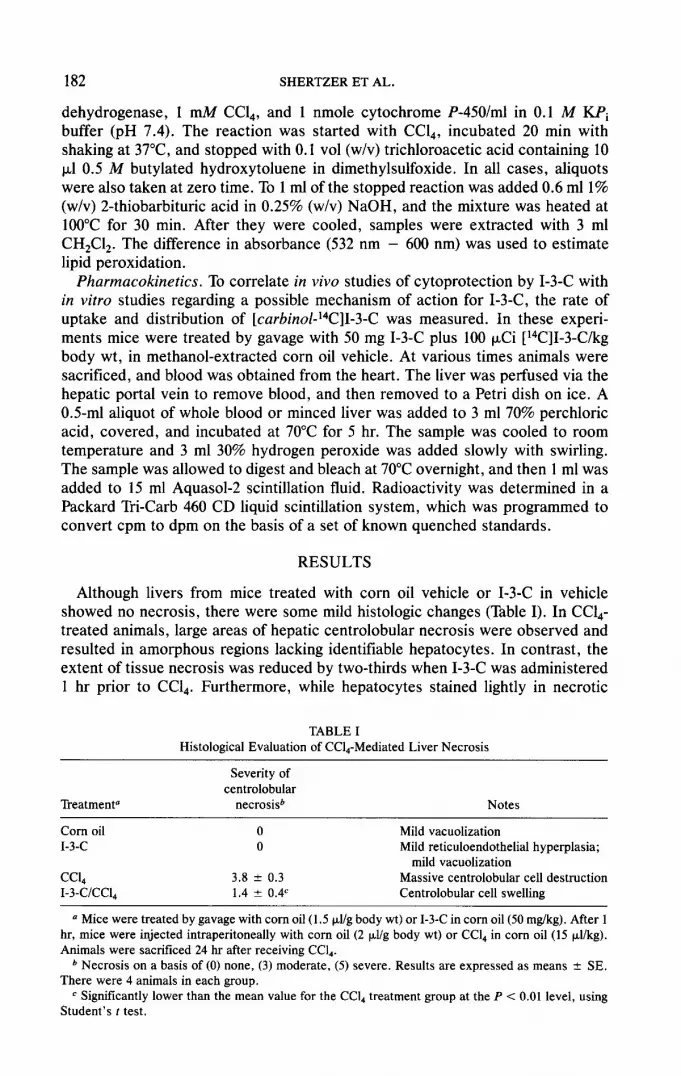
Although livers from mice treated with corn oil vehicle or I-3-C in vehicle showed no necrosis, there were some mild histologic changes (Table I). In Ccl,- treated animals, large areas of hepatic centrolobular necrosis were observed and resulted in amorphous regions lacking identifiable hepatocytes. In contrast, the extent of tissue necrosis was reduced by two-thirds when I-3-C was administered 1 hr prior to Ccl,. Furthermore, while hepatocytes stained lightly in necrotic
TABLE I Histological Evaluation of Ccl,-Mediated Liver Necrosis
Treatment”
Severity of centrolobular
necrosis6 Notes
Corn oil 0 I-3-C 0
ccl, 3.8 k 0.3 I-3-C/Ccl, 1.4 f 0.4’
Mild vacuolization Mild reticuloendothelial hyperplasia;
mild vacuolization Massive centrolobular cell destruction Centrolobular cell swelling
a Mice were treated by gavage with corn oil (1.5 p,l/g body wt) or I-3-C in corn oil (50 mg/kg). After 1 hr, mice were injected intraperitoneally with corn oil (2 pJg body wt) or CC& in corn oil (15 PI/kg). Animals were sacrificed 24 hr after receiving CCL.
b Necrosis on a basis of (0) none, (3) moderate, (5) severe. Results are expressed as means + SE. There were 4 animals in each group.
c Significantly lower than the mean value for the Ccl, treatment group at the P < 0.01 level, using Student’s f test.

INDOLE CARBINOL PROTECTION AGAINST CCL, TOXICITY 183
TABLE II General Parameters of Tissue Damage
Parameter
Serum OTC Serum ALT Serum ALK
Control CCL,
-1-3-C +1-3-c -1-3-c + I-3-C
10.6 i 3.1 6.9 2 0.7 2720 + 251a 1812 2 46W 0.02 2 0.01 0.03 + 0.01 1.89 i 0.4 0.78 + 0.14”*” 13.0 2 0.7 12.9 2 1.5 22.9 * 2.ga 17.5 2 1.60,~
NOW. Mice were treated with I-3-C and/or Ccl, as described in footnote a to Table I. Values are expressed as means 2 SE, with 8-12 animals per group. Units: serum omithine transcarbamylase (OTC), U/ml serum; serum alkaline phosphatase (ALK) and serum alanine transaminase (ALT), +mole/min/ml serum. Statistical analyses were performed by an analysis of variance (ANOVA), fol- lowed by Duncan’s multiple-range test, using an Apple 2e computer.
n Significantly different from control (first column) values at the P < 0.05 level.
b Significantly different from CC&-treated (third column) values at the P < 0.05 level.
regions, general cell structure was preserved and amorphous regions were not observed.
The data in Table II summarize the serum levels of three enzymes that were used routinely to assess liver damage in this study. Although liver specificity of these enzymes varies, with the liver specificity of ornithine transcarbamylase > alanine transaminase > alkaline phosphatase, the trend was the same with each enzyme. I-3-C had no effect by itself in altering serum activities. However, as expected, Ccl, produced a dramatic rise in levels of serum enzymes, with the greatest increase in the liver specific enzyme, OTC, and the smallest increase in the non-tissue-specific enzyme, alkaline phosphatase. In each case, pretreatment with I-3-C resulted in a 50% reduction in the levels of serum enzymes observed following Ccl, exposure. This reduction in serum enzymes was not as great as the decrease in necrosis observed histologically.
Since cytochrome P-450 is the major activation enzyme of Ccl,, it was of in- terest to determine the “suicidal inactivation” of this enzyme by Ccl, in the absence and presence of I-3-C. The data in Table III show that I-3-C was a modest inducer of cytochrome P-450 and aminopyrine N-demethylase, while the high-af- finity NDMA N-demethylase activity was not significantly affected. Ccl, pro- duced a significant decrease in hepatic cytochrome P-450 levels and associated mixed-function oxidase activities. The decrease in NDMA demethylase activity
TABLE III Parameters of Oxidative Biotransformation
Parameter
Control CCI,
-1-3-c + I-3-C - I-3-C + I-3-C
P-450 43.1 2 0.7 47.1 f 1.0” 21.4 T 1.5” 28.5 2 2.8”~~ Aminopyrine demethylase 8.2 f 0.9 9.4 It 0.1 6.4 2 0.6” 8.9 2 0.7-b NDMA demethylase 4.6 2 0.4 4.9 + 0.6 1.7 4 0.2” 3.1 k 0.3”sb
Nore. Mice were treated with I-3-C and/or Ccl, as described in footnote a to Table I. Values are expressed as means + SE, with 8-12 animals per group. Units: cytochrome P-450, nmole/g tissue; aminopyrine N-demethylase and NDMA N-demethylase, nmole/min . mg protein.
On6 Statistical evaluation as described in footnotes a and b to Table II.

184 SHERTZER ET AL.
was greater than that observed for aminopyrine demethylase activity. This was expected since the high-affinity cytochrome P-450 NDMA demethylase isozyme is apparently the same as the isozyme that metabolizes Ccl, to phosgene, a toxic metabolite (English and Anders, 1985). Treatment with I-3-C prior to CCL, admin- istration diminished the loss of cytochrome P-450 and related enzyme activity (Table III). Figure 1 shows that I-3-C treatment also resulted in a dose-dependent increase of total cytochrome P-450.
Endogenous cytoprotective agents against oxidative hepatotoxins collectively constitute the chemical reducing capacity of the cell. These compounds include NAD(P)H, GSH, and ascorbate. Therefore, total hepatic reducing equivalents and GSH and ascorbate levels were determined in the experimental groups (Table IV). While I-3-C produced no effect on hepatic levels of these parameters, Ccl, produced a significant reduction in all parameters but ascorbate. Pretreatment with I-3-C prevented the decreases in the levels of reducing equivalents, GSH, and protein sulfhydryls following Ccl, exposure.
Since the production of toxic metabolites from Ccl, is mediated by cytochome P-450, we examined whether I-3-C prevented the toxicity of CC& by simply inhib- iting metabolism. This was assessed indirectly in two ways. First, the data in Table V show that Ccl, produced a rapid decrease in cytochrome P-450 levels. If I-3-C inhibited metabolism of Ccl,, then in the presence of 1-3-C cytochrome P-450 would be protected from CCL,-mediated loss. This was not the case. Two hours after CC& administration, hepatic cytochrome P-450 levels were decreased to the same extent in control animals and in I-3-C-pretreated mice. Second, we assumed that metabolic inhibition would be proportional to the degree of binding of I-3-C to cytochrome P-450 in vivo. It was found (data not shown) that I-3-C displayed a type II binding absorbance spectrum, with an absorbance maximum
00 -
SB-
40 -
30 -
20 -
10 -
t I I I I I 4
0 50 100 150
I-3-C (MG/KG BODY WEIGHT)
FIG. 1. Effects of I-3-C on hepatic cytochrome P-450 levels. Mice were treated with 15 p,l CCl,/kg body wt in corn oil vehicle or with vehicle alone, as described under Methods. One hour prior to receiving Ccl.,, mice were administered I-3-C in corn oil vehicle by gavage; at the indicated dosages. The volume of corn oil administered was constant at 2 PI/g body wt. Mice were sacrificed 24 hr after receiving Ccl.,, and hepatic cytochrome P-450 levels were measured in whole homogenate, as de- scribed under Methods. Each point represents the average of three independent determinations (three mice); the bars, standard error.

INDOLE CARBINOL PROTECTION AGAINST CCL4 TOXICITY
TABLE IV Parameters of Cellular Reduction Potential
185
Parameter
Total reducing equivalents Ascorbate Nonprotein sulfhydryls Total protein sulfhydryls
Control CC&
-1-3-C + I-3-C - I-3-C + I-3-C
7.3 k 0.7 7.2 k 0.4 5.5 + 0.4a 7.4 * 0.46 1.25 f 0.08 1.28 f 0.06 1.12 k 0.07 1.19 + 0.09 9.7 If- 0.2 9.8 2 0.3 7.8 5 0.20 8.9 + 0.3b
43.1 -+ 2.8 39.2 k 0.7 30.9 k 1.8” 40.8 k l.lb
Nore. Mice were treated with I-3-C and/or Ccl, as described in footnote a to Table I. Values are expressed as means 2 SE, with 8- 12 animals per group. Units: total reducing equivalents, pmolelg tissue; ascorbate and nonprotein sulfbydryls, mM; total protein sulfbydryls, pmole/g protein.
a*b Statistical evaluation as described in footnotes a and b to Table II.
of 423 nm, an absorbance minimum of 383 nm, and a KS value of 237 pM. This is the concentration of I-3-C that produces a half-maximal type II binding spectrum in oxidized liver microsomes. To estimate the level of I-3-C binding to cy- tochrome P-450 in vivo, we measured blood and tissue concentrations of I-3-C versus time. When mice were treated by gavage with a chemoprotective dose (50 mg/kg body wt) of [14C]I-3-C, blood and tissue concentrations increased rapidly. Figure 2 shows that, for each time point, the liver concentration of I-3-C was about five times the concentration measured in blood. The highest liver concen- tration (120 t&f) was attained 30-60 min after treatment with I-3-C. If the KS of I-3-C binding to cytochrome P-450 is approximately equal to the Ki for I-3-C inhibition of NDMA N-demethylation, then 120 pYU I-3-C would bind to only 33% of hepatic cytochrome P-450 in vivo. This value represents an upper limit, since other cytochrome P-450 substrates would tend to displace I-3-C from its binding site.
To monitor the effects of I-3-C from on Ccl,-induced lipid peroxidation, we measured the change in malondialdehyde (MDA) levels in an in vitro system con- taining mouse liver microsomes and an NADPH regenerating system. Pretreat- ment with I-3-C resulted in a dose-dependent decrease in the rate of MDA forma-
TABLE V Hepatic Cytochrome P-450 Levels 2 hr after Treatment with Ccl,
Treatment
Control I-3-C ccl, I-3-C/C/Cl,
Cytochrome P-450 (nmole/g tissue)
50.6 f 3.0” 47.9 f 1.8 34.2 5 2.4 34.8 f 1.0
Note. Mice were treated with I-3-C and/or CC& as described in footnote a to Table I. Animals were sacrificed 2 hr after receiving Ccl,, and hepatic cytochrome P-450 levels were determined on whole homogenates as described under Methods. The values are means 2 SE. There were 3 animals in each group.
0 This experiment was performed on mice weighing about 40 g. This resulted in control levels of tissue cytochrome P-450 somewhat higher than those observed in Table III and Fig. 1, where 25- to 30-g animals were employed.

186 SHERTZER ET AL.
tion (slope) following Ccl, exposure, with 50% inhibition at a concentration of 35-40 pJ4 (Fig. 3).
DISCUSSION
In spite of the intense effort to define the molecular mechanisms involved in eliciting tissue necrosis from CC&-reactive metabolites, the specific mechanisms remain speculative (Brattin ef al., 1985). However, it is quite certain that reactive free radicals, formed by reductive metabolism of Ccl, by cytochrome P-450, are responsible for initiating the cytologic events which result in liver necrosis. In this paper, we show that 1-3-C, administered by gavage prior to Ccl,, protected against histological and biochemical parameters of tissue necrosis.
Vegetables containing I-3-C and I-3-C alone have been shown previously to have a variety of biological effects. A general correlation exists between their abilities to induce various cellular enzymes, such as NAD(P)H:(quinone ac- ceptor) oxidoreductase (M. J. DeLong, personal communication; Salbe and Bjel- danes, in press) mixed-function oxidases, and glutathione S-transferases, and their ability to inhibit various forms of chemically induced neoplasia and toxicity in experimental animals (Bradfield et al., 1985; Godlewski et al., 1985; Sparnins et al., 1982; Stoewsand et al., 1978; Wattenberg et al., 1976). The pattern of isozyme induction has not been elucidated, but apparently I-3-C induces the p- naphthoflavone inducible forms of cytochrome P-450 (Shertzer 1982). It is pos- sible that enzyme induction was responsible, in part, for the higher levels of cy- tochrome P-450 and associated enzyme activity observed when I-3-C was admin- istered prior to Ccl,. In addition, I-3-C has been shown to inhibit the binding of benzo[a]pyrene and NDMA metabolites to cellular protein and DNA, in vivo and in vitro (Shertzer, 1983, 1984). These reductions in covalent binding were not accompanied by significant alterations in metabolic activation of the chemical toxicant.
Although Ccl, toxicity can be inhibited by cytochrome P-450 inhibitors (Berger
3 loo- R . r CD m- : . z z 602
LIVER
E UJ 40. 5
----------a BLOOD .
1 2 no& t&R GA”R5GE
6 7
FIG. 2. Blood and liver concentrations of radiolabel after mice received [‘4ClI-3-C. Mice were treated by gavage with [carbinoP4C]I-3-C, and subsequent blood and liver concentrations of radio- label were determined as described under Methods. The values shown are the averages from two experiments.

INDOLE CARBINOL PROTECTION AGAINST CCL, TOXICITY 187
0.6 CoNTRa. SLOPE
0 25utlI3C
0.5 A SBUHIX
P 0 160 utl 1x 0 266 ul IX
; 0.4
I
5 0.3
tY g 0.2
5 10 IS 20 2s
MINUTES
FIG. 3. Effects of I-3-C on CC&-initiated lipid peroxidation in mouse liver microsomes. The time course for CC&-mediated lipid peroxidation was determined as described under Methods. Five micro- liters of DMSO or I-3-C in DMSO was included per milliliter of the reaction mixture to produce the final I-3-C concentrations as shown. The line of best tit (linear least-squares regression analysis) is shown for each I-3-C concentration. The inset table shows the percentage of the rate of lipid peroxi- dation determined in the absence of I-3-C (control slope).
et al., 1986), this does not appear to be the mechanism of protection afforded by I-3-C. More likely, I-3-C protection is in part mediated by its ability to act as a free radical scavenger. Pharmacokinetic studies with I-3-C indicate that the com- pound is rapidly absorbed into the blood and partitions into the liver. We recognize that with time, increasing amounts of radiolabel will represent I-3-C metabolites, rather than the parent compound. However, the concentration of indole com- pound present in the liver 1 hr after I-3-C administration is about three times the concentration of I-3-C necessary to inhibit CC&-initiated lipid peroxidation by 50%, as measured in our in vitro assay. Furthermore, the metabolites of I-3-C may have greater, less, or equal ability than I-3-C in scavenging radical metabo- lites of Ccl,. Therefore, it appears that sufficient I-3-C is present in the liver to function as a radical trap in vivo.
The ability of I-3-C to trap radical metabolites of CC& may be distinct from or intimately related to the findings of Wang et al. (1985). In that study, tryptophan and/or cysteine, administered 6 hr after Ccl,, protected against CCL, hepatotox- icity assessed at 24 hr. The authors suggested that protection was related to the prevention of Ccl,-induced impairment of protein synthesis. Since the hepatic concentration of I-3-C and/or its metabolites exceeded 60 )IM 6 hr after treat- ment, a potential for protection involving protein synthesis may be operative.
The concept of antioxidants intervening in Ccl, toxicity has been proposed previously for the case of vitamin E (Pesh-Iman and Recknagel, 1977) and pro- methazine (Berger et al., 1986). We propose that a major component of the pro- tection afforded by I-3-C against Ccl, hepatotoxicity is mediated by the ability of the indole to scavenge radicals. As such, we predict that I-3-C would be chemo- protective against other toxins and chemical carcinogens that function by a rad- ical mechanism.

188 SHERTZER ET AL.
ACKNOWLEDGMENTS
This investigation was supported by USPHS Grants ES-03373 and CA-38277 and USPHS Training Grant ES-07073. The authors thank Dr. Steele Mattingly, James Buchanan, Steve Batson, and Lydia Elam for their assistance.
REFERENCES
ALBANO, E., RUNDGREN, M., HARVISON, P. J., NELSON, S. D. and MOLDEUS, I? (1985). Mecha- nisms of N-acetyl-p-benzoquinone imine cytotoxicity. Mol. Pharmacol. 28, 306-311.
BERGER, M. L., BHAT~, H., COMBES, B., and ESTABROOK, R. W. (1986). CC&-induced toxicity in isolated hepatocytes: The importance of direct solvent injury. Hepatology 6, 36-45.
BOWERS, G. N., JR., and MCCOMB, R. B. (1975). Measurement of total alkaline phosphatase activity in human serum. C&I. Chem. 21, 1988-1995.
BRADFIELD, C. A., CHANG, Y., and BJELDANES, L. E (1985). Effects of commonly consumed vege- tables on hepatic xenobiotic-metabolizing enzymes in the mouse. Food Chem. Toxicol. 23,
899-904. BRA-~-~IN, W. J., GLENDE, E. A., JR. and RECKNAGEL, R. 0. (1985). Pathological mechanisms in
carbon tetrachloride hepatotoxicity. J. Free Rad. Biol. Med. 1, 27-38.
ENGLISH, J. C. and ANDERS, M. W. (1985). Evidence for the metabolism of N-nitrosodimethylamine and carbon tetrachloride by a common isozyme of cytochrome P-450. Drug Metab. Dispos. 13,
449-452. GODLEWSKI, C. E., BOYD, J. N., SHERMAN, W. K., ANDERSON, J. L., and STOEWSAND, G. S.
(1985). Hepatic glutathione S-transferase activity and aflatoxin B,-induced enzyme altered foci in rats fed fractions of Brussels sprouts. Cancer Lett. 28, 151-157.
JOHANNESEN, K. A. M., and DEPIERRE, J. W. (1978). Measurement of cytochrome P-450 in the pres- ence of large amounts of contaminating hemoglobin and methemoglobin. Anal. Biochem. 86,
725-732. MANN, A. H., PRICE, S. C., MITCHELL, F. E., GRASSO, P., HINTON, R. H., and BRIDGES, J. W.
(1985). Comparison of the short-term effects of di(2-ethylhexyl)phthalate, di(n-hexyl)phthalate, and di(n-octyl)phthalate in rats. Toxicol. Appl. Pharmacol. 77, 116- 132.
PESH-INMAN, M., and RECKNAGEL, R.O. (1977). Lipid peroxidation and the concept of antioxygenic potential: Vitamin E changes in acute experimental Ccl,-, BrCCI,-, and ethanol-induced liver in- jury. Toxicol. Appl. Pharmacol. 42, 463-475.
RECKNAGEL, R. 0. and GLENDE, E. A., JR. (1973). Carbon tetrachloride hepatotoxicity: An ex- ample of lethal cleavage. CRC Crit. Rev. Toxicol. 2, 263-297.
SALBE, A. D., and BJELDANES, L. E Dietary influences on rat hepatic and intestinal DT-diaphorase activity. Food Chem. Toxicol., in press.
SEDLAK, J., and LINDSAY, R.H. (1968). Estimation of total, protein-bound, and nonprotein sulfbydryl groups in tissue with Ellman’s reagent. Anal. Biochem. 25, 192-205.
SEGAL, H. L., and MATSUZAWA, T. (1970). L-Alanine aminotransferase (rat liver). In “Methods in Enzymology” (H. Tabor and C. W. Tabor, Eds.), Vol. 17, Part A, pp. 153-159. Academic Press, New York.
SHERTZER, H. G. (1982). Indole-3-carbinol and indole-3-acetonitrile influence on hepatic microsomal metabolism. Toxicol. Appl. Pharmacol. 64, 353-361.
SHERTZER, H. G. (1983). Protection by indole-3-carbinol against covalent binding of benzo(a)pyrene metabolites to mouse liver DNA and protein. Food Chem. Toxicol. 21, 31-35.
SHERTZER, H. G. (1984). Indole-3-carbinol protects against covalent binding of benzo(a)pyrene and N-nitrosodimethylamine metabolites to mouse liver macromolecules. Chem.-Biol. Interact. 48, 81-90.
SHERTZER, H. G., and DUTHU, G. S. (1979). Nitrite binding to rabbit liver microsomes and effects on aminopyrine demethylation. Biochem. Pharmacol. 28, 873-879.
SHERTZER, H. G., and TABOR, M. W. (1985). Peroxide removal from organic solvents and vegetable oils. J. Environ. Sci. Health Part A 20, 845-855.
SPARNINS, V. L., VENEGAS, P. L., and WAT~ENBERG, L. W. (1982). Glutathione S-transferase ac- tivity: Enhancement by compounds inhibiting chemical carcinogenesis and by dietary constituents. J. Natl. Cancer Inst. 68, 493-496.
STOEWSAND, G. S., BABISH, J. G., and WIMBERLY, H. C. (1978). Inhibition of hepatic toxicities from

INDOLE CARBINOL PROTECTION AGAINST CCL4 TOXICITY 189
polybrominated biphenyls and aflatoxin B, in rats fed cauliflower. J. Environ. P&ho/. Toxicol. 2,
399-406.
VASSEF, A. A. (1978). Direct micromethod for calorimetry of serum omithine carbamoyltransferase activity, with use of a linear standard curve. Clin. Chem. 24, 101-107.
WANG, D., VERNEY, E., and SIDRANSKY, H. (1985). Protection effect of tryptophan and cysteine against carbon tetrachloride-induced liver injury. Exp. Mol. Pathol. 43, 375-387.
WAT~ENBERG, L. W., LOUB, W. D., LAM, L. K., and SPEIER, J. L. (1976). Dietary constituents al- tering the responses to chemical carcinogens. Fed. Proc. 35, 1327-1331.
ZANNONI, V., LYNCH, M., GOLDSTEIN, S., and SATO, P (1974). A rapid micromethod for the determi- nation of ascorbic acid in plasma and tissues. Biochem. Med. 11, 41-48.


