Embed Size (px)
Citation preview

Diphtheria Toxin-Mediated Cell Ablation RevealsInterregional Communication during ArabidopsisSeed Development1
Dolf Weijers2, Jan-Piet van Hamburg, Erwin van Rijn3, Paul J.J. Hooykaas, and Remko Offringa*
Developmental Genetics, Institute of Biology, Leiden University, Clusius Laboratory, Wassenaarseweg 64,2333 AL Leiden, The Netherlands
Fertilization of the female gametophyte in angiosperm plants initiates a process of coordinated development of embryo,endosperm, and seed coat that ensures the production of a viable seed. Mutant analysis has suggested that communicationbetween the endosperm and the seed coat is an important determinant in this process. In addition, cell groups within theembryo, derived from the apical and from the basal cell, respectively, after zygote division, concertedly establish a functionalroot meristem, and cells in the apical region of the embryo are hypothesized to repress cell divisions in the basal cell-derivedsuspensor. The available evidence for these interregional communication events mostly relies on the analysis of mutantphenotypes in Arabidopsis. To provide independent and direct evidence for communication events, we used conditionaldomain-specific expression of the diphtheria toxin A chain (DTA) in developing Arabidopsis seeds. By using a collection ofcell- or tissue-type-specific promoters, we show that the mGAL4:VP16/UAS two-component gene expression allows reliablespatiotemporal and conditional expression of the GFP:GUS reporter and the DTA gene in the developing embryo andendosperm. Expression of DTA in the protoderm of the embryo proper led to excessive proliferation of suspensor cells,sometimes resulting in the formation of secondary embryos. Endosperm-specific expression of DTA caused completecessation of seed growth, followed by pattern defects in the embryo and embryo arrest. Taken together, the results presentedhere substantiate the evidence for and underline the importance of interregional communication in embryo and seeddevelopment and demonstrate the usefulness of conditional toxin expression as a method complementary to phenotypicanalysis of developmental mutants.
Seed development in higher plants is characterizedby the coordinated development of distinct tissues.Seed tissues mainly arise from cells and tissues of thefemale gametophyte that are formed before fertiliza-tion. The multinucleate female gametophyte is en-closed by several sporophytic maternal cell layersthat constitute the ovule. In angiosperms, doublefertilization by fusion of the egg cell and the centralcell of the female gametophyte with the two spermcells delivered by the pollen tube generates the dip-loid embryo and triploid endosperm, respectively.The embryo and endosperm both develop within theconfines of the maternal tissue, now referred to as theseed coat, but each follows a different developmentalprogram (for review on embryo and endosperm de-velopment, see Jurgens and Mayer, 1994; Berger,
2003, respectively). Within the embryo, a complexbut precise pattern of organs and cell types is laiddown from a single cell, the zygote (Jurgens andMayer, 1994). Genetic controls are required to estab-lish the embryo pattern and to ensure the properinitiation and relative positioning of distinct cellgroups and organs, such as the meristems and vas-culature. The endosperm first undergoes a series ofsynchronized nuclear divisions and forms a free-nuclear syncytium divided in three domains. Themicropylar endosperm surrounds the developingembryo, the peripheral endosperm fills up most ofthe seed volume, and a dense nucleocytoplasmic do-main, the chalazal endosperm, extends to the nucel-lar region of the seed. As soon as the embryo reachesthe heart stage, endosperm nuclei are partitioned intoindividual units by cell walls during a process calledcellularization. Only a remnant of the endosperm ispresent in the mature seed as a thin layer that sepa-rates the embryo from the seed coat tissues (Berger,2003). The relative growth of seed coat, endosperm,and embryo must be stringently regulated to securedevelopment of a viable seed.
Several Arabidopsis mutants in seed and en-dosperm development and embryo pattern forma-tion have been isolated, and detailed phenotypicanalysis of a number of them has brought new insightsinto the importance of communication between differ-ent functional/cellular domains—referred to here as
1 This work was supported by the Research Council for Earthand Life Sciences (ALW), with financial aid from the NetherlandsOrganisation for Scientific Research (NWO).
2 Present address: Developmental Genetics, Zentrum fur Mole-kularbiologie der Pflanzen, Universitat Tubingen, Auf der Mor-genstelle 3, D–72076 Tubingen, Germany.
3 Present address: Department of Cell Biology and Histology,University of Amsterdam, Academic Medical Center, Meiberg-dreef 15, 1105 AZ Amsterdam, The Netherlands.
* Corresponding author; e-mail [email protected];fax 31–71–5274999.
Article, publication date, and citation information can be foundat http://www.plantphysiol.org/cgi/doi/10.1104/pp.103.030692.
1882 Plant Physiology, December 2003, Vol. 133, pp. 1882–1892, www.plantphysiol.org © 2003 American Society of Plant Biologists www.plant.org on July 15, 2015 - Published by www.plantphysiol.orgDownloaded from Copyright © 2003 American Society of Plant Biologists. All rights reserved.

interregional communication—in plant seeds and,specifically, in embryos. For example, mutants of thefis (fertilization-independent seed) category and seedswith an excess of maternal genomes resulting fromcrosses between parents with different ploidy levelsshow reduced endosperm development and reducedseed size. In some cases, endosperm defects in suchgenotypes are accompanied by aberrant embryo de-velopment (Ohad et al., 1996; Scott et al., 1998). Therecent isolation of the haiku mutants reinforces thehypothesis that endosperm development plays an im-portant role in overall seed growth and also has afunction in late embryo development (Garcia et al.,2003). The role of the endosperm in early embryodevelopment, however, is as yet unclear.
Phenotypic analysis of the large set of Arabidopsisembryo patterning mutants (e.g. Mayer et al., 1991)has highlighted the importance of interregional com-munication in the regulation of pattern formation.Analysis of the sus and twin mutants classes, in whichthe basally located suspensor cells proliferate aftercellular defects become visible in the embryo proper(Vernon and Meinke, 1994; Schwartz et al., 1994;Zhang and Somerville, 1997) and of bdl (bodenlos) andmp (monopteros) mutants, which show initial apicalcell division defects and subsequent failure to initiatea root meristem from a clonally distinct cell popula-tion (Berleth and Jurgens, 1993; Hamann et al., 1999),led to the hypothesis that the apical region of theembryo exerts significant control over the develop-ment of the basal region of the embryo and thesuspensor.
Although cloning of the BDL (Hamann et al., 2002)and MP (Hardtke and Berleth, 1998) genes has givenimportant insight into the possible mechanism un-derlying this interregional communication, identifi-cation of the TWN2 (Zhang and Somerville, 1997) andSUS1 (Golden et al., 2002) genes has not significantlyimproved our understanding of the mechanism ofembryo-suspensor interactions.
Generally, in the mentioned cases of hypothesizedregulatory interactions between cells or domains, it isdifficult to conclude that a mutation affects interre-gional communication, e.g. between the endospermand the embryo proper, unless the expression of themutated gene is known to be confined to specific cellsor domains. Therefore, independent evidence shouldcome from experiments where a strictly defined setof cells within the seed or embryo is disabled, e.g. byexpression of a toxin. Subsequent non-cell-autonomous effects on development then directlyshow functional interactions. Until now, only a singlestudy has made use of toxin expression in the em-bryo to study cellular interactions. However, the ob-served non-cell-autonomous effects were mild anddid not provide evidence for repression of suspensordevelopment by the embryo proper (Baroux et al.,2001).
Here, we present an analysis of cellular communi-cation within the seed by local expression of thediphtheria toxin A chain (DTA). This protein ishighly toxic (Yamaizumi et al., 1978), and the encod-ing gene has been used successfully to ablate root capcells (Tsugeki and Fedoroff, 1999), petal and stamenprimordia in flowers (Day et al., 1995), or even entireflowers (Nilsson et al., 1998). To circumvent any ex-pression of the toxin before fertilization, we adoptedthe mGAL4:VP16/UAS two-component gene expres-sion system. Two-component gene expression tech-nology (transactivation) makes use of two plantlines—one that expresses a heterologous transcrip-tion factor under control of a chosen plant promoterand another that contains a gene of interest undercontrol of a silent promoter that is only activated bythe heterologous transcription factor. Only uponcrossing the two lines, the gene of interest will beexpressed in the zygotic tissues.
The mGAL4:VP16/UAS system has been opti-mized for Arabidopsis (Haseloff, 1999) and has beenused by several researchers to express reporter genesor genes of interest during postembryonic stages ofdevelopment (e.g. Kiegle et al., 2000; Benjamins et al.,2001; Sabatini et al., 2003) or in the endosperm(Boisnard-Lorig et al., 2001). No data were availableon its reliability in the embryo; therefore, we firstanalyzed this aspect in detail. The analysis showsthat mGAL4:VP16/UAS technology is reliable in em-bryos. Here, we use the technology to express DTAtoxin in specific tissues in the embryo or in the en-dosperm, and we provide direct evidence for thepreviously hypothesized cellular interactions in em-bryo and seed development.
RESULTS
Plant Lines for Conditional Domain-SpecificExpression in Seed or Embryo
An experimental strategy for cell ablation by ex-pressing the DTA cytotoxin in limited domains of thedeveloping seed needs to fulfill two important crite-ria. First, it requires a selected set of gene promoterswith a well-defined spatiotemporal expression spec-ificity in the seed; second, the expression must beconditional, i.e. expression of DTA must not takeplace during generation of lines or at any time beforebut should be readily activated upon fertilization. Asfor the first requirement, we have selected a set ofpreviously described promoters that providedomain-specific expression in the embryo or the en-dosperm. The Arabidopsis RPS5A (RIBOSOMALPROTEIN S5A) promoter is strongly expressed individing cells, starting as early as the one-cell embryostage (Weijers et al., 2001a). The Arabidopsis LTP1(LIPID TRANSFER PROTEIN 1) promoter marks theL1 layer of all newly formed organs and is preferen-tially expressed in the apical domain of the embryo(Thoma et al., 1994). The synthetic auxin-responsive
Communication during Arabidopsis Seed Development
Plant Physiol. Vol. 133, 2003 1883 www.plant.org on July 15, 2015 - Published by www.plantphysiol.orgDownloaded from Copyright © 2003 American Society of Plant Biologists. All rights reserved.

DR5(7x) promoter has an expression peak in the cen-tral root cap cells of the torpedo stage Arabidopsisembryo (Sabatini et al., 1999). The enhancer-trappedmGAL4:VP16 gene (see below) in line KS22I was usedas a tool to drive expression in the developing en-dosperm (Boisnard-Lorig et al., 2001).
To obtain conditional expression of DTA, weadopted the mGAL4:VP16/UAS two-component geneexpression system (Fig. 1A; Haseloff, 1999). ACT lineswere generated that harbor the mGAL4:VP16 codingsequence coupled to the RPS5A, LTP1, or DR5(7x)promoters. For all ACT constructs, lines were obtainedwith a single T-DNA insertion that did not displayabnormal embryo- or seedling development at fre-quencies exceeding those in wild type (Table I). ThemGAL4:VP16 mRNA was easily detected in ACTRPS5A, ACT 35S (pCaMV35S::mGAL4:VP16; J. Hasel-off, unpublished data), ACT LTP1 (Fig. 1B), and ACTDR5(7x) (not shown) seedlings. In addition, themGAL4:VP16 protein was detectable in extracts fromACT 35S seedlings (Fig. 1C).
Separate EF DTA plant lines were generated con-taining the DTA gene driven by the mGAL4:VP16-
responsive UAS promoter (Fig. 1A). As a control, EFGGi lines containing a UAS::GFP:GUS reporter genewere generated. No GUS activity could be detected in30 independent EF GFP:GUS lines, and the develop-ment of all EF DTA lines (n � 28; Table I; data notshown) was indistinguishable from that of wild-typeArabidopsis plants. These results, together withthose of Bougourd et al. (2000), show that the UASpromoter is inactive in the absence of mGAL4:VP16and, therefore, not a target of endogenous transcrip-tion factors.
Reliable Domain-Specific Reporter GeneExpression after mGAL4:VP16/UAS Transactivation
To first assess the reliability of cell-specific trans-activation in the seed, homozygous EF GGi plantswere crossed with the homozygous ACT lines andwith the KS022I enhancer trap line. The ACT lineswere consistently used as female parents in crosseswith EF lines because we observed that this providedthe strongest transactivation during early embryo-genesis (Weijers et al., 2001b).
Transactivation of the UAS::GFP:GUS gene inRPS5A��GGi F1 (the notation promoter Y �� gene X isused to distinguish transactivation from direct pro-moter Y::gene X fusions) seeds was detectable in theembryo and endosperm in the one-cell embryo stage(Fig. 2A), and the intensity of GUS staining increaseduntil the heart stage (Fig. 2, A–D). Although theexpression level in RPS5A��GGi embryos was muchstronger than in RPS5A::GUS reporter lines (compareFig. 2C with Weijers et al., 2001a), the spatiotemporalpattern did not differ.
As expected for an L1 layer-specific promoter, GUSactivity in LTP1��GGi F1 embryos was detectable inprotodermal cells in dermatogen stage embryos (Fig.2G). Subsequently, the expression spread to cover theentire apical region of the heart stage embryo (Fig.2H), after which the GUS signal became exclusivelyconfined to the L1 layer of the cotyledons (Fig. 2I). InF1 seedlings, the GUS pattern did not differ from thereported LTP1::GUS pattern (Thoma et al., 1994; datanot shown), and analysis of GFP fluorescence re-vealed strict epidermal specificity (Fig. 2, J and K).
Similarly, GFP:GUS transactivation in DR5(7x)��GGi(Fig. 2F) embryos mirrored the direct promoter::GUSfusion activity (Fig. 2E). In KS22I��GGi F1 plants, weobserved endosperm specific expression of both GFP:GUS on the EF GGi construct (Fig. 2, M and N) andmGFP5-ER on the enhancer trap construct (Fig. 2L),indicating coregulation of both UAS-controlled re-porter genes. In summary, ACT line-driven UAS::GGiexpression conserved the spatiotemporal expressionof each promoter and provided stronger GGi expres-sion than a corresponding direct promoter::GGi fu-sion. Notably, for reasons that we do not know, asignificant variation in gene expression levels wasalways observed between sibling embryos (Fig. 2, Dand I, insets).
Figure 1. Expression of mGAL4:VP16 in Arabidopsis. A, mGAL4:VP16/UAS transactivation approach: A promoter of interest (pro-moter X) is fused to the mGAL4:VP16 coding sequence in activator(ACT) line X. The mGAL4:VP16 protein is then produced only in cellsin which promoter X is active. In effector (EF) line Y, both a targetgene (gene Y) and eGFP:GUS::intron are controlled by the GAL4:VP16-responsive UAS promoter (UAS; contains five GAL4-bindingsites placed upstream of the �47 CaMV 35S promoter). No transcrip-tion of these genes occurs in the absence of GAL4:VP16. Uponcrossing ACT X and EF Y, transactivation of Y and GGi only occursin those cells of F1 (and F2) progeny in which promoter Y is active. B,mGAL4:VP16 mRNA accumulation in seedlings of the ACT RPS5A(nos. 5, 10, 17, and 18), ACT 35S#2 and ACT LTP1#8 lines. Colum-bia (Col), Wild-type control. Twenty micrograms of total RNA wasloaded for ACT LTP1 and 10 �g for all other samples. Ethidiumbromide-stained gel to show loading differences. C, Western blotincubated with anti-VP16 antibody shows the expected band of 26kD in total protein extracts of ACT 35S seedlings (30 �g of protein)but not in wild type (Col).
Weijers et al.
1884 Plant Physiol. Vol. 133, 2003 www.plant.org on July 15, 2015 - Published by www.plantphysiol.orgDownloaded from Copyright © 2003 American Society of Plant Biologists. All rights reserved.
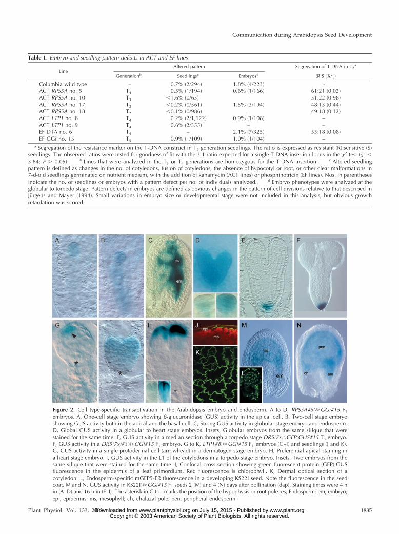
Figure 2. Cell type-specific transactivation in the Arabidopsis embryo and endosperm. A to D, RPS5A#5��GGi#15 F1
embryos. A, One-cell stage embryo showing �-glucuronidase (GUS) activity in the apical cell. B, Two-cell stage embryoshowing GUS activity both in the apical and the basal cell. C, Strong GUS activity in globular stage embryo and endosperm.D, Global GUS activity in a globular to heart stage embryos. Insets, Globular embryos from the same silique that werestained for the same time. E, GUS activity in a median section through a torpedo stage DR5(7x)::GFP:GUS#15 T3 embryo.F, GUS activity in a DR5(7x)#3��GGi#15 F1 embryo. G to K, LTP1#8��GGi#15 F1 embryos (G–I) and seedlings (J and K).G, GUS activity in a single protodermal cell (arrowhead) in a dermatogen stage embryo. H, Preferential apical staining ina heart stage embryo. I, GUS activity in the L1 of the cotyledons in a torpedo stage embryo. Insets, Two embryos from thesame silique that were stained for the same time. J, Confocal cross section showing green fluorescent protein (GFP):GUSfluorescence in the epidermis of a leaf primordium. Red fluorescence is chlorophyll. K, Dermal optical section of acotyledon. L, Endosperm-specific mGFP5-ER fluorescence in a developing KS22I seed. Note the fluorescence in the seedcoat. M and N, GUS activity in KS22I��GGi#15 F1 seeds 2 (M) and 4 (N) days after pollination (dap). Staining times were 4 hin (A–D) and 16 h in (E–I). The asterisk in G to I marks the position of the hypophysis or root pole. es, Endosperm; em, embryo;epi, epidermis; ms, mesophyll; ch, chalazal pole; pen, peripheral endosperm.
Table I. Embryo and seedling pattern defects in ACT and EF lines
LineAltered pattern Segregation of T-DNA in T2
a
Generationb Seedlingsc Embryosd (R:S ��2�)
Columbia wild type – 0.7% (2/294) 1.8% (4/223) -ACT RPS5A no. 5 T4 0.5% (1/194) 0.6% (1/166) 61:21 (0.02)ACT RPS5A no. 10 T3 �1.6% (0/63) – 51:22 (0.98)ACT RPS5A no. 17 T2 �0.2% (0/561) 1.5% (3/194) 48:13 (0.44)ACT RPS5A no. 18 T2 �0.1% (0/986) – 49:18 (0.12)ACT LTP1 no. 8 T4 0.2% (2/1,122) 0.9% (1/108) –ACT LTP1 no. 9 T4 0.6% (2/355) – –EF DTA no. 6 T4 – 2.1% (7/325) 55:18 (0.08)EF GGi no. 15 T5 0.9% (1/109) 1.0% (1/104) –
a Segregation of the resistance marker on the T-DNA construct in T2 generation seedlings. The ratio is expressed as resistant (R):sensitive (S)seedlings. The observed ratios were tested for goodness of fit with the 3:1 ratio expected for a single T-DNA insertion locus in the �2 test (�2 �3.84; P � 0.05). b Lines that were analyzed in the T3 or T4 generations are homozygous for the T-DNA insertion. c Altered seedlingpattern is defined as changes in the no. of cotyledons, fusion of cotyledons, the absence of hypocotyl or root, or other clear malformations in7-d-old seedlings germinated on nutrient medium, with the addition of kanamycin (ACT lines) or phosphinotricin (EF lines). Nos. in parenthesesindicate the no. of seedlings or embryos with a pattern defect per no. of individuals analyzed. d Embryo phenotypes were analyzed at theglobular to torpedo stage. Pattern defects in embryos are defined as obvious changes in the pattern of cell divisions relative to that described inJürgens and Mayer (1994). Small variations in embryo size or developmental stage were not included in this analysis, but obvious growthretardation was scored.
Communication during Arabidopsis Seed Development
Plant Physiol. Vol. 133, 2003 1885 www.plant.org on July 15, 2015 - Published by www.plantphysiol.orgDownloaded from Copyright © 2003 American Society of Plant Biologists. All rights reserved.

Protoderm Ablation Reveals Apical Control of BasalEmbryo Development
A striking example of interregional communicationin embryo development is the hypothesized suppres-sion of cell divisions in the suspensor and the regu-lation of hypophysis division by the embryo proper(Schwartz et al., 1994; Vernon and Meinke, 1994;Zhang and Somerville, 1997; Jurgens, 2001). A previ-ous study has challenged the hypothesis of apicalcontrol of basal embryo development (Baroux et al.,2001). Upon LTP1-driven expression of the BARNASE(Bacillus amyloliquefaciens ribonuclease) gene, em-bryos showed defects in the basal tier of the embryoproper and in hypophysis division. Surprisingly,however, no signs for deregulation of suspensor de-velopment were found. Possibly, the effectiveness ofBARNASE-induced cell lethality (Baroux et al., 2001)was not sufficient to induce such defects in theembryo proper that would allow the detection ofembryo-suspensor interregional communication. Al-ternatively, it may be that the defects in suspensordevelopment observed in Arabidopsis sus and twnmutants are not a result of improper regulation bythe embryo proper. Here, we used the ACT LTP1 andEF DTA lines to challenge this hypothesis.
First, we confirmed that the UAS::DTA gene en-codes a functional toxin by analyzing RPS5A��DTAF1 embryos. Cell ablation was visible in Schiffreagent-stained ovules shortly after fertilization asbright nuclear fluorescence and degeneration of theyoung embryo and endosperm (Fig. 3B). To confirmthat the DTA protein works cell autonomously,LTP1��DTA embryos were generated and analyzedat early developmental stages. The first obvious ab-errations were found during the 16-cell stage. At thisstage, protodermal cells but not inner cells showedthe hallmarks of cell ablation, ranging from stronguniform nuclear fluorescence (not shown) to thepresence of nucleus-free cellular remnants (Fig. 3H).
Cell ablation in LTP1��DTA embryos showed sig-nificant variation in timing, and in the severity of theinduced defects. Extreme defects were observed in8.7% of the analyzed embryos (n � 92), includingcomplete protoderm ablation during the 16-cell stage(Fig. 3, E and F), whereas in the remaining embryos,the defects originated from post-globular ablationevents (Fig. 3D). Interestingly, preglobular ablationof the protoderm induced excessive divisions in thesuspensor (Fig. 3, E, F, and H). In one class of em-bryos, excessive divisions were in appropriate orien-tation, which merely anticipated later suspensor di-visions (Fig. 3H). In another class of embryos,suspensor cells were more obviously released fromrepression because a dense cell group developed inthe middle of the suspensor. Often, these cell groupsattained division patterns common to the embryo(Fig. 3, E and F). In the remaining embryos, cellulardefects suggested the ablation events to have takenplace after the globular to heart stage transition. In
these embryos, the ablation events resulted in strongdefects in cotyledon development. Embryos showedonly rudimentary cotyledons at the apical end of theaxis. Surprisingly, such embryos had no visible de-fects in root pole patterning (Fig. 3D), which indi-cates that the apical control over suspensor develop-ment, evident at early stages, loses its effect aftertransition to the heart stage.
The sites that are marked by cell ablation in post-globular LTP1��DTA embryos are exactly thosewhere LTP1 promoter activity is reported by GUSactivity in LTP1��GGi embryos. Moreover, in con-trast to DR5(7x)��DTA embryos (Friml et al., 2003),no signs of cell ablation were found in LTP1��DTAsuspensor cells or in the hypophysis, This indicatesthat the aberrant basal cell divisions after early abla-tion events in the embryo proper are not a result oflocal DTA expression but are induced by a lack of“controlling” signals from the apical cell lineage.
Endosperm Ablation Reveals Its Role in Seed SizeRegulation and Embryo Patterning
Several Arabidopsis mutants and transgenic linesand seeds resulting from crosses between parentplants with different ploidies show reduced en-dosperm development and reduced seed size (Scottet al., 1998; Luo et al., 2000; Garcia et al., 2003). The
Figure 3. DTA expression in domains of the Arabidopsis embryo. A,C, and G, Wild-type two-cell (A), heart (C), and dermatogen stage (G)embryos. B, RPS5A#5��DTA#6 F1 seed containing only remnants ofembryo and endosperm. D, Heart stage LTP1#8��DTA#6 F1 em-bryos with normal pattern but defective cotyledons. E and F, Glob-ular stage LTP1#8��DTA#6 F1 embryos showing arrest of the em-bryo proper and proliferation of the suspensor. The clonal boundaryof the first embryonic cell division is marked by an arrow. H,Dermatogen stage LTP1#8��DTA#6 F1 embryo showing ablation ofthe protoderm (arrow, compare with wild-type embryo in G). Note thatthe number of suspensor cells is doubled in the LTP1#8��DTA#6embryo. Em, Embryo; es, endosperm; c, cotyledon.
Weijers et al.
1886 Plant Physiol. Vol. 133, 2003 www.plant.org on July 15, 2015 - Published by www.plantphysiol.orgDownloaded from Copyright © 2003 American Society of Plant Biologists. All rights reserved.

reduction in seed size in these cases is zygoticallydetermined, suggesting that the regulation of seedsize is controlled by the endosperm. This regulatoryinteraction between the endosperm, embryo, andseed coat represents another example of interregionalcommunication within the seed. Because the mutantgenotype may not only affect the endosperm, it isdifficult to draw unequivocal conclusions as for therole of endosperm in seed growth and embryo de-velopment. To obtain such direct evidence, we ana-lyzed seed growth and development in seeds thatexpress DTA under control of the endosperm-expressed mGAL4:VP16 activator in enhancer trapline KS22I.
Based on fluorescence of the UAS::mGFP5-ER genepresent on the mGAL4:VP16 enhancer trap T-DNA,KS22I enhancer expression commences after the sec-ond round of nuclear divisions (M. Ingouff and F.Berger, personal communication). In accordance with
this expression pattern, endosperm development ar-rested almost immediately in KS22I��DTA seeds,and the endosperm soon degenerated, leaving only afew remnant cellular structures (Fig. 4, E–G). Themost striking phenotype of KS22I��DTA F1 seedswas the complete cessation of seed expansion. Wild-type seeds underwent rapid and continuous growthshortly after fertilization (Fig. 4, A–D), whereas seedexpansion was impaired in 57% (47/83) of theKS22I��DTA seeds analyzed 2 dap, in 56% (46/83) at4 dap, and in 97% (131/135) at 6 dap (Fig. 4, E–H).This result probably reflects the variability in theonset of DTA expression in independent F1 seeds, butit also shows that the induced effect is fully penetrantat stages when the phenotype is unambiguouslyidentifiable (6 dap, under these experimental condi-tions more or less corresponding to wild-type seedswith heart stage embryos). The absence of seedgrowth upon genetic endosperm ablation presents
Figure 4. Consequences of endosperm ablation on seed development. A to D, Wild-type seed development. A, Seedcontaining a two-celled embryo (arrow marks endosperm nucleus). B, Seed containing an eight-celled embryo. The insetshows a magnification of endosperm nuclei with surrounding cytoplasm and organelles. C, Seed containing a globular stageembryo. The endosperm domains (ch, pen, and mp) are clearly recognizable along the anterio-posterior axis of the seed. D,Seed containing a torpedo stage embryo. E to K, KS22I��DTA#6 F1 seed development. E, Seed of a stage comparable withA. Note the intense red fluorescence and the tracheid-like structure in the degraded endosperm (arrow). The embryo is notvisible in this optical section. F, Seed containing a globular stage embryo. The endosperm is absent, as shown by amagnification of the endosperm cavity in the inset. G, Seed containing a globular stage embryo that is directly surroundedby seed coat tissues. The seed starts to collapse at this stage, which is visible because the seed coat tissues are in the samefocal plane as the embryo. H, Collapsed seed containing a “heart stage” embryo. Magnification and stage are the same asin D. Inset, Magnification of the micropylar region containing the embryo. I, Higher magnification of the embryo in F showsa normal embryo pattern, except for a slight bulging of the epidermal cells (arrow). J, Higher magnification of the embryoin G, which shows severe lateral compression, indicating aberrant patterning. K, Heart stage embryos enclosed by seed coattissues show abnormal patterns of cell division. Note that a structure resembling an embryo proper develops in the suspensorof the embryo in the inset (arrowhead marks the boundary between two embryo structures). L and M, RPS5A::GFP:GUS#20activity in phenotypically wild-type (L) and KS22I��DTA#6 F1 sibling seeds (M). Note that GUS activity is still detectablein the embryo in (M) but only in the basal domain that does not yet show signs of physical obstruction. The KS22I line washeterozygous for the transgene. N and O, DR5(7x)::GFP:GUS#13 activity in wild-type (N) and phenotypically affected (O)sibling KS22I��DTA embryos. The embryo in O is delayed and does not show GUS activity. Seeds were stained for GUSactivity for 4 h. es, Endosperm; em, embryo; ch, chalazal pole; pen, peripheral endosperm; mp, micropylar pole.
Communication during Arabidopsis Seed Development
Plant Physiol. Vol. 133, 2003 1887 www.plant.org on July 15, 2015 - Published by www.plantphysiol.orgDownloaded from Copyright © 2003 American Society of Plant Biologists. All rights reserved.

direct evidence that development of the endospermis stringently required for seed growth. Similar re-sults were obtained with enhancer trap line M003B(F. Berger and J. Haseloff, unpublished data) that alsoprovides expression of mGAL4:VP16 in the en-dosperm (data not shown).
In addition to the defects in seed growth, embryosin KS22I��DTA F1 seeds showed defects in growthand cell division patterns. However, less than 30% ofthe embryos (10/38 at preglobular stage and 8/46 atglobular stage) were affected. Preglobular embryosdisplayed swollen protodermal cells (Fig. 4I) or al-tered cell division planes (Fig. 4J), but embryos nevershowed any sign of cell death. Several embryo phe-notypes, such as presence of two stacked embryoproper structures (Fig. 4K, inset), were found thatindicated defects originating from early preglobularstages. This suggests that the presence of a develop-ing endosperm is at least to some extent required forproper early embryo development.
In the absence of endosperm development,KS22I��DTA post-globular stage embryos becamedirectly enclosed by the seed coat tissues, and, pre-sumably as a result of this physical obstruction, em-bryos showed heavily distorted division patterns(Fig. 4K), and seeds collapsed after transition stage(Fig. 4H) and did not germinate (not shown). Cellswithin “arrested” embryos showed reduced expres-sion of the RPS5A::GUS marker (Weijers et al., 2001a;Fig. 4M) that is normally active in dividing cells (Fig.4L). Interestingly, expression of the RPS5A::GUSmarker always ceased first in the apical cells that arein direct contact with the seed coat. Because thephenotypes at the basal pole of KS22I��DTA em-bryos resembled those of embryos with defects inauxin response or transport (Mayer et al., 1991; Ber-leth and Jurgens, 1993; Hamann et al., 1999; Hobbie etal., 2000), the DR5(7x)::GUS reporter was crossed intothe KS22I line. Upon crossing KS22I;DR5(7x)::GUSlines with EF DTA lines, F1 embryos consistentlyfailed to activate the DR5(7x)::GUS marker (Fig. 4O),which is normally active in the hypophyseal cellgroup from the mid-heart stage on (Fig. 4N), suggest-ing that arrested embryos fail to establish the auxinmaximum that is associated with patterning of theroot meristem (Sabatini et al., 1999). Whether thisreflects a defect in auxin-related processes or that theabsence of the auxin response maximum is merelythe result of auxin-independent defects in embryopatterning due to the endosperm ablation cannot bedistinguished.
Our analysis provides direct evidence for the pre-viously suggested role for endosperm developmentin promoting seed growth and facilitating properembryo development (Ohad et al., 1996; Scott et al.,1998; Luo et al., 2000; Garcia et al., 2003). The resultsclearly show that endosperm development is not re-quired per se for proper embryo development duringthe early preglobular stages because only a part of
the nonenlarged seeds contain defective embryos.Whether the patterning defects observed inKS22I��DTA embryos at later stages are caused bythe absence of the endosperm or merely by the phys-ical constraints of the underdeveloped integumentlayers awaits further studies.
DISCUSSION
Intercellular communication in plants, with thecentral role of protein or peptide movement in rootradial patterning (Nakajima et al., 2001), floral pat-terning (Sessions et al., 2000), and stem cell ho-meostasis in the shoot meristem (Lenhard and Laux,2003) as recent examples, attracts increasing atten-tion. Cellular interactions take place during postem-bryonic and embryo development, where, for exam-ple, embryonic root initiation depends on signalingacross the clonal boundary set during the first celldivision. Previously, communication had been pro-posed between embryo proper and suspensor andbetween embryo, endosperm, and seed coat tissues.In most cases, models were based on the phenotypesof developmental mutants; thus, direct evidence forthese communication events was still lacking. Here,we provide direct evidence for and confirm the pres-ence of all previously proposed communication andregulatory interactions in the seed by local expres-sion of the DTA toxin in domains of the developingseed or embryo.
mGAL4:VP16/UAS Transactivation in DevelopingArabidopsis Seed
GAL4-based transactivation technologies are rou-tine methods in several species, including fruitfly(Drosophila melanogaster), zebra fish (Danio rezio), andXenopus laevis (Brand and Perrimon, 1993; Scheer andCampos-Ortega, 1999; Hartley et al., 2002), to spa-tially regulate expression of transgenes. GAL4 com-ponents have been optimized for Arabidopsis (Ha-seloff, 1999), and examples of their successfulapplication have been reported previously (Kiegle etal., 2000; Benjamins et al., 2001; Boisnard-Lorig et al.,2001; Sabatini et al., 2003). However, unlike the Lacoperator/repressor system (Moore et al., 1998) thatwas tested for embryo-specific expression of GUS orthe BARNASE toxin in Arabidopsis embryos (Barouxet al., 2001), a systematic analysis of the use of themGAL4:VP16/UAS system for conditional region-specific expression in developing embryos and seedshas not been presented.
Our analysis using three different cell type-specificpromoters and an endosperm-specific GAL4 en-hancer trap line shows that transactivation of GUS isreliable in that the expression patterns mimic thoseseen in direct promoter-GUS fusion lines. In addi-tion, reliable cell type-specific transactivation hasbeen observed with three other promoters (D. Wei-
Weijers et al.
1888 Plant Physiol. Vol. 133, 2003 www.plant.org on July 15, 2015 - Published by www.plantphysiol.orgDownloaded from Copyright © 2003 American Society of Plant Biologists. All rights reserved.

jers and R. Offringa, unpublished data). It is partic-ularly noteworthy that all of the promoters used here(RPS5A, LTP1, DR5, and KS22I) are also active insporophytic maternal tissues. Nonetheless, two-component gene expression technology ensures theexpression of GUS (or any other gene) only in the F1zygotic tissues, thereby circumventing the sporo-phytic expression pattern of the chosen promoter.
One very striking observation was that embryosshowed variable levels of transactivated gene expres-sion regardless of which set of homozygous ACT andEF parent lines was used for crossings. This wasobvious when GUS activity was monitored but exag-gerated when the DTA toxin was expressed. Variabil-ity in onset of transactivated gene expression also hasbeen reported for the pOp/LhG4 system (Baroux etal., 2001). Concentrations of the cytosine-methylationinhibitor 5-azacytidine that are sufficient to derepressUAS-dependent expression in tobacco (Nicotianatabacum; Galweiler et al., 2000) leave the variabilityand the maximal gene expression level unaffected inArabidopsis (D. Weijers and R. Offringa, unpub-lished data). Therefore, it is unlikely that methylationplays a significant role in the variability of geneexpression. An alternative explanation is that GAL4/UAS transactivation enhances naturally occurringfluctuations in gene expression levels. Along theselines, it is interesting to note that real-time measure-ment of transgene expression, as monitored bypromoter-luciferase fusions, showed that gene ex-pression in itself is a highly stochastic process (vanLeeuwen et al., 2001). The amplification of expressionlevels by GAL4/UAS, superimposed on this stochas-tic behavior of transgenes, could easily explain thevariability in gene expression. Nonetheless, in thissystem, the variability in gene expression is advan-tageous in that it can be visualized by coregulatedreporter gene expression and, therefore, allows oneto create dose-response relationships for transgeneexpression without the need to analyze a multitudeof transgenic lines.
For the mGAL4:VP16/UAS system to be generallyapplicable in studies on plant development, it is im-portant that transgenic lines do not show phenotypiceffects in the absence of transactivation. That is, themGAL4:VP16 and UAS components should not bythemselves interfere with the process under study.As a previous study indicated (Bougourd et al., 2000),we show that in seeds, the UAS promoter is inactivein the absence of GAL4 because UAS::GFP:GUS linesdo not show any GUS activity and UAS::DTA plantsare wild type but conditionally lethal in the presenceof mGAL4:VP16. Expression of a fusion betweenGAL4:VP16 and the rat (Rattus norvegicus) glucocor-ticoid receptor domain in Arabidopsis was shown tocause stress-related molecular and developmentalphenotypes upon treatment with dexamethasone(Kang et al., 1999). In this study, we focused ouranalysis on embryo patterning and found it to be
unaffected in all mGAL4:VP16-expressing lines. Inthe course of our work, ACT 35S lines were gener-ated that are phenotypically normal and show appre-ciable postembryonic transactivation levels (Y.Xiong, D. Weijers, and R. Offringa, unpublisheddata). This corroborates previous observations thatmGAL4:VP16 expression does not affect postembry-onic Arabidopsis development (Kiegle et al., 2000;Boisnard-Lorig et al., 2001; Sabatini et al., 2003).
DTA-Mediated Ablation Shows Apical Control of BasalEmbryo Development
The first zygotic division in Arabidopsis embryo-genesis is asymmetric and yields two cells with dis-tinct fates. One cell produces most of the matureembryo, whereas the other cell gives rise to a fila-mentous structure, the suspensor, which in the earlystages connects the embryo to the maternal seed coattissues. During the globular stage, the uppermostsuspensor cell joins the developing pro-embryo andgives rise to the distal root meristem region, includ-ing the quiescent center. Thus, cell lineages that areclonally separated during the first division have tocoordinate their development to collectively initiate aroot meristem. Detailed analysis of the mp and bdlmutants showed that the failure to initiate a rootmeristem in these mutants is preceded by defects inspecification of the apical zygote daughter cell. Inaddition, the MP and BDL mRNAs are detected in theembryo proper but not in the hypophysis (Hamannet al., 2002), confirming the idea that hypophysisspecification and division requires gene activities inthe adjacent pro-embryo. Earlier studies on the susand twn classes of Arabidopsis mutants had sug-gested that repression of embryonic potential in thesuspensor requires a vital and functional pro-embryo. An elegant study by Baroux et al. (2001)showed that expression of BARNASE in the pro-embryo caused hypophysis defects similar to thosefound in mp and bdl mutants. However, they did notfind indications for derepression of suspensor divi-sion activity, which challenged the concept of activerepression of the suspensor by the embryo proper. Inour analyses, expression of DTA in the LTP1 expres-sion domain resulted in the phenotypes reported byBaroux et al. (2001) and in derepression of suspensordivision. The stronger phenotypes observed in ourstudy can be explained by higher expression levels ofthe mGAL4:VP16-UAS system in the embryo as com-pared with the pOp/LhG4 transactivation system.Alternatively, the toxicity of DTA used in our exper-iments may be higher than that of BARNASE used byBaroux and coworkers.
After protoderm ablation in preglobularLTP1��DTA embryos, ectopic cell divisions in thesuspensor were either ordered in a linear file, super-ficially random, or in embryo-like patterns. This re-sult provides strong direct evidence for the hypoth-
Communication during Arabidopsis Seed Development
Plant Physiol. Vol. 133, 2003 1889 www.plant.org on July 15, 2015 - Published by www.plantphysiol.orgDownloaded from Copyright © 2003 American Society of Plant Biologists. All rights reserved.

esis that the pro-embryo represses suspensordivision. Apparently, two important aspects of em-bryo development, the initiation of the root meristemat the basal pole of the embryo axis and maintenanceof the initial fate specification upon the first asym-metric division of the zygote, requires pro-embryoactivity. Interestingly, ablation of cotyledons did notaffect root pole patterning, which is in accordancewith the observation that the root meristem is auton-omous at this stage (Schiavone and Racusen, 1990)and with the phenotype of the Arabidopsis gurkemutant that lacks cotyledons but shows normal basalpole patterning (Torres-Ruiz et al., 1996). In conclu-sion, the communication across the clonal apical-basal lineage boundary is essential for the establish-ment but not for the maintenance of the embryopattern.
The Role of Endosperm in Seed Development
Our ablation studies also support the proposed roleof the endosperm in seed growth (Ohad et al., 1996;Scott et al., 1998; Garcia et al., 2003). Although it isimpossible to draw mechanistic conclusions fromthese experiments, the advantage of our expressionsystem is that the cytological consequences of DTA-mediated ablation are unambiguous; therefore, it isjustified to state that only the endosperm is manip-ulated. In other studies based on mutants or crossesbetween plants with different ploidy levels, it cannotbe excluded that the effects on embryo and seeddevelopment are indirectly caused by the geneticbackground, rather than by the (lack of) endospermdevelopment. Therefore, our experiments providemore direct evidence for a causal relationship be-tween endosperm development and seed expansion.The possibility that endosperm ablation inhibits em-bryo development, which in turn inhibits seedgrowth, can be excluded because the arrest in seedgrowth occurs very early after the onset of KS22Iexpression, whereas embryo growth is not severelyhampered at these stages.
Although previous reports have postulated andshown a role for endosperm in growth of the embryo(Scott et al., 1998; Garcia et al., 2003), it remainedunclear as to whether the endosperm functions inregulating embryo development before the heartstage. Given the fact that a limited percentage ofKS22I��DTA embryos showed embryo defects thatoriginated in early embryogenesis, it seems that theendosperm is at least to some extent involved in earlyembryo development. However, these defects werevariable and pleiotropic; therefore, it seems unlikelythat the endosperm regulates a specific embryo pat-terning event. If the endosperm nurtures the youngembryo, then endosperm ablation could lead to a lackof sufficient nutrients, thereby causing pleiotropicembryo defects. Alternatively, early endosperm ab-lation might lead to early loss of mechanical buffer-
ing of the seed coat, thereby leading to early physicalobstruction of the developing embryo. The use of aGAL4 line that gives even earlier expression duringendosperm development may provide insight intothe function of the endosperm in early embryo de-velopment. Alternatively, this problem could be ap-proached by embryo-specific complementation of amutation that affects both embryo and endospermdevelopment.
MATERIALS AND METHODS
Plant Lines, Growth, and Crosses
Arabidopsis plants were grown as previously described (Weijers et al.,2001a). ACT lines were germinated on medium supplemented with 25 mgL�1 kanamycin (Duchefa, Haarlem, The Netherlands), and EF lines weregerminated on medium supplemented with 15 mg L�1 phosphinotricin(Duchefa). The KS22I line (C24 ecotype; Boisnard-Lorig et al., 2001) wasintrogressed into Col by two backcrosses. The RPS5A::GFP:GUS andDR5(7x)::GFP:GUS lines have been described previously (Benjamins et al.,2001; Weijers et al., 2001a).
Genetic crosses were performed as described by Weijers et al. (2001a)using homozygous ACT lines as female parents and homozygous EF lines asa male parents. The RPS5A::GFP:GUS and DR5(7x)::GFP:GUS lines werecrossed with KS22I, and F1 plants were selected that contained bothT-DNAs. These lines were crossed subsequently with EF DTA lines. Twoindependent transgenic lines were used for each construct.
DNA Cloning
DNA cloning was performed following standard procedures (Sambrooket al., 1989) using Escherichia coli strain DH5�. The Arabidopsis LTP1 pro-moter was isolated as a 1,150-bp HinDIII/BamHI fragment from pMT121(Toonen et al., 1997) and used to replace the CaMV 35S promoter in pBIN35S::mgal4:vp16 (J. Haseloff, unpublished data; here named ACT 35S;pSDM1600). The resulting construct was named ACT LTP1 (pSDM7019).The ACT DR5(7x) construct (pSDM7028) was generated by introducing a100-bp HinDIII/SalI fragment containing seven tandem DR5 repeats (Ulma-sov et al., 1997) from pSDM6215 (R. Offringa, unpublished data) into con-struct pSDM7027, which contains a �47 35S promoter upstream of mGAL4:VP16. For generating the ACT RPS5A construct (pSDM7040), the 35Spromoter in ACT 35S was replaced with a HinDIII/BamHI fragment fromAtRPS5A::GGi (Weijers et al., 2001a; pSDM7041) containing the AtRPS5Apromoter.
The UAS promoter was excised as a 200-bp SphI/BamHI fragment frompBIN 35S::mgal4:vp16-UAS::mgfp5-ER (J. Haseloff, unpublished data) andcloned into pIC20H, resulting in pIC UAS (pSDM7000). Subsequently, thetranscriptional terminator from the NOS (nopaline synthase) gene wasisolated as a 300-bp EcoRI-KpnI fragment from pMOG690 (Romano et al.,1991) and ligated into pSDM7000 to yield pIC UAS-tNOS (pSDM7022). A2.7-kb NcoI-EcoRI fragment from pMP3625 (Quaedvlieg et al., 1998) contain-ing the egfp/gusA::intron (GGi) fusion gene was cloned into pSDM7022 toyield pIC UAS::GGi::tNOS (pSDM7003). A 3.2-kb HinDIII-SacI fragmentcontaining UAS::GGi::tNOS was then inserted in the T-DNA region ofpGPTV-BAR (Becker et al., 1992) to yield EF GGi (pSDM7006). The DT-Acoding sequence from pTH1 (Breitman et al., 1987) was isolated as an 800-bpBamHI-KpnI fragment from pSDM6023 (R. Offringa, unpublished data) andcloned into pSDM7000 to result in pIC UAS::DTA::t35S (pSDM7020). A1.3-kb HinDIII fragment from pSDM7020 was cloned into pSDM7006 toyield EF DTA (pSDM7021). The use of BamHI for cloning removed theoriginal translation start codon from the DTA coding sequence. Althoughthis leads to the production of a truncated DT-A protein from the nextin-frame ATG codon 15 downstream from the original translation start, ourstudy shows that the resulting protein is still active in cell ablation.
Transformation of Constructs and Selection of Lines
All binary vector constructs were introduced into Agrobacterium tumefa-ciens LBA1115, and Col plants were transformed by floral dip (Clough and
Weijers et al.
1890 Plant Physiol. Vol. 133, 2003 www.plant.org on July 15, 2015 - Published by www.plantphysiol.orgDownloaded from Copyright © 2003 American Society of Plant Biologists. All rights reserved.

Bent, 1998). The ACT DR5(7x) construct was transformed into EF GGi#15lines directly. Primary transformants were selected on medium containing100 mg L�1 timentin and 30 mg L�1 phosphinotricin or 70 mg L�1 kana-mycin, respectively, and inspected for growth aberrations during theirfurther development. In the T2 generation, lines were selected that showeda 3:1 segregation ratio for the transgene. To preselect EF GGi lines, T2
seedlings were infected with A. tumefaciens containing the ACT 35S con-struct, and GUS activity was scored 3 d later. Lines with the highest stainingfrequency and intensity were used. ACT lines were selected by crossing T2
plants with EF GGi#15 plants and monitoring GUS activity in heart stage F1
embryos. Homozygous lines were selected in the T3 generation.
Blotting and Hybridization
Total RNA was isolated from seedlings as described (Weijers et al.,2001a). A 600-bp BamHI-SacI fragment from ACT 35S spanning the mGAL4:VP16 coding sequence was used as a probe. For protein isolation, 50 to 100mg of seedling tissue was quickly frozen in liquid nitrogen and ground toa fine powder. After incubation in an equal volume of extraction buffer (50mm Na-phosphate [pH 7.2], 5 mm dithiothreitol, 5% [v/v] glycerol, 10 mmEDTA, and 0.1% [v/v] Triton X-100) for 20 min on ice and centrifugation,the supernatant was used as a protein extract. Protein concentration in totalextracts was quantified using a Bradford assay. Thirty micrograms of pro-tein was separated on a 7.5% (w/v) SDS-polyacrylamide gel according tostandard procedures. Protein was then blotted onto Immobilon-P mem-branes (Millipore, Billerica, MA). Blots were incubated with �-GAL4-DBD(sc-510; Santa Cruz Biotechnology, Santa Cruz, CA) or �-VP16 (1–21; sc-7545; Santa Cruz Biotechnology) as primary antibody. An alkalinephosphatase-linked conjugate was used as a secondary antibody, and de-tection of phosphatase activity was performed according to the manufac-turer’s procedures.
GUS Activity Assays and Microscopy
Histochemical staining of plant tissues and phenotypic embryo analysiswas performed as described by Weijers et al. (2001a). Staining times areindicated where appropriate. For analysis of GFP fluorescence, seedlingswere mounted in 10% (v/v) glycerol and viewed on an Axioplan micro-scope (Zeiss, Jena, Germany) equipped with a Bio-Rad MRC1024 confocalmicroscope (Bio-Rad Laboratories, Hercules, CA). The 488-nm laser linefrom the Kr/Ar laser was used to excite GFP, and the fluorescent signal wasdetected through a 510-nm bandpass filter. Images were collected in Laser-sharp software (Bio-Rad Laboratories). Confocal laser scanning microscopyon whole-mount seeds was performed as described (Sørensen et al., 2001).All images were recorded using a DKC5000 camera and digital recorder(Sony, Tokyo) and compiled in Adobe Photoshop 5.5 (Adobe Systems,Mountain View, CA).
ACKNOWLEDGMENTS
The authors thank Jim Haseloff for generously sharing the mGAL4:VP16/UAS constructs before publication; Fred Berger and Jim Haseloff forproviding the KS22I and M003B lines; Sacco de Vries and Herman Spainkfor providing DNA clones; Quirien Boone, Nick Wierckx, Olivier Meurette,and Ab Quint for invaluable technical assistance; Peter Hock for digitalartwork; Enrico Scarpella, Haico van Attikum, and Rene Benjamins forhelpful discussions; and Fred Berger, Jiri Friml, and Niko Geldner forhelpful comments on the manuscript.
Received July 23, 2003; returned for revision September 15, 2003; acceptedSeptember 15, 2003.
LITERATURE CITED
Baroux C, Blainvillain R, Moore IR, Gallois P (2001) Transactivation ofBARNASE under the AtLTP1 promoter affects the basal pole of theembryo and shoot development of the adult plant in Arabidopsis. Plant J28: 503–515
Becker D, Kemper E, Schell J, Masterson R (1992) New plant binary vectorswith selectable markers located proximal to the left T-DNA border. PlantMol Biol 20: 1195–1197
Benjamins R, Quint A, Weijers D, Hooykaas PJJ, Offringa R (2001) ThePINOID protein kinase regulates organ development in Arabidopsis byenhancing polar auxin transport. Development 128: 4057–4067
Berger F (2003) Endosperm: the crossroad of seed development. Curr OpinPlant Biol 6: 42–50
Berleth T, Jurgens G (1993) The role of the monopteros gene in organising thebasal body region of the Arabidopsis embryo. Development 118: 575–587
Boisnard-Lorig C, Colon-Carmona A, Bauch M, Hodge S, Doerner P,Bancharel E, Dumas C, Haseloff J, Berger F (2001) Dynamic analyses ofthe expression of the HISTONE:YFP fusion protein in Arabidopsis showthat syncytial endosperm is divided in mitotic domains. Plant Cell 13:495–509
Bougourd S, Marrison J, Haseloff J (2000) An aniline-blue staining proce-dure for confocal microscopy and 3D imaging of normal and perturbedcellular phenotypes in mature Arabidopsis embryos. Plant J 24: 543–550
Brand A, Perrimon N (1993) Targeted gene expression as a means ofaltering cell fates and generating dominant phenotypes. Development118: 401–415
Breitman ML, Clapoff S, Rossant J, Tsui LC, Glode LM, Maxwell IH,Bernstein A (1987) Genetic ablation: targeted expression of a toxin genecauses microphtalmia in transgenic mice. Science 238: 1563–1565
Clough SJ, Bent AF (1998) Floral dip: a simplified method forAgrobacterium-mediated transformation of Arabidopsis thaliana. Plant J 16:735–743
Day CD, Galgoci BF, Irish VF (1995) Genetic ablation of petal and stamenprimordia to elucidate cell interactions during floral development. De-velopment 121: 2887–2895
Friml J, Vieten A, Sauer M, Weijers D, Schwarz H, Offringa R, Jurgens G(2003) Efflux-dependent auxin gradients establish the apical-basal axis ofArabidopsis. Nature (in press)
Galweiler L, Conlan RS, Mader P, Palme K, Moore I (2000) The DNA-binding activity of Gal4 is inhibited by methylation of the Gal4 bindingsite in plant chromatin. Plant J 23: 143–157
Garcia D, Saingery V, Chambrier P, Mayer U, Jurgens G, Berger F (2003)Arabidopsis haiku mutants reveal new controls of seed size by en-dosperm. Plant Physiol 131: 1661–1670
Golden TA, Schauer SE, Lang JD, Pien S, Mushegian AR, Grossniklaus U,Meinke DW, Ray A (2002) SHORT INTEGUMENTS/SUSPENSOR1/CARPEL FACTORY, a Dicer homolog, is a maternal effect gene requiredfor embryo development in Arabidopsis. Plant Physiol 130: 808–822
Hamann T, Benkova E, Baurle I, Kientz M, Jurgens G (2002) The Arabi-dopsis BODENLOS gene encodes an auxin-response protein inhibitingMONOPTEROS-mediated embryo patterning. Genes Dev 16: 1610–1615
Hamann T, Mayer U, Jurgens G (1999) The auxin-insensitive bodenlosmutation affects primary root formation and apical-basal patterning inthe Arabidopsis embryo. Development 126: 1387–1395
Hardtke CS, Berleth T (1998) The Arabidopsis gene MONOPTEROS encodesa transcription factor mediating embryo axis formation and vasculardevelopment. EMBO J 17: 1405–1411
Hartley KO, Nutt SL, Amaya E (2002) Targeted gene expression in trans-genic Xenopus using the binary Gal4-UAS system. Proc Natl Acad SciUSA 99: 1377–1382
Haseloff J (1999) GFP variants for multispectral imaging of living cells.Methods Cell Biol 58: 139–151
Hobbie L, McGovern M, Hurwitz LR, Pierro A, Liu NY, BandyopadhyayA, Estelle M (2000) The axr6 mutants of Arabidopsis define a gene in-volved in auxin response and early development. Development 127:23–32
Jurgens G (2001) Apical-basal pattern formation in Arabidopsis embryogen-esis. EMBO J 20: 3609–3616
Jurgens G, Mayer U (1994). Arabidopsis. In JBL Bard, ed, Embryos, ColorAtlas of Development. Wolffe Publishing, London, pp 7–22
Kang H-G, Fang Y, Singh KB (1999) A glucocorticoid-inducible transcrip-tion system causes severe growth defects in Arabidopsis and inducesdefence-related genes. Plant J 20: 127–133
Kiegle E, Moore CA, Haseloff J, Tester MA, Knight MR (2000) Cell-type-specific calcium responses to drought, salt and cold in the Arabidopsisroot. Plant J 23: 267–278
Communication during Arabidopsis Seed Development
Plant Physiol. Vol. 133, 2003 1891 www.plant.org on July 15, 2015 - Published by www.plantphysiol.orgDownloaded from Copyright © 2003 American Society of Plant Biologists. All rights reserved.

Lenhard M, Laux T (2003) Stem cell homeostasis in the Arabidopsis shootmeristem is regulated by intercellular movement of CLAVATA3 and itssequestration by CLAVATA1. Development 130: 3163–3173
Luo M, Bilodeau P, Dennis ES, Peacock WJ, Chaudhury A (2000) Expres-sion and parent-of-origin effects for FIS2, MEA, and FIE in the en-dosperm and embryo of developing Arabidopsis seeds. Proc Natl AcadSci USA 97: 10637–10642
Mayer U, Torrez Ruiz RA, Berleth T, Misera S, Jurgens G (1991) Mutationsaffecting body organisation in the Arabidopsis embryo. Nature 353:402–407
Moore I, Galweiler L, Grosskopf D, Schell J, Palme K (1998) A transcrip-tion activation system for regulated gene expression in transgenic plants.Proc Natl Acad Sci USA 95: 376–381
Nakajima K, Sena G, Nawy T, Benfey PN (2001) Intercellular movement ofthe putative transcription factor SHR in root patterning. Nature 413:307–311
Nilsson O, Wu E, Wolfe DS, Weigel D (1998) Genetic ablation of flowers intransgenic Arabidopsis. Plant J 15: 799–804
Ohad N, Margossian L, Hsu YC, Williams C, Repetti P, Fischer RL (1996)A mutation that allows endosperm development without fertilization.Proc Natl Acad Sci USA 93: 5319–5324
Quaedvlieg NEM, Schlaman HRM, Admiraal PC, Wijting SE, Stougaard J,Spaink HP (1998) Fusions between green fluorescent protein and�-glucuronidase as sensitive and vital bifunctional reporters in plants.Plant Mol Biol 37: 715–727
Romano CP, Hein MB, Klee HJ (1991) Inactivation of auxin in tobaccotransformed with the indoleacetic acid-lysine synthetase gene of Pseudo-monas savastanoi. Genes Dev 5: 438–446
Sabatini S, Beis D, Wolkenfelt H, Murfett J, Guilfoyle T, Malamy J,Benfey P, Leyser O, Bechtold N, Weisbeek P et al. (1999) An auxin-dependent distal organizer of pattern and polarity in the Arabidopsis root.Cell 99: 463–472
Sabatini S, Heidstra R, Wildwater M, Scheres B (2003) SCARECROW isinvolved in positioning the stem cell niche in the Arabidopsis rootmeristem. Genes Dev 17: 354–358
Sambrook J, Fritsch EF, Maniatis T (1989) Molecular Cloning, A LaboratoryManual. Cold Spring Harbor Laboratory Press, Cold Spring Harbor, NY
Scheer N, Campos-Ortega JA (1999) Use of the Gal4-UAS technique fortargeted gene expression in the zebrafish. Mech Dev 80: 153–158
Schiavone FM, Racusen RH (1990) Microsurgery reveals regional capabil-ities for pattern reestablishment in somatic carrot embryos. Dev Biol 141:211–219
Schwartz BW, Yeung EC, Meinke DW (1994) Disruption of morphogenesisand transformation of the suspensor in abnormal suspensor mutants ofArabidopsis. Development 120: 3235–3245
Scott RJ, Spielman M, Bailey J, Dickinson HG (1998) Parent-of-origineffects on seed development in Arabidopsis thaliana. Development 125:3329–3341
Sessions A, Yanofsky MF, Weigel D (2000) Cell-cell signaling and move-ment by the floral transcription factors LEAFY and APETALA1. Science289: 779–782
Sørensen MB, Chaudhury AM, Robert H, Bancharel E, Berger F (2001).Polycomb group genes control pattern formation in plant seed. Curr Biol11: 277–281
Thoma S, Hecht U, Kippers A, Botella J, de Vries S, Somerville C (1994)Tissue-specific expression of a gene encoding a cell wall-localized lipidtransfer protein from Arabidopsis. Plant Physiol 105: 35–45
Toonen MA, Verhees JA, Schmidt ED, van Kammen A, de Vries SC (1997)AtLTP1 luciferase expression during carrot somatic embryogenesis. PlantJ 12: 1213–1221
Torres-Ruiz RA, Lohner A, Jurgens G (1996) The GURKE gene is requiredfor normal organization of the apical region in the Arabidopsis embryo.Plant J 10: 1005–1016
Tsugeki R, Fedoroff NV (1999) Genetic ablation of root cap cells in Arabi-dopsis. Proc Natl Acad Sci USA 96: 12941–12946
Ulmasov T, Murfett J, Hagen G, Guilfoyle TJ (1997) Aux/IAA proteinsrepress expression of reporter genes containing natural and highly activesynthetic auxin response elements. Plant Cell 9: 1963–1971
van Leeuwen W, Ruttink T, Borst-Vrenssen AW, van der Plas LH, van derKrol AW (2001) Characterization of position-induced spatial and tempo-ral regulation of transgene promoter activity in plants. J Exp Bot 52:949–959
Vernon DM, Meinke DW (1994) Embryonic transformation of the suspen-sor in twin polyembryonic mutant of Arabidopsis. Dev Biol 165: 566–573
Weijers D, Franke-van Dijk M, Vencken R-J, Quint A, Hooykaas P,Offringa R (2001a) An Arabidopsis Minute-like phenotype caused by asemi-dominant mutation in A Ribosomal Protein S5 gene. Development128: 4289–4299
Weijers D, Geldner N, Offringa R, Jurgens G (2001b) Paternal gene activityduring Arabidopsis embryogenesis. Nature 414: 709–710
Yamaizumi M, Mekada E, Uchida T, Okada Y (1978) One molecule ofdiphtheria toxin fragment A introduced into a cell can kill the cell. Cell15: 245–250
Zhang JZ, Somerville CS (1997) Suspensor-derived polyembryony causedby altered expression of valyl-tRNA synthetase in the twn2 mutant ofArabidopsis. Proc Natl Acad Sci USA 94: 7349–7355
Weijers et al.
1892 Plant Physiol. Vol. 133, 2003 www.plant.org on July 15, 2015 - Published by www.plantphysiol.orgDownloaded from Copyright © 2003 American Society of Plant Biologists. All rights reserved.


![[Adverse events following diphtheria, pertussis and tetanus vaccinations and factors associated with severity]](https://img.pdfslide.net/doc/110x75/635475e9c1c6b761fb0eab55/adverse-events-following-diphtheria-pertussis-and-tetanus-vaccinations-and-factors.jpg)


