Embed Size (px)
Citation preview

BRAINA JOURNAL OF NEUROLOGY
Disease duration and the integrity of thenigrostriatal system in Parkinson’s diseaseJeffrey H. Kordower,1,* C. Warren Olanow,2,* Hemraj B. Dodiya,1 Yaping Chu,1
Thomas G. Beach,3 Charles H. Adler,4 Glenda M. Halliday5 and Raymond T. Bartus6,*
1 Department of Neurological Sciences, Rush University Medical Centre, Chicago, Illinois, USA
2 Department of Neurology and Neuroscience, Mount Sinai School of Medicine, New York, NY, USA
3 Civin Laboratory for Neuropathology, Banner Sun Health Research institute, Sun City, AZ, USA
4 Parkinson’s Disease and Movement Disorders Centre, Mayo Clinic, Scottsdale, AZ, USA
5 Neuroscience Research Australia and the University of New South Wales, Randwick NSW 2031 Australia, USA
6 Ceregene Inc, San Diego, CA, USA
*These authors contributed equally to this work.
Correspondence to: Jeffrey H. Kordower, Ph.D.
Department of Neurological Sciences,
Rush University Medical Centre,
1735 West Harrison Street,
Chicago Illinois 60612
E-mail: [email protected]
The pace of nigrostriatal degeneration, both with regards to striatal denervation and loss of melanin and tyrosine hydroxylase-
positive neurons, is poorly understood especially early in the Parkinson’s disease process. This study investigated the extent of
nigrostriatal degeneration in patients with Parkinson’s disease at different disease durations from time of diagnosis. Brains
of patients with Parkinson’s disease (n = 28) with post-diagnostic intervals of 1–27 years and normal elderly control subjects
(n = 9) were examined. Sections of the post-commissural putamen and substantia nigra pars compacta were processed for
tyrosine hydroxylase and dopamine transporter immunohistochemistry. The post-commissural putamen was selected due to
tissue availability and the fact that dopamine loss in this region is associated with motor disability in Parkinson’s disease.
Quantitative assessments of putaminal dopaminergic fibre density and stereological estimates of the number of melanin-
containing and tyrosine hydroxylase-immunoreactive neurons in the substantia nigra pars compacta (both in total and in sub-
regions) were performed by blinded investigators in cases where suitable material was available (n = 17). Dopaminergic markers
in the dorsal putamen showed a modest loss at 1 year after diagnosis in the single case available for study. There was variable
(moderate to marked) loss, at 3 years. At 4 years post-diagnosis and thereafter, there was virtually complete loss of staining in
the dorsal putamen with only an occasional abnormal dopaminergic fibre detected. In the substantia nigra pars compacta, there
was a 50–90% loss of tyrosine hydroxylase-positive neurons from the earliest time points studied with only marginal additional
loss thereafter. There was only a �10% loss of melanized neurons in the one case evaluated 1 year post-diagnosis, and variable
(30 to 60%) loss during the first several years post-diagnosis with more gradual and subtle loss in the second decade. At all
time points, there were more melanin-containing than tyrosine hydroxylase-positive cells. Loss of dopaminergic markers in the
dorsal putamen occurs rapidly and is virtually complete by 4 years post-diagnosis. Loss of melanized nigral neurons lags behind
the loss of dopamine markers. These findings have important implications for understanding the nature of Parkinson’s disease
neurodegeneration and for studies of putative neuroprotective/restorative therapies.
doi:10.1093/brain/awt192 Brain 2013: 136; 2419–2431 | 2419
Received January 23, 2013. Revised May 22, 2013. Accepted May 24, 2013
� The Author (2013). Published by Oxford University Press on behalf of the Guarantors of Brain. All rights reserved.
For Permissions, please email: [email protected]
at Library of R
ush University on July 26, 2013
http://brain.oxfordjournals.org/D
ownloaded from

Keywords: Parkinsons disease; human brain; morphometry; substantia nigra; neuroscience
Abbreviation: SNc = substantia nigra pars compacta
IntroductionParkinson’s disease is a synucleinopathy characterized by degen-
eration of the dopaminergic nigrostriatal system, resulting in the
classic motor features of the disease (Hughes et al., 2002). In
advanced Parkinson’s disease, it is well known that neuronal loss
in the substantia nigra pars compacta (SNc) is severe and most
pronounced in the caudal and ventrolateral tier (Jellinger, 2012).
Information regarding the magnitude and rate of nigral degener-
ation at different stages of the illness, particularly in the first few
years after diagnosis, is limited, even though this information is
crucial for understanding the natural history of Parkinson’s disease
and for planning clinical trials with putative neuroprotective/re-
storative agents (Ehringer and Hornykiewicz, 1960; Rajput et al.,
2008). Although striatal dopamine levels are reported to be
reduced by 44–98% in advanced Parkinson’s disease (Ehringer
and Hornykiewicz, 1960; Bernheimer et al., 1973; Rajput et al.,
2008) and �50% of tyrosine hydroxylase and vesicular mono-
amine transporter 2 (VMAT2) staining is lost in subjects with in-
cidental Lewy bodies (Beach et al., 2008a; DelleDonne et al.,
2008; Dickson et al., 2008), there are no studies to our knowledge
that sequentially examined the extent of dopaminergic terminal
degeneration at different disease time points in Parkinson’s dis-
ease, and little information exists regarding the status of dopamine
SNc neurons throughout the full course of the disease. The present
study attempts to fill these important information gaps.
Materials and methodsTwenty-eight Parkinson’s disease brains were examined. Twenty two
cases (1–27 years post-diagnosis) were obtained from the Arizona
Parkinson Disease Consortium/Banner Sun Health Research Institute
Brain and Body Donation Program (Arizona cohort) (Beach et al.,
2008b) and six cases (3–5 years post-diagnosis) from the Sydney
Brain Bank (Australia cohort). In each case, the diagnosis of
Parkinson’s disease was made by a movement disorder specialist
based on UK brain bank criteria (Lang and Lozano, 1998) and con-
firmed by a neuropathologist based on the loss of nigral neurons with
free neuromelanin, and the presence of alpha synuclein positive Lewy
bodies. Age-matched control brains (n = 9) with no clinical or patho-
logical evidence of Parkinson’s disease were obtained from the Rush
University Brain Bank (Rush cohort). Brains were fixed in 4% paraf-
ormaldehyde (Arizona and Rush cohort) or 10% formaldehyde
(Australia cohort), sectioned at 40-mm thickness, and stored in
cryoprotectant. Based on the material available and how tissues
were fixed and stored, histological investigations were limited to the
post-commissural putamen and the SNc, and quantitative assessments
were performed on 17 brains from the Arizona cohort.
ImmunohistochemistrySections containing the putamen and SNc were immunostained for tyro-
sine hydroxylase or dopamine transporter as previously described
(Kordower et al., 2008). In these sections melanin was readily discerned
and not obscured by the tyrosine hydroxylase-immunoreactivity.
Control sections were processed in the same manner except that an ir-
relevant IgG was substituted for the primary antibodies. Staining for all
cases in each of the cohorts was performed simultaneously. Because the
Australian cases had been in fixative for a relatively long period of time,
antigen retrieval procedures were performed as previously described
(Chu et al., 2002) thereby preventing quantitative assessments.
Quantification of putamenal dopaminefibre densityQuantification of dopaminergic innervation of the post-commissural
putamen using optical densities was performed in 17 patients in the
Arizona cohort for whom adequate tissue was available (Table 1). All
assessments were performed by an investigator blinded to diagnosis
and disease duration using a densitometry software program and
standardized procedures (Mufson et al., 1995).
Stereological estimates of tyrosinehydroxylase-immunoreactivity andmelanin containing nigral neuronsStereological estimates of tyrosine hydroxylase-immunoreactivity
(blue–black reaction product) and melanin-containing neurons
(brown melanin granules) in the SNc were performed unilaterally in
17 cases from the Arizona cohort for which sufficient tissue was avail-
able. The side chosen was ipsilateral to the putamenal sections eval-
uated. Estimates were made for the SNc in total and for each
subregion as defined by Fearnley and Lees (1991). All quantitative
studies were performed by a single blinded investigator using Stereo-
Investigator (MicroBrightField, MBFBioscience) using standard optical
fractionator principles (West, 1993). Numerical density was measured
by the Stereo Investigator software and coefficients of error were
calculated (Gundersen and Jensen, 1987). Quantitative analyses
were not performed for the Australian cohort as samples were not
suitably prepared for stereological analysis.
ResultsPatient demographics and post-mortem intervals are provided in
Table 1. The number of specimens at different time points after
diagnosis were: 1 year: n = 1; 3 years: n = 4; 4 years: n = 4;
5 years: n = 3; 6–11 years: n = 4; 12–16 years: n = 5; 17–21
years: n = 5; 5 22 years: n = 2.
Post-commissural putamen: tyrosinehydroxylase and dopamine transporterimmunostaining as a function of timefrom diagnosisIn each age-matched control, there was robust tyrosine hydro-
xylase and dopamine transporter fibre and terminal network
2420 | Brain 2013: 136; 2419–2431 J. H. Kordower et al.
at Library of R
ush University on July 26, 2013
http://brain.oxfordjournals.org/D
ownloaded from

staining throughout the dorsal and ventral putamen (Figs 1A and
2A). In contrast, there was a clear diminution in tyrosine hydro-
xylase and dopamine transporter staining in all patients with
Parkinson’s disease (Figs 1 and 2). In the single case with disease
duration of 1 year, there was only a mild reduction in tyrosine
hydroxylase and dopamine transporter staining. In the four cases
with disease duration of 3 years, reductions in staining were
variable ranging from moderate to marked (Fig. 3). By 4 years
following diagnosis and thereafter, minimal, if any, tyrosine
hydroxylase or dopamine transporter staining remained through-
out the dorsal and mid-to-lateral aspects of the putamen (Figs
1D–L and 2D–L). In all cases, a relatively high density band of
Table 1 Characteristics of patients with Parkinson’s disease
Cases Arizona cohort Age (years) Post-mortem interval (hours.min) Disease duration
1 98-15 88 NA 1
2 96-36* 88 NA 3
3 03-45 88 1.83 4
4 95-19* 84 NA 4
5 99-08 64 3.75 5
6 93-19* 80 NA 5
7 04-01^ 89 6.5 7
8 98-38* 64 1 11
9 99-26 79 6 11
10 07-40 69 4.16 11
11 01-39 85 2 13
12 04-10^ 77 1.66 14
13 04-27 79 3.5 14
14 01-42 85 4 15
15 07-01*^ 63 18.5 15
16 03-20^ 81 4 18
17 05-26 73 7.16 18
18 06-62 82 4.16 19
19 99-17^ 83 12 21
20 02-18 74 4 21
21 02-17 82 3 22
22 98-03 72 3 27
Mean 78.59 5.01
SD 8.15 4.21
Cases Australia cohort Age (years) Post-mortem interval (hours.min) Disease duration
1 Case#1 67 8 3
2 Case#2 86 13 3
3 Case#3 80 12 3
4 Case#4 84 4.5 4
5 Case#5 43 36 4
6 Case#6 53 5 5
Mean 68.83 13.08
SD 17.72 11.76
Cases Control cohort Age (years) Post-mortem interval (hours.min) Disease duration
1 B98-57 91 4
2 B98-115 81 4
3 B97-39 83 5.5
4 B98-54 91 10.7
5 B00-76 71 4.1
6 B96-50 88 7
7 B96-98 72 7.1
8 B98-15 81 6.1
9 B01-39 89 4.2
Mean 83 5.86
SD 8 2.21
Demographic data with age, disease duration and autopsy interval time for all Parkinson’s disease cases as well as control subjects.
*Cases where SNc dopaminergic cell count analysis could not be performed (n = 5).^Cases from the Arizona cohorts where tyrosine hydroxylase and DAT optical density analysis was not performed as sufficient tissue was not available (n = 5).
Parkinson’s disease and disease duration Brain 2013: 136; 2419–2431 | 2421
at Library of R
ush University on July 26, 2013
http://brain.oxfordjournals.org/D
ownloaded from

tyrosine hydroxylase and dopamine transporter stained fibres was
observed in the ventral putamen, which coursed dorsally across
the medial aspect of the putamen likely en route to the caudate
nucleus. This fibre pathway gradually diminished in size with
increasing disease duration becoming a thin band by Year 12,
but was still present through to Year 27 (Figs 1 and 2). Under
high power, morphologically intact dopaminergic fibres were
seen in the dorsal portion of the striatum in Parkinson’s disease
Figure 1 (A) Tyrosine hydroxylase immunoreactivity in putamen region in control subjects and patients with Parkinson’s disease with
varying disease duration from time of diagnosis (n = 28). Representative tyrosine hydroxylase-stained post-commissural putamen sections
from a control (A) and patients with Parkinson’s disease, with 1 year (B), 3 years (C), 4 years (D), 5 years (E), 11 years (F), 14 years (G),
15 years (H), 18 years (I), 19 years (J), 22 years (K) and 27 years (L) disease duration. Note the distinct intact tyrosine hydroxylase stained
fibre band pattern in the medial putamen. Scale bar in L = 500 mm.
2422 | Brain 2013: 136; 2419–2431 J. H. Kordower et al.
at Library of R
ush University on July 26, 2013
http://brain.oxfordjournals.org/D
ownloaded from

cases with disease duration of 3 years or less (Fig. 4). By 4
years, only a few remaining tyrosine hydroxylase or dopamine
transporter fibres could be detected in the dorsal putamen, and
morphologically they had thick, enlarged, swollen varicosities
suggesting ongoing degeneration (Fig. 4 insets). The comprehen-
sive loss of dopamine markers in the dorsal putamen by Years
4–5 was observed in both younger and older patients in both
the Arizona and Australia cohorts (Fig. 4).
Figure 2 Dopamine transporter immunoreactivity in the putamen region in control subjects and patients with Parkinson’s disease with
varying disease duration from time of diagnosis (n = 28). Representative dopamine transporter-stained post-commissural putamen
sections from a control (A) and patients with Parkinson’s disease, with 1 year (B), 3 years (C), 4 years (D), 5 years (E), 11 years (F), 14 years
(G), 15 years (H), 18 years (I), 19 years (J), 22 years (K) and 27 years (L) disease durations. Scale bar in L = 500 mm.
Parkinson’s disease and disease duration Brain 2013: 136; 2419–2431 | 2423
at Library of R
ush University on July 26, 2013
http://brain.oxfordjournals.org/D
ownloaded from

At all time points, greater numbers of tyrosine hydroxylase and
dopamine transporter stained fibres were observed in the ventral
compared to the dorsal putamen, consistent with the relative pres-
ervation of the medial portion of the nigra/ventral tegmental area
which projects to this region (Hirsch et al., 1988; McRitchie et al.,
1997) (Fig. 4). With disease progression, there was a gradual re-
duction in staining in the ventral putamen as well, but not to the
extent observed in the dorsal putamen, and even 27 years post-
diagnosis some fibres remained. The relative preservation of dopa-
mine staining in the ventral putamen (Fig. 4) confirms the integrity
of the staining procedure.
Quantitative analyses revealed a 35–75% loss of tyrosine
hydroxylase and dopamine transporter optical fibre density at
Years 1–3 after diagnosis, �70–90% reduction by Year 5, and
relatively little change thereafter. The relatively stable levels of
tyrosine hydroxylase and dopamine transporter optical densities
for cases with disease durations 44–5 years suggest that loss of
putamenal dopaminergic function, at least as evidenced by these
markers, is largely complete by this time point. The pattern and
magnitude of degeneration seen with tyrosine hydroxylase and
dopamine transporter in the Australian cohort was identical to
that observed in the Arizona cohort (Figs 3, 4 and 6). These
cases provide samples from a different brain bank and include
younger patients with relatively early disease (3–5 years post-
diagnosis) when nigrostriatal degeneration appears to occur most
rapidly and where few autopsy studies have previously been re-
ported (Table 1). Specifically, the Australia cohort included two
patients aged 43 and 53 years who died within 5 years following
diagnosis and showed a marked reduction in dopamine transporter
and tyrosine hydroxylase staining identical to that seen in cases
from the Arizona cohort with short post-diagnosis durations
(Fig. 6). Importantly the dramatic reductions observed in the
dorsal putamen at these time points in these younger patients
indicate that these changes do not simply reflect the effects of
age, but rather the pathogenic pattern seen in Parkinson’s disease.
Substantia nigra pars compacta
Dopaminergic staining and stereological neuronalcounts as a function of time from diagnosis
In age-matched control subjects, numerous neurons were detected
in the SNc. In each case there were far more melanin-containing
than tyrosine hydroxylase-positive (Figs 7 and 8a) or dopamine
transporter-positive (data not shown) cells. In patients with
Parkinson’s disease, there were reductions in neurons expressing
all three markers, relative to age-matched control subjects, al-
though residual SNc dopamine neurons and fibres were still de-
tected even at 27 years post diagnosis. Stereological estimates of
SNc dopamine neurons were performed in the nine control sub-
jects and 17 patients with Parkinson’s disease from the Arizona
cohort (Table 1). In control subjects, the mean density (�SEM) of
SNc melanin-containing neurons was 1730.53 � 120.3/mm3 and
that of tyrosine hydroxylase-immunoreactivity neurons was
1412.32 � 105.4/mm3 (Fig. 8b), reflecting an 18% decrease in
phenotypic expression of tyrosine hydroxylase in SNc melanized
dopamine neurons in normal control subjects (P50.01), likely as
a consequence of ageing (Chu and Kordower, 2007).
Figure 3 Tyrosine hydroxylase staining from three cases with disease duration of 3 years following diagnosis: Scale bar in C = 500mm.
PD = Parkinson’s disease.
2424 | Brain 2013: 136; 2419–2431 J. H. Kordower et al.
at Library of R
ush University on July 26, 2013
http://brain.oxfordjournals.org/D
ownloaded from

In Parkinson’s disease cases where the entire nigra was con-
sidered, (Fig. 8b) the number of tyrosine hydroxylase stained neu-
rons in the SNc was reduced at all post-diagnosis time points. This
reduction was marked but highly variable (50–90%) in comparison
to control subjects, even from the earliest time points examined
(with several of the short-duration subjects showing comparable,
severe loss of tyrosine hydroxylase-positive neurons to that seen in
subjects 20 years, post-diagnosis). There was relatively little
Figure 4 Comparison between the tyrosine hydroxylase immunoreactivity in the putamen of Arizona versus Australia cases. High
magnification photomicrographs of representative dorsal and ventral post-commissural putamen sections comparing Arizona cohort
Parkinson’s disease (PD) cases (A) of diseasedurations 3 years, 4 years and 5 years with Australia cohort Parkinson’s disease cases (B) of
disease durations 3 years, 4 years and 5 years. Note that the reduction in tyrosine hydroxylase staining at Years 4 and 5 is comparable in
the two cohorts, with Year 3 showing more variable results as described in text. Inset demonstrates a single but relatively normal dopamine
fibre at Year 3 and isolated morphologically abnormal fibres at Years 4 and 5. Note the relative preservation of the ventral versus dorsal
putamen. Scale bars: A and B = 100 mm; insets = 20 mm.
Parkinson’s disease and disease duration Brain 2013: 136; 2419–2431 | 2425
at Library of R
ush University on July 26, 2013
http://brain.oxfordjournals.org/D
ownloaded from

additional loss of tyrosine hydroxylase-positive neurons (i.e.
520%) over the remaining course of the illness, and a residual
population of tyrosine hydroxylase-positive neurons still was
observed decades after diagnosis. In contrast there was minimal
reduction in the number of melanized neurons 1-year post-diag-
nosis case (albeit one case) and a highly variable 33–80% reduc-
tion over the next several years, post-diagnosis. Beyond the first
decade, there was relatively little further loss, and there remained
a population of residual SNc melanin-containing neurons. At all
time points in patients with Parkinson’s disease and control sub-
jects, the number of SNc melanin-containing neurons was greater
than the number of tyrosine hydroxylase-stained neurons until
more than two decades post-diagnosis was reached; Fig. 8). A
similar histological pattern of tyrosine hydroxylase-staining in the
SNc was observed in the Australia and Arizona cases.
We also stereologically examined cell loss in subregions of the
SNc as delineated by Fearley and Lees (1991) (Table 2). The
ventrolateral tier was the most devastated subregion (Table 2;
Fig. 9) as has been previously reported (Fearnley and Lees,
1991; Ma et al., 1997; Greffard et al., 2006). Indeed, these
papers have shown previously an inverse association between
nigral cell number and clinical function (Ma, 1997; Greffard
et al., 2006) but an attempt to find a correlation of nigral cell
counts with disease duration were confounded by outliers (Ma,
1997; Greffard et al., 2006) and now appreciated flawed counting
procedures (Fearnley and Lees, 1991). In each subregion there
was a greater loss of tyrosine hydroxylase-immunoreactivity neu-
rons relative to melanin-containing neurons (Table 2 and Fig. 9).
Interestingly, when the nigra was considered as a whole, or when
the dorsomedial, dorsolateral or ventromedial subdivisions were
considered, there was little to no loss of melanin-containing cells
in the case that was 1 year post-diagnosis. In contrast, this case
showed extensive melanin positive cell loss in the ventrolateral tier
and the pars lateralis region of the nigra. In the ventrolateral tier
(Fig. 9), there was considerable variability in melanin-positive cell
loss across disease duration with no general pattern emerging. In
contrast, this subregion displayed an almost comprehensive loss of
tyrosine hydroxylase-immunoreactivity neurons beginning at 1
year disease duration and continuing through the time from diag-
nosis assessment period (Fig. 9).
DiscussionOur study attempts to fill an important knowledge gap involving
the status of the nigrostriatal system in Parkinson’s disease at
varying time points after diagnosis. We found that dopamine mar-
kers in the fibres of the Parkinson’s disease dorsal putamen are
variably reduced (moderate to marked) in the initial 4 years, post-
diagnosis, and virtually absent by Years 4 and thereafter. It should
be noted that all patients had been diagnosed by a movement
disorders expert and the diagnosis confirmed at post-mortem.
These findings were confirmed by optical density measurements;
however, the presence of a small optical density signal at these
time-points reflects a general background signal (even with
Figure 5 Optical density of putaminal (A) tyrosine hydroxylase
and (B) dopamine transporter immunohistochemistry with the
progression of Parkinson’s disease (PD; Arizona cohort, n = 17).
The data illustrate the modest loss at Year 1–3 after diagnosis,
the rapid decline through Years 4–5 and the relatively stable
numbers thereafter.
Figure 6 Parkinson’s disease cases 5 years after disease
diagnosis from Arizona cohort (82 years) (A) and Australia
cohort (53 years) (B).
2426 | Brain 2013: 136; 2419–2431 J. H. Kordower et al.
at Library of R
ush University on July 26, 2013
http://brain.oxfordjournals.org/D
ownloaded from
subtracting out the optical density from the corpus callosum) and
does not reflect preservation of genuine fibre staining. Although
some ventral and medial putaminal fibres are relatively preserved,
they are most likely traversing the putamen en route to the caud-
ate nucleus. The observations in the present paper provide a
unique glimpse into the degenerative changes that occur in the
putamen soon after Parkinson’s disease diagnosis, offering a po-
tentially important observation of an early and substantial reduc-
tion in dopamine function in the terminal areas that heretofore has
been unrecognized. The same pattern was seen in both Arizona
and Australia cohorts and was not influenced by age. Furthermore,
our patterns of striatal changes are similar to those reported when
specific dopamine transporter ligands are employed (Nandhagopal
et al., 2009; Piccini, personal communication) or through neuro-
chemical measures in more advanced cases (Riederer and
Wuketich, 1976; Kish et al., 1988; Piggott et al., 1999).
Replication and extension of our novel findings will be important
as additional rare, early-stage cases become available for study.
Quantitative optical density was reduced �50% at Years 1–3
suggesting that this level of dopamine reduction is required to
produce clinical symptoms sufficient to make a diagnosis of
Parkinson’s disease. Reductions in optical density were maximal
by 4–5 years post-diagnosis and tyrosine hydroxylase and dopa-
mine transporter staining at this time point suggests there are
negligible fibres efficiently synthesizing dopamine in the dorsal
putamen. However, it remains uncertain whether the loss of tyro-
sine hydroxylase and dopamine transporter staining in the dorsal
striatum is due to the fibre degeneration or dopaminergic dysfunc-
tion that could be reflected by a profound loss of dopaminergic
phenotype in those fibres. It is impossible using post-mortem ma-
terial to distinguish between these two possibilities as generalized
markers of axons would be influenced by the massive innervation
to the striatum from non-nigral regions such as the cortex, thal-
amus, and raphe as well as efferents emanating from striatal pro-
jections neurons. Answers to this fundamental question require
that additional tools be developed to help distinguish these two
alternatives, as well as that far more early-stage tissue becomes
available than can currently be found.
Several previous studies reported a marked reduction in mela-
nized and tyrosine hydroxylase-immunoreactive neurons in the
SNc in patients with advanced Parkinson’s disease (Bernheimer
et al., 1973; Hirsch et al., 1988; Halliday et al., 1996; McRitchie
et al., 1997). Although consistent with these general observations,
our results provide far more information than has heretofore been
available. Most reviews estimate a loss of 50–70% of SNc dopa-
mine neurons at the time when symptoms emerge (Marsden,
1990; Damier et al., 1999; Rajput et al., 2008), though little pri-
mary data are presented to support these estimates. Fearnley and
Lees (1991) used logistic regression analysis to estimate a 31%
reduction in the number of melanized SNc neurons at the time of
symptom onset (Fearnley and Lees, 1991). However, only cells in
single sections through the caudal nigra were counted and modern
unbiased stereological counting techniques were not yet available.
Ma et al. (1997) calculated similar estimates using a quantitative
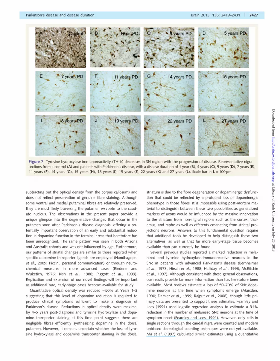
Figure 7 Tyrosine hydroxylase immunoreactivity (TH-ir) decreases in SN region with the progression of disease. Representative nigra
sections from a control (A) and patients with Parkinson’s disease, with a disease duration of 1 year (B), 4 years (C), 5 years (D), 7 years (E),
11 years (F), 14 years (G), 15 years (H), 18 years (I), 19 years (J), 22 years (K) and 27 years (L). Scale bar in L = 100mm.
Parkinson’s disease and disease duration Brain 2013: 136; 2419–2431 | 2427
at Library of R
ush University on July 26, 2013
http://brain.oxfordjournals.org/D
ownloaded from

morphometric approach and a linear regression analysis, but also
examined few cases, especially in the early stages of the disease.
In their study, Ma et al. (1997) performed stereological estimates
of pigmented cells in the substantia nigra of control subjects and
patients with Parkinson’s disease. As seen in other studies, they
found a 51% reduction in melanin containing nigral neurons in
Parkinson’s disease. In contrast to our findings, they found a stat-
istically significant correlation between disease duration and the
number of melanin-containing neurons. However, examination
of their data reveals that this correlation was due to two cases
with �17 years disease duration. If these cases were removed
from the analysis, the loss of melanin-positive cells as a function
of disease duration would occur in a manner virtually identical to
what is seen in the present study. Greffard et al. (2006) also
examined nigral cell loss as a function of disease duration. They
claim—based upon a linear regression analysis—that motor symp-
tom onset occurs after a 30% loss of nigral neurons. However,
this figure is based upon control values and one Parkinson’s dis-
ease case with 1 year disease duration that displayed a 70% loss
of nigral neurons. We quantified both tyrosine hydroxylase-posi-
tive and melanin-containing neurons in the SNc in a relatively
large number of subjects over a multi-decade post-diagnostic
time frame, including several rare cases where death occurred
relatively soon after diagnosis. We observed a profound but vari-
able loss of tyrosine hydroxylase-positive neurons in the earliest
cases with modest-to-negligible loss for decades thereafter. This
was true whether we assessed the nigra as a whole or the most
vulnerable ventrolateral tier. This suggests that the SNc, like the
terminal field in the putamen, suffers a profound loss of dopamin-
ergic function soon after diagnosis and that the entire neuron is
apparently impaired. These findings suggest that clinical deterior-
ation after this time likely represents a loss of a compensatory
mechanism or degeneration of non-dopaminergic neurons. The
pattern of degeneration seen in melanin-containing SNc cells
seems somewhat slower. In this case, there were minimal reduc-
tions when the nigra was assessed as a whole, but extensive loss
of melanin cells in the ventrolateral tier and the pars lateralis
subregion. Interestingly, this same case showed a profound loss
of tyrosine hydroxylase-positive neurons in both analyses. It is
noted that there was considerable variability in the numbers
of melanin-positive neurons in early-stage disease with several
early patients exhibiting profound loss (Fig. 8A). From 3–7 years
Figure 8 Stereologic Estimates of (A) melanin-containing and
(B) tyrosine hydroxylase-immunoreactivity (TH-ir) neurons in
patients with Parkinson’s disease (PD) of the whole SNc at dif-
ferent disease durations (n = 20).
Figure 9 Stereological estimates of (A) melanin-containing and
(B) tyrosine hydroxylase-immunoreactivity (TH-ir) neurons in
patients with Parkinson’s disease in the ventrolateral tier of the
SNc at different disease durations (n = 16).
2428 | Brain 2013: 136; 2419–2431 J. H. Kordower et al.
at Library of R
ush University on July 26, 2013
http://brain.oxfordjournals.org/D
ownloaded from

post-diagnosis, there was a highly variable 33–80% reduction in
melanin-containing neurons, and thereafter, a substantial loss at all
time points, albeit with some variability. After the first decade
following diagnosis, there was a slower rate of degeneration,
and further loss was subtle, gradual and with much less variability.
Nonetheless, loss of melanized neurons was less than the loss of
tyrosine hydroxylase neurons at all time points, suggesting that
some cells may be dysfunctional but still potentially capable of
being restored by trophic therapies or similar therapies.
Our findings, analysing both the cell bodies and terminal areas
of the nigrostriatal system, suggest that at symptom onset, there is
a moderate loss of at least 50% of fibres in the putamen that
continue to express sufficient tyrosine hydroxylase to be detected
and a comparable loss of tyrosine hydroxylase-positive stained
cells in the SNc. Although the loss tyrosine hydroxylase-positive
staining in the SNc neurons was profound at the earliest time
points, the loss of melanin containing neurons occurred more
gradually over the course of the first decade following diagno-
sis for most subregions (except the ventrolateral tier and pars
lateralis subregion), and the difference in the number of tyrosine
hydroxylase-positive and melanin-positive neurons continued at all
post-diagnostic time points. As Parkinson’s disease progresses
beyond the first decade, there are relatively stable numbers of
residual tyrosine hydroxylase-positive and melanin containing neu-
rons persisting, suggesting that there may be two subpopulations
of substantia nigra neurons with substantially different vulnerabil-
ities to Parkinson’s disease neurodegeneration and anatomical
targets.
Collectively, these findings indicate that phenotypic changes in
the nigrostriatal systems are greater than structural changes and
suggest that clinical dysfunction in the early stages is likely due
to a severe loss of function of nigrostriatal neurons. This
may be related to degeneration of dopamine terminals, but it is
also possible that SNc neurons may be dysfunctional but still
potentially viable. The loss of a dopaminergic phenotype is well
described in primates as the loss of phenotypic markers occurs as a
function of normal ageing and Parkinson’s disease (Bartus et al.,
2010). This has obvious implications for the timing of studies
testing putative neuroprotective or trophic therapies where it
might be preferable to study patients at a relatively early stage
of the disease when there is a greater chance of surviving
dopamine neurons that could benefit from therapy. Indeed,
degeneration of dopamine terminals, or severe dysfunction of
dopamine terminals that limit axonal transport to the SNc could
explain why adeno-associated viral delivery of the trophic factor
neurturin (CERE-120) into the putamen failed to improve Unified
Parkinson’s Disease Rating Scale motor scores at 12 months in
patients with moderately advanced Parkinson’s disease who
were 45 years after diagnosis, but did provide Unified
Parkinson’s Disease Rating Scale motor benefit for the subpopula-
tion that was followed for 18 month (Marks et al., 2010). In
support of the concept that axonal dysfunction may have contrib-
uted to a delayed trophic response, we observed minimal SNc
neuronal staining for neurturin with only sparse tyrosine hydroxy-
lase staining in the putamen in autopsies performed 2–3 months
following gene delivery of CERE-120 (Bartus et al., 2010), but
more robust SNc neurturin expression with dense tyrosine hydro-
xylase-immunoreactive fibre network at autopsy performed four
years following treatment (Marks et al., 2010). These observations
suggest that dopaminergic fibres that do not stain for tyrosine
hydroxylase and dopamine transporter may be dysfunctional but
not completely degenerated and potentially recoverable.
Table 2 Subregional analysis of neuromelanin and tyrosine hydroxylase-immunoreactive neurons as a function of diseaseduration
DM DL VM VL PL
No. Diseasedurations
NM TH NM TH NM TH NM TH NM TH
1 Controls (n = 8) 984 �95.40
748 �71.51
813 �70.02
688 �67.33
1368 �87.68
1115 �93.49
1879 �167.87
1499 �207.70
1645 �383.42
932 �341.97Cells/mm3
� SEM
2 1 1292 386 1104 431 1463 484 828 291 598 322
3 3 1065 1022 962 612 2323 1971 1267 1145 408 272
4 4 703 595 696 562 1261 705 746 361 263 n/a
5 5 946 398 510 185 2109 465 1439 428 607 303
6 7 602 212 277 126 586 247 657 143 318 n/a
7 11 974 299 799 439 1321 237 1031 356 578 n/a
8 13 269 134 642 84 371 50 232 46 603 133
9 14 435 14 423 13 801 59 659 98 513 125
10 14 434 334 474 297 502 188 497 213 379 n/a
11 15 640 471 490 453 1294 388 1160 394 333 190
12 18 601 95 337 61 437 26 372 106 169 84
13 18 885 531 545 385 1148 395 1775 596 439 293
14 19 713 475 832 185 422 162 283 40 628 523
15 21 516 196 370 148 614 116 255 108 681 143
16 21 677 150 489 139 777 104 1062 354 495 123
17 22 454 319 248 198 619 364 592 269 305 85
DM = dorsomedial; DL = dorsolateral; VM = ventromedial; VL = ventrolateral; PL = pars lateralis.
Parkinson’s disease and disease duration Brain 2013: 136; 2419–2431 | 2429
at Library of R
ush University on July 26, 2013
http://brain.oxfordjournals.org/D
ownloaded from

These findings have important implications for considering
which patients should be included in studies of putative neuropro-
tective agents. Although our findings may reflect a phenotypic
downregulation of dopaminergic markers, it remains equally plaus-
ible that absence of staining for dopamine terminals in the puta-
men at 4 years and beyond reflects complete degeneration of
dopamine terminals and a time point at which no benefit could
be expected, even from a potentially effective agent. These data
suggest that there will be a better chance of detecting a beneficial
effect of a putative neuroprotective agent if patients with
Parkinson’s disease are enrolled at earlier time points in their ill-
ness, and further emphasize the importance of trying to define
patients with pre-motor or prodromal Parkinson’s disease where
it is likely that there is a greater number of preserved dopamine
neurons and a greater chance of a neuroprotective or restorative
agent providing a clinically meaningful benefit.
Several caveats must be considered when interpreting the data
from our study. Pathological studies are potentially biased depend-
ent upon the population of individuals studied. In the present
report, Parkinson’s disease cases in the Arizona cohort were
obtained from a retirement community with an environment
that facilitates rapid autopsy. This cohort is comprised primarily
of elderly individuals with a higher socio-economic status and
greater education than a random Parkinson’s disease population.
However, we found the same pattern of results with a dramatic
and early loss of striatal dopaminergic terminals in the Australia
cohort, which included younger patients with relatively short
disease duration. Thus, it appears that neither socio-economic
factors, nor an age-related inability to maintain compensatory
mechanisms accounts for our data. We had limited numbers of
brains, particularly in the early stages of the illness when it appears
that degeneration occurs at the most rapid rate. Finally, we were
not able to make firm conclusions regarding how our data are
associated with clinical status.
In summary we have performed one of the largest pathology
studies to date, evaluating the integrity of the nigrostriatal dopa-
mine system in patients with Parkinson’s disease at multiple time
points following diagnosis. Using markers for dopamine function,
we show that fibres in the dorsal striatum, which underlie motor
function in Parkinson’s disease, are only mildly affected at 1 year,
are variably affected at 3 years, and are virtually absent by Years
4–5 and thereafter. A similarly profound loss of the dopaminergic
marker, tyrosine hydroxylase, was seen in the SNc neuronal cell
bodies, demonstrating that the entire neuron is affected early after
diagnosis. Importantly, the number of tyrosine hydroxylase-posi-
tive neurons is less than the number of melanized neurons at all
time points, suggesting that some nerve cells are likely to be
damaged but to have not yet degenerated, and thus might be
capable of responding to a protective or trophic factor therapy.
We also demonstrate a population of dopamine neurons that
continue to express tyrosine hydroxylase and persist for decades
after diagnosis. These neurons presumably innervate the ventral
striatum and other non-striatal regions and are not vulnerable to
the Parkinson’s disease degenerative process.
Our findings suggest that there may be significant challenges
for ‘neuroprotective’ therapies that include patients with 44 years
disease duration given the substantial loss of dopamine markers in
the nigrostriatal terminals that has occurred by this time point and
supports the use of patients in early stages of the disease for
clinical trials. On the other hand, the persistence of populations
of melanin containing neurons in comparison to the number of
tyrosine hydroxylase-immunoreactive neurons for decades after
diagnosis, suggests that trophic or regenerative therapies might
still have value even in the later stages of the illness. A determin-
ation as to whether the loss of dopaminergic staining in fibres of
the dorsal putamen is due to frank neurodegeneration or pheno-
typic downregulation will help guide the development of novel
experimental therapies. Finally, the early changes in dopaminergic
markers that we observe in both the SNc perikarya and in fibres
projecting to the dorsal putamen emphasize the importance of
efforts aimed at developing biomarkers that permit diagnosing
Parkinson’s disease at early time points, perhaps even before the
emergence of the classical motor features of the disease when
there is greater preservation of dopaminergic markers and a
greater potential for a beneficial effect with neuroprotective or
trophic therapies.
FundingThis research was supported in part by a Centre Grant from the
Parkinson’s Disease Foundation to Rush University Medical Centre.
The Sun Health Research Institute Brain and Body Donation
Program is supported by the National Institute of Neurological
Disorders and Stroke (U24 NS072026 National Brain and Tissue
Resource for Parkinson’s Disease and Related Disorders), the
National Institute on Aging (P30 AG19610 Arizona Alzheimer’s
Disease Core Centre), the Arizona Department of Health
Services (contract 211002, Arizona Alzheimer’s Research Center),
the Arizona Biomedical Research Commission (contracts 4001,
0011, 05-901 PubMed and 1001 to the Arizona Parkinson’s
Disease Consortium) and the Michael J. Fox Foundation for
Parkinson’s Research. The Human brain tissue samples received
from the Sydney Brain Bank was supported by Neuroscience
Research Australia, the University of New South Wales and the
National Health and Medical Research Council of Australia
(NHMRC). GMH has a NHMRC Senior Principal Research
Fellowship 630434.
ReferencesBartus RT, Herzog CD, Chu Y, Wilson A, Brown L, Siffert J, et al.
Bioactivity of AAV2-neurturin gene therapy (CERE-120): differences
between Parkinson’s disease and nonhuman primate brains. Mov
Disord 2010; 26: 27–36.
Beach TG, Adler CH, Sue LI, Peirce JB, Bachalakuri J, Dalsing-
Hernandez JE, et al. Reduced striatal tyrosine hydroxylase in incidental
Lewy body disease. Acta Neuropathol 2008a; 115: 445–51.
Beach TG, Sue LI, Walker DG, Roher AE, Lue L, Vedders L, et al. The
Sun Health Research Institute Brain Donation Program: description and
experience, 1987-2007. Cell Tissue Bank 2008b; 9: 229–45.Bernheimer H, Birkmayer W, Hornykiewicz O, Jellinger K, Seitelberger F.
Brain dopamine and the syndromes of Parkinson and Huntington.
Clinical, morphological and neurochemical correlations. J Neurol Sci
1973; 20: 415–55.
2430 | Brain 2013: 136; 2419–2431 J. H. Kordower et al.
at Library of R
ush University on July 26, 2013
http://brain.oxfordjournals.org/D
ownloaded from

Chu Y, Kompoliti K, Cochran EJ, Mufson EJ, Kordower JH. Age-relateddecreases in Nurr1 immunoreactivity in the human substantia nigra.
J Comp Neurol 2002; 450: 203–14.
Chu Y, Kordower JH. Age-associated increases of alpha-synuclein in
monkeys and humans are associated with nigrostriatal dopaminedepletion: Is this the target for Parkinson’s disease? Neurobiol Dis
2007; 25: 134–49.
Damier P, Hirsch EC, Agid Y, Graybiel AM. The substantia nigra of the
human brain. II. Patterns of loss of dopamine-containing neurons inParkinson’s disease. Brain 1999; 122 (Pt 8): 1437–48.
DelleDonne A, Klos KJ, Fujishiro H, Ahmed Z, Parisi JE, Josephs KA, et al.
Incidental Lewy body disease and preclinical Parkinson disease. ArchNeurol 2008; 65: 1074–80.
Dickson DW, Fujishiro H, DelleDonne A, Menke J, Ahmed Z, Klos KJ,
et al. Evidence that incidental Lewy body disease is pre-symptomatic
Parkinson’s disease. Acta Neuropathol 2008; 115: 437–44.Ehringer H, Hornykiewicz O. Distribution of noradrenaline and dopamine
(3-hydroxytyramine) in the human brain and their behavior in diseases
of the extrapyramidal system [in German]. Klin Wochenschr 1960; 38:
1236–9.Fearnley JM, Lees AJ. Ageing and Parkinson’s disease: substantia nigra
regional selectivity. Brain 1991; 114 (Pt 5): 2283–301.
Greffard S, Verny M, Bonnet AM, Beinis JY, Gallinari C, Meaume S, et al.
Motor score of the Unified Parkinson Disease Rating Scale as a goodpredictor of Lewy body-associated neuronal loss in the substantia
nigra. Arch Neurol 2006; 63: 584–8.
Gundersen HJ, Jensen EB. The efficiency of systematic sampling instereology and its prediction. J Microsc 1987; 147: 229–63.
Halliday GM, McRitchie DA, Cartwright H, Pamphlett R, Hely MA,
Morris JG. Midbrain neuropathology in idiopathic Parkinson’s
disease and diffuse Lewy body disease. J Clin Neurosci 1996; 3:52–60.
Herzog CB, Kordower JH, Chu Y, Wilson A, Brown L, Siffert J, et al.
Long-term neurturin gene expression and upregulation of tyrosine
hydroxylase in a PD patient with CERE-120. Soc Neurosci Abstr, inpress.
Hirsch E, Graybiel AM, Agid YA. Melanized dopaminergic neurons are
differentially susceptible to degeneration in Parkinson’s disease. Nature1988; 334: 345–8.
Hughes AJ, Daniel SE, Ben-Shlomo Y, Lees AJ. The accuracy of diagnosis
of parkinsonian syndromes in a specialist movement disorder service.
Brain 2002; 125: 861–70.
Jellinger KA. Neuropathology of sporadic Parkinson’s disease: Evaluationand changes of concepts. Mov Disord 2012; 27: 8–30.
Kish SJ, Shannak K, Hornykiewicz O. Uneven pattern of dopamine loss
in the striatum of patients with idiopathic Parkinson’s disease.
Pathophysiologic and clinical implications. N Engl J Med 1988; 318:876–80.
Kordower JH, Chu Y, Hauser RA, Freeman TB, Olanow CW. Lewy body-
like pathology in long-term embryonic nigral transplants in Parkinson’s
disease. Nat Med 2008; 14: 504–6.Lang AE, Lozano AM. Parkinson’s disease. First of two parts. N Engl J
Med 1998; 339: 1044–53.
Ma SY, Roytta M, Rinne JO, Collan Y, Rinne UK. Correlation betweenneuromorphometry in the substantia nigra and clinical features in
Parkinson’s disease using disector counts. J Neurol Sci 1997; 151:
83–7.
Marks WJ Jr, Bartus RT, Siffert J, Davis CS, Lozano A, Boulis N, et al.Gene delivery of AAV2-neurturin for Parkinson’s disease: a double-
blind, randomised, controlled trial. Lancet Neurol 2010; 9: 1164–72.
Marsden CD. Parkinson’s disease. Lancet 1990; 335: 948–52.
McRitchie DA, Cartwright HR, Halliday GM. Specific A10 dopaminergicnuclei in the midbrain degenerate in Parkinson’s disease. Exp Neurol
1997; 144: 202–13.
Mufson EJ, Conner JM, Kordower JH. Nerve growth factor in Alzheimer’s
disease: defective retrograde transport to nucleus basalis. Neuroreport1995; 6: 1063–6.
Nandhagopal R, Kuramoto L, Schulzer M, Mak E, Cragg J, Lee CS, et al.
Longitudinal progression of sporadic Parkinson’s disease: a multi-tracerpositron emission tomography study. Brain 2009; 132: 2970–9
PubMed.
Piggott MA, Marshall EF, Thomas N, Lloyd S, Court JA, Jaros E, et al.
Striatal dopaminergic markers in dementia with Lewy bodies,Alzheimer’s and Parkinson’s diseases: rostrocaudal distribution. Brain
1999; 122 (Pt 8): 1449–68.
Rajput AH, Sitte HH, Rajput A, Fenton ME, Pifl C, Hornykiewicz O.
Globus pallidus dopamine and Parkinson motor subtypes: clinical andbrain biochemical correlation. Neurology 2008; 70: 1403–10 PubMed.
Riederer P, Wuketich S. Time course of nigrostriatal degeneration in
parkinson’s disease. A detailed study of influential factors in humanbrain amine analysis. J Neural Transm 1976; 38: 277–301.
West MJ. New stereological methods for counting neurons. Neurobiol
Aging 1993; 14: 275–85.
Parkinson’s disease and disease duration Brain 2013: 136; 2419–2431 | 2431
at Library of R
ush University on July 26, 2013
http://brain.oxfordjournals.org/D
ownloaded from


