Embed Size (px)
Citation preview
Brain (1999),122,111–120
Visual control of locomotion in Parkinson’s diseaseJean-Philippe Azulay,1,2 Serge Mesure,3 Bernard Amblard,1 Olivier Blin,3 Iban Sangla2 andJean Pouget2,3
1UPR Neurobiologie et Mouvement, CNRS,2Department of Correspondence to: Jean-Philippe Azulay, Service deNeurology, University Hospital La Timone and3UPRES Neurologie et Maladies Neuromusculaires CHU de LaPhysiopathologie du Syste`me Nerveux et du Muscle, Timone, 13 385 Marseille cedex 05, FranceMarseille, France
SummaryThe effect of placing parallel lines on the walking surfaceon parkinsonian gait was evaluated. To identify the kindof visual cues (static or dynamic) required for the controlof locomotion, we tested two visual conditions: normallighting and stroboscopic illumination (three flashes/s), thelatter acting to suppress dynamic visual cues completely.Sixteen subjects with idiopathic Parkinson’s disease (ninemales, seven females; mean age 68.8 years) and thesame number of age-matched controls (seven males; ninefemales, mean age 67.5 years) were studied. During thebaseline phase, Parkinson’s disease patients walked witha short-stepped, slow velocity pattern. The double limbsupport duration was increased and the step cadence wasreduced relative to normal. Under normal lighting, visualcues from the lines on the walking surface induced asignificant improvement in gait velocity and stride length
Keywords: Parkinson’s disease; locomotion; optic flow
IntroductionSince the original description by Parkinson (1817),Parkinson’s disease has been recognized as a motor disorderresulting from neuropathological changes affecting the basalganglia (Barbeau, 1986). However, sensorial deficits havealso been reported (Snideret al., 1976; Koller, 1984). Morespecifically, visual deficits have been demonstrated withrespect to visual evoked potentials (Bodis-Wollner and Yahr,1978; Onofrj et al., 1986) and spatiotemporal contrastsensitivity (Bulenset al., 1986; Tagliatiet al., 1992). Theinterest in the visual defect in parkinsonism is enhanced bythe possible relationships between gait disorders and visualperception, inasmuch as gait problems such as festinationand the freezing phenomenon are strongly influenced byvisual stimulation (Mestreet al., 1992).
The earliest detailed analysis of gait in parkinsonism wasperformed by Martin (1967), who described mainly theconsequences on gait of encephalitis lethargica. Martin wasalso the first to report the effectiveness of utilizing vision tofacilitate locomotor activity. Moreover, he showed that onlycertain visual stimuli were effective in improving gait:
© Oxford University Press 1999
in Parkinson’s disease patients. With stroboscopicillumination and without lines, both groups reduced theirstride length and velocity but the changes were significantonly in the Parkinson’s disease group, indicating greaterdependence on dynamic visual information. Whenstroboscopic light was used with stripes on the floor, theimprovement in gait due to the stripes was suppressed inparkinsonian patients. These results demonstrate that theperceived motion of stripes, induced by the patient’swalking, is essential to improve the gait parameters andthus favour the hypothesis of a specific visual–motorpathway which is particularly responsive to rapidlymoving targets. Previous studies have proposed acerebellar circuit, allowing the visual stimuli to by-passthe damaged basal ganglia.
transverse lines, an inch or more wide, 18 inches or so apart,and of a colour contrasting with that of the floor (white lineson a dark ground). Zigzag lines, lines parallel to the line ofmovement, very narrow lines, lines wider than 6 feet orstripes without contrast of colour had no influence. Laterstudies confirmed the positive influence of visual guidanceon gait movements in Parkinson’s disease patients (Forssberget al., 1984; Azulayet al., 1996). Other sensory cues mayalso improve parkinsonian gait, e.g. rhythmic auditory cues(Richardset al., 1992; Thautet al., 1996; McIntoshet al.,1997). While the influence of vision on gait control inparkinsonian patients has been established, the questionsregarding the mechanisms of action of the visual cues arestill controversial (Morriset al., 1996). One suggestion isthat stripes on the floor improve gait by drawing attention tothe stepping process. Another is that each stripe may triggera step during locomotion. A third is that, when patients walk,the stripes move downward in the visual field and inducespecific dynamic visual stimuli that may improve motorperformance. Dynamic visual cues have been shown to
by guest on June 13, 2013http://brain.oxfordjournals.org/
Dow
nloaded from
112 J.-P. Azulayet al.
provide an important contribution to body balance, in standing(Amblard et al., 1985; Cre´mieux et Mesure, 1994) as wellas in walking (Assaianteet al., 1989) in healthy adults. Theaim of the present study was to evaluate the type of visualcues (static or dynamic) required for the control of locomotionin parkinsonian patients. With this aim, we tested two visualconditions: normal lighting and stroboscopic illumination,the latter serving to suppress dynamic visual cues completely(Amblardet al., 1985; Assaianteet al., 1989). The comparisonof locomotor performance observed with normal vision withthat obtained under stroboscopic illumination allowed us todetermine the specific contribution of dynamic visual cuesin parkinsonian gait. We also evaluated the benefit of placingparallel lines on the walking surface in both visual conditions.This was done to determine whether motion of the lines isnecessary to improve locomotor performance in Parkinson’sdisease patients, i.e. if transverse lines induce an improvementin gait performance under normal lighting, the persistence orremoval of the effect under stroboscopic illumination woulddetermine whether or not it is linked to the perceived motionof the lines.
MethodsSubjectsThirty-two subjects were included in the study: 16 Parkinson’sdisease patients (nine males, seven females; mean age 68.864 years) and the same number of age-matched normal controls(seven males, nine females; mean age 67.56 5 years). Allpatients were clinically diagnosed as having ‘idiopathic’Parkinson’s disease according to the UK Brain Bankdiagnostic criteria (Gibb and Lees, 1988), and had a sustainedimprovement with dopaminergic treatments.
Eleven parkinsonian patients were Hoehn and Yahr stageII and five were Hoehn and Yahr stage III. The mean diseaseduration was 6.3 years. All patients and controls had a visualacuity of 20/20, with correction if necessary. The recordingswere carried out at the same hour in the morning. Parkinson’sdisease patients had fasted overnight and were withouttreatment for at least 12 h. All patients and controls gaveinformed consent according to the declaration of Helsinkiand the protocol was approved by the ethics committee ofThe University of Marseille.
Procedures of gait analysisAll observations were performed on a 12 m walkway.Kinematic gait analysis was performed with a commerciallyavailable automatic motion analyser (Ferrigno and Pedotti,1985), with four cameras at a sampling frequency of 100 Hzand with two high-resolution force platforms which measurethe timing amplitude, direction and location of the forceexerted on the support level. Nine markers were placedsymmetrically on the subjects at the following sites: fifthmetatarsal joint, external malleolus, tibial plate and
posteriosuperior iliac crest, with the last marker being placedon the sacrum to determine hip, knee and ankle movements.In the present study, only some of the gait parameters(velocity, stride length, cadence, double limb supportduration) were analysed.
Subjects were instructed to perform three consecutivewalks, either on a uniformly grey flat surface or on the samesupport with parallel transverse high contrasting white lines(5 cm wide) spaced at 45 cm intervals, this having beendemonstrated previously to be the most effective pattern(Azulay et al., 1996). They were instructed to walk at theirnatural speed, looking straight ahead without any specificationregarding foot positioning. The analysis started after walkinga distance of 4 m.
Two visual conditions were tested: normal lighting andstroboscopic (electronic) illumination at 3 Hz (flash duration,0.2 ms, flash energy: 0.3 J). Both types of lighting conditionswere provided by the same sources and were adjusted to beperceived as having an equivalent brightness. All the lightsources were placed on the ceiling of the experimental roomto avoid any dazzle. The first three trials under stroboscopicillumination had the aim of familiarizing the subjects withthe testing conditions. In each experiment, each of the supportsituations (grey floor or transverse lines) combined with eachof the two illumination situations was presented accordingto a pseudo-random design. The results of the three trials ineach situation were averaged.
Statistical analysisANOVA (analysis of variance) was used to compare thedifferent parameters (stride length, velocity, cadence) betweenthe two groups (patients and control subjects) and betweenthe different conditions (with and without stripes, normaland stroboscopic light). The minimum 0.05 level ofsignificance was adopted throughout the data analysis. Resultsare expressed as mean6 SD.
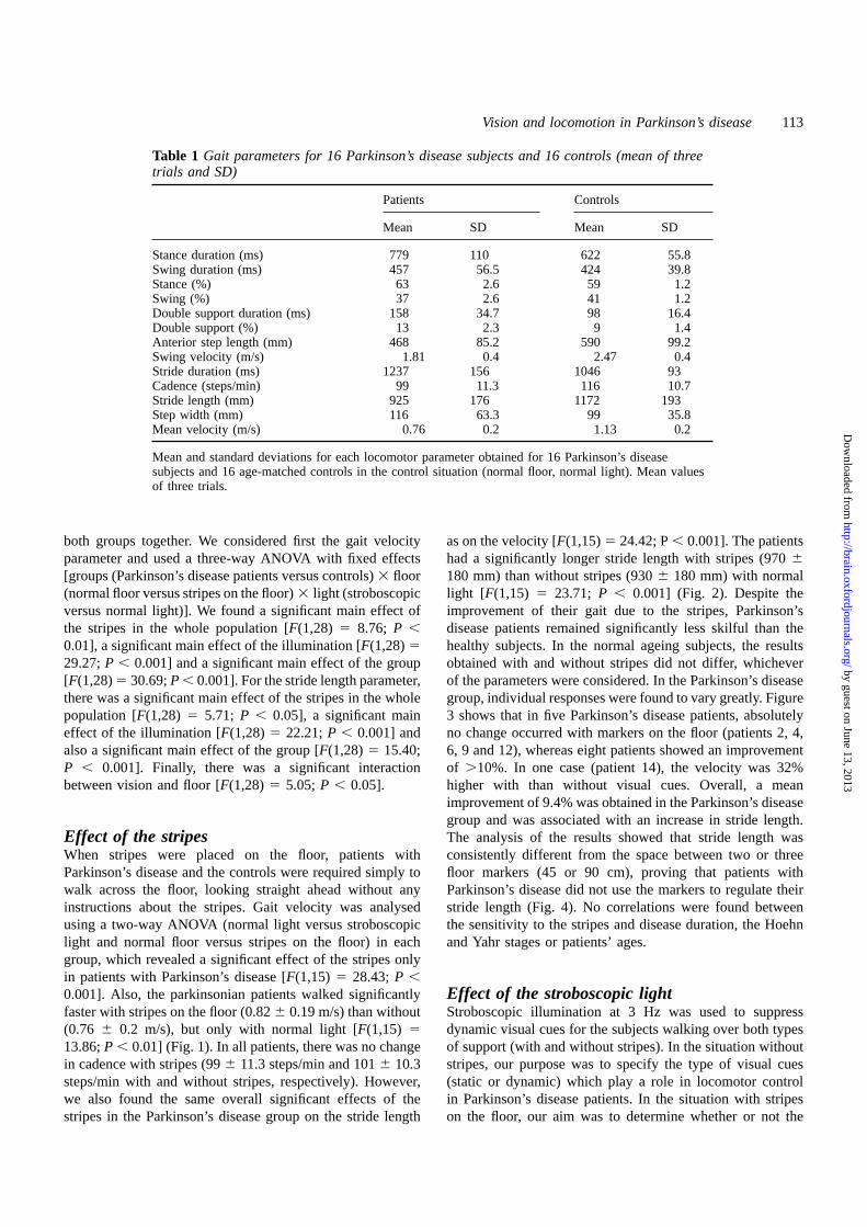
ResultsAnalysis of gait parameters (Table 1)During the control situation (normal light, normal ground),the mean gait velocity of patients with Parkinson’s disease(0.76 6 0.2 m/s) was slower than that of controls (1.1360.2 m/s) [F(1,30)5 27.75; P , 0.001]; the mean stridelength was shorter (9256 176 mm versus 11726 193 mm,respectively) [F(1,30)5 14.48;P , 0.001]; the cadence wasreduced (996 11.3 steps/min versus 1166 10.7, respectively)[F(1,30) 5 19.79;P , 0.001]; and the relative double limbsupport duration was greater (136 2.3% versus 96 1.4%,respectively) [F(1,30)5 26.63; P , 0.001]. These resultswere obtained for patients and controls walking at theirpreferred speed.
Analysis of the main effectsBefore analysing the effects of stripes and illumination ineach group separately, we analysed these conditions in
by guest on June 13, 2013http://brain.oxfordjournals.org/
Dow
nloaded from
Vision and locomotion in Parkinson’s disease 113
Table 1 Gait parameters for 16 Parkinson’s disease subjects and 16 controls (mean of threetrials and SD)
Patients Controls
Mean SD Mean SD
Stance duration (ms) 779 110 622 55.8Swing duration (ms) 457 56.5 424 39.8Stance (%) 63 2.6 59 1.2Swing (%) 37 2.6 41 1.2Double support duration (ms) 158 34.7 98 16.4Double support (%) 13 2.3 9 1.4Anterior step length (mm) 468 85.2 590 99.2Swing velocity (m/s) 1.81 0.4 2.47 0.4Stride duration (ms) 1237 156 1046 93Cadence (steps/min) 99 11.3 116 10.7Stride length (mm) 925 176 1172 193Step width (mm) 116 63.3 99 35.8Mean velocity (m/s) 0.76 0.2 1.13 0.2
Mean and standard deviations for each locomotor parameter obtained for 16 Parkinson’s diseasesubjects and 16 age-matched controls in the control situation (normal floor, normal light). Mean valuesof three trials.
both groups together. We considered first the gait velocityparameter and used a three-way ANOVA with fixed effects[groups (Parkinson’s disease patients versus controls)3 floor(normal floor versus stripes on the floor)3 light (stroboscopicversus normal light)]. We found a significant main effect ofthe stripes in the whole population [F(1,28)5 8.76; P ,0.01], a significant main effect of the illumination [F(1,28)529.27;P , 0.001] and a significant main effect of the group[F(1,28)5 30.69;P , 0.001]. For the stride length parameter,there was a significant main effect of the stripes in the wholepopulation [F(1,28)5 5.71; P , 0.05], a significant maineffect of the illumination [F(1,28)5 22.21;P , 0.001] andalso a significant main effect of the group [F(1,28)5 15.40;P , 0.001]. Finally, there was a significant interactionbetween vision and floor [F(1,28)5 5.05;P , 0.05].
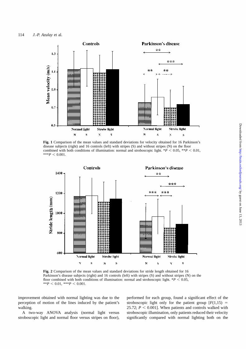
Effect of the stripesWhen stripes were placed on the floor, patients withParkinson’s disease and the controls were required simply towalk across the floor, looking straight ahead without anyinstructions about the stripes. Gait velocity was analysedusing a two-way ANOVA (normal light versus stroboscopiclight and normal floor versus stripes on the floor) in eachgroup, which revealed a significant effect of the stripes onlyin patients with Parkinson’s disease [F(1,15) 5 28.43;P ,0.001]. Also, the parkinsonian patients walked significantlyfaster with stripes on the floor (0.826 0.19 m/s) than without(0.76 6 0.2 m/s), but only with normal light [F(1,15)513.86;P , 0.01] (Fig. 1). In all patients, there was no changein cadence with stripes (996 11.3 steps/min and 1016 10.3steps/min with and without stripes, respectively). However,we also found the same overall significant effects of thestripes in the Parkinson’s disease group on the stride length
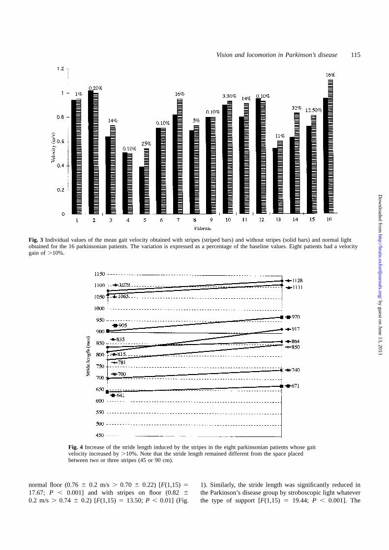
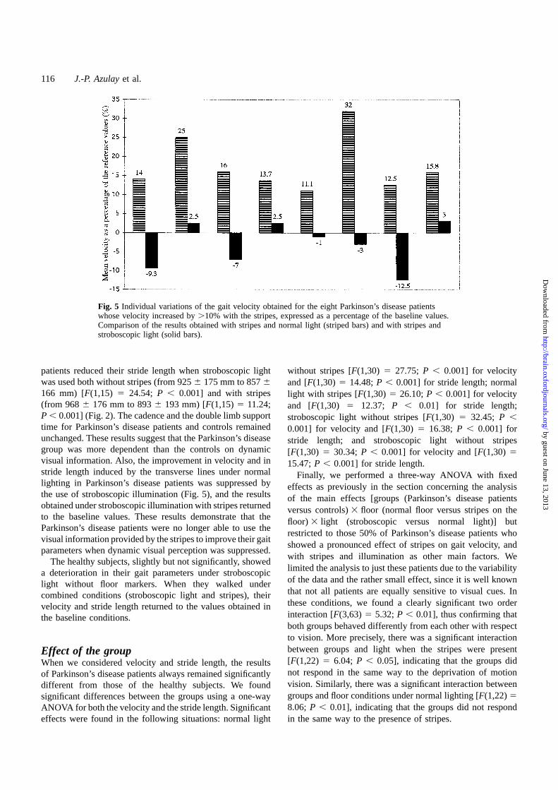
as on the velocity [F(1,15)5 24.42; P, 0.001]. The patientshad a significantly longer stride length with stripes (9706180 mm) than without stripes (9306 180 mm) with normallight [F(1,15) 5 23.71; P , 0.001] (Fig. 2). Despite theimprovement of their gait due to the stripes, Parkinson’sdisease patients remained significantly less skilful than thehealthy subjects. In the normal ageing subjects, the resultsobtained with and without stripes did not differ, whicheverof the parameters were considered. In the Parkinson’s diseasegroup, individual responses were found to vary greatly. Figure3 shows that in five Parkinson’s disease patients, absolutelyno change occurred with markers on the floor (patients 2, 4,6, 9 and 12), whereas eight patients showed an improvementof .10%. In one case (patient 14), the velocity was 32%higher with than without visual cues. Overall, a meanimprovement of 9.4% was obtained in the Parkinson’s diseasegroup and was associated with an increase in stride length.The analysis of the results showed that stride length wasconsistently different from the space between two or threefloor markers (45 or 90 cm), proving that patients withParkinson’s disease did not use the markers to regulate theirstride length (Fig. 4). No correlations were found betweenthe sensitivity to the stripes and disease duration, the Hoehnand Yahr stages or patients’ ages.
Effect of the stroboscopic lightStroboscopic illumination at 3 Hz was used to suppressdynamic visual cues for the subjects walking over both typesof support (with and without stripes). In the situation withoutstripes, our purpose was to specify the type of visual cues(static or dynamic) which play a role in locomotor controlin Parkinson’s disease patients. In the situation with stripeson the floor, our aim was to determine whether or not the
by guest on June 13, 2013http://brain.oxfordjournals.org/
Dow
nloaded from
114 J.-P. Azulayet al.
Fig. 1 Comparison of the mean values and standard deviations for velocity obtained for 16 Parkinson’sdisease subjects (right) and 16 controls (left) with stripes (S) and without stripes (N) on the floorcombined with both conditions of illumination: normal and stroboscopic light. *P , 0.05, **P , 0.01,***P , 0.001.
Fig. 2 Comparison of the mean values and standard deviations for stride length obtained for 16Parkinson’s disease subjects (right) and 16 controls (left) with stripes (S) and without stripes (N) on thefloor combined with both conditions of illumination: normal and stroboscopic light. *P , 0.05,**P , 0.01, ***P , 0.001.
improvement obtained with normal lighting was due to theperception of motion of the lines induced by the patient’swalking.
A two-way ANOVA analysis (normal light versusstroboscopic light and normal floor versus stripes on floor),
performed for each group, found a significant effect of thestroboscopic light only for the patient group [F(1,15)525.72;P , 0.001]. When patients and controls walked withstroboscopic illumination, only patients reduced their velocitysignificantly compared with normal lighting both on the
by guest on June 13, 2013http://brain.oxfordjournals.org/
Dow
nloaded from
Vision and locomotion in Parkinson’s disease 115
Fig. 3 Individual values of the mean gait velocity obtained with stripes (striped bars) and without stripes (solid bars) and normal lightobtained for the 16 parkinsonian patients. The variation is expressed as a percentage of the baseline values. Eight patients had a velocitygain of .10%.
Fig. 4 Increase of the stride length induced by the stripes in the eight parkinsonian patients whose gaitvelocity increased by.10%. Note that the stride length remained different from the space placedbetween two or three stripes (45 or 90 cm).
normal floor (0.766 0.2 m/s. 0.70 6 0.22) [F(1,15)517.67; P , 0.001] and with stripes on floor (0.8260.2 m/s. 0.74 6 0.2) [F(1,15)5 13.50; P , 0.01] (Fig.
1). Similarly, the stride length was significantly reduced inthe Parkinson’s disease group by stroboscopic light whateverthe type of support [F(1,15)5 19.44; P , 0.001]. The
by guest on June 13, 2013http://brain.oxfordjournals.org/
Dow
nloaded from
116 J.-P. Azulayet al.
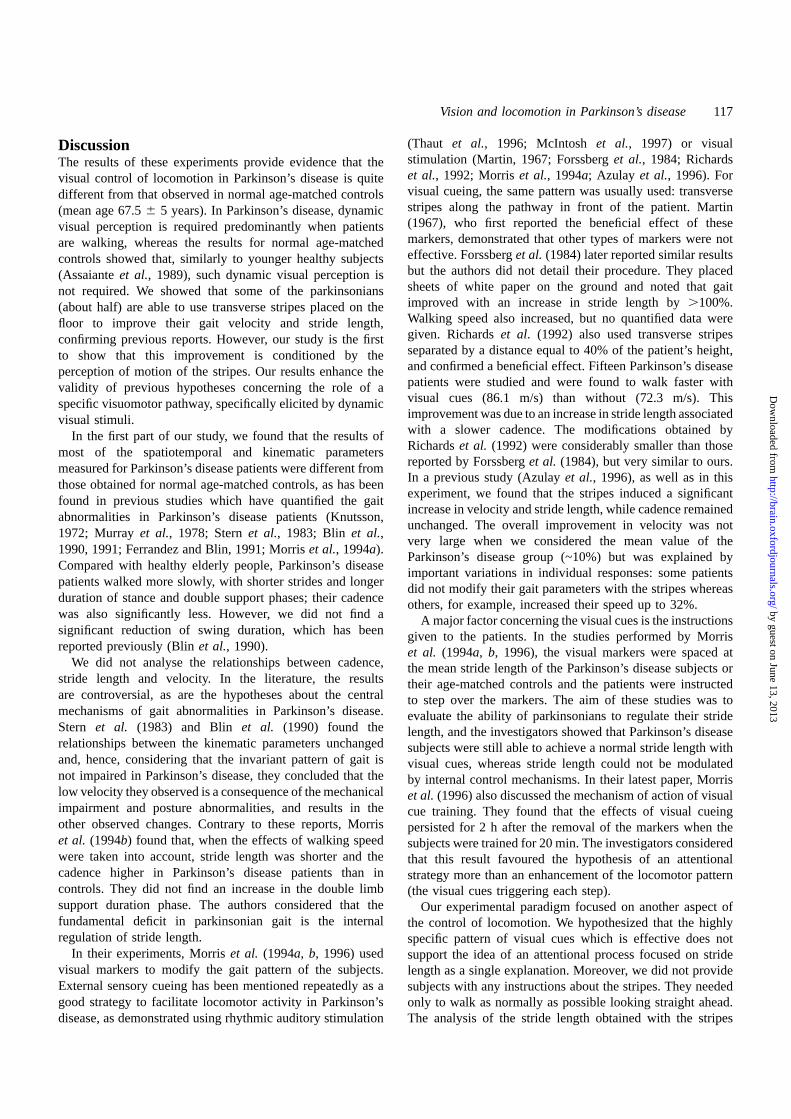
Fig. 5 Individual variations of the gait velocity obtained for the eight Parkinson’s disease patientswhose velocity increased by.10% with the stripes, expressed as a percentage of the baseline values.Comparison of the results obtained with stripes and normal light (striped bars) and with stripes andstroboscopic light (solid bars).
patients reduced their stride length when stroboscopic lightwas used both without stripes (from 9256 175 mm to 8576166 mm) [F(1,15)5 24.54; P , 0.001] and with stripes(from 9686 176 mm to 8936 193 mm) [F(1,15)5 11.24;P , 0.001] (Fig. 2). The cadence and the double limb supporttime for Parkinson’s disease patients and controls remainedunchanged. These results suggest that the Parkinson’s diseasegroup was more dependent than the controls on dynamicvisual information. Also, the improvement in velocity and instride length induced by the transverse lines under normallighting in Parkinson’s disease patients was suppressed bythe use of stroboscopic illumination (Fig. 5), and the resultsobtained under stroboscopic illumination with stripes returnedto the baseline values. These results demonstrate that theParkinson’s disease patients were no longer able to use thevisual information provided by the stripes to improve their gaitparameters when dynamic visual perception was suppressed.
The healthy subjects, slightly but not significantly, showeda deterioration in their gait parameters under stroboscopiclight without floor markers. When they walked undercombined conditions (stroboscopic light and stripes), theirvelocity and stride length returned to the values obtained inthe baseline conditions.
Effect of the groupWhen we considered velocity and stride length, the resultsof Parkinson’s disease patients always remained significantlydifferent from those of the healthy subjects. We foundsignificant differences between the groups using a one-wayANOVA for both the velocity and the stride length. Significanteffects were found in the following situations: normal light
without stripes [F(1,30)5 27.75; P , 0.001] for velocityand [F(1,30)5 14.48;P , 0.001] for stride length; normallight with stripes [F(1,30)5 26.10;P , 0.001] for velocityand [F(1,30) 5 12.37; P , 0.01] for stride length;stroboscopic light without stripes [F(1,30)5 32.45; P ,
0.001] for velocity and [F(1,30)5 16.38; P , 0.001] forstride length; and stroboscopic light without stripes[F(1,30) 5 30.34;P , 0.001] for velocity and [F(1,30)515.47;P , 0.001] for stride length.
Finally, we performed a three-way ANOVA with fixedeffects as previously in the section concerning the analysisof the main effects [groups (Parkinson’s disease patientsversus controls)3 floor (normal floor versus stripes on thefloor) 3 light (stroboscopic versus normal light)] butrestricted to those 50% of Parkinson’s disease patients whoshowed a pronounced effect of stripes on gait velocity, andwith stripes and illumination as other main factors. Welimited the analysis to just these patients due to the variabilityof the data and the rather small effect, since it is well knownthat not all patients are equally sensitive to visual cues. Inthese conditions, we found a clearly significant two orderinteraction [F(3,63)5 5.32;P , 0.01], thus confirming thatboth groups behaved differently from each other with respectto vision. More precisely, there was a significant interactionbetween groups and light when the stripes were present[F(1,22) 5 6.04; P , 0.05], indicating that the groups didnot respond in the same way to the deprivation of motionvision. Similarly, there was a significant interaction betweengroups and floor conditions under normal lighting [F(1,22)5
8.06; P , 0.01], indicating that the groups did not respondin the same way to the presence of stripes.
by guest on June 13, 2013http://brain.oxfordjournals.org/
Dow
nloaded from
Vision and locomotion in Parkinson’s disease 117
DiscussionThe results of these experiments provide evidence that thevisual control of locomotion in Parkinson’s disease is quitedifferent from that observed in normal age-matched controls(mean age 67.56 5 years). In Parkinson’s disease, dynamicvisual perception is required predominantly when patientsare walking, whereas the results for normal age-matchedcontrols showed that, similarly to younger healthy subjects(Assaianteet al., 1989), such dynamic visual perception isnot required. We showed that some of the parkinsonians(about half) are able to use transverse stripes placed on thefloor to improve their gait velocity and stride length,confirming previous reports. However, our study is the firstto show that this improvement is conditioned by theperception of motion of the stripes. Our results enhance thevalidity of previous hypotheses concerning the role of aspecific visuomotor pathway, specifically elicited by dynamicvisual stimuli.
In the first part of our study, we found that the results ofmost of the spatiotemporal and kinematic parametersmeasured for Parkinson’s disease patients were different fromthose obtained for normal age-matched controls, as has beenfound in previous studies which have quantified the gaitabnormalities in Parkinson’s disease patients (Knutsson,1972; Murrayet al., 1978; Sternet al., 1983; Blinet al.,1990, 1991; Ferrandez and Blin, 1991; Morriset al., 1994a).Compared with healthy elderly people, Parkinson’s diseasepatients walked more slowly, with shorter strides and longerduration of stance and double support phases; their cadencewas also significantly less. However, we did not find asignificant reduction of swing duration, which has beenreported previously (Blinet al., 1990).
We did not analyse the relationships between cadence,stride length and velocity. In the literature, the resultsare controversial, as are the hypotheses about the centralmechanisms of gait abnormalities in Parkinson’s disease.Stern et al. (1983) and Blin et al. (1990) found therelationships between the kinematic parameters unchangedand, hence, considering that the invariant pattern of gait isnot impaired in Parkinson’s disease, they concluded that thelow velocity they observed is a consequence of the mechanicalimpairment and posture abnormalities, and results in theother observed changes. Contrary to these reports, Morriset al. (1994b) found that, when the effects of walking speedwere taken into account, stride length was shorter and thecadence higher in Parkinson’s disease patients than incontrols. They did not find an increase in the double limbsupport duration phase. The authors considered that thefundamental deficit in parkinsonian gait is the internalregulation of stride length.
In their experiments, Morriset al. (1994a,b, 1996) usedvisual markers to modify the gait pattern of the subjects.External sensory cueing has been mentioned repeatedly as agood strategy to facilitate locomotor activity in Parkinson’sdisease, as demonstrated using rhythmic auditory stimulation
(Thaut et al., 1996; McIntoshet al., 1997) or visualstimulation (Martin, 1967; Forssberget al., 1984; Richardset al., 1992; Morriset al., 1994a; Azulayet al., 1996). Forvisual cueing, the same pattern was usually used: transversestripes along the pathway in front of the patient. Martin(1967), who first reported the beneficial effect of thesemarkers, demonstrated that other types of markers were noteffective. Forssberget al. (1984) later reported similar resultsbut the authors did not detail their procedure. They placedsheets of white paper on the ground and noted that gaitimproved with an increase in stride length by.100%.Walking speed also increased, but no quantified data weregiven. Richardset al. (1992) also used transverse stripesseparated by a distance equal to 40% of the patient’s height,and confirmed a beneficial effect. Fifteen Parkinson’s diseasepatients were studied and were found to walk faster withvisual cues (86.1 m/s) than without (72.3 m/s). Thisimprovement was due to an increase in stride length associatedwith a slower cadence. The modifications obtained byRichardset al. (1992) were considerably smaller than thosereported by Forssberget al. (1984), but very similar to ours.In a previous study (Azulayet al., 1996), as well as in thisexperiment, we found that the stripes induced a significantincrease in velocity and stride length, while cadence remainedunchanged. The overall improvement in velocity was notvery large when we considered the mean value of theParkinson’s disease group (~10%) but was explained byimportant variations in individual responses: some patientsdid not modify their gait parameters with the stripes whereasothers, for example, increased their speed up to 32%.
A major factor concerning the visual cues is the instructionsgiven to the patients. In the studies performed by Morriset al. (1994a,b, 1996), the visual markers were spaced atthe mean stride length of the Parkinson’s disease subjects ortheir age-matched controls and the patients were instructedto step over the markers. The aim of these studies was toevaluate the ability of parkinsonians to regulate their stridelength, and the investigators showed that Parkinson’s diseasesubjects were still able to achieve a normal stride length withvisual cues, whereas stride length could not be modulatedby internal control mechanisms. In their latest paper, Morriset al. (1996) also discussed the mechanism of action of visualcue training. They found that the effects of visual cueingpersisted for 2 h after the removal of the markers when thesubjects were trained for 20 min. The investigators consideredthat this result favoured the hypothesis of an attentionalstrategy more than an enhancement of the locomotor pattern(the visual cues triggering each step).
Our experimental paradigm focused on another aspect ofthe control of locomotion. We hypothesized that the highlyspecific pattern of visual cues which is effective does notsupport the idea of an attentional process focused on stridelength as a single explanation. Moreover, we did not providesubjects with any instructions about the stripes. They neededonly to walk as normally as possible looking straight ahead.The analysis of the stride length obtained with the stripes
by guest on June 13, 2013http://brain.oxfordjournals.org/
Dow
nloaded from
118 J.-P. Azulayet al.
clearly confirmed that the patients did not use the stripes asa target for foot positioning. Furthermore, the random orderof the different situations and the small number of recordingsrender the hypothesis of a training effect unlikely. In theseconditions, we evaluated the role of the motion of stripes asproduced by the patient’s own movement. During locomotion,it is possible to differentiate static visual cues that areavailable within a single flash of stroboscopic light, namelyposition and orientation visual cues, from dynamic visualcues that are perceptible under permanent illumination andare involved in the visual perception of movement producedby the subject’s own actions (Assaianteet al., 1989), alsocalled optic flow. The role of dynamic visual cues in visuallyguided locomotion in normal young adults appears essentialonly when the conditions of equilibrium are compromised(Assaianteet al., 1989), whereas we found that in Parkinson’sdisease patients, and not in the normal age-matched controls,stroboscopic light produced a deterioration of the gait velocityand the stride length even in unperturbed conditions ofequilibrium, suggesting that the patients were highlydependent on dynamic visual information for the control oftheir gait velocity. Moreover, when stroboscopic light wasused in combination with stripes, Parkinson’s disease patientswho had improved their gait parameters with the floormarkers no longer benefited from the stripes. Consideringthat we used a methodology which avoided any dazzlingeffect, this result supports the hypothesis that the stripesgenerated optic flow which influenced the gait velocity andstride length in patients. Recently, Prokopet al. (1997) haveshown that optic flow modulates walking velocity in normalsubjects and that this effect was related to a modulation ofstride length without a modulation in stride frequency, resultsin line with those we obtained with the Parkinson’s diseasepatients. In their experiments, Prokopet al. (1997) used anartificial optic flow which resulted in a mismatch betweenthe leg proprioceptive and the visual velocity information.Their results suggest that the adjustment of the gait velocityis the result of a summation of visual and leg proprioceptivevelocity information. The fact that the strategy of Parkinson’sdisease patients to control their walking velocity relies moreon information originating from dynamic visual cues thanfrom proprioceptive feed-back may be due to a reducedkinesthetic feed-back which has been established recently byDemirciet al.(1997). We can therefore suggest the hypothesisthat the visual dependence may be the consequence of anadaptative process, in a long-standing degenerative diseasesuch as Parkinson’s disease, to compensate for an impairedkinaesthetic feed-back.
It is well established that movements driven by externalstimuli employ different pathways from those driven byinternal decisions (Goldberg, 1985; Passinghamet al., 1989).Marsden and Obeso (1994) proposed that the cerebellummay be used in Parkinson’s disease to compensate for thebasal ganglia deficit. Glickstein and Stein (1991) hypothesizedthat information concerning the motion of stripes may use aspecific visuomotor pathway, relaying through the cerebellum
and thus by-passing the damaged basal ganglia. Theirspeculation derived from results obtained in animal models(Glickstein et al., 1985) showing that cells in the cerebralcortex which are especially sensitive to moving targetsprovide the cerebellum with the major visual input by wayof cells of the pontine nuclei. The suspected role of cerebellarpathways as an alternative motor pathway was confirmed byRascol et al. (1997) in another motor task. Using singlephoton emission tomography, they recently reported thatpatients not on medication exhibited an overactivation in theipsilateral cerebellar hemisphere during a finger-to-thumbmotor task. This task was performed under sensorydeprivation conditions (eyes closed), suggesting that visualinformation, especially concerning moving targets, may bethe most powerful but not the sole input involving thecerebellar pathway.
In our study, only half of the patients improved their gaitparameters using stripes. We were not able to correlate theresponsiveness to the floor markers with the characteristicsof the patients (age, sex) or the disease (duration, severity).The interactions between a perceptive visual fielddependence–independence and the visual contribution topostural control were demonstrated recently (Isableuet al.,1997). They should be addressed in the visual control oflocomotion as well.
In conclusion, the results presented here suggest that inParkinson’s disease, visual cueing can facilitate locomotionand that this facilitation is linked to the visual perception ofmotion rather than to position or orientation. We do not knowwhether or not conscious perception of visual motion isnecessary for the effects to occur. In fact, the dynamic visualeffects may occur subconsciously perhaps using mechanismssimilar to that which controls velocity in pursuit eyemovements. The fact that only some Parkinson’s diseasepatients improved their gait under visual cueing seems morelikely to be attributable to differences at the perceptual thanat the motor level.
AcknowledgementsWe wish to thank Dr J. Massion for critically reviewing themanuscript, Dr C. Francklin-Mestre for revising the Englishand Mr F. Dumaine for technical assistance. This study wassupported by a grant from the French Ministry of Health(Programme Hospitalier de Recherche Clinique 1996).
ReferencesAmblard B, Cremieux J, Marchand AR, Carblanc A. Lateralorientation and stabilization of human stance: static versus dynamicvisual cues. Exp Brain Res 1985; 61: 21–37.
Assaiante C, Marchand AR, Amblard B. Discrete visual samplesmay control locomotor equilibrium and foot positioning in man.J Mot Behav 1989; 21: 72–91.
by guest on June 13, 2013http://brain.oxfordjournals.org/
Dow
nloaded from
Vision and locomotion in Parkinson’s disease 119
Azulay JP, Van den Brand C, Mestre D, Blin O, Sangla I, PougetJ, et al. Analyse cine´matique de la marche du parkinsonien: effetsde la levodopa et de stimulations visuelles. Rev Neurol (Paris)1996; 152: 128–34.
Barbeau A. Parkinson’s disease: clinical features and etiopathology.In: Vinken PJ, Bruyn GW, Klawans HL, editors. Handbook ofclinical neurology, Vol. 49. Amsterdam: Elsevier; 1986. p. 87–152.
Blin O, Ferrandez AM, Serratrice G. Quantitative analysis of gaitin Parkinson patients: increased variability of stride length. J NeurolSci 1990; 98: 91–7.
Blin O, Ferrandez AM, Pailhous J, Serratrice G. Dopa-sensitive anddopa-resistant gait parameters in Parkinson’s disease. J Neurol Sci1991; 103: 51–4.
Bodis-Wollner I, Yahr MD. Measurements of visual evokedpotentials in Parkinson’s disease. Brain 1978; 101: 661–71.
Bronstein AM. Suppression of visually evoked postural responses.Exp Brain Res 1986; 63: 655–8.
Bulens C, Meerwaldt JD, van der Wildt GJ, Keemink CJ. Contrastsensitivity in Parkinson’s disease. Neurology 1986; 36: 1121–5.
Cremieux J, Mesure S. Differential sensitivity to static visual cuesin the control of postural equilibrium in man. Percept Motor Skills1994; 78: 67–74.
Demirci M, Grill S, McShane L, Hallett M. A mismatch betweenkinesthetic and visual perception in Parkinson’s disease. Brain 1997;41: 781–8.
Dichgans J, Mauritz KH, Allum JH, Brandt T. Postural swayin normals and atactic patients: analysis of the stabilizing anddestabilizing effects of vision. Agressologie 1976; 17 (C Spec No):15–24.
Ferrandez AM, Blin O. A comparison between the effect ofintentional modulations and the action ofL-dopa on gait inParkinson’s disease. Behav Brain Res 1991; 45: 177–83.
Ferrigno G, Pedotti A. ELITE: a digital dedicated hardware systemfor movement analysis via real-time TV signal processing. IEEETrans Biomed Eng 1985; 32: 943–50.
Forssberg H, Johnels B, Steg G. Is parkinsonian gait caused by aregression to an immature walking pattern? In: Hasller RG, ChristJF, editors. Parkinson-specific motor and mental disorders. AdvNeurol, Vol. 40. New York: Raven Press; 1984. p. 375–9.
Gibb WR, Lees AJ. The relevance of the Lewy body to thepathogenesis of idiopathic Parkinson’s disease. [Review]. J NeurolNeurosurg Psychiatry 1988; 51: 745–52.
Glickstein M, Stein J. Paradoxical movement in Parkinson’s disease.[Review]. Trends Neurosci 1991; 14: 480–2.
Glickstein M, May JG 3rd, Mercier BE. Corticopontine projectionin the macaque: the distribution of the labelled cortical cells afterlarge injections of horseradish peroxidase in the pontine nuclei.J Comp Neurol 1985; 235: 343–59.
Goldberg G. Supplementary motor area: structure and function:review and hypotheses. Behav Brain Sci 1985; 8:567–615.
Isableu B, Ohlmann Th, Cre´mieux J, Amblard B. Selection ofspatial frame of reference and postural control variability. Exp BrainRes 1997; 114: 584–9.
Knutsson E. An analysis of parkinsonian gait. Brain 1972; 95:475–86.
Koller WC. Sensory symptoms in Parkinson’s disease. Neurology1984; 34: 957–9.
Lee DN, Aronson E. Visual proprioceptive control of standing inhuman infants. Percept Psychophys 1974; 15: 529–32.
Marsden CD, Obeso JA. The functions of the basal ganglia and theparadox of stereotaxic surgery in Parkinson’s disease [seecomments]. [Review]. Brain 1994; 117: 877–97. Comment in: Brain1995; 118: 822, Comment in: Brain 1995; 118: 1613–7.
Martin JP. The basal ganglia and posture. Philadelphia:Lippincott; 1967.
McIntosh GC, Brown SH, Rice RR, Thaut MH. Rhythmic auditory–motor facilitation of gait patterns in patients with Parkinson’s disease[see comments]. J Neurol Neurosurg Psychiatry 1997; 62: 2–26.Comment in: J Neurol Neurosurg Psychiatry 1997;63: 556–7.
Mestre D, Blin O, Serratrice G. Contrast sensitivity is increased ina case of nonparkinsonian freezing gait. Neurology 1992; 42:189–94.
Morris ME, Iansek R, Matyas A, Summers JJ. Ability to modulatewalking cadence remains intact in Parkinson’s disease. J NeurolNeurosurg Psychiatry 1994a; 57: 1532–4.
Morris ME, Iansek R, Matyas TA, Summers JJ. The pathogenesisof gait hypokinesia in Parkinson’s disease. Brain 1994b; 117:1169–81.
Morris ME, Iansek R, Matyas TA, Summers JJ. Stride lengthregulation in Parkinson’s disease: normalization strategies andunderlying mechanisms. Brain 1996; 119: 551–68.
Murray MP, Sepic SB, Gardner GM, Downs WJ. Walking patternsof men with parkinsonism. Am J Phys Med 1978; 57: 278–94.
Onofrj M, Ghilardi MF, Basciani M, Gambi D. Visual evokedpotentials in parkinsonism and dopamine blockade reveal a stimulus-dependent dopamine function in humans. J Neurol NeurosurgPsychiatry 1986; 49: 1150–9.
Parkinson J. An essay on the shaking palsy. London: Sherwood,Neely, and Jones; 1817.
Passingham RE, Chen YC, Thaler D. Supplementary motor cortexand self-iniated movement. In: Ito M, editor. Neural programming.Basel: Karger; 1989. p. 13–24.
Prokop T, Schubert M, Berger M. Visual influence on humanlocomotion. Modulation to changes in optic flow. Exp Brain Res1997; 114: 63–70.
Rascol O, Sabatini U, Fabre N, Brefel C, Loubinoux I, Celsis P,et al. The ipsilateral cerebellar hemisphere is overactive duringhand movements in akinetic parkinsonian patients. Brain 1997; 120:103–10.
Richards CL, Malouin F, Be´dard PJ, Cioni M. Changes induced byL-dopa and sensory cues on the gait of parkinsonian patients. In:
by guest on June 13, 2013http://brain.oxfordjournals.org/
Dow
nloaded from
120 J.-P. Azulayet al.
Wollacot M, Horak F, editors. Posture and gait: control mechanisms.Vol. II. Eugene (OR): University of Oregon Books; 1992. p. 126–9.
Snider SR, Fahn S, Isgreen WP, Cote LJ. Primary sensory symptomsin parkinsonism. Neurology 1976; 26: 423–9.
Stern GM, Franklyn SE, Imms FJ, Prestidge SP. Quantitativeassessments of gait and mobility in Parkinson’s disease. J NeuralTransm 1983; Suppl 19: 210–4.
Tagliati M, Brannan JR, Bodis-Wollner I. Contrast sensitivity in PD
[letter; comment]. Neurology 1992; 42: 1126–7. Comment in:Neurology 1991; 41: 1200–2.
Thaut MH, McIntosh GC, Rice RR, Miller RR, Rathbun J, BraultJM. Rhythmic auditory stimulation in gait training for Parkinson’sdisease patients. Mov Disord 1996; 11: 193–200.
Received November 10, 1997. Revised August 17, 1998.Accepted August 27, 1998
by guest on June 13, 2013http://brain.oxfordjournals.org/
Dow
nloaded from


