Embed Size (px)
Citation preview
E:FoodEngineering
&PhysicalProperties
Disintegration Efficiency of Pulsed Electric FieldInduced Effects on Onion (Allium cepa L.) Tissuesas a Function of Pulse Protocol and Determinationof Cell Integrity by 1H-NMR RelaxometrySeda Ersus, Mecit Halil Oztop, Michael J. McCarthy, and Diane M. Barrett
Abstract: The influence of electrical pulse protocol parameters on cell rupture of onion tissues was investigated inorder to improve fundamental understanding and to enhance the processing of plant tissues with pulsed electric fields(PEFs). The impact of PEF parameters on cell integrity of 20 mm dia, 4-mm thick disks of Don Victor onions (Alliumcepa L.) was determined by ion leakage measurements. Electric field strength, pulse width, total pulse duration, andfrequency effects were determined in relation to their effects on cell damage as a function of pulse protocol. Electricfield strengths up to 500 V/cm increased the damage efficiency but there was no significant difference in efficiencybeyond this field strength. Larger pulse widths increased the degree of tissue disintegration at a constant pulse number.Higher PEF efficiency was achieved with shorter pulse widths and a larger number of pulses at a constant total treatmenttime. Lower frequencies caused a greater degree of disintegration at constant number of pulses. 1H-NMR experimentswere performed to determine the proton relaxation components of the PEF-treated onion samples and to obtain celldamage information nondestructively. Paramagnetic ion uptake by the onion sample was used to identify different protonrelaxation components. Five different proton relaxation components were observed and changes in the 2 componentsrepresenting different proton environments showed high correlations with ion leakage results (R2 = 0.99), indicating thatT2 distributions can be used to obtain information about cell membrane integrity in PEF-treated samples. 1H-NMRproved to be an effective method for nondestructive quantification of cell membrane rupture in onions.
Keywords: cell disintegration, frequency, membrane integrity, nuclear magnetic resonance (NMR), onion tissue, pulsedelectric fields, pulse width, T2 distributions
IntroductionThe irreversible permeabilization of cell membranes in plant
tissues offers a wide range of process applications where cell mem-brane disruption is required, including drying, extraction or dif-fusion of plant metabolites, and permeabilization will also affectthe mass and heat transfer of food products (Knorr and Angers-bach 1998; Estiaghi and Knorr 1999; Bazhal and Vorobiev 2000;Bouzzara and Vorobiev 2000; Bazhal and others 2001; Eshtiaghiand Knorr 2002; Vorobiev and Lebovka 2006; Lebovka and oth-ers 2007). Electrical treatment of food materials, which may bereferred to as electroplasmolysis (McLellan and others 1991) orelectropermeabilization (Angersbach and others 2000), is an ef-fective method of rupturing cellular membranes to facilitate sub-sequent extraction. The permeabilization of the plant cell mem-brane can be reversible (cell membrane reseals) or irreversible (cellruptures) as a function of the electrical protocols used (Zim-mermann and others 1974). The most important electric pulse
MS 20090981 Submitted 10/2/2009, Accepted 3/10/2010. Authors Ersus, Mc-Carthy, and Barrett are with Dept. of Food Science and Technology, Univ. of Cali-fornia Davis, One Shields Ave., Davis, CA 95616, U.S.A. Author Ersus is alsowith Dept. of Food Engineering, Faculty of Engineering, Ege Univ., 35100 Bornova,Izmir, Turkey. Authors Oztop and McCarthy are with Dept. of Biological and Agricul-tural Engineering, Univ. of California Davis, One Shields Ave., Davis, CA 95616,U.S.A. Direct inquiries to author Barrett (E-mail: [email protected]).
parameters are amplitude, duration, number, and repetition orfrequency. To date, the basic mechanism for electroporation hasbeen studied primarily at the single cell level, in cell suspensions,and most studies have been carried out with mammalian cells.These studies have focused on applications for electrochemother-apy and gene electrotransfer (Rols and Tessie 1990; Macek-Laberand Miklavcic 2001), and while other authors have studied pulsedelectric field (PEF) effects on liquid foods (Zhang and others1995; Barbosa-Canovas and others 1998), there is limited infor-mation on electrical field applications to intact plant tissues inliterature.
According to the aim of the application, the main challengeto utilization of electric field processing is the choice of an opti-mal PEF treatment mode, that is, electric field intensity (E), pulseshape, number of pulses (n), pulse duration (ti) and pulse repetitiontime (�t). The goal of the process optimization procedure is in ob-taining quantifiable correlations between the processing protocoland the resulting plasmolysis or degree of damage (Lebovka andothers 2002). The secondary effects of electrical processing canalso depend on other process parameters, such as pulse duration,energy input and the number of pulses utilized. Very little infor-mation is available regarding membrane permeabilization kinetics,or on the reversible-irreversible structural changes cells in real foodsystems during and after the application of high intensity electricfield pulse. A fundamental understanding of these phenomena isessential for optimal process design and for the characterization
C© 2010 Institute of Food Technologists R©E444 Journal of Food Science � Vol. 75, Nr. 7, 2010 doi: 10.1111/j.1750-3841.2010.01769.x
Further reproduction without permission is prohibited
E:Fo
odEn
gine
erin
g&
Phys
ical
Prop
ertie
s
Pulsed electric field effect on cell integrity of onion tissues . . .
of the critical process parameters of PEFs in the food industry(Angersbach and others 2000).
Estimation of the degree of damage can be defined as the ratioof the ruptured cells to the total number of cells. A conventionalmethod of damage degree estimation is based on electrical con-ductivity measurements (Lebovka and others 2000; Angersbachand others 2002; Voroboev and Lebovka 2006). Another reason-able method for determination of the cell disintegration index ofplant tissues, electrolyte leakage (ion leakage), has long been usedas a measurement of the intactness and permeability of cell mem-branes (Salveit 1989; Vasquez-Tello and others 1990; Gonzalez2009; Milczarek and others 2009; Ersus and Barrett 2010). Sincethe leakage of electrolytes is from high concentration inside thecell to a low concentration outside it, the efflux may be consideredto be passive diffusion, but the influx must be due to active trans-port. Increased injury, as indicated by the net leakage, may resultfrom either an increased efflux due to damage to the permeabilityof the plasmalemma, which surrounds the entire cell, or a de-creased influx due to damage to the active transport system (Paltaand others 1977). When the tonoplast membrane that surroundsthe plant cell vacuole is ruptured, the ions located in the vac-uole diffuse through the extracellular liquid and by measuring thechange in conductivity it is possible to estimate the degree of cellrupture. Cell rupture can also be estimated by using nuclear mag-netic resonance (NMR) relaxometry. 1H NMR relaxation timemeasurements are being used extensively to investigate the watercompartmentalization in plant tissues. Each compartment can becharacterized by an intrinsic relaxation rate as long as there is noexchange of magnetization between the compartments (that is,immiscible phases such as water and oil) or the diffusive exchangebetween the compartments is extremely slow on the observationaltime scale of NMR experiments (Hills 1998). Within plant cells,several proton compartments have been identified previously byresearchers (Hills and Duce 1990; Hills and others 1991; Snaar andVan As 1992; Raffo and others 2005). Between these compart-ments, water molecules or protons are in exchange, resulting inaveraging of the intrinsic relaxation times, preventing the assign-ment of relaxation times to a particular compartment. In order tomake the assignment of compartments, the change in relaxationtimes in different proton compartments after drying and freez-ing were investigated (Hills and Remigereau 1997). The effect ofhigh pressure applications on strawberries (Marigheto and oth-ers 2004), starch and potato (Hills and others 2005), and onion(Gonzalez 2009) were studied by NMR relaxometry to investi-gate the intactness of cells. NMR relaxometry was also used totrack cell permeability changes in plants during osmotic stress (VanDer Weerd and others 2001). Paramagnetic ions that enhance therelaxation rate were used for the assignments of proton compart-ments to cellular organelles based on the diffusion rate of the ion.(Snaar and Van As 1992; Marigheto and others 2004).
The objective of this work is to investigate the effect of PEF pa-rameters, for example, external electric field strength, pulse width-pulse duration, and frequency, on the degree of cell rupture inintact onion tissues and to monitor the changes in membrane in-tegrity of PEF-treated onion tissues using nondestructive NMRrelaxation measurements.
Materials and Methods
Raw materialSpanish yellow onions cv. Don Victor (Allium cepa L.) were
provided by Gills Onions (Oxnard, Calif., U.S.A.) and used for
PEF treatments. Onions were shipped to the UC Davis FoodScience and Technology Dept. and stored at 4 ◦C until processed.Large yellow Spanish bulb cultivars that mature in late May wereused. The bulb shape was globe to deep grano with the excellentfirmness.
Sample preparationThe outer papery scales and the 1st and 2nd fleshy scale (layer)
of the onions were removed. Starting from the 3rd scale, which isundamaged by the mechanical harvesting and postharvest condi-tions, samples were prepared by cutting tissue with a cork borerinto disks that were 20 mm in dia and 4-mm thick. The lowerepidermis of the onion tissues was separated manually. Each singledisk was processed in the PEF system and was considered as onereplicate sample. Eight replicate disks were processed for all exper-iments. Samples were frozen (−18 ◦C) and thawed twice, resultingin 100% ruptured cells for NMR and total conductivity measure-ments. Control samples were prepared by placing disks into thePEF sample chamber for the same time as those processed, but notapplying the electric field treatment.
PEF treatmentPEF treatments were carried out using a system developed at the
Univ. of California, Davis (Asavasanti and others 2010). The plex-iglass cylindrical sample holder consists of a top and bottom cham-ber, and the bottom chamber has a well (gap) of a specific depth.The top chamber is assembled with a 2 cm dia flat stainless steelelectrode. The well of the bottom chamber, used in these studies is0.3 cm deep and 2 cm in dia and has a flat stainless steel electrodefixed inside the bottom. An onion sample of the same thicknessis placed between the 2 electrodes of the sample chambers withthe convex plane facing down. To ensure an airtight condition, o-ring gaskets are present between the top and the bottom electrodeassemblies and a constant clamping force is applied to the sampleholder using an Arbor press with a fixed deadweight. The PEFsystem consisted of a high voltage power supply (PowerPAC HV,Bio-Rad, Hercules, Calif., U.S.A.), a function generator (model33220A, Agilent, Santa Clara, Calif., U.S.A.), a PEF generator,sample holder, and an oscilloscope (model TDS1012B, Tektronix,Beaverton, Oreg., U.S.A.) for signal monitoring. Experimentswere carried out with monopolar positive pulses (for example,the current from one of the electrodes toward the grounding elec-trode) of rectangular shape.
Experimental design of PEF applicationsThe experimental design parameters are summarized in Table 1.Influence of electric field strength, E (V/cm). To investi-
gate the effect of electric field strength (E) on the cell disintegrationefficiency, experiments were carried out using an E up to 1500V/cm (125, 250, 500, 750, 1000, 1250, and 1500 V/cm) at oneconstant frequency (1 Hz), pulse number (10) and pulse width(100 μs).
Influence of pulse width (ti) alone and in combinationwith different pulse numbers (n) to result in constant pulseduration time. Experiments were performed using 5 differentpulse widths (20, 40, 60, 80, and 100 μs) applied at constantelectric field strength of 500 V/cm and frequency of 1 Hz for10 pulses. In these trials, total pulse duration (pulse width × 10pulses) varied from 200, 400, 600, and 800 μs to 1000 μs for eachtrial, respectively.
In order to better understand the pulse width effect, anotherexperiment was conducted. To separate the pulse width effect fromthe effect of the total pulse duration, the total pulse duration was
Vol. 75, Nr. 7, 2010 � Journal of Food Science E445
E:FoodEngineering
&PhysicalProperties
Pulsed electric field effect on cell integrity of onion tissues . . .
kept constant at 1000 μs by changing the pulse numbers for eachpulse width. Protocols were designed such that the pulse width(μs) to pulse number combinations were 20/50, 50/20, 100/10,500/2, and 1000/1.
Influence of frequency. To determine the frequency effecton disintegration of cells, the experiments were conducted insuch a way that pulse width of 50 μs and 100 μs were chosen.Frequency trials were carried out at 0.5, 1, 2, 4, and 8 Hz andconstant electric field strength of 500 V/cm. The pulse numberwas kept constant at 8 pulses.
Ion leakage (%) determinationOne of each control and PEF-treated onion disks were placed
directly into a 50 mL centrifuge tube containing 10 mL of anisotonic solution (0.2 M mannitol) following treatment. Ion leak-age was measured as electrical conductivity (σ , mS/cm) changesover a 4 h time period at 25 ◦C (Salveit 1989) in a 0.2 M man-nitol solution, which was found to be isotonic for onion tissues,using a conductivity meter (Accument portable AP65 Fisher Sci-entific). For all treatments (data not shown), the change in percentconductivity reaches a plateau after 240 min of ion leakage mea-surements. Ion leakage was calculated as a percentage of electricalconductivity of the sample. Total conductivity was measured fromsamples that were frozen (−18 ◦C) and thawed twice, resulting in100% ruptured cells. Four replicates were used for the ion leakageanalyses.
Ion leakage(%) = Conductivity(ti )Total Conductivity
× 100 (1)
NMR measurementsRelaxation measurements. The NMR relaxation measure-
ments were carried out by using a 1.03T (43.8 MHz) NMR spec-trometer (Aspect-AI, Netanya, Israel). Spin-spin relaxation times(T2) were measured using a Carr-Purcell-Meiboom-Gill (CPMG)pulse sequence (Echo time: 0.5 ms; number of acquisition points:16; number of echoes: 16383; number of averages: 16). Two oniondisks, with one on top of the other, were utilized in order to in-crease the signal to noise ratio. The disks were placed in the sameorientation for each measurement and experiments were repeated4 times. The samples were located in the center of the coil. Mul-tiexponential decay analysis of the CPMG curves was done byusing the Nonnegative-Least-Square (NNLS) macro of the soft-ware PROSPA (Magritek, New Zealand). The macro is based onLawson and Hanson algorithm that uses a regularization functionthat seeks to find a smooth spectrum of exponentials that satisfiesthe data in a chi-squared sense. It minimizes the function,
min
{χ2 =
n∑j=1
(Sj −
m∑i=1
A(T2i ) exp(−t j /T2i )
)2
+ Cm∑
i=1
(2Ai − Ai−1 + Ai+1)2}
where S is the measured data, T2i is the possible relaxation times,and A is the relaxation time distribution or the spectrum. The 2ndterm in the expression corresponds to the regularization functionand is an expression of the sharpness of the spectral features. Theparameter C can be used to control the amount of smoothing ap-plied to the spectrum. C is expressed in terms of 1/(α) , which isreferred as the smoothing parameter. In order to prevent spuriouspeaks, α should be chosen such that the chi-squared value (χ2)is approximately equal to the sum of the variances in the origi-nal dataset. In this study, another macro available in the softwarePROSPA was used to determine the value of α. More informationabout the macro can be found in PROSPA user manual (Magritek,New Zealand).
Paramagnetic ion study. In order to identify the protonrelaxation components, the onion disks were soaked in 50 mMMnCl2 to 200 mM mannitol solution. Thirty onions disks wereplaced into a constantly stirred mannitol solution of 750 mL. Ateach time, 2 onion disks were removed, blotted with tissue paper,placed into the magnet, and the relaxation times were measured.The experiment continued for 7 h.
Statistical analysisData were analyzed by using SPSS 11.5 package program (SPSS
Inc., Chicago, Ill., U.S.A.). One-way ANOVA and Duncan’s mul-tiple range tests were used for determination of the differencesbetween the PEF conditions (P < 0.05).
Results and Discussion
Effect of electric field strength on onion cell ruptureElectrical field strength is an important parameter that may be
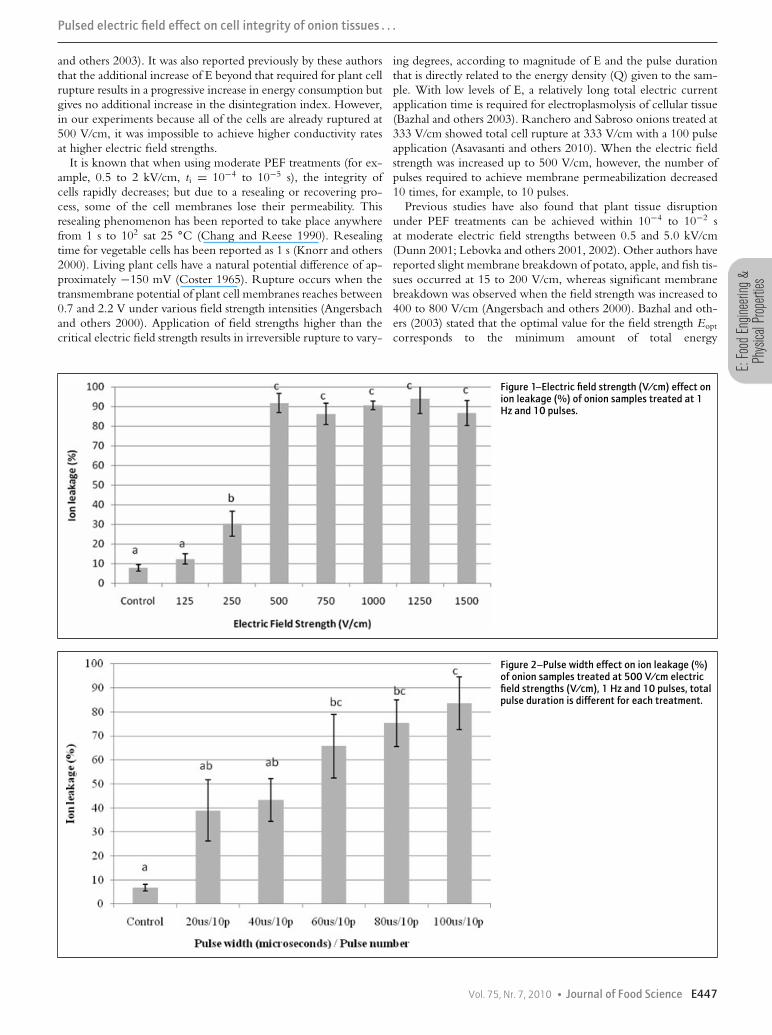
utilized to control the efficiency of plant and animal cell rupture.The influence of electric field strength, E (V/cm) on percent ionleakage is shown in Figure 1. Ion leakage of untreated (control)samples in an isotonic solution of 0.2 M mannitol is 7.8 ± 1.7%.It is thought that the increase in electrical conductivity of controlsamples was due to leakage of electrolytes from cells cut duringsampling and/or from the apoplastic region between the cells of thetissue. At a constant number of pulses (10) and frequency (1 Hz),application of electric field strengths of 250 and 500 V/cm bothresulted in significant (P < 0.05) increases in ion leakage as com-pared to the control. Additional increases in field strength above500 V/cm did not result in any increase in ion leakage, indicatingthat cells were ruptured already at 500 V/cm. Approximately 12%and 30% cell rupture were obtained with 125 and 250 V/cm PEFapplications. Figure 1 indicates that PEF treatments between 500and 1500 V/cm showed no significant difference in their effect onion leakage (P < 0.05). In a separate report (Ersus and Barrett2010), we evaluated the effect of PEF treatments with differentelectric field strengths and pulse numbers of various cell type thatwere distributed in a heterogeneous manner within plant tissues.
For different plant tissues, the critical level of electric fieldstrength required for cell rupture is reported to be in the range of200 to 500 V/cm, but this depends on the type of tissue (Bazhal
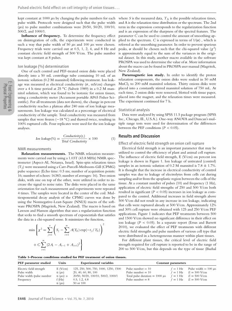
Table 1–Process conditions studied for PEF treatment of onion tissues.
PEF parameter studied Units Experimental variables Constant parameters
Electric field strength E (V/cm) 125, 250, 500, 750, 1000, 1250, 1500 Pulse number = 10 f = 1 Hz Pulse width = 100 μsPulse width ti (μs) 20, 40, 60, 80, 100 Pulse number = 10 f = 1 Hz E = 500 V/cmPulse width /pulse number ti (μs): n 20/50, 50/20, 100/10, 500/2, 1000/1 Total pulse duration = 1000 μs f = 1 Hz E = 500 V/cmFrequency f (Hz) 0.5, 1.2, 4.8 Pulse number = 8 f = 1 Hz E = 500 V/cm
ti (μs) 50 or 100
E446 Journal of Food Science � Vol. 75, Nr. 7, 2010
E:Fo
odEn
gine
erin
g&
Phys
ical
Prop
ertie
s
Pulsed electric field effect on cell integrity of onion tissues . . .
and others 2003). It was also reported previously by these authorsthat the additional increase of E beyond that required for plant cellrupture results in a progressive increase in energy consumption butgives no additional increase in the disintegration index. However,in our experiments because all of the cells are already ruptured at500 V/cm, it was impossible to achieve higher conductivity ratesat higher electric field strengths.
It is known that when using moderate PEF treatments (for ex-ample, 0.5 to 2 kV/cm, ti = 10−4 to 10−5 s), the integrity ofcells rapidly decreases; but due to a resealing or recovering pro-cess, some of the cell membranes lose their permeability. Thisresealing phenomenon has been reported to take place anywherefrom 1 s to 102 sat 25 ◦C (Chang and Reese 1990). Resealingtime for vegetable cells has been reported as 1 s (Knorr and others2000). Living plant cells have a natural potential difference of ap-proximately −150 mV (Coster 1965). Rupture occurs when thetransmembrane potential of plant cell membranes reaches between0.7 and 2.2 V under various field strength intensities (Angersbachand others 2000). Application of field strengths higher than thecritical electric field strength results in irreversible rupture to vary-
ing degrees, according to magnitude of E and the pulse durationthat is directly related to the energy density (Q) given to the sam-ple. With low levels of E, a relatively long total electric currentapplication time is required for electroplasmolysis of cellular tissue(Bazhal and others 2003). Ranchero and Sabroso onions treated at333 V/cm showed total cell rupture at 333 V/cm with a 100 pulseapplication (Asavasanti and others 2010). When the electric fieldstrength was increased up to 500 V/cm, however, the number ofpulses required to achieve membrane permeabilization decreased10 times, for example, to 10 pulses.
Previous studies have also found that plant tissue disruptionunder PEF treatments can be achieved within 10−4 to 10−2 sat moderate electric field strengths between 0.5 and 5.0 kV/cm(Dunn 2001; Lebovka and others 2001, 2002). Other authors havereported slight membrane breakdown of potato, apple, and fish tis-sues occurred at 15 to 200 V/cm, whereas significant membranebreakdown was observed when the field strength was increased to400 to 800 V/cm (Angersbach and others 2000). Bazhal and oth-ers (2003) stated that the optimal value for the field strength Eopt
corresponds to the minimum amount of total energy
Figure 1–Electric field strength (V/cm) effect onion leakage (%) of onion samples treated at 1Hz and 10 pulses.
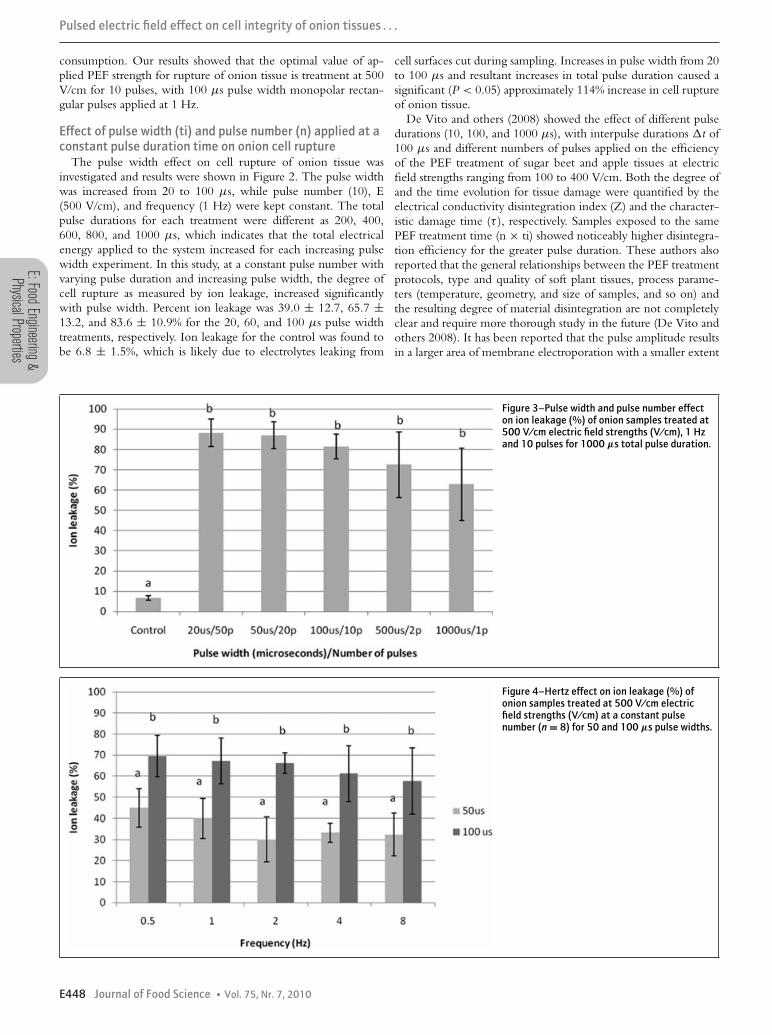
Figure 2–Pulse width effect on ion leakage (%)of onion samples treated at 500 V/cm electricfield strengths (V/cm), 1 Hz and 10 pulses, totalpulse duration is different for each treatment.
Vol. 75, Nr. 7, 2010 � Journal of Food Science E447
E:FoodEngineering
&PhysicalProperties
Pulsed electric field effect on cell integrity of onion tissues . . .
consumption. Our results showed that the optimal value of ap-plied PEF strength for rupture of onion tissue is treatment at 500V/cm for 10 pulses, with 100 μs pulse width monopolar rectan-gular pulses applied at 1 Hz.
Effect of pulse width (ti) and pulse number (n) applied at aconstant pulse duration time on onion cell rupture
The pulse width effect on cell rupture of onion tissue wasinvestigated and results were shown in Figure 2. The pulse widthwas increased from 20 to 100 μs, while pulse number (10), E(500 V/cm), and frequency (1 Hz) were kept constant. The totalpulse durations for each treatment were different as 200, 400,600, 800, and 1000 μs, which indicates that the total electricalenergy applied to the system increased for each increasing pulsewidth experiment. In this study, at a constant pulse number withvarying pulse duration and increasing pulse width, the degree ofcell rupture as measured by ion leakage, increased significantlywith pulse width. Percent ion leakage was 39.0 ± 12.7, 65.7 ±13.2, and 83.6 ± 10.9% for the 20, 60, and 100 μs pulse widthtreatments, respectively. Ion leakage for the control was found tobe 6.8 ± 1.5%, which is likely due to electrolytes leaking from
cell surfaces cut during sampling. Increases in pulse width from 20to 100 μs and resultant increases in total pulse duration caused asignificant (P < 0.05) approximately 114% increase in cell ruptureof onion tissue.
De Vito and others (2008) showed the effect of different pulsedurations (10, 100, and 1000 μs), with interpulse durations �t of100 μs and different numbers of pulses applied on the efficiencyof the PEF treatment of sugar beet and apple tissues at electricfield strengths ranging from 100 to 400 V/cm. Both the degree ofand the time evolution for tissue damage were quantified by theelectrical conductivity disintegration index (Z) and the character-istic damage time (τ ), respectively. Samples exposed to the samePEF treatment time (n × ti) showed noticeably higher disintegra-tion efficiency for the greater pulse duration. These authors alsoreported that the general relationships between the PEF treatmentprotocols, type and quality of soft plant tissues, process parame-ters (temperature, geometry, and size of samples, and so on) andthe resulting degree of material disintegration are not completelyclear and require more thorough study in the future (De Vito andothers 2008). It has been reported that the pulse amplitude resultsin a larger area of membrane electroporation with a smaller extent
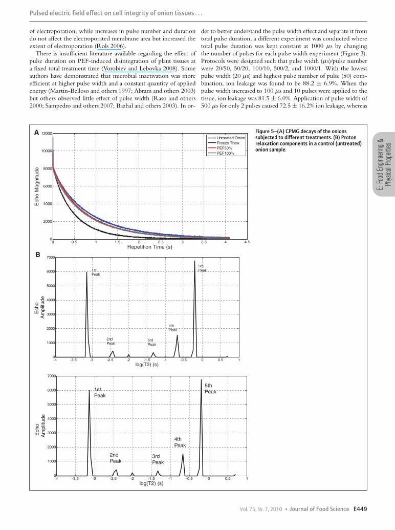
Figure 3–Pulse width and pulse number effecton ion leakage (%) of onion samples treated at500 V/cm electric field strengths (V/cm), 1 Hzand 10 pulses for 1000 μs total pulse duration.
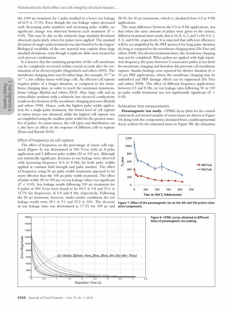
Figure 4–Hertz effect on ion leakage (%) ofonion samples treated at 500 V/cm electricfield strengths (V/cm) at a constant pulsenumber (n = 8) for 50 and 100 μs pulse widths.
E448 Journal of Food Science � Vol. 75, Nr. 7, 2010
E:Fo
odEn
gine
erin
g&
Phys
ical
Prop
ertie
s
Pulsed electric field effect on cell integrity of onion tissues . . .
of electroporation, while increases in pulse number and durationdo not affect the electroporated membrane area but increased theextent of electroporation (Rols 2006).
There is insufficient literature available regarding the effect ofpulse duration on PEF-induced disintegration of plant tissues ata fixed total treatment time (Vorobiev and Lebovka 2008). Someauthors have demonstrated that microbial inactivation was moreefficient at higher pulse width and a constant quantity of appliedenergy (Martin-Belloso and others 1997; Abram and others 2003)but others observed little effect of pulse width (Raso and others2000; Sampedro and others 2007; Bazhal and others 2003). In or-
der to better understand the pulse width effect and separate it fromtotal pulse duration, a different experiment was conducted wheretotal pulse duration was kept constant at 1000 μs by changingthe number of pulses for each pulse width experiment (Figure 3).Protocols were designed such that pulse width (μs)/pulse numberwere 20/50, 50/20, 100/10, 500/2, and 1000/1. With the lowestpulse width (20 μs) and highest pulse number of pulse (50) com-bination, ion leakage was found to be 88.2 ± 6.9%. When thepulse width increased to 100 μs and 10 pulses were applied to thetissue, ion leakage was 81.5 ± 6.0%. Application of pulse width of500 μs for only 2 pulses caused 72.5 ± 16.2% ion leakage, whereas
0 0.5 1 1.5 2 2.5 3 3.5 4 4.50
2000
4000
6000
8000
10000
12000
Repetition Time (s)
Ech
o M
ag
nitu
de
Untreated Onion
Freeze Thaw
PEF50%
PEF100%
A
-4 -3.5 -3 -2.5 -2 -1.5 -1 -0.5 0 0.5 10
1000
2000
3000
4000
5000
6000
7000
log(T2) (s)
Ech
o
Am
plit
ud
e
1stPeak
4thPeak
5thPeak
2ndPeak
3rdPeak
-4 -3.5 -3 -2.5 -2 -1.5 -1 -0.5 0 0.5 10
1000
2000
3000
4000
5000
6000
7000
log(T2) (s)
Echo
A
mp
litu
de
1stPeak
2ndPeak
3rdPeak
4thPeak
5thPeak
B
Figure 5–(A) CPMG decays of the onionssubjected to different treatments. (B) Protonrelaxation components in a control (untreated)onion sample.
Vol. 75, Nr. 7, 2010 � Journal of Food Science E449
E:FoodEngineering
&PhysicalProperties
Pulsed electric field effect on cell integrity of onion tissues . . .
the 1000 μs treatment for 1 pulse resulted in a lower ion leakageof 62.9 ± 17.9%. Even though the ion leakage values decreasedwith decreasing pulse numbers and increasing pulse widths, nosignificant change was observed between each treatment (P <
0.05). This may be due to the relatively large standard deviationsobserved, particularly when few pulses were applied. The standarddeviation of single-pulse treatment was also found to be the largest.Biological variability of the raw material may explain these largestandard deviations, even though 4 replicate disks were treated foreach process combination.
It is known that the insulating properties of the cell membranecan be completely recovered within several seconds after the ter-mination of an electrical pulse (Angersbach and others 2000). Themembrane charging time may be rather large, for example, 10−5 to10−4 s, for cellular tissues with large cells. An efficient cell rupturerequires pulses of a longer duration, as compared to the mem-brane charging time, in order to reach the maximum transmem-brane voltage (Bazhal and others 2003). Also, large cells and anextracellular medium with a relatively low electrical conductivityresults in deceleration of the membrane charging processes (Kotnikand others 1998). Hence, with the highest pulse width applica-tion for a single-pulse treatment, the lowest level of cell rupturein onion tissues was obtained, while the highest cell rupture wasaccomplished using the smallest pulse width for the greatest num-ber of pulses. In onion tissues, the cell types and distribution cane also have an effect on the response of different cells to rupture(Ersus and Barrett 2010).
Effect of frequency on cell ruptureThe effect of frequency on the percentage of onion cells rup-
tured (Figure 4) was determined at 500 V/cm with an 8 pulseapplication and 2 different pulse widths (50 to 100 μs). Althoughnot statistically significant, decreases in ion leakage were observedwith increasing frequency (0.5 to 8 Hz) for both pulse widthsapplied at constant field strength and pulse number. The effectof frequency using 50 μs pulse width treatments appeared to bemore effective than the 100 μs pulse width treatment. The effectof pulse width (50 to 100 μs) on ion leakage values was significant(P < 0.05). Ion leakage results following 100 μs treatments for8 pulses at 500 V/cm were found to be 69.5 ± 9.8 and 57.6 ±15.7% for frequencies of 0.5 and 8 Hz, respectively. Followingthe 50 μs treatment, however, under similar conditions the ionleakage results were 45.1 ± 9.1 and 32.3 ± 10%. The decreasein ion leakage ratio was determined as 17.1% for 100 μs and
28.4% for 50 μs treatments, which is calculated from 0.5 to 8 Hzapplications.
The main difference between the 0.5 to 8 Hz applications, wasthat when the same amount of pulses were given to the system,different treatment times result, that is 16, 8, 4, 2, and 1 s for 0.5, 1,2, 4, and 8 Hz, respectively. It is expected that sufficient efficiencywill be accomplished by the PEF protocol for long pulse duration(ti) long as compared to the membrane charging time (De Vito andothers 2008). For shorter treatment times, the membrane chargingmay not be completed. When pulses are applied with high repeti-tion frequency, the pause between 2 consecutive pulses is too shortfor membrane charging and therefore this prevents cell membranerupture. Similar findings were reported for shorter duration (ti =10 μs) PEF applications, where the membrane charging may beunfinished and PEF damage effects can be suppressed (De Vitoand others 2008). The effect of different frequency applications,between 0.5 and 8 Hz, on ion leakage rates following 50 or 100μs pulse width treatments was not significantly significant (P <
0.05).
Relaxation time measurementsParamagnetic ion study. CPMG decay plots for the control
(untreated) and treated samples of onion tissue are shown in Figure5A along with the components calculated from a multiexponentialdecay analysis for the untreated tissue in Figure 5B, the relaxation
Figure 7–Effect of the paramagnetic ion on the 4th and 5th proton relax-ation components.
0 0.5 1 1.5 2 2.5 3 3.5 4 4.50
2000
4000
6000
8000
10000
12000
14000
16000
18000
Repetition Time (s)
Echo
Ma
gn
itud
e
(0,15min,30min,1hrs,2hrs,3hrs,4hr,5hr,6hr,7hrs)
Figure 6–CPMG curves obtained at differenttimes of paramagnetic ion soaking.
E450 Journal of Food Science � Vol. 75, Nr. 7, 2010
E:Fo
odEn
gine
erin
g&
Phys
ical
Prop
ertie
s
Pulsed electric field effect on cell integrity of onion tissues . . .
spectrum. The CPMG curves obtained during paramagnetic ionuptake are also shown in Figure 6. The decay of the signal is veryrapid initially and then slows. The complete decay is well describedby 5 components in Figure 5B (the small peak in the middle wasthought to be spurious since the contribution was so small andnot present in all experiments).
The paramagnetic ions were used to decrease proton relaxationtimes enabling peaks in the relaxation spectrum to be assigned tospecific cell compartments (Snaar and Van As 1992). Shown inFigure 7 are the changes observed in the relaxation spectra forpeaks 4 and 5 (Figure 5B) as a function of soaking time in MnCl2-mannitol solution. The percentage change of peaks 4 and 5 areshown in Figure 8. It is found that the change in the peaks aresignificantly different from each other and with respect to soakingtimes (P < 0.05).
The largest compartments in an onion tissue are the cytoplasmand the vacuole. The cytoplasm compartment should be impactedfirst by paramagnetic ion diffusion and the vacuole second.
The vacuole will contain the largest number of protons andwill have greater amplitude in the relaxation spectrum. Peak 4 isassigned to cytoplasm since the changes in T2 values are faster andthe amplitude lower than peak 5, which is assigned to vacuole.However, it is important to note that a change in the relaxationtime of vacuole was observed at the end of 15 min in contrastto findings of Snaar and Van As (1992). They found out that inapple parenchyma tissue it took almost 16 h to see the change inthe relaxation time of the vacuole in the presence of paramagneticions. On the other hand, Hills and Duce (1990) found out thatin an onion tissue, in contrast to an apple tissue, relaxation isdominated by the various combinations of fast proton exchangebetween water and biopolymers. These finding are consistent withthe behavior observed for peak 4 and peak 5.
Peaks 1, 2, and 3 are virtually unchanged by paramagnetic iondiffusion and hence associated with the protons in solid-like com-partment in the cell (for example, macromolecules and mem-branes).
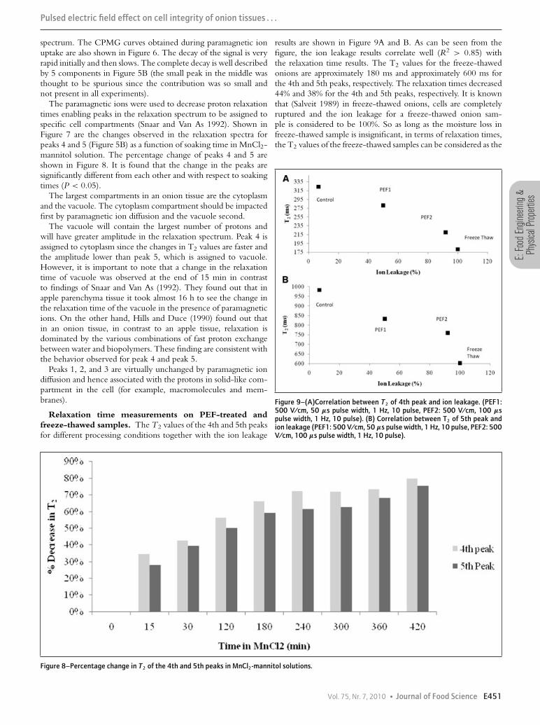
Relaxation time measurements on PEF-treated andfreeze-thawed samples. The T2 values of the 4th and 5th peaksfor different processing conditions together with the ion leakage
results are shown in Figure 9A and B. As can be seen from thefigure, the ion leakage results correlate well (R2 > 0.85) withthe relaxation time results. The T2 values for the freeze-thawedonions are approximately 180 ms and approximately 600 ms forthe 4th and 5th peaks, respectively. The relaxation times decreased44% and 38% for the 4th and 5th peaks, respectively. It is knownthat (Salveit 1989) in freeze-thawed onions, cells are completelyruptured and the ion leakage for a freeze-thawed onion sam-ple is considered to be 100%. So as long as the moisture loss infreeze-thawed sample is insignificant, in terms of relaxation times,the T2 values of the freeze-thawed samples can be considered as the
Figure 9–(A)Correlation between T2 of 4th peak and ion leakage. (PEF1:500 V/cm, 50 μs pulse width, 1 Hz, 10 pulse, PEF2: 500 V/cm, 100 μspulse width, 1 Hz, 10 pulse). (B) Correlation between T2 of 5th peak andion leakage (PEF1: 500 V/cm, 50 μs pulse width, 1 Hz, 10 pulse, PEF2: 500V/cm, 100 μs pulse width, 1 Hz, 10 pulse).
Figure 8–Percentage change in T2 of the 4th and 5th peaks in MnCl2-mannitol solutions.
Vol. 75, Nr. 7, 2010 � Journal of Food Science E451
E:FoodEngineering
&PhysicalProperties
Pulsed electric field effect on cell integrity of onion tissues . . .
limit for a ruptured cell and can be used as an intactness measurefor the cells. On the other hand, it is interesting to note that therelaxation time measurements for the freeze-thawed samples givestill the same number of peaks as the untreated onions but withshorter relaxation times. The presence of the same peaks showsthat the exchange of water by molecular diffusion between thedifferent relaxation components is slow on the NMR relaxationtime scale and implies that there are still significant permeabilitybarriers to water exchange (Hills and Remigereau 1997) althoughthe cell is significantly damaged.
Relaxation time measurements were done for 2 different PEFconditions (PEF1: 500 V/cm, 50 μs pulse width, 1 Hz, 10 pulse,PEF2: 500 V/cm, 100 μs pulse width, 1 Hz, 10 pulse). Accord-ing to ion leakage results, PEF2 conditions resulted in 91.83 ±4.86% rupture that is consistent with higher T2 values compared tofreeze-thawed samples. PEF1 treatment that resulted in 50% rup-ture had higher T2 values for both peaks compared to PEF1 andfreeze-thawed samples. The high correlation between ion leakageand the T2 values indicates that relaxation time measurements canbe used for characterizing cell intactness.
ConclusionPEF processing can be optimized to achieve varying levels of
disruption to cellular tissues. PEF systems designed to achieve highlevels of cellular damage should employ high voltage applicationsfor short times with high repetition rates or frequencies. Valida-tion of PEF efficiency can be achieved using either ion leakagemeasurements or nondestructive NMR relaxation spectrum anal-ysis. In-line control of PEF processing may be achieved by employ-ing NMR relaxation measurements to optimize PEF processingparameters.
NomenclatureE electric field strength (V/cm)n number of pulsef frequency (Hz)ti pulse width (μs)
nti total pulse duration (μs)
ReferencesAbram F, Smelt JPPM, Bos R, Wouters PC. 2003. Modeling and optimization of inactiva-
tion of Lactobacillus plantarum by pulsed electric field treatment. J Appl Microbiol 94:571–9.
Angersbach A, Heinz V, Knorr D. 2000. Effects of pulsed electric fields on cell membranes inreal food systems. Innovative Food Sci Emerg Technol 1:135–49.
Asavasanti S, Ersus S, Ristenpart W, Stroeve P, Barrett DM. 2010. Onion tissues and pulsedelectric fields: indication for different critical field strengths. J Food Sci. 75:E433–43.
Barbosa-Canovas GV, Pothakamury UR, Palou E, Swanson BG. 1998. Biological effects andapplications of pulsed electric fields for the preservation of foods. In: Nonthermal preservationof foods. New York: Marcel Dekker Inc. p 73–112.
Bazhal MI, Lebovka NI, Vorobiev E. 2001. Pulsed electric field treatment of apple tissue duringcompression for juice extraction. J Food Eng 50(3):129–39.
Bazhal MI, Vorobiev E. 2000. Electrical treatment of apple cossettes for intensifying juice pressing.J Sci Food Agric 80:1668–74.
Bazhal MI, Lebovka NI, Vorobiev EI. 2003. Optimization of pulsed electric field strength forelectroplasmolysis of vegetable tissues. Biosyst Eng 86:339–45.
Bouzzara H, Vorobiev E. 2000. Beet juice extraction by pressing and pulsed electric fields. IntSugar J CII 1216:194–200.
Chang DC, Reese TS. 1990. Changes in membrane structure induced by electroporation asrevealed by rapid freezing electron microscopy. Biophys J 58:1–12.
Coster HGL. 1965. A quantative analysis of the voltage-current relationships of fixedcharged membranes and the associated property of punch-through. Biophys J 5:669–86.
De Vito F, Ferrari G, Lebovka NI, Shynkaryk NV, Vorobiev E. 2008. Pulse duration andefficiency of soft cellular tissue disintegration by pulsed electric fields. Food Bioprocess Technol1:307–13
Dunn J. 2001. Pulsed electric field processing: an overview. In: Barbosa-Canovas GV, ZhangQH, editors. Pulsed electric fields in food processing: fundamental aspects and applications.Lancaster, Pa.: Technomic Publishing Co. p 1–30.
Ersus S, Barrett DM. 2010. Determination of membrane integrity in onion tissues treated bypulsed electric fields: use of microscopic images and ion leakage measurements. InnovativeFood Sci and Emerging Tech. Forthcoming.
Estiaghi MN, Knorr D. 1999. Method for treating sugar beet. Intl. Patent, No. WO 99/6434.Gonzalez ME. 2009. Quantification of cell membrane integrity and its relevance to texture
quality of onions: effects of high hydrostatic pressure and thermal processes [PhD diss.]. Davis,Calif.: Univ. of California. 138 p.
Hills B. 1998. Magnetic resonance imaging in food science. New York: Wiley.Hills B, Duce SL. 1990. The influence of chemical and diffusive exchange on water proton
transverse relaxation in plant tissues. Magn Reson Imaging 8(3):321–31.Hills B, Remigereau B. 1997. NMR studies of changes in subcellular water compartmentation
in parenchyma apple tissue during drying and freezing. Int J Food Sci Tech 32:51–61.Hills B, Costa A, Marigheto N, Wright K. 2005. T1-T2 NMR correlation studies of high
pressure processed starch and potato tissue. Appl Magn Reson 28:13–27.Hills B, Cano C, Belton PS. 1991. Proton NMR relaxation studies of aqueous polysaccharide
systems. Macromolecules 24(10):2944–50.Knorr D, Heinz V, Angersbach A, Lee D. 2000. Membrane permeabilization and inactivation
mechanisms of biological systems by emerging technologies. Proceedings of the 8th Intl.Congress on Engineering and Food (p.15). Puebla, Mexico.
Knorr D, Angersbach A. 1998. Impact of high-intensity electrical field pulses on plant membranepermeabilization. Trends Food Sci Technol 9:185–91.
Kotnik T, Miklavcic D, Slivnik T. 1998. Time course of transmembrane voltage induced by time-varying electric fields—a method for theoretical analysis and its application. BioelectrochemBioenerg 45:3–16.
Lebovka NI, Bazhal MI, Vorobiev E. 2000. Simulation and experimental investigation of foodmaterial breakage using pulsed electric field treatment. J Food Eng 44:213–23.
Lebovka NI, Bazhal MI, Vorobiev E. 2001. Pulsed electric field breakage of cellular tissues:visualization of percolative properties. Innovative Food Sci Emerg Technol 2:113–25.
Lebovka NI, Bazhal MI, Vorobiev E. 2002. Estimation of characteristic damage time of foodmaterials in pulsed-electric fields. J Food Eng 54:337–46.
Lebovka NI, Shynkaryk MV, El-Belghiti K, Benjelloun H, Vorobiev E. 2007. Plasmolysis ofsugarbeet: pulsed electric fields and thermal treatment. J Food Eng 80(2):639–44.
Macek-Lebar A, Miklavcic D. 2001. Cell electropermeabilization to small molecules in vitro:control by pulse parameters. Radiol Oncol 35:193–202.
Marigheto N, Vial A, Wright K, Hills B. 2004. A combined NMR and microstructural studyof effect of high pressure processing on strawberries. Appl Magn Reson 26:521–31.
Martin-Belloso O, Vega-Mercado H, Qin BL, Chang BJ, Barbosa-Canovas GV, Swanson BG.1997. Inactivation of Escherichia coli suspended in liquid egg using pulsed electric fields. J FoodProcess Preserv 21(3):193–208.
McLellan MR, Kime RL, Lind LR. 1991. Electroplasmolysis and other treatments to improveapple juice yield. J Sci Food Agric 57:303–6.
Milczarek RR, Salveit ME, Garvey TC, McCarthy MJ. 2009. Assessment of tomato pericarpmechanical damage using multivariate analysis of magnetic resonance images. Postharvest BiolTechnol 52:189–95.
Palta J, Levitt J, Stadelmann EJ. 1977. Freezing injury in onion bulb cells. Plant Physiol60:398–401.
Raffo A, Gianferri R, Barbieri R, Brosio E. 2004. Ripening of banana fruit monitored by waterrelaxation and diffusion H-1-NMR measurements. Food Chem 89(1):149–58.
Raso J, Alvarez I, Condon S, Sala FJ. 2000. Predicting inactivation of Salmonella senftenberg bypulsed electric fields. Innovative Food Sci Emerg Technol 1:21–30.
Rols MP, Tessie J. 1990. Electropermeabilization of mammalian cells. Quantitative analysis ofphenomenon. Biophys J 58:1089–98.
Rols MP. 2006. Electropermeabilization, a physical method for the delivery of therapeuticmolecules into cells. Biochim Biophys Acta 1758:423–8.
Salveit ME. 1989. A kinetic examination of ion leakage from chilled tomato pericarp discs. ActaHorticulturae 258:617–22.
Sampedro F, Rivas A, Rodrigo D, Martinez A, Rodrigo M. 2007. Pulsed electric fields in-activation of Lactobacillus plantarum in an orange juice-milk based beverage: effect of processparameters. J Food Eng 80:931–8.
Snaar JEM, Van As H. 1992. Probing water compartments and membrane permeability in plantcells by 1H NMR relaxation measurements. Biophys J 63:1654–8.
Van Der Weerd L, Claessens M, Ruttnik T, Vergeldt F, Schaafsma T, Van As H. 2001. Quan-titative NMR microscopy of osmotic stress response in maize and pearl millet. J Exp Bot52:2333–43.
Vasquez-Tello A, Zuily-Fodil Y, Pham Thi AT, Viera da Silva JB. 1990. Electrolyte and Pileakages and soluble sugar content as physiological tests for screening resistance to water stressin Phaseolus and Vigna species. J Exp Bot 228:827–32.
Vorobiev E, Lebovka NI. 2006. Extraction of intercellular components by pulsed electric fields.In: Raso J, Heinz H, editors. Pulsed electric field technology for the food industry. Funda-mentals and applications. New York: Springer. p 153–94.
Vorobiev E, Lebovka NI. 2008. Pulsed-electric-fields-induced effects in plant tissues: funda-mental aspects and perspectives of applications. In: Vorobiev E, Lebovka N, editors. Elec-trotechnologies for extraction from food plants and biomaterials. New York: Springer.p 39–81.
Zhang QH, Qin BL, Barbosa-Canovas GV, Swanson BG. 1995. Inactivation of Escherichia colifor pasteurization by high-strength pulsed electric fields. J Food Preservation 19:103–18.
Zimmermann U, Pilwat G, Riemann F. 1974. Dielectric breakdown of cell membranes. BiophysJ 14:881–99.
E452 Journal of Food Science � Vol. 75, Nr. 7, 2010