Embed Size (px)
Citation preview

Dislocation resistance of ProRoot Endo Sealer, acalcium silicate-based root canal sealer, fromradicular dentine
B. P. Huffman1, S. Mai2, L. Pinna3, R. N. Weller4, C. M. Primus5, J. L. Gutmann6, D. H. Pashley7
& F. R. Tay4,7
1School of Dentistry, Medical College of Georgia, Augusta, GA, USA; 2Guanghua School of Stomatology & Institute of
Stomatological Research, Sun Yat-sen University, Guangzhou, China; 3Universita degli Studi di Cagliari, Reparto di Odontoiatria
Conservatrice, Sardinia, Italy; 4Department of Endodontics, School of Dentistry, Medical College of Georgia, Augusta, GA, USA;5Primus Consulting, Bradenton, FL, USA; 6Department of Endodontics, Baylor College of Dentistry, Texas A&M University System
Health Science Center, Dallas, TX, USA; and 7Department of Oral Biology, School of Dentistry, Medical College of Georgia, Augusta,
GA, USA
Abstract
Huffman BP, Mai S, Pinna L, Weller RN, Primus CM,
Gutmann JL, Pashley DH, Tay FR. Dislocation resistance
of ProRoot Endo Sealer, a calcium silicate-based root
canal sealer, from radicular dentine. International Endodontic
Journal, 42, 34–46, 2009.
Aim To examine the dislocation resistance of three
root canal sealers from radicular dentine with and
without immersion in a simulated body fluid (SBF),
using a modified push-out test design that produced
simulated canal spaces of uniform dimensions under
identical cleaning and shaping conditions.
Methodology Sixty single-rooted caries-free human
canine teeth were used. Standardized simulated canal
spaces were created using 0.04 taper ProFile instru-
ments along the coronal, middle and apical thirds of
longitudinal tooth slabs. Following NaOCl/ethylenedi-
amine tetra-acetic acid cleaning, the cavities were filled
with ProRoot Endo Sealer, AH Plus Jet or Pulp Canal
Sealer. After setting, half of the cavities were tested
with a fibre-optic light-illuminated push-out testing
device. The rest were immersed in SBF for 4 weeks
before push-out evaluation. Failure modes were exam-
ined with stereomicroscopy and field emission (FE)-
scanning electron microscopy.
Results Location of the sealer-filled cavities did not
affect push-out strengths. ProRoot Endo Sealer exhibited
higher push-out strengths than the other two sealers
particularly after SBF storage (P < 0.001). Failure
modes were predominantly adhesive and mixed for Pulp
Canal Sealer and AH Plus Jet, and predominantly
cohesive for ProRoot Endo Sealer. Spherical amorphous
calcium phosphate-like phases that spontaneously trans-
formed into apatite-like phases were seen in the fractured
specimens of ProRoot Endo Sealer after SBF storage.
Conclusions When tested in bulk without a main
core, both ‘sealer type’ and ‘SBF storage’ were signif-
icant in affecting push-out results. The ProRoot Endo
Sealer demonstrated the presence of spherical amor-
phous calcium phosphate-like phases and apatite-like
phases (i.e. ex vivo bioactivity) after SBF storage.
Keywords: calcium silicate-based sealer, dislocation
resistance, in vitro bioactivity, thin-slice push-out test.
Received 1 June 2008; accepted 16 September 2008
Introduction
The use of a sealer and a thermoplastic core material
for filling root canals is the accepted norm in contem-
porary root canal procedures. As leakage from the
apical or coronal direction is a possible cause of root
treatment failure (Madison & Wilcox 1988, De Moor &
Hommez 2000), a root canal sealer should exhibit good
sealing (Laghios et al. 2000) and adhesive properties
(Wennberg & Ørstavik 1990, Gettleman et al. 1991,
Timpawat et al. 2001, Lee et al. 2002a,b, Saleh et al.
2003, Tagger et al. 2003). A sealer may be
Correspondence: Dr Franklin R. Tay, Department of Endodon-
tics, School of Dentistry, Medical College of Georgia, Augusta,
GA, 30912-1129, USA (Tel.: +1 706 7212033; fax:
+1 706 7216252; e-mail: [email protected]).
doi:10.1111/j.1365-2591.2008.01490.x
International Endodontic Journal, 42, 34–46, 2009 ª 2009 International Endodontic Journal34

conceptualized as a joint created between the radicular
dentine and the filling material. Similar to other
prosthetic joints in the body, the ability to resist
dislocation during function is crucial to their survival
(Scifert et al. 1999, Weale et al. 2002, He et al. 2007).
For a root canal sealer, the ability to resist disruption of
the established seal via micromechanical retention or
friction is highly desirable during intraoral tooth
flexure (Panitvisai & Messer 1995) or preparation of
cores or postspaces along the coronal- and middle-
thirds of canal walls (Munoz et al. 2007).
Predictable clinical results have been reported with
the use of gutta-percha in conjunction with zinc oxide
eugenol or epoxy resin-based root canal sealers (Saleh-
rabi & Rotstein 2004, Tilashalski et al. 2004). Never-
theless, there is a continuous quest for alternative
sealers or root filling materials with better seal and dis-
location resistance. Although the correlation between
the sealing property of a root canal sealer and its
adhesive characteristics has not been firmly esta-
blished, it is essential that the dislocation resistance of
a root canal sealer to dentine is not adversely affected
by the seepage of body fluids when there is a breach of
either the apical or coronal seal.
ProRoot Endo Sealer (Dentsply Tulsa Dental Special-
ties, Tulsa, OK, USA) is an experimental calcium
silicate-based root canal sealer that is designed to be
used in conjunction with a root filling material in either
the cold lateral, warm vertical or carrier-based filling
techniques. The major components of the powder
component are tricalcium silicate and dicalcium sili-
cate, with the inclusion of calcium sulphate as a setting
retardant, bismuth oxide as a radiopacifier and a small
amount of tricalcium aluminate. The liquid component
consists of a viscous aqueous solution of a water-
soluble polymer. Similar to other tricalcium silicate and
dicalcium silicate-containing biomaterials, the sealer
produces calcium hydroxide on reaction with water
(Gou et al. 2005, Camilleri & Pitt Ford 2006, Wang
et al. 2008). It is also anticipated that release of
calcium and hydroxyl ions from the set sealer will
result in the formation of apatites as the material comes
into contact with phosphate-containing fluids (Sarkar
et al. 2005), via spontaneous transformation from
initial amorphous calcium phosphate phases (Tay et al.
2007, Tay & Pashley 2008).
Whereas the retentive potential of geosynthetics
(Marques 2005), concrete reinforcements (Lee et al.
2002a,b) and rigid postsystems within canal spaces
(Mitchell et al. 1994, Teixeira et al. 2006) may be
evaluated en masse using conventional pull-out test
designs, thermoplastic root filling materials and sealers
are not amendable to gripping that is a prerequisite for
this type of mechanical testing (Goracci et al. 2007).
Thus, the thin-slice push-out test has been used quite
frequently for evaluating the dislocation resistance of
root filling materials (Gesi et al. 2005, Sousa-Neto et al.
2005, Gancedo-Caravia & Garcia-Barbero 2006, Skid-
more et al. 2006, Ungor et al. 2006, Bouillaguet et al.
2007, Fisher et al. 2007, Jainaen et al. 2007, Nagas
et al. 2007, Sly et al. 2007, Ureyen Kaya et al. 2008).
The strength of that experimental design is that each
horizontal root slab being tested is derived from a root
filled canal and contains a cross-section of the thermo-
plastic root filling material and sealer to be investigated.
In the present study, a modified thin-slice push-out test
was designed to evaluate the dislocation resistance of
root canal sealers that were applied in bulk to simu-
lated canal spaces without the use of thermoplastic
material cores. The null hypothesis tested was that
there are no differences in the dislocation resistance of
three root canal sealers from radicular dentine when
the set sealers are tested with and without immersion
in a simulated body fluid (SBF).
Materials and methods
Preparation of simulated canal spaces
Sixty intact, caries-free human canine teeth were
collected after the patients’ informed consents were
obtained under a protocol reviewed and approved by
the Human Assurance Committee of the Medical
College of Georgia, Georgia, USA. For each tooth, a
0.90 ± 0.05 mm thick longitudinal slab was prepared
by making buccolingual sections parallel to the longi-
tudinal axis of the tooth using a slow-speed diamond
saw (Isomet; Buehler Ltd, Lake Bluff, IL, USA) under
water-cooling. A Plexiglas platform containing a cylin-
drical well was affixed to the base of a mini drill press
to generate vertically oriented, truncated cavities of
uniform dimensions within the tooth slab (Fig. 1a).
A 0.6 mm drill bit was first used to prepare pilot holes
in the radicular dentine adjacent to the dental pulp.
Each pilot hole was carefully drilled so that it was
equidistant from the cementum and the canal wall.
Two pilot holes each were prepared in the coronal,
middle and apical thirds of the root.
Each hole was subsequently enlarged using a size 40,
25 mm long 0.04 taper ProFile nickel titanium rotary
instrument (Dentsply Tulsa Dental Specialties). To
ensure optimal cutting efficacy, a new instrument
Huffman et al. Dislocation resistance of ProRoot Endo Sealer
ª 2009 International Endodontic Journal International Endodontic Journal, 42, 34–46, 2009 35
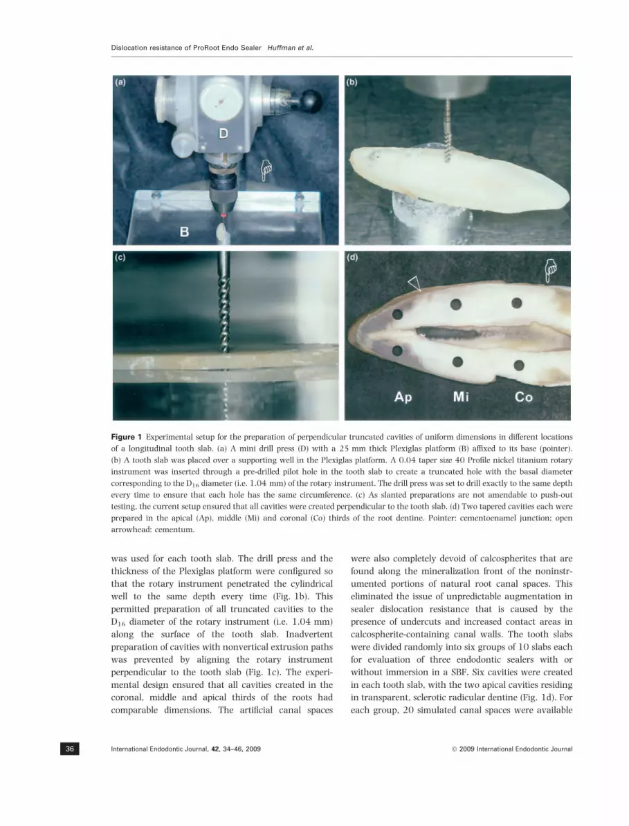
was used for each tooth slab. The drill press and the
thickness of the Plexiglas platform were configured so
that the rotary instrument penetrated the cylindrical
well to the same depth every time (Fig. 1b). This
permitted preparation of all truncated cavities to the
D16 diameter of the rotary instrument (i.e. 1.04 mm)
along the surface of the tooth slab. Inadvertent
preparation of cavities with nonvertical extrusion paths
was prevented by aligning the rotary instrument
perpendicular to the tooth slab (Fig. 1c). The experi-
mental design ensured that all cavities created in the
coronal, middle and apical thirds of the roots had
comparable dimensions. The artificial canal spaces
were also completely devoid of calcospherites that are
found along the mineralization front of the noninstr-
umented portions of natural root canal spaces. This
eliminated the issue of unpredictable augmentation in
sealer dislocation resistance that is caused by the
presence of undercuts and increased contact areas in
calcospherite-containing canal walls. The tooth slabs
were divided randomly into six groups of 10 slabs each
for evaluation of three endodontic sealers with or
without immersion in a SBF. Six cavities were created
in each tooth slab, with the two apical cavities residing
in transparent, sclerotic radicular dentine (Fig. 1d). For
each group, 20 simulated canal spaces were available
Figure 1 Experimental setup for the preparation of perpendicular truncated cavities of uniform dimensions in different locations
of a longitudinal tooth slab. (a) A mini drill press (D) with a 25 mm thick Plexiglas platform (B) affixed to its base (pointer).
(b) A tooth slab was placed over a supporting well in the Plexiglas platform. A 0.04 taper size 40 Profile nickel titanium rotary
instrument was inserted through a pre-drilled pilot hole in the tooth slab to create a truncated hole with the basal diameter
corresponding to the D16 diameter (i.e. 1.04 mm) of the rotary instrument. The drill press was set to drill exactly to the same depth
every time to ensure that each hole has the same circumference. (c) As slanted preparations are not amendable to push-out
testing, the current setup ensured that all cavities were created perpendicular to the tooth slab. (d) Two tapered cavities each were
prepared in the apical (Ap), middle (Mi) and coronal (Co) thirds of the root dentine. Pointer: cementoenamel junction; open
arrowhead: cementum.
Dislocation resistance of ProRoot Endo Sealer Huffman et al.
International Endodontic Journal, 42, 34–46, 2009 ª 2009 International Endodontic Journal36

from each of the three respective radicular dentine
locations (n = 20).
Filling of root canal sealers
The tooth slabs were immersed in 17% ethylenedia-
mine tetra-acetic acid (EDTA) and ultrasonicated for
5 min to dissolve the smear layer created during the
hole-shaping procedures. The slabs were further
immersed in 6.15% sodium hypochlorite and ultraso-
nicated for 5 min to remove organic debris and the
demineralized collagen matrix created during EDTA
application. The rationale for en masse cleaning was to
further ensure that differences in dislocation resistance
of the sealers from different dentine locations were not
caused by inadequate cleaning of the apical radicular
dentine.
The three sealers investigated in this study were Pulp
Canal Sealer (SybronEndo; Sybron Dental Specialties
Inc., Orange, CA, USA), AH Plus Jet (Dentsply Caulk,
Milford, DE, USA) and the experimental ProRoot Endo
Sealer. The former two sealers were mixed according to
the manufacturers’ instructions. The calcium silicate-
based sealer was mixed with a liquid-to-powder ratio of
1 : 2 and covered with moist gauze to avoid evapora-
tion of the water component. All cavities from one
tooth slab were filled with one type of sealer. Each tooth
slab was placed over a Mylar strip (Angst & Pfister,
Geneva, Switzerland), which in turn was placed over a
microscope glass slide. For Pulp Canal Sealer and
ProRoot Endo Sealer, the sealer was mixed and placed
inside a 19-gauge AccuDose Needle Tube (Centrix,
Shelton, CT, USA). The sealer was dispensed into the
cavities so that each hole was filled with excess sealer.
For AH Plus Jet, the sealer was dispensed directly from
the double-barrel mixing syringe via an intraoral tip
attached to an auto-mixing tip. The surface of the tooth
slab was then covered with another Mylar strip and a
glass slide. The assembly was secured with binder clips
so that excess sealer was expressed laterally from the
surface and bottom Mylar strips. The assemblies were
transferred to humidors and stored under 100%
relative humidity for 1 week until all the sealers had
completely set.
The binder clips were released and the Mylar strips
were removed from the tooth slab to expose the set
sealers. The top and bottom surfaces of each tooth slab
were polished with 800-grit silicon carbide papers
under running water to remove the excess sealer. For
each sealer, one subgroup of 10 tooth slabs was tested
immediately after polishing, whilst the other subgroup
of 10 tooth slabs was immersed for 4 weeks at 37 �C in
a phosphate-containing SBF prior to testing. The SBF
contained 136.8 mmol L)1 NaCl, 3.0 mmol L)1 KCl,
2.5 mmol L)1 CaCl2Æ6 H2O, 1.5 mmol L)1 MgCl2Æ6-
H2O, 0.5 mmol L)1 Na2SO4Æ10 H2O, 4.2 mmol L)1
NaHCO3 and 1.0 mmol L)1 K2HPO4Æ3H2O in deionized
water (pH 7.4). To prevent bacterial growth, 0.02%
sodium azide was also included in the SBF.
Dislocation resistance evaluation
The dislocation resistance of the set root canal sealers
was evaluated using a thin-slice push-out test design
(Chandra & Ananth 1995, Chandra & Ghonem 2001).
Prior to testing, the thickness of each tooth slab was
measured to the nearest 0.01 mm using a pair of
digital calipers. A 0.7 mm diameter carbon steel
cylindrical plunger was used for the push-out test.
The plunger had a clearance of about 0.1 mm from
either side of the dentinal wall when it is perfectly
aligned with the apical part of the truncated hole. The
plunger was attached to a 10 kg load cell that was
connected to a universal testing machine (Vitrodyne
V1000 universal tester; Liveco Inc., Burlington, VT,
USA). The push-out device consisted of a clear
Plexiglas platform with a vertical cylindrical channel,
which served as the support for the tooth slab and
provided space for the vertical movement of the
plunger through the truncated hole (Fig. 2a). To
ensure optimal alignment of the plunger with the
sealer-filled hole, a horizontal channel was drilled
through the Plexiglas platform into the vertical chan-
nel (Fig. 2b). A fibre-optic light guide was inserted into
the horizontal channel to provide high intensity
illumination of the truncated hole during the align-
ment procedure. Each root slab was secured with
sticky wax in an apical-coronal direction to the
supporting Plexiglas platform, so that the smaller
diameter apical side of the sealer-filled hole was facing
the plunger. Each sealer-filled hole was subjected to
compressive loading at a cross-head speed of
10 lm s)1 in order to displace the set sealer toward
the coronal aspect of the hole. As the plunger contacts
the set sealer on loading, shear stresses were intro-
duced along the sealer-dentine interface, causing the
set sealer to be dislocated from the walls of the
radicular dentine. Failure was confirmed by the
appearance of a sharp drop along the load/displace-
ment curve recorded by the testing machine. After
performing push-out testing of the first hole, the tooth
slab was carefully removed and realigned with the
Huffman et al. Dislocation resistance of ProRoot Endo Sealer
ª 2009 International Endodontic Journal International Endodontic Journal, 42, 34–46, 2009 37

second hole. The procedures were repeated until the
set sealers were dislodged from all the six cavities
within a tooth slab. After the push-out test, each root
slab was examined with a stereomicroscope at 30·magnification to determine the mode of failure. Failure
modes were classified as: adhesive failure along the
sealer-dentine interface; cohesive failure within the
sealer, and mixed failure that consisted of partial
adhesive failure along the dentinal walls and partial
cohesive failure within the sealer (Fig. 2c).
Digitized photographs of each tested hole were taken
from the coronal and apical aspects of the tooth slab
together with a millimetre scale for calibration purpose.
Such a procedure was performed after completion of
the push-out test as this permitted better contrast of the
circumference of the cavities. The circumferences of the
coronal (C) and apical aspects (A) of each cavity were
measured from the digitized images using image
analysis software (Image 4.01; Scion Corp., Frederick,
MA, USA). The area of the sealer-dentine interface was
approximated by 0.5 · (C + A) · h, where h represents
the thickness of the tooth slab. Dislocation resistance of
the sealer, as represented by the push-out strength, was
computed by dividing the maximum load (N) derived
from the load displacement curve with the sealer-
dentine interfacial area (mm2) and expressed in mega-
Pascals (MPa). The same procedures were applied to
those tooth slabs that had been immersed in SBF for
4 weeks.
Statistical analysis
Each sealer-filled hole was treated as a statistical unit.
For each of the six subgroups, data (n = 20) from the
three radicular dentine locations (i.e. coronal, middle
and apical thirds) were analysed using one-way anova
to determine if dislocation resistance of a particular
sealer was affected by the location of the sealer. As there
were no differences in the dislocation resistance amongst
dentine locations in all the six subgroups, data from the
coronal, middle and apical aspects of each subgroup
were pooled together for further analysis (n = 60). As
the pooled data were not normally distributed, log10-
transformation of the data was performed to normalize
the data before statistical evaluation. The transformed
pooled data were evaluated using a two-way anova
design, with sealer type and SBF storage as independent
variables. Post hoc pair-wise comparisons were per-
formed using Tukey multiple comparisons. The Student
paired t-test was conducted within the same sealer type
to examine if there was difference between the subgroup
that was tested without SBF immersion and the other
Figure 2 Experimental setup for the thin-slice push-out test. (a) The plunger (P) was connected to a 10 kg load cell (L). The
plunger was aligned with the cylindrical well (arrow) of a clear Plexiglas stage. The latter had a side channel (open arrowhead) for
the fitting of a fibre-optic light guide. (b) Each tooth slice was placed on top of the cylindrical well. The plunger had a diameter of
0.7 mm whilst the truncated hole had diameters of about 0.94 and 1.04 mm along its top and base. The use of light illumination
ensured that the plunger was aligned with the centre of the hole so that the sealer was pushed out without the plunger contacting
the wall of the hole. (c) Examples of adhesive failure, mixed failure and cohesive failure of the sealers, as observed through a
stereomicroscope after the push-out test.
Dislocation resistance of ProRoot Endo Sealer Huffman et al.
International Endodontic Journal, 42, 34–46, 2009 ª 2009 International Endodontic Journal38

that was tested after SBF immersion. Statistical signifi-
cance was set at a = 0.05.
Scanning electron microscopy
After push-out testing, two slabs from each of the six
subgroups were air-dried, sputter-coated with gold/
palladium, and examined using a field emission scan-
ning electron microscope (Model XL-30 FEG; Philips,
Eindhoven, The Netherlands) at 15 KeV. The objective
of the morphologic examination was not to reiterate
the assessment of failure modes that had been per-
formed using stereomicroscopy. Rather, the higher
resolution of a field emission microscope was utilized to
substantiate whether calcium phosphate-like phases
and their phase transformation could be identified after
the calcium silicate-based sealer was immersed in the
phosphate-containing SBF.
Results
Representative load–displacement curves of the three
root canal sealers are shown in Fig. 3a. Despite the
differences in the magnitude of the maximum load
achieved in the three sealers, their load–displacement
curves demonstrated were characterized by four
regions. There was an initial linear increase in load
(zone I) that corresponded with the increase in shear
stresses along the sealer-dentine interfaces as
the compressive load was applied from the base of the
inverted truncated sealer core. Prior to reaching the
maximum compressive load, the shear stresses reached
a critical value whereupon delamination was initiated
from the top of the inverted core. The increase in
Poisson’s ratio along the nondelaminated part of the
core (i.e. expansion) resulted in increased work to
continue the delamination and hence a change in the
slope of the load–displacement curve (zone II). Upon
reaching the maximum load, propagation of shear
stresses toward the bottom of the interface resulted in
complete interfacial delamination and a sudden sharp
drop in the recorded load (zone III). During the final
push-out phase (zone IV), resistance to displacement by
sliding friction and surface roughness of the delami-
nated sealer core resulted in a progressive, less abrupt
decline in the recorded load as the delaminated core
was displaced out of the truncated hole.
For each sealer with or without SBF immersion, no
significant differences were observed amongst the push-
out strengths obtained from different dentine locations
(Fig. 3b). Thus, data from the apical, middle and
coronal thirds of the roots were pooled to provide a
more robust analysis of the effects of sealer type and
SBF immersion on push-out strengths (Fig. 3c). When
the specimens were tested without SBF immersion,
significant differences (P < 0.001) were observed
amongst the three sealers, with the calcium silicate-
based sealer producing the highest push-out strength
(16.2 ± 6.5 MPa) followed by AH Plus Jet (3.5 ±
1.7 MPa) and Pulp Canal Sealer (0.7 ± 0.6 MPa) in
decreasing order. Significant differences in push-out
strength was also observed for specimens that were
tested after they were immersed in SBF for 4 weeks
(P < 0.001), following the same order as previously
described (calcium silicate-based sealer 22.4 ±5.0 MPa;
AH Plus Jet 6.6 ± 1.7 MPa; Pulp Canal Sealer
0.4 ± 0.3 MPa). Interaction of these two factors were
also significant (P < 0.001). For the AH Plus Jet and
the calcium silicate-base sealer, Student paired t-tests
revealed significant differences (P < 0.05) between the
push-out strengths generated from specimens that were
tested without SBF immersion and those that were
tested after immersion in SBF.
The per cent distribution of failure modes amongst
the six subgroups is presented in Fig. 4. No cohesive
failure was observed for Pulp Canal Sealer. This sealer
also exhibited an increase in the percentage of adhesive
failure after storage in SBF. A preponderance of mixed
failures was seen in AH Plus under the two storage
conditions, whilst cohesive failures within the sealer
were predominantly identified for the calcium silicate-
based sealer.
Under scanning electron microscopy, failures classi-
fied as adhesive failures in the Pulp Canal Sealer groups
invariably contained some sealer remnants along the
dentinal walls (not shown). However, the overall
impressions of those dentinal walls were still relatively
smooth when compared with the mixed failures
observed in the other sealer groups. A cohesive failure
in AH Plus Jet after SBF immersion is shown in Fig. 5a.
A high magnification view of the fractured sealer
surface revealed characteristic multi-faceted fillers that
were partially embedded, amongst other smaller fillers,
within a resinous matrix (Fig. 5b). A mixed failure
mode in the calcium silicate-based sealer after SBF
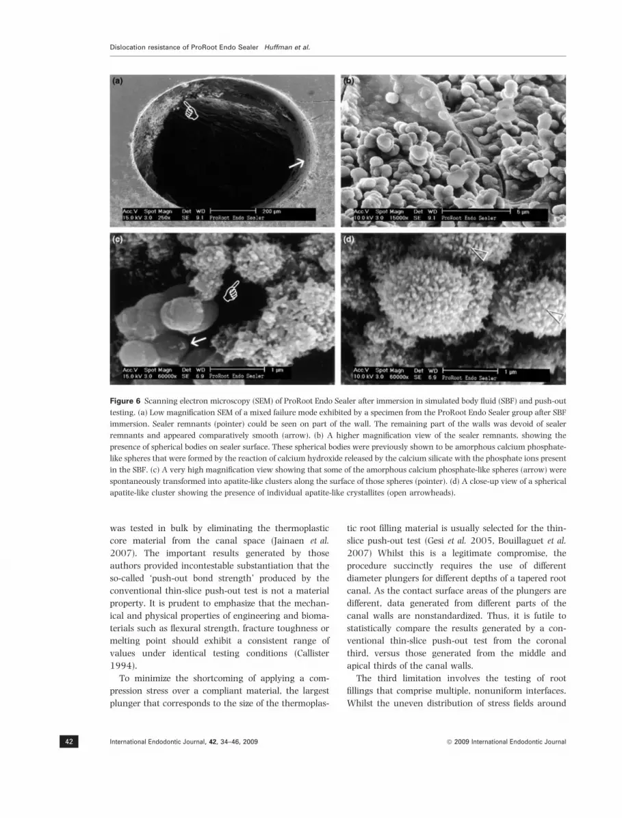
immersion is depicted in Fig. 6a. Spherical bodies were
identified along the sealer–dentin interface as well as
the surface of the fractured sealer (Fig. 6b). These
spherical phases were not observed from fractured
specimens of the same sealer that had not been
immersed in SBF (not shown). Very high magnification
views of the specimens that had been immersed in SBF
Huffman et al. Dislocation resistance of ProRoot Endo Sealer
ª 2009 International Endodontic Journal International Endodontic Journal, 42, 34–46, 2009 39

before testing revealed phase transformation of the
spherical bodies to spherules with clustered polycrys-
talline surfaces (Fig. 6c). Individual crystallites that
protruded from the surface of these spherules were
about 40–70 nm in diameter (Fig. 6d).
Discussion
This study utilized a modified push-out protocol that
was designed specifically to examine the retentive
potential of sealers in radicular dentine. Although the
study design is far removed from clinical practice, the
results indicate that under identical cleaning and
shaping conditions that may not be easily achieved
under a clinical setting, the dislocation resistance of a
particular sealer is independent of the location of the
radicular dentine. Moreover, the dislocation resistance
of the three sealers were significantly different from
each other and that two of the three sealers exhibited
higher dislocation resistance after immersion in SBF.
Thus, the null hypothesis has to be rejected. Although
a modified push-out test design was used in this study,
it is interesting to note that the relatively low push-out
strengths for AH Plus and Pulp Canal Sealer were
similar to the range reported for similar sealers
(2.00 ± 0.65 MPa for AH Plus and 0.79 ± 0.52 MPa
for Kerr EWT sealer) in a previous study (Fisher et al.
2007).
Although testing designs that involve the use of
natural canal spaces have obvious pragmatic appeal to
clinicians, there are severe limitations from a materials
science perspective. First, the application of a compres-
sive load on top of a thermoplastic material, which has
Load-displacement curves of endodontic sealers(a)
(b)
(c)
Push out strength of endodontic sealers
Push out strength of endodontic sealers(pooled results; N = 60)
Displacement (µm)
Ap: Apical thirdMi: Middle thirdCo: Coronal third
Before immersion in SBFAfter immersion in SBF for4 weeks
Before immersion in SBFAfter immersion in SBF for4 weeks
70
60
50
40 I
II
III
IV30
20
10
00 200 400
ProRoot Endo SealerAH Plus Jet Pulp Canal Sealer
600 800 1000
30
25
20
15
10
5
0
Lo
ad (
N)
MP
a
25
20
15
10
5
0Pulp Cancal
SealerAH Plus Jet ProRoot Endo
Sealer
Pulp Cancal Sealer
Ap Mi Co Ap Mi Co Ap Mi CoAH Plus Jet ProRoot Endo
Sealer
3 C
2
B
1A
Dis
loca
tio
n r
esis
tan
ce (
MP
a)
Figure 3 Push-out strength results. (a) Representative load-
displacement curves of the three sealers that were tested in
bulk without an accompanying gutta-percha core. Load is
expressed as Newtons (N) and displacement is expressed as
microns (lm). Zone I: initial linear increase in load; zone II:
change in slope of the load–displacement curve before
reaching maximum load; zone III: initial sudden sharp drop
in recorded load upon interfacial delamination; zone IV: final
push-out phase. When magnified, the four regions described
for ProRoot Endo Sealer could also be seen in the load–
displacement curves of AH Plus Jet and Pulp Canal Sealer. (b)
dislocation resistance (expressed as MPa) of the three sealers in
the apical third (Ap), middle third (Mi) and coronal third (Co)
of the root dentine with and without storage in a simulated
body fluid (SBF) (n = 20/location/storage subgroup). As there
were no statistical differences in the push-out strengths of each
sealer amongst different locations at each time period, data
from the three locations were pooled (n = 60) for subsequent
statistical comparisons. (c) The pooled data was analysed
using a two-way anova design with sealer type and SBF
storage as independent variables. For specimens tested without
SBF immersion, sealers with different numerals above their
corresponding data columns represent significant differences
(P < 0.001). For specimens that were tested after they were
immersed in SBF, sealers with different upper case letters
above their corresponding data columns represent significant
differences (P < 0.001). For each sealer type, a horizontal bar
above the respective columns for the two immersion protocols
indicates no statistical difference (P > 0.05).
Dislocation resistance of ProRoot Endo Sealer Huffman et al.
International Endodontic Journal, 42, 34–46, 2009 ª 2009 International Endodontic Journal40

the tendency to flow during testing generates results
that are susceptible to erroneous interpretation. Unless
the rheological properties of the materials being com-
pressed are equivalent (Kohyama et al. 2003, Tornqvist
et al. 2004), statistical comparison of the results
derived from two thermoplastic root filling materials
is virtually meaningless. This could also have been
responsible, in part, for the recent report that sealers
tested in thin films using the thin-slice push-out test
were considerably weaker than when the same sealer
Failure modes of push out tests
Before immersionin SBF
PulpCanalSealer
AH PlusJet
ProRootEndoSealer
61.7%38.3%
20%
80%
6.7%
36.6%
56.7%74.1%
8.6%17.3%
100% 95%
5%
After immersion in SBFfor 4 weeks
Adhesive Mixed Cohesive
Figure 4 Distribution of adhesive, mixed
and cohesive failures of the three sealers
in specimens that were tested without
immersion in simulated body fluid (SBF)
and specimens that were tested after
immersion in SBF for 4 weeks.
Figure 5 Scanning electron microscopy (SEM) of AH Plus after immersion in simulated body fluid (SBF) and push-out testing. (a)
Low magnification SEM of a cohesive failure mode exhibited by a specimen from the AH Plus Jet group after SBF immersion. (b) A
higher magnification view showing the presence of large, multi-facet fillers (open arrowheads) that are characteristic of the AH
Plus sealer. These fillers were embedded in a resinous matrix together with other fine filler particles.
Huffman et al. Dislocation resistance of ProRoot Endo Sealer
ª 2009 International Endodontic Journal International Endodontic Journal, 42, 34–46, 2009 41
was tested in bulk by eliminating the thermoplastic
core material from the canal space (Jainaen et al.
2007). The important results generated by those
authors provided incontestable substantiation that the
so-called ‘push-out bond strength’ produced by the
conventional thin-slice push-out test is not a material
property. It is prudent to emphasize that the mechan-
ical and physical properties of engineering and bioma-
terials such as flexural strength, fracture toughness or
melting point should exhibit a consistent range of
values under identical testing conditions (Callister
1994).
To minimize the shortcoming of applying a com-
pression stress over a compliant material, the largest
plunger that corresponds to the size of the thermoplas-
tic root filling material is usually selected for the thin-
slice push-out test (Gesi et al. 2005, Bouillaguet et al.
2007) Whilst this is a legitimate compromise, the
procedure succinctly requires the use of different
diameter plungers for different depths of a tapered root
canal. As the contact surface areas of the plungers are
different, data generated from different parts of the
canal walls are nonstandardized. Thus, it is futile to
statistically compare the results generated by a con-
ventional thin-slice push-out test from the coronal
third, versus those generated from the middle and
apical thirds of the canal walls.
The third limitation involves the testing of root
fillings that comprise multiple, nonuniform interfaces.
Whilst the uneven distribution of stress fields around
Figure 6 Scanning electron microscopy (SEM) of ProRoot Endo Sealer after immersion in simulated body fluid (SBF) and push-out
testing. (a) Low magnification SEM of a mixed failure mode exhibited by a specimen from the ProRoot Endo Sealer group after SBF
immersion. Sealer remnants (pointer) could be seen on part of the wall. The remaining part of the walls was devoid of sealer
remnants and appeared comparatively smooth (arrow). (b) A higher magnification view of the sealer remnants, showing the
presence of spherical bodies on sealer surface. These spherical bodies were previously shown to be amorphous calcium phosphate-
like spheres that were formed by the reaction of calcium hydroxide released by the calcium silicate with the phosphate ions present
in the SBF. (c) A very high magnification view showing that some of the amorphous calcium phosphate-like spheres (arrow) were
spontaneously transformed into apatite-like clusters along the surface of those spheres (pointer). (d) A close-up view of a spherical
apatite-like cluster showing the presence of individual apatite-like crystallites (open arrowheads).
Dislocation resistance of ProRoot Endo Sealer Huffman et al.
International Endodontic Journal, 42, 34–46, 2009 ª 2009 International Endodontic Journal42

interfaces with variable circumferential thickness can-
not be over-emphasized (Shirazi-Adl & Forcione 1992,
Mequid & Zhu 1995), the uncertainty with respect to
which interface was consistently dislodged imposes
rigorous challenges when specific hypotheses such as
the dislocation resistance of sealers from radicular
dentine are to be tested.
The fourth limitation is that one is almost certain to
find noninstrumented areas that co-exist with instru-
mented areas in an oval-shaped canal that has been
cleaned and shaped (Peters 2004). For the noninstru-
mented areas that are treated with sodium hypochlorite
as an irrigant, one should expect increases in both
undercut retention and surface contact areas within
the calcospherite-containing regions (Wakabayashi
et al. 1993, Tatsuta et al. 1999) that inadvertently
augments the dislocation resistance of the sealer being
investigated. For example, comparing the results gen-
erated from a natural canal space with 50% noninstr-
umented canal walls versus one that has 20%
noninstrumented canal walls may result in erroneous
conclusions on the dislocation resistance of various
sealers from radicular dentine. It is unrealistic to
quantify the extent of noninstrumented natural canal
walls from a root slab either before or after a push-out
test. Because of these limitations, a modified push-out
strength testing design was utilized in the present
study.
Even without SBF immersion, the calcium silicate-
based sealer was approximately 16 times as difficult to
be dislodged from the radicular dentine walls as Pulp
Canal Sealer, and almost four times as resistant to
dislodging as AH Plus Jet. This may be due, in part, to
the hardness of the calcium silicate-based sealer after
setting in the presence of 100% relative humidity. As
natural root canals cannot be completely dehydrated
(Amyra et al. 2000, Hosoya et al. 2000) due to the
retention of moisture within the dentinal tubules,
similar hardness should be expected of the set sealer
when it is used for filling natural canals. The tenacity
of this sealer to radicular dentine cannot be solely
attributed to sealer penetration into the dentinal
tubules following depletion of the smear layer, as the
dentine from the apical third of the roots is often highly
sclerotic. It is beyond the scope of this study to provide
definitive annotations on whether the increased dislo-
cation resistance is caused by the frictional resistance
or micromechanical/chemical adhesion of the sealer to
dentine (Shirazi-Adl 1992, Goracci et al. 2005). This
issue should be further investigated in the future using
more advanced transmission electron microscopy and
chemoanalytical techniques. Nevertheless, the
increased dislocation resistance of the calcium
silicate-based sealer to radicular dentine should be
advantageous in maintaining the integrity of the
sealer–dentine interface during tooth flexure, as well
as during the preparation of postholes within the filled
canal spaces.
The concern on whether the dislocation resistance of
root canal sealers is adversely affected by the contam-
ination of body fluids was simulated in the present
study by immersing the specimens in a SBF. This is an
exaggerated simulation as the entire tooth slab was
immersed in the SBF after the cavities were filled with
sealers. The increase in dislocation resistance of the AH
Plus Jet is probably caused by swelling of the epoxy
resin component after water sorption (Fernandez-Gar-
cıa & Chiang 2002, Domotor & Hentschke 2004). For
the calcium silicate-based sealer, continuous matura-
tion of the material (Andriamanantsilavo & Amziane
2004) may also have increased the material’s disloca-
tion resistance. However, the occurrence of spherical
phases along the sealer–dentine interface and within
the remnant fractured sealer after the specimens were
immersed in the phosphate-containing SBF is notable.
These spherical phases have previously been identified
as amorphous calcium phosphate when Portland
cement was immersed in a phosphate-containing fluid
(Tay et al. 2007). Amorphous calcium phosphate
phases undergo spontaneous transformation to car-
bonated apatites (Gadaleta et al. 1996), producing
hollow spherules of apatite clusters (Eanes 2001, Tay
& Pashley 2008) that contributed to the ex vivo
bioactivity of calcium silicate-containing materials
when they interact with phosphate ions. Similar
apatite-containing clusters had been observed when
Mineral Trioxide Aggregate was immersed in phos-
phate-containing fluids (Sarkar et al. 2005). The apat-
itic composition in these spherules has also been
established using x-ray diffraction (XRD) and Fourier
transform-infrared spectroscopy (FT-IR) (Tay et al.
2007). No attempt was made to analyse the compar-
atively smooth spherical phases and the crystallite-
containing spherules in this study, as these phases were
present adjacent to calcium-phosphate rich dentine and
on the surface of the fractured sealer. The use of energy
dispersive X-ray analysis to analyse these surface
phases would have yielded information that includes
the subsurface elemental composition of the dentine
and sealer components. Likewise, these phases were not
amendable for collection and purification for XRD and
FT-IR analyses. Thus, they are only referred to as
Huffman et al. Dislocation resistance of ProRoot Endo Sealer
ª 2009 International Endodontic Journal International Endodontic Journal, 42, 34–46, 2009 43

amorphous calcium phosphate-like and apatite-like in
the present study. Generation of these reaction phases
only in specimens that were immersed in the SBF could
also have resulted in the increase in frictional resis-
tance of the sealer–dentin interface. Although it is
presumptuous to correlate the ‘in vitro bioactivity’ (i.e.
the ability to form carbonate hydroxyapatite on the
surface of a biomaterial when it is exposed to SBF)
(LeGeros 2002, Zhao et al. 2005, Panzavolta et al.
2008) observed in the present study with ‘clinical
bioactivity’ (i.e. the property of the material to develop
a direct, adherent and strong bonding with the bone
tissue) (Hench et al. 1978, Hench 1994), the issue of
‘clinical bioactivity’ associated with the use of endo-
dontic sealers in general is of practical clinical interest
and should be duly investigated.
Conclusion
Within the limits of the modified push-out testing
design utilized in the present ex vivo study, it may be
concluded that:
• Under identical cleaning and shaping conditions,
the dislocation resistance of ProRoot Endo Sealer, AH
Plus Jet and Pulp Canal Sealer are independent of the
location of the radicular dentine.
• The dislocation resistance of the three sealers are in
descending order: ProRoot Endo Sealer, AH Plus Jet and
Pulp Canal Sealer.
• Both ProRoot Endo Sealer and AH Plus Jet exhibited
higher dislocation resistance after immersion in a SBF.
• ProRoot Endo Sealer exhibited amorphous calcium
phosphate-like phases that spontaneously transformed
into apatite-like phases after immersion in the phos-
phate-containing SBF. This phenomenon probably
accounts for the in vitro bioactivity of this calcium
silicate-based sealer.
Acknowledgements
This study was supported by Dentsply Tulsa Dental
Specialties. Dr Primus and Dr Gutmann served as
consultants for Dentsply Tulsa Dental Specialties. The
authors are grateful to Miss Anna Lam for her
secretarial support.
References
Amyra T, Walsh LT, Walsh LJ (2000) An assessment of
techniques for dehydrating root canals using infrared laser
radiation. Australian Endodontic Journal 26, 78–80.
Andriamanantsilavo NR, Amziane S (2004) Maturation of
fresh cement paste within 1- to 10-m-large formworks.
Cement and Concrete Research 34, 2141–52.
Bouillaguet S, Bertossa B, Krejci I, Wataha JC, Tay FR, Pashley
DH (2007) Alternative adhesive strategies to optimize
bonding to radicular dentin. Journal of Endodontics 33,
1227–30.
Callister WD Jr. (1994) Materials Science and Engineering: an
Introduction, 3rd edn. New York: John Wiley & Sons, pp. 1–5.
Camilleri J, Pitt Ford TR (2006) Mineral trioxide aggregate: a
review of the constituents and biological properties of the
material. International Endodontic Journal 39, 747–54.
Chandra N, Ananth CR (1995) Analysis of interfacial behavior
in MMCs and IMCs using thin slice push-out tests. Compos-
ites Science and Technology 54, 87–100.
Chandra N, Ghonem H (2001) Interfacial mechanics of push-
out tests: theory and experiments. Composites Part A: Applied
Science and Manufacturing 32, 578–84.
De Moor R, Hommez G (2000) The importance of apical and
coronal leakage in the success or failure of endodontic
treatment. Revue Belge de Medecine Dentaire 55, 334–44.
Domotor G, Hentschke R (2004) Equilibrium swelling of an
epoxy-resin in contact with water – a molecular dynamics
simulation study. Macromolecular Theory and Simulations 13,
506–11.
Eanes ED (2001) Amorphous calcium phosphate. In: Chow LC,
Eanes ED, eds. Monographs in Oral Science, Vol. 18, Basel:
Karger, pp. 130–47.
Fernandez-Garcıa M, Chiang MYM (2002) Effect of
hygrothermal aging history on sorption process, swelling,
and glass transition temperature in a particle-filled
epoxy-based adhesive. Journal of Applied Polymer Science
84, 1581–91.
Fisher MA, Berzins DW, Bahcall JK (2007) An in vitro
comparison of bond strength of various obturation materials
to root canal dentin using a push-out test design. Journal of
Endodontics 33, 856–8.
Gadaleta SJ, Paschalis EP, Betts F, Mendelsohn R, Boskey AL
(1996) Fourier transform infrared spectroscopy of the
solution-mediated conversion of amorphous calcium phos-
phate to hydroxyapatite: new correlations between X-ray
diffraction and infrared data. Calcified Tissue International 58,
9–16.
Gancedo-Caravia L, Garcia-Barbero E (2006) Influence of
humidity and setting time on the push-out strength of
mineral trioxide aggregate obturations. Journal of Endodon-
tics 32, 894–6.
Gesi A, Raffaelli O, Goracci C, Pashley DH, Tay FR, Ferrari M
(2005) Interfacial strength of Resilon and gutta-percha to
intraradicular dentin. Journal of Endodontics 31, 809–13.
Gettleman BH, Messer HH, ElDeeb ME (1991) Adhesion of
sealer cements to dentin with and without the smear layer.
Journal of Endodontics 17, 15–20.
Goracci C, Fabianelli A, Sadek FT, Papacchini F, Tay FR,
Ferrari M (2005) The contribution of friction to the
Dislocation resistance of ProRoot Endo Sealer Huffman et al.
International Endodontic Journal, 42, 34–46, 2009 ª 2009 International Endodontic Journal44

dislocation resistance of bonded fiber posts. Journal of
Endodontics 31, 608–12.
Goracci C, Grandini S, Bossu M, Bertelli E, Ferrari M (2007)
Laboratory assessment of the retentive potential of adhesive
posts: a review. Journal of Dentistry 35, 827–35.
Gou Z, Chang J, Zhai W, Wang J (2005) Study on the self-
setting property and the in vitro bioactivity of beta-Ca2SiO4.
Journal of Biomedical Materials Research. Part B, Applied
Biomaterials 73, 244–51.
He RX, Yan SG, Wu LD, Wang XH, Dai XS (2007) Position of
the prosthesis and the incidence of dislocation following
total hip replacement. Chinese Medical Journal (English) 120,
1140–4.
Hench LL (1994) Bioceramics: from concept to clinic. Journal
of the American Ceramics Society 74, 1487–510.
Hench LL, Splinter RJ, Allen WC, Greenlee TK (1978) Bonding
mechanisms at the interface of ceramic prosthetic materials.
Journal of Biomedical Materials Research 2, 117–41.
Hosoya N, Nomura M, Yoshikubo A, Arai T, Nakamura J, Cox
CF (2000) Effect of canal drying methods on the apical seal.
Journal of Endodontics 26, 292–4.
Jainaen A, Palamara JE, Messer HH (2007) Push-out bond
strengths of the dentine-sealer interface with and without a
main cone. International Endodontic Journal 40, 882–90.
Kohyama K, Sasaki T, Dan H (2003) Active stress during
compression testing of various foods measured using a
multiple-point sheet sensor. Bioscience, Biotechnology, and
Biochemistry 67, 1492–8.
Laghios CD, Cutler CW, Gutmann JL (2000) In vitro evidence
that lipopolysaccharide of an oral pathogen leaks from root-
end filled teeth. International Endodontic Journal 33, 333–9.
Lee HS, Noguchi T, Tomosawa F (2002a) Evaluation of the
bond properties between concrete and reinforcement as a
function of the degree of reinforcement corrosion. Cement
and Concrete Research 32, 1313–8.
Lee KW, Williams MC, Camps JJ, Pashley DH (2002b)
Adhesion of endodontic sealers to dentin and gutta-percha.
Journal of Endodontics 28, 684–8.
LeGeros RZ (2002) Properties of osteoconductive biomaterials:
calcium phosphates. Clinical Orthopaedics and Related
Research 395, 81–98.
Madison S, Wilcox LR (1988) An evaluation of coronal
microleakage in endodontically treated teeth. Part III. In
vivo study. Journal of Endodontics 14, 455–8.
Marques JMM (2005) Finite element modelling of the pull-out
test of geosynthetics. In: Onate E, Owen DRJ, eds. VIII
International Conference on Computational Plasticity COMPLAS
VIII. Barcelona: CIMNE, pp. 1–4.
Mequid SA, Zhu ZH (1995) Stress distribution in dissimilar
materials containing inhomogeneities near the interface
using a novel finite element. Finite Elements in Analysis and
Design 20, 283–98.
Mitchell CA, Orr JF, Connor KN, Magill JP, Maguire GR (1994)
Comparative study of four glass ionomer luting cements
during post pull-out tests. Dental Materials 10, 88–91.
Munoz HR, Saravia-Lemus GA, Florian WE, Lainfiesta JF
(2007) Microbial leakage of Enterococcus faecalis after post
space preparation in teeth filled in vivo with RealSeal versus
gutta-percha. Journal of Endodontics 33, 673–5.
Nagas E, Cehreli ZC, Durmaz V, Vallittu PK, Lassila LV (2007)
Regional push-out bond strength and coronal microleakage
of Resilon after different light-curing methods. Journal of
Endodontics 33, 1464–8.
Panitvisai P, Messer HH (1995) Cuspal deflection in molars in
relation to endodontic and restorative procedures. Journal of
Endodontics 21, 57–61.
Panzavolta S, Torricelli P, Sturba L, Bracci B, Giardino R,
Bigi A (2008) Setting properties and in vitro bioactivity
of strontium-enriched gelatin–calcium phosphate bone
cements. Journal of Biomedical Materials Research A 84,
965–72.
Peters OA (2004) Current challenges and concepts in the
preparation of root canal systems: a review. Journal of
Endodontics 30, 559–67.
Saleh IM, Ruyter IE, Haapasalo MP, Ørstavik D (2003)
Adhesion of endodontic sealers: scanning electron micros-
copy and energy dispersive spectroscopy. Journal of End-
odontics 29, 595–601.
Salehrabi R, Rotstein I (2004) Endodontic treatment outcomes
in a large patient population in the USA: an epidemiological
study. Journal of Endodontics 30, 846–50.
Sarkar NK, Caicedo R, Ritwik P, Moiseyeva R, Kawashima I
(2005) Physicochemical basis of the biologic properties of
mineral trioxide aggregate. Journal of Endodontics 31, 97–
100.
Scifert CF, Brown TD, Lipman JD (1999) Finite element
analysis of a novel design approach to resisting total hip
dislocation. Clinical Biomechanics (Bristol, Avon) 14, 6697–
703.
Shirazi-Adl A (1992) Finite element stress analysis of a push-
out test. Part 1: fixed interface using stress compatible
elements. Journal of Biomechanical Engineering 114, 111–8.
Shirazi-Adl A, Forcione A (1992) Finite element stress analysis
of a push-out test. Part II: free interface with nonlinear
friction properties. Journal of Biomechanical Engineering 114,
155–61.
Skidmore LJ, Berzins DW, Bahcall JK (2006) An in vitro
comparison of the intraradicular dentin bond strength of
Resilon and gutta-percha. Journal of Endodontics 32, 963–6.
Sly MM, Moore BK, Platt JA, Brown CE (2007) Push-out bond
strength of a new endodontic obturation system (Resilon/
Epiphany). Journal of Endodontics 33, 160–2.
Sousa-Neto MD, Silva Coelho FI, Marchesan MA, Alfredo E,
Silva-Sousa YT (2005) Ex vivo study of the adhesion of an
epoxy-based sealer to human dentine submitted to irradia-
tion with Er:YAG and Nd:YAG lasers. International Endo-
dontic Journal 38, 866–70.
Tagger M, Tagger E, Tjan AH, Bakland LK (2003) Shearing
bond strength of endodontic sealers to gutta-percha. Journal
of Endodontics 29, 191–3.
Huffman et al. Dislocation resistance of ProRoot Endo Sealer
ª 2009 International Endodontic Journal International Endodontic Journal, 42, 34–46, 2009 45

Tatsuta CT, Morgan LA, Baumgartner JC, Adey JD (1999)
Effect of calcium hydroxide and four irrigation regimens on
instrumented and uninstrumented canal wall topography.
Journal of Endodontics 25, 93–8.
Tay FR, Pashley DH (2008) Guided tissue remineralisation of
partially demineralised human dentine. Biomaterials 29,
1127–37.
Tay FR, Pashley DH, Rueggeberg FA, Loushine RJ, Weller RN
(2007) Calcium phosphate phase transformation produced
by interaction of the Portland cement component of white
MTA with a phosphate-containing fluid. Journal of Endodon-
tics 33, 1347–51.
Teixeira EC, Teixeira FB, Piasick JR, Thompson JY (2006) An
in vitro assessment of prefabricated fiber post systems.
Journal of the American Dental Association 137, 1006–12.
Tilashalski KR, Gilbert GH, Boykin MJ, Shelton BJ (2004) Root
canal treatment in a population-based adult sample: status
of teeth after endodontic treatment. Journal of Endodontics
30, 577–81.
Timpawat S, Harnirattisai C, Senawongs P (2001) Adhesion of
a glass-ionomer root-canal sealer to the root-canal wall.
Journal of Endodontics 27, 168–71.
Tornqvist R, Sunderland P, Manson JAE (2004) Determination
of the rheological properties of thermoplastic composites for
compression flow molding. Polymer Composites 21, 779–88.
Ungor M, Onay EO, Orucoglu H (2006) Push-out bond
strengths: the Epiphany–Resilon endodontic obturation
system compared with different pairings of Epiphany,
Resilon, AH Plus and gutta-percha. International Endodontic
Journal 39, 643–7.
Ureyen Kaya B, Kececi AD, Orhan H, Belli S (2008) Micro-
push-out bond strengths of gutta-percha versus thermo-
plastic synthetic polymer-based systems – an ex vivo study.
International Endodontic Journal 41, 211–8.
Wakabayashi H, Matsumoto K, Nakamura Y, Shirasuka T
(1993) Morphology of the root canal wall and arrangement
of underlying dentinal tubules. International Endodontic
Journal 26, 153–8.
Wang X, Sun H, Chang J (2008) Characterization of
Ca(3)SiO(5)/CaCl(2) composite cement for dental applica-
tion. Dental Materials 24, 74–82.
Weale AE, Feikes J, Prothero D, O’Connor JJ, Murray D,
Goodfellow J (2002) In vitro evaluation of the resistance to
dislocation of a meniscal-bearing total knee prosthesis
between 30� and 90� of knee flexion. Journal of Arthroplasty
17, 475–83.
Wennberg A, Ørstavik D (1990) Adhesion of root canal sealers
to bovine dentine and gutta-percha. International Endodontic
Journal 23, 13–9.
Zhao W, Wang J, Zhai W, Wang Z, Chang J (2005) The self-
setting properties and in vitro bioactivity of tricalcium
silicate. Biomaterials 26, 6113–21.
Dislocation resistance of ProRoot Endo Sealer Huffman et al.
International Endodontic Journal, 42, 34–46, 2009 ª 2009 International Endodontic Journal46


