Embed Size (px)
Citation preview
Disrupted small-world networks in schizophreniaYong Liu1Meng Liang12 Yuan Zhou1 Yong He13 Yihui Hao4 Ming Song1 Chunshui Yu5 Haihong Liu4
Zhening Liu4 and Tianzi Jiang1
1National Laboratory of Pattern Recognition Institute of Automation Chinese Academy of Sciences Beijing 100080 PeoplersquosRepublic of China 2Department of Physiology Anatomy and Genetics University of Oxford Oxford OX13QXUK3McConnell Brain Imaging Centre Montreal Neurological Institute McGill University Montreal Quebec H3A 2B4 Canada4Institute of Mental Health Second Xiangya Hospital Central South University Changsha 410011 Hunan China and5Department of Radiology Xuanwu Hospital of Capital Medical University Beijing 100053 Peoplersquos Republic of China
Correspondence to Tianzi Jiang National Laboratory of Pattern Recognition Institute of Automation Chinese Academy ofSciences Beijing 100080 ChinaE-mail jiangtznlpriaaccn
The human brain has been described as a large sparse complex network characterized by efficient small-worldproperties which assure that the brain generates and integrates informationwith high efficiencyMany previousneuroimaging studies have provided consistent evidence of lsquodysfunctional connectivityrsquo among the brain regionsin schizophrenia however little is known about whether or not this dysfunctional connectivity causes disruptionof the topological properties of brain functional networksTo this end we investigated the topological propertiesof human brain functional networks derived from resting-state functional magnetic resonance imaging (fMRI)Data was obtained from 31 schizophrenia patients and 31healthy subjects then functional connectivity between90 cortical and sub-cortical regions was estimated by partial correlation analysis and thresholded to construct aset of undirected graphs Our findings demonstrated that the brain functional networks had efficient small-world properties in the healthy subjects whereas these properties were disrupted in the patients withschizophrenia Brain functional networks have efficient small-world properties which support efficient parallelinformation transfer at a relatively low cost More importantly in patients with schizophrenia the small-worldtopological properties are significantly altered in many brain regions in the prefrontal parietal and temporallobes These findings are consistent with a hypothesis of dysfunctional integration of the brain in this illnessSpecifically we found that these altered topologicalmeasurements correlate with illness duration in schizophre-nia Detection and estimation of these alterations could prove helpful for understanding the pathophysiologicalmechanism as well as for evaluation of the severity of schizophrenia
Keywords efficient small-world brain functional networks functional connectivity resting-state fMRI schizophrenia
Abbreviations BOLD=blood oxygenation level dependent EPI=echo planar imaging fMRI= functional magneticresonance imaging PANSS=Positive and Negative Syndrome Scale
Received September 14 2007 Revised January 4 2008 Accepted January 25 2008 Advance Access publication February 25 2008
IntroductionThe human brain has evolved to support rapid real-timeintegration of information across segregated sensory brainregions (Sporns and Zwi 2004) to confer resilience againstpathological attack (Achard et al 2006) and to maximizeefficiency at a minimal cost for effective information pro-cessing between different brain regions (Achard andBullmore 2007) Small-world networks offer a structuralsubstrate for functional segregation and integration of thebrain (Sporns and Zwi 2004) and facilitate rapid adaptivereconfiguration of neuronal assemblies in support ofchanging cognitive states (Bassett and Bullmore 2006)
Efficiency provides a vital measure of how well informationis transformed over a network (Achard and Bullmore 2007)The combination of these factors makes efficient small-worldtopology an attractive model for brain functional networks
In terms of the pathophysiology of schizophreniadysfunctional connectivity has been hypothesized to be thepathophysiological mechanism of cognitive dysfunctionWidely distributed dysfunctional connectivity such asfrontal-frontalfronto-temporal disconnections (Friston andFrith 1995 Andreasen et al 1998 Tan et al 2006) reducedconnectivity between the fronto-parietal (Paulus et al 2002Kim et al 2003) occipito-temporal (Kim et al 2005) and
doi101093brainawn018 Brain (2008) 131 945^961
The Author (2008) Published by Oxford University Press on behalf of the Guarantors of Brain All rights reserved For Permissions please email journalspermissionsoxfordjournalsorg
dorsolateral prefrontal-anterior cingulate (Spence et al 2000)have been reported Also disrupted interregional connectivitywithin the cortico-cerebellar-thalamo-cortical circuit (Honeyet al 2005) and aberrant connectivity within default modenetwork (Bluhm et al 2007 Garrity et al 2007 Zhou et al2007b) have been reported The disruptions of interregionalbrain connectivity may lead to the failure of functionalintegration within the brain in schizophrenia This failuremay partially account for the deficits in cognition andbehaviour of schizophrenia patients So far however little isknown about changes in the globallocal structure of the brainfunctional network in schizophrenia except for the results oftwo recent studies using fMRI (Liang et al 2006a) and EEGdata (Micheloyannis et al 2006a) Liang et al (2006a)suggested altered small-world properties in schizophreniabased on resting-state fMRI data However a key problemwith that study is that only two networks (one for each group)were constructed thus the results were descriptive and nostatistical conclusion was able to be drawn Micheloyanniset al (2006a) reported disrupted small-world properties ofbrain networks in different bands of EEG signals inschizophrenia Although EEG supplies a high temporalresolution it cannot reveal information about the exactactivities of specific sub-cortical brain regions thus EEGscannot be used to construct a complete brain networkTo investigate directly the hypothesis that the brain
network of schizophrenia is characterized by disruption ofefficient small-world topological properties based on resting-state fMRI data we divided the cerebrum into 90 brainregions Functional connectivities were then estimated bycalculating the partial correlation between the mean timeseries of each pair of brain regions for each subject Theresulting partial correlation matrices were thresholded togenerate a set of undirected binary graphs Topologicalparameters of brain networks were evaluated as a function ofconnectivity threshold T and the degree of connectivity KStatistical analyses were performed to explore the differencesbetween patients and healthy subjects Pearsonrsquos correla-tion coefficients between these topological properties andclinical variables were used to evaluate the relationship inschizophrenia
Materials and MethodsSubjectsThe study included 31 patients with schizophrenia (mean age of24 years) who were recruited from the Institute of Mental HealthSecond Xiangya Hospital China Confirmation of the diagnosis forall patients was made by clinical psychiatrists using the StructuredClinical Interview for DSM-IV Patient Version (First et al 1995)During the time of the experiments trained and experiencedpsychiatrists assessed the symptoms of these patients using thePositive and Negative Syndrome Scale (PANSS) The meantreatment was 442mg chlorpromazine-equivalent antipsychotic(21 subjects were receiving atypical antipsychotic medications and10 were not receiving any medical treatment at the time ofexamination) (Table 1) Thirty-one age and gender-matched
healthy subjects were recruited from similar geographic anddemographic regions (Table 1)All subjects were right-handed The exclusion criteria for all the
subjects were as follows no history of neurological or significantphysical disorders no history of alcohol or drug dependence and
no history of receiving electroconvulsive therapy All the healthysubjects had no history of psychiatric illness Some of these subjectshave been used in the previous studies (Liang et al 2006a bZhou et al 2007a b) All subjects gave voluntary and informedconsent according to the standards set by the Ethics Committee ofthe Second Xiangya Hospital Central South University
Data acquisition and preprocessingImaging was performed on a 15 Tesla GE scanner in the SecondXiangya Hospital Blood oxygenation level dependent (BOLD)
images of the whole brain using an echo planar imaging (EPI)sequence were acquired in 20 axial slices (TR= 2000ms TE= 40msflip angle = 90 FOV=24 cm 5mm thickness and 1mm gap) ThefMRI scanning was done in darkness All the subjects wereinstructed to keep their eyes closed not to think about anything
in particular and to move as little as possible For each subject thefMRI scanning lasted 6min Structural sagittal images wereobtained using a magnetization prepared rapid acquisition gradientecho three-dimensional T1-weighted sequence for each subject(TR= 2045ms TE= 96ms flip angle = 90 FOV=24 cm)Unless specifically stated otherwise all the preprocessing was
carried out using statistical parametric mapping (SPM2 httpwwwfilionuclacukspm) To allow for magnetization equilib-rium the first 10 images were discarded The remaining 170 imageswere first corrected for the acquisition time delay among differentslices and then the images were realigned to the first volume for
head-motion correction The time course of head motions wasobtained by estimating the translations in each direction and therotations in angular motion about each axis for each of the 170consecutive volumes All the subjects included in this studyexhibited a maximum displacement of less than 15mm at eachaxis and an angular motion of less than 15 for each axis We also
evaluated the group differences in translation and rotation of headmotion according to the following formula
HeadMotion=Rotation frac14
1
M 1
XMifrac142
ffiffiffiffiffiffiffiffiffiffiffiffiffiffiffiffiffiffiffiffiffiffiffiffiffiffiffiffiffiffiffiffiffiffiffiffiffiffiffiffiffiffiffiffiffiffiffiffiffiffiffiffiffiffiffiffiffiffiffiffiffiffiffiffiffiffiffiffiffiffiffiffiffiffiffiffiffiffijxi xi1j
2 thorn jyi yi1j2 thorn jzi zi1j
2
q
where M is the length of the time series (M=170) in this studyxi yi and zi are translationsrotations at the ith time point in
Table 1 Demographic and clinical details of the subjects
Controls(n=31)
Schizophrenia(n=31)
P-value
Gender (male) 16 17 08a
Age (years) 26 4 24 6 020b
Duration of illness (months) ^ 27 24 ^Medication dose (mg) ^ 442 208c ^PANSS ^ 8320 ^
aThe P-value was obtained by Pearson Chi-squarebThe P-value was obtained by two-sample two-tailed t-testcChlorpromazine equivalent excluding 10 non-medications
946 Brain (2008) 131 945^961 Y Liu et al
the x y and z directions respectively (Liang et al 2006b) The resultsshowed that the two groups had no significant differences in headmotion (two sample two-tailed t-test P= 059 for translationalmotion and P= 055 for rotational motion) The fMRI images werefurther spatially normalized to the Montreal Neurological Institute(MNI) EPI template and resampled to a 3mm cubic voxel Finallytemporal band-pass filtering (0015f5008Hz) was performed inorder to reduce the effects of low-frequency drift and high-frequencynoise (Fox et al 2005 Liang et al 2006b Liu et al 2007)
Anatomical parcellationThe registered fMRI data were segmented into 90 regions (45 for eachhemisphere Table 2) using the anatomically labelled templatereported by Tzourio-Mazoyer et al (2002) which has been used inseveral previous studies (Salvador et al 2005a b Achard et al 2006Achard and Bullmore 2007 Liang et al 2006a b Liu et al 2007) Foreach subject the representative time series of each individual regionwas then obtained by simply averaging the fMRI time series over allvoxels in this region Each regional mean time-series was furthercorrected for the effect of head movement on the partial correlationcoefficients by regression on the translations and rotations of the headestimated in the procedure of image realignment The residuals ofthese regressions constituted the set of regional mean time-series usedfor undirected graph analysis (Salvador et al 2005b)
Estimation of the interregional partialcorrelationsFunctional connectivity examines interregional correlations inneuronal variability (Friston et al 1993) Partial correlation canbe used as a measure of the functional connectivity between a given
pair of regions by attenuating the contribution of other sourcesof covariance (Whittaker 1990 Hampson et al 2002) In this casewe used partial correlations to reduce indirect dependencies byother brain areas and built undirected graphs Given a set of Nrandom variables the partial correlation matrix is a symmetricmatrix in which each off-diagonal element is the correlationcoefficient between a pair of variables after filtering out thecontributions of all other variables included in the dataset In thepresent study therefore the partial correlation between any pair ofregions filters out the effects of the other 88 brain regions (Salvadoret al 2005a)The first step is to estimate the sample covariance matrix S from
the data matrix Y= (xi)i=1 90 of observations for each individualHere xi is the mean time series of each brain region If we introduceX= (xj xk) to denote the average over time of the observations inthe jth and kth regions Z=YX denotes the other 88 mean timeseries matrices Each component of S contains the samplecovariance value between two regions (say j and k) If thecovariance matrix of [XZ] is
S frac14S11 S12
ST12 S22
in which S11 is the covariance matrix of X S12 is the covariancematrix of X and Z and S22 is the covariance matrix of Z then thepartial correlation matrix of X controlling for Z can be definedformally as a normalized version of the covariance matrix
Sxy frac14 S11 S12S122 S
T12
Finally a Fisherrsquos r-to-z transformation is used on the partialcorrelation matrix in order to improve the normality of the partialcorrelation coefficients
Table 2 Cortical and sub-cortical regions defined in Automated Anatomical Labeling template image in standardstereotaxic space
Region name Abbreviation Region name Abbreviation
Superior frontal gyrus dorsolateral SFGdor Superior parietal gyrus SPGSuperior frontal gyrus orbital SFGorb Paracentral lobule PCLSuperior frontal gyrus medial SFGmed Postcentral gyrus PoCGSuperior frontal gyrus medial orbital SFGmorb Inferior parietal gyrus IPGMiddle frontal gyrus MFG Supramarginal gyrus SMGMiddle frontal gyrus orbital MFGorb Angular gyrus ANGInferior frontal gyrus opercular IFGoper Precuneus PCNUInferior frontal gyrus triangular IFGtri Posterior cingulate gyrus PCCInferior frontal gyrus orbital IFGorbGyrus rectus REG Insula INSAnterior cingulate gyrus ACC Thalamus THAOlfactory cortex OLF
Superior temporal gyrus STGPrecentral gyrus PreCG Superior temporal gyrus temporal pole STGpSupplementary motor area SMA Middle temporal gyrus MTGRolandic operculum ROL Middle temporal gyrus temporal pole MTGpMedian- and para-cingulate gyrus MCC Inferior temporal gyrus ITG
Heschl gyrus HESCalcarine fissure and surrounding cortex CAL Hippocampus HIPCuneus CUN Parahippocampal gyrus PHIPLingual gyrus LING Amygdala AMYGSuperior occipital gyrus SOGMiddle occipital gyrus MOG Caudate nucleus CAUInferior occipital gyrus IOG Lenticular nucleus putamen PUTFusiform gyrus FG Lenticular nucleus pallidum PAL
Disrupted small-world networks Brain (2008) 131 945^961 947
Graph theoretical analysisTopological properties of the brain functional networksAn NN (N=90 in the present study) binary graph G consistingof nodes (brain regions) and undirected edges (functionalconnectivity) between nodes can be constructed by applying acorrelation threshold T (Fisherrsquos r-to-z) to the partial correlationcoefficients
eij frac141 if jzethi jTHORNj T0 otherwise
That is if the absolute z(i j) (Fisher r-to-z of the partial correlationcoefficient) of a pair of brain regions i and j exceeds a giventhreshold T an edge is said to exist otherwise it does not existWe define the subgraph Gi as the set of nodes that are the directneighbours of the ith node ie directly connected to the ithnode with an edge The degree of each node Kii=12 90 is definedas the number of nodes in the subgraph Gi The degree ofconnectivity Kp of a graph is the average of the degrees of all thenodes in the graph
Kp frac141
N
Xi2G
Ki
which is a measure to evaluate the degree of sparsity of a networkThe total number of edges in a graph divided by the maximumpossible number of edges N(N 1)2
Kcost frac141
NethN 1THORN
Xi2G
Ki
is called the cost of the network which measures how expensive itis to build the network (Latora and Marchiori 2003) The con-nectivity strength of the ith node is
Ei corr frac141
Ki
Xj2Gi
jzethi jTHORNj eij
Ei_corr is a measure of the strength of the functional connectivitybetween the ith node and the nodes in the subgraph Gi Thestrength of the functional connectivity of a graph is
Ecorr frac141
N
Xi2G
Ei corr
The larger the Ei_corr the stronger the functional connectivity of thebrain functional networkThe absolute clustering coefficient of a node is the ratio of the
number of existing connections to the number of all possibleconnections in the subgraph Gi
Ci frac14Ei
KiethKi 1THORN=2
where Ei is the number of edges in the subgraph Gi (Watts andStrogatz 1998 Strogatz 2001) The absolute clustering coefficientof a network is the average of the absolute clustering coefficients ofall nodes
Cp frac141
N
Xi2G
Ci
Cp is a measure of the extent of the local density or cliquishness ofthe network
The mean shortest absolute path length of a node is
Li frac141
N 1
Xi 6frac14j2G
minfLi jg
in which min Li j is the shortest absolute path length between theith node and the jth node and the absolute path length is thenumber of edges included in the path connecting two nodesThe mean shortest absolute path length of a network is the averageof the shortest absolute path lengths between the nodes
Lp frac141
N
Xi2G
Li
Lp is a measure of the extent of average connectivity or overallrouting efficiency of the networkCompared with random networks small-world networks have
similar absolute path lengths but higher absolute clustering coeffi-cients that is frac14 Creal
p =Crandp gt 1 frac14 Lreal
p =Lrandp 1 (Watts and
Strogatz 1998) These two conditions can also be summarized into ascalar quantitative measurement small-worldness = which istypically 41 for small-world networks (Achard et al 2006Humphries et al 2006 He et al 2007) To examine the small-world properties the values of Creal
p and Lrealp of the functional brain
network need to be compared with those of random networks Thetheoretical values of these two measures for random networks areCrand
p frac14 K=N and Lrandp lnethNTHORN=lnethKTHORN (Achard et al 2006 Bassett
and Bullmore 2006 Stam et al 2007) However as suggested byStam et al (2007) statistical comparisons should generally beperformed between networks that have equal (or at least similar)degree sequences however theoretical random networks haveGaussian degree distributions that may differ from the degreedistribution of the brain networks that we discovered in this studyTo obtain a better control for the functional brain networkswe generated 100 random networks for each K and threshold Tof each individual network by a Markov-chain algorithm (Maslovand Sneppen 2002 Milo et al 2002 Sporns and Zwi 2004)In the original matrix if i1 was connected to j1 and i2 was con-nected to j2 for random matrices we removed the edge betweeni1 and j1 but added an edge between i1 and j2 That means thata pair of vertices (i1 j1) and (i2 j2) was selected forwhich ei1j1 frac14 1 ei2j2 frac14 1 ei1 j2 frac14 0 and ei2 j1 frac14 0 Thenei1j1 frac14 0 ei2j2 frac14 1 ei1 j2 frac14 1 and ei2j1 frac14 0 Then we randomlypermuted the matrix which assured that the random matrix hadthe same degree distribution as the original matrix This procedurewas repeated until the topological structure of the original matrixwas randomized (Achard et al 2006) Then we averaged across all100 generated random networks to obtain a mean Crand
p and a meanLrandp for each degree K and threshold T
Efficiency of small-world brain networksIt has been shown that brain functional networks have efficientsmall-world properties which support the efficient transfer ofparallel information at a relatively low cost (Achard and Bullmore2007) Eglobal a measure of the global efficiency of parallel infor-mation transfer in the network is defined by the inverse of theharmonic mean of the minimum absolute path length between eachpair of nodes (Latora and Marchiori 2001 2003 Achard andBullmore 2007)
Eglobal frac141
NethN 1THORN
Xi 6frac14j2G
1
Li j
948 Brain (2008) 131 945^961 Y Liu et al
We can calculate the local efficiency of the ith node
Ei local frac141
NGiethNGi
1THORN
Xj k2Gi
1
Lj k
In fact since the ith node is not an element of the subgraph Gi thelocal efficiency can also be understood as a measure of the faulttolerance of the network indicating how well each subgraphexchanges information when the index node is eliminated (Achardand Bullmore 2007) In addition based on its definition it is ameasure of the global efficiency of the subgraph Gi The mean localefficiency of a graph Elocal frac14 eth1=NTHORN
Pi2G Ei local is the mean of all
the local efficiencies of the nodes in the graph We can also calculatethe global efficiency (Eglobal) and local efficiency (Elocal) as a functionof KcostTable 3 presents the measurements we have introduced and
illustrates their meaning in human brain functional networks
Statistical analysisStatistical comparisons of Ecorr C
realp Lreal
p Eglobal and Elocalbetween the two groups were performed by using a two-sampletwo-tailed t-test for each value over a wide range of T or K (Kcost)For each selected threshold value we also computed the meandegree of each subject and used a two-sample two-tailed t-test todetermine if the degree of connectivity was significantly differentbetween the two groups If any change in the topological propertieswas found between the two groups we investigated the distributionof the regions which showed significant differences in thesetopological properties
Relationship between topological measuresand clinical variablesWe used Pearsonrsquos correlation coefficient to evaluate the relation-ship between the topological properties (Ecorr C
realp Lreal
p Eglobal
and Elocal) of the brain functional networks and various clinicalvariables (illness duration PANSS scores and medication doses) foreach T or K in the schizophrenia group Because these analyses wereexploratory in nature we used a statistical significance level ofP5005 (uncorrected)
ResultsDirect comparisons between schizophreniaand healthy subjectsThe mean functional connectivity matrix of each group wascalculated by averaging the NN (N= 90 in the presentstudy) absolute connection matrix of all the subjects withinthe group In the normal group most of the strongfunctional connectivities (large z-scores) were betweeninter-hemispheric homogenous regions within a lobe andbetween anatomically adjacent brain areas (Fig 1) Thisfunctional connectivity pattern was consistent with manyprevious studies of whole brain functional connectivity in theresting-state (Salvador et al 2005a Achard et al 2006) Theschizophrenia group showed a similar functional connectiv-ity pattern to that of the healthy group however the strengthof the functional connectivity was lower in the schizophreniagroup [F (160) = 1076 P= 0002]
Direct comparisons of all possible connections between thetwo groups were also performed to test the between-groupdifferences We found that the altered functional connectiv-ities are distributed throughout the entire brain which isconsistent with a previous study by Liang et al (2006b)Extended details about the methods and results can be foundin part I of the supplemental material
Efficient small-world properties ofthe two groups
Efficient small-world regime of brain functional networksFollowing the studies by Stam and colleagues (2007) weinvestigated the topological properties of brain functionalnetworks as a function of T or K (Kcost) Clearly the choice ofa threshold value will have a major effect on the topologicalproperties of the resulting networks conservative thresholdsT 1 will generate sparsely connected graphs (with smallKp more lenient thresholds T 0 will generate moredensely connected graphs (with large Kp for T= 0 Kp= 90Creal
p frac14 Lrealp frac14 1) inevitably including a number of edges
representing spurious or statistically non-significant correla-tions between regions In the present study we adopted thefollowing complementary approaches to choose thethresholds
(1) We thresholded all matrices using a single conserva-tive threshold chosen to construct a sparse graph withmean degree Kp5 2log N 9 (total number of edgesK5 405) In addition the maximum threshold (T)must also assure that each network is fully connectedwith N= 90 nodes This allowed us to compare the
Table 3 Introduction of measurements and their meaningin the brain functional network
Character Meaning
z(i j) z score of Fisher r-to-z transform of partialcorrelation coefficients
Gi the set of nodes that are nearest neighbors of theith node
Kp degree of connectivity which evaluates the level ofsparseness of a network
Kcost cost of networkEcorr mean z score of a brain functional networkCp clustering coefficient which measures the extent of
a local cluster of the networkLp path length which measures of the extent of
average connectivity of the network frac14 Creal
p =Crandp the ratio of the clustering
coefficients between real and random network frac14 Lreal
p =Lrandp the ratio of the path length between
real and random network = scalar quantitative measurement of the
small-wordness of a networkEglobal a measure of the global efficiency of parallel
information transfer in the networkElocal a measure of the fault tolerance of the network
Disrupted small-world networks Brain (2008) 131 945^961 949
topological properties between the two groups in away that was relatively independent of the size of thenetwork
(2) The minimum threshold must ensure that the brainnetworks have a lower global efficiency and a larger localefficiency compared to random networks with relativelythe same distribution of the degrees of connectivity assuggested by Achard and Bullmore (2007)
We selected the threshold range Tmin4T4Tmax byintersecting the upper criteria Then we repeated the fullanalysis for each value of T in the range Tmin4T4Tmaxwith increments of 0002 However if the topological indicesof the brain functional networks are only computed as afunction of threshold T the results could be influenced bydifferences in the number of edges between the two groups(Stam et al 2007) To control this effect we repeated theanalysis by computing the topological indices of each brainfunctional network as a function of the degree ofconnectivity Kmin4K4Kmax with steps of 022 (edgestepnode = 2090) where the range was determined in amanner similar to that described earlier As shown in Fig S5brain networks were accepted as fully in both groupsif the threshold T4 0316 (Fig S5) brain networkswere accepted as fully connected if the total connectededge 5490 (that is Kcost5eth490=4005THORN 0122) (Fig S6)Consequently we selected the small-world interval as01224Kcost4 0267 (the corresponding degree of connec-tivity threshold is 1094K4 238 and the corresponding
connectivity threshold is 02684T4 0316) (Fig 2B) Sucha range is similar to that used in Achard and Bullmorersquosstudy (2007)
Altered topological properties of brain functionalnetworks in schizophreniaThe distributions of the degree of connectivity and thestrength of the functional connectivity of the brain networkas a function of threshold within each group are shown inFig 3 With an increase in the threshold the degree ofconnectivity decreases whereas the strength of the functionalconnectivity starts to increase because more and more edgeswith lower strength of the functional connectivities are beinglost (providing a corresponding value of z-score4T) Overthe whole range of threshold values (0268ndash0316) the degree(Fig 3A) and strength of functional connectivity (Fig 3B)were significantly lower in the schizophrenia group com-pared with the healthy group The schizophrenia group alsoshowed a significantly lower strength of functional con-nectivity than the healthy group over the whole range of K(Fig 3C)
As shown in Fig 4 the higher threshold resulted in a lowermean absolute clustering coefficient and a longer mean shortestabsolute path length for both groups as expected Over thewhole range of threshold values investigated (0268ndash0316) theabsolute clustering coefficients of the healthy subjects wereslightly higher than those of the schizophrenia group (Fig 4A)For all values of T the absolute path length was significantlylonger in the schizophrenia group compared with the healthy
Fig 1 Mean absolute z-score matrices for normal and schizophrenia group Each figure shows a 90 90 square matrix where thex and y axes correspond to the regions listed inTable 2 and where each entry indicates the mean strength of the functional connectivitybetween each pair of brain regions The diagonal running from the lower left to the upper right is intentionally set to zeroThe z scoreof the functional connectivity is indicated with a coloured bar
950 Brain (2008) 131 945^961 Y Liu et al
subjects (Fig 4B) When looking at the absolute clusteringcoefficients and the absolute path length as a function of K wefound the absolute clustering coefficient increased and theshortest absolute path length decreased with an increase in the
degree K and only the absolute clustering coefficient showed asignificant difference between the two groups (Fig 4C)
The small-world attribute is evident in the brain networksof both groups is significantly greater than 1 while is near
Fig 3 (A) Mean degree of connectivity Krealp and (B) strength of functional connectivity Ecorr for schizophrenic (blue squares) and
healthy (red dots) subjects as a function of threshold T (C) strength of functional connectivity Ecorr for schizophrenic (blue squares)and healthy (red dots) subjects as a function of degree K Error bars correspond to standard error of the mean Black triangles indicatewhere the difference between the two groups is significant (t-test P5005)
Fig 2 Economic small-world human brain functional networks (A) Global and (B) local efficiency for a random graph and brainnetworks as a function of costOn average over all subjects in each group normal (blue line) and schizophrenia (red line) brain networkshave lower efficiency than the limiting cases of random networks (black dashed line) The small-world regime is conservatively defined asthe range of costs 01224Kcost4 0267 for which the global efficiency curve for the normal networks is less than the global efficiencycurve for the random networks
Disrupted small-world networks Brain (2008) 131 945^961 951
the value of 1 over the whole range of T or K Due to the largevariance there were no statistically significant differences inthe values of or between the two groups when the samethreshold was used (Fig 5AndashC) and were significantlydecreased in the patients with schizophrenia when usingthe same K for both groups (Fig 5D and F) Only at a smallnumber of connectivity values was found to showstatistically significant differences between the two groups(Fig 5E) The fact that we found significant differencesbetween the two groups indicates that small-world propertiesare disrupted in patients with schizophreniaGlobal efficiency is a measure of the transfer speed of
parallel information in the brain whereas local efficiency isa measure of the information exchange of each subgraph(Achard and Bullmore 2007) Global efficiency of brainfunctional networks were compared with the parametersestimated in a random graph with the same degree dis-tribution over a range of network costs As expectedefficiency monotonically increased as a function of cost inall networks the random graph had higher globalefficiency and lower local efficiency than the healthy subjectsrsquo(Fig 2A and B) which is consistent with the previous study(Achard and Bullmore 2007) The global and localefficiencies of the brain functional networks in the patients
with schizophrenia were disrupted as a function ofthreshold (Fig 6A and B) And the local efficiencies ofthe brain functional networks in the patients with schizo-phrenia were disrupted as a function of degree of con-nectivity (Fig 6D) However there was no significantdifference in global and local efficiency between the twogroups (Fig 6C)
Distribution of the altered regions in the brainWe used a two-sample two-tailed t-test to detect statisticaldifferences in the small-world topological properties betweenschizophrenic patients and the healthy subjects for each brainregion at each selected threshold (or degree of connectivity)We found the pattern of small-world topological propertiesto be significantly altered in many brain regions in theprefrontal parietal and temporal lobes (Figs S7 and S8)Since all thresholds were similar in their trends with respectto the differences in their small-world properties between theschizophrenic patients and the healthy subjects we havechosen to report only one typical threshold (T= 0316the top of the threshold range) (Fig 7) and degree ofconnectivity (K= 109 the smallest degree in this study)(Fig 8) in the main text Details can be found in part III ofthe Supplementary Material
Fig 4 (A) Mean absolute clustering coefficient Crealp and (B) absolute path length Lreal
p for schizophrenic (blue squares) and healthy(red dots) subjects as a function forT (C) Mean absolute clustering coefficient C real
p and (D) absolute path length Lrealp for schizophrenic
(blue squares) and healthy (red dots) subjects as a function of K Error bars correspond to standard error of the mean Black trianglesindicate where the difference between the two groups is significant (t-test P5005)
952 Brain (2008) 131 945^961 Y Liu et al
Relationship between the altered topologicalmeasurements and the clinical variablesWe found that the degree of connectivity Ecorr Kp C
realp
Eglobal and Elocal were negatively correlated however Lrealp was
positively correlated with illness duration when thesemeasurements were computed as a function of each threshold(Fig S9) We show this pattern at a typical threshold T= 0316in Fig 9 A similar pattern was found when the relationshipbetween these topological measurements and the clinicalvariables was investigated as a function of the degrees ofconnectivity (Figs 10 and S10) (For further detail please seeFigs S9 and S10 of part IV in the Supplementary Material)
DiscussionIn the present study resting-state fMRI data were used toconstruct functional brain networks in schizophrenic andhealthy subjects We thresholded the partial correlationmatrices to construct a set of undirected binary graphs foreach subject and compared the topological properties ofbrain functional networks between the two groups Thebrain functional networks of the healthy subjects showedefficient small-world structure which is consistent withseveral previous studies (Stam 2004 Achard et al2006 Bassett and Bullmore 2006 Bassett et al 2006Achard and Bullmore 2007) Nevertheless the patients with
Fig 5 (A) (B) and (C) for schizophrenic (blue squares) and healthy (red dots) subjects as a function of threshold (D) (E) and(F) for schizophrenic (blue squares) and healthy (red dots) subjects as a function of K Error bars correspond to standard error of themean Black triangles indicate where the difference between the two groups is significant (t-test P5005)
Disrupted small-world networks Brain (2008) 131 945^961 953
schizophrenia showed disturbed topological properties suchas a lower degree of connectivity a lower strength of con-nectivity a lower absolute clustering coefficient and a longerabsolute path length compared with those of healthysubjects All these findings support the hypothesis thatschizophrenia is a disorder of dysfunctional integrationamong large distant brain regions (Bullmore et al 19971998 Friston 2005) These results are consistent with aprevious EEG study in which disrupted small-world proper-ties were found in the alpha beta and gamma bands duringresting-state and working memory tasks in patients withschizophrenia (Micheloyannis et al 2006a) More impor-tantly we found that the topological measurements ofefficient small-world brain functional networks were corre-lated with illness duration in schizophrenia
Disrupted efficient small-world propertiesin schizophrenia
Efficient small-world brain networkThe human brain is a large dynamic functional network withan economical small-world architecture and is characterizedby high local clustering of connections between neighbouring
nodes but with short path lengths between any pair of nodes(Sporns and Honey 2006 Achard and Bullmore 2007)Functional segregation and integration are two majororganizational principles of the human brain In otherwords an optimal brain requires a suitable balance betweenlocal specialization and global integration of brain functionalactivity (Tononi et al 1998) This is supported by higherabsolute clustering coefficients (an index of functionalsegregation) and shorter absolute path length (an index offunctional integration) in the functional brain networksof healthy subjects
The small-world attributes reflect the need of thefunctional brain network to satisfy the opposing demandsof local and global processing (Kaiser and Hilgetag 2006)This issue is supported by many previous studies based onMEG (Stam 2004) EEG (Micheloyannis et al 2006b) andfMRI (Eguiluz et al 2005 Salvador et al 2005a Achardet al 2006) In the present study we found that the restingbrain functional network of the healthy subjects had salientsmall-world properties (Figs 5 and 6) Our results are in linewith those of several previous brain network studies (Hilgetaget al 2000 Stam 2004 Salvador et al 2005a Achard et al2006 Micheloyannis et al 2006a Stam et al 2007) (Table 4)
Fig 6 (A) Mean global efficiency Eglobal and (B) local efficiency Elocal for schizophrenic (blue squares) and healthy (red dots) subjects asa function of T (C) Mean global efficiency Eglobal and (D) local efficiency Elocal for schizophrenic (blue squares) and healthy (red dots)subjects as a function of degree Error bars correspond to standard error of the mean Black triangles indicate where the differencebetween the two groups is significant (t-test P5005)
954 Brain (2008) 131 945^961 Y Liu et al
Disrupted efficient small-world characters inschizophreniaMore importantly over a wide range of thresholds and the
degree of connectivity K the absolute clustering coefficients
Crealp showed significantly smaller values in schizophrenia
implying relatively sparse local connectedness of the brain
functional networks in schizophrenia (Micheloyannis et al
2006a) In addition the strength of functional connectivity
showed significantly lower values in patients with schizo-
phrenia This means that the extent of local connectedness
decreased in the schizophrenia group Information interac-
tions between interconnected brain regions are believed to be a
basis of human cognitive processes (Pastor et al 2000
Horwitz 2003 Stam et al 2007) Short absolute path lengths
have been demonstrated to promote effective interactions
between and across different cortical regions (Bassett and
Bullmore 2006 Achard and Bullmore 2007) The longer
absolute path lengths may indicate that information interac-
tions between interconnected brain regions are slower and
less efficient in schizophrenia Thus the lower degree of
Table 4 Small-world properties of brain networks shownin the present study and previous studies
Present studya Healthy subjects 157 102Schizophrenia patients 151 102
Hilgetag et al (2000) Healthy subjects 158 107Cat whole cortex 199 107
Stam (2004)b Healthy subjects 189 119Salvador et al (2005a) Healthy subjects 208 109Archard et al (2006)c Healthy subjects 238 108Stam et al (2007)d AD patients 16 112
Healthy subjects 158 107Micheloyannis et al (2006a)e Healthy subjects 195 119
Schizophrenia patients 182 113
aData are shown for the same K=109 (N=90)bData are shown for the gamma band of EEG signals and K=20cThe mean cluster coefficients and the minimum path length werecomputed from the 003^006Hz resting state fMRI signalsdData are shown for the beta band of EEG signals and N=21 K=3eData are shown for the theta band of EEG signals in a workingmemory task at K=5 (N=28)
Fig 7 Distribution of brain regions in which small-world properties [(A) Krealp (B) Ecorr (C) Creal
p (D) Lrealp (E) Eglobal and (F) Elocal] altered
significantly in healthy subjects (black) and schizophrenia patients (white) at a selected threshold T (T=0316) Bars indicate that the areashows significant altered in the relative measurement and the length of the bar indicates the mean value of the relative measurementbetween the two groups
Disrupted small-world networks Brain (2008) 131 945^961 955
connectivity the lesser strength of connectivity the lowerabsolute clustering coefficients and the longer absolute pathlength as a function of T or K indicate a dysfunctionalorganization of the brain functional network in schizophreniaWe found that the values did not show significant
differences between groups when the same thresholds wereapplied to both groups and at some thresholds the evenshowed lower values in healthy subjects which may be due tothe higher degree of the corresponding random networks forhealthy subjects (Stam et al 2007) To control this effect wecompared the values between the two groups as a function ofK ie each subject has the same number of edges for bothgroups The results showed that schizophrenia patients had asignificantly lower rsquos compared with healthy subjects at thesame degree Our findings are consistent with the previousfindings of the presence of a small-world brain functionalnetwork in healthy subjects however these topologicalstructures of the brain functional network are disrupted inschizophrenia (Liang et al 2006b Micheloyannis et al 2006a)
Our results (Figs 3 and 4) are in line with a previousEEG study in which schizophrenic patients showed smallerabsolute clustering coefficients but relatively longer absolutepath lengths during the resting-state Moreover it should benoted that the reduction of the absolute clustering coefficientfor schizophrenia patients in the earlier EEG study is largerthan in our study (Micheloyannis et al 2006a) The mostlikely explanation for this difference is a longer duration ofthe illness of the patients in the study by Micheloyannis et al(2006a) The mean illness duration was 120 months in thatstudy whereas the mean illness duration of our patientswas 27 months (Table 1) This was also consistent with ourfinding that there was a significant correlation between theabsolute clustering coefficient and the duration of the illnessa longer duration of the illness induced a smaller absoluteclustering coefficient (Figs 7 and 8) Another possible reasonis that differences in data acquisition techniques or networksize could cause changes in the topological properties of thenetwork
Fig 8 Distribution of brain regions in which small-world properties [(A) Krealp (B) Ecorr (C) Creal
p (D) Lrealp (E) Eglobal and (F) Elocal] altered
significantly in healthy subjects (black) and schizophrenia patients (white) at a selected degree of connectivity K (K=109) Bars indicatethat the area shows significant altered in the relative measurement and the length of the bar indicates the mean value of the relativemeasurement between the two groups
956 Brain (2008) 131 945^961 Y Liu et al
Networks with small-world attributes confer resilienceagainst pathological attack and support parallel segregatedand distributed information processing at a relatively highefficiency (Achard and Bullmore 2007) The efficiencymeasure provides us with a precise quantitative analysisof the information transfer among brain regions it alsoindicates that in the neural cortex each region is intermingledwith others allowing a perfect balance between local neces-sities and a wide scope of interactions (Latora and Marchiori2001) Our results not only demonstrate that interregionalrelationships in brain activity are indeed disrupted inschizophrenia but also show for the first time that patientswith schizophrenia have lower efficiency in parallel informa-tion transfer in the brain network (Fig 6) This is consistentwith the increasing evidence that schizophrenia can beconsidered as a disorder of dysfunctional integration amongdifferent brain regionsIn the current study we found that in many brain regions
in the frontal parietal and temporal lobes the small-world
properties were significantly altered (Figs 7 and 8 andFigs S7 and S8) For example we found that the degree ofconnectivity is smaller in many brain areas in the frontal lobein the patient group (Fig 7) which indicates a lowerconnectivity between the regions in this lobe with other brainregions This might lead to longer absolute path lengths inmany regions of the frontal lobes in patients with schizo-phrenia This is consistent with many previous studies inwhich the functional connectivity of the frontal (Fletcheret al 1999a Meyer-Lindenberg et al 2001 Tan et al 2006)parietal (Danckert et al 2004) and temporal lobes (Fletcheret al 1999b Garrity et al 2007) were disturbed in thisdisorder
Relationship between topological measurements andduration of illnessImportantly we found that the topological measurements ofthe small-world brain functional networks were correlatedwith illness duration A longer duration of the illness induced
Fig 9 Scatter plots with trend line showing the topological properties (Krealp Creal
p Lrealp Eglobal Elocal) of the brain functional networks
(open blue squares) as a function of illness duration for a selected T (T=0316) in patients with schizophrenia Pearson correlationcoefficient for Kreal
p (R=0399 P=0026) (A) Crealp (R=0443 P=0013) (B) Lreal
p (R=0426 P=0017) (C) Eglobal (R=0418 P=0020)(D) and Elocal (R=0446 P=0012) (E) were significant
Disrupted small-world networks Brain (2008) 131 945^961 957
a smaller absolute clustering coefficient a lower degree ofconnectivity a lower global and local efficiency and alonger absolute path length of brain functional networks(Figs 9 and 10 and Figs S9 and S10) These findingsappear to demonstrate that the severity of the disruptionof the efficient small-world structure of the brain is relatedto the duration of the illness In addition the significantnegative correlation between the strength of the functionalconnectivity and the chlorpromazine-equivalent medi-cation dosage (Figs 10A and S10) may indicate that inpatients with schizophrenia the more severe the illness theweaker the functional connectivity between the regions ofthe brainThe findings in the present study provide further evidence
for schizophrenia as a disconnection syndrome (Friston andFrith 1995 Bullmore et al 1997 Friston 2002 2005)Detection and estimation of the small-world topologicalmeasurements could help us better understand the patho-physiological mechanism of schizophrenia
Methodological considerations
Effect of morphometric changes on functional connectivityanalysisChanges in the volume of the frontal lobe temporal lobe andtheir sub-regions over the course of illness have been wellstudied in patients with schizophrenia (Giedd et al 1999Bachmann et al 2004 Molina et al 2004 2006 Premkumar
et al 2006) A recent meta-analysis indicated that the leftsuperior temporal gyrus and the left medial temporal lobeare the key regions of structural alteration in patients withschizophrenia (Honea et al 2005) To assess whether thestructural alterations have an influence on altered small-world properties in patients with schizophrenia we analysedour data for structural differences between the two groupsWe divided the grey matter of the entire brain according toan anatomically labelled template and then investigated thedifference at each brain region There were no significantdifferences found in brain regions between the two groups(P5005 FDR corrected) Although other studies havereported such differences (Giedd et al 1999 Bachmannet al 2004 Molina et al 2004 2006 Premkumar et al2006) our failure to detect them does not mean that they donot exist Our analyses and methodologies may havedisguised such changes For example our use of ananatomically labelled template may have caused atrophiedareas to be separated into different but adjacent brainregions making them difficult to detect To further reducethe effects of grey matter atrophy on our results we regressedout the confounding factor of grey matter atrophy and thenconstructed a set of functional brain networks using a widerange of thresholds (or degrees of connectivity) The resultsobtained after eliminating the possible influence of greymatter atrophy were similar to those obtained withouteliminating this influence Extended details can be found inthe part V of the supplemental material
Fig 10 Scatter plots with trend line showing the topological properties (Ecorr Crealp Elocal) of the brain functional networks (open blue
squares) as a function of illness duration for a selected K in patients with schizophrenia Pearson correlation coefficient between Ecorrand medication dosed (R=0470 P=0032) (A) were significant in the medication patients Pearson correlation coefficient for Ecorr(R=0407 P=0023) (B) and Creal
p (R=0528 P=0002) (C) and Elocal (R=0436 P=0014) (D) were significant
958 Brain (2008) 131 945^961 Y Liu et al
Low-frequency fluctuations of resting-state fMRILow frequency (501Hz) fluctuations of resting-state fMRIsignals have been strongly suggested to be neurobiologicallyinteresting and related to spontaneous neural activity (Biswalet al 1995) and endogenousbackground neurophysiologicprocess of the human brain (Raichle and Gusnard 2005Raichle and Mintun 2006) Furthermore resting-state fMRIhas practical advantages for clinical applications because nostimulation and response are required thus it can beperformed easily by subjects especially patients Because ofthis although resting-state fMRI is a relatively youngtechnique many exciting findings have been reported inthe past several years Functional correlation may reflectendogenously coordinated dynamic activity in large-scaleneuronal populations in healthy subjects In addition thealtered patterns of functional connectivity based on resting-state fMRI have been suggested to be pathophysiologicallymeaningful in some diseases (Fox and Raichle 2007) Forthese reasons we believe that it is significant to investigatethe topological properties of the brain functional network ofpatients with schizophrenia based on resting-state fMRI data
Effect of the length of the time seriesIt should also be noted that we used relatively shorter timeseries (170 volumes per subject) compared to severalprevious resting-state fMRI studies (eg 2048 volumes inBullmore and colleaguesrsquo previous studies) (Salvador et al2005a Achard et al 2006) However our results on healthysubjects are compatible with these previous studies (extendeddetails can be found in the part VI in the supplementalmaterial) In addition we compared many earlier resting-state fMRI studies that used either short or long time series(80ndash2048 volumes of resting-state fMRI signals) and foundthat they produced replicable results in normal people(Greicius et al 2003 Fox et al 2005 Salvador et al 2005aAchard and Bullmore 2007) and in some cognitivedisorders such as in Alzheimerrsquos disease (Greicius et al2004 Sorg et al 2007 Wang et al 2007) major depression(Greicius et al 2007) and schizophrenia (Liang et al 2006bZhou et al 2007a b) Consequently this suggests that arelatively small number of volumes may be sufficient to getenough information during rest Nonetheless determiningthe appropriate number of volumes remains an interestingtopic for future resting-state fMRI studies
Some limitationsIt should be noted that there are some additional limitationsin methodology and materials in the present study Like mostfunctional connectivity studies based on resting-state fMRIwe cannot eliminate the effects of physiologic noise becausewe used a relatively low sampling rate (TR= 2 s) for multi-slice acquisitions Under this sampling rate respiratory andcardiac fluctuations may be present in the fMRI timeseries although a band-pass filtering of 001008Hz wasused to reduce physiological noise These respiratory and
cardiac fluctuations may reduce the specificity of lowfrequency fluctuations to functional connected regions(Lowe et al 1998)
Another limitation is that from the perspective ofmaterials we cannot eliminate the effects of heterogeneitywith respect to clinical symptoms duration of illnessseverity of symptoms and medication among the patientsMany of these factors such as clinical symptoms (Strouset al 2004 Hazlett et al 2007) duration of non-treatment(Perkins et al 2005) and medication (Strous et al 2004Davis et al 2005 Lieberman et al 2005) have been shownto be related to brain functioning in patients A large sampleof first episode schizophrenic patients is needed in futureresearch to support the findings of the present study
ConclusionOur results support the concept that the brain functionalnetwork is a large complex of networks with optimaleconomical small-world topological properties Specificallythe present study shows that the spatial topological pattern ofthe brain functional network is altered in the frontal parietaland temporal lobes in patients with schizophrenia whichlends itself to an interpretation of disorganization of neuralnetworks in this illness The smaller degree of connectivityand the lower strength of the functional connectivity notonly demonstrate the sparse connectedness but also indicatea decreased synchronization of functionally related brainregions in schizophrenia A longer absolute path length witha smaller absolute clustering coefficient suggests a loss ofcomplexity and a less than optimal organization of the brainfunctional network This disruption may partially accountfor the reduced globallocal efficiency of informationprocessing within the brain which may lead to the deficitsof cognition and behaviour of patients with schizophreniaThe current study has identified deficits in the spatialorganization of the human brain functional network inpatients with schizophrenia These findings are comparablewith contemporary dysfunctional integration theoriesregarding the pathophysiological basis of schizophreniaThe correlation between the topological measures of theefficient small-world attributes and illness duration inschizophrenia leads us to believe that this method could behelpful for understanding the dysfunction syndrome inschizophrenia This approach may also be able to be used inother disorders such as Alzheimerrsquos disease which can alsobe taken as a disconnection syndrome and in whichabnormal functional connectivity plays a role
Supplementary materialSupplementary material is available at Brain online
AcknowledgementsThe authors are grateful to Prof Edward Bullmore KunWang and Lijuan Xu for their constructive comments and
Disrupted small-world networks Brain (2008) 131 945^961 959
suggestions The authors are grateful to the anonymousreferees for their significant and constructive commentsand suggestions which greatly improved the paper Theauthors express appreciation to Drs Rhoda E andEdmund F Perozzi for English language and editingassistance The authors also thank Fan Kuang for helpingin collecting samples This work was partially supported bythe Natural Science Foundation of China Grant Nos30425004 30530290 and 30670752 and the National KeyBasic Research and Development Program (973) Grant No2004CB318107
ReferencesAchard S Bullmore E Efficiency and cost of economical brain functional
networks PLoS Comput Biol 2007 3 e17
Achard S Salvador R Whitcher B Suckling J Bullmore E A resilient low-
frequency small-world human brain functional network with highly
connected association cortical hubs J Neurosci 2006 26 63ndash72
Andreasen NC Paradiso S OrsquoLeary DS lsquolsquoCognitive dysmetriarsquorsquo as an
integrative theory of schizophrenia a dysfunction in cortical-subcortical-
cerebellar circuitry Schizophr Bull 1998 24 203ndash18
Bachmann S Bottmer C Pantel J Schroder J Amann M Essig M et al
MRI-morphometric changes in first-episode schizophrenic patients at 14
months follow-up Schizophr Res 2004 67 301ndash3
Bassett DS Bullmore E Small-world brain networks Neuroscientist 2006
12 512ndash23
Bassett DS Meyer-Lindenberg A Achard S Duke T Bullmore E Adaptive
reconfiguration of fractal small-world human brain functional networks
Proc Natl Acad Sci USA 2006 103 19518ndash23
Biswal B Yetkin FZ Haughton VM Hyde JS Functional connectivity in
the motor cortex of resting human brain using echo-planar MRI Magn
Reson Med 1995 34 537ndash41
Bluhm RL Miller J Lanius RA Osuch EA Boksman K Neufeld RW et al
Spontaneous low-frequency fluctuations in the BOLD signal in
schizophrenic patients anomalies in the default network Schizophr
Bull 2007 33 1004ndash12
Bullmore ET Frangou S Murray RM The dysplastic net hypothesis an
integration of developmental and dysconnectivity theories of schizo-
phrenia Schizophr Res 1997 28 143ndash56
Bullmore ET Woodruff PW Wright IC Rabe-Hesketh S Howard RJ
Shuriquie N et al Does dysplasia cause anatomical dysconnectivity in
schizophrenia Schizophr Res 1998 30 127ndash35
Danckert J Saoud M Maruff P Attention motor control and motor
imagery in schizophrenia implications for the role of the parietal cortex
Schizophr Res 2004 70 241ndash61
Davis CE Jeste DV Eyler LT Review of longitudinal functional
neuroimaging studies of drug treatments in patients with schizophrenia
Schizophr Res 2005 78 45ndash60
Eguiluz VM Chialvo DR Cecchi GA Baliki M Apkarian AV Scale-free
brain functional networks Phys Rev Lett 2005 94 018102
First M Spitzer R Gibbon M Williams J Structured clinical interview for
DSM-IV Axis I Disorder-Patient Edition (SCID-IPVersion 20)
Biometrics Research Department New York State Psychiatric Institute
New York 1995
Fletcher P Buchel C Josephs O Friston K Dolan R Learning-related
neuronal responses in prefrontal cortex studied with functional
neuroimaging Cereb Cortex 1999a 9 168ndash78
Fletcher P McKenna PJ Friston KJ Frith CD Dolan RJ Abnormal
cingulate modulation of fronto-temporal connectivity in schizophrenia
Neuroimage 1999b 9 337ndash42
Fox MD Raichle ME Spontaneous fluctuations in brain activity observed
with functional magnetic resonance imaging Nat Rev Neurosci 2007 8
700ndash11
Fox MD Snyder AZ Vincent JL Corbetta M Van Essen DC Raichle ME
The human brain is intrinsically organized into dynamic anticorrelated
functional networks Proc Natl Acad Sci USA 2005 102 9673ndash8
Friston KJ Dysfunctional connectivity in schizophrenia World Psychiatry
2002 1 66ndash71
Friston KJ Disconnection and cognitive dysmetria in schizophrenia Am J
Psychiatry 2005 162 429ndash32
Friston KJ Frith CD Schizophrenia a disconnection syndrome Clin
Neurosci 1995 3 89ndash97
Friston KJ Frith CD Liddle PF Frackowiak RS Functional connectivity
the principal-component analysis of large (PET) data sets J Cereb Blood
Flow Metab 1993 13 5ndash14
Garrity AG Pearlson GD McKiernan K Lloyd D Kiehl KA Calhoun VD
Aberrant lsquolsquodefault modersquorsquo functional connectivity in schizophrenia Am J
Psychiatry 2007 164 450ndash7
Giedd JN Jeffries NO Blumenthal J Castellanos FX Vaituzis AC
Fernandez T et al Childhood-onset schizophrenia progressive brain
changes during adolescence Biol Psychiatry 1999 46 892ndash8
Greicius MD Flores BH Menon V Glover GH Solvason HB Kenna H
et al Resting-state functional connectivity in major depression
abnormally increased contributions from subgenual cingulate cortex
and thalamus Biol Psychiatry 2007 62 429ndash37
Greicius MD Krasnow B Reiss AL Menon V Functional connectivity in
the resting brain a network analysis of the default mode hypothesis
Proc Natl Acad Sci USA 2003 100 253ndash8
Greicius MD Srivastava G Reiss AL Menon V Default-mode
network activity distinguishes Alzheimerrsquos disease from healthy aging
evidence from functional MRI Proc Natl Acad Sci USA 2004 101
4637ndash42
Hampson M Peterson BS Skudlarski P Gatenby JC Gore JC Detection
of functional connectivity using temporal correlations in MR images
Hum Brain Mapp 2002 15 247ndash62
Hazlett EA Romero MJ Haznedar MM New AS Goldstein KE
Newmark RE et al Deficient attentional modulation of startle eyeblink
is associated with symptom severity in the schizophrenia spectrum
Schizophr Res 2007 93 288ndash95
He Y Chen ZJ Evans AC Small-world anatomical networks in the human
brain revealed by cortical thickness from MRI Cereb Cortex 2007 17
2407ndash19
Hilgetag CC Burns GA OrsquoNeill MA Scannell JW Young MP Anatomical
connectivity defines the organization of clusters of cortical areas in the
macaque monkey and the cat Philos Trans R Soc Lond B Biol Sci 2000
355 91ndash110
Honea R Crow TJ Passingham D Mackay CE Regional deficits in brain
volume in schizophrenia a meta-analysis of voxel-based morphometry
studies Am J Psychiatry 2005 162 2233ndash45
Honey GD Pomarol-Clotet E Corlett PR Honey RA McKenna PJ
Bullmore ET et al Functional dysconnectivity in schizophrenia
associated with attentional modulation of motor function Brain 2005
128 2597ndash611
Horwitz B The elusive concept of brain connectivity Neuroimage 2003
19 466ndash70
Humphries MD Gurney K Prescott TJ The brainstem reticular formation
is a small-world not scale-free network Proc Biol Sci 2006 273
503ndash11
Kaiser M Hilgetag CC Nonoptimal component placement but short
processing paths due to long-distance projections in neural systems
PLoS Comput Biol 2006 2 e95
Kim JJ Ho Seok J Park HJ Soo Lee D Chul Lee M Kwon JS Functional
disconnection of the semantic networks in schizophrenia Neuroreport
2005 16 355ndash9
Kim JJ Kwon JS Park HJ Youn T Kang DH Kim MS et al Functional
disconnection between the prefrontal and parietal cortices during
working memory processing in schizophrenia a[15(O)]H2O PET
study Am J Psychiatry 2003 160 919ndash23
Latora V Marchiori M Economic small-world behavior in weighted
networks Eur Phys J B 2003 32 249ndash63
960 Brain (2008) 131 945^961 Y Liu et al
Latora V Marchiori M Efficient behavior of small-world networks Phys
Rev Lett 2001 87 198701
Liang M Jiang T Tian L Liu B Zhou Y Liu H et al An information-
theoretic based method for constructing the complex brain functional
network with fMRI and the analysis of small world property In
Frangi A and Delingette H editors MICCAI 2006 Workshop
Proceedings From Statistical Atlases to Personalized Models
Understanding Complex Diseases in Populations and Individuals
Copenhagen Denmark 2006a p 23ndash6
Liang M Zhou Y Jiang T Liu Z Tian L Liu H et al Widespread
functional disconnectivity in schizophrenia with resting-state functional
magnetic resonance imaging Neuroreport 2006b 17 209ndash13
Lieberman JA Stroup TS McEvoy JP Swartz MS Rosenheck RA
Perkins DO et al Effectiveness of antipsychotic drugs in patients with
chronic schizophrenia N Engl J Med 2005 353 1209ndash23
Liu Y Yu C Liang M Li J Tian L Zhou Y et al Whole brain functional
connectivity in the early blind Brain 2007 130 2085ndash96
Lowe MJ Mock BJ Sorenson JA Functional connectivity in single and
multislice echoplanar imaging using resting-state fluctuations
Neuroimage 1998 7 119ndash32
Maslov S Sneppen K Specificity and stability in topology of protein
networks Science 2002 296 910ndash3
Meyer-Lindenberg A Poline JB Kohn PD Holt JL Egan MF
Weinberger DR et al Evidence for abnormal cortical functional
connectivity during working memory in schizophrenia Am J
Psychiatry 2001 158 1809ndash17
Micheloyannis S Pachou E Stam CJ Breakspear M Bitsios P Vourkas M
et al Small-world networks and disturbed functional connectivity in
schizophrenia Schizophr Res 2006a 87 60ndash6
Micheloyannis S Pachou E Stam CJ Vourkas M Erimaki S Tsirka V
Using graph theoretical analysis of multi channel EEG to evaluate the
neural efficiency hypothesis Neurosci Lett 2006b 402 273ndash7
Milo R Shen-Orr S Itzkovitz S Kashtan N Chklovskii D Alon U
Network motifs simple building blocks of complex networks Science
2002 298 824ndash7
Molina V Sanz J Sarramea F Benito C Palomo T Lower prefrontal gray
matter volume in schizophrenia in chronic but not in first episode
schizophrenia patients Psychiatry Res 2004 131 45ndash56
Molina V Sanz J Sarramea F Luque R Benito C Palomo T Dorsolateral
prefrontal and superior temporal volume deficits in first-episode
psychoses that evolve into schizophrenia Eur Arch Psychiatry Clin
Neurosci 2006 256 106ndash11
Pastor J Lafon M Trave-Massuyes L Demonet JF Doyon B Celsis P
Information processing in large-scale cerebral networks the causal
connectivity approach Biol Cybern 2000 82 49ndash59
Paulus MP Hozack NE Zauscher BE Frank L Brown GG McDowell J
et al Parietal dysfunction is associated with increased outcome-related
decision-making in schizophrenia patients Biol Psychiatry 2002 51
995ndash1004
Perkins DO Gu H Boteva K Lieberman JA Relationship between
duration of untreated psychosis and outcome in first-episode schizo-
phrenia a critical review and meta-analysis Am J Psychiatry 2005 162
1785ndash804
Premkumar P Kumari V Corr PJ Sharma T Frontal lobe volumes in
schizophrenia effects of stage and duration of illness J Psychiatr Res
2006 40 627ndash37
Raichle ME Gusnard DA Intrinsic brain activity sets the stage for
expression of motivated behavior J Comp Neurol 2005 493 167ndash76
Raichle ME Mintun MA Brain work and brain imaging Annu Rev
Neurosci 2006 29 449ndash76
Salvador R Suckling J Coleman MR Pickard JD Menon D Bullmore E
Neurophysiological architecture of functional magnetic resonance
images of human brain Cereb Cortex 2005a 15 1332ndash42
Salvador R Suckling J Schwarzbauer C Bullmore E Undirected graphs of
frequency-dependent functional connectivity in whole brain networks
Philos Trans R Soc Lond B Biol Sci 2005b 360 937ndash46
Sorg C Riedl V Muhlau M Calhoun VD Eichele T Laer L et al Selective
changes of resting-state networks in individuals at risk for Alzheimerrsquos
disease Proc Natl Acad Sci USA 2007 104 18760ndash5
Spence SA Liddle PF Stefan MD Hellewell JS Sharma T Friston KJ et al
Functional anatomy of verbal fluency in people with schizophrenia and
those at genetic risk Focal dysfunction and distributed disconnectivity
reappraised Br J Psychiatry 2000 176 52ndash60
Sporns O Honey CJ Small worlds inside big brains Proc Natl Acad Sci
USA 2006 103 19219ndash20
Sporns O Zwi JD The small world of the cerebral cortex
Neuroinformatics 2004 2 145ndash62
Stam CJ Functional connectivity patterns of human magnetoencepha-
lographic recordings a rsquosmall-worldrsquo network Neurosci Lett 2004
355 25ndash8
Stam CJ Jones BF Nolte G Breakspear M Scheltens P Small-world
networks and functional connectivity in Alzheimerrsquos disease Cereb
Cortex 2007 17 92ndash9
Strogatz SH Exploring complex networks Nature 2001 410 268ndash76
Strous RD Alvir JM Robinson D Gal G Sheitman B Chakos M et al
Premorbid functioning in schizophrenia relation to baseline symptoms
treatment response and medication side effects Schizophr Bull 2004
30 265ndash78
Tan HY Sust S Buckholtz JW Mattay VS Meyer-Lindenberg A Egan MF
et al Dysfunctional prefrontal regional specialization and compensation
in schizophrenia Am J Psychiatry 2006 163 1969ndash77
Tononi G Edelman GM Sporns O Complexity and coherency integrating
information in the brain Trends Cogn Sci 1998 2 474ndash84
Tzourio-Mazoyer N Landeau B Papathanassiou D Crivello F Etard O
Delcroix N et al Automated anatomical labeling of activations in SPM
using a macroscopic anatomical parcellation of the MNI MRI single-
subject brain Neuroimage 2002 15 273ndash89
Wang K Liang M Wang L Tian L Zhang X Li K et al Altered
functional connectivity in early Alzheimerrsquos disease a resting-state fMRI
study Hum Brain Mapp 2007 28 967ndash78
Watts DJ Strogatz SH Collective dynamics of lsquosmall-worldrsquo networks
Nature 1998 393 440ndash2
Whittaker J Graphical models in applied multivariate statistics Chichester
Wiley 1990
Zhou Y Liang M Jiang T Tian L Liu Y Liu Z et al Functional
dysconnectivity of the dorsolateral prefrontal cortex in first-episode
schizophrenia using resting-state fMRI Neurosci Lett 2007a 417
297ndash302
Zhou Y Liang M Tian L Wang K Hao Y Liu H et al Functional
disintegration in paranoid schizophrenia using resting-state fMRI
Schizophr Res 2007b 97 194ndash205
Disrupted small-world networks Brain (2008) 131 945^961 961
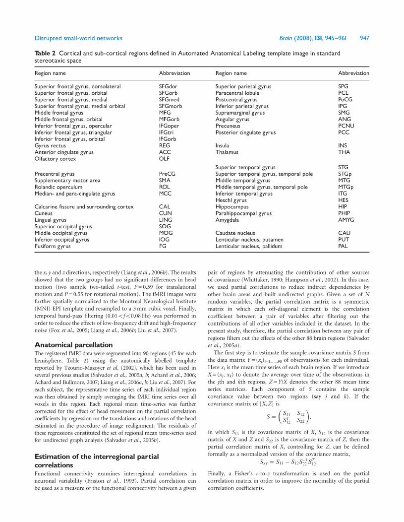
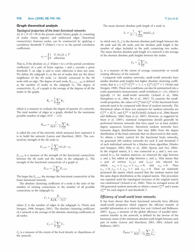
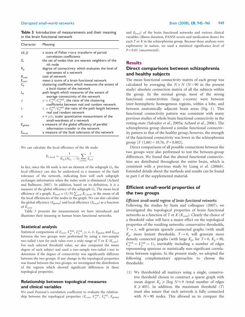
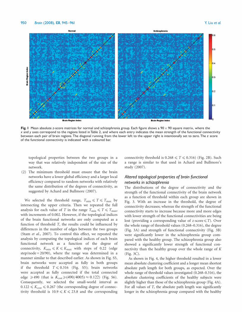
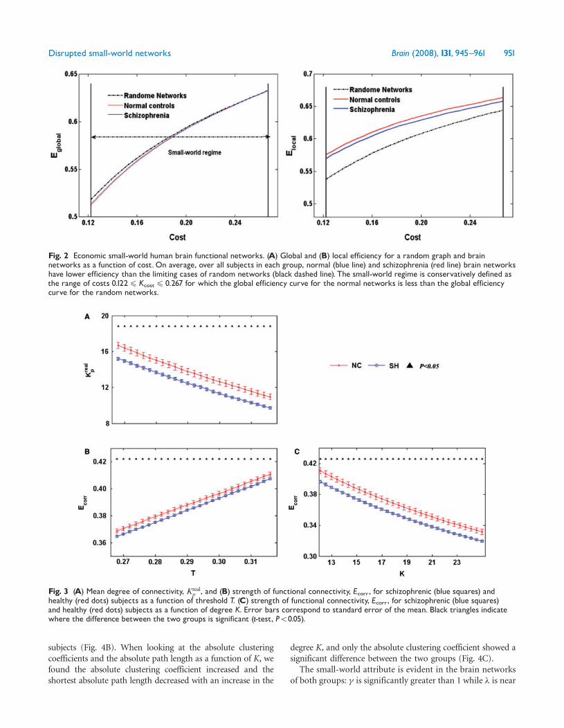
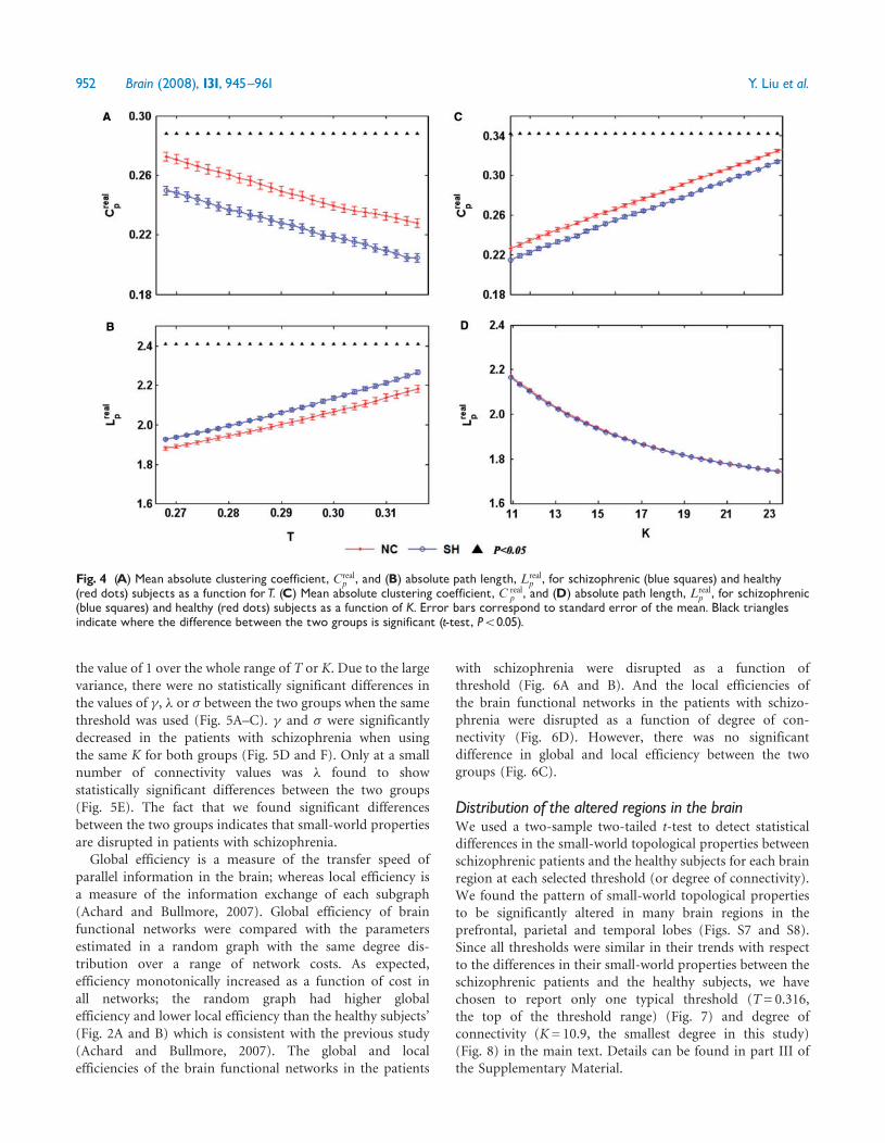
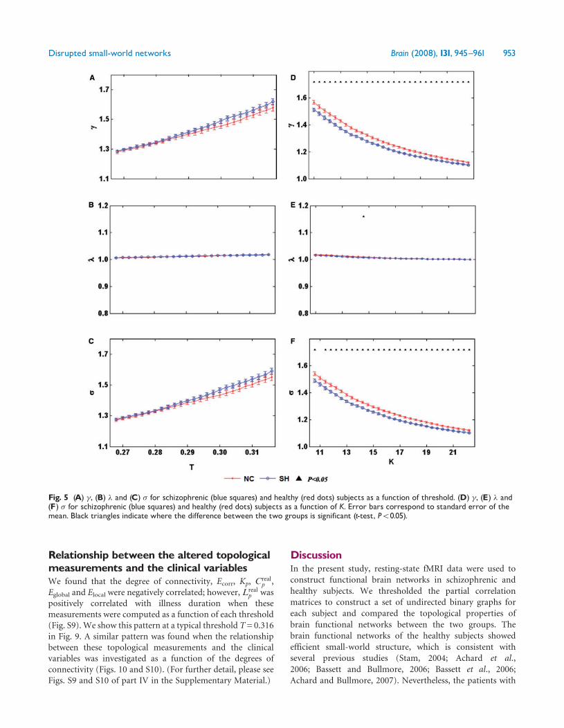
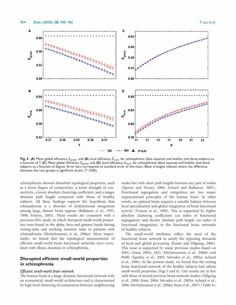
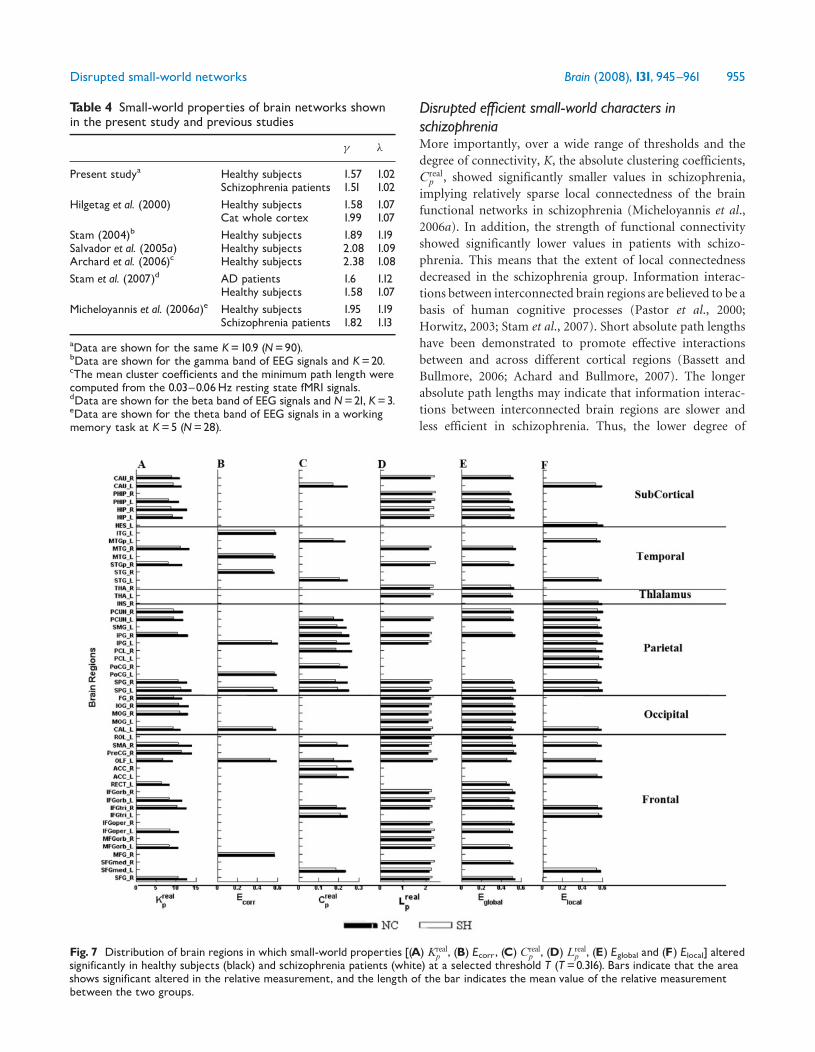
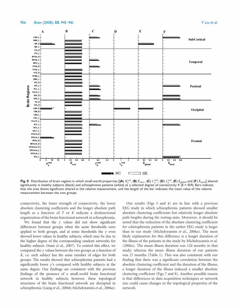
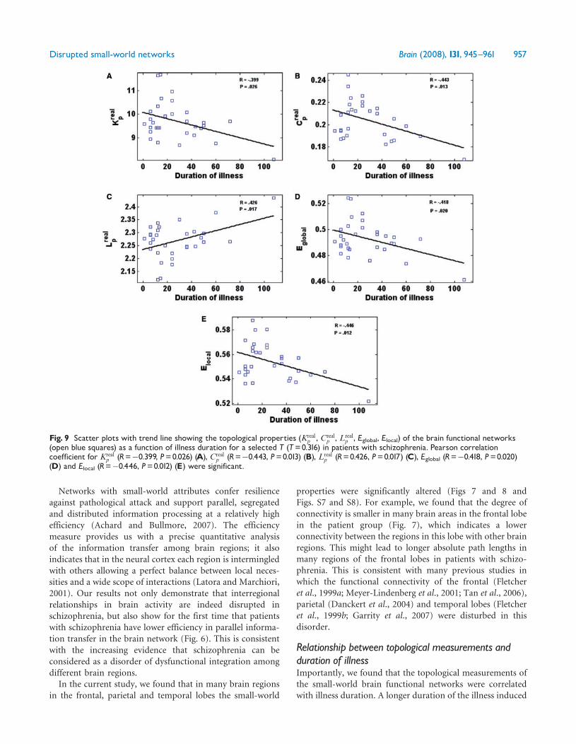
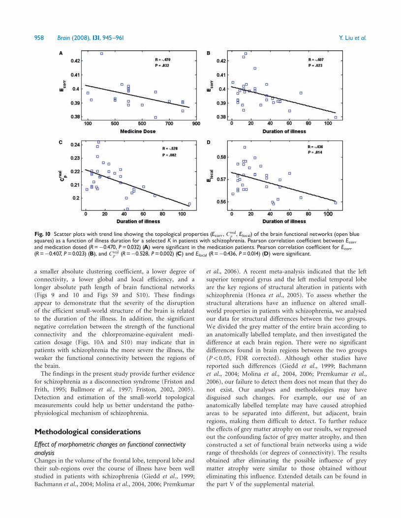