Embed Size (px)
Citation preview
1996, 16(9):5026. Mol. Cell. Biol.
and A C ChanG Kong, M Dalton, J Bubeck Wardenburg, D Straus, T Kurosaki regulation of antigen receptor function.ZAP-70 mediate activation and negative Distinct tyrosine phosphorylation sites in
http://mcb.asm.org/content/16/9/5026Updated information and services can be found at:
These include:
CONTENT ALERTS more»cite this article),
Receive: RSS Feeds, eTOCs, free email alerts (when new articles
http://journals.asm.org/site/misc/reprints.xhtmlInformation about commercial reprint orders: http://journals.asm.org/site/subscriptions/To subscribe to to another ASM Journal go to:
on October 12, 2013 by guest
http://mcb.asm
.org/D
ownloaded from
on O
ctober 12, 2013 by guesthttp://m
cb.asm.org/
Dow
nloaded from
on October 12, 2013 by guest
http://mcb.asm
.org/D
ownloaded from
on O
ctober 12, 2013 by guesthttp://m
cb.asm.org/
Dow
nloaded from
on October 12, 2013 by guest
http://mcb.asm
.org/D
ownloaded from
on O
ctober 12, 2013 by guesthttp://m
cb.asm.org/
Dow
nloaded from
on October 12, 2013 by guest
http://mcb.asm
.org/D
ownloaded from
on O
ctober 12, 2013 by guesthttp://m
cb.asm.org/
Dow
nloaded from
on October 12, 2013 by guest
http://mcb.asm
.org/D
ownloaded from
on O
ctober 12, 2013 by guesthttp://m
cb.asm.org/
Dow
nloaded from
on October 12, 2013 by guest
http://mcb.asm
.org/D
ownloaded from
MOLECULAR AND CELLULAR BIOLOGY, Sept. 1996, p. 5026–5035 Vol. 16, No. 90270-7306/96/$04.0010Copyright q 1996, American Society for Microbiology
Distinct Tyrosine Phosphorylation Sites in ZAP-70 MediateActivation and Negative Regulation of Antigen
Receptor FunctionGUANGHUI KONG,1 MARK DALTON,1 JULIANE BUBECK WARDENBURG,2 DAVID STRAUS,3
TOMOHIRO KUROSAKI,4 AND ANDREW C. CHAN1,2,5,6*
Howard Hughes Medical Institute,1 Center for Immunology,2 Department of Pathology,5 and Division ofRheumatology, Department of Internal Medicine,6 Washington University School of Medicine,
St. Louis, Missouri 63110; Department of Medicine, University of Chicago, Chicago,Illinois 606373; and Department of Oncology and Immunology,
Wyeth-Ayerst Research, Pearl River, New York 109654
Received 5 April 1996/Returned for modification 18 May 1996/Accepted 17 June 1996
Biochemical and genetic evidence has implicated two families of protein tyrosine kinases (PTKs), the Src-and Syk-PTKs, in T- and B-cell antigen receptor signaling. ZAP-70 is a member of the Syk-PTKs thatassociates with the T-cell antigen receptor and undergoes tyrosine phosphorylation following receptor activa-tion. Three tyrosine residues, Tyr-292, -492, and -493, have been identified as sites of phosphorylation followingT-cell antigen receptor engagement. Utilizing ZAP-70- and Syk-deficient lymphocytes (Syk2 DT40 cells), weprovide biochemical and functional evidence that heterologous trans-phosphorylation of Tyr-493 by a Src-PTKis required for antigen receptor-mediated activation of both the calcium and ras pathways. In contrast, cellsexpressing mutations at Tyr-292 or -492 demonstrate hyperactive T- and B-cell antigen receptor phenotypes.Thus, phosphorylation of ZAP-70 mediates both activation and inactivation of antigen receptor signaling.
Stimulation of T and B lymphocytes through the T-cell re-ceptor (TCR) and B-cell receptor (BCR), respectively, acti-vates a cascade of protein tyrosine kinases (PTKs) that isrequired for lymphocyte cellular responses (5, 43). T and Bcells appear to utilize analogous proximal signal transductionpathways in mediating these responses. Two distinct families ofcytoplasmic PTKs, the Src- and Syk-PTK families, have beenimplicated in the proximal activation events for both BCRs andTCRs (6). In T cells, the Src-PTKs Lck and/or Fyn are thoughtto phosphorylate the two tyrosine residues within a 16-amino-acid motif (YXXLX6–8YXXL, the immunoreceptor tyrosine-based activation motif [ITAM]) located in the cytoplasmicdomains of CD3 and z chains. In B cells, the Src-PTKs Lyn,Fyn, and/or Blk are thought to phosphorylate the immunoglob-ulin a chain (Iga) and Igb ITAM sequences. Phosphorylationof the ITAM provides docking sites for the two SH2 domainswithin the Syk-PTKs (ZAP-70 and Syk) which associate withthe phosphorylated TCR and BCR complexes. The phosphor-ylation and activation of ZAP-70 and Syk following TCR andBCR engagement, respectively, are in part mediated by theSrc-PTKs. Coexpression of Lck or Fyn with ZAP-70 or Syk ininsect and COS cells results in catalytic activation of the Syk-PTKs (7, 9, 10, 12, 19, 24, 38, 41). Recent evidence also sug-gests that binding of Syk to the Iga, Igb, or FcεRIg ITAMs canmediate Syk activation and phosphorylation presumably by anallosteric mechanism (33, 34).Expression of dominant negative forms or deletion of Src or
Syk-PTKs abrogates thymocyte development and/or antigenreceptor function (2, 3, 8, 11, 14, 21, 28, 29, 35, 36, 39). Acti-vation of both families of PTKs is thought to be required toactivate at least two distinct pathways, ras and calcium, to
mediate transcriptional activation of cytokine genes. Recentevidence in anergic T cells has demonstrated that activation ofthese two pathways may be differentially regulated (15, 25). Weexamine here the role of ZAP-70 phosphorylation and acti-vation in regulating the divergent pathways activated by thelymphocyte antigen receptor (20). In addition, we provide evi-dence that the catalytic activation of ZAP-70 and the phos-phorylation of downstream effector molecules is initiated bythe heterologous trans-phosphorylation of ZAP-70 at Tyr-493by Lck. Finally, we provide evidence that phosphorylation ofZAP-70 at Tyr-492 and -292 mediates two distinct mechanismsfor down regulation of antigen receptor function.
MATERIALS AND METHODS
Cells and antibodies. DT40 Syk2 B cells, Jurkat T cells, and Sf9 cells (Pharm-ingen) were maintained as previously described (10, 23, 38). The M4 monoclonalantibody (MAb) directed against the chicken BCR was provided courtesy of M.Cooper, 2F3.2 is an anti-ZAP-70 MAb (Upstate Biotechnology Inc.), 9E10 is ananti-myc epitope MAb, 12CA5 is an anti-hemagglutinin epitope MAb (Boehr-inger Mannheim), and 4G10 is an antiphosphotyrosine MAb (Upstate Biotech-nology Inc.). The anti-ZAP-70 immunoprecipitating antiserum used was raisedagainst a peptide encoding amino acids 282 to 307 of human ZAP-70 as previ-ously described (9). Fluorescein isothiocyanate-conjugated anti-mouse IgM andIgG were purchased from Sigma.Construction of plasmids. ZAP-70 mutants Y292F, Y492F, and Y493F were
constructed as previously described (10). A kinase-inactive version of ZAP-70[designated ZAP-70(KD)] was produced by PCR-directed mutagenesis of Lys-369 to Arg. All constructs were subcloned into the EcoRI and SalI sites of amodified version of the pApuro vector to generate hemagglutinin-tagged ver-sions of human ZAP-70 (23). In addition, myc-tagged versions of wild-typeZAP-70 and ZAP-70(Y292F) were also subcloned into the p409 vector fortetracycline-regulated gene expression (16). Glutathione S-transferase (GST)–ZAP-70(KD) was constructed in the PVIKS vector (10). All constructs wereconfirmed by standard DNA sequencing methods. The interleukin 2 (IL-2)-luciferase and cytomegalovirus-chloramphenicol acetyltransferase (CMV-CAT)reporter genes were gifts from K. Murphy (Washington University, St. Louis,Mo.); the AP-1–luciferase reporter gene was a gift from T. Chatila (WashingtonUniversity).Cellular expression and biochemical analysis. Protocols for the generation of
stable clones and the conditions for transient and stable transfections have beenpreviously described (16, 23). For expression of genes under the control of the
* Corresponding author. Mailing address: Washington UniversitySchool of Medicine, Box 8022, 660 S. Euclid Ave., St. Louis, MO63110. Phone: (314) 362-9012. Fax: (314) 454-0175. Electronic mailaddress: [email protected].
5026
tetracycline-regulated promoter (16), a parental Jurkat T-cell line (449-3) thatexpressed a tetracycline-controlled transactivator was established. This parentalline was then transfected with ZAP-70 mutants under the control of a tetracy-cline-dependent promoter. Clones were selected in G418 (1 mg/ml; Gibco),hygromycin (1 mg/ml), and tetracycline (1 mg/ml). For analysis, stable cloneswere washed and grown in the absence of tetracycline for 24 to 48 h prior toanalysis.For luciferase assays, cells were electroporated with 20 mg of IL-2– or AP-1–
luciferase cDNA and 5 mg of CMV-CAT cDNA as previously described (23).Luciferase activity was determined with an OptocompII automated luminometer(MGM Instruments, Hamden, Conn.) and normalized for transfection efficiencywith CMV-CAT. Generation of baculovirus-encoded proteins was performed aspreviously described (10). Stimulation of B cells, in vitro kinase assays, flowcytometric analysis, and calcium fluorimetry were performed as previously de-scribed (10, 23).Analysis of 32P-labeled ZAP-70(KD) tryptic peptides. GST–ZAP-70(KD) and
GST-Lck(K) were immobilized on glutathione-Sepharose and incubated with[g-32P]ATP for 5 min in the presence of 20 mM Tris (pH 7.4), 10 mMMgCl2, and10 mM MnCl2. The in vitro-labeled proteins were then separated by sodiumdodecyl sulfate-polyacrylamide gel electrophoresis, transferred to nitrocellulosemembrane, and visualized by autoradiography. 32P-labeled GST–ZAP-70 wasdigested with trypsin, and the resultant peptides were separated by two-dimen-sional thin-layer chromatography as previously described (10). Extraction ofradiolabeled peptides and manual sequencing of separated peptides were per-formed as previously described (4, 26, 37).Measurement of inositol phosphate generation. Incorporation of [3H]inositol
into phospholipids was achieved by incubating cells (106/ml) with 10 mCi ofmyo-[3H]inositol (37 MBq/ml; Amersham) per ml for 18 h in inositol-free RPMI1640 supplemented with 10% dialyzed fetal calf serum at 378C. The cells werethen washed extensively, and 5 3 106 cells were stimulated with an anti-BCRMAb (M4; 4 mg/ml) at 378C in the presence of 10 mM LiCl. Extraction andseparation of inositol phosphates were performed as previously described (18).Briefly, cells were extracted with chloroform-methanol, and soluble fractionswere applied to a 0.6-ml AG 1-X8 (formate form) ion-exchange column (Bio-Rad) that was preequilibrated with 0.1 M formic acid. The columns were washedwith 60 mM sodium formate and eluted with 0.2, 0.4, and 1 M ammoniumformate to yield the IP1, IP2, and IP3 fractions, respectively.
RESULTS
Positive and negative regulation of ZAP-70 by tyrosine phos-phorylation. Peptide analysis of ZAP-70 has demonstratedthat tyrosine residues 292, 492, and 493 are phosphorylatedfollowing TCR stimulation (10, 42). We have previously dem-onstrated that phosphorylation of Tyr-493 is required for cat-alytic activation of ZAP-70 and for efficient induction of cyto-kine release (10). To further explore the role of these threetyrosine residues in antigen receptor function, we analyzed aSyk2 chicken B-cell line (DT40) that had been transfected withZAP-70 cDNAs in which tyrosine 292, 492, or 493 had beenmutated to phenylalanine, [designated ZAP-70(Y292F), ZAP-70(Y492F), or ZAP-70(Y493F), respectively]. While loss ofSyk in these cells abrogates BCR-mediated signaling (38), ex-pression of transfected wild-type ZAP-70 reconstitutes BCR-me-diated signaling (23). Expression of wild-type ZAP-70 in Syk2
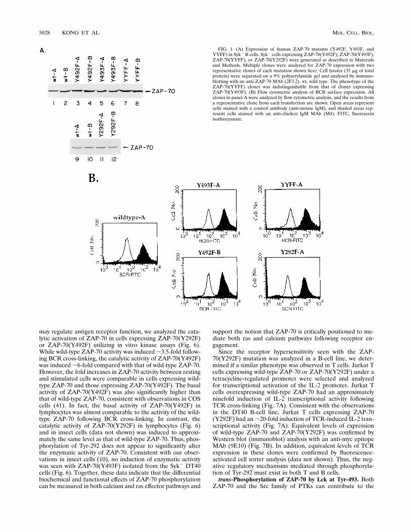
DT40 cells reconstitutes the ability of the BCR to inducecellular tyrosine phosphoproteins, mobilize the intracellularCa21 concentration ([Ca21]i), and induce IL-2 promoter ac-tivity following receptor cross-linking. A minimum of twoclones expressing equivalent levels of ZAP-70 protein andBCR surface expression (Fig. 1) for each ZAP-70 mutation wasanalyzed.To confirm our previous result that Tyr-493 is required for
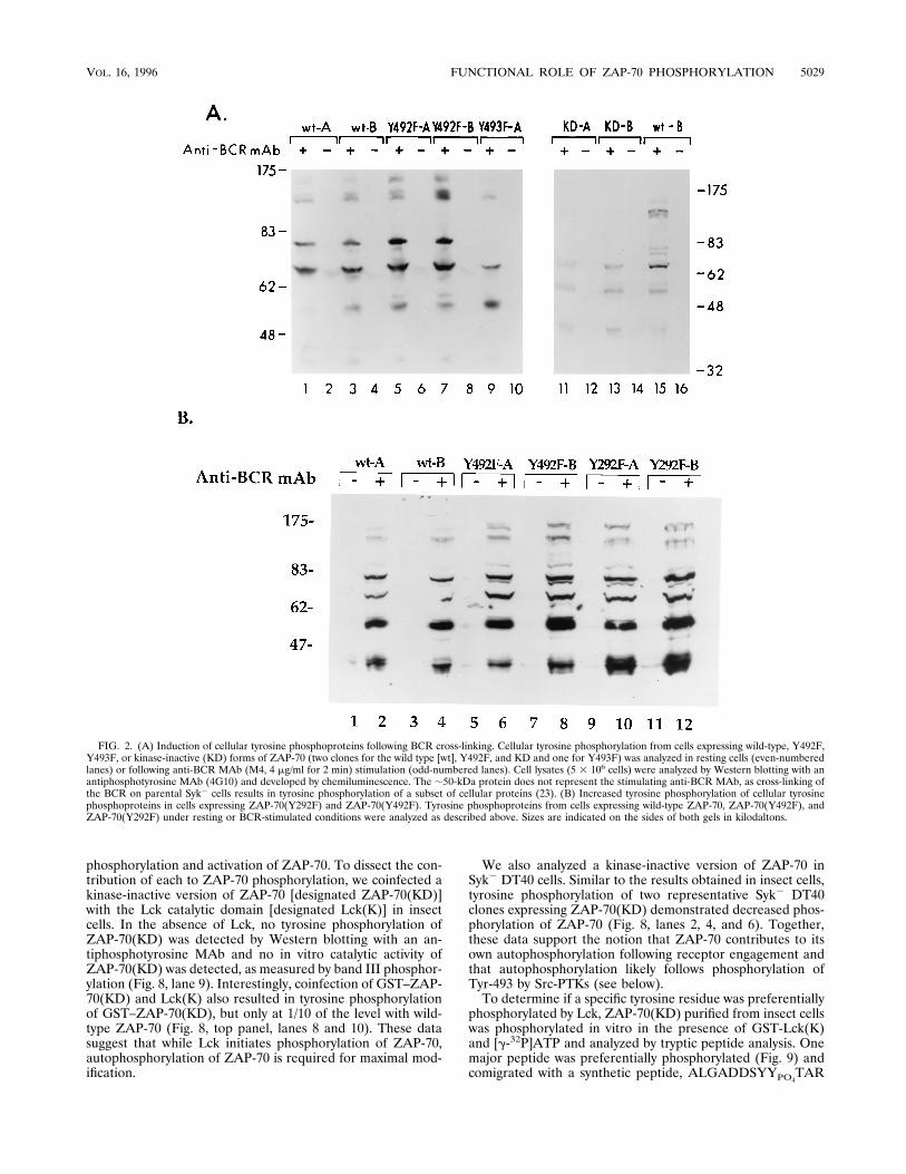
ZAP-70 activation, ZAP-70(Y493F) was expressed in DT40Syk2 cells. Cell lysates from resting or BCR-stimulated cellswere analyzed by immunoblotting with an antiphosphotyrosineMAb (4G10). Analysis of a representative clone expressingZAP-70(Y493F) demonstrated a reduction in tyrosine phos-phorylation of proteins with Mrs of 170,000, 150,000, 120,000,and 80,000 compared with that with wild-type ZAP-70 (Fig.2A, lanes 1 to 4, 9, and 10). A similar reduction in the cellulartyrosine phosphoproteins induced was also observed in cellsexpressing ZAP-70(KD) (Fig. 2A, lanes 11 to 14). Thus, the
induction of these cellular tyrosine phosphoproteins is depen-dent on Tyr-493 as well as the catalytic activity of ZAP-70.In contrast to Tyr-493, Tyr-292 and -492 serve to attentuate
antigen receptor function. In two representative clones ex-pressing ZAP-70(Y492F) or ZAP-70(Y292F), a qualitativelysimilar pattern of cellular tyrosine phosphoproteins was in-duced following BCR cross-linking (Fig. 2A, lanes 5 to 8, and2B). Quantitatively, however, BCR cross-linking of cells ex-pressing ZAP-70(Y492F) and ZAP-70(Y292F) consistently re-sulted in a small increase in tyrosine phosphorylation of cellu-lar proteins compared with that in cells expressing wild-typeZAP-70. In view of these small differences, we analyzed morequantitative measures of the two major signaling pathwaysactivated by antigen receptor cross-linking, calcium and ras.Consistent with the ability of ZAP-70(Y492F) to induce cellu-
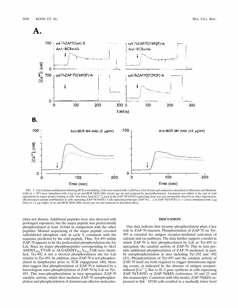
lar tyrosine phosphoproteins, expression of ZAP-70(Y492F)also induced increases in [Ca21]i following BCR cross-linking(Fig. 3A). However, with lower concentrations of anti-BCRMAb, the increase in [Ca21]i flux was greater in cells express-ing ZAP-70(Y492F) than in cells expressing wild-type ZAP-70 (Fig. 3B). Expression of ZAP-70(Y292F) also inducedincreases in [Ca21]i following BCR cross-linking (Fig. 3A),though no dosage differences in the stimulating antibody werefound compared with cells expressing wild-type ZAP-70 (datanot shown). Notably, cells expressing ZAP-70(Y493F) wereunable to exhibit a [Ca21]i response.Since mobilization of [Ca21]i is the result of activation of the
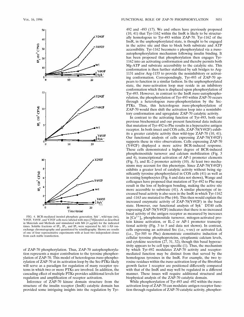
phosphoinositide pathway, we measured the accumulation ofphosphoinositide metabolites following BCR cross-linking(Fig. 4). Cells expressing ZAP-70(Y492F) or ZAP-70(Y292F)accumulated, respectively, approximately three- or fivefoldmore IP1, IP2, and IP3 over the 20-min course of BCR stimu-lation compared with cells expressing wild-type ZAP-70. Asexpected, the phosphoinositide pathway was not activated incells expressing ZAP-70(Y493F). Together, these data supportthe notion that phosphorylation of Tyr-292 and -492 serves todown regulate antigen receptor function.These negative regulatory functions were also reflected in
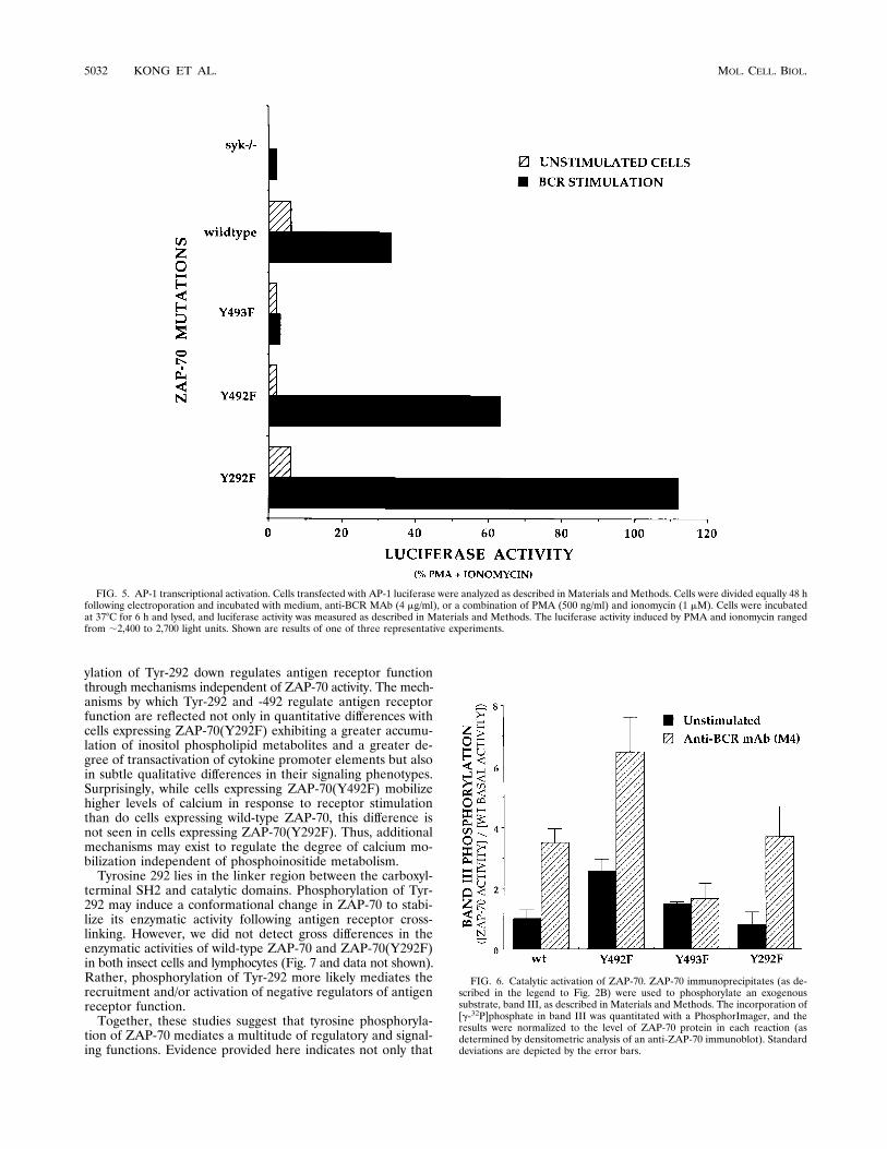
the ras pathway as measured by AP-1 transcriptional activation(Fig. 5). BCR cross-linking of cells expressing ZAP-70(Y492F)resulted in an approximately twofold greater induction of AP-1transcriptional activation compared with that in cells express-ing wild-type ZAP-70 (Fig. 5) [33% of phorbol myristate ace-tate (PMA) and ionomycin for the wild type versus 63% forZAP-70(Y492F)]. As expected, cells expressing ZAP-70(Y493F)were unresponsive. The AP-1 measurements extend and areconsistent with our previous observations utilizing the IL-2promoter wherein cells expressing ZAP-70(Y492F) had ap-proximately twofold greater IL-2 promoter activity in the pres-ence of ionomycin and anti-BCR MAbs (10). However, in thisstudy, the AP-1 measurements required only BCR cross-link-ing and did not have a corequirement for ionophore. Thus, thedegree of AP-1 transcriptional activation can be ascribed solelyto the true sensitivity of BCR-mediated signaling events with-out the contribution of exogenous pharmacologic agents.Cells expressing ZAP-70(Y292F) had an even greater de-
gree of both AP-1 and IL-2 promoter transcriptional activationcompared with cells expressing wild-type ZAP-70 or ZAP-70(492F) (Fig. 5 and data not shown). An ;3.5-fold greaterinduction of AP-1 transcriptional activity was found followingBCR activation in cells expressing ZAP-70(Y292F) comparedwith that in cells expressing wild-type ZAP-70 [33% for thewild type versus 112% for ZAP-70(Y292F)]. Therefore, thenegative regulatory functions of Tyr-292 and -492 are reflectedin both calcium and ras signaling pathways.To determine the mechanisms by which Tyr-292 and -492
VOL. 16, 1996 FUNCTIONAL ROLE OF ZAP-70 PHOSPHORYLATION 5027
may regulate antigen receptor function, we analyzed the cata-lytic activation of ZAP-70 in cells expressing ZAP-70(Y292F)or ZAP-70(Y492F) utilizing in vitro kinase assays (Fig. 6).While wild-type ZAP-70 activity was induced;3.5-fold follow-ing BCR cross-linking, the catalytic activity of ZAP-70(Y492F)was induced ;6-fold compared with that of wild-type ZAP-70.However, the fold increases in ZAP-70 activity between restingand stimulated cells were comparable in cells expressing wild-type ZAP-70 and those expressing ZAP-70(Y492F). The basalactivity of ZAP-70(Y492F) was also significantly higher thanthat of wild-type ZAP-70, consistent with observations in COScells (41). In fact, the basal activity of ZAP-70(Y492F) inlymphocytes was almost comparable to the activity of the wild-type ZAP-70 following BCR cross-linking. In contrast, thecatalytic activity of ZAP-70(Y292F) in lymphocytes (Fig. 6)and in insect cells (data not shown) was induced to approxi-mately the same level as that of wild-type ZAP-70. Thus, phos-phorylation of Tyr-292 does not appear to significantly alterthe enzymatic activity of ZAP-70. Consistent with our obser-vations in insect cells (10), no induction of enzymatic activitywas seen with ZAP-70(Y493F) isolated from the Syk2 DT40cells (Fig. 6). Together, these data indicate that the differentialbiochemical and functional effects of ZAP-70 phosphorylationcan be measured in both calcium and ras effector pathways and
support the notion that ZAP-70 is critically positioned to me-diate both ras and calcium pathways following receptor en-gagement.Since the receptor hypersensitivity seen with the ZAP-
70(Y292F) mutation was analyzed in a B-cell line, we deter-mined if a similar phenotype was observed in T cells. Jurkat Tcells expressing wild-type ZAP-70 or ZAP-70(Y292F) under atetracycline-regulated promoter were selected and analyzedfor transcriptional activation of the IL-2 promoter. Jurkat Tcells overexpressing wild-type ZAP-70 had an approximatelyninefold induction of IL-2 transcriptional activity followingTCR cross-linking (Fig. 7A). Consistent with the observationsin the DT40 B-cell line, Jurkat T cells expressing ZAP-70(Y292F) had an;20-fold induction of TCR-induced IL-2 tran-scriptional activity (Fig. 7A). Equivalent levels of expressionof wild-type ZAP-70 and ZAP-70(Y292F) was confirmed byWestern blot (immunoblot) analysis with an anti-myc epitopeMAb (9E10) (Fig. 7B). In addition, equivalent levels of TCRexpression in these clones were confirmed by fluorescence-activated cell sorter analysis (data not shown). Thus, the neg-ative regulatory mechanisms mediated through phosphoryla-tion of Tyr-292 must exist in both T and B cells.trans-Phosphorylation of ZAP-70 by Lck at Tyr-493. Both
ZAP-70 and the Src family of PTKs can contribute to the
FIG. 1. (A) Expression of human ZAP-70 mutants (Y492F, Y493F, andYYFF) in Syk2 B cells. Syk2 cells expressing ZAP-70(Y492F), ZAP-70(Y493F),ZAP-70(YYFF), or ZAP-70(Y292F) were generated as described in Materialsand Methods. Multiple clones were analyzed for ZAP-70 expression with tworepresentative clones of each mutation shown here. Cell lysates (35 mg of totalprotein) were separated on a 9% polyacrylamide gel and analyzed by immuno-blotting with an anti-ZAP-70 MAb (2F3.2). wt, wild type. The phenotype of theZAP-70(YYFF) clones was indistinguishable from that of clones expressingZAP-70(Y493F). (B) Flow cytometric analysis of BCR surface expression. Allclones in panel A were analyzed by flow cytometric analysis, and the results froma representative clone from each transfection are shown. Open areas representcells stained with a control antibody (anti-mouse IgM), and shaded areas rep-resent cells stained with an anti-chicken IgM MAb (M4). FITC, fluoresceinisothiocyanate.
5028 KONG ET AL. MOL. CELL. BIOL.
phosphorylation and activation of ZAP-70. To dissect the con-tribution of each to ZAP-70 phosphorylation, we coinfected akinase-inactive version of ZAP-70 [designated ZAP-70(KD)]with the Lck catalytic domain [designated Lck(K)] in insectcells. In the absence of Lck, no tyrosine phosphorylation ofZAP-70(KD) was detected by Western blotting with an an-tiphosphotyrosine MAb and no in vitro catalytic activity ofZAP-70(KD) was detected, as measured by band III phosphor-ylation (Fig. 8, lane 9). Interestingly, coinfection of GST–ZAP-70(KD) and Lck(K) also resulted in tyrosine phosphorylationof GST–ZAP-70(KD), but only at 1/10 of the level with wild-type ZAP-70 (Fig. 8, top panel, lanes 8 and 10). These datasuggest that while Lck initiates phosphorylation of ZAP-70,autophosphorylation of ZAP-70 is required for maximal mod-ification.
We also analyzed a kinase-inactive version of ZAP-70 inSyk2 DT40 cells. Similar to the results obtained in insect cells,tyrosine phosphorylation of two representative Syk2 DT40clones expressing ZAP-70(KD) demonstrated decreased phos-phorylation of ZAP-70 (Fig. 8, lanes 2, 4, and 6). Together,these data support the notion that ZAP-70 contributes to itsown autophosphorylation following receptor engagement andthat autophosphorylation likely follows phosphorylation ofTyr-493 by Src-PTKs (see below).To determine if a specific tyrosine residue was preferentially
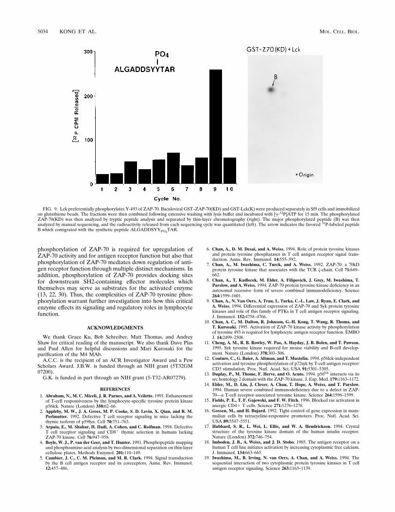
phosphorylated by Lck, ZAP-70(KD) purified from insect cellswas phosphorylated in vitro in the presence of GST-Lck(K)and [g-32P]ATP and analyzed by tryptic peptide analysis. Onemajor peptide was preferentially phosphorylated (Fig. 9) andcomigrated with a synthetic peptide, ALGADDSYYPO4TAR
FIG. 2. (A) Induction of cellular tyrosine phosphoproteins following BCR cross-linking. Cellular tyrosine phosphorylation from cells expressing wild-type, Y492F,Y493F, or kinase-inactive (KD) forms of ZAP-70 (two clones for the wild type [wt], Y492F, and KD and one for Y493F) was analyzed in resting cells (even-numberedlanes) or following anti-BCR MAb (M4, 4 mg/ml for 2 min) stimulation (odd-numbered lanes). Cell lysates (5 3 106 cells) were analyzed by Western blotting with anantiphosphotyrosine MAb (4G10) and developed by chemiluminescence. The ;50-kDa protein does not represent the stimulating anti-BCR MAb, as cross-linking ofthe BCR on parental Syk2 cells results in tyrosine phosphorylation of a subset of cellular proteins (23). (B) Increased tyrosine phosphorylation of cellular tyrosinephosphoproteins in cells expressing ZAP-70(Y292F) and ZAP-70(Y492F). Tyrosine phosphoproteins from cells expressing wild-type ZAP-70, ZAP-70(Y492F), andZAP-70(Y292F) under resting or BCR-stimulated conditions were analyzed as described above. Sizes are indicated on the sides of both gels in kilodaltons.
VOL. 16, 1996 FUNCTIONAL ROLE OF ZAP-70 PHOSPHORYLATION 5029
(data not shown). Additional peptides were also detected withprolonged exposures, but the major peptide was preferentiallyphosphorylated at least 10-fold in comparison with the otherpeptides. Manual sequencing of the major peptide revealedradiolabeled phosphate only in cycle 9, consistent with thesequence predicted by the cold peptide. Thus, Tyr-493 withinZAP-70 appears to be the preferential phosphorylation site forLck. Since no major phosphopeptides corresponding to ALGADDSYPO4YTAR or ALGADDSYPO4YPO4TAR were identi-fied, Tyr-492 is not a favored phosphorylation site for Lckrelative to Tyr-493. In addition, since ZAP-70 is not phosphor-ylated in lymphocytes prior to TCR engagement (40), thesedata suggest that phosphorylation of ZAP-70 is initiated by aheterologous trans-phosphorylation of ZAP-70 by Lck on Tyr-493. This trans-phosphorylation in turn upregulates ZAP-70catalytic activity, which then mediates ZAP-70 autophosphor-ylation and phosphorylation of downstream effector molecules.
DISCUSSION
Our data indicate that tyrosine phosphorylation plays a keyrole in ZAP-70 function. Phosphorylation of ZAP-70 on Tyr-493 is essential for antigen receptor-mediated activation ofcalcium and ras pathways. The data further support a model inwhich ZAP-70 is first phosphorylated by Lck at Tyr-493 toupregulate the catalytic activity of ZAP-70. This in turn per-mits additional phosphorylation of ZAP-70 mediated, in part,by autophosphorylation at sites including Tyr-292 and -492(41). Phosphorylation of Tyr-493 and the catalytic activity ofZAP-70 itself are both required to trigger downstream signal-ing events, as indicated by the absence of antigen receptor-induced [Ca21]i flux or IL-2 gene synthesis in cells expressingZAP-70(Y493F) or ZAP-70(KD) (references 10 and 23 andthis manuscript). Consistent with this model, ZAP-70(KD) ex-pressed in Syk2 DT40 cells resulted in a markedly lower level
FIG. 3. (A) Calcium mobilization following BCR cross-linking. Cells were loaded with 3 mM Fura-2 for 40 min and analyzed as described in Materials and Methods.Cells (6 3 106) were stimulated with 4 mg of an anti-BCR MAb (M4; arrow) per ml and analyzed by spectrofluorimetry. Ionomycin was added at the end of eachexperiment to ensure proper loading of cells. The lower basal [Ca21]i seen in the ZAP-70(Y493F)-expressing clone was not consistently observed in other experiments.(B) Increased calcium mobilization in cells expressing ZAP-70(Y492F). Cells expressing wild-type ZAP-70 (. . .) or ZAP-70(Y492F) (——) were stimulated with 2 mg(left) or 1.2 mg (right) of an anti-BCR MAb (M4; arrow) per ml and analyzed as described above.
5030 KONG ET AL. MOL. CELL. BIOL.
of ZAP-70 phosphorylation. Thus, ZAP-70 autophosphoryla-tion represents a major contribution to the tyrosine phosphor-ylation of ZAP-70. This model of heterologous trans-phospho-rylation of ZAP-70 at its activation loop by the Src-PTKs likelywill serve as a paradigm for regulation of many receptor sys-tems in which two or more PTKs are involved. In addition, thecascading effect of multiple PTKs provides additional levels forregulation and amplification of receptor activation.Inference of ZAP-70 kinase domain structure from the
structure of the insulin receptor (InsR) catalytic domain hasprovided some intriguing insights into the regulation by Tyr-
492 and -493 (17). We and others have previously proposed(10, 41) that Tyr-1162 within the InsR is likely to be structur-ally homologous to Tyr-493 within ZAP-70. Tyr-1162 of theInsR, in the unphosphorylated state, is thought to be engagedin the active site and thus to block both substrate and ATPaccessibility. Tyr-1162 becometo s phosphorylated via a trans-autophosphorylation mechanism following insulin binding. Ithas been proposed that phosphorylation then engages Tyr-1162 into an activating conformation and thereby permits bothMg-ATP and substrate accessibility to the catalytic site. Thisconformation is then further stabilized by salt bridges to Arg-1131 and/or Arg-1155 to provide the noninhibitory or activat-ing conformation. Correspondingly, Tyr-493 of ZAP-70 ap-pears to function in a similar fashion. In the unphosphorylatedstate, the trans-activation loop may reside in an inhibitoryconformation which then is displaced upon phosphorylation ofTyr-493. However, in contrast to the InsR trans-autophospho-rylation, the phosphorylation of Tyr-493 within ZAP-70 occursthrough a heterologous trans-phosphorylation by the Src-PTKs. Thus, this heterologous trans-phosphorylation ofZAP-70 would then shift the activation loop into a noninhibi-tory conformation and upregulate ZAP-70 catalytic activity.In contrast to the activating function of Tyr-493, both our
previous biochemical and our present functional data indicatethat mutation of Tyr-492 to Phe results in a hyperactive antigenreceptor. In both insect and COS cells, ZAP-70(Y492F) exhib-its a greater catalytic activity than wild-type ZAP-70 (10, 41).Our functional analysis of cells expressing ZAP-70(Y492F)supports these in vitro observations. Cells expressing ZAP-70(Y492F) displayed a more active BCR-induced response.These cells demonstrated a higher degree of BCR-inducedphosphoinositide turnover and calcium mobilization (Fig. 3and 4), transcriptional activation of AP-1 promoter elements(Fig. 5), and IL-2 promoter activity (10). At least two mecha-nisms may account for this phenotype. Since ZAP-70(Y492F)exhibits a greater level of catalytic activity without being sig-nificantly tyrosine phosphorylated in COS cells (41) as well asin resting lymphocytes (Fig. 6 and data not shown), Wange andcolleagues have proposed that mutation of Tyr-492 to Phe mayresult in the loss of hydrogen bonding, making the active sitemore accessible to substrate (41). A similar phenotype of in-creased basal activity is also seen in the InsR in which Tyr-1162and -1163 are mutated to Phe (44). This then would explain theincreased enzymatic activity of ZAP-70(Y492F) in the basalstate. However, our functional analysis of Syk2 DT40 cellsexpressing ZAP-70(Y492F) indicates that there is no increasedbasal activity of the antigen receptor as measured by increasesin [Ca21]i, phosphoinositide turnover, mitogen-activated pro-tein kinase activation, or IL-2 or AP-1 promoter transcrip-tional activity (Fig. 3 to 5 and data not shown). In contrast,cells expressing an activated Src (i.e., v-src) or activated Lck(i.e., Tyr-505 to Phe) demonstrate constitutive induction ofcellular tyrosine phosphoproteins, cytoplasmic calcium levels,and cytokine secretion (27, 31, 32), though this basal hyperac-tivity appears to be cell type specific (1). Thus, the mechanismby which Tyr-492 modulates ZAP-70 activity and receptor-mediated function may be distinct from that served by thehomologous tyrosines in the InsR. For example, the two ty-rosine residues within the trans-activation loop of the fibroblastgrowth factor 1 receptor are positioned differently comparedwith that of the InsR and may well be regulated in a differentmanner. These issues will require additional structural andbiophysical analysis of the ZAP-70 catalytic domain.While phosphorylation of Tyr-493 and -492 within the trans-
activation loop of ZAP-70 can modulate antigen receptor func-tion through regulation of ZAP-70 catalytic activity, phosphor-
FIG. 4. BCR-mediated inositol phosphate generation. Syk2, wild-type (wt),Y492F, Y493F, and Y292F cells were labeled with myo-[3H]inositol as describedin Materials and Methods and stimulated with M4 (4 mg/ml) for the indicatedtimes. Soluble fractions of IP1, IP2, and IP3 were separated by AG 1-X8 ion-exchange chromatography and quantitated by scintillography. Shown are resultsof one of four representative experiments with at least two independent clonesfrom each stable transfection.
VOL. 16, 1996 FUNCTIONAL ROLE OF ZAP-70 PHOSPHORYLATION 5031
ylation of Tyr-292 down regulates antigen receptor functionthrough mechanisms independent of ZAP-70 activity. The mech-anisms by which Tyr-292 and -492 regulate antigen receptorfunction are reflected not only in quantitative differences withcells expressing ZAP-70(Y292F) exhibiting a greater accumu-lation of inositol phospholipid metabolites and a greater de-gree of transactivation of cytokine promoter elements but alsoin subtle qualitative differences in their signaling phenotypes.Surprisingly, while cells expressing ZAP-70(Y492F) mobilizehigher levels of calcium in response to receptor stimulationthan do cells expressing wild-type ZAP-70, this difference isnot seen in cells expressing ZAP-70(Y292F). Thus, additionalmechanisms may exist to regulate the degree of calcium mo-bilization independent of phosphoinositide metabolism.Tyrosine 292 lies in the linker region between the carboxyl-
terminal SH2 and catalytic domains. Phosphorylation of Tyr-292 may induce a conformational change in ZAP-70 to stabi-lize its enzymatic activity following antigen receptor cross-linking. However, we did not detect gross differences in theenzymatic activities of wild-type ZAP-70 and ZAP-70(Y292F)in both insect cells and lymphocytes (Fig. 7 and data not shown).Rather, phosphorylation of Tyr-292 more likely mediates therecruitment and/or activation of negative regulators of antigenreceptor function.Together, these studies suggest that tyrosine phosphoryla-
tion of ZAP-70 mediates a multitude of regulatory and signal-ing functions. Evidence provided here indicates not only that
FIG. 5. AP-1 transcriptional activation. Cells transfected with AP-1 luciferase were analyzed as described in Materials and Methods. Cells were divided equally 48 hfollowing electroporation and incubated with medium, anti-BCR MAb (4 mg/ml), or a combination of PMA (500 ng/ml) and ionomycin (1 mM). Cells were incubatedat 378C for 6 h and lysed, and luciferase activity was measured as described in Materials and Methods. The luciferase activity induced by PMA and ionomycin rangedfrom ;2,400 to 2,700 light units. Shown are results of one of three representative experiments.
FIG. 6. Catalytic activation of ZAP-70. ZAP-70 immunoprecipitates (as de-scribed in the legend to Fig. 2B) were used to phosphorylate an exogenoussubstrate, band III, as described in Materials and Methods. The incorporation of[g-32P]phosphate in band III was quantitated with a PhosphorImager, and theresults were normalized to the level of ZAP-70 protein in each reaction (asdetermined by densitometric analysis of an anti-ZAP-70 immunoblot). Standarddeviations are depicted by the error bars.
5032 KONG ET AL. MOL. CELL. BIOL.
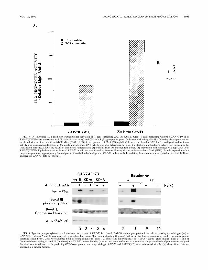
FIG. 7. (A) Increased IL-2 promoter transcriptional activation of T cells expressing ZAP-70(Y292F). Jurkat T cells expressing wild-type ZAP-70 (WT) orZAP-70(Y292F) were transfected with IL-2–luciferase (20 mg) and CMV-CAT (5 mg) reporter genes. Cells were divided equally 48 h following electroporation andincubated with medium or with anti-TCR MAb (C305, 1:1,000) in the presence of PMA (500 ng/ml). Cells were incubated at 378C for 6 h and lysed, and luciferaseactivity was measured as described in Materials and Methods. CAT activity was also determined for each transfection, and luciferase activity was normalized fortransfection efficiency. Shown are results of one of two representative experiments from two independent clones. (B) Expression of the induced wild-type ZAP-70 orZAP-70(Y292F). Equivalent levels of induced ZAP-70 protein were confirmed by Western blotting with an anti-myc epitope MAb (9E10). Protein expression of theexogenous genes was approximately fivefold greater than the level of endogenous ZAP-70 in these cells. In addition, these clones express equivalent levels of TCR andendogenous ZAP-70 (data not shown).
FIG. 8. Tyrosine phosphorylation of a kinase-inactive version of ZAP-70 is reduced. ZAP-70 immunoprecipitates from cells expressing the wild type (wt) orZAP-70(KD) clones A and B were analyzed by antiphosphotyrosine MAb immunoblotting (top row) and by in vitro kinase assays using band III as an exogenoussubstrate (second row). Cells were analyzed both in resting conditions (lanes 1, 3, and 5) and following BCR (M4 MAb, 4 mg/ml) cross-linking (lanes 2, 4, and 6).Coomassie blue staining of band III (third row) and ZAP-70 immunoblotting (bottom row) were performed to ensure that comparable levels of protein were analyzed.Baculovirus-infected insect cells producing GST-fusion proteins encoding wild-type ZAP-70 and ZAP-70(KD) were coinfected with Lck(K) (lanes 8 and 10) andanalyzed in a similar fashion.
VOL. 16, 1996 FUNCTIONAL ROLE OF ZAP-70 PHOSPHORYLATION 5033
phosphorylation of ZAP-70 is required for upregulation ofZAP-70 activity and for antigen receptor function but also thatphosphorylation of ZAP-70 mediates down regulation of anti-gen receptor function through multiple distinct mechanisms. Inaddition, phosphorylation of ZAP-70 provides docking sitesfor downstream SH2-containing effector molecules whichthemselves may serve as substrates for the activated enzyme(13, 22, 30). Thus, the complexities of ZAP-70 tyrosine phos-phorylation warrant further investigation into how this criticalenzyme effects its signaling and regulatory roles in lymphocytefunction.
ACKNOWLEDGMENTS
We thank Grace Ku, Bob Schreiber, Matt Thomas, and AndreyShaw for critical reading of the manuscript. We also thank Dave Plasand Paul Allen for helpful discussions and Mari Kurosaki for thepurification of the M4 MAb.A.C.C. is the recipient of an ACR Investigator Award and a Pew
Scholars Award. J.B.W. is funded through an NIH grant (5T32GM07200).G.K. is funded in part through an NIH grant (5-T32-AR07279).
REFERENCES1. Abraham, N., M. C. Miceli, J. R. Parnes, and A. Veilette. 1991. Enhancementof T-cell responsiveness by the lymphocyte-specific tyrosine protein kinasep56lck. Nature (London) 350:62–66.
2. Appleby, M. W., J. A. Gross, M. P. Cooke, S. D. Levin, X. Qian, and R. M.Perlmutter. 1992. Defective T cell receptor signaling in mice lacking thethymic isoform of p59fyn. Cell 70:751–763.
3. Arpaia, E., M. Shahar, H. Dadi, A. Cohen, and C. Roifman. 1994. DefectiveT cell receptor signaling and CD81 thymic selection in humans lackingZAP-70 kinase. Cell 76:947–958.
4. Boyle, W. J., P. van der Geer, and T. Hunter. 1991. Phosphopeptide mappingand phosphoamino acid analysis by two-dimensional separation on thin-layercellulose plates. Methods Enzymol. 201:110–149.
5. Cambier, J. C., C. M. Pleiman, and M. R. Clark. 1994. Signal transductionby the B cell antigen receptor and its coreceptors. Annu. Rev. Immunol.12:457–486.
6. Chan, A., D. M. Desai, and A. Weiss. 1994. Role of protein tyrosine kinasesand protein tyrosine phosphatases in T cell antigen receptor signal trans-duction. Annu. Rev. Immunol. 14:555–592.
7. Chan, A., M. Iwashima, C. Turck, and A. Weiss. 1992. ZAP-70: a 70kDprotein tyrosine kinase that associates with the TCR z-chain. Cell 71:649–662.
8. Chan, A., T. Kadlecek, M. Elder, A. Filipovich, J. Grey, M. Iwashima, T.Parslow, and A. Weiss. 1994. ZAP-70 protein tyrosine kinase deficiency in anautosomal recessive form of severe combined immunodeficiency. Science264:1599–1601.
9. Chan, A., N. Van Oers, A. Tran, L. Turka, C.-L. Law, J. Ryan, E. Clark, andA. Weiss. 1994. Differential expression of ZAP-70 and Syk protein tyrosinekinases and role of this family of PTKs in T cell antigen receptor signaling.J. Immunol. 152:4758–4766.
10. Chan, A. C., M. Dalton, R. Johnson, G.-H. Kong, T. Wang, R. Thoma, andT. Kurosaki. 1995. Activation of ZAP-70 kinase activity by phosphorylationof tyrosine 493 is required for lymphocyte antigen receptor function. EMBOJ. 14:2499–2508.
11. Cheng, A. M., R. B. Rowley, W. Pao, A. Hayday, J. B. Bolen, and T. Pawson.1995. Syk tyrosine kinase required for mouse viability and B-cell develop-ment. Nature (London) 378:303–306.
12. Couture, C., G. Baier, A. Altman, and T. Mustelin. 1994. p56lck-independentactivation and tyrosine phosphorylation of p72syk by T-cell antigen receptor/CD3 stimulation. Proc. Natl. Acad. Sci. USA 91:5301–5305.
13. Duplay, P., M. Thome, F. Herve, and O. Acuto. 1994. p56lck interacts via itssrc homology 2 domain with the ZAP-70 kinase. J. Exp. Med. 179:1163–1172.
14. Elder, M., D. Lin, J. Clever, A. Chan, T. Hope, A. Weiss, and T. Parslow.1994. Human severe combined immunodeficiency due to a defect in ZAP-70—a T-cell receptor-associated tyrosine kinase. Science 264:1596–1599.
15. Fields, P. E., T. F. Gajewski, and F. W. Fitch. 1996. Blocked ras activation inanergic CD41 T cells. Science 271:1276–1278.
16. Gossen, M., and H. Bujard. 1992. Tight control of gene expression in mam-malian cells by tetracycline-responsive promoters. Proc. Natl. Acad. Sci.USA 89:5547–5551.
17. Hubbard, S. R., L. Wei, L. Ellis, and W. A. Hendrickson. 1994. Crystalstructure of the tyrosine kinase domain of the human insulin receptor.Nature (London) 372:746–754.
18. Imboden, J. B., A. Weiss, and J. D. Stobo. 1985. The antigen receptor on ahuman T cell line initiates activation by increasing cytoplasmic free calcium.J. Immunol. 134:663–665.
19. Iwashima, M., B. Irving, N. van Oers, A. Chan, and A. Weiss. 1994. Thesequential interaction of two cytoplasmic protein tyrosine kinases in T cellantigen receptor signaling. Science 263:1163–1139.
FIG. 9. Lck preferentially phosphorylates Y-493 of ZAP-70. Baculoviral GST–ZAP-70(KD) and GST-Lck(K) were produced separately in Sf9 cells and immobilizedon glutathione beads. The fractions were then combined following extensive washing with lysis buffer and incubated with [g-32P]ATP for 15 min. The phosphorylatedZAP-70(KD) was then analyzed by tryptic peptide analysis and separated by thin-layer chromatography (right). The major phosphorylated peptide (B) was thenanalyzed by manual sequencing, and the radioactivity released from each sequencing cycle was quantitated (left). The arrow indicates the favored 32P-labeled peptideB which comigrated with the synthetic peptide ALGADDSYYPO4TAR.
5034 KONG ET AL. MOL. CELL. BIOL.
20. Izquierdo Pastor, M., K. Rief, and D. Cantrell. 1995. The regulation andfunction of p21ras during T-cell activation and growth. Immunol. Today16:159–164.
21. Karnitz, L., S. L. Sutor, T. Torigoe, J. C. Reed, M. P. Bell, D. J. McKean,P. J. Leibson, and R. T. Abraham. 1992. Effects of p56lck deficiency on thegrowth and cytolytic effector function of an interleukin-2 dependent cyto-toxic T-cell line. Mol. Cell. Biol. 12:4521–4530.
22. Katzav, S., M. Sutherland, G. Packham, T. Yi, and A. Weiss. 1994. Theprotein tyrosine kinase ZAP-70 can associate with the SH2 domain of proto-Vav. J. Biol. Chem. 269:32579–32585.
23. Kong, G. H., J. Y. Bu, T. Kurosaki, A. S. Shaw, and A. C. Chan. 1995.Reconstitution of syk function by the ZAP-70 protein tyrosine kinase. Im-munity 2:485–492.
24. Kurosaki, T., M. Takata, Y. Yamanashi, T. Inazu, T. Taniguchi, T.Yamamoto, and H. Yamamura. 1994. Syk activation by the Src-family ty-rosine kinase in the B cell receptor signaling. J. Exp. Med. 179:1725–1729.
25. Li, W., C. D. Whaley, A. Mondino, and D. L. Mueller. 1996. Blocked signaltransduction to the erk and JNK protein kinases in anergic CD41 T cells.Science 271:1272–1276.
26. Luo, K., T. R. Hurley, and B. M. Sefton. 1991. Cyanogen bromide cleavageand proteolytic peptide mapping of proteins immobilized to membranes.Methods Enzymol. 201:149–152.
27. Luo, K., and B. M. Sefton. 1992. Activated lck tyrosine protein kinasestimulates antigen-independent interleukin-2 production in T cells. Mol CellBiol. 12:4724–4732.
28. Molina, T. J., K. Kishihara, D. P. Siderovski, W. van Ewijk, A. Narendran,E. Timms, A. Wakeham, C. J. Paige, K. U. Hartmann, A. Veillette, D.Davidson, and T. W. Mak. 1992. Profound block in thymocyte developmentin mice lacking p56lck. Nature (London) 357:161–164.
29. Negishi, I., N. Motoyama, K.-I. Nakayama, K. Nakayama, S. Senju, S.Hatakeyama, Q. Zhang, A. C. Chan, and D. Y. Loh. 1995. Essential role forZAP-70 in both positive and negative selection of thymocytes. Nature (Lon-don) 376:435–438.
30. Neumeister, E. N., Y. Zhu, S. Richard, C. Terhorst, A. C. Chan, and A. S.Shaw. 1995. Binding of ZAP-70 to phosphorylated T-cell receptor z and henhances its autophosphorylation and generates specific binding sites forSH2 domain-containing proteins. Mol. Cell. Biol. 15:3171–3178.
31. Niklinska, B. B., H. Yamada, J. J. O’Shea, C. H. June, and J. D. Ashwell.1992. Tyrosine kinase-regulated and inositol phosphate-independent Ca21elevation and mobilization in T cells. J. Biol. Chem. 267:7154–7159.
32. O’Shea, J. J., J. D. Ashwell, T. L. Bailey, S. L. Cross, L. E. Samelson, andR. D. Klausner. 1991. Expression of v-src in a murine T-cell hybridoma
results in constitutive T-cell receptor phosphorylation and interleukin 2production. Proc. Natl. Acad. Sci. USA 88:1741–1745.
33. Rowley, R. B., A. L. Burkardt, H.-G. Chao, G. R. Matsueda, and J. B. Bolen.1995. Syk protein-tyrosine kinase is regulated by tyrosine-phosphorylatedIga/Igb immunoreceptor tyrosine activation motif binding and autophos-phorylation. J. Biol. Chem. 270:11590–11594.
34. Shiue, L., M. J. Zoller, and J. S. Brugge. 1995. Syk is activated by phospho-tyrosine-containing peptides representing the tyrosine-based activation mo-tifs of the high affinity receptor for IgE. J. Biol. Chem. 270:10498–10502.
35. Stein, P. L., H.-M. Lee, S. Rich, and P. Soriano. 1992. pp59fyn mutant micedisplay differential signalling in thymocyte and peripheral T cells. Cell 70:741–750.
36. Straus, D., and A. Weiss. 1992. Genetic evidence for the involvement of thelck tyrosine kinase in signal transduction through the T cell antigen receptor.Cell 70:585–593.
37. Sullivan, S., and T. W. Wong. 1991. A manual sequencing method foridentification of phosphorylated amino acids in phosphopeptides. Anal. Bio-chem. 197:65–68.
38. Takata, M., H. Sabe, H. A., T. Inazu, Y. Homma, T. Nukada, H. Yamamura,and T. Kurosaki. 1994. Tyrosine kinases lyn and syk regulate B cell receptor-coupled Ca21 mobilization through distinct pathways. EMBO J. 13:1341–1349.
39. Turner, M., P. J. Mee, P. S. Costello, O. Williams, A. A. Price, L. P. Duddy,M. T. Furlong, R. L. Geahlen, and V. L. J. Tybulewicz. 1995. Perinatallethality and blocked B-cell development in mice lacking the tyrosine kinaseSyk. Nature (London) 378:298–302.
40. van Oers, N. S. C., N. Killeen, and A. Weiss. 1995. ZAP-70 is constitutivelyassociated with tyrosine-phosphorylated TCR z in murine thymocytes andlymph node T cells. Immunity 1:675–685.
41. Wange, R. L., R. Guitian, N. Isakov, J. D. Watts, R. Aebersold, and L. E.Samelson. 1995. Activating and inhibitory mutations in adjacent tyrosines inthe kinase domain of ZAP-70. J. Biol. Chem. 270:18730–18733.
42. Watts, J. D., M. Affolter, D. L. Krebs, R. L. Wange, L. E. Samelson, and R.Aebersold. 1994. Identification by electrospray ionization mass spectrometryof the sites of tyrosine phosphorylation induced in activated Jurkat T cells onthe protein tyrosine kinase ZAP-70. J. Biol. Chem. 25:29520–29529.
43. Weiss, A., and D. R. Littman. 1994. Signal transduction by lymphocyteantigen receptors. Cell 76:263–274.
44. Zhang, B., J. M. Tavare, L. Ellis, and R. A. Roth. 1991. The regulatory roleof known tyrosine autophosphorylation sites of the insulin receptor kinasedomain. J. Biol. Chem. 266:990–996.
VOL. 16, 1996 FUNCTIONAL ROLE OF ZAP-70 PHOSPHORYLATION 5035


