Embed Size (px)
Citation preview
The
Journ
al o
f Exp
erim
enta
l M
edic
ine
ARTICLE
JEM © The Rockefeller University Press $15.00
Vol. 204, No. 6, June 11, 2007 1431–1440 www.jem.org/cgi/doi/10.1084/jem.20062642
1431
The discovery of vascular endothelial growth factors (VEGFs) and their receptors has facili-tated the understanding of the development and function of the vasculature (1–3). Each VEGF family member appears to have a spe-cifi c function. Whereas VEGF induces angio-genesis, i.e., growth of new blood vessels from preexisting ones, placenta growth factor (PlGF) mediates both angiogenesis and arteriogenesis, i.e., the formation of collateral arteries from preexisting arterioles (1, 2). VEGF-C and -D are primarily lymphangiogenic factors, which can also trigger angiogenesis in some condi-tions (3). Overall, the members of the VEGF
family and their receptors appear to provide promising and versatile tools for therapeutic manipulation of the vascular system (1–3).
VEGF is one of the most important regula-tors of both physiological and pathological angiogenesis, and its activity is mediated via VEGF receptor (VEGFR) 1 and 2. VEGF, act-ing mainly via VEGFR-2, is an endothelial cell mitogen, motogen, chemoattractant, and sur-vival factor that increases the permeability of blood vessels (1). The importance of VEGF in the development of the vascular and hemato-poietic systems is exemplifi ed by the fact that inactivation of even one VEGF allele leads to early embryonic lethality caused by defects in angiogenesis and hematopoiesis (4, 5).
Genes with sequence homology to VEGF have been discovered in Orf and pseudocowpox
Distinct vascular endothelial growth factor signals for lymphatic vessel enlargement and sprouting
Maria Wirzenius,1 Tuomas Tammela,1 Marko Uutela,1 Yulong He,1 Teresa Odorisio,2 Giovanna Zambruno,2 Janice A. Nagy,3 Harold F. Dvorak,3 Seppo Ylä-Herttuala,4 Masabumi Shibuya,5 and Kari Alitalo1
1Molecular/Cancer Biology Laboratory and Ludwig Institute for Cancer Research, Haartman Institute and Helsinki University
Hospital, Biomedicum Helsinki, University of Helsinki, 00014 Helsinki, Finland2Laboratory of Molecular and Cell Biology, Istituto Dermopatico dell’Immacolata, Istituto di Ricovero e Cura a Caraterre
Scientifi co, 00167 Rome, Italy3Department of Pathology, Beth Israel Deaconess Medical Center and Harvard Medical School, Boston, MA 022154A.I. Virtanen Institute, University of Kuopio, 70211 Kuopio, Finland5Division of Genetics, Institute of Medical Science, University of Tokyo, Minato-ku, Tokyo 108-8639, Japan
Lymphatic vessel growth, or lymphangiogenesis, is regulated by vascular endothelial growth
factor-C (VEGF-C) and -D via VEGF receptor 3 (VEGFR-3). Recent studies suggest that
VEGF, which does not bind to VEGFR-3, can also induce lymphangiogenesis through
unknown mechanisms. To dissect the receptor pathway that triggers VEGFR-3–independent
lymphangiogenesis, we used both transgenic and adenoviral overexpression of placenta
growth factor (PlGF) and VEGF-E, which are specifi c activators of VEGFR-1 and -2,
respectively. Unlike PlGF, VEGF-E induced circumferential lymphatic vessel hyperplasia, but
essentially no new vessel sprouting, when transduced into mouse skin via adenoviral vectors.
This effect was not inhibited by blocking VEGF-C and -D. Postnatal lymphatic hyperplasia,
without increased density of lymphatic vessels, was also detected in transgenic mice
expressing VEGF-E in the skin, but not in mice expressing PlGF. Surprisingly, VEGF-E induced
lymphatic hyperplasia postnatally, and it did not rescue the loss of lymphatic vessels in
transgenic embryos where VEGF-C and VEGF-D were blocked. Our data suggests that
VEGFR-2 signals promote lymphatic vessel enlargement, but unlike in the blood vessels,
are not involved in vessel sprouting to generate new lymphatic vessels in vivo.
CORRESPONDENCE
Kari Alitalo:
Abbreviations used: E,
em bryonic day; P, postnatal day;
PAE, porcine aortic endothe-
lial; PECAM, platelet endo-
thelial cell adhesion molecule;
PlGF, placenta growth factor;
SMA, smooth muscle α-actin;
SMC, smooth muscle cell;
VEGF, vascular endothelial
growth factor; VEGFR,
VEGF receptor.
T. Tammela and M. Uutela contributed equally to this paper.
Y. He’s present address is Model Animal Research Institute,
Nanjing University, Nanjing 210061, China.
The online version of this article contains supplemental material.
1432 VEGF RECEPTORS IN LYMPHATIC HYPERPLASIA | Wirzenius et al.
viruses. These virus -encoded VEGFs, which are commonly called VEGF-E, cause highly vascularized and pustular dermati-tis in sheep, in goats, and, occasionally, in humans (6–8). The virus-encoded VEGF-Es can be separated into two groups, with VEGF-ED1701 and -ENZ2 most closely related to VEGF and PlGF, whereas VEGF-ENZ7 is similar to VEGF-C and -D (9). In this article, VEGF-E refers to VEGF-ENZ7. The virus- encoded VEGFs bind to VEGFR-2 and induce its autophos-phorylation to almost the same extent as VEGF, but do not bind to VEGFR-1 (7, 8, 10). Although VEGF-E does not play a role in vascular physiology, it can be used as a VEGFR-2–specifi c agonist in experimental models of angiogenesis in vitro and in vivo. Such studies have indicated that VEGF-E expression in the skin of transgenic mice results in an angiogenic phenotype (11).
Recent studies have suggested that at least the most com-monly expressed isoform of VEGF can also induce lymphatic hyperplasia (12, 13). However, the signaling mechanisms mediating this response have been unclear. In this study, we wanted to determine if signals mediated via VEGFR-1 or -2 can trigger lymphangiogenesis in embryonic or adult tissues. For our analysis, we used adenoviral transduction of VEGF-E, as well as transgenic overexpression of PlGF and VEGF-E, to activate VEGFR-1 and -2, respectively.
RESULTS
Characteristics of VEGF-induced lymphatic hyperplasia
VEGF-C has been shown to induce excessive sprouting of lymphatic vessels 4 d after adenoviral delivery and new
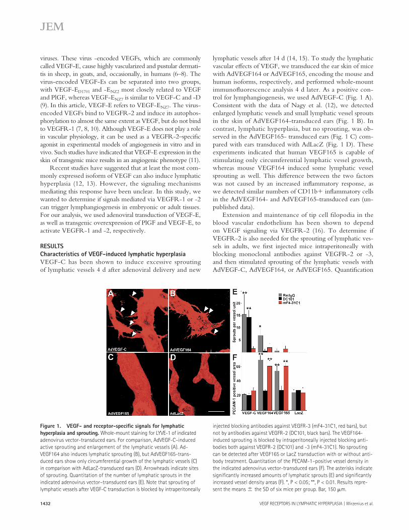
lymphatic vessels after 14 d (14, 15). To study the lymphatic vascular eff ects of VEGF, we transduced the ear skin of mice with AdVEGF164 or AdVEGF165, encoding the mouse and human isoforms, respectively, and performed whole-mount immunofl uorescence analysis 4 d later. As a positive con-trol for lymphangiogenesis, we used AdVEGF-C (Fig. 1 A). Consistent with the data of Nagy et al. (12), we detected enlarged lymphatic vessels and small lymphatic vessel sprouts in the skin of AdVEGF164-transduced ears (Fig. 1 B). In contrast, lymphatic hyperplasia, but no sprouting, was ob-served in the AdVEGF165- transduced ears (Fig. 1 C) com-pared with ears transduced with AdLacZ (Fig. 1 D). These experiments indicated that human VEGF165 is capable of stimulating only circumferential lymphatic vessel growth, whereas mouse VEGF164 induced some lymphatic vessel sprouting as well. This diff erence between the two factors was not caused by an increased infl ammatory response, as we detected similar numbers of CD11b+ infl ammatory cells in the AdVEGF164- and AdVEGF165-transduced ears (un-published data).
Extension and maintenance of tip cell fi lopodia in the blood vascular endothelium has been shown to depend on VEGF signaling via VEGFR-2 (16). To determine if VEGFR-2 is also needed for the sprouting of lymphatic ves-sels in adults, we fi rst injected mice intraperitoneally with blocking monoclonal antibodies against VEGFR-2 or -3, and then stimulated sprouting of the lymphatic vessels with AdVEGF-C, AdVEGF164, or AdVEGF165. Quantifi cation
Figure 1. VEGF- and receptor-specifi c signals for lymphatic
hyperplasia and sprouting. Whole-mount staining for LYVE-1 of indicated
adenovirus vector-transduced ears. For comparison, AdVEGF-C–induced
active sprouting and enlargement of the lymphatic vessels (A). Ad-
VEGF164 also induces lymphatic sprouting (B), but AdVEGF165-trans-
duced ears show only circumferential growth of the lymphatic vessels (C)
in comparison with AdLacZ-transduced ears (D). Arrowheads indicate sites
of sprouting. Quantitation of the number of lymphatic sprouts in the
indicated adenovirus vector–transduced ears (E). Note that sprouting of
lymphatic vessels after VEGF-C transduction is blocked by intraperitoneally
injected blocking antibodies against VEGFR-3 (mF4-31C1, red bars), but
not by antibodies against VEGFR-2 (DC101, black bars). The VEGF164-
induced sprouting is blocked by intraperitoneally injected blocking anti-
bodies both against VEGFR-2 (DC101) and -3 (mF4-31C1). No sprouting
can be detected after VEGF165 or LacZ transduction with or without anti-
body treatment. Quantitation of the PECAM-1–positive vessel density in
the indicated adenovirus vector-transduced ears (F). The asterisks indicate
signifi cantly increased amounts of lymphatic sprouts (E) and signifi cantly
increased vessel density areas (F). *, P < 0.05; **, P < 0.01. Results repre-
sent the means ± the SD of six mice per group. Bar, 150 μm.
JEM VOL. 204, June 11, 2007 1433
ARTICLE
of lymphatic sprouting showed that the sprouts induced by AdVEGF-C were not inhibited by nonspecifi c rat IgG or antibodies against VEGFR-2 (DC101; Fig. 1 E), but were strongly inhibited (16-fold) by antibodies against VEGFR-3 (mF4-31C1). Both antibodies blocked the considerably weaker sprouting induced by AdVEGF164, whereas essentially no sprouting was obtained after AdVEGF165 or AdLacZ trans-duction. Quantifi cation of the platelet endothelial cell ad-hesion molecule (PECAM) 1–positive vessel density in the AdVEGF164- and AdVEGF165-transduced ears indicated that both VEGF vectors promoted angiogenesis (Fig. 1 F). In contrast, AdVEGF-C or AdLacZ had very little or no eff ect on the blood vessels. Collectively, these results suggested that VEGFR-2 signaling may not be necessary for the sprouting of lymphatic vessels in adults. To study this further, we chose to overexpress VEGF-E in the skin.
VEGF-ENZ7 does not act via the VEGFR-3 pathway
VEGF-E has been shown to bind to VEGFR-2, but not to VEGFR-3, in stimulated cells (7, 16). To ensure that VEGF-E is not able to induce phosphorylation of VEGFR-3 in endothelial cells, we stimulated porcine aortic endo-thelial (PAE) cells stably expressing either VEGFR-2 or -3 with increasing amounts of recombinant VEGF-E and ana-lyzed receptor phosphorylation. Although VEGF-E induced VEGFR-2 phosphorylation at low concentrations, it was not able to induce phosphorylation of VEGFR-3, even at high concentrations (Fig. S1 A, available at http://www.jem.org/cgi/content/full/jem.20062642/DC1). Furthermore, soluble VEGFR-2-Ig fusion proteins or rat monoclonal antibodies against human VEGFR-2 inhibited VEGFR-2 stimulation by VEGF-E, whereas VEGFR-3-Ig did not. In contrast, all three reagents blocked VEGFR-2 stimulation by VEGF-C
(Fig. S1 B). These results indicate that VEGF-E does not interact with the VEGFR-3 signal transduction pathway in vitro.
Adenovirally transduced VEGF-E induces lymphatic
hyperplasia in vivo
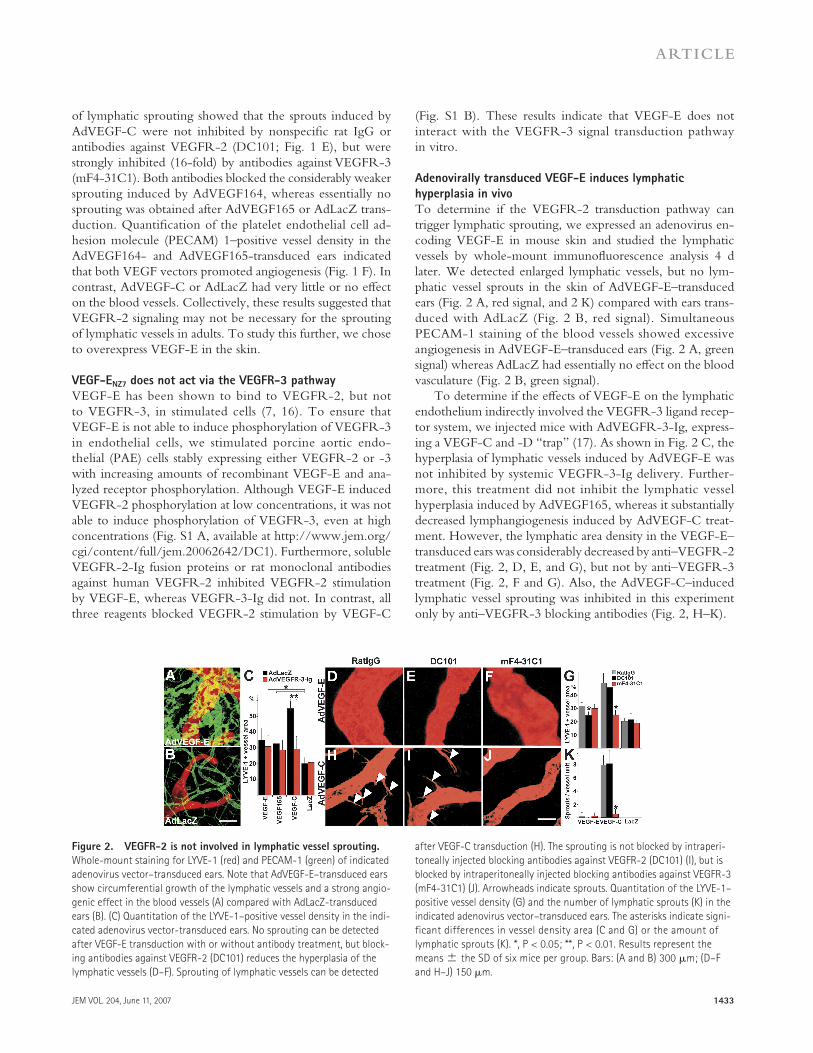
To determine if the VEGFR-2 transduction pathway can trigger lymphatic sprouting, we expressed an adenovirus en-coding VEGF-E in mouse skin and studied the lymphatic vessels by whole-mount immunofl uorescence analysis 4 d later. We detected enlarged lymphatic vessels, but no lym-phatic vessel sprouts in the skin of AdVEGF-E–transduced ears (Fig. 2 A, red signal, and 2 K) compared with ears trans-duced with AdLacZ (Fig. 2 B, red signal). Simultaneous PECAM-1 staining of the blood vessels showed excessive angiogenesis in AdVEGF-E–transduced ears (Fig. 2 A, green signal) whereas AdLacZ had essentially no eff ect on the blood vasculature (Fig. 2 B, green signal).
To determine if the eff ects of VEGF-E on the lymphatic endothelium indirectly involved the VEGFR-3 ligand recep-tor system, we injected mice with AdVEGFR-3-Ig, express-ing a VEGF-C and -D “trap” (17). As shown in Fig. 2 C, the hyperplasia of lymphatic vessels induced by AdVEGF-E was not inhibited by systemic VEGFR-3-Ig delivery. Further-more, this treatment did not inhibit the lymphatic vessel hyperplasia induced by AdVEGF165, whereas it substantially decreased lymphangiogenesis induced by AdVEGF-C treat-ment. However, the lymphatic area density in the VEGF-E–transduced ears was considerably decreased by anti–VEGFR-2 treatment (Fig. 2, D, E, and G), but not by anti–VEGFR-3 treatment (Fig. 2, F and G). Also, the AdVEGF-C–induced lymphatic vessel sprouting was inhibited in this experiment only by anti–VEGFR-3 blocking antibodies (Fig. 2, H–K).
Figure 2. VEGFR-2 is not involved in lymphatic vessel sprouting.
Whole-mount staining for LYVE-1 (red) and PECAM-1 (green) of indicated
adenovirus vector–transduced ears. Note that AdVEGF-E–transduced ears
show circumferential growth of the lymphatic vessels and a strong angio-
genic effect in the blood vessels (A) compared with AdLacZ-transduced
ears (B). (C) Quantitation of the LYVE-1–positive vessel density in the indi-
cated adenovirus vector-transduced ears. No sprouting can be detected
after VEGF-E transduction with or without antibody treatment, but block-
ing antibodies against VEGFR-2 (DC101) reduces the hyperplasia of the
lymphatic vessels (D–F). Sprouting of lymphatic vessels can be detected
after VEGF-C transduction (H). The sprouting is not blocked by intraperi-
toneally injected blocking antibodies against VEGFR-2 (DC101) (I), but is
blocked by intraperitoneally injected blocking antibodies against VEGFR-3
(mF4-31C1) (J). Arrowheads indicate sprouts. Quantitation of the LYVE-1–
positive vessel density (G) and the number of lymphatic sprouts (K) in the
indicated adenovirus vector–transduced ears. The asterisks indicate signi-
ficant differences in vessel density area (C and G) or the amount of
lymphatic sprouts (K). *, P < 0.05; **, P < 0.01. Results represent the
means ± the SD of six mice per group. Bars: (A and B) 300 μm; (D–F
and H–J) 150 μm.
1434 VEGF RECEPTORS IN LYMPHATIC HYPERPLASIA | Wirzenius et al.
Expression of VEGFR-2 and -3 in lymphatic capillaries
and collecting vessels
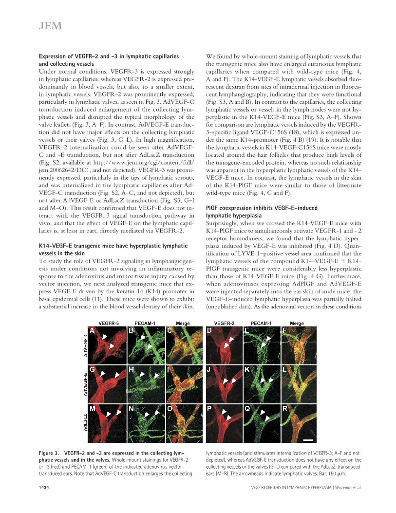
Under normal conditions, VEGFR-3 is expressed strongly in lymphatic capillaries, whereas VEGFR-2 is expressed pre-dominantly in blood vessels, but also, to a smaller extent, in lymphatic vessels. VEGFR-2 was prominently expressed, particularly in lymphatic valves, as seen in Fig. 3. AdVEGF-C transduction induced enlargement of the collecting lym-phatic vessels and disrupted the typical morphology of the valve leafl ets (Fig. 3, A–F). In contrast, AdVEGF-E transduc-tion did not have major eff ects on the collecting lymphatic vessels or their valves (Fig. 3, G–L). In high magnifi cation, VEGFR-2 internalization could be seen after AdVEGF-C and -E transduction, but not after AdLacZ transduction (Fig. S2, available at http://www.jem.org/cgi/content/full/jem.20062642/DC1, and not depicted). VEGFR-3 was promi-nently expressed, particularly in the tips of lymphatic sprouts, and was internalized in the lymphatic capillaries after Ad-VEGF-C transduction (Fig. S2, A–C, and not depicted), but not after AdVEGF-E or AdLacZ transduction (Fig. S3, G–I and M–O). This result confi rmed that VEGF-E does not in-teract with the VEGFR-3 signal transduction pathway in vivo, and that the eff ect of VEGF-E on the lymphatic capil-laries is, at least in part, directly mediated via VEGFR-2.
K14-VEGF-E transgenic mice have hyperplastic lymphatic
vessels in the skin
To study the role of VEGFR-2 signaling in lymphangiogen-esis under conditions not involving an infl ammatory re-sponse to the adenovirus and minor tissue injury caused by vector injection, we next analyzed transgenic mice that ex-press VEGF-E driven by the keratin 14 (K14) promoter in basal epidermal cells (11). These mice were shown to exhibit a substantial increase in the blood vessel density of their skin.
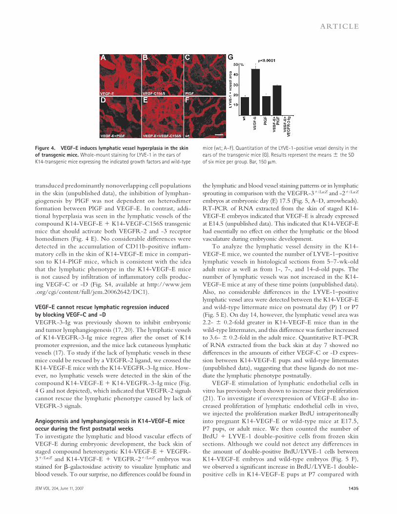
We found by whole-mount staining of lymphatic vessels that the transgenic mice also have enlarged cutaneous lymphatic capillaries when compared with wild-type mice (Fig. 4, A and F). The K14-VEGF-E lymphatic vessels absorbed fl uo-rescent dextran from sites of intradermal injection in fl uores-cent lymphangiography, indicating that they were functional (Fig. S3, A and B). In contrast to the capillaries, the collecting lymphatic vessels or vessels in the lymph nodes were not hy-perplastic in the K14-VEGF-E mice (Fig. S3, A–F). Shown for comparison are lymphatic vessels induced by the VEGFR-3–specifi c ligand VEGF-C156S (18), which is expressed un-der the same K14-promoter (Fig. 4 B) (19). It is notable that the lymphatic vessels in K14-VEGF-C156S mice were mostly located around the hair follicles that produce high levels of the transgene-encoded protein, whereas no such relationship was apparent in the hyperplastic lymphatic vessels of the K14-VEGF-E mice. In contrast, the lymphatic vessels in the skin of the K14-PlGF mice were similar to those of littermate wild-type mice (Fig. 4, C and F).
PlGF coexpression inhibits VEGF-E–induced
lymphatic hyperplasia
Surprisingly, when we crossed the K14-VEGF-E mice with K14-PlGF mice to simultaneously activate VEGFR-1 and - 2 receptor homodimers, we found that the lymphatic hyper-plasia induced by VEGF-E was inhibited (Fig. 4 D). Quan-tifi cation of LYVE-1–positive vessel area confi rmed that the lymphatic vessels of the compound K14-VEGF-E + K14-PlGF transgenic mice were considerably less hyperplastic than those of K14-VEGF-E mice (Fig. 4 G). Furthermore, when adenoviruses expressing AdPlGF and AdVEGF-E were injected separately into the ear skin of nude mice, the VEGF-E–induced lymphatic hyperplasia was partially halted (unpublished data). As the adenoviral vectors in these conditions
Figure 3. VEGFR-2 and -3 are expressed in the collecting lym-
phatic vessels and in the valves. Whole-mount stainings for VEGFR-2
or -3 (red) and PECAM-1 (green) of the indicated adenovirus vector–
transduced ears. Note that AdVEGF-C transduction enlarges the collecting
lymphatic vessels (and stimulates internalization of VEGFR-3; A–F and not
depicted), whereas AdVEGF-E transduction does not have any effect on the
collecting vessels or the valves (G–L) compared with the AdLacZ-transduced
ears (M–R). The arrowheads indicate lymphatic valves. Bar, 150 μm.
JEM VOL. 204, June 11, 2007 1435
ARTICLE
transduced predominantly nonoverlapping cell populations in the skin (unpublished data), the inhibition of lymphan-giogenesis by PlGF was not dependent on heterodimer formation between PlGF and VEGF-E. In contrast, addi-tional hyperplasia was seen in the lymphatic vessels of the compound K14-VEGF-E + K14-VEGF-C156S transgenic mice that should activate both VEGFR-2 and -3 receptor homodimers (Fig. 4 E). No considerable diff erences were detected in the accumulation of CD11b-positive infl am-matory cells in the skin of K14-VEGF-E mice in compari-son to K14-PlGF mice, which is consistent with the idea that the lymphatic phenotype in the K14-VEGF-E mice is not caused by infi ltration of infl ammatory cells produc-ing VEGF-C or -D (Fig. S4, available at http://www.jem.org/cgi/content/full/jem.20062642/DC1).
VEGF-E cannot rescue lymphatic regression induced
by blocking VEGF-C and -D
VEGFR-3-Ig was previously shown to inhibit embryonic and tumor lymphangiogenesis (17, 20). The lymphatic vessels of K14-VEGFR-3-Ig mice regress after the onset of K14 promoter expression, and the mice lack cutaneous lymphatic vessels (17). To study if the lack of lymphatic vessels in these mice could be rescued by a VEGFR-2 ligand, we crossed the K14-VEGF-E mice with the K14-VEGFR-3-Ig mice. How-ever, no lymphatic vessels were detected in the skin of the compound K14-VEGF-E + K14-VEGFR-3-Ig mice (Fig. 4 G and not depicted), which indicated that VEGFR-2 signals cannot rescue the lymphatic phenotype caused by lack of VEGFR-3 signals.
Angiogenesis and lymphangiogenesis in K14-VEGF-E mice
occur during the fi rst postnatal weeks
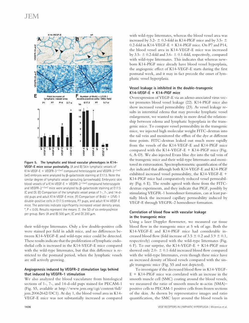
To investigate the lymphatic and blood vascular eff ects of VEGF-E during embryonic development, the back skin of staged compound heterozygotic K14-VEGF-E + VEGFR-3+/LacZ and K14-VEGF-E + VEGFR-2+/LacZ embryos was stained for β-galactosidase activity to visualize lymphatic and blood vessels. To our surprise, no diff erences could be found in
the lymphatic and blood vessel staining patterns or in lymphatic sprouting in comparison with the VEGFR-3+/LacZ and -2+/LacZ embryos at embryonic day (E) 17.5 (Fig. 5, A–D, arrowheads). RT-PCR of RNA extracted from the skin of staged K14-VEGF-E embryos indicated that VEGF-E is already expressed at E14.5 (unpublished data). This indicated that K14-VEGF-E had essentially no eff ect on either the lymphatic or the blood vasculature during embryonic development.
To analyze the lymphatic vessel density in the K14-VEGF-E mice, we counted the number of LYVE-1–positive lymphatic vessels in histological sections from 5–7-wk-old adult mice as well as from 1-, 7-, and 14-d-old pups. The number of lymphatic vessels was not increased in the K14-VEGF-E mice at any of these time points (unpublished data). Also, no considerable diff erences in the LYVE-1–positive lymphatic vessel area were detected between the K14-VEGF-E and wild-type littermate mice on postnatal day (P) 1 or P7 (Fig. 5 E). On day 14, however, the lymphatic vessel area was 2.2- ± 0.2-fold greater in K14-VEGF-E mice than in the wild-type littermates, and this diff erence was further increased to 3.6- ± 0.2-fold in the adult mice. Quantitative RT-PCR of RNA extracted from the back skin at day 7 showed no diff erences in the amounts of either VEGF-C or -D expres-sion between K14-VEGF-E pups and wild-type littermates (unpublished data), suggesting that these ligands do not me-diate the lymphatic phenotype postnatally.
VEGF-E stimulation of lymphatic endothelial cells in vitro has previously been shown to increase their proliferation (21). To investigate if overexpression of VEGF-E also in-creased proliferation of lymphatic endothelial cells in vivo, we injected the proliferation marker BrdU intraperitoneally into pregnant K14-VEGF-E or wild-type mice at E17.5, P7 pups, or adult mice. We then counted the number of BrdU + LYVE-1 double-positive cells from frozen skin sections. Although we could not detect any diff erences in the amount of double-positive BrdU/LYVE-1 cells between K14-VEGF-E embryos and wild-type embryos (Fig. 5 F), we observed a signifi cant increase in BrdU/LYVE-1 double-positive cells in K14-VEGF-E pups at P7 compared with
Figure 4. VEGF-E induces lymphatic vessel hyperplasia in the skin
of transgenic mice. Whole-mount staining for LYVE-1 in the ears of
K14-transgenic mice expressing the indicated growth factors and wild-type
mice (wt; A–F). Quantitation of the LYVE-1–positive vessel density in the
ears of the transgenic mice (G). Results represent the means ± the SD
of six mice per group. Bar, 150 μm.
1436 VEGF RECEPTORS IN LYMPHATIC HYPERPLASIA | Wirzenius et al.
their wild-type littermates. Only a few double-positive cells were stained per fi eld in adult mice, and no diff erences be-tween K14-VEGF-E and wild-type mice could be detected. These results indicate that the proliferation of lymphatic endo-thelial cells is increased in the K14-VEGF-E mice compared with the wild-type littermates, but that this diff erence is re-stricted to the postnatal period, when the lymphatic vessels are still actively growing.
Angiogenesis induced by VEGFR-2 stimulation lags behind
that induced by VEGFR-1 stimulation
We also analyzed the blood vasculature from histological sections of 1-, 7-, and 14-d-old pups stained for PECAM-1 (Fig. S5, available at http://www.jem.org/cgi/content/full/jem.20062642/DC1). At day 1, the blood vessel area in K14-VEGF-E mice was not substantially increased as compared
with wild-type littermates, whereas the blood vessel area was increased by 3.2- ± 0.3-fold in K14-PlGF mice and by 3.5- ± 0.2-fold in K14-VEGF-E + K14-PlGF mice. On P7 and P14, the blood vessel area in K14-VEGF-E mice was increased by 3.5- ± 0.2-fold and 3.6- ± 0.1-fold, respectively, compared with wild-type littermates. This indicates that whereas new-born K14-PlGF mice already have blood vessel hyperplasia, the angiogenic eff ect of K14-VEGF-E starts during the fi rst postnatal week, and it may in fact precede the onset of lym-phatic vessel hyperplasia.
Vessel leakage is inhibited in the double-transgenic
K14-VEGF-E + K14-PlGF mice
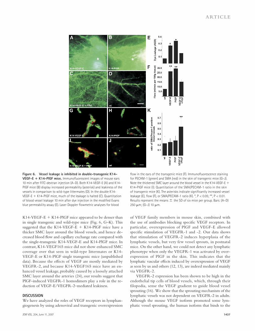
Overexpression of VEGF-E via an adeno-associated virus vec-tor promotes blood vessel leakage (22). K14-PlGF mice also show increased vessel permeability (23). As vessel leakage re-sults in interstitial edema that may provoke lymphatic vessel enlargement, we wanted to study in more detail the relation-ship between edema and lymphatic hyperplasia in the trans-genic mice. To compare vessel permeability in the transgenic mice, we injected high molecular weight FITC-dextran into the tail vein and monitored the effl ux of the dye at diff erent time points. FITC-dextran leaked out much more rapidly from the vessels of the K14-VEGF-E and K14-PlGF mice compared with the K14-VEGF-E + K14-PlGF mice (Fig. 6, A–D). We also injected Evans blue dye into the tail vein of the transgenic mice and their wild-type littermates and moni-tored its extravasation. Spectrophotometric quantifi cation of the dye indicated that although both K14-VEGF-E and K14-PlGF exhibited increased vessel permeability, the K14-VEGF-E + K14-PlGF mice had comparatively reduced vessel permeabil-ity (Fig. 6 E). The results agreed with those from the FITC-dextran experiments, and they indicate that PlGF, possibly by stimulating VEGFR-1 homodimer formation, can at least par-tially block the increased capillary permeability induced by VEGF-E through VEGFR-2 homodimer formation.
Correlation of blood fl ow with vascular leakage
in the transgenic mice
Using a laser Doppler fl owmeter, we measured ear tissue blood fl ow in the transgenic mice at 5 wk of age. Both the K14-VEGF-E and K14-PlGF mice had considerable in-creased blood fl ow (fold increase of 3.5 ± 0.2 and 3.9 ± 0.1, respectively) compared with the wild-type littermates (Fig. 6 F). To our surprise, the K14-VEGF-E + K14-PlGF mice showed only 2.0- ± 0.1-fold increased blood fl ow compared with the wild-type littermates, even though these mice have an increased density of blood vessels compared with the sin-gle transgenic mice (Fig. S5 and not depicted).
To investigate if the decreased blood fl ow in K14-VEGF-E + K14-PlGF mice was correlated with an increase in the smooth muscle cell (SMC) coating around the blood vessels, we measured the ratio of smooth muscle α-actin (SMA)–positive cells to PECAM-1 positive cells from frozen sections of the skin. As shown by representative images and ratio quantifi cation, the SMC layer around the blood vessels in
Figure 5. The lymphatic and blood vascular phenotypes in K14-
VEGF-E mice occur postnatally. (A and B) Skin lymphatic vessels of
K14-VEGF-E + VEGFR-3+/LacZ compound heterozygote and VEGFR-3+/LacZ
(wt) embryos were analyzed by β-galactoside staining at E17.5. Note the
similar degree of lymphatic vessel sprouting (arrowheads). Embryonic skin
blood vessels of a K14-VEGF-E + VEGFR-2+/LacZ compound heterozygote
and VEGFR-2+/LacZ mice were analyzed by β-galactoside staining at E17.5
(C and D). (E) Comparison of the lymphatic vessel areas of 1-, 7-, and 14-d-
old pups and adult K14-VEGF-E mice. (F) Comparison of BrdU + LYVE-1
double-positive cells in E17.5 embryos, P7 pups, and adult K14-VEGF-E
mice. The asterisks indicate signifi cantly increased vessel density areas.
*, P < 0.05. Results represent the means ± the SD of six embryos/mice
per group. Bars: (A and B) 500 μm; (C and D) 200 μm.
JEM VOL. 204, June 11, 2007 1437
ARTICLE
K14-VEGF-E + K14-PlGF mice appeared to be denser than in single transgenic and wild-type mice (Fig. 6, G–K). This suggested that the K14-VEGF-E + K14-PlGF mice have a thicker SMC layer around the blood vessels, and hence de-creased blood fl ow and capillary exchange rate compared with the single-transgenic K14-VEGF-E and K14-PlGF mice. In contrast, K14-VEGF165 mice did not show enhanced SMC coverage over that seen in wild-type littermates or K14-VEGF-E or K14-PlGF single transgenic mice (unpublished data). Because the eff ects of VEGF are mostly mediated by VEGFR-2, and because K14-VEGF165 mice have an en-hanced vessel leakage, probably caused by a loosely attached SMC layer around the arteries (24), our results suggest that PlGF-induced VEGFR-1 homodimers play a role in the re-duction of VEGF-E/VEGFR-2–mediated leakiness.
D I S C U S S I O N
We have analyzed the roles of VEGF receptors in lymphan-giogenesis by using adenoviral and transgenic overexpression
of VEGF family members in mouse skin, combined with the use of antibodies blocking specifi c VEGF receptors. In particular, overexpression of PlGF and VEGF-E allowed specifi c stimulation of VEGFR-1 and -2. Our data shows that stimulation of VEGFR-2 induces hyperplasia of the lymphatic vessels, but very few vessel sprouts, in postnatal mice. On the other hand, we could not detect any lymphatic phenotype when only the VEGFR-1 was activated by over-expression of PlGF in the skin. This indicates that the lymphatic vascular eff ects induced by overexpression of VEGF as seen by us and others (12, 13), are indeed mediated mainly via VEGFR-2.
VEGFR-2 expression has been shown to be high in the endothelial tip cells of blood vessels, which, through their fi lopodia, sense the VEGF gradient to guide blood vessel sprouting (16). We show that the sprouting mechanism of the lymphatic vessels was not dependent on VEGFR-2 in adults. Although the mouse VEGF isoform promoted some lym-phatic vessel sprouting, the human isoform that binds to the
Figure 6. Vessel leakage is inhibited in double-transgenic K14-
VEGF-E + K14-PlGF mice. Immunofl uorescent images of mouse ears
10 min after FITC-dextran injection (A–D). Both K14-VEGF-E (A) and K14-
PlGF mice (B) display increased permeability (asterisk) and leakiness of the
vessels in comparison to wild-type littermates (D). In the double K14-
VEGF-E + K14-PlGF mice, much of the leakage is halted (C). Quantitation
of blood vessel leakage 10 min after dye injection in the modifi ed Evans
blue permeability assay (E). Laser Doppler fl owmetric analyses for blood
fl ow in the ears of the transgenic mice (F). Immunofl uorescence staining
for PECAM-1 (green) and SMA (red) in the skin of transgenic mice (G–J).
Note the thickened SMC layer around the blood vessel in the K14-VEGF-E +
K14-PlGF mice (I). Quantitation of the SMA/PECAM-1 ratio in the skin
of transgenic mice (K). The asterisks indicate signifi cantly increased vessel
leakage (E), fl ow (F), or SMA/PECAM-1 ratio (K). *, P < 0.05; **, P < 0.01.
Results represent the means ± the SD of six mice per group. Bars: (A–D)
250 μm; (G–J) 10 μm.
1438 VEGF RECEPTORS IN LYMPHATIC HYPERPLASIA | Wirzenius et al.
same receptors did not. However, both isoforms induced enlargement of lymphatic vessels and proliferation of blood vessels. Furthermore, overexpression of the VEGFR-2–specifi c ligand VEGF-E did not lead to sprouting of the lymphatic vessels, but instead to circumferential growth and the sprout-ing of the lymphatic vessels induced by adenoviral VEGF-C, which could not be blocked with neutralizing antibodies against VEGFR-2. These results suggest that the sprouting mechanisms of lymphatic vessels in adults do not rely on VEGFR-2, and thus diff er from those of the blood vessels.
Surprisingly, VEGF-E did not induce changes in the lymphatic vasculature during embryonic development. Al-though VEGF-E stimulated lymphatic vessel enlargement in postnatal mice, we did not detect increased numbers of lym-phatic vessels in the K14-VEGF-E mice at any age analyzed. This indicates that signaling through VEGFR-2 does not induce lymphangiogenesis during embryonic development, but its eff ects become apparent during the postnatal period upon maturation of the blood and lymphatic vessels. As with the adenovirus vectors, we also detected circumferential lym-phatic vessel hyperplasia without additional sprouting in the transgenic mice after 1 wk of postnatal growth. One pos-sibility was that at least some of the hyperplasia was a second-ary eff ect resulting from the vascular leakage promoted by VEGF-E overexpression. The lymphatic hyperplasia started after the eff ects of VEGF-E in the blood vessels became apparent, and both the vascular leakage and the lymphatic hyperplasia could be reduced by PlGF overexpression that apparently reduced blood flow via the increased coating of small vessels by SMCs. However, from additional ex-periments, we learned that vascular leakage alone was not suffi cient for the hyperplasia, as the lymphatic vessels of the K14-PlGF mice appeared to be normal, although their blood vessels were even leakier than in the K14-VEGF-E mice. We cannot currently explain the late onset of the ef-fects of the K14-VEGF-E transgene, despite its high-level expression starting during the embryonic period. It may be speculated that in vivo particular pericellular matrix proteins and integrins are required for specifi c VEGFR-2 activa-tion by VEGF-E, and that they are not present until later in development. Importantly, we also found that adenoviral coexpression of PlGF inhibits VEGF-E-induced, but not VEGF-C156S–induced, lymphatic hyperplasia (this study and unpublished data).
We also found that VEGF-E overexpression was not able to rescue the lymphatic regression induced by blocking VEGF-C and -D (the VEGF-C/D trap). This result indi-cates that VEGFR-2 cannot substitute for VEGFR-3 sig-nals during the onset of lymphangiogenesis. In accordance with these data, treatment with AdVEGFR-2-Ig does not aff ect the postnatal development of lymphatic vessels (25). VEGFR-3 was expressed strongly in lymphatic capillaries, and it was internalized into the lymphatic endothelial cells after adenoviral VEGF-C stimulation, but not after VEGF-E stimulation, indicating that VEGF-E does not lead to sig-naling via this receptor. VEGFR-2 was expressed weakly in
lymphatic capillaries and more strongly in collecting lym-phatic vessels in wild-type mice. The fact that essentially no VEGF-E–induced changes were observed in the collecting lymphatic vessels or lymph nodes was surprising considering the strong constitutive VEGFR-2 expression in these vessels. However, VEGFR-2 was internalized in both types of ves-sels upon AdVEGF-E or -C transduction, indicating that the receptor was functional.
In conclusion, we show here that overexpression of a VEGFR-2–specifi c ligand induces circumferential hyper-plasia of the lymphatic vessels in adult, but not in embryonic, skin. However, VEGFR-2 activation is not suffi cient for the generation of new lymphatic vessels, and it was not able to rescue the lymphatic regression induced by blocking the VEGFR-3 ligands VEGF-C and -D. These results specify the contribution of the diff erent VEGFR pathways to lymphan-giogenesis and reveal previously unknown postnatal changes that occur in the sensitivity of both blood and lymphatic ves-sels toward VEGF family ligands.
MATERIALS AND METHODSCell stimulation and Western blot analysis. PAE cells stably overex-
pressing VEGFR-2 (26) or -3 (17) were starved overnight in serum-free
medium and incubated for 20 min with soluble VEGFR-2-Ig or VEGFR-
3-Ig (357-kD and 349-F4; R&D Systems) fusion proteins or rat monoclonal
antibodies against human VEGFR-2 (1121B; a gift from B. Pytowski, Im-
Clone Systems, New York, NY) at a concentration of 1 μg/ml. The cells
were stimulated for 10 min with recombinant VEGF-E (27) or -C (28) at the
indicated concentrations, washed with PBS, and lysed in ice-cold PLCLB
lysis buff er (150 mM NaCl, 5% glycerol, 1% Triton X-100, 1.5 M MgCl2,
and 50 mM Hepes, pH 7.5) containing 2 mM sodium vanadate, 2 mM
PMSF, 10 μg/ml leupeptin, and 0.07 U/ml aprotinin. Clarifi ed lysates were
immunoprecipitated with antibodies against 2 μg/ml VEGFR-3 (9D9f9
[29]) or 2 μg/ml VEGFR-2, separated by SDS-PAGE, and transferred to a
nitrocellulose fi lter. Detection was performed with phosphotyrosine-specifi c
antibodies (clone 4G10; Millipore) and an enhanced chemiluminescence
detection system (Pierce Chemical Co.). Antibodies against VEGFR-2
(C-1158; Santa Cruz Biotechnology) or -3 were used for detection of
VEGFR-2 and -3 in the Western blot, respectively.
Generation and in vitro analysis of recombinant adenoviruses. Full-
length VEGF-E cDNA (GenBank accession no. AF106020) was cloned into
the pAdapt vector, and the adenovirus was produced as previously described
(30). The adenoviruses encoding VEGF164, VEGF165, VEGF-C, VEGFR-
3-Ig, and nuclear-targeted β-galactosidase (LacZ) were constructed and pro-
duced as previously described (13, 30–32). Analysis of protein expression was
carried out as previously reported (32).
In vivo use of the viral vectors. 2.5 × 10⁸ PFU of recombinant adeno-
viruses encoding VEGF-C, VEGF-E, VEGF164, VEGF165, or LacZ were
injected intradermally into 4–6-wk-old female NMRI nu/nu mice (Harlan).
The skin was analyzed 4 d later. For inhibition experiments, 109 PFU of
VEGFR-3-Ig or LacZ adenoviruses were injected intravenously 3 d before
the ligand-encoding adenoviruses.
For sprouting inhibition experiments, the mice were injected intraperi-
toneally with 600 μg of mF4-31C1, which is a rat monoclonal antibody
against mouse VEGFR-3 (33), DC101, which is a rat monoclonal antibody
against mouse VEGFR-2 (34), or rat IgG every second day in a volume of
200 μl. 1 d later, 2.5 × 10⁸ PFU of recombinant adenoviruses encoding
VEGF-C, VEGF-C156S, VEGF-E, or LacZ were injected intradermally
into the ears of the same mice. The skin was analyzed 3–4 d later.
JEM VOL. 204, June 11, 2007 1439
ARTICLE
Transgenic mice. The K14-VEGF-E, K14-PlGF, K14-VEGF-C156S,
K14-VEGF-C, K14-VEGFR-3-Ig, K14-VEGF165, VEGFR-3+/LacZ, and
VEGFR-2+/LacZ mice were previously described (11, 17, 19, 23, 24, 35–37).
The Provincial State Offi ce of Southern Finland approved all experiments
involving mice, and they were performed in accordance with institu-
tional guidelines.
Analysis of lymphatic and blood vessels. Whole-mount staining was
performed as previously published (15). Blood and lymphatic vessels were
stained with rabbit antiserum against LYVE-1 (38), α-hamster monoclonal
anti–mouse PECAM-1 antibody (clone 2H8, MAB-13982Z; CHEMICON
International, Inc.), α-goat polyclonal anti–VEGFR-2 antibody (AF644;
R&D Systems), or α-goat polyclonal anti–mouse VEGFR-3 antibody
(AF743; R&D Systems), followed by appropriate fl uorochrome-conjugated
secondary antibody (Alexa Fluor 488 or 594 [Invitrogen] or FITC [Jackson
ImmunoResearch Laboratories]). Frozen sections were fi xed with −20°C
acetone, incubated with anti-LYVE-1, the hamster monoclonal antibody
against PECAM-1 (CHEMICON International, Inc.), a rat monoclonal an-
tibody against CD11b (BD Biosciences), or mouse anti–SMA-Cy3 (Sigma-
Aldrich), followed by the appropriate fl uorochrome-conjugated secondary
antibodies and analysis with a compound fl uorescent microscope (Zeiss 2;
Carl Zeiss MicroImaging, Inc.; 10× objective/NA 0.30), or a confocal
microscope (Zeiss LSM 510; 20× objective/NA 1.3 and 40× objective/
NA 1.4). Three-dimensional projections were digitally constructed from
confocal z stacks.
Paraffi n sections were stained with a rabbit antibody against LYVE-1
and a rat anti–monoclonal antibody against PECAM-1 (BD Biosciences)
using the tyramide signal amplifi cation kit (PerkinElmer). Lymphangiography
and visualization of blood vessels with FITC-dextran was performed as pre-
viously described (22, 32). Quantitation of the lymphatic sprouts and the
area covered by lymphatic or blood vessels in the skin was performed as pre-
viously described (25). The area covered by lymphatic or blood vessels in the
skin was quantifi ed from photomicrographs of LYVE-1– or PECAM-1–
stained sections (6 photomicrographs/mouse, and 6 mice of each genotype
at each time point) using the Image-Pro Plus program (Media Cybernetics).
All statistical analyses were performed using the unpaired Student’s t test.
A P value of <0.05 was considered statistically signifi cant.
𝛃-Galactosidase staining of vessels. Staged embryos of the compound
K14-VEGF-E + VEGFR-2+/LacZ and K14-VEGF-E + VEGFR-3+/LacZ
mice were dissected, fi xed in 0.2% glutaraldehyde, and stained with X-gal
(Sigma-Aldrich) for β-galactosidase activity at 37°C.
BrdU incorporation and immunostaining. 20 mM BrdU in PBS was
injected intraperitoneally at a volume of 20 μl/g into adult mice, P7
pups, or pregnant mice at E17.5. Mice were sacrifi ed 4 h later, and frozen
sections from the skin were stained with a monoclonal rat anti-BrdU anti-
body (Abcam Ltd.).
RT-PCR. RNA extracted from the skin of staged K14-VEGF-E embryos
and their wild-type littermates was reverse transcribed using oligo-dT
(Boehringer) and Superscript II reverse transcriptase (Invitrogen). PCR anal-
ysis using a pair of primers specifi c for VEGF-E or β-actin primers was per-
formed as previously published (22).
Quantitative RT-PCR. RNA was extracted from K14-VEGF-E and wild-
type littermate pups at P7, and quantitative PCR reactions were performed as
previously described (25) with the DyNAmo HS SYBR Green qPCR kit
(Finnzymes) using the ABI 7500 SDS real-time PCR instrument (Applied
Biosystems). The oligonucleotide primers used are as follows: 5′-C A C A G T G-
T C A G G C A G C T A A C -3′ and 5′-T C C A C A G A C A T C A T G G A A T C -3′ for
VEGF-C; and 5′-C T T G C T G G A A C A G A A G A C C A -3′ and 5′-C T C T G A-
G G A C T G G A A G C T G T -3′ for VEGF-D, as well as 5′-A C A A C T T T G G C A-
T T G T G G A A -3′ and 5′-G A T G C A G G G A T G A T G T T C T G -3′ for GAPDH.
The expression of the genes was normalized to GAPDH expression.
Measurement of vascular permeability and tissue blood fl ow. A modi-
fi ed Evans blue permeability assay was performed as previously described
(32). Microcirculation was measured from the base of the ear by a HL-N1451
Flowprobe (Transonic Systems, Inc.) connected to a laser Doppler fl ow-
meter (model BLF21; Transonic Systems, Inc.).
Online supplemental material. Fig. S1 shows ligand-dependent VEGFR-2
and -3 phosphorylation and its inhibition by antibodies. Fig. S2 shows in-
ternalization of VEGFR-2 and -3 upon ligand stimulation in the lymphatic
capillaries. Fig. S3 shows collecting lymphatic vessels and lymph node vascu-
larity in K14-VEGF-E mice. Fig. S4 shows a CD11b staining of the skin of
the diff erent transgenic mice. Fig. S5 shows PECAM-1 staining of the skin
of transgenic mice at diff erent ages. The online version of this article is avail-
able at http://www.jem.org/cgi/content/full/jem.20062642/DC1.
We thank Dr. Bronislaw Pytowski for 1121B, DC101, and mF4-31C1 antibodies,
Dr. Michael Detmar for kindly supplying tissues from K14-VEGF164 mice, Drs. Caroline
Heckman and Terhi Karpanen for critical comments on the manuscript, and Tapio
Tainola, Mari Helanterä, Sanna Karttunen, Paula Hyvärinen, and Kaisa Makkonen
for excellent technical assistance. The Biomedicum Molecular Imaging Unit is
acknowledged for its expertise on confocal microscopy.
This study was supported by grants from the Finnish Cancer Organizations,
the Academy of Finland (202852 and 204312), National Institutes of Health
(5 R01 HL075183-02), the Novo Nordisk Foundation, the European Union
(Lymphangiogenomics LSHG-CT-2004-503573; the Louis Jeantet Foundation; and
Tumor-Host Genomics LSHC-CT-2005-518198). M. Wirzenius obtained personal
grants from Svenska Kulturfonden, Biomedicum Helsinki Foundation, and
the K.A. Johanssons Foundation.
K. Alitalo and S. Ylä -Herttuala are minority shareholders of Lymphatix Ltd.,
and K. Alitalo is a scientifi c advisory board member of Vegenics Ltd.
The authors have no other confl icting fi nancial interests.
Submitted: 18 December 2006
Accepted: 9 May 2007
R E F E R E N C E S 1. Ferrara, N., and R.S. Kerbel. 2005. Angiogenesis as a therapeutic target.
Nature. 438:967–974. 2. Carmeliet, P. 2005. Angiogenesis in life, disease and medicine. Nature.
438:932–936. 3. Alitalo, K., T. Tammela, and T.V. Petrova. 2005. Lymphangiogenesis
in development and human disease. Nature. 438:946–953. 4. Ferrara, N., K. Carver-Moore, H. Chen, M. Dowd, L. Lu, K.S. O’Shea,
L. Powell-Braxton, K.J. Hillan, and M.W. Moore. 1996. Heterozygous embryonic lethality induced by targeted inactivation of the VEGF gene. Nature. 380:439–442.
5. Carmeliet, P., V. Ferreira, G. Breier, S. Pollefeyt, L. Kieckens, M. Gertsenstein, M. Fahrig, A. Vandenhoeck, K. Harpal, C. Ebenhardt, et al. 1996. Abnormal blood vessel development and lethality in embryos lacking a single VEGF allele. Nature. 380:435–439.
6. Lyttle, D.J., K.M. Fraser, S.B. Fleming, A.A. Mercer, and A.J. Robinson. 1994. Homologs of vascular endothelial growth factor are encoded by the poxvirus orf virus. J. Virol. 68:84–92.
7. Ogawa, S., A. Oku, A. Sawano, S. Yamaguchi, Y. Yazaki, and M. Shibuya. 1998. A novel type of vascular endothelial growth factor, VEGF-E (NZ-7 VEGF) preferentially utilizes KDR/Flk-1 receptor and carries a potent mitotic activity without heparin-binding domain. J. Biol. Chem. 273:31273–31282.
8. Meyer, M., M. Clauss, A. Lepple-Wienhues, J. Waltenberger, H.G. Augustin, M. Ziche, C. Lanz, M. Böttner, H.-J. Rziha, and C. Dehio. 1999. A novel vascular endothelial growth factor encoded by Orf virus, VEGF-E, mediates angiogenesis via signaling through VEGFR-2 (KDR) but not VEGFR-1 (Flt-1) receptor tyrosine kinases. EMBO J. 18:363–374.
9. Holmes, D.I., and I. Zachary. 2005. The vascular endothelial growth factor (VEGF) family: angiogenic factors in health and disease. Genome Biol. 6:209.
1440 VEGF RECEPTORS IN LYMPHATIC HYPERPLASIA | Wirzenius et al.
10. Wise, L.M., T. Veikkola, A.A. Mercer, L.J. Savory, S.B. Fleming, C. Caesar, A. Vitali, T. Makinen, K. Alitalo, and S.A. Stacker. 1999. Vascular endothelial growth factor (VEGF)-like protein from orf virus NZ2 binds to VEGFR2 and neuropilin-1. Proc. Natl. Acad. Sci. USA. 96:3071–3076.
11. Kiba, A., H. Sagara, T. Hara, and M. Shibuya. 2003. VEGFR-2-specifi c ligand VEGF-E induces non-edematous hyper-vascularization in mice. Biochem. Biophys. Res. Commun. 301:371–377.
12. Nagy, J.A., E. Vasile, D. Feng, C. Sundberg, L.F. Brown, M.J. Detmar, J.A. Lawitts, L. Benjamin, X. Tan, E.J. Manseau, et al. 2002. Vascular permeability factor/vascular endothelial growth factor induces lymphan-giogenesis as well as angiogenesis. J. Exp. Med. 196:1497–1506.
13. Hirakawa, S., S. Kodama, R. Kunstfeld, K. Kajiya, L.F. Brown, and M. Detmar. 2005. VEGF-A induces tumor and sentinel lymph node lymphangiogenesis and promotes lymphatic metastasis. J. Exp. Med. 201:1089–1099.
14. Enholm, B., T. Karpanen, M. Jeltsch, H. Kubo, F. Stenback, R. Prevo, D.G. Jackson, S. Yla-Herttuala, and K. Alitalo. 2001. Adenoviral ex-pression of vascular endothelial growth factor-C induces lymphangio-genesis in the skin. Circ. Res. 88:623–629.
15. Tammela, T., A. Saaristo, M. Lohela, T. Morisada, J. Tornberg, C. Norrmen, Y. Oike, K. Pajusola, G. Thurston, T. Suda, et al. 2005. Angiopoietin-1 promotes lymphatic sprouting and hyperplasia. Blood. 105:4642–4648.
16. Gerhardt, H., M. Golding, M. Fruttiger, C. Ruhrberg, A. Lundkvist, A. Abramsson, M. Jeltsch, C. Mitchell, K. Alitalo, D. Shima, and C. Betsholtz. 2003. VEGF guides angiogenic sprouting utilizing endothe-lial tip cell fi lopodia. J. Cell Biol. 161:1163–1177.
17. Makinen, T., L. Jussila, T. Veikkola, T. Karpanen, M.I. Kettunen, K.J. Pulkkanen, R. Kauppinen, D.G. Jackson, H. Kubo, S. Nishikawa, et al. 2001. Inhibition of lymphangiogenesis with resulting lymphedema in transgenic mice expressing soluble VEGF receptor-3. Nat. Med. 7:199–205.
18. Joukov, V., V. Kumar, T. Sorsa, E. Arighi, H. Weich, O. Saksela, and K. Alitalo. 1998. A recombinant mutant vascular endothelial growth factor-C that has lost vascular endothelial growth factor receptor-2 binding, activation, and vascular permeability activities. J. Biol. Chem. 273:6599–6602.
19. Veikkola, T., L. Jussila, T. Makinen, T. Karpanen, M. Jeltsch, T.V. Petrova, H. Kubo, G. Thurston, D.M. McDonald, M.G. Achen, et al. 2001. Signalling via vascular endothelial growth factor receptor-3 is suffi -cient for lymphangiogenesis in transgenic mice. EMBO J. 20:1223–1231.
20. Karpanen, T., M. Egeblad, M.J. Karkkainen, H. Kubo, D.G. Jackson, S. Ylä-Herttuala, M. Jäättelä, and K. Alitalo. 2001. Vascular endothelial growth factor C promotes tumor lymphangiogenesis and intralymphatic tumor growth. Cancer Res. 61:1786–1790.
21. Veikkola, T., M. Lohela, K. Ikenberg, T. Makinen, T. Korff , A. Saaristo, T. Petrova, M. Jeltsch, H.G. Augustin, and K. Alitalo. 2003. Intrinsic versus microenvironmental regulation of lymphatic endothelial cell phenotype and function. FASEB J. 17:2006–2013.
22. Uutela, M., M. Wirzenius, K. Paavonen, I. Rajantie, Y. He, T. Karpanen, M. Lohela, H. Wiig, P. Salven, K. Pajusola, et al. 2004. PDGF-D in-duces macrophage recruitment, increased interstitial pressure, and blood vessel maturation during angiogenesis. Blood. 104:3198–3204.
23. Odorisio, T., C. Schietroma, M.L. Zaccaria, F. Cianfarani, C. Tiveron, L. Tatangelo, C.M. Failla, and G. Zambruno. 2002. Mice overexpress-ing placenta growth factor exhibit increased vascularization and vessel permeability. J. Cell Sci. 115:2559–2567.
24. Zheng, Y., M. Murakami, H. Takahashi, M. Yamauchi, A. Kiba, S. Yamaguchi, N. Yabana, K. Alitalo, and M. Shibuya. 2006. Chimeric
VEGF-E(NZ7)/PlGF promotes angiogenesis via VEGFR-2 without signifi cant enhancement of vascular permeability and infl ammation. Arterioscler. Thromb. Vasc. Biol. 26:2019–2026.
25. Karpanen, T., M. Wirzenius, T. Makinen, T. Veikkola, H.J. Haisma, M.G. Achen, S.A. Stacker, B. Pytowski, S. Yla-Herttuala, and K. Alitalo. 2006. Lymphangiogenic growth factor responsiveness is modulated by postnatal lymphatic vessel maturation. Am. J. Pathol. 169:708–718.
26. Waltenberger, J., L. Claesson-Welsh, A. Siegbahn, M. Shibuya, and C.H. Heldin. 1994. Diff erent signal transduction properties of KDR and Flt1, two receptors for vascular endothelial growth factor. J. Biol. Chem. 269:26988–26995.
27. Jeltsch, M., T. Karpanen, T. Strandin, K. Aho, H. Lankinen, and K. Alitalo. 2006. Vascular endothelial growth factor (VEGF)/VEGF-C mosaic molecules reveal specifi city determinants and feature novel re-ceptor binding patterns. J. Biol. Chem. 281:12187–12195.
28. Karkkainen, M.J., P. Haiko, K. Sainio, J. Partanen, J. Taipale, T.V. Petrova, M. Jeltsch, D.G. Jackson, M. Talikka, H. Rauvala, et al. 2004. Vascular endothelial growth factor C is required for sprouting of the fi rst lymphatic vessels from embryonic veins. Nat. Immunol. 5:74–80.
29. Jussila, L., R. Valtola, T.A. Partanen, P. Salven, P. Heikkila, M.T. Matikainen, R. Renkonen, A. Kaipainen, M. Detmar, E. Tschachler, et al. 1998. Lymphatic endothelium and Kaposi’s sarcoma spindle cells detected by antibodies against the vascular endothelial growth factor receptor-3. Cancer Res. 58:1599–1604.
30. Puumalainen, A.M., M. Vapalahti, R.S. Agrawal, M. Kossila, J. Laukkanen, P. Lehtolainen, H. Viita, L. Paljarvi, R. Vanninen, and S. Yla-Herttuala. 1998. Beta-galactosidase gene transfer to human malig-nant glioma in vivo using replication-defi cient retroviruses and adeno-viruses. Hum. Gene Ther. 9:1769–1774.
31. Laitinen, M., I. Zachary, G. Breier, T. Pakkanen, T. Hakkinen, J. Luoma, H. Abedi, W. Risau, M. Soma, M. Laakso, et al. 1997. VEGF gene transfer reduces intimal thickening via increased production of ni-tric oxide in carotid arteries. Hum. Gene Ther. 8:1737–1744.
32. Saaristo, A., T. Veikkola, T. Tammela, B. Enholm, M.J. Karkkainen, K. Pajusola, H. Bueler, S. Yla-Herttuala, and K. Alitalo. 2002. Lymphangiogenic gene therapy with minimal blood vascular side eff ects. J. Exp. Med. 196:719–730.
33. Pytowski, B., J. Goldman, K. Persaud, Y. Wu, L. Witte, D.J. Hicklin, M. Skobe, K.C. Boardman, and M.A. Swartz. 2005. Complete and spe-cifi c inhibition of adult lymphatic regeneration by a novel VEGFR-3 neutralizing antibody. J. Natl. Cancer Inst. 97:14–21.
34. Prewett, M., J. Huber, Y. Li, A. Santiago, W. O’Connor, K. King, J. Overholser, A. Hooper, B. Pytowski, L. Witte, et al. 1999. Antivascular endothelial growth factor receptor (fetal liver kinase 1) monoclonal an-tibody inhibits tumor angiogenesis and growth of several mouse and human tumors. Cancer Res. 59:5209–5218.
35. Dumont, D.J., L. Jussila, J. Taipale, A. Lymboussaki, T. Mustonen, K. Pajusola, M. Breitman, and K. Alitalo. 1998. Cardiovascular failure in mouse embryos defi cient in VEGF receptor-3. Science. 282:946–949.
36. Shalaby, F., J. Rossant, T.P. Yamaguchi, M. Gertsenstein, X.F. Wu, M.L. Breitman, and A.C. Schuh. 1995. Failure of blood-island forma-tion and vasculogenesis in Flk-1-defi cient mice. Nature. 376:62–66.
37. Jeltsch, M., A. Kaipainen, V. Joukov, X. Meng, M. Lakso, H. Rauvala, M. Swartz, D. Fukumura, R.K. Jain, and K. Alitalo. 1997. Hyperplasia of lymphatic vessels in VEGF-C transgenic mice. Science. 276:1423–1425.
38. Laakkonen, P., K. Porkka, J.A. Hoff man, and E. Ruoslahti. 2002. A tumor-homing peptide with a targeting specifi city related to lymphatic vessels. Nat. Med. 8:751–755.


