Embed Size (px)
Citation preview

1 Supported by NIH grant HL18645.2 To whom correspondence should be addressed.
819
0022-1554/891$3.30
The Journal of Histochemistry and CytochemistryCopyright © 1989 by The Histochemical Society. Inc.
Vol. 37, No. 6, pp. 819-829. 1989
Printed in U.S.A.
Original Article
Distribution of the Calcium-binding Protein SPARC InTissues of Embryonic and Adult Mice’
HELENE SAGE,2 ROBERT B. VERNON, JAY DECKER, SARAH FUNK,
and MARIA LUISA IRUELA-ARISPEDepartment ofB:ological Structure, University of Washington, Seattle, Washington 98195.
Received for publication August 29, 1988 and in revised form December 20, 1988; accepted December 27, 1988 (8A1470).
SPARC (Secreted Protein that is Acidic and Rich in Cysteine),
a Ca�-bin&ng glycoprotein also known as osteonectin, isproduced in significant amounts by injured or proliferatingcells in vitro. To elucidate the possible function of SPARCin growth and remodeling, we examined its distribution in
embryonic and adult murine tissues. Immunohistochemis-try on adult mouse tissues revealed a preferential associa-tion of SPARC protein with epitheia exhibiting high ratesof turnover (gut, skin, and glandular tissue). Fetal tissuescontaining high levels of SPARC induded heart, thymus,lung, and gut. In the 14-18-day developing fetus, SPARCexpression was particularly enhanced in areas undergoing
Introduction
SPARC is a secreted Ca�-binding protein which is induced in con-
junction with cell differentiation, proliferation, stress, and certain
developmental signals (Holland et al., 1987; Mason et al., 1986a,b;
Sage et al. , 1986). Molecular cloning and protein sequence analysis
have shown that SPARC is identical to osteonectin (ON), originally
described as a major noncollagenous protein ofbone which bound
Ca�, hydnoxyapatite, and type I collagen (Bolanden et al., 1988;
Mason et al., 1986b; Young et al., 1986; Rombeng et al., 1985; Ten-
mine et al., 1981). SPARC is also identical to BM-40, a product
of a basement membrane-secreting tumor cell line (Mann et al.,
1987; Dziadek et al., 1986), and to an Mr 43 KD glycopnotein, syn-
thesized in vitro by cells ofthe vessel wall as well as by other normal
and transformed cells derived from all primordial germ layers (Sage
et al., 1981, 1984). ON was also found in dentin, periodontal liga-
ment (Tung et al., 1985), and platelets (Stennen et al., 1986), and
was a synthetic product ofgingival and ligament fibnoblasts (Zung
et al., 1986; Wasi et al., 1984) and calvarial cells in vitro (Otsuka
et al., 1984). The Mr 43 KD protein was identified as a “culture
shock” protein which bound bovine serum albumin (BSA) and a
70 KD serum protein, and which was associated with endothelial
chondrogenesis, osteogenesis, and somitogenesis, whereas
10-day embryos exhibited selective staining for this proteinin Reichert’s membrane, maternal sinuses, and trophoblas-
tic giant cells. SPARC displayed a Ca�-dependent affinityfor hydrophobic surfaces and was not incorporated into the
extraceilular matrix produced by cells in vitro. We proposethat in some tissues SPARC associates with cell surfaces to
facilitate proliferation during embryonic morphogenesis andnormal cell turnover in the adult. (J Histochem Cytochem37:819-829, 1989)KEY WORDS: SPARC; Calcium binding; Mouse embryo; Morpho-
genesis; Trophoblast.
cell migration, proliferation, serum deprivation, and endotoxin-
mediated injury (Sage, 1986a; Sage et al., 1986).
Since bone ON/SPARC is now known to be a potent inhibitor
of hydroxyapatite formation (Rombeng et al., 1985), it has been
suggested that the protein might inhibit (Engel et al., 1987) rather
than promote (Tenmine et al. , 1981) extracellular mineralization.
Although SPARC mRNA levels are decreased in virally transformed
cells (Mason et al., 1986a; Young et al., 1986), it has been observed
that SPARC is up-regulated in normal cells undergoing prolifena-
tion (Sage et al., i986). The location of SPARC protein in vivo,
however, has remained controversial. By immunofluorescence his-
tochernistry, ON has been identified in the extracellular matrix and
in cells ofligament and mineralizing tissues (Tung et al., 1985; Wasi
et al., 1984; Termine et al., 1981), while BM-40 was observed in
a limited number of basement membranes (Dziadek et al. , 1986).
In contrast, SPARC mRNA has been localized in a variety of tis-
sues. A recent study has shown tissue-specific and developmen-
tally related expression of SPARC mRNA in fetal and adult mice
(Holland et al. , 1987). These results suggested the association of
SPARC with proliferating cells in several tissues.
In this study we developed highly specific polyclonal antibod-
ies to examine the distribution ofSPARC protein in embryonic and
adult mice. Its association with monphogenesis and proliferation
in certain tissues resembled in part the immunohistochemical dis-
tribution ofltF43 protein in the mouse embryo (Heine et al., i987).
We have also shown that SPARC has a Ca�-sensitive affinity for
by guest on December 15, 2015jhc.sagepub.comDownloaded from

.�tL34 5 6
I.
s�
III
820 SAGE, VERNON, DECKER, FUNK, IRUELA-ARISPE
hydrophobic surfaces. SPARC may therefore function as an extnacel-
lular modulator of Ca� at the cell surface which either initiates
or facilitates cell migration and/or growth.
Materials and Methods
Cell Culture
Human femoral head osteoblasts in primary culture were a gift from Dr.
W. Lanzer (University of Washington). MG63 human osteosarcoma cells
were obtained from American Type Culture Collection (Rockville, MD).
Mouse PYS-2 cells, a teratocarcinoma line derived from panietal yolk sac
endoderm, were gifts from Drs. B. Hogan (Vanderbilt University, Nash-
yule, TN) and J. Lehman (Albany Medical College, Albany, NY). Meta-
bolic labeling ofcells in vitro with [355J.Met was according to Sage et al.
(1986) and Sage (1986a).
Purification of SPARC Protein
PYS-2 cells were primarily used as a source of SPARC. Subconfluent cells
(15-20 x 106) were incubated in Dulbecco Modified Eagle’s Medium
(DMEM) in the absence of fetal calf serum; 21 hr later the medium was
collected, clarified of cell debris, and stirred overnight at 4’C in the pres-
ence of 50% (w/v) ammonium sulfate. The precipitated protein was pelleted
by centrifugation, dissolved in and dialyzed against a 4 M urea-SO mM
Tnis-HCI buffer(pH 8), and chromatographed on diethylaminoethyl(DEAE)-cellulose at 4’C. The flow-through was discarded; laminin and BSA were
eluted both with 75 mM and 175 mM NaC1 in the same buffer, and SPARCwas eluted with 175 mM NaCl (Sage et al., 1984, 1986). Dialysis of this
second eluate peak against distilled water caused SPARC to precipitate
quantitatively, while the BSA remained in solution. This precipitation
occurred in a relatively narrow pH range (3.5-5.5) and could be inhib-
ited by 10 mM ethylenediamine tetraacetic acid (EDTA). SPARC was
purified further by chromatography on Sephadex G-200 in Tnis-buffered
saline (TBS) at 4’C or, alternatively, by carboxymethyl (CM)-cellulose chro-
matography at 20’C in a 2 M urea, 40 mM Na acetate buffer (pH 4.5),
after an initial denaturation step at 40’C for 5-10 mm. Pure SPARC (250-500
�tg) was recovered from 50 x 106 PYS cells. To monitor the purification,
one dish ofcells was often radiolabeled with [35S].Met. However, SPARCused for Western blotting was nonradioactive.
Sodium dodecylsulfate polyacrylamide gel electrophoresis (SDS-PAGE),
protein staining, fluorescence autoradiography, and scanning densitome-
try were performed as previously described (Sage et al., 1986; Laemmli, 1970).
incubated with the blot in MT buffer overnight at room temperature. The
blot was subsequently washed five times for 5 mm each in MT buffer, in-
cubated 2-3 hr with 100 p1 [ ‘2511-protein A (approximately 10 �.tCi) (New
England Nuclear; Boston, MA) in 35 ml MT buffer, washed sequentiallyin 1% MT buffrr (twice for 10 mm) and PBS-Tween (eight times, for a
minimum of 2 hr), and dried before exposure to X-ray film.
Structural Studies and Binding Assays
Circular Dichroism. Far ultraviolet circular dichroism spectra were ob-
tamed at 20’C from a Cary 61 spectrometer, calibrated with D( - )-panto-
Preparation and Characterization of Antibodies
Both native SPARC (purified on G-200) and denatured SPARC (purified
on CM-cellulose) were used to generate polyclonal antibodies in adult rab-
bits. lgG was precipitated from whole antisera by addition of ammonium
sulfate to a final concentration of 20% (w/v). IgG specific for SPARC was
purified by affinity chromatography on Sepharose CL.4B (Pharmacia; Pis-
cataway, NJ) coupled with purified SPARC, according to the manufacturer’s
instructions. Affinity-purified anti-SPARC IgG(typical concentrations ranged
from 0.1-0.225 mg/mI) was reactive by enzyme-linked immunosorbent as-
say (ELISA) at dilutions in excess of 1:2048 and exhibited no reactivity to-
wards BSA or fetal calf serum. Antibodies were characterized by ELISA,Western blotting, and radioimmune precipitation (Sage et al. , 1984, 1986).
Proteins were transferred to nitrocellulose for 1-3 hr at 500 mA, stained
with Amido black, dc-stained, and blocked in 1% non-fat dry milk-0.05%
Tween in PBS at pH 8.0 (MT buffer), with gentle shaking at room tempera-
rune for 1-2 hr or at 4C overnight. Anti-SPARC antisera (diluted at a mini-
mum of 1:1000) or affinity-purified antibodies (diluted 1250-500) were
0:
Figure 1. Fractionation and identification of SPARC in PYS culture media.
Serum-free culture media were collected from subconfluent PYS cells. Lanes1 and 2 show Ccomassie blue-stained proteins, fractionated initially by ammo-nium sulfate precipitation and DEAE-cellulose chromatography, which were di-alyzed against water at pH 5.5. Lane 1 (supernatant) contains BSA (A) and lane2 (precipitate) contains primarily SPARC (5). Western blotting of lane 2 mate-rial with affinity-purified anti-SPARC IgG in conjunction with [125l1-protein A isshown in lane 4; lane 3 is a duplicate sample incubated with antibody that waspro-absorbed with SPARC. [asS]�Met.labeIed proteins secreted by PYS cells(lane 5) yield purified SPARC after immunoprecipitation with anti-SPARC lgG(lane 6). Proteins in the presence of OTT were resolved by SDS-PAGE on5%/10% gels. Molecular weight markers, shown on the far left, are, in descend-ing order, 200 KO, 97 KD, 68 KD, 43 KD, and 28 KD.
by guest on December 15, 2015jhc.sagepub.comDownloaded from

DISTRIBUTION OF SPARC IN MURINE flSSUES 821
cent reports which have proposed a basement membrane- or matrix-
lactone, with a quartz cell of 0.1-mm path length. Protein concentrations
ranged from 0.15-0.45 mg/mI (5.1-15 riM) in TBS containing 2.5-5 mM
CaCI2 or 12 mM EDTA. The molar ellipticity 0 at 220 nm (deg cm2 /dmol)was calculated from a mean residue molecular weight of 110 determined
from the amino acid composition ofmurine SPARC (Mason et al., 1986b).Protein concentration was determined by amino acid analysis (performed
by Dr. D. Eyre, University ofWashington) or from an extinction coefficient
(E#{176}’’1280. 1 cm) of 1.076 calculated from the compositional data and the
molecular weight of 33,062 predicted from the cDNA sequence (Mason
et al., 1986b).
Chelex Ca�-binding Assay. This procedure followed closely the meth-
odology described by Waisman et al. (1986) and Waisman and Rasmussen
(1983). Chelex 100 (minus 400 mesh; Bio-Rad, Richmond, CA)-treated
buffers were used throughout the four separate experiments. In a typical
set, 249.3 �zg (7.54 nmol) of purified native SPARC was dissolved in 100
�tl 100 mM Tris-HC1, pH 7.4-50 mM NaCI buffer. Purified bovine testicu-
lar calmodulin (a gift from Dr. R. Klevit, University of Washington) at a
similar concentration was examined for 45Ca-binding activity in parallel
with SPARC. To 25 �tl Chelex resin were added various amounts (10-150
�tl) of SPARC or calmodulin and from 1-25 �zl [45Caj-Cl2 (2 mCi or 93
Mg Ca/mI; Amersham International, Poole, UK), in a final volume of 500�tl Chelexed buffer. Solutions were vortexed frequently, microfuged, and2-100 �tl aliquots of the supernatant were counted by liquid scintillation
spectrometry (Waisman et al., 1986). In each set ofexperiments, the ionic
strength was kept at a constant value. Controls included BSA and reaction
mixtures without protein. Protein concentrations were determined by amino
acid analysis.
Affinity Chromatography
Metal chelate affinity chromatography (iminodiacetic acid coupled to epoxy-activated Sepharose 6B; Pharmacia) was performed according to the manufac-
turer’s instructions. The column was equilibrated in CuSO4 (2.5 mg/mI)
and samples were eluted with 0.05 M EDTA-O.S M NaCI, followed by 2 M
EDTA, pH 7.0, and 1% SDS. Phenyl Sepharose chromatography (Pharma-
cia) was according to Davis et al. (1986) with modifications as described
in the figure legend.
Immunohistological Studies
Immunofluorescence microscopy was performed on cells grown on glass coy-erslips in vitro as previously described (Sage et al., 1984, 1986). Antibody
concentrations were determined by absorbance at 280 nm and extrapola-
tion to a standard curve for normal rabbit IgG.
Timing of mouse embryos was as follows: females were injected with
pregnant mare serum at 1700 hr the first day and human chonionic gonad-
otropin at 1700 hr on the second day; midnight on the following (third)
day (24 hr after appearance of the vaginal plug) marked the end of day 1.
Tissues and embryos from Swiss-Webster mice were either frozen im-mediately after dissection in OCT (Tissue-Tek) in liquid N2-cooled Freon
12 or immersed for 4-6 hr in Bouin’s fixative (0.9% picnic acid and 9%
formaldehyde by volume in 5 % acetic acid). Some of the fixed specimenswere demineralized in 8 N formic acid: 1 N Na formate (11) for 12-18
hr. Fixed tissues were dehydrated in ascending concentrations of ethanol,
cleared in toluene, and embedded in paraffin. Sections were cut at a thick-ness of 6 �tm (paraffin blocks) or 8 �tm (cryostat sections) and were placed
on slides pre-coated with a 0.1% solution ofpoly-L.lysine in water. Paraffin
sections were dried overnight at 4PC and stored at - 20CC for a maximumof 1 week before use. Paraffin sections were deparaffinized in two washes
( S mm each) ofxylene, rehydrated through a graded series ofethanol solu-tions to 70%, and washed four times for 3 mm in PBS. Frozen sections
were fixed for 5 mm in 2% paraformaldehyde at 4C and were dried for
0.5 hr at 24C (Sage et al., in press).
Sections intended for immunoperoxidase staining were incubated 30mm in 70% methanol containing 3% H2O2, followed by PBS, to macti-
vate endogenous peroxidases. Nonspecific binding of immunoglobulinswas minimized by incubating sections in PBS-10% normal goat serum. Sec-
tions were exposed sequentially to anti-SPARC IgG, diluted in 1% normal
goat serum-PBS (dilutions varied with the titer of the antibody, as deter-mined by ELISA, and were generally from 150-i00 for ammonium sulfate
precipitates and 1 :2-5 for affinity-purified fractions), and rhodamine- or
fluorescein isothiocyanate(RI1E, FITC)-conjugated to goat antibodies againstrabbit IgG (diluted 11#{174}in 10% goat serum)(Sage et al., in press). Altema-
tively, sections were exposed sequentially to anti-SPARC IgG, biotinylated
goat antibodies against rabbit IgG, avidin-biotin-peroxidase complex (ABCReagent; Vector Labs, Burlingame, CA), and 3,3’-diaminobenzidine-4 HCI(3 mg/mI in 0.05 M Tris-HCI, pH 7.6, containing 0.02% H2O2)(8-10 mm).
Sections were washed in PBS (four times for 3 mm) after each addition.
Kodak Tri-X film was used for black-and-white photography, Kodak SOT
type EPY was used for brightfield color photomicroscopy, and Kodak ASA
400 Ektachrome was used for color photography with a Zeiss Photomicro-
scope equipped for epifluorescence. Specificity controls included (a) sub-
stitution of normal rabbit IgG at the same concentration used for anti-SPARC IgG, (b) use of second antibody alone, (c) incubation of primary
antibody with excess purified SPARC or BSA, (d) use ofa series of concen-trations of several preparations of primary antibody, and (e) use of rabbit
anti-human plasma fibronectin (FN) IgG (a gift from Dr. P. Bornstein,
University of Washington) on serial sections.
Results
Previous studies by others and from our own laboratory had shown
SPARC to be secreted by cells derived from all germ layers under
a variety ofcultune conditions. A common observation was the as-
sociation ofSPARC with cells undergoing proliferation, migration,
attachment on detachment, and modulation of biosynthetic pheno-
type in response to a foreign substrate and/or factors present in
serum (Sage, 1986a,b). In an effort to understand the function of
this protein in vivo, we produced antibodies to murine SPARC and
examined its distribution in fetal and adult mice. As shown in Fig-
ure 1 (lane 4), polyclonal affinity-purified anti-SPARC IgG reacted
on a Western blot specifically with SPARC protein. Lanes 1 and
2 ofFigure 1 show that dialysis ofthe 175 mM NaCl eluate against
water (pH 5.5) caused SPARC to precipitate (lane 2), while the ma-
jon contaminant, BSA, remained in the supernatant (lane 1). This
behavior of SPARC, which is Ca� dependent, is probably owing
to the acidic p1 ofthe protein, 4.3 (Mason et al., 1986b). The pro-
tein fraction shown in lane 2 was further purified by native chno-
matography on Sephadex G-200 on under denaturing conditions
(which resulted in higher yields) on CM-cellulose. These pnepana-
tions contained a single Mr 43,000 component (shown in later
figures) which was used for affinity purification ofanti-SPARC IgG
and for absorption ofthe antibody as a specificity control (as shown
in Figure 1, lane 3). Radioimmune precipitation of [355]-Met-la-
beled PYS cell culture medium protein (lane 5) produced a single
species of Mr 43,000 (lane 6).
In earlier studies we had shown that SPARC (as 43 KD protein)
was not incorporated in vitro into the extracellular matrix of BAE
cells, smooth muscle cells, fibroblasts (Sage et al., 1981, 1986), or
panietal endoderm (Mason et al. , 1986b). However, in view of re-
by guest on December 15, 2015jhc.sagepub.comDownloaded from

822 SAGE, VERNON, DECKER, FUNK, IRUELA-ARISPE
Figure 2. Localization of SPARC protein in
vitro. PYS (A) and human osteosarcoma
cells(B,C)were permeabilized and exposedto anti-SPARC lgG (A,B)or to antibody pre-absorbed with SPARC (C), followed byFlit-secondary antibody. Both cells ex-hibited cytoplasmic vesicular staining, withno reactivity in the extracellular matrix. Pro-
teins secreted from human osteoblasts a-beled with [3HJ-Pro were fractionated onDEAE-cellulose and resolved by SDS-PAGEon a 5%tIO% gel under reducing conditions.Lanes 1-3 denote step gradient fractionseluted from the column of 0, 75, and 175 mMNaCI, respectively. Arrowhead identifiesSPARC at Mr 43,000. Original magnifica-
tions: A-C x 250. Bars = 25 sm.
associated role for this protein (Mann et al., 1987; Tung et al., 1985;
Termine et al., 1981), we examined the distribution of SPARC in
PYS cells and a human osteosarcoma-denived cell line, MG63. As
shown in Figures 2A and 2B, immunoneactive SPARC protein was
confined to cytoplasmic granules ofthese penmeabilized cells. Dc-
spite a significant level of SPARC in the medium from MG63 cell
cultures (Figure 2D, lane 3), SPARC protein was noticeably absent
from the spaces between or under the cells. Figure 2C shows that
pre-incubation ofthe antibody with SPARC protein completely in-
hibited the staining reaction.
SPARC Is Associated with Certain Proliferating
Cells and with Morphogenic Processes During
Embryogenesis
We examined the distribution of SPARC protein in fetal, newborn,
and adult mouse tissues by immunofluonescence and immunopenox-
idase staining with affinity-purified anti-SPARC IgG. Reactivity
of the antibody preparations was highest on frozen sections and
lowest on paraffin-embedded tissue fixed in paraformaldehyde. Tis-
sue fixed in Bouin’s solution, on the other hand, retained antige-
by guest on December 15, 2015jhc.sagepub.comDownloaded from

+
J Tissues were from 14-18-day embryos; chondrogenic tissue from 12-day em-bryos was also positive.
DISTRIBUTION OF SPARC IN MURINE TISSUES 823
Table I . Distribution oJSPARC protein in mouse
Fetus” Adult
Alimentary tract
Tongue + +
Oral epithelium +
Esophagus + +
Stomach +
Small intestine + +
Glandular Tissue
Submaxillary gland +
Parotid gland +
Adrenal gland - -
Pancreas - -
Mammary gland +
Thymus + -
Muscle and NerveSkeletal muscle (tongue, diaphragm) + +
Olfactory nerve +
Somites +
Cartilage and Bone
Calvaria, including epithelium +
Vertebrae (chondrocytes and perichondrium) #{247}
Osteoid/osteoblasts +
Sagittal sutures of skull +
Marrow + +
Reproductive Tissue
Ovary +
Testis +
Epididymis +
Other Organs
Heart + -
Liver - -
Kidney - -
Lung + -
Spleen -
Skin +
nicity towards anti-SPARC IgG and had better morphology. Results
of this tissue survey are presented in Table 1; the distribution of
SPARC in newborn mouse was similar to that seen in the adult and
was therefore not included. In most cases, both frozen and Bouin’s-
fixed tissues were examined by RITC- and/on FIlE-based immu-
nofluonescence and by immunoperoxidase staining, with a mini-
mum of two different antibody preparations. The distribution of
SPARC in the respective tissues under these various experimental
conditions was nearly coincident. All tissues were checked for au-
tofluorescence and reactivity towards anti-FN and normal rabbit IgG.
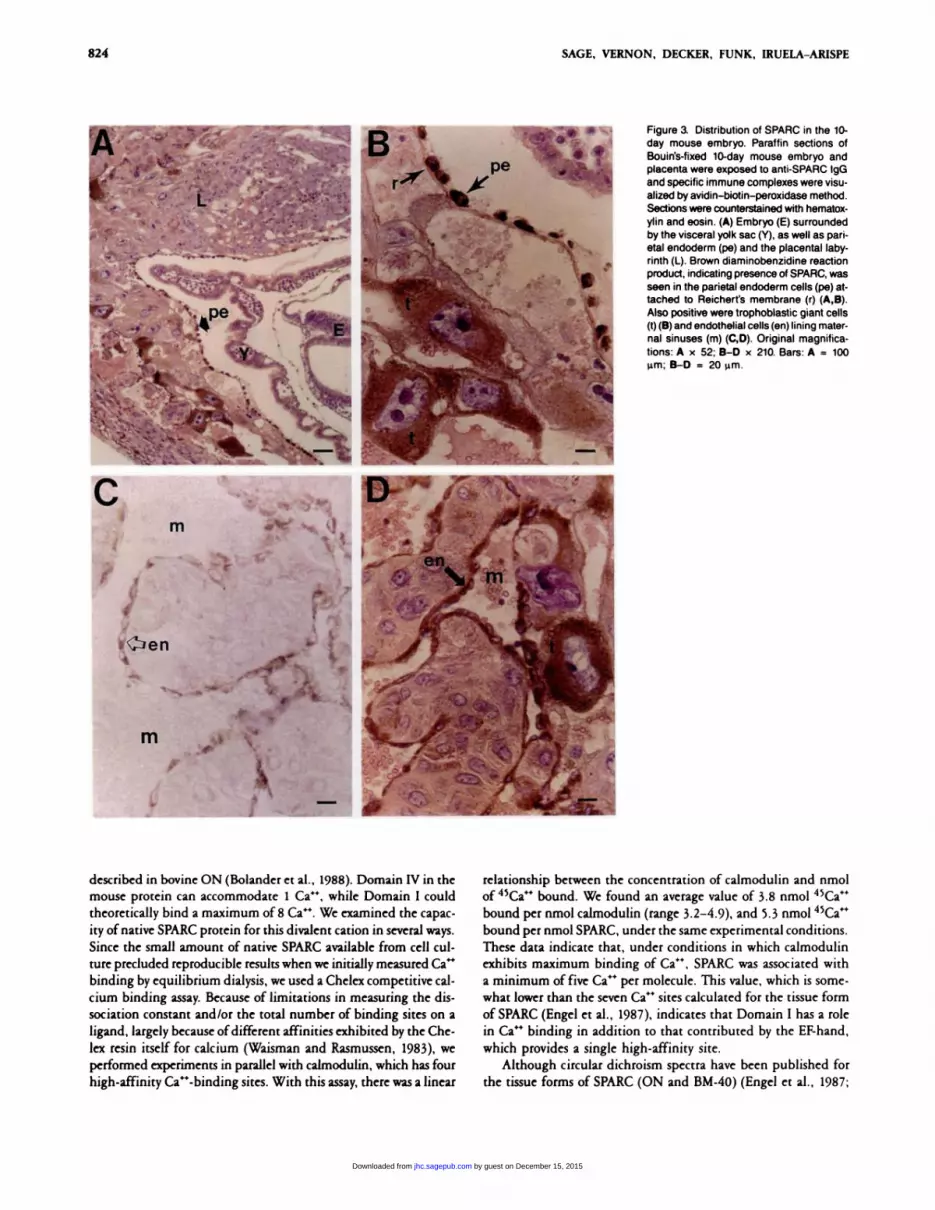
To further confirm the specificity of the affinity-purified anti-
SPARC polyclonal antibodies, we first tested the preparation on
sections of 10 day mouse embryos. As shown in Figures 3A and 3B,
a positive reaction was observed in the cells of the parietal endo-
derm that secrete Reichent’s membrane. Trophoblastic giant cells
and endothelia lining the maternal sinuses also exhibited a strong
staining reaction indicative of the presence of SPARC (Figures
3B-3D).
Representative tissues from embryonic and newborn mice are
shown in Figures 4 and 5. Alveolar epithelium (and probably en-
dothelium), as well as ductal epithelium, stained positively for
SPARC in 18-day fetal lung (Figure 4A); this tissue, however, was
negative in the adult (Table 1). Thoracic (and caudal, not shown)
somites from 14-day embryos expressed SPARC antigen (Figure 4B).
In developing bone, SPARC was localized primarily to the perichon.
drium/peniosteum in 18-day embryos (Figure 5A), but a more genen-
alized distribution was seen in newborn (4-5-day) long bone, where
SPARC was associated with nonhypertnophic chondrocytes (Figure
SB). In the newborn, staining for SPARC was also seen in peniosteumand penichondnium, and in association with osteoblasts and newly
synthesized osteoid (Figure SC). Chondnogenic tissues from 12-day
embryos were also positive for SPARC (Table 1, footnote); however,
tongue and intestine from these animals were negative.
Some ofthe other tissues listed as positive for SPARC in I#{224}ble1
are shown in Figure 6. SPARC was cleanly associated with prolifen-
ating epithelia in several tissues. In Figure 6A, the epidermal cover-
ing of newborn skull stained intensely with anti-SPARC IgG; for
comparison, anti-FN IgG identified antigen in the connective tis-
sue intenstitium ofthe skin (Figure 6B). In newborn thymus, SPARC
was associated with the epithelial processes that provide support
for T-cell populations (Figure 6C). At this stage of development,
both epidermal and thymic epithelial cells are undergoing gener-
alized proliferation. In lactating mammary gland, SPARC was
associated with both the ductal and alveolar epithelia that proliferate
in response to hormonal stimuli during late stages of the neproduc-
tive cycle (Figure 6D).
Anti-SPARC antibodies consistently did not stain the extracel-
lular matrix ofmany different cell types in vitro (Sage et al., 1986)
(Figure 2). Therefore, it appears that SPARC protein visualized by
immunostaining in vivo (Figures 3-6) was located primarily in thecells. Additional explanations are that SPARC was closely apposed
to the surface of the cell that secreted it, was bound to a compo-
nent ofthe interstitium, on was immobilized by the fixation proce-
dune en route to another destination.
The presence of SPARC in mesenchymal cells (e.g. , chondro-
cytes, osteoblasts, osteo- and fibroprogenitor cells of the perichon-
dnium, somitic cells), as well as cells derived from primitive ecto-
derm (pneumocytes, thymic epithelium) and extra-embryonic
(parietal)endoderm (Mason et al., 1986a,b), is suggestive ofa gener.alized rather than a lineage-specific function in cell behavior. In
some cases this function may be related to proliferation or migra-
tion, as seen in certain embryonic tissues but not in their adult
counterparts (e.g. , thymus, heart, lung). SPARC is present in adult
tissues that exhibit high rates of proliferation (e.g. , gut mucosa,salivary gland) (Table 1). However, both the temporal and spatial
restriction of SPARC in the mouse suggests that expression of the
protein, although not lineage-specific, may be related to certain
stages of cell differentiation and/or cell cycle.
Calcium-binding Properties of SPARC
The amino acid sequence predicted from mouse SPARC cDNA re-
vealed two putative Ca�-binding domains - a unique, acidic, Glu-
rich N-terminal region (Domain I), and an EF-hand-containing re-
gion (Domain IV) which is homologous to Ca4�-binding loops in
calmodulin (Engel et al., 1987). An additional EF-hand has been
by guest on December 15, 2015jhc.sagepub.comDownloaded from
CR
pe
‘� -.0��t.4d . ‘ -�
I�__� ��‘:3 �
\ -... �)
� - -� .
� �
� ‘���‘t\� �,,
4 ..
.\4�‘I’-
r
it,
- .
m ‘S4r�
824 SAGE, VERNON, DECKER, FUNK, LRUELA-ARISPE
. � � � � .
� ,. � - -��‘ � � .-.. . .. . . �-; � . . ::�I��
� -.‘� .: �.p � ��i-� � .. �.‘;.. T- -:��:�
�. L L �
- -�
‘ .. . � � �
/.
described in bovine ON (Bolander et al., 1988). Domain IV in the
mouse protein can accommodate 1 Ca�, while Domain I could
theoretically bind a maximum of 8 Ca�. We examined the capac-
ity ofnative SPARC protein for this divalent cation in several ways.
Since the small amount of native SPARC available from cell cul-
tune precluded reproducible results when we initially measured Ca�
binding by equilibrium dialysis, we used a Chelex competitive cal-
cium binding assay. Because of limitations in measuring the dis-
sociation constant and/on the total number of binding sites on a
ligand, largely because ofdifferent affinities exhibited by the Che-
lex resin itself for calcium (Waisman and Rasmussen, 1983), we
performed experiments in parallel with calmodulin, which has four
high-affinity Ca�-binding sites. With this assay, there was a linear
Figure a Distribution of SPARC in the 10-
day mouse embryo. Paraffin sections ofBouin’s-fixed 10-day mouse embryo andplacenta were exposed to anti-SPARC lgGand specific immune complexes were visu-alized by avidin-biotin-peroxidase method.Sections were counterstained with hematox-ylin and eosin. (A) Embryo (E) surrounded
by the visceral yolk sac (Y), as well as pan-etal endoderm (pe) and the placental laby-ninth (L). Brown diaminobenzidine reactionproduct, indicating presence of SPARC, wasseen in the parietal endoderm cells (pe) at-tached to Reichert’s membrane (r) (A,B).Also positive were trophoblastic giant cells(t)(B) and endothelial cells (en) lining mater-nal sinuses (m) (C,D). Original magnifica-tions: A x 52; B-D x 210. Bans: A = 100�tm; B-D = 20 �tm.
relationship between the concentration of calmodulin and nmol
of 45CC bound. We found an average value of 3.8 nmol 45Ca�
bound per nmol calmodulin (range 3.2-4.9), and 5.3 nmol 45Ca�
bound per nmol SPARC, under the same experimental conditions.
These data indicate that, under conditions in which calmodulinexhibits maximum binding of Ca�, SPARC was associated with
a minimum of five Ca� pen molecule. This value, which is some-
what lower than the seven Ca� sites calculated for the tissue formof SPARC (Engel et al., 1987), indicates that Domain I has a role
in Ca� binding in addition to that contributed by the EF-hand,
which provides a single high-affinity site.
Although circular dichnoism spectra have been published for
the tissue forms of SPARC (ON and BM-40) (Engel et al., 1987;
by guest on December 15, 2015jhc.sagepub.comDownloaded from

I
�..
�, �, a
, ,
� �..
‘0
- 4 � ,t_�__.� �
p. #{149}� . ..
�, 1�
a’
;,�_.
I
�“H’
I’#{149}_,
��w_ �‘
.‘ (�,
. ,.a�
.�. __)‘� ..�e
DISTRIBUTION OF SPARC IN MURINE TISSUES 825
Romberg et al., 1985), their disparity prompted us to examine the
structure of the native, intact SPARC molecule secreted from PYS
cells. As shown in Figure 7A, SPARC produced a spectrum similar
to that shown for intact BM-40 (Engel et al., 1987), with a promi.
nent shoulder at 220 nm. In the presence of 2.5 mM CaCl2 at pH
7.4, the mean residue molar ellipticity calculated for SPARC was
-4519 deg-cm2 dmol’. This value agrees rather well with that
of - 5600 deg-cm2 dmol’ found for BM-40 by Engel et al. (1987),
who also reported an a-helical content of 25-30% for the intact
molecule. Addition of 12 mM EDTA resulted in a marked decrease
in negative ellipticity at 220 nm. Furthermore, a flattened curve
with a trough at 205 nm was obtained with denatured SPARC Pu.
nified on CM-cellulose (data not shown).
A further indication of the Ca�-sensitive nature of SPARC is
shown in Figure 7B. In the presence of 4 mM CaCl2, SPARC (5)
bound selectively to hydrophobic phenyl-Sepharose and was elutedin the presence of 0.5 M NaCI (lane 2) on 12.5 mM EDTA (lane
3). These results are interesting in light ofpreliminary studies that
demonstrate binding of SPARC to endothelial cells (H. Sage and
S. Funk, unpublished observations) and suggest that a hydropho-
bic surface is exposed when SPARC adopts a native conformation
in the presence of Ca�.
Discussion
The distribution of SPARC in mouse tissues can be summarized
as follows: (a)in the adult, SPARC was associated with certain epithe-ha that exhibit high rates of turnover (gut, skin, glandular tissue);
(b) SPARC was not detected in adult heart, liven, pancreas, spleen,
and kidney; (c) fetal tissues that exhibited high levels of SPARC
included thymus, heart, lung, and gut; and (d) in the developing
embryo, SPARC expression was particularly enhanced in areas of
chondrogenesis, osteogenesis, and somitogenesis. Our studies are
in general agreement with the in situ hybridization data published
by Hogan and coworkers (Nomura et al., 1988; Holland et al., 1987).
The earliest appearance ofSPARC mRNA was reported in the 8.5 day
mouse embryo (Reichert’s membrane, trophoblastic giant cells)
(Holland et al., 1987). It was seen in embryonic chondnogenic tis-
sues at day 12 (Table 1), and was rather widely distributed after
13.5 days (Nomuna et al., 1988; Mason et al., 1986a) (l#{224}ble1).
We note an interesting coincidence between the distributionof SPARC and fos mRNA in embryonic mouse bone (Dony and
Gruss, 1987). Our data show SPARC protein in the penichon-dnium/peniosteum and in the mesodermal web of the 18-day paw,
a tissue that exhibits a rapid proliferative phase at this stage of em-
bryogenesis(Figure SA). Furthermore, de Togni et al. (1988) showed
nuclear staining offos in cartilage of 17-day rat embryos. In new-
born mouse bone, SPARC was positive primarily in the zones of
Figure 4. Distribution of SPARC protein in tissues of fetal mouse. Paraffin see-tions of Bouin’s-fixed 18-dayfetallung(A)and 14-dayfetal thoracic somites(B)were exposed to anti-SPARC lgG. The antigen-antibody complex was visual-ized bythe immunoperoxidase technique (A) or by Alit indirect immunofluo-rescence(B). Ductal epithelium(d)and alveolar epithelium(a)and endothelium(e)stained positively for SPARC(A). Thoracic somites also expressed SPARC,
which was seen inthe cytoplasm ofcells comprising each somite(S)(B). Onigi-
nal magnification x 180. Bars � 30 �tm.
by guest on December 15, 2015jhc.sagepub.comDownloaded from

826 SAGE, VERNON, DECKER, FUNK, LRUELA-ARISPE
w�
:�,�
‘.�, �
Figure 5. Expression of SPARC protein in developing osseous tissues. Paraffin sections of Bouin’s-fixed 18-day fetal mouse forepaw (A) and 4-5-day newbornmouse joint (B,C)were exposed to anti-SPARC lgG. (A) Antigen-antibody complex visualized by the immunoperoxidase technique. (BC) FITC indirect immunofluo-rescence. In embryonic bone, SPARC was concentrated primarily in the penichondrium/peniosteum (arrow) of the phalanx (p), as well as the mesodermal web
tissue (w) (A). The secondary center of ossification, shown in a carpal epiphysis (B), exhibited staining for SPARC in resting chondrocytes (r) as well as thosecomprising the zones of proliferation (p) and degeneration (d); the hypertrophic zone appeared negative (h). (C) Marrow cavity (m) and newly formed osteoid(o) with SPARC-positive osteoblasts (arrow). Original magnifications: A x 45; B,C x 150. Bars: A = 160 �tm; B,C = 35 �tm.
proliferation and degeneration. Nomuna et al. (1988) also observed
a restricted distribution ofSPARC mRNA in 14.5-day peniosteum,
as well as a more generalized expression in hypentrophic cartilage
and in cells associated with bone matrix in later embryos. Although
we saw SPARC protein in the hypertnophic zone offetal mouse bone
(not shown), it was conspicuously absent from these chondrocytes
in the newborn tissue (Figure SB). Jundt et al. (1987), in studies
of human fracture callus, failed to identify ON (SPARC) in any
cartilage except that of “chondroid bone:’ although ON was pres-
ent in osteopnogeniton (peniosteal) and active osteoblastic cells. We
have no explanation for the lack of staining of non-mineralized
tissues reported by these investigators. Although there are com-
pelling similarities between the immunohistological distributions
of SPARC and ‘rt;F-(3 in mouse embryos (most notably in connec-
tive tissues, cartilage, bone, teeth, hair follicles, skin, limb buds,
and submandibular gland) (Heine et al. , 1987), recent data do not
support an induction of SPARC by 1XF-13, as suggested by these
authors, in the morphogenesis on remodeling of these coincident
tissues (Wrana et al., 1988; H. Sage and R. Penttinen, unpublished
observations).
Although a function for SPARC (ON) was originally proposed
in the mineralization of bone (Tenmine et al. , 1981), Romberg et
al. (1985) have shown that this protein inhibits hydroxyapatite for-
mation. Since SPARC is associated with areas of cell proliferation
in many tissues, it could function as a general inhibitor of mineral-
ization in all extracellular matrices. As a corollary, SPARC could
regulate cellular migration and growth by facilitating the disper-
sion of, on preventing the formation of, excessive matrix. In this
respect, SPARC might resemble the tissue inhibitor of metallopno-
teinases (TIMP) (Gavnilovic et al., 1987). By neutralizing specific
enzymes, TIMP has been proposed to regulate the turnover of the
ECM during cell perturbation.
We propose that SPARC might participate in a proliferative re-
sponse by interacting at the cell surface with other Ca�2-sensitive
proteins. Intermolecular regulation is exemplified by the blood clot-
ting proteins, which show calcium-mediated binding to lipids and
modulation ofpnotease activity by conformational changes in Ca�-
binding domains (Rodgers, 1988). Mellgnen (1987) has proposed
that cell membranes are preferred sites for Ca-dependent protease
action: the pnotease, a specific inhibitor, and the membrane itself
by guest on December 15, 2015jhc.sagepub.comDownloaded from

DISTRIBUTION OF SPARC IN MURINE TISSUES 827
Figure 6. Location of SPARC protein inproliferating epithelia. Frozen sections ofnewborn mouse skull skin (A,B), newbornthymus (C), and adult lactating mammarygland (D)were exposed to anti-SPARC lgG(A,C,D) orto rabbit anti-human FN (B), andwere visualized by RITC indirect immuno-fluorescence. In contrast to FN, which wasdistributed primarily in the denmal inter-stitium (B), SPARC exhibited a preferentiallocalization in the epidermis (A). SPARCwas also apparent in the epithelial reticularcells of newborn thymic stroma (C) �nd inboth ductal and alveolar epithelia of lactat-ing mammary gland (0). Original magnifi-cation x 180. Bans � 30 �tm.
form a regulatory complex which activates membrane-associated
protein kinase C after exposure of certain cells to phorbol esters
or other mitogens. Transduction of a signal from such an associa-
tion would lead to the activation of other genes and, indirectly,
to cell proliferation. An example of a cell-surface protein that has
been claimed to be essential for proliferation of smooth muscle
cells is the secreted Ca�-binding protein thnombospondin (Ma-
jack et al., 1988).
An analogous example of extnacellulan protein/matrix regula-
tion of cell growth might be somitogenesis, in which both Ca�-
independent and Ca�-dependent mechanisms are operative (Du-
band et al., 1987). We have observed that SPARC protein was pneva-
lent in 14-day embryo somites and are pursuing the effect of SPARC
depletion on cell compaction and dispersion in this system. Since
constituents ofthe plasma membrane and extracellular matrix, such
as pnoteoglycans, can sequester significant levels of Ca� , it is not
unreasonable to assume that reservoirs ofCa4� could modulate pro-
teins with otherwise high-affinity binding sites. In addition, SPARC
might be proteolytically processed in the extracellular space. Exci-
sion of the high-affinity EF-hand in vivo could result in an mdc-
by guest on December 15, 2015jhc.sagepub.comDownloaded from

A
0-
-8-
�1 -16#{149}0
� -24-
32 -
B123�
I
828 SAGE, VERNON, DECKER, FUNK, IRUELA-ARISPE
Davis TN, Urdea MS, Masiarz FR, ThornerJ (1986): Isolation of the yeast Holland P, Harper 5, McVey J, Hogan BLM (1987): In vivo expression of
-.w I I -1 1 I I I I I U 1
205 215 225 235 245 255
WavelengthFigure 7. Calcium-sensitive properties of the SPARC protein. (A) Circular
dichnoism spectrum of SPARC in the presence of calcium. Native SPARC(0.15 mg/mI, 5.1 pM) in TBS containing 2.5 mM CaCl2 was measured at20#{176}C.The shoulder at 220 nm is indicative of a-helical content. O��o wascalculated as -4519 degcm2 dmol1 . (B) Phenyl-Sepharose affinity chro-matography. P�Sl-Met-labeled proteins in PYS cell culture medium weredialyzed against PBS-4 mM CaCl2 and applied to phenyl-Sepharoseequilibrated in the same buffer. Bound proteins, eluted successively with0.5 M NaCI (lane 2) and 12.5 mM EDTA (lane 3), were analyzed by SDS-PAGE and fluorescence autoradiography. Lane 1 represents total startingmaterial. Native SPARC (S), which was selectively retained on the column,was dissociated from the hydrophobic resin by salt and/or EDTA. Averagerecovery from this column was 27%.
pendent function for Domain I with a lower Ca�-binding poten-
tial. Alternatively, increased levels of SPARC might disrupt the
equilibrium among other secreted proteins to effect the conforma-
tional changes necessary for facilitation ofcell migration on pnolifer-
ation.
Acknowledgments
We thank Dr B. Hogan for pmviding the mouse SPARC cDNA, P #{237}#{224},-
nemeyerfor histology, Dr R. Kievit and G. Parragafor help with the circu-
lar dichroism studies, Dr D. Eyre andS. Aponeforperform:ng amino acid
analyses, Dr L Fouser and E. Everiti forpreparations ofSPARC, and our
colleagues in the Departments ofBiological Structure and Pathology for
discussions andsuggestions. Specialappreciat:on is due B. Cmckettforprep-
aration of the manuscript.
Literature Cited
Bolander, ME, Young MF, Fisher LW, Yamada U, TermineJ (1988): Osteonec-
tin cDNA sequencing reveals potential binding regions for calcium and
hydroxyapatite and shows homologies with both a basement membrane
protein (SPARC) and a senine protease inhibitor (ovomucoid). Proc Nati
Acad Sci USA 85:2919
I.�s
calmodulin gene: calmodulin is an essential protein. Cell 47:423
de Togni P, Niman H, Raymond V, Sawchenko P. Verma IM (1988): Detec-
non offos protein during osteogenesis by monoclonal antibodies. Mol Cell
Biol 8:2251
Dony C, Gruss P (1987): Proto-oncogene c-fos expression in growth regions
of fetal bone and mesodermal web tissue. Nature 328:711
DubandJ-L, Dufour 5, Hatta K, l#{224}keichiM, Edelman GM, ThieryJP(1987):Adhesion molecules during somitogenesis in the avian embryo. J Cell Biol104:1361
Dziadek M, Paulsson M, Aumailley M, Timpl R (1986): Purification and
tissue distribution of a small protein (BM-40) extracted from a basement
membrane tumor. EurJ Biochem 161:455
EngelJ, 1�ylor W, Paulsson M, Sage H, Hogan B (1987): Calcium-binding
domains and calcium-induced transition in SPARC (osteonectin, BM-4O),
an extracellular glycoprotein expressed in mineralized and nonmineralized
tissues. Biochemistry 26:6958
GavrilovicJ, Hembry RM, ReynoldsJJ, Murphy G (1987): Tissue inhibitorof metalloproteinases (TIMP) regulates extracellular type I collagen degra-
dation by chondrocytes and endothelial cells. J Cell Sd 87:357
Heine VL, Munoz EF, Flanders KC, Ellingsworth LR, Lam H-YP, Thomp-son NL, Roberts AB, Sporn MB (1987): Role oftransforming growth factor-a
in the development of the mouse embryo. J Cell Biol 105:2861
by guest on December 15, 2015jhc.sagepub.comDownloaded from

DISTRIBUTION OF SPARC IN MURINE TISSUES 829
mRNA for the Ca� binding protein SPARC (osteonectin) revealed by in protein associated with endothelial cell injury and proliferation.J Mol Cellsitu hybridization. J Cell Biol 105:473 Cardiol, in press
Jundt G, Berghauser K-H, TermineJD, Schulz A (1987): Osteonectin-a Sage H,Johnson C, Bonnstein P (1984): Characterization ofa novel serumdifferentiation marker of bone cells. Cell Tissue Res 248:409 albumin-binding glycoprotein secreted by endothelial cells in culture.J Biol
Chem 259:3993Laemmli UK (1970): Cleavage ofstructural proteins during the assembly
of the head of bacteriophage T4. Nature 277:680 Sage H, Pnitzl P, Bornstein P (1981): Secretory phenotypes of endothelialcells in culture: a comparison of aortic, venous, capillary, and corneal en-
Majack RA, Goodman LV, Dixit VM (1988): Cell surface thrombospondin dothelium. Arteriosclerosis 1:427is functionally essential for vascular smooth muscle cell proliferation. J Cell
Biol 106:415 Sage H, TupperJ, Bramson R (1986): Endothelial cell injury in vitro is as-
Mann K, Deutzmann R, Paulsson M, Timpl R (1987): Solubilization of sociated with increased secretion of an Mr 43,000 glycoprotein ligand. JCell Physiol 127:373protein BM-40 from a basement membrane tumor with chelating agents
and evidence for its identity with osteonectin and SPARC. FEBS Lett 218:167 Stenner DD, Tracy RP, Riggs BL, Mann KG (1986): Human platelets con-tam and secrete osteonectin, a major protein of mineralized bone. Proc
Mason U Murphy D, Munke M, Francke U, Elliott R, Hogan BLM (1986a): Natl Acad Sci USA 83:6892Developmental and transformation-sensitive expression of the SPARC geneon mouse chromosome 11. EMBO J 5:1831 TermineJD, Kleinman HK, Whitson SW, Conn KM. McGarvey ML, and
Mason IJ� ‘I#{224}ylorA, WilliamsJG, Sage H, Hogan B (1986b): Evidence from Martin GR (1981): Osteonectin, a bone-specific protein linking mineralmolecular cloning that SPARC, a major product of mouse embryo parietal to collagen. Cell 26:99endoderm, is related to an endothelial cell “culture shock” glycoprotein. Tung PS, Domenicucci C, Wasi S. Sodek J (1985): Specific immuno-EMBO ) 5:1465 histochemical localization of osteonectin and collagen types I and III in
fetal and adult porcine dental tissues. J Histochem Cytochem 33:531Mellgren RL (1987): Calcium-dependent proteases: an enzyme system ac-tive at cellular membranes? FASEB J 1:110 Waisman DM, Khanna NC, Tokuda M (1986): Identification of a major
bovine heart �2. binding protein. Biochem Biophys Res Commun 139:5%Nomura 5, Wills AJ, Edwards DR, HeathJK, Hogan BLM (1988): Develop-mental expression of 2ar (osteopontin) and SPARC (osteonectin) RNA as Waisman DM, Rasmussen H (1983): A reexamination of the chelex com-
revealed by in situ hybridization. J Cell Biol 106:441 petitive calcium binding assay. Cell Calcium 4:89
Otsuka K, Yao K-L, Wasi S, Tung PS, AubinjE, SodekJ, TermineJD(1984): Wasi S, Otsuka K, Yao K-L, lung PS, AubinjE, SodekJ, TermineJD (1984):
Biosynthesis of osteonectin by fetal porcine calvanial cells in vitro. J Biol An osteonectin-like protein in porcine periodontal ligament and its syn-
Chem 259:9805 thesis by periodontal ligament fibroblasts. Biochem Cell Biol 62:470
Rodgers GM (1988): Hemostatic properties of normal and perturbed vas- WranajL, Macno M, Hawrylshyn B, Yao K-L, Domenicucci C, SodekJ (1988):
cular cells. FASEB J 2:116 Differential effects oftransforming growth factor-(� on the synthesis of cx-tracellular matrix proteins by normal fetal rat calvarial bone cell popula-
Romberg RW, Werness PG, Lollar P, Riggs EL, Mann KG (1985): Isolationand characterization of native adult osteonectin. J Biol Chem 260:2728 tions. J Cell Biol 106:915
Sage H (1986a): Culture shock: selective uptake and rapid release ofa novel Young, MF, Bolander ME, Day AA, Ramis CI, Robey PG, Yamada Y, Ter-
serum protein by endothelial cells in vitro. J Biol Chem 261:7082 mine JD (1986): Osteonectin mRNA: distribution in normal and trans-formed cells. Nucleic Acids Res 14:4483
Sage H (1986b): Characterization and modulation of extracellular glyco-proteins secreted by endothelial cells in culture. In Gimbrone M, ed. Con- Zung P, Domenicucci C, Wasi S. Kuwata F, Sodek J (1986): Osteonectin
is a minor component of mineralized connective tissues in rat. Biochemtemporary issues in haemostasis and thrombosis. Vol 2. Vascularendothelium Cell Biol 64:356in haemostasis and thrombosis. New York, Churchill-Livingstone, 187
Sage H, DeckerJ, Funk S, Chow M: SPARC: a Ca�2-binding extracellular
by guest on December 15, 2015jhc.sagepub.comDownloaded from


