Embed Size (px)
Citation preview

Chapter 6
DNA, RNA, AND PROTEINSYNTHESIS
A typical human cell contains about 10,000 differentproteins, which are synthesized according to instructionsthat are sent from the chromosome to the ribosomein the form of messenger ribonucleic acid (mRNA).Therefore gene expression requires two steps (Fig. 6.1):
1. Transcription is the synthesis of an mRNA moleculein the nucleus. The mRNA is the carbon copy of adeoxyribonucleic acid (DNA) strand.
2. Translation is the synthesis of the polypeptide by theribosome, guided by the base sequence of the mRNA.
A gene is a length of DNA that directs the synthesis of amessenger RNA and polypeptide, or of a functionalRNA that is not translated into a polypeptide. It consistsof a transcribed sequence and regulatory sites. A chromo-some is a very long DNA molecule with hundreds orthousands of genes.
As it is expressed, the genetic message is amplified.A single gene can be transcribed into thousands ofmRNAmolecules, and each mRNA can be translated intothousands of polypeptides. For example, a red blood cellcontains 5 108 copies of the hemoglobin b-chain, butthe nucleated red blood cell precursors that make thehemoglobin have only two copies of the b-chain gene.
ALL LIVING ORGANISMS USE DNA AS THEIRGENETIC DATABANK
Living things are grouped into two major branches on thebasis of their cell structure. The prokaryotes include bacte-ria, actinomycetes, blue-green algae, and archaea, and theeukaryotes include protozoa, plants, and animals. Onlyeukaryotic cells are compartmentalized into organelles byintracellular membranes. Structures that are present ineukaryotic but not prokaryotic cells include the following:
1. A nucleus surrounded by a twofold membrane.2. Mitochondria, with a twofold membrane, are the
powerhouses of the cell. They turn food and oxygeninto adenosine triphosphate (ATP).
3. The endoplasmic reticulum, bounded by a singlemembrane, processes membrane proteins, membranelipids, and secreted proteins.
4. The Golgi apparatus is a sorting station that sendssecreted proteins, lysosomal enzymes, and mem-brane components to their proper destinations.
5. Lysosomes are vesicles filled with hydrolytic enzymes.They degrade many cellular macromolecules as wellas substances that the cell engulfs by endocytosis.
6. Peroxisomes contain enzymes that generate anddestroy toxic hydrogen peroxide.
7. Cytoskeletal fibers give structural support to the cell.They are also required for cell motility and intracel-lular transport.
Differences between prokaryotes and eukaryotes aresummarized in Figure 6.2 and Table 6.1. Despite thesedifferences, all living cells have three features in common:
1. All cells are surrounded by a plasmamembrane,whichis a flimsy, fluid, flexible structure that forms a diffu-sion barrier between the cell and its environment.
2. All cells generate metabolic energy, which they usefor biosynthesis, maintenance of cell structure, andcell motility.
3. All cells store genetic information in the form ofDNA, which the cells use to reproduce themselvesand to direct the synthesis of RNA and protein.
This chapter describes DNA replication and proteinsynthesis in prokaryotes. The corresponding processesin eukaryotes (see Chapter 7) are similar but are oftenmore complex.
DNA CONTAINS FOUR BASES
DNA is a polymer of nucleoside monophosphates (alsocalled nucleotides) (Fig. 6.3, B). Its structural backboneconsists of alternating phosphate and 2-deoxyriboseresidues that are held together by phosphodiester bondsinvolving carbon-3 and carbon-5 of the sugar. Carbon-1forms a b-N-glycosidic bond with one of the four basesshown in Figure 6.4.
One end of the DNA strand has a free hydroxylgroup at C-5 of the last 2-deoxyribose. The other endhas a free hydroxyl group at C-3. The carbons of2-deoxyribose are numbered by a prime (0) to distinguish
64

Polypeptide Polypeptide
mRNADNA
A B
Ribosome
Ribosome
mRNA
Nucleus Cytoplasm
Transcription
Translation
Nuclearmembrane
DNA(in chromosome)
mRNA
Figure 6.1 Expression of genetic information. In all organisms, the DNA of the gene is first copied into a single-stranded
molecule of messenger RNA (mRNA). This process is called transcription. During ribosomal protein synthesis, the base
sequence of the mRNA specifies the amino acid sequence of a polypeptide. This is called translation. A, In prokaryotic cells,
translation starts before transcription is completed. B, Eukaryotic cells have a nuclear membrane. Therefore transcription
and translation take place in different compartments: transcription in the nucleus and translation in the cytoplasm.
DNA(chromosome)
Ribosomes
Plasma membrane
Cell wall
MIT
L
L
LL
L
L
L
L
LL
EV
EV
EVEV
EV
SV
1 2 3 4 5 µm
SV
SVSV
SV
G
R
ER
Cytoskeletalfibers
P
P
P
P
P
P
Nucleus
Nuc leo lus
1 µmA
B
Figure 6.2 Typical elements of prokaryotic and eukaryotic cell structure. A, Typical bacterial (prokaryotic) cell.
B, Typical human (eukaryotic) cell. ER, Endoplasmic reticulum; EV, endocytotic vesicle; G, Golgi apparatus; L, lysosome;
MIT, mitochondrion; P, peroxisome; R, ribosome; SV, secretory vesicle.
65DNA, RNA, and Protein Synthesis

them from the carbon and nitrogen atoms of the bases;therefore, each strand has a 50 end and a 30 end. By con-vention, the 50 terminus of a DNA (or RNA) strand iswritten at the left end and the 30 terminus at the rightend. Thus the tetranucleotide in Figure 6.4 can be writ-ten as ACTG but not GTCA.
The variability of DNA structure is produced by itsbase sequence. With four different bases, there are 42
(or 16) different dinucleotides and 43 (or 64) differenttrinucleotides, and 4100 possibilities exist for a sequenceof 100 nucleotides.
DNA FORMS A DOUBLE HELIX
Cellular DNA is double stranded, and almost all of it ispresent as a double helix, as first described by JamesWatson and Francis Crick in 1953. The most prominentfeatures of the Watson-Crick double helix (Figs. 6.5to 6.7) are as follows:
Table 6.1 Typical Differences between Prokaryotic and
Eukaryotic Cells
Property Prokaryotes Eukaryotes
Typical size 0.4–4 mm 5–50 mm
Nucleus " þ
Membrane-bounded
organelles
" þ
Cytoskeleton " þ
Endocytosis and
exocytosis
" þ
Cell wall þ (some ") þ (plants)
" (animals)
No. of chromosomes 1 (þplasmids) >1
Ploidy Haploid Haploid or
diploid
Histones " þ
Introns " þ
Ribosomes 70S 80S
CH2OH
H
HOH
OH
H
β-D-2-deoxyribose Phosphate
A
5′
4′
3′ 2′
1′
O
NH
NH2
Adenine(A)
2
34
56
7
8
9
1N
N
N
NH
H2N
Guanine(G)
Cytosine(C)
HN
N
N CH3
NH
2
16
5
4
3N
O
NH
Thymine(T)
NH
HN
O
CH2
N
H
HOH
H
2-deoxy-AMP(dAMP)B
NH2
N
N
N
O
O–
O O
O
–O
O–
O
OHP–O
OP
Figure 6.3 The building blocks of DNA. A, Structures of the four bases, 2-deoxyribose, and phosphate. The bases A and G are
purines, and C and T are pyrimidines. B, Structure of 2-deoxy-adenosine monophosphate (dAMP), one of the four
2-deoxyribonucleoside monophosphates (also called 2-deoxynucleotides) in the repeat structure of DNA. Note that a nitrogen
atom of the base is bound by a b-N-glycosidic bond to C-1 of 2-deoxyribose, whereas C-5 forms a phosphate ester bond.
66 GENETIC INFORMATION: DNA, RNA, AND PROTEIN SYNTHESIS

1. The two strands of the double helix have oppositepolarity, meaning they run in opposite directions.Base pairing is always antiparallel, not only in theDNA double helix but also in other base-pairedstructures formed by DNA or RNA.
2. The 2-deoxyribose/phosphate backbones of the twostrands form two ridges on the surface of the mole-cule. The phosphate groups are negatively charged.
3. The bases face inward to the helix axis, but theiredges are exposed. They form the lining of twogrooves that are framed by the ridges of the sugar-phosphate backbone. Because theN-glycosidic bondsare not exactly opposite each other (see Fig. 6.7), thetwo grooves are of unequal size. They are calledthe major groove and the minor groove.
4. In each of the two strands, successive bases lie flat,one on top of the other, like a stack of pancakes.The flat surfaces of the bases are hydrophobic, and
successive bases in a strand form numerous van derWaals interactions.
5. Bases in opposite strands interact by hydrogen bonds.Adenine (A) always pairs with thymine (T) in theopposite strand, and guanine (G) with cytosine (C).Therefore the molar amount of adenine in thedouble-stranded DNA always equals that of thymine,and the amount of guanine equals that of cytosine.A-T base pairs are held together by two hydrogenbonds, and G-C base pairs by three. Most important,the base sequence of one strand predicts exactly thebase sequence of the opposite strand. This is essentialfor DNA replication and DNA repair.
6. The double strand is wound into a right-handedhelix. Each turn of the helix has about 10.4 basepairs and advances about 3.4 nm along the helixaxis. The double helix is rather stiff, but it can bebent and twisted by DNA-binding proteins.
HO
5′-terminusN
N
A
C
T
G
NH2
N
N
N
O
N
O
NH2
N
N
OH
O
HN
N
N
O
HN
O
O
OO
O
O
O
O
O
OCH3
H2N
3′-terminus
CH2–O OP
CH2
CH2
–O OP
CH2–O OP
Figure 6.4 Structure of the (2-deoxy-)tetranucleotide ACTG. The DNA strands in chromosomes are far larger, with lengths
of many million nucleotide units. A, Adenine; C, cytosine; G, guanine; T, thymine.
67DNA, RNA, and Protein Synthesis

DNA CAN BE DENATURED
Like other noncovalent structures, the Watson-Crickdouble helix disintegrates at high temperatures. Heatdenaturation of DNA is also called melting. BecauseA-T base pairs are held together by two hydrogenbonds and G-C base pairs by three, A-T–rich sectionsof the DNA unravel more easily than G-C–rich regionswhen the temperature is raised (Figs. 6.8 and 6.9). Atphysiological pH and ionic strength, this typically hap-pens between 85$C and 95$C.
Heat denaturation decreases the viscosity ofDNA solu-tions because the single strands are more flexible than thestiff, resilient double helix. It also increases the ultravioletlight absorbance at 260 nm, which is caused by the bases,because base pairing and base stacking are disrupted.
Other ways to denature DNA include decreased saltconcentration, extreme pH, and chemicals that disrupthydrogen bonding or base stacking.
When cooled slowly, denatured DNA “renatures”spontaneously. This process is called annealing.Whereassmall DNA molecules anneal almost instantaneously,large molecules require seconds to minutes.
DNA IS SUPERCOILED
Many naturally occurring DNA molecules are circular.When a linear duplex is partially unwound by oneor several turns before it is linked into a circle, the
number of base pairs per turn of the helix is greaterthan the usual 10.4. The torsional strain in this mole-cule leads to supercoiling of the duplex around itsown axis, much as a telephone cord twists arounditself. This is called a negative supertwist. The oppo-site situation, in which the helix is overwound, iscalled a positive supertwist.
Most cellular DNAs are negatively supertwisted,with 5% to 7% fewer right-handed turns than expec-ted from the number of their base pairs. This under-wound condition favors the unwinding of the doublehelix during DNA replication and transcription.
The supertwisting of DNA is regulated bytwo types of topoisomerase. Type I topoisomerasescleave one strand of the double helix, creating amolecular swivel that relaxes supertwists passively.Type II topoisomerases are more complex. They cleaveboth strands and allow an intact helix to passthrough this transient double-strand break, beforeresealing the break. Type II topoisomerases hydrolyzeATP to pump negative supertwists into the DNA(Fig. 6.10).
Majorgroove
Minorgroove
Sugar-phosphatebackbone
Figure 6.6 Space-filling model of the Watson-Crick double
helix (B-DNA).
5′
3′
3′
5′
T A
G C
A T
C G
P
P
P
P
P
P
P
P
P
P
Figure 6.5 Schematic view of the DNA double strand. Note
that the strands are antiparallel and that only A-T and G-C
base pairs are permitted. Therefore the base sequence of one
strand predicts the base sequence of the opposite strand.
A, Adenine; C, cytosine; G, guanine; P, phosphate; T, thymine.
68 GENETIC INFORMATION: DNA, RNA, AND PROTEIN SYNTHESIS

DNA REPLICATION IS SEMICONSERVATIVE
DNAmakes identical copies of itself, which are transmit-ted to the daughter cells during mitosis and even to thenext generation through the gametes. In this sense, DNAis the only immortal molecule in the body. The organismis best understood as an artificial environment, createdby genes for the benefit of their own continued existence.
DNA is replicated in two steps (Fig. 6.11):
1. The double helix unwinds to produce two singlestrands. This requires ATP-dependent enzymes tobreak the hydrogen bonds between bases. DNAunwinding creates the replication fork. This is theplace where the new DNA strands are synthesized.
2. A new complementary strand is synthesized for eachof the two old strands. This is possible because thebase sequence of each strand predicts the basesequence of the complementary strand.
DNA replication is called semiconservative because onestrand in the daughter molecule is always old and theother strand is newly synthesized.
DNA IS SYNTHESIZED BY DNA POLYMERASES
The steps inDNAreplication are best known inEscherichiacoli, an intestinal bacterium that has enjoyed the unfal-tering affection of generations of molecular biologists.
The key enzymes of DNA replication in E. coli, as inall other cells, are the DNA polymerases. DNA poly-merases synthesize the new DNA strand stepwise,nucleotide by nucleotide, in the 50!30 direction. Theprecursors are the deoxyribonucleoside triphosphates:deoxy-adenosine triphosphate (dATP), deoxy-guano-sine triphosphate (dGTP), deoxy-cytosine triphosphate(dCTP), and deoxy-thymidine triphosphate (dTTP).DNA polymerase elongates DNA strands by linkingthe proximal phosphate of an incoming nucleotide tothe 30-hydroxyl group at the end of the growing strand(Fig. 6.12). The pyrophosphate formed in this reactionis rapidly cleaved to inorganic phosphate by cellularpyrophosphatases.
Prior unwinding of the double helix is required be-cause the DNA polymerases require a single-strandedDNA as a template. While synthesizing the new strand
Mi
no
r g r o ov
e
M
aj
or g r o o v e
N
NHH
N N
N
N
CH3
Adenine
Thymine
N
O
O
N
NN
N
N
H
H
H
H
H
N
N
Guanine
Cytosine
N
O
O
H
O
O
CH2
O
5′
3′
O
OCH2
O
5′
3′
Mi
no
r g r o o ve
M
aj
or g r o o v e
O
O
CH2
O
5′
3′
O
OCH2
O
5′
3′
Figure 6.7 Cross-sections through an adenine-thymine (A-T) and a guanine-cytosine (G-C) base pair in the DNA duplex.
The A-T base pair is held together by two hydrogen bonds [ ] and the G-C base pair by three.
69DNA, RNA, and Protein Synthesis

in the 50!30 direction, the enzyme moves along thetemplate strand in the 30!50 direction.
DNA polymerases are literate enzymes. They readthe base sequence of their template and make sure thateach base that they add to the new strand pairs with the
base in the template strand. Therefore the new strand isexactly complementary to the template strand. TheDNA polymerases are lacking in creative spirit. Theyare like the scribe monks in medieval monasteries,who worked day and night copying old manuscriptswithout understanding their content.
BACTERIAL DNA POLYMERASES HAVEEXONUCLEASE ACTIVITIES
A nuclease is an enzyme that cleaves phosphodiesterbonds in a nucleic acid. Deoxyribonucleases (DNases)cleave DNA, and ribonucleases (RNases) cleave RNA.Nucleases that cleave internal phosphodiester bondsare called endonucleases, and those that cleave bondsat the 50 end or the 30 end are called exonucleases.
Nobody is perfect, and even DNA polymerase some-times incorporates a wrong nucleotide in the newstrand. This can create a lasting mutation, which canbe deadly if it leads to the synthesis of a faulty protein.To minimize such mishaps, the bacterial DNA poly-merases are equipped with a 30-exonuclease activitythat they use for proofreading. When the nucleotidethat has been added to the 30 end of a growing chainfails to pair with the base in the template strand, it isremoved by the 30-exonuclease activity (Fig. 6.13). Thisproofreading mechanism reduces the error rate from 1in 104 or 1 in 105 to less than 1 in 107.
Most bacterial DNA polymerases also have a50-exonuclease activity (see Fig. 6.13). This activity isnot used for proofreading, but it cleaves damagedDNA strands during DNA repair and erases the RNAprimer during DNA replication.
Escherichia coli has three DNA polymerases. Theydiffer in their affinity for the DNA template and con-sequently in their processivity, the number of nucleotidesthey polymerize before dissociating from the template:
||
|
| | | | | |
| | | |
Figure 6.8 Melting of DNA.
70% G-C
30% G-C
50% G-C
Temperature (°C)
Rela
tive
absorb
ance
1.4
1.3
1.2
1.1
60 70 80 90
Figure 6.9 Melting of DNA, monitored by the increase of
ultraviolet light absorbance at 260 nm. The melting
temperature increases with increased guanine-cytosine (G-C)
content of the DNA. It is also affected by ionic strength and
pH. The melting temperature is the temperature at which the
increase in ultraviolet absorbance is half-maximal.
Positivelysupercoiled DNA
After relaxation of the supertwist,the ends of the DNA are linked by topoisomerase
The supercoiled DNAis cleaved by topoisomerase, thefree ends rotate torelax the supertwist
Figure 6.10 Relaxation of positive supertwists in DNA by a
type II topoisomerase.
70 GENETIC INFORMATION: DNA, RNA, AND PROTEIN SYNTHESIS

+
Parentalduplex
DNAsynthesis
Replicateddaughter molecules
Replicationfork
Movementof thereplicationfork
Figure 6.11 Semiconservative mechanism of DNA replication.
CH2 A TO
P
New strand
5′
CH2
CH2
P
5′
CH2
P
3′
P
O
OH
O
G
O
OH
T
PP
P
PP
P
O
OH
C
C CH2
P
P P
2 Pi
O
A CH2
P
O
G CH2
CH2
P
O
C CH2
P
Templatestrand
O
CH2 A TO
P
5′
CH2
P
5′
CH2
P
3′
3′
3′
P
O
CH2
P
O
G
O
OH
T
C CH2
P
O
A CH2
P
O
G CH2
P
O
C CH2
P
O
Figure 6.12 Template-directed synthesis of DNA by DNA polymerases. A, Adenine; C, cytosine; G, guanine; P, phosphate;
T, thymine.
71DNA, RNA, and Protein Synthesis
l Poly I has low processivity and falls off its templateafter polymerizing only a few dozen nucleotides.This enzyme is used for DNA repair (see Chapter 9)and plays only an accessory role in DNA replication.
l Poly II has a somewhat higher processivity. It isinvolved with DNA repair as well.
l Poly III is the major enzyme of DNA replication.With the help of a specialized clamp protein, it bindstightly to its template and polymerizes hundredsof thousands of nucleotides in one sitting, at a rate ofabout 800 nucleotides per second.
UNWINDING PROTEINS PRESENT A SINGLE-STRANDED TEMPLATE TO THE DNAPOLYMERASES
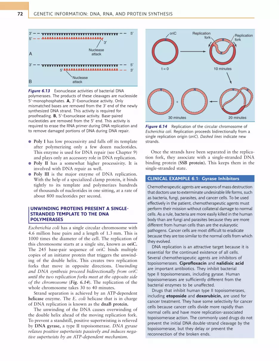
Escherichia coli has a single circular chromosome with4.6 million base pairs and a length of 1.3 mm. This is1000 times the diameter of the cell. The replication ofthis chromosome starts at a single site, known as oriC.The 245 base-pair sequence of oriC binds multiplecopies of an initiator protein that triggers the unwind-ing of the double helix. This creates two replicationforks that move in opposite directions. Unwindingand DNA synthesis proceed bidirectionally from oriCuntil the two replication forks meet at the opposite sideof the chromosome (Fig. 6.14). The replication of thewhole chromosome takes 30 to 40 minutes.
Strand separation is achieved by an ATP-dependenthelicase enzyme. The E. coli helicase that is in chargeof DNA replication is known as the dnaB protein.
The unwinding of the DNA causes overwinding ofthe double helix ahead of the moving replication fork.To prevent a standstill, positive supertwisting is relievedby DNA gyrase, a type II topoisomerase. DNA gyraserelaxes positive supertwists passively and induces nega-tive supertwists by an ATP-dependent mechanism.
Once the strands have been separated in the replica-tion fork, they associate with a single-stranded DNAbinding protein (SSB protein). This keeps them in thesingle-stranded state.
CLINICAL EXAMPLE 6.1: Gyrase Inhibitors
Chemotherapeutic agents areweaponsofmass destruction
that doctors use to exterminate undesirable life forms, such
as bacteria, fungi, parasites, and cancer cells. To be used
effectively in the patient, chemotherapeutic agents must
perform their mission without collateral damage to normal
cells. As a rule, bacteria are more easily killed in the human
body than are fungi and parasites because they are more
different from human cells than are the eukaryotic
pathogens. Cancer cells are most difficult to eradicate
because they are too similar to the normal cells fromwhich
they evolved.
DNA replication is an attractive target because it is
essential for the continued existence of all cells.
Several chemotherapeutic agents are inhibitors of
topoisomerases. Ciprofloxacin and nalidixic acid
are important antibiotics. They inhibit bacterial
type II topoisomerases, including gyrase. Human
topoisomerases are sufficiently different from the
bacterial enzymes to be unaffected.
Drugs that inhibit human type II topoisomerases,
including etoposide and doxorubicin, are used for
cancer treatment. They have some selectivity for cancer
cells because cancer cells divide more rapidly than
normal cells and have more replication-associated
topoisomerase action. The commonly used drugs do not
prevent the initial DNA double-strand cleavage by the
topoisomerase, but they delay or prevent the
reconnection of the broken ends.
| | | | | | | | | | | | | | | | | | | | | | | | | |
| | | | | | | | | | | | | | | | | | | | | | | | | | | | | | | | 3′
3′ 5′
A
B
5′3′
Nucleaseattack
3′ 5′
Nucleaseattack
5′
Figure 6.13 Exonuclease activities of bacterial DNA
polymerases. The products of these cleavages are nucleoside
50-monophosphates. A, 30-Exonuclease activity. Only
mismatched bases are removed from the 30 end of the newly
synthesized DNA strand. This activity is required for
proofreading. B, 50-Exonuclease activity. Base-paired
nucleotides are removed from the 50 end. This activity is
required to erase the RNA primer during DNA replication and
to remove damaged portions of DNA during DNA repair.
t = 0 10 minutes
Replicationfork
oriCReplicationfork
30 minutes 20 minutes
+
Figure 6.14 Replication of the circular chromosome of
Escherichia coli. Replication proceeds bidirectionally from a
single replication origin (oriC). Dashed lines indicate new
strands.
72 GENETIC INFORMATION: DNA, RNA, AND PROTEIN SYNTHESIS

ONE OF THE NEW DNA STRANDS ISSYNTHESIZED DISCONTINUOUSLY
None of the known DNA polymerases can assemble thefirst nucleotides of a new chain. This task is left to pri-mase (dnaG protein), a specialized RNA polymerasethat is tightly associated with the dnaB helicase inthe replication fork. Primase synthesizes a small pieceof RNA, only about 10 nucleotides long. This smallRNA, base paired with the DNA template strand, isthe primer for poly III (Fig. 6.15, A).
DNA polymerases synthesize only in the 50!30 direc-tion, reading their template 30!50. Because the parentaldouble strand is antiparallel, only one of the new DNAchains, the leading strand, can be synthesized by a polyIII molecule that simply travels with the replicationfork. The other strand, called the lagging strand, has tobe synthesized piecemeal.
This requires the repeated action of the primase, fol-lowed by poly III. Together they produce DNA strandsof about 1000 nucleotides, each with a tiny piece ofRNA at the 50 end. The pieces are called Okazakifragments. The RNA primer is soon removed by the50-exonuclease activity of poly I, and the gaps are filledby its polymerase activity.
Poly I cannot connect the loose ends of two Okazakifragments. This is the task of a DNA ligase, which linksthe phosphorylated 50 terminus of one fragment with
the free 30 terminus of another. The hydrolysis of aphosphoanhydride bond in NADH (in bacteria) or ATP(in humans) is required for this reaction (Fig. 6.16).
Figure 6.15, B, shows that the enzymes of DNA rep-lication are aggregated in large complexes. The helicaseis associated with the primase to ensure that strandseparation is followed almost immediately by synthesisof the new strand. DNA is synthesized by DNA poly-merase III holoenzyme, a large complex with twocopies of the catalytically active core enzyme (subunitstructure aey) held together by two t subunits. Onecopy of the core enzyme synthesizes the leading strand,and the other synthesizes the lagging strand. Thisdimeric core enzyme associates with other polypeptidesand finally with the clamp protein b to form the holo-enzyme with a subunit structure of (aey)2t2g2dd
0wcb2and a molecular weight of 900,000.
RNA PLAYS KEY ROLES IN GENE EXPRESSION
There are only two strictly chemical differencesbetween DNA and RNA: RNA contains ribose insteadof 2-deoxyribose, and it contains uracil instead ofthymine. Thymine and uracil are distinguished only bya methyl group. Because this methyl group does notparticipate in base pairing, both uracil and thyminepair with adenine (Table 6.2).
||
||
||
||
||
||
||
||
||
||
||
||
||
||
||
||
||
|
|
|
|
|
|
|
|
|
|
|
|
|
|
|
|
|
|
|
|
|
|
|
|
|
|
|
|
|
|
|
|
|
|
|
|
|
|
|
|
dnaGdnaB
RNAprimers
5′
5′
5′
5′
3′
3′
3′
5′3′
5′3′
3′
Okazakifragments
RNAprimers
dnaB (helicase)Unwinding ofthe replication fork
dnaG (primase)
Poly III
Poly III
Poly III
Leadingstrand
Okazakifragment inlagging strand
Poly I (erasesprimer, fills gap)
DNA ligase (connects
loose ends)
A B
Leadingstrand
Laggingstrand
β β
γ
Figure 6.15 Replication fork of Escherichia coli. A, Because new DNA can be synthesized only in the 50!30 direction, one of
the two new strands (the “lagging strand”) is synthesized piecemeal. The primer has to be removed from the lagging strand
by DNA polymerase I (Poly I), and the Okazaki fragments have to be connected by DNA ligase. B, Model for the actual assembly
of proteins in the bacterial replication fork. Note that the DNA template for the lagging strand has to spool through the b clamp
backward to account for the direction of DNA synthesis. b, Clamp protein; g, clamp loader; dnaB, helicase; dnaG, primase;
Poly III, DNA polymerase III.
73DNA, RNA, and Protein Synthesis

RNA can form a double helix with dimensions simi-lar to those of DNA but is present in single-strandedform in cells. RNA strands can nevertheless fold backon themselves to form short antiparallel double-helicalsegments between complementary sequences. MostRNA molecules contain both unpaired and base-paired
portions. RNA can also base pair with DNA to form aDNA-RNA hybrid. Hybridization is the base pairing(or annealing) of different kinds of nucleic acid (e.g.,messenger RNAwith genomic DNA) or of nucleic acidsfrom different origins (e.g., human DNAwith chimpan-zee DNA).
Most RNAs play roles in gene expression andprotein synthesis, but only mRNA is translated intoprotein. Ribosomal RNA (rRNA) is a major constituentof the ribosome, and transfer RNAs (tRNAs) are smallcytoplasmic RNAs that bind amino acids covalentlyand deliver them to the ribosome for protein synthesis(Table 6.3). More than 80% of all RNA in E. coli isrRNA and only 3% is mRNA, although about onethird of the RNA synthesized in this organism ismRNA. This is because bacterial mRNA has an averagelifespan of only 3 minutes. Most human mRNAs, onthe other hand, live for 1 to 10 hours before they suc-cumb to cellular nucleases.
Table 6.2 Differences between DNA and RNA
Property DNA RNA
Sugar 2-Deoxyribose Ribose
Bases A, G, C, T A, G, C, U
Strandedness in vivo Double strand Single strand
Typical size Often >106 base
pairs
60–20,000
bases
Function Genetic information Gene expression
A, Adenine; C, cytosine; DNA, deoxyribonucleic acid; G, guanine; RNA,
ribonucleic acid; T, thymine; U, uracil.
CH2
Base
O
O
OH
P
O–
O
O
CH2
Base
NAD+
ATP
Bacteria
Eukaryotes
AMPNMN
AMPPPi
O
O
CH2
Base
O
O
O
O
CH2
Base
O
O
–O P O–O
Figure 6.16 Reaction of DNA ligase. The two DNA strands have to be base paired with a complementary strand in a DNA
duplex. AMP, Adenosine monophosphate; ATP, adenosine triphosphate; NADþ, nicotinamide adenine dinucleotide; NMN,
nicotinamide mononucleotide (contains nicotinamide þ ribose þ phosphate); PPi, inorganic pyrophosphate.
Table 6.3 Properties of rRNA, tRNA, and mRNA
Property rRNA tRNA mRNA
Relative abundance Most abundant Less abundant Least abundant
Molecular weight (in
Escherichia coli)
1.2 106 2–3 104 Heterogeneous
0.55 106
3.6 104
Location (in eukaryotes) Ribosomes, nucleolus Cytoplasm Nucleus, cytoplasm
Function Structure of ribosomal subunits, peptidyl
transferase activity
Brings amino acids to
the ribosome
Transmits information for
protein synthesis
mRNA, Messenger ribonucleic acid; rRNA, ribosomal ribonucleic acid; tRNA, transfer ribonucleic acid.
74 GENETIC INFORMATION: DNA, RNA, AND PROTEIN SYNTHESIS

THE s SUBUNIT RECOGNIZES PROMOTERS
RNA is synthesized by RNA polymerase (Table 6.4).Bacterial RNA polymerase consists of a core enzymewith the subunit structure a2bb
0o and a s (sigma) sub-unit that is only loosely bound to the core enzyme. Thes subunit recognizes transcriptional start sites, and thecore enzyme synthesizes RNA.
Before starting transcription, RNA polymerase has tobind to a promoter, a sequence of about 60 base pairs atthe start of the gene. The promoter is recognized by thes subunit, which binds to the promoter and positionsthe core enzyme over the transcriptional start site.
The RNA polymerase then separates the DNA dou-ble helix over a length of about 18 base pairs, startingat a conserved A-T–rich sequence about 10 base pairsupstream of the transcriptional start site. Strand separa-tion is essential because transcription, like DNA repli-cation, requires a single-stranded template.
The s subunit separates from the core enzyme afterthe formation of the first 5 to 15 phosphodiester bonds.This marks the transition from the initiation phase tothe elongation phase of transcription. The core enzymenow moves along the template strand of the gene whilesynthesizing the RNA transcript at a rate of about 50nucleotides per second.
The promoters of different genes look quite differ-ent. Only two short segments, located about 10 basepairs and 35 base pairs upstream of the transcriptionalstart site, are similar in all promoters. Even thesesequences are variable, but we can define a consensussequence of the most commonly encountered bases(Fig. 6.17).
This diversity is required because genes must be tran-scribed at different rates. Some are transcribed up to 10times per minute, whereas others are transcribedonly once every 10 to 20 minutes. The rate of trans-criptional initiation depends on the base sequence ofthe promoter. In general, the more the promoterresembles the consensus sequence, the higher is the rateof transcription.
DNA IS FAITHFULLY COPIED INTO RNA
RNA synthesis resembles DNA synthesis in mostrespects (Fig. 6.18). ATP, GTP, CTP, and uridine tri-phosphate (UTP) are the precursors, and RNA issynthesized in the 50!30 direction (Fig. 6.19). However,unlike the DNA polymerases, RNA polymerase can dowithout a primer. It starts a new chain simply by plac-ing a nucleotide in the first position.
Table 6.4 Comparison of Bacterial DNA Polymerases and RNA Polymerase
Property DNA Polymerase I DNA Polymerase III RNA Polymerase
Subunit structure Single polypeptide 8 Subunits aa2 bb0os
Molecular weight &103,000 &170,000* &450,000
Substrates dATP, dGTP, dCTP, dTTP dATP, dGTP, dCTP, dTTP ATP, GTP, CTP, UTP
Direction of synthesis 50!30 50!30 50!30
Template required DNA DNA DNA
Primer required Yes Yes No
Speed (bases/s) 10–20 600–1000 &50
30-Exonuclease activity Yes Yes No
50-Exonuclease activity Yes No No
ATP, Adenosine triphosphate, CTP, cytosine triphosphate; dATP, deoxy-adenosine triphosphate, dCTP, deoxy-cytosine triphosphate; dGTP, deoxy-
guanosine triphosphate; DNA, deoxyribonucleic acid; dTTP, deoxy-thymidine triphosphate; GTP, guanosine triphosphate; RNA, ribonucleic acid;
UTP, uridine triphosphate.
*Core enzyme (a þ e þ y subunits) only. Several other polypeptides are associated with this core enzyme in vivo.
PR
T7Al
lac
araC
trp
bioB
tRNAtyr
Str
Tet
C
C
G
A
C
A
T
A
G
A
C
A
A
A
G
T
G
C
C
A
T
C
T
T
C
A
C
G
T
A
G
A
C
A
A
C
T
G
A
T
T
T
T
A
C
G
A
T
A
A
C
G
A
A
A
G
C
A
T
A
A
G
G
T
C
G
C
T
T
T
A
G
T
T
A
A
T
G
A
G
C
A
G
C
C
C
T
T
T
T
T
T
T
T
T
T
T
T
T
T
A
T
T
T
T
T
T
G
G
T
G
G
G
T
G
G
G
A
A
A
A
A
T
A
A
A
A
C
C
C
C
C
A
C
C
C
C
T
T
A
A
A
A
A
A
A
A
T
T
C
C
A
A
G
C
G
A
A
T
T
T
C
C
C
C
T
A
T
T
T
C
G
T
T
T
A
T
T
A
A
G
T
T
G
G
A
T
A
A
C
T
A
A
T
T
G
T
A
G
T
T
A
C
G
T
C
T
C
C
C
T
T
C
T
A
T
G
G
A
A
A
T
A
T
G
T
G
T
A
A
T
C
C
A
C
C
C
A
C
C
G
G
A
A
A
G
A
C
C
C
A
A
T
T
A
T
T
G
G
A
A
T
C
T
T
A
G
T
C
G
T
G
A
G
T
C
T
T
A
G
C
A
G
A
T
T
A
T
A
C
G
G
G
C
T
G
T
C
C
G
T
G
T
T
T
T
T
T
T
T
A
A
A
G
T
A
A
A
T
A
A
T
T
T
A
G
T
A
T
T
A
A
G
C
A
G
G
A
A
A
T
C
T
A
C
T
A
A
A
A
T
T
T
T
T
T
T
T
T
T
T
T
G
G
A
T
G
T
G
G
A
T
G
G
A
C
C
C
A
C
G
C
T
C
G
G
G
C
A
T
T
A
A
C
G
G
T
G
G
T
C
A
C
C
T
C
C
G
T
G
G
C
G
A
A
C
A
G
C
T
C
T
G
A
A
A
G
A
C
G
C
T
C
T
T
T
A
T
C
C
T
G
T
C
G
A
T
T
T
Consensussequence
Figure 6.17 Consensus
sequence for promoters in
Escherichia coli. All of these
promoters are recognized by
the major s subunit of E. coli.
Some belong to E. coli genes,
and others belong to
bacteriophages infecting
E. coli. Only the base sequence
of the coding strand
(nontemplate strand) is
shown. Colored bases to the
right of the TATAAT consensus
indicate start of transcription.
75DNA, RNA, and Protein Synthesis

A
UU
U
A C G C G U A A U C C C
G
P P P
A U C G G G A
G A T C G G G A T A G C AA
C T A G C C C T T
TT
T
GG
A
A C G C G T A
RNA polymerase
Movement ofRNA polymerase
Codingstrand
RNA
Templatestrand
A T C C
T G C G C A T T A G G G
3′
3′
5′
5′
5′
3′
CCC
T
AA
A A T C G T
Figure 6.19 Elongation phase of transcription. RNA polymerase separates the double helix on a length of about 18 base pairs
to form a “transcription bubble.” Only one of the two DNA strands is used as a template. A, Adenine; C, cytosine; G, guanine;
P, phosphate; T, thymine; U, uracil.
C
A
OHOH
OH
U
OH
O
OH
U
OH
O
OH
OH
O
O
O
O
O–
O–
O
O
O
O
P
O– O–
O
O
CH2
OH
O
O
O
O
CH2
A
OHOH
O
O
O
CH2
C
PO O–
PO O–
PO O–
PO O–
PO O–
O–
O–PO
CH2
PO
P– O
O–
P O
OO
OO
–
O–
O
CH2
PO
–OP
O–
PO
O
O
O
O–
O–
O
CH2
PO
– OP
O–
PO
O CH2
T
O
O
3′
DNA Template
5′
O
O
O CH2
G
O
O
O CH2
A
O
O
O CH2
T
O
O
3′
DNA Template
5′
O
O
GRNA polymerase
PPiO CH
2
O
O
O CH2
A
O
O
PO O– PO O–
PO O–
PO O–
PO O–PO O–
PO O–
PO O–
Figure 6.18 Formation of the first phosphodiester bond during transcription. The nucleotide at the 50 terminus of the RNA
remains in the 50-triphosphate form. Compare this with the mechanism of DNA synthesis shown in Figure 6.12. A, Adenine;C, cytosine; G, guanine; PPi, inorganic pyrophosphate; T, thymine.

The template strand of the DNA is antiparallel andcomplementary to the RNA. The opposite DNA strand,which has the same polarity and base sequence as theRNA transcript (TreplacingU), is called the coding strand(see Fig. 6.19). RNA polymerase has no proofreadingnuclease activity, and thus it has an error rate of about1 per 10,000, which is 1000 times higher than the errorrate of poly III. This can be tolerated because the damagecaused by a single faulty RNA molecule is not nearly asgreat as the damage caused by a faulty DNA.
Transcription continues until the RNA polymeraseruns into a terminator sequence at the end of the gene.Most terminators contain a palindrome, which is a typeof symmetrical sequence in which the base sequence ofone DNA strand traced in one direction from the sym-metry axis is the same as the sequence of the oppositestrand traced in the opposite direction. A palindromeis a word or sentence that reads the same in both direc-tions, as in “Madam, I’m Adam.”
When a palindrome is transcribed, the RNA tran-script forms a hairpin loop by internal base pairing(Fig. 6.20), causing the RNA polymerase to dissociatefrom the DNA template and release the RNA.
CLINICAL EXAMPLE 6.2: Rifampicin
Rifampicin inhibits transcription by tight binding to the
b subunit of bacterial RNA polymerase. This does not kill
the bacteria, but it prevents their growth: The effect is
bacteriostatic, not bactericidal. Rifampicin causes no
collateral damage because eukaryotic RNA polymerases
are not affected.
Rifampicin can be used for the treatment of bacterial
infections, including tuberculosis. Its main limitation is
the rapid development of drug resistance, because a
point mutation that changes the rifampicin binding site
on the b subunit can make the bacteria resistant.
Drug resistance mutations are not induced by the
drug. They pop up randomly in bacterial populations but
remain at very low frequency because they offer no
advantage (or even a slight disadvantage) in the absence
of the drug. However, when the bacteria are exposed to
the drug and the susceptible cells are killed, the few
drug-resistant mutants survive and take over the
ecosystem. This is evolution in action, with organisms
changing rapidly through mutation and selection. It
makes the drug treatment of infectious diseases an
arms race between pharmaceutical chemists designing
new drugs and bacteria evolving resistance to the drugs.
CLINICAL EXAMPLE 6.3: Actinomycin D
Transcription can be inhibited by the microbial toxin
actinomycin D. A planar phenoxazone ring in the
molecule (Fig. 6.21) becomes intercalated (sandwiched)
Continued
Symmetry axis
5′
3′
A
TA
B
C
G
A
T
A
T
T
A
T
A
A
T
A
T
A
T
G
C
G
C
C
G
T
A
C
G
C
G
T
A
T
A
T
A
T
A
G
C
G
C
A
T
C
C
U
C
G
G
A
A
U U
U U
G
G
A
G
C
C
U
U
G
C
C
G
C
G
T
A
T
A
T
A
T
A
T
A
T
A
T
A
T
A
T
A
G
C
G
C
A
T
5′ A C A A U U A U U U U U U U G G A 3′
3′
5′
Coding strand
Template strand
RNA transcript
Figure 6.20 Termination sequence of a viral gene that is transcribed by the bacterial RNA polymerase. A, Sequence of DNA
double strand. B, RNA transcript forms a hairpin loop. A, Adenine; C, cytosine; G, guanine; T, thymine; U, uracil.
CH3
NH2
C O O
L-Methylvaline
Methylglycine
L-Proline
D-Valine
L-Threonine
CH3
C
C
O
OO
N
Phenoxazone ring
L-Methylvaline
Methylglycine
L-Proline
D-Valine
L-Threonine
C
O
Figure 6.21 Structure of actinomycin D.
77DNA, RNA, and Protein Synthesis

CLINICAL EXAMPLE 6.3: Actinomycin D—cont’d
between two G-C base pairs in double-stranded
DNA, and two oligopeptide tails in the molecule
clamp the drug to the minor groove of the double
helix. RNA polymerase cannot transcribe past the
bound drug.
Human and bacterial DNA have the same structure.
Therefore actinomycin D binds equally to human
and bacterial DNA, and it cannot be used for the
treatment of bacterial infections. However, for
unknown reasons, it is very effective in the treatment
of Wilms tumor (nephroblastoma), a rare childhood
cancer.
SOME RNAs ARE CHEMICALLY MODIFIEDAFTER TRANSCRIPTION
The chemical modification of RNA after its synthesisby RNA polymerase is called posttranscriptional pro-cessing. Of the three major RNA types, mRNA is rarelyprocessed in prokaryotes. It is translated as soon as it issynthesized, leaving no time for posttranscriptionalmodifications. In fact, ribosomes attach to the 50 endof bacterial mRNA and start translation long beforethe synthesis of the mRNA has been completed. Ineukaryotes, however, mRNA is processed extensively(see Chapter 7).
Ribosomal RNA is modified posttranscriptionally inboth prokaryotes and eukaryotes. Each bacterial ribo-some contains three molecules of rRNA: 5S, 16S, and23S RNA. The “S” refers to the behavior of the molecule
in the ultracentrifuge, and its numerical value is roughlyrelated to its size.
These ribosomal RNAs are derived from a singlelong precursor RNA, which is cleaved into the threerRNAs by specific endonucleases. The ribosome con-tains a single copy of each rRNA, and this mechanismof synthesis guarantees that the three rRNAs are pro-duced in equimolar amounts. Eukaryotes use a similarstrategy for the synthesis of their rRNA (Fig. 6.22).
Bacterial rRNA contains methylated bases, andeukaryotic rRNA contains methylated ribose residues.Eukaryotic rRNA also contains a rather large amountof pseudouridine (Fig. 6.23). The methylation of basesand ribose residues requires S-adenosyl methionine(SAM) as a methyl group donor.
tRNA is modeled from the original transcript by theconcerted action of endonucleases and exonucleases,and chemically modified bases are formed by the actionof enzymes on the tRNA or tRNA precursors, both inprokaryotes and in eukaryotes (see Fig. 6.23).
THE GENETIC CODE DEFINES THE RELATIONSHIPBETWEEN BASE SEQUENCE OF mRNA ANDAMINO ACID SEQUENCE OF POLYPEPTIDE
mRNA has only four bases, but polypeptides contain20 amino acids. Therefore a single base in the mRNAcannot specify an amino acid in a polypeptide. A sequenceof two bases can specify 42 (16) amino acids, and asequence of three bases can specify 43 (64) amino acids.
In fact, a sequence of three bases on the mRNAcodes for an amino acid. The ribosome reads these basetriplets, or codons, in the 50!30 direction, the same
Nucleasecleavage
Nucleasecleavage
5′
A
16S rRNA
+
B + +
+ +
3′
3′5′
5S rRNAtRNA 23S rRNA
18S rRNA 5.8S rRNA 28S rRNA
Figure 6.22 Processing of ribosomal RNA (rRNA) precursors in prokaryotes and eukaryotes. A, In Escherichia coli. B, In Homo
sapiens. tRNA, Transfer RNA.
78 GENETIC INFORMATION: DNA, RNA, AND PROTEIN SYNTHESIS

direction in which the mRNA is synthesized by RNApolymerase. As the ribosome moves along the mRNAin the 50!30 direction, it synthesizes the polypeptidein the amino!carboxyl terminal direction.
The important properties of the genetic code (Fig. 6.24)are as follows:
1. It is colinear. The sequence of amino acids in thepolypeptide, from amino end to carboxyl end, corre-sponds exactly to the sequence of their codons in themRNA, read from 50 to 30.
2. It is nonoverlapping and “commaless.”The codons arealigned without overlap and without empty spaces inbetween. Each base belongs to one and only one codon.
3. It contains 61 amino acid coding codons and the threestop codons UAA, UAG, and UGA.One of the aminoacid coding codons, AUG, is also used as a start codon.
4. It is unambiguous. Each codon specifies one andonly one amino acid.
5. It is degenerate. More than one codon can code foran amino acid.
6. It is universal. With the minor exception of the startcodon AUG, which determines N-formyl methioninein prokaryotes and methionine in eukaryotes, the codeis identical in prokaryotes and eukaryotes. Otherminor variations occur in the small genomes of mito-chondria and chloroplasts and in some single-celledeukaryotes.
N
OH
5-methyluridine(= ribothymidine)
(tRNA)
HN
O
O
N
N
CH3H3C
N
N
N
O
O
CH3
CH2
O N
OH
5,6-dihydrouridine(tRNA)
HN
O
O
O
CH2
O
OH
Pseudouridine(tRNA and
eukaryotic rRNA)
HN NH
O
O
O
CH2
O
OHO
N6,N6-dimethyladenosine
OCH2
O
N
HN
N
N
OHO
Inosine(tRNA)
OCH2
Hypoxanthine
O
Base
O CH3O
Methylated ribose(eukaryotic rRNA)
OCH2
O
O O O
Figure 6.23 Some posttranscriptional modifications in transfer RNA (tRNA) and ribosomal RNA (rRNA). Hypoxanthine is
introduced into tRNA by replacement of adenine. The other unusual bases shown here are produced by the enzymatic
modification of existing bases.
79DNA, RNA, and Protein Synthesis

The near universality of the genetic code shows that allsurviving life on earth is descended from a commonancestor. There is no way that a complex and arbitrarysystem such as the genetic code could have evolvedindependently in two lineages. Aliens from other pla-nets would also need replicating genetic moleculesbecause inheritance is an essential attribute of life, butthere is no reason to expect that they would use the ter-restrial genetic code. It is even doubtful that they woulduse DNA.
The universality of the code is also interesting forgenetic engineers. It implies that coding sequences ofeukaryotic genes that are artificially introduced intoprokaryotic cells can be expressed correctly.
One final problem is the three possible reading framesfor a colinear, nonoverlapping, and commaless tripletcode. The ribosome decides among these three possibili-ties by searching the 50-terminal region of the mRNA forthe initiation codon AUG. Starting with AUG, it thenreads successive base triplets as codons until it reaches astop codon that signals the end of the polypeptide. Thisimplies that the 50 and 30 ends of the mRNA are not trans-lated (Fig. 6.25).
U
U
U
U
Phe
Leu
U
U
U
U
U
C
A
G
C
C
C
C
Leu
U
U
U
U
U
C
A
G
A
A
A
A
Ile
Met(Start)
U
U
U
U
U
C
A
G
G
G
G
G
Val
U
U
U
U
U
C
A
G
U
U
U
U
Ser
C
C
C
C
U
C
A
G
C
C
C
C
Pro
C
C
C
C
U
C
A
G
A
A
A
A
Thr
C
C
C
C
U
C
A
G
G
G
G
G
Ala
C
C
C
C
U
C
A
G
U
U
U
U
Tyr
Stop
A
A
A
A
U
C
A
G
C
C
C
C
His
Gln
A
A
A
A
U
C
A
G
A
A
A
A
Asn
Lys
A
A
A
A
U
C
A
G
G
G
G
G
Asp
Glu
A
A
A
A
U
C
A
G
U
U
U
U
Cys
Stop
Trp
G
G
G
G
U
C
A
G
C
C
C
C
Arg
G
G
G
G
U
C
A
G
A
A
A
A
Ser
Arg
G
G
G
G
U
C
A
G
G
G
G
G
Gly
G
G
G
G
U
C
A
G
Figure 6.24 The genetic code. A, Adenine; Ala, alanine; Arg,
arginine;Asn, asparagine; Asp, aspartate; C, cytosine;G, guanine;
Cys, cysteine; Gln, glutamine; Glu, glutamate; Gly, glycine; His,
histidine; Ile, isoleucine; Leu, leucine; Lys, lysine;Met,
methionine; Phe, phenylalanine; Pro, proline; Ser, serine; Thr,
threonine; Trp, tryptophan; Tyr, tyrosine; U, uracil; Val, valine.
| | | | | | | | | | | | | | | | | | | | | | | | | | | | | |
C C A G A A
AU
GC
UA
GC
CA
UU
CC
GU
CG
5′-untranslatedregion
Ribosomerecognition
Translated sequence
StartAUG
3′-untranslatedregion
DNA template 3′
StopUAA
or UAGor UGA
C G G T A A G G C A G C G G T
Glu
C
RNA polymerase
T T A C C T T T G A G T C T
5′
39
ProSer
Pro
Ile
Ala
Leu
fMet
(start) mRNA
5′
Ribosome
B
A
395′
Figure 6.25 Reading frame of messenger RNA (mRNA) and importance of the start and stop codons. A, Determination of the
reading frame. The ribosome identifies the start codon AUG, then reads successive base triplets as codons. The cotranscriptional
initiation of translation depicted here is specific for prokaryotes. B, Overall structure of mRNA. mRNAs have a 50-untranslated
sequence upstream of the start codon and a 30-untranslated sequence downstream of the stop codon. The 50-untranslated
region is required for initial binding of the mRNA to the ribosome during the initiation of translation. A, Adenine; C, cytosine;
G, guanine; T, thymine; U, uracil.
80 GENETIC INFORMATION: DNA, RNA, AND PROTEIN SYNTHESIS

TRANSFER RNA IS THE ADAPTER MOLECULEIN PROTEIN SYNTHESIS
The tRNAs are small RNAs, about 80 nucleotides long,that present amino acids to the ribosome for proteinsynthesis. Some of their common structural and func-tional features (Fig. 6.26) are as follows:
1. The molecule is folded into a cloverleaf structure,with three base-paired stem portions and three loopswhose bases are unpaired.
2. The 30 terminus is the attachment site for an aminoacid. It ends with the sequence CCA, and the aminoacid is bound to the ribose of the last nucleotide.
3. One of the three loops of the cloverleaf contains theanticodon. The three bases of the anticodon pair withthe codon on the mRNA during protein synthesis.
The unique feature of tRNA is that it possesses bothan anticodon to recognize the codon on the mRNAand a covalently bound amino acid. Thus the tRNAmatches the amino acid to the appropriate codon onthe mRNA.
AMINO ACIDS ARE ACTIVATED BY AN ESTERBOND WITH THE 30 TERMINUS OF THE tRNA
A cytoplasmic aminoacyl-tRNA synthetase attaches anamino acid to the 30 end of the tRNA, thereby convert-ing it into an aminoacyl-tRNA (Fig. 6.27). The balanceof the reaction is
Amino acidþ tRNA þ ATP
#
Aminoacyl-tRNA þ AMPþ PPi
where AMP ¼ adenosine monophosphate, and PPi ¼inorganic pyrophosphate. The ester bond betweenamino acid and tRNA is almost as energy rich as aphosphoanhydride bond in ATP, but the reaction isnevertheless irreversible because the pyrophosphateis quickly hydrolyzed in the cell.
Aminoacyl-tRNA synthetases must be highly accu-rate because attachment of the wrong amino acid to atRNA leads to the incorporation of a wrong amino acid
A
A ψ
ψ
G30 40
45
Variableloop
50
55
6065
70
P
5
10
5mC
7mG
5mC
6mA
2m2G
2mG
A U
U
C G
GC
CGA
AU
U
U
A
G
G
C
G
C
U
U
A
A
G
G
A C A
A
C
C
U
U
G U G
C
CT
G
C
A
C
C
A
75 64
54
56
20
44
32
38
25
12
7 69
723′
5′
3′
5′
TψC stem
Acceptor stem
TψC loop
TψC loop
UH2 loop
UH2 loop
Variableloop
Anticodonstem
Anticodonloop
UH2 stem
A
A
G
CUC
15
20
25G
G
G
G
UH2
UH2
G
Gm A
Cm
U Y
A
35
AnticodonA
B
Anticodonloop
Acceptorstem
Figure 6.26 Structure of a typical transfer ribonucleic acid (tRNA), yeast tRNAPhe. Note the wide separation between the
amino acid binding site (“acceptor stem”) and the anticodon. A, “Cloverleaf” structure. Conserved bases are indicated by dark
color. B, Tertiary structure. The three stem-loop structures of the cloverleaf are shown in orange. A, Adenine; C, cytosine;
G, guanine; T, thymine; U, uracil. Modified nucleosides: c2, Pseudouridine; Cm, 20-O-methyl cytidine; Gm, 20-O-methylguanosine;
m2G, 2-methylguanosine; m2G, 2,2-dimethylguanosine; m5C, 5-methylcytidine; m6A, 6-methyladenosine; m7G,
7-methylguanosine; UH2, dihydrouridine; Y, “hypermodified” purine.
81DNA, RNA, and Protein Synthesis

in the protein. In fact, aminoacyl-tRNA synthetasesmake only about one mistake in every 40,000 cou-plings. When a mutation alters the specificity of anaminoacyl-tRNA synthetase, causing it to attach thewrong amino acid to a tRNA, the result will be a changein the genetic code. A mutation in the anticodon of atRNA changes the genetic code as well, but such muta-tions are rarely observed because they are rapidly fatal.
MANY TRANSFER RNAs RECOGNIZE MORETHAN ONE CODON
During protein synthesis, the codon of the mRNA basepairs with the anticodon of the tRNA in an antiparallelorientation.With strictWatson-Crick base pairing, at least
61 different tRNAs would be needed for the 61 aminoacid coding codons. However, most bacteria have fewerthan 61 different tRNAs, and human mitochondria haveonly 22.
This is possible because the rules of base pairing arerelaxed for the third codon base. Uracil at the 50 end ofthe anticodon can pair not only with adenine (A) but alsowith guanine (G) at the 30 end of the codon, and a G inthis position can pair with cytosine (C) or uracil (U).Some tRNAs have hypoxanthine as their first anticodonbase (the corresponding nucleoside is called inosine).Hypoxanthine can pair with A, U, or C. This freedomof base pairing is called wobble.
Wobble contributes to the degeneracy of the geneticcode. As shown in Figure 6.24, codons specifying the
C O
CH
R
CH COO–H3N+
H3N+
R O
R
CHH3N+
C
Adenine
OHO
Aminoacyl-tRNA
OCH2
O
P
Ribose
ATP Aminoacyl-AMPAmino acid
Adenine
PPi
2 Pi
Ribose
Adenine
O– O–
O O
O–
O
O–
O
Adenine
OH
tRNA(3′-terminus)
OH
OCH2
O
P
+
AMP
Ribose
Adenine
O–
O
–O OP
–O OP
O OPOP OP G
Figure 6.27 Activation of the amino acid for protein synthesis. The activation reactions are catalyzed by highly selective
aminoacyl-tRNA synthetases in the cytoplasm. The aminoacyl-tRNA is the immediate substrate for ribosomal protein synthesis.
AMP, Adenosine monophosphate; ATP, adenosine triphosphate; Pi, inorganic phosphate; PPi, inorganic pyrophosphate.
82 GENETIC INFORMATION: DNA, RNA, AND PROTEIN SYNTHESIS

same amino acid usually differ in the third codonbase. This is the “wobble position,” and in many casesthe alternative codons are indeed read by the sametRNA.
RIBOSOMES ARE THE WORKBENCHES FORPROTEIN SYNTHESIS
Although they enjoy the prestigious status of organelles,ribosomes are not surrounded by a membrane anddo not form a separate cellular compartment. Theyare merely large, catalytically active ribonucleoproteinparticles. In bacteria, they are either free floating inthe cytoplasm or attached to the plasma membrane. Ineukaryotes they are either free floating in the cytoplasmor attached to the membrane of the endoplasmic reticu-lum. Escherichia coli contains about 16,000 ribosomes,whereas a typical eukaryotic cell has more than onemillion.
Ribosomes consist of a large subunit and a smallsubunit. According to their sedimentation rate in theultracentrifuge, bacterial ribosomes are described as70S ribosomes, with 30S and 50S subunits. The ribo-somes in the cytoplasm and on the endoplasmic reticu-lum of eukaryotes are a bit larger: 80S, with 40S and60S subunits.
The composition of bacterial and eukaryotic ribo-somes is summarized in Table 6.5. Each ribosomalRNA molecule and, with one exception, each ribo-somal protein is present in only one copy per ribosome.The components are held together noncovalently, andribosomal subunits can be assembled in the test tubeby mixing ribosomal proteins and RNAs. In the cell,
ribosomes are assembled in the cytoplasm (prokar-yotes) or the nucleolus (eukaryotes).
Some features of ribosomal protein synthesis are asfollows:
1. To start protein synthesis, the ribosome binds to asite near the 50 terminus of the mRNA.
2. mRNA is read in the 50!30 direction while thepolypeptide is synthesized in the amino!carboxylterminal direction.
3. The ribosome has a binding site for the tRNA thatcarries the growing polypeptide chain (P site) andanother binding site for the incoming aminoacyl-tRNA (A site).
4. The ribosome forms the peptide bond, while pepti-dyl-tRNA and aminoacyl-tRNA are bound to thesesites.
5. Energy-dependent steps in ribosomal protein synthe-sis require GTP, not ATP.
6. Some steps in protein synthesis require soluble cyto-plasmic proteins known as initiation factors, elonga-tion factors, and termination factors.
7. Each ribosome synthesizes only one polypeptide at atime, but an mRNA molecule is read simultaneouslyby many ribosomes.
THE INITIATION COMPLEX BRINGS TOGETHERRIBOSOME, MESSENGER RNA, AND INITIATORtRNA
Positioning the mRNA correctly on the ribosome requiresthe conserved Shine-Dalgarno sequence in the 50-untrans-lated region of the mRNA (consensus: AGGAGGU),about 10 nucleotides upstream of the AUG start codon.It base pairs with a complementary sequence on the 16SRNA in the small ribosomal subunit. Now the initiatortRNA can bind to the small ribosomal subunit while pair-ing its anticodon with the AUG start codon of the mRNA(Fig. 6.28). The initiator tRNA carries themodified aminoacid N-formylmethionine (fMet), and therefore all bacte-rial proteins are synthesized with fMet at the aminoterminus.
This 30S initiation complex also contains three initia-tion factors. IF-1 and IF-3 prevent the ribosomalsubunits from associating into a complete 70S ribosome.IF-2, which contains a bound GTP molecule, is requiredfor binding of fMet-tRNA to the 30S initiation complex.
The 70S initiation complex is formed when the 50Sribosomal subunit binds to the 30S initiation complex.The initiation factors are released while the boundGTP is hydrolyzed to GDP and inorganic phosphate.
The binding site for the initiator tRNA on the ribo-some is called the P site because it is occupied by apeptidyl-tRNA during the elongation phase. A secondtRNA binding site, the A site (A ¼ aminoacyl-tRNA, oracceptor) is still empty. It receives incoming aminoacyl-tRNA molecules during the elongation phase.
Table 6.5 Features of Prokaryotic and Eukaryotic
Ribosomes
Property
Escherichia
coli
Homo sapiens
(Cytoplasmic)*
Diameter (nm) 20 25
Mass (kD) 2700 4200
Sedimentation coefficient{
Complete ribosome 70S 80S
Small subunit 30S 40S
Large subunit 50S 60S
RNA content 65% 50%
Protein content 35% 50%
rRNA, small subunit 16S 18S
rRNA, large subunit 5S, 23S 5S, 5.8S, 28S
No. of proteins
Small subunit 21 34
Large subunit 34 50
RNA, Ribonucleic acid; rRNA, ribosomal ribonucleic acid.
*For mitochondrial ribosomes, see Chapter 7.{S, Svedberg unit, describes the behavior of particles in the
ultracentrifuge. Higher S values are associated with heavier particles,
but they are not additive.
83DNA, RNA, and Protein Synthesis

POLYPEPTIDES GROW STEPWISE FROMTHE AMINO TERMINUS TO THE CARBOXYLTERMINUS
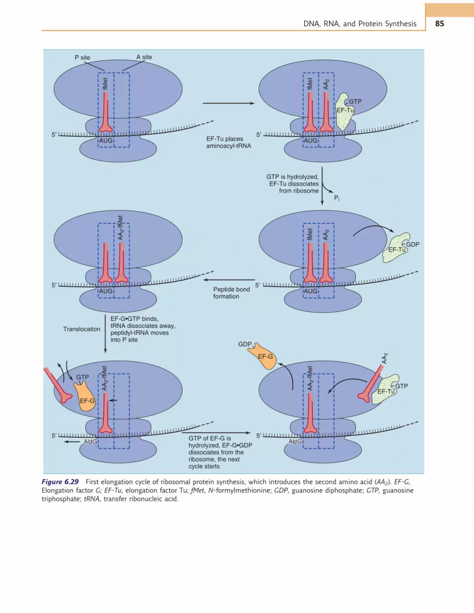
To start the elongation cycle of protein synthesis(Fig. 6.29), an aminoacyl-tRNA is placed in the A site ofthe ribosome along with the GTP-binding elongationfactor Tu (EF-Tu). If (and only if) codon and anticodonmatch, the bound GTP is hydrolyzed to GDP, and EF-Tuwith its boundGDP vacates the ribosome. The aminoacyl-tRNA remains in the A site, ready for peptide bondformation.
The first peptide bond is formed when fMet is trans-ferred from the initiator tRNA to the amino acid residueon the aminoacyl-tRNA in the A site (Fig. 6.30). Thisreaction requires no external energy source because thefree energy content of the ester bond in the fMet-tRNA(&7 kcal/mol) exceeds that of the peptide bond(&1 kcal/mol). The peptidyl transferase of the large ribo-somal subunit that catalyzes peptide bond formation isnot a ribosomal protein, but it is an enzymatic activity
of the 23SRNA in the large ribosomal subunit.Thereforethe ribosome is an RNA enzyme, or ribozyme.
Peptide bond formation leaves a free tRNA in theP site and a peptidyl-tRNA in the A site. The free tRNAmoves from the P site to an E site (E ¼ exit) on the largeribosomal subunit before leaving the ribosome alto-gether, and the peptidyl-tRNA moves from the A siteinto the P site. Codon-anticodon pairing remains intact.Therefore the ribosome moves along the mRNA bythree bases. This step is called translocation. It requiresthe GTP-binding elongation factor EF-G and is accom-panied by the hydrolysis of the bound GTP (seeFig. 6.29). The speed of ribosomal protein synthesis isabout 20 amino acids per second, and the error rate isabout 1 for every 10,000 amino acids.
The stop codons UAA, UAG, and UGA have nomatch-ing tRNAs. Instead they are recognized by proteins calledtermination factors or release factors,which induce cleav-age of the bond between polypeptide and tRNA. GTPhydrolysis takes place during translational termination.
IF-3
IF-1
IF-1
30S
IF-1IF-3
P30S
fMet
AUG
fMet
AUG
70S initiation complex 30S initiation complex
5′
PP
GTP
IF-3
IF-2•GTP,fMet-tRNA,mRNA
mRNA
IF-2
P
5′
PP
50S subunitIF-1, Pi,
IF-2•GDP
50SA site
P site
Figure 6.28 Formation of the 70S initiation complex in prokaryotes. AUG is the intiation codon. fMet, N-formylmethionine; IF-1,
IF-2, and IF-3, initiation factors 1, 2, and 3, respectively; mRNA, messenger RNA; Pi, inorganic phosphate; tRNA, transfer RNA.
84 GENETIC INFORMATION: DNA, RNA, AND PROTEIN SYNTHESIS
GTP is hydrolyzed,EF-Tu dissociates
from ribosome
EF-Tu placesaminoacyl-tRNA
P site
Pi
AUG
fMet
A site
5′AUG
fMet
AA
2
5′
Translocation
EF-G•GTP binds,tRNA dissociates away,peptidyl-tRNA movesinto P site
EF-Tu
Peptide bondformation
AUG
AA
2-f
Met
5′ AUG
fMet
AA
2
5′
GTP of EF-G ishydrolyzed, EF-G•GDPdissociates from the ribosome, the nextcycle starts
AUG AUG
AA
2-f
Met
AA
3
5′AUG
5′
EF-Tu
GTP
GTP
EF-TuGDP
GTP
EF-G
GDP
EF-G
AA
2-f
Met
AUG
Figure 6.29 First elongation cycle of ribosomal protein synthesis, which introduces the second amino acid (AA2). EF-G,
Elongation factor G; EF-Tu, elongation factor Tu; fMet, N-formylmethionine; GDP, guanosine diphosphate; GTP, guanosine
triphosphate; tRNA, transfer ribonucleic acid.
85DNA, RNA, and Protein Synthesis

PROTEIN SYNTHESIS IS ENERGETICALLYEXPENSIVE
GTP hydrolysis during initiation and termination areone-time expenses, but each elongation cycle requiresthe recurrent expense of two GTP bonds: one for
placement, and the other for translocation. This addsto the two high-energy phosphate bonds in ATP thatare expended for the synthesis of each aminoacyl-tRNA.
Therefore at least four high-energy bonds are con-sumed for the synthesis of each peptide bond. Rapidly
CLINICAL EXAMPLE 6.4: Streptomycin Resistance
Cells stop growing immediately when their protein
synthesis is inhibited, and they die slowly when worn-out
cellular proteins can no longer be replaced by new ones.
Therefore it is not surprising that many of the antibiotics
that microorganisms have invented for chemical warfare
against their competitors are inhibitors of ribosomal
protein synthesis.
These antibiotics bind to various sites on the ribosome
and interfere with individual steps in protein synthesis
(Table 6.6). Most are very selective, inhibiting protein
synthesis in either prokaryotes or eukaryotes but not in both.
Bacteria can become resistant to ribosomally acting
antibiotics by mutations that change the target of drug
action. For example, streptomycin resistance can be
produced by mutations in the gene for S12, a protein of
the small ribosomal subunit to which this antibiotic
binds. Even streptomycin-dependent bacterial mutants
can be selected in the laboratory. In other cases of
ribosomally acting antibiotics, mutations that alter the
sequence of one or another ribosomal RNA can make the
bacterium drug resistant.
Table 6.6 Some Antibiotic Inhibitors of Ribosomal Protein Synthesis
Drug Target Organisms Ribosomal Subunit Bound Effect on Protein Synthesis
Streptomycin Prokaryotes 30S Inhibits initiation, causes misreading of mRNA
Tetracycline Prokaryotes 30S Inhibits aminoacyl-tRNA binding
Chloramphenicol Prokaryotes 50S Inhibits peptidyl transferase
Cycloheximide Eukaryotes 60S Inhibits peptidyl transferase
Erythromycin Prokaryotes 50S Inhibits translocation
Puromycin Prokaryotes, eukaryotes 50S, 60S Terminates elongation
mRNA, Messenger ribonucleic acid; tRNA, transfer messenger ribonucleic acid.
O H
C
C
NH
O
O H
C
NH
S CH(CH2)2 H3C
CO
S CH(CH2)2H3C
CO
NH3+
CHR CHR
NH
CO
fMet-tRNA Aminoacyl-tRNA
O O O
Peptidyl-tRNAtRNA
OH
Figure 6.30 Formation of the first peptide bond in the peptidyl transferase reaction. fMet, N-Formylmethionine; tRNA, transfer
ribonucleic acid.
86 GENETIC INFORMATION: DNA, RNA, AND PROTEIN SYNTHESIS

growing bacteria devote 30% to 50% of their total meta-bolic energy to protein synthesis, but the human bodyspends only about 5% of its energy for this purpose.
GENE EXPRESSION IS TIGHTLY REGULATED
Some proteins are needed at all times, so the genes thatencode them are transcribed at a fairly constant rate atall times. These proteins are called constitutive pro-teins, and their genes are called housekeeping genesbecause they have to work continuously. Inducible pro-teins, in contrast, are synthesized only when they areneeded. Their genes are transcribed only in responseto external stimuli that signal a requirement for theencoded protein.
Bacteria have to adjust their metabolic activities tothe nutrient supply. When a bacterium falls into a glassof milk, in which lactose is the major carbohydrate, thebacterium needs enzymes for lactose metabolism. In aglass of lemonade, in which sucrose is abundant, thebacterium needs enzymes of sucrose metabolism. In aglass of beer, the bacterium needs enzymes for alcoholoxidation. In short, having the enzymes for a catabolic,energy-generating pathway makes sense only when thesubstrate of the pathway is available.
The enzymes of a biosynthetic pathway, on the otherhand, are required only when the end product is not avail-able from external sources. For example, the enzymes oftryptophan synthesis are required only when the cell hasto grow on a tryptophan-free medium.
Humans have to adjust their metabolism not only tonutrient supply and need for biosynthetic products, butalso according to cell type. All cells of the body have thesame genes, but different cells make different proteins.
For example, hemoglobin is synthesized by erythroid pre-cursor cells in the bone marrow but not by neurons inthe cerebral cortex. Such differences are the result ofcell-specific gene expression.
A REPRESSOR PROTEIN REGULATESTRANSCRIPTION OF THE LAC OPERON IN E. COLI
Escherichia coli can use the disaccharide lactose (milksugar) as a source of metabolic energy. Lactose is firsttransported across the plasma membrane by the mem-brane carrier lactose permease, then it is cleaved to freeglucose and galactose by the enzyme b-galactosidase(Fig. 6.31). A third protein, b-galactoside transacetylase,is not required for lactose catabolism, but it acetylatesseveral other b-galactosides. It probably is involved in theremoval of nonmetabolizable b-galactosides from the cell.
As an intestinal bacterium, E. coli needs these threeproteins only when its host drinks milk. In the absenceof lactose, the cell contains only about 10 molecules ofb-galactosidase, but several thousand molecules arepresent when lactose is the only carbon source. Thelevels of the permease and the transacetylase parallelexactly those of b-galactosidase.
The genes for these three proteins are lined up headto tail in the bacterial chromosome. They are regulatedin concert because they are transcribed from a singlepromoter. The product of transcription is a polycis-tronic mRNA (from cistron meaning “gene”). The ribo-some can synthesize three different polypeptides fromthis large mRNA because the stop codons of the firsttwo genes are followed by a Shine-Dalgarno sequenceand a start codon at which the synthesis of the nextpolypeptide is initiated.
CH2OH
HO
OH
OH
O
CH2OH
OH
OH
O
CH2OH
HO
HO
OH
OH
OH
O
O CH2
OHHO
OH
OH
OO
CH2OH
OH
OH
OH
O
CH2OH
OH
OH
OHHO
O
1,6-Allolactose
Lactose
Galactose Glucose
+
H2O
Figure 6.31 b-Galactosidase reaction. A small percentage of the substrate is not hydrolyzed but rather is isomerized to
1,6-allolactose in a minor side reaction.
87DNA, RNA, and Protein Synthesis

The array of protein-coding genes, shared promoter,and associated regulatory sites is called an operon, andthe protein-coding genes of the operon are called struc-tural genes.
Wedged between the three structural genes and theirshared promoter is an operator (Fig. 6.32), a shortregulatory DNA sequence that binds the lac repressor.The repressor binding site (operator) overlaps with thebinding site for RNA polymerase (promoter). Thereforethe RNA polymerase cannot bind to the promoter whenthe lac repressor is bound to the operator.
The lac repressor is a tetrameric (from Greek tetra
meaning “four” and meros meaning “part”) proteinwith four identical subunits, encoded by a regulatorygene that is constitutively transcribed at a low rate. Thisgene is located immediately upstream of the lac operon.
In the absence of lactose, the lac repressor bindstightly to the operator and prevents transcription ofthe structural genes. In the presence of lactose, however,a small amount of 1,6-allolactose is formed. This minorside product of the b-galactosidase reaction (seeFig. 6.31) binds tightly to the lac repressor, changingits conformation by an allosteric mechanism. Therepressor-allolactose complex no longer binds to the
operator, and the structural genes can be transcribed.Thus 1,6-allolactose functions as an inducer of the lacoperon.
ANABOLIC OPERONS ARE REPRESSEDBY THE END PRODUCT OF THE PATHWAY
The tryptophan (trp) operon of E. coli codes for a setof five enzymes that are required for the synthesis oftryptophan. Thus the bacteria can synthesize theirown tryptophan, but this energetically expensive bio-synthetic pathway is required only when external tryp-tophan is not available.
The repressor of the trp operon is a dimeric proteinthat binds to an operator site about 20 to 30 nucleo-tides upstream of the transcriptional start site, in themiddle of the promoter. It thereby prevents the bindingof RNA polymerase (Fig. 6.33).
Unlike the lac repressor, the trp repressor cannotbind its operator without outside help. It is an allostericprotein that becomes an active repressor only when itbinds tryptophan. Therefore the trp operon is repressedwhen tryptophan is abundant. In this system, the re-pressor protein is called an aporepressor, and tryptophan
Structural genes
I P O Z Y A
Lactose operon
Figure 6.32 Structure of the lactose (lac) operon of Escherichia coli. Promoter (P), operator (O), and structural genes are
contiguous in all bacterial operons. The regulatory gene that encodes the lac repressor (I) may or may not be located next to the
operon. A, Gene for b-galactoside transacetylase; Y, gene for lactose permease; Z, gene for b-galactosidase.
Holorepressor
Tryptophan
B PP O
Structural genes
Structural genes
PP OR
R
Aporepressor
A
RNA polymerase
Figure 6.33 Regulation of the tryptophan (trp) operon of Escherichia coli. A, Tryptophan is absent. The aporepressor does
not bind to the operator, and the structural genes are transcribed. B, Tryptophan is abundant. The “holorepressor” (aporepressor
þ tryptophan) binds to the operator. The binding of RNA polymerase is prevented, and the structural genes are not transcribed.
O, Operator; P, promoter; R, regulatory gene.
88 GENETIC INFORMATION: DNA, RNA, AND PROTEIN SYNTHESIS

is called a corepressor. This regulatory strategy is typicalfor biosynthetic operons.
GLUCOSE REGULATES THE TRANSCRIPTIONOF MANY CATABOLIC OPERONS
When given the choice between glucose and lactose,E. colimetabolizes glucose first. The levels of b-galactosidase andthe other products of the lac operon are very low as long asboth sugars are present in the medium, and the enzymes oflactose metabolism are induced only when glucose isdepleted (Fig. 6.34).
Glucose is the favored tasty treat of bacteria becauseit is more easily metabolized than lactose. The thrifty(or lazy) bacterium saves the expense for the synthesisof lactose-metabolizing enzymes by metabolizing glu-cose first. Not only the lac operon but also many othercatabolic operons are repressed in the presence of glu-cose. This is called catabolite repression.
Catabolite repression is mediated by cyclic adenosinemonophosphate (cAMP). This small molecule serves asa second messenger of hormone action in humans (seeChapter 17), but in bacteria it is regulated by glucose.The intracellular cAMP level is low when glucose isplentiful and high when it is scarce.
When glucose is depleted and cAMP is abundant,cAMP binds to the dimeric catabolite activator protein
(CAP). CAP alone does not bind to DNA, but the CAP-cAMP complex binds at the promoters of many cata-bolic operons, including the lac operon (Figs. 6.35and 6.36, A). The DNA-bound CAP-cAMP complexprovides additional sites of interaction for RNA poly-merase, thereby facilitating its binding to the promoterand the initiation of transcription.
Time (hours)
Glucosedepletion
Gro
wth
(lo
garith
mic
scale
)
Number ofbacteria
Concentration ofβ-galactosidase(units/mg protein)
1 2 3 4 5
Figure 6.34 Growth of Escherichia coli bacteria on a mixture
of glucose and lactose.
Membrane
Glucose depletion
cAMP
cAMP
Operator
Promoter
Structuralgenes
CAP site
ATPAdenylatecyclase
+
CAP
cAMP
CAP
CAPCAP
CAP
cAMPcAMP
CAP
Figure 6.35 Mechanism of catabolite repression. The promoter of the lac operon is intrinsically weak and permits a high rate
of transcription only when the complex of catabolite activator protein (CAP) and cyclic adenosine monophosphate (cAMP) is
bound to the DNA of the promoter. The cAMP level is low in the presence of glucose but rises in the absence of glucose when
the cAMP-forming enzyme adenylate cyclase is activated. The CAP binds the promoter only when it is complexed with cAMP.
89DNA, RNA, and Protein Synthesis

TRANSCRIPTIONAL REGULATION DEPENDS ONDNA-BINDING PROTEINS
The lac operon and the trp operon illustrate the follow-ing important features of transcriptional regulation:
1. Gene expression is most commonly regulated at thelevel of transcription. Regulation of mRNA processing
and translation also occur, especially in eukaryotes, buttranscriptional regulation is of prime importance.
2. Prokaryotes coordinate the transcription of function-ally related genes by arranging them in operons.Eukar-yotes, however, do not use this strategy. They workwith monocistronic mRNAs, and functionally relatedgenes need not be close together in the genome.
D
–90 –80 –70 –60 –50 –40 –30 –20 –10 0 +10 +20
5′
5′
5′
5′
RNA
polymerase
RNApolymerase
Repressor withbound inducer
C
B
A
Very little mRNA
Abundant mRNA
CAP-cAMP
RNA polymeraseCAP
Repressor
CAPsite
cAMPStart of
transcription
Structural genesRepressor bound
to operator
Promoter
Figure 6.36 Regulation of the lactose operon. A, Binding sites for RNA polymerase, repressor, and catabolite activator protein
(CAP)-cyclic AMP (cAMP). B, Lactose absent, glucose present. RNA polymerase cannot start transcription. C, Lactose present,
glucose present. Weak binding of RNA polymerase to the promoter. D, Lactose present, glucose absent. Strong binding of RNA
polymerase to promoter; high rate of transcription.
90 GENETIC INFORMATION: DNA, RNA, AND PROTEIN SYNTHESIS

3. Transcription is controlled by proteins that bindto regulatory DNA sequences in the vicinity of thetranscriptional start site. The proteins can recognizethese sites because the edges of the DNA bases areexposed in the major and minor grooves of thedouble helix.
4. Many transcriptional activators and repressors areallosterically controlled by small molecules such as1,6-allolactose, cAMP, and tryptophan. However,other control mechanisms are possible. These includeinteractions with other regulatory proteins and cova-lent modification by protein phosphorylation.
5. DNA-binding gene regulator proteins act as eitheractivators or repressors. This action is called positivecontrol or negative control of transcription, res-pectively. The stimulation of transcription by CAP-cAMP is an example of positive control, and theactions of the lac repressor and the trp repressorare examples of negative control.
6. Many transcriptional regulators are either dimers(CAP, trp repressor) or larger oligomers (lac repressor)with identical or slightly different subunits. Thereforetheir structures are symmetrical. The symmetry of theproteins is reflected in the DNA sequences to whichthey bind, which in many cases are palindromic(Fig. 6.37). The oligomeric nature of the gene regula-tors facilitates allosteric changes in their conforma-tion, and their responses to effector molecules can beaccentuated by positive cooperativity.
SUMMARY
DNA as it occurs in cells is a large double-strandedmolecule with a length of several million nucleotides.It contains the four bases adenine (A), guanine (G),cytosine (C), and thymine (T), bound to a backboneof 2-deoxyribose and phosphate. Bases in oppositestrands interact, forming A-T base pairs and G-C basepairs. Therefore the base sequence of one strand pre-dicts the base sequence of the opposite strand.
For replication, the parental DNA double helix isunwound and new complementary strands are synthe-sized; one of the old strands is always used as a tem-plate. The new strands are synthesized by DNApolymerases, with deoxyribonucleoside triphosphatesas precursors.
For gene expression, DNA is copied into RNA byRNA polymerase. This process is called transcription.The gene is defined as the length of DNA that codesfor a polypeptide or for a functional RNA in cases inwhich the RNA is not translated into protein (e.g.,rRNA and tRNA). The RNA transcript of a protein-coding gene is called a messenger ribonucleic acid(mRNA).
The most important principle discussed so far isthat all nucleic acid synthesis requires a DNA tem-plate. DNA is the sovereign master of the organismbecause it alone controls the synthesis of DNA,RNA, and proteins.
The correspondence between the base sequence ofthe mRNA and the amino acid sequence of theencoded polypeptide is called the genetic code. Theamino acids are specified by base triplets calledcodons. There are 61 amino acid coding codons andthree stop codons. The amino acid coding codons arerecognized by tRNAs during protein synthesis, andeach tRNA presents the appropriate amino acid tothe ribosome. During protein synthesis, the ribosomemoves along the mRNA in the 50!30 direction whilepolymerizing the polypeptide in the amino!carboxylterminal direction.
Genes contain regulatory DNA sequences near thetranscriptional start site that bind regulatory proteins.The enhancement of transcription by a DNA-bindingprotein is called positive control, and the inhibitionof transcription is called negative control.
Further Reading
Benkovic SJ, Valentine AM, Salinas F: Replisome-mediatedDNA replication, Annu Rev Biochem 70:181–208, 2001.
Froelich-Ammon SJ, Osheroff N: Topoisomerase poisons: har-nessing the dark side of enzyme mechanism, J Biol Chem270:21429–21432, 1995.
Gilmour DS, Fan R: Derailing the locomotive: transcriptiontermination, J Biol Chem 283:661–664, 2008.
5′-
3′-
A
B
C
T
A
G
C
T
A
G
C
T
A
G
C
G
C
Start oftranscription Symmetry axis
Start oftranscriptionSymmetry axis
Symmetry axis
A
T
A
T
T
A
T
A
G
C
T
A
G
C
A
T
G
C
C
G
G
C
G
C
A
T
T
A
A
T
A
T
C
G
A
T
A
T
T
A
T
A
T
A
C
G
A
T
C
G
A
T
C
G
A
T
-3′
-5′
5′-
3′-
C
G
G
C
C
G
A
T
A
T
T
A
T
A
A
T
A
T
T
A
G
C
T
A
G
C
A
T
G
C
T
A
T
A
A
T
G
C
C
G
T
A
C
G
A
T
C
G
T
A
C
G
A
T
T
A
T
A
A
T
G
C
G
C
-3′
-5′
5′-
3′-
C
G
G
C
A
T
A
T
C
G
T
A
A
T
G
C
T
A
T
A
A
T
A
T
C
G
T
A
A
T
G
C
T
A
A
T
C
G
G
C
C
G
A
T
A
T
G
C
-3′
-5′
Figure 6.37 Incomplete palindromic sequences in the
binding sites of transcriptional regulator proteins. Most
transcriptional regulators bind their cognate DNA sites in a
dimeric form, each subunit interacting with one leg of the
palindrome left and right of the symmetry axis. A, The lac
operator. B, The trp operator. C, The catabolite activator
protein-cAMP binding site of the lac operon. A, Adenine;
C, cytosine; G, guanine; T, thymine.
91DNA, RNA, and Protein Synthesis

Kisselev LL, Buckingham RH: Translational termination comesof age, Trends Biochem Sci 25:561–566, 2000.
Kunkel TA: DNA replication fidelity, J Biol Chem 279:16895–16898, 2004.
Vassylyev DG: Elongation by RNA polymerase: a race throughroadblocks, Curr Opin Struct Biol 19:691–700, 2009.
Wilson DN: The A-Z of bacterial translation inhibitors, CritRev Biochem Mol Biol 44:393–433, 2009.
QUESTIONS
1. The base triplet 50-GAT-30 on the templatestrand of DNA is transcribed into mRNA. Theanticodon that recognizes this sequence duringtranslation is
A. 50-GAT-30
B. 50-GAU-30
C. 50-UAG-30
D.50-AUC-30
E. 50-ATC-30
2. The high fidelity of DNA replication in E. coliwould not be possible without
A. The high processivity of DNA polymerase IIIB. The S subunit of DNA polymerase IC. The 50-exonuclease activity of DNA polymerase ID. The 30-exonuclease activity of DNA polymerase IIIE. The extremely high accuracy of the aminoacyl-
tRNA synthetases
3. Stop codons are present on
A. The coding strand of DNA, where they signal theend of transcription
B. The template strand of DNA, where they signalthe end of transcription
C. The mRNA, where they signal the end oftranslation
D. The tRNA, where they signal the end oftranslation
E. Termination factors, where they signal the end oftranslation
4. As a result of a mutation, an E. coli cellproduces an aberrant aminoacyl-tRNAsynthetase that attaches not leucine butisoleucine to one of the leucine tRNAs. Thiskind of mutation would lead to
A. A disruption of codon-anticodon pairing duringprotein synthesis
B. Premature chain termination during ribosomalprotein synthesis
C. Impaired initiation of ribosomal protein synthesisD. Inability of the aminoacyl-tRNA to bind to the
ribosomeE. A change in the genetic code
5. Like lactose, the pentose sugar arabinose canbe used as a source of metabolic energy byE. coli. The most reasonable prediction for theregulation of the arabinose-catabolizingenzymes would be that
A. Enzymes of arabinose catabolism are inducedwhen glucose is plentiful
B. A repressor protein prevents the synthesis ofarabinose-catabolizing enzymes in the absence ofarabinose
C. The catabolite activator protein prevents thesynthesis of arabinose-catabolizing enzymes inthe absence of arabinose
D.Arabinose acts as a corepressor that is requiredfor the binding of the repressor to the operator
E. Arabinose stimulates the synthesis of arabinose-catabolizing enzymes by raising the cellular cAMPlevel
92 GENETIC INFORMATION: DNA, RNA, AND PROTEIN SYNTHESIS


