Embed Size (px)
Citation preview
Development/Plasticity/Repair
Dopamine, through the Extracellular Signal-RegulatedKinase Pathway, Downregulates CD4�CD25� RegulatoryT-Cell Activity: Implications for Neurodegeneration
Jonathan Kipnis, Michal Cardon, Hila Avidan, Gil M. Lewitus, Sharon Mordechay, Asya Rolls, Yael Shani, andMichal SchwartzDepartment of Neurobiology, The Weizmann Institute of Science, 76100 Rehovot, Israel
Fighting off neuronal degeneration requires a well controlled T-cell response against self-antigens residing in sites of the CNS damage.The ability to evoke this response is normally suppressed by naturally occurring CD4�CD25 � regulatory T-cells (Treg). No physiologicalcompound that controls Treg activity has yet been identified. Here, we show that dopamine, acting via type 1 dopamine receptors (foundhere to be preferentially expressed by Treg), reduces the suppressive activity and the adhesive and migratory abilities of Treg. Tregactivity was correlated with activation of the ERK1/2 (extracellular signal-regulated kinase 1/2) signaling pathway. Systemic injection ofdopamine or an agonist of its type 1 receptors significantly enhanced, via a T-cell-dependent mechanism, protection against neuronaldeath after CNS mechanical and biochemical injury. These findings shed light on the physiological mechanisms controlling Treg andmight open the way to novel therapeutic strategies for downregulating Treg activity (e.g., in neuronal degeneration) or for strengtheningit (in autoimmune diseases).
Key words: CD4 �CD25 � regulatory T-cells; neurotransmitters; dopamine; dopamine receptors; ERK1/2; autoimmune response; neuro-protection; neurodegeneration; CNS
IntroductionIt is becoming increasingly clear that an autoimmune responseagainst self-antigens residing in the site of insult (Moalem et al.,1999) can protect the body against CNS neurodegeneration. Nor-mally, autoimmunity is suppressed by naturally occurring regu-latory CD4�CD25� T-cells (Treg) (Shevach et al., 2001). There-fore, to elicit the desired autoimmune response for protection ofCNS neurons at risk of degeneration, the Treg-imposed suppres-sion must be alleviated. Depletion of Treg promotes survival ofneurons after CNS insults (Kipnis et al., 2002) as well as boostsantitumor autoimmunity (Sakaguchi et al., 2001). Treg-imposedsuppression is a multifactorial process, involving cell-to-cell con-tacts (Nakamura et al., 2001) and the activity of soluble factors[which presumably include interleukin (IL)-10 (Sundstedt et al.,2003) and TGF-� (Piccirillo et al., 2002)]. Studies have shownthat the suppressive activity of Treg can be inhibited by the addi-tion of exogenous IL-2 (Thornton and Shevach, 1998), or block-ing of the cytotoxic T-lymphocyte-associated antigen receptor 4(CTLA-4) (Nakamura et al., 2001), or activation of the newly
discovered glucocorticoid-induced TNF-� receptor (McHugh etal., 2002).
Some key adhesion molecules are more abundant on the sur-faces of Treg than of effector (CD4�CD25�) T-cells (Teff)(Kohm et al., 2002). The ability of Treg to enter tissues might helpprevent autoimmune disease progression. In fighting off neuro-degeneration or cancer, however, the presence of Treg is a liabil-ity. Compounds capable of reducing the trafficking ability (adhe-sion and migration) of Treg, or their suppressive activity, or both,might therefore be promising candidates for therapy against bothcancer and CNS insults. As a corollary, compounds capable ofupregulating the inhibitory or trafficking activity of Treg, orboth, might be potential candidates for therapy against autoim-mune diseases. A fine balance would then be needed to fight offthe conditions leading to neuronal degeneration without creatingconditions that foster neural tissue-specific autoimmune dis-eases. Up to now, however, no physiological compounds havebeen discovered that can control the activity of Treg.
The present study was undertaken in an attempt to identifyphysiological compounds potentially capable of controlling theTreg activity after CNS injury. We postulated that because stress-or pain-related physiological compounds are increased after CNSinjury (Rothblat and Schneider, 1998; Thiffault et al., 2000), oneor more of them might transmit an early signal to Treg, withconsequent reduction of the trafficking or suppressive activity, orboth, of the latter. We reasoned that likely candidate compoundsmight be key neurotransmitters such as dopamine, norepineph-rine, serotonin, and substance P, all of which have been shown to
Received Feb. 19, 2004; revised May 28, 2004; accepted May 28, 2004.This work was supported by Proneuron Ltd. Industrial Park (Ness-Ziona, Israel) to M.S. M.S. holds the Maurice and
Ilse Katz Professorial Chair in Neuroimmunology. We thank S. Smith for editing this manuscript, A. Shapira for animalmaintenance, H. Avital for graphical assistance, and Dr. R. Eilam for histological assistance.
Correspondence should be addressed to Dr. Michal Schwartz, Department of Neurobiology, The Weizmann Insti-tute of Science, 76100 Rehovot, Israel. E-mail: [email protected].
DOI:10.1523/JNEUROSCI.0600-04.2004Copyright © 2004 Society for Neuroscience 0270-6474/04/246133-11$15.00/0
The Journal of Neuroscience, July 7, 2004 • 24(27):6133– 6143 • 6133
participate in interactions between the brain and the immunesystem (Edgar et al., 2002).
Of all the tested neurotransmitters, dopamine was the onlyone that reduced the activity of Treg, and it did so via an ERK(extracellular signal-regulated kinase)-dependent pathway. Do-pamine affected both the suppressive and the trafficking activitiesof Treg, via dopamine type 1 (D1-R and D5-R) receptors, foundhere to be preferentially expressed by Treg. Using mouse modelsof neurodegenerative conditions caused by partial crush injury ofthe optic nerve or glutamate intoxication in the eye, we showedthat systemic administration of dopamine or its D1-type agonistcan induce neuroprotection after mechanical and chemical CNSinjury by alleviating the suppression imposed by Treg.
Materials and MethodsAnimals. Inbred adult wild-type, severe combined immunodeficient(scid), and nu/nu BALB/c and C57Bl/6 mice were supplied by the AnimalBreeding Center of The Weizmann Institute of Science. All animals werehandled according to the regulations formulated by the InstitutionalAnimal Care and Use Committee.
Antibodies and reagents. The antibodies and reagents used included:mouse recombinant IL-2, anti-mouse �-CD3, anti-mouse CTLA-4, andpurified rabbit anti-mouse ERK2 antibody (R & D Systems, Minneapolis,MN); rat anti-mouse phycoerythrin (PE)-conjugated CD25 antibody(PharMingen, Becton-Dickinson, Franklin Lakes, NJ); FITC-conjugatedanti-CD4 antibody (Serotec, Oxford, UK); anti D1-R (Calbiochem,Darmstadt, Germany); 3-hydroxytyramine (dopamine), norepineph-rine, SKF-38393, SCH-23390, quinpirole, clozapine, genistein, andPD98059 (Sigma-Aldrich, Rehovot, Israel); phosphatidyl serine detec-tion kit (IQ Products, Houston, TX). Purified anti-pERK1/2 antibodywas a gift from Prof. R. Seger from The Weizmann Institute of Science.
Intravitreal glutamate injection. The right eyes of anesthetized micewere punctured with a 27 gauge needle in the upper part of the sclera, anda 10 �l Hamilton syringe with a 30 gauge needle was inserted as far as thevitreal body. A total volume of 1 �l of L-glutamate (400 nmol) dissolvedin saline was injected into the eye (Schori et al., 2001).
Retrograde labeling of retinal ganglion cells. Mice were anesthetized andplaced in a stereotactic device. The skull was exposed and kept dry andclean. The bregma was identified and marked. The designated point ofinjection was at a depth of 2 mm from the brain surface, 2.92 mm behindthe bregma in the anteroposterior axis, and 0.5 mm lateral to the midline.The neurotracer dye FluoroGold (5% solution in saline; Fluorochrome,Denver, CO) was applied (1 �l, at a rate of 0.5 �l/min in each hemi-sphere) using a Hamilton syringe, and the skin over the wound wassutured.
Crush injury of the optic nerve in mice. Animals were anesthetizeddeeply by intraperitoneal injection of 2% Xyl-M (xylazine, 10 mg/kg;VMD, Arendonk, Belgium) and Ketaset (ketamine, 50 mg/kg; FortDodge Laboratories, Fort Dodge, IA) and subjected to severe crush injuryof the intraorbital portion of the optic nerve. The uninjured contralateralnerve was left undisturbed. The optic nerve was crushed 3 d after retro-grade labeling of retinal ganglion cells with FluoroGold, as describedabove (Fisher et al., 2001).
Enzyme-linked immunosorbent assay. Treg or Teff (0.5 � 10 6 cells/ml)were cultured for 48 hr in the presence of anti-CD3 and anti-CD28. After48 hr, the cells were centrifuged and their supernatants were collectedand sampled. Concentrations of IL-2 in the samples were determined bythe use of sandwich ELISA kits (R & D Systems). For detection of secretedIL-10, cells were centrifuged every 24 hr and replaced with a fresh me-dium. Supernatants obtained from cells after 24, 48, and 72 hr in culturewere subjected to an ELISA kit (Diaclone Research, Fleming, France).
Purification of murine CD4�CD25�/CD4�CD25- T-cells. Lymphnodes (axillary, inguinal, superficial cervical, mandibular, and mesen-teric) and spleens were harvested and mashed. T-cells were purified (en-riched by negative selection) on T-cell columns (R & D Systems). Theenriched T-cells were incubated with anti-CD8 microbeads (MiltenyiBiotec, Bergisch Gladbach, Germany), and negatively selected CD4 �
T-cells were incubated with PE-conjugated anti-CD25 (30 �g/10 8 cells)in PBS/2% FCS. They were then washed and incubated with anti-PEmicrobeads and subjected to magnetic separation with AutoMACS. Theretained cells were eluted from the column as purified CD4 �CD25 �
cells. The negative fraction consisted of CD4�CD25� T-cells. Purified cellswere cultured in 24-well plates (1 ml) with T-cell-depleted spleen cellsas accessory cells (irradiated with 3000 rad) and 0.5 �g/ml anti-CD3,supplemented with 100 U of mouse recombinant IL-2.
T-cell adhesion. Adhesion of activated CD4�CD25� and CD4�CD25�
T-cells to chondroitin sulfate proteoglycans (CSPG) was analyzed asdescribed previously (Ariel et al., 1998). Briefly, flat-bottomed microtiter(96-well) plates were precoated with CSPG (1 �g/well, 40 min, 37°C).51Cr-labeled T-cells were left untreated or were preincubated (30 min,37°C) with dopamine or the specified agonist or antagonist (10 �5 M). THE
CELLS (10 5 cells in 100 �l of RPMI medium containing 0.1% BSA) werethen added to the CSPG-coated wells, incubated (30 min, 37°C), andwashed. Adherent cells were lysed, and the resulting supernatants wereremoved and counted in a gamma counter. Results were expressed as themean percentage of the total population before adhesion of boundT-cells from quadruplicate wells for each experimental group.
Chemotaxis assay. The migration of T-cells across polycarbonate filters(pore size, 5 �m; diameter, 6.5 mm) toward stromal cell-derived factor-1(SDF-1) and macrophage-derived chemokine (MDC) (CCL22) was as-sayed in 24-well Transwell chambers (Costar, Corning, Corning, NY).T-lymphocytes (1.67 � 10 6 cells/ml) were suspended in RPMI medium/0.1% BSA, and 150 �l of the cell suspension was added to the upperchamber after incubation with or without dopamine (90 min, 37°C).Chemokines were added to the lower chamber at concentrations of 1�g/ml SDF-1 (CytoLab, Ness-Ziona, Israel) and 0.25 �g/ml MDC (R & DSystems). The plates were incubated for 90 min at 37°C in 9.5% CO2.T-cells that migrated to the lower chambers were collected and stainedwith anti-CD4 and anti-CD25 antibodies. The numbers of migratingT-cells were measured by flow cytometer acquisition for a fixed time (60sec). To calculate specific migration, the number of cells in each sub-population in the absence of chemokine was subtracted from the numberin the corresponding cell subpopulation that migrated in the presence ofchemokines. The number of migrating CD4 �CD25 � T-cells was calcu-lated as a percentage of the total T-cell population before migration. Formigration of purified population, we used a similar protocol.
Activation of Treg. Purified Treg (0.5 � 10 6 cells/ml) were activated inRPMI medium supplemented with L-glutamine (2 mM),2-mercaptoethanol (5 � 10 �5
M), sodium pyruvate (1 mM), penicillin(100 IU/ml), streptomycin (100 �g/ml), nonessential amino acids (1ml/100 ml), and autologous serum 2% (v/v) in the presence of mouserecombinant IL-2 (mrIL-2; 5 ng/ml) and soluble anti-CD3 antibodies (1ng/ml). Irradiated (2500 rad) splenocytes (1.5 � 10 6 cells/ml) wereadded to the culture. Cells were activated for 24 or 96 hr. In some of the96 hr experiments, fresh dopamine was added to the culture every 24 hrduring activation.
Inhibition assay (coculturing of Teff with Treg). Naive Teff (50 � 10 3
cells/well) were cocultured with decreasing numbers of activated Treg for72 hr in 96-well flat-bottomed plates in the presence of irradiated spleno-cytes (10 6/ml) supplemented with anti-CD3 antibodies. [ 3H]-thymidine(1 �Ci) was added for the last 16 hr of culture. After the cells wereharvested, their [ 3H]-thymidine content was analyzed by the use of agamma counter.
Immunocytochemistry. T-cells were fixed for 10 min with a mixture(1:1) of methanol and acetone at �20°C, incubated in blocking solution(PBS containing 0.3% Triton-X100 and 1% of normal rabbit serum) for60 min at room temperature, and then incubated overnight with a spe-cific antibody (dilution, 1:1000) in the blocking solution. The T-cellswere then washed and incubated with the secondary antibody (PE-labeled goat anti-rabbit IgG) for 30 min at room temperature, thenwashed, and analyzed by fluorescence and confocal microscopy.
Western blotting. Cells were stimulated for 20 min with anti-CD3 andanti-CD28 antibodies in the presence or absence of dopamine or SKF-38393. Cell lysates were prepared using radioimmunoprecipitation assaylysis buffer (50 mM Tris, pH 8, 0.1% SDS, 0.5% deoxycholate, 1% NP-40,500 mM NaCl, and 10 mM MgCl2). Supernatants were collected, and 5�
6134 • J. Neurosci., July 7, 2004 • 24(27):6133– 6143 Kipnis et al. • Dopamine Downregulates Treg Activity
sample buffer (containing 25 mM Tris, pH 6.8, 2% SDS, 10% glycerol,0.1% bromophenol blue, and 0.5 M �-mercaptoethanol) was added be-fore boiling. Activated ERK1/2 was detected by probing blots with amonoclonal antibody. Total ERK protein was detected by using a poly-clonal rabbit antibody. The blots were developed by HRP-conjugatedanti-mouse or anti-rabbit Fab and ECL (Amersham Biosciences, Freiburg,Germany). Signals were quantified using NIH Image version 1.62.
Polymerase chain reaction. Total RNA was purified with the RNeasyMini kit (Qiagen, Germantown, MD). For PCR, the following primerswere used (for dopamine receptors, primers were used from Lemmer etal., 2002); CCR-4: sense 5�-GTGCAGTCCTGAAGGACTTCAAGCTC-CACCAG-3�, antisense 5�-GGCAAGGACCCTGACCTATGGGGTCA-TCAC-3�; FOXP3: sense 5�-CAGCTGCCTACAGTGCCCCTAG-3�,antisense 5�-CATTTGCCAGCAGTGGGTAG-3�.
Signals were quantified using a Gel-Pro analyzer 3.1 (Media Cybernet-ics, Silver Spring, MD). Real-time PCR was performed with a LightCyclerinstrument (Roche, Mannheim, Germany) using the FastStart DNAMaster SYBR Green 1 kit (catalog #3003230; Roche) as described by themanufacturer. The following primers were used: D5-R: sense 5�-CCTTTATCCCGGTCCA-3�, antisense 5�-GATACGGCGGATCTGAA-3�; IL-10: sense 5�-ACCTGGTAGAAGTGATGCCCCAGGCA-3�, anti-sense 5�-CTATGCAGTTGATGAAGATGTCAAA-3�.
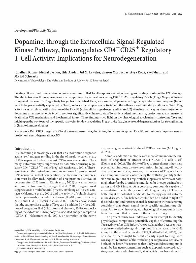
ResultsDopamine reduces the suppression imposed by TregCoculturing of Teff with Treg isolated from naive mice results insuppression of Teff proliferation. The suppressive potency de-pends on the Treg/Teff ratio and the state of Treg activation; thesuppression is significantly increased, for example, if the Treg areactivated before being added to Teff (Thornton and Shevach,1998). Inhibition of Teff proliferation, assayed by [ 3H]-thymidine incorporation, can therefore be taken as a measure ofthe suppressive effect. We examined the ability of major neuro-transmitters and neuropeptides (dopamine, norepinephrine,substance P, and serotonin) to alleviate the Treg-induced sup-pression of Teff in vitro. Each compound was tested at severalconcentrations. Proliferation of Teff was significantly inhibitedby cocultivation of Teff with naive Treg or with Treg that hadbeen activated by incubation for 24 hr with anti-CD3 antibodiesand IL-2 in the presence of antigen-presenting cells (APCs; le-thally irradiated splenocytes) (Fig. 1). After incubating the acti-vated Treg for 2 hr with a neurotransmitter or a neuropeptide, wewashed the cells and then cocultured them with Teff. Prolifera-tion of Teff cocultured with activated Treg that had been incu-bated with dopamine (10�5
M) was more than twofold higherthan proliferation in coculture with activated Treg not incubatedwith dopamine (Fig. 1a). A significant effect on Treg-suppressiveactivity was also obtained with 10�7
M dopamine (Fig. 1a),whereas 10�9
M had no significant effect (data not shown). Theinhibitory effect of dopamine at 10�5 and 10�7
M on Treg activitywas reproduced when freshly isolated (nonactivated) Treg wereused (Fig. 1b). At the dopamine concentration of 10�9
M, theobtained effect was slight and not statistically significant (Fig. 1b).It should be noted, however, that the effect of dopamine on Treg-suppressive activity was only partial and that complete blockingwas not seen at any of the concentrations tested. We also exam-ined the effect of dopamine on the activity of Treg that had beenactivated as described above (Fig. 1a), but for 96 hr, and to whichdopamine (10�5
M) was added for 2 hr at the end of the activationperiod and then washed off before the activated cells were cocul-tured with naive Teff. Again, Teff proliferation was significantlyhigher in the presence of activated Treg treated with dopaminethan in the presence of activated Treg without dopamine (Fig.1c). A direct effect of dopamine on Teff proliferation was ruledout by incubation of Teff for 2 hr with 10�5
M dopamine, then
washing off the dopamine and adding activated Treg withoutdopamine. The resulting proliferation of Teff did not differ fromthat seen in cultures of Teff in the absence of dopamine. More-over, the inhibitory effects of Treg on naive Teff and on Teffexposed to dopamine were similar (Fig. 1c), indicating that do-pamine did not alter the susceptibility of Teff to Treg suppres-sion. The uptake of thymidine by Teff and the Treg-inducedinhibition of such uptake varied from one experiment to another.In all experiments, however, the effect of dopamine on Treg(tested �20 times) was consistent, and in most cases, the prolif-eration of Teff cocultured with Treg treated with dopamine wasmore than twofold higher than that in the absence of dopaminetreatment. The Treg used in this study were always obtained fromnaive animals, therefore, it is unlikely that they contained anyactivated effector T-cells. The purity of the Treg population usedin all experiments was high (between 92 and 98% of the totalCD4� population). Moreover, the use of anti-CD25 antibodiesto isolate Treg reportedly does not interfere with either the sup-pressive activity or the state of activation of Treg (Thornton andShevach, 1998).
Figure 1. Dopamine (DA) reduces the suppressive activity mediated by CD4 �CD25 � reg-ulatory T-cells. Proliferation of Teff (a CD4 �CD25 � population) was assayed by incorporationof [ 3H]-thymidine into Teff cocultured with naturally occurring Treg. Recorded values are fromone of three representative experiments and are expressed as means � SD of four replicates. a,Treg were activated by incubation for 24 hr with anti-CD3 antibodies in the presence of mrIL-2.Incubation of the activated Treg for 2 hr with dopamine (10 �5 or 10 �7
M) before their co-culturing with Teff reduced their suppression of Teff compared with that obtained with Treg notexposed to dopamine. b, Dopamine (10 �5, 10 �7, or 10 �9
M) added to freshly purified Treg.Dopamine (10 �5 and 10 �7
M) had a similar effect on activity of naive Treg to that of toactivated Treg, whereas the effect of dopamine at 10 �9
M on Treg-mediated suppression wasnot significant. c, Activation of Treg for 96 hr, followed by the addition of dopamine (10 �5
M) for2 hr at the end of activation, significantly reduced the suppressive activity of Treg on Teff.Incubation of Teff with dopamine (10 �5
M) for 2 hr did not affect their susceptibility to Treg-induced suppression.
Kipnis et al. • Dopamine Downregulates Treg Activity J. Neurosci., July 7, 2004 • 24(27):6133– 6143 • 6135
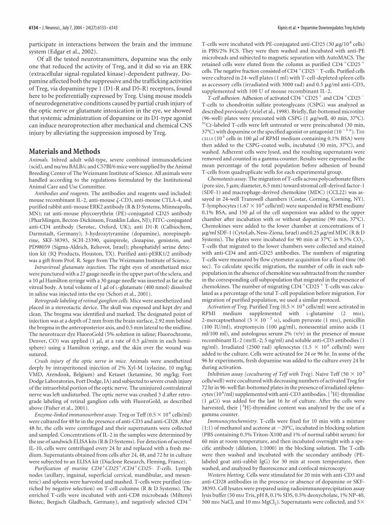
In contrast to the effect seen with dopamine, no effect on theability of Treg to suppress Teff proliferation could be detectedwhen Treg were preincubated with different concentrations ofnorepinephrine (another member of the catecholamine family)(Fig. 2a), substance P (a pain- and stress-related neurotransmit-ter; data not shown), or serotonin (data not shown).
To establish whether the observed effect of dopamine on Tregis exerted through a receptor-mediated pathway, we used specificagonists and antagonists of dopamine receptors. Incubation ofTreg with 10�5
M SKF-38393, an agonist of the type 1 family ofdopamine receptors (consisting of D1-R and D5-R), reproducedthe dopamine effect (Fig. 2b). The specific D1-type antagonistSCH-23390 (10�5
M), when added together with dopamine
(10�5M), prevented the dopamine effect, further substantiating
the contention that the effect of dopamine on Treg is mediatedthrough the type 1 receptor family. Also in line with this conten-tion was the finding that incubation of Treg with 10�5
M quinpi-role, an agonist of the type 2 family of dopamine receptors (com-prising D2-R, D3-R, and D4-R), had no effect on the suppressiveactivity of Treg.
To exclude the possibility that dopamine exerts its effect bycausing the death of Treg, we examined whether dopamine at theconcentrations used here cause Treg apoptosis. No signs of apo-ptosis were detectable in Treg, which, after being incubated withdopamine, were stained with propidium iodide and analyzed forapoptotic cells (sub-G1) by flow cytometry (Fig. 2c). To furtherverify the absence of apoptotic death in Treg, after incubatingTreg with dopamine, we stained them for phosphatidylserinewith annexin V. Again, we could not detect any signs of apoptosisin Treg beyond the background levels seen in the absence ofdopamine (Fig. 2d). Thus, the reduction in Treg activity aftertheir encounter with dopamine or a related agonist evidentlyresults not from the death of Treg but rather from alteration oftheir behavior.
Because dopamine reduced the suppressive activity of Treg onTeff but did not alter the susceptibility of Teff to suppression byTreg, we examined the possibility that Teff and Treg express dif-ferent subtypes or different amounts of the relevant dopaminereceptors. This was done by assaying the expression of the dopa-mine type 1 receptors, D1-R and D5-R, in Treg and Teff. PCRassays showed that Treg expressed significantly more D1-R andD5-R transcripts (4-fold and 14-fold, respectively) than Teff (Fig.3a,b). To further verify the differences in expression of dopaminereceptors by Teff and Treg, we performed real-time PCR, whichshowed that the amounts of D1-R and D5-R in Treg were 5-foldand 13-fold higher, respectively, than in Teff (Fig. 3c).
We also used PCR to assay the expression of dopamine type 2family receptors, namely D2-R, D3-R, and D4-R, in Treg andTeff. Although the expression of D4-R was somewhat moreabundant in Teff than in Treg, the difference between the expres-sion of each of these receptors in the two T-cell subpopulationswas not significant (Fig. 3d,e). To verify that the difference inD1-R between Treg and Teff observed at the transcript level ismanifested also at the protein level, we subjected the cells toimmunocytochemical analysis. D1-R immunoreactivity was de-tected in naive Treg but not in naive Teff (Fig. 3f).
To gain additional insight into the mechanism whereby dopa-mine affects Treg activity, we examined CTLA-4, a moleculecharacteristic of Treg (Im et al., 2001). Expression of this mole-cule was slightly, but consistently, decreased on exposure of Tregto dopamine. A similar effect on CTLA-4 expression was ob-tained with the D1-type-specific agonist SKF-38393 (Fig. 4a).Another molecule that participates in the suppressive activity ofTreg is IL-10 (Maloy et al., 2003). It was therefore of interest tomeasure the production of IL-10 by Treg after their exposure todopamine. Media collected after incubation of Treg with dopa-mine (10�5
M) for 24, 48, and 72 hr showed a significant decreasein the amount of IL-10 at all time points examined (Fig. 4b).Dopamine did not, however, alter the anergic state of Treg; pro-duction of IL-2 was not detected in Treg that had been incubatedin the presence of dopamine, as verified by ELISA for a secretedcytokine in media conditioned for 48 hr by activated Treg (Fig.4c). Teff, as expected, secreted IL-2, the level of which was notaffected by dopamine (Fig. 4c). It should be noted that activationof both T-cell populations was performed in the absence ofmrIL-2.
Figure 2. Dopamine effect on Treg is mediated via D1-type receptor family. Proliferation ofTeff (a CD4 �CD25 � population) was assayed by incorporation of [ 3H]-thymidine into Teffcocultured with naturally occurring Treg. Recorded values are from one of three representativeexperiments and are expressed as means � SD of four replicates. a, Addition of norepinephrine(NE; 10 �5 or 10 �7
M) to Treg for 2 hr after their activation for 24 hr did not affect the suppres-sive activity of Treg. Significant differences between groups were analyzed by Student’s t test( p � 0.001). b, The inhibitory effect of dopamine (DA) on the suppressive activity of Treg wasmimicked by SKF-38393, a specific agonist of the D1-type family. The D2-type agonist quinpi-role did not alter the effect of dopamine on Treg. SCH 23390, a specific D1-type antagonist,wiped out the dopamine effect on the suppressive activity of Treg. Each experiment was per-formed at least five times, and representative results are shown. c, Incubation of Treg or Teffwith dopamine did not cause apoptosis, as shown by propidium iodide staining for DNA contentand FACS analysis of Treg and Teff, 48 hr after their incubation for 2 hr with dopamine. d,Staining for apoptosis with annexin V for phosphatidylserine on a surface membrane. No in-crease in annexin V-labeled cells was detected on incubation of Treg with dopamine or with theD1-type agonist SKF-38393.
6136 • J. Neurosci., July 7, 2004 • 24(27):6133– 6143 Kipnis et al. • Dopamine Downregulates Treg Activity
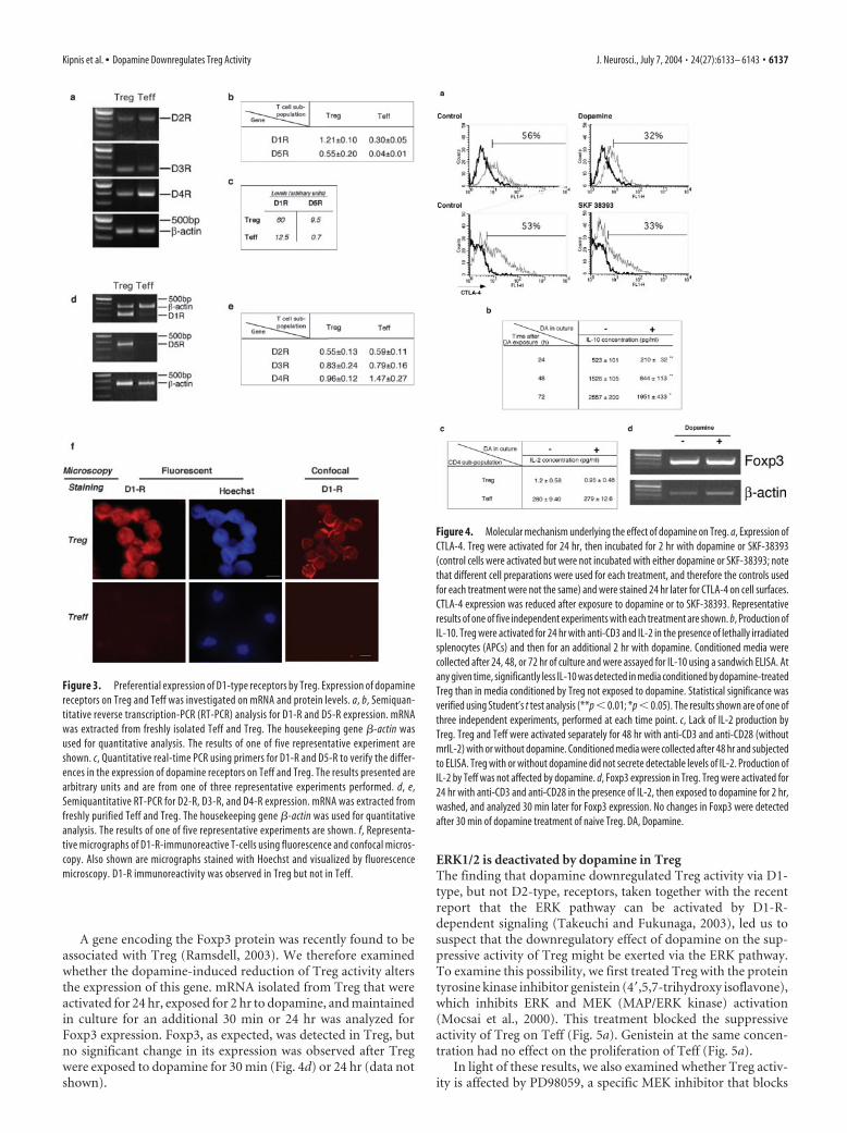
A gene encoding the Foxp3 protein was recently found to beassociated with Treg (Ramsdell, 2003). We therefore examinedwhether the dopamine-induced reduction of Treg activity altersthe expression of this gene. mRNA isolated from Treg that wereactivated for 24 hr, exposed for 2 hr to dopamine, and maintainedin culture for an additional 30 min or 24 hr was analyzed forFoxp3 expression. Foxp3, as expected, was detected in Treg, butno significant change in its expression was observed after Tregwere exposed to dopamine for 30 min (Fig. 4d) or 24 hr (data notshown).
ERK1/2 is deactivated by dopamine in TregThe finding that dopamine downregulated Treg activity via D1-type, but not D2-type, receptors, taken together with the recentreport that the ERK pathway can be activated by D1-R-dependent signaling (Takeuchi and Fukunaga, 2003), led us tosuspect that the downregulatory effect of dopamine on the sup-pressive activity of Treg might be exerted via the ERK pathway.To examine this possibility, we first treated Treg with the proteintyrosine kinase inhibitor genistein (4�,5,7-trihydroxy isoflavone),which inhibits ERK and MEK (MAP/ERK kinase) activation(Mocsai et al., 2000). This treatment blocked the suppressiveactivity of Treg on Teff (Fig. 5a). Genistein at the same concen-tration had no effect on the proliferation of Teff (Fig. 5a).
In light of these results, we also examined whether Treg activ-ity is affected by PD98059, a specific MEK inhibitor that blocks
Figure 3. Preferential expression of D1-type receptors by Treg. Expression of dopaminereceptors on Treg and Teff was investigated on mRNA and protein levels. a, b, Semiquan-titative reverse transcription-PCR (RT-PCR) analysis for D1-R and D5-R expression. mRNAwas extracted from freshly isolated Teff and Treg. The housekeeping gene �-actin wasused for quantitative analysis. The results of one of five representative experiment areshown. c, Quantitative real-time PCR using primers for D1-R and D5-R to verify the differ-ences in the expression of dopamine receptors on Teff and Treg. The results presented arearbitrary units and are from one of three representative experiments performed. d, e,Semiquantitative RT-PCR for D2-R, D3-R, and D4-R expression. mRNA was extracted fromfreshly purified Teff and Treg. The housekeeping gene �-actin was used for quantitativeanalysis. The results of one of five representative experiments are shown. f, Representa-tive micrographs of D1-R-immunoreactive T-cells using fluorescence and confocal micros-copy. Also shown are micrographs stained with Hoechst and visualized by fluorescencemicroscopy. D1-R immunoreactivity was observed in Treg but not in Teff.
Figure 4. Molecular mechanism underlying the effect of dopamine on Treg. a, Expression ofCTLA-4. Treg were activated for 24 hr, then incubated for 2 hr with dopamine or SKF-38393(control cells were activated but were not incubated with either dopamine or SKF-38393; notethat different cell preparations were used for each treatment, and therefore the controls usedfor each treatment were not the same) and were stained 24 hr later for CTLA-4 on cell surfaces.CTLA-4 expression was reduced after exposure to dopamine or to SKF-38393. Representativeresults of one of five independent experiments with each treatment are shown. b, Production ofIL-10. Treg were activated for 24 hr with anti-CD3 and IL-2 in the presence of lethally irradiatedsplenocytes (APCs) and then for an additional 2 hr with dopamine. Conditioned media werecollected after 24, 48, or 72 hr of culture and were assayed for IL-10 using a sandwich ELISA. Atany given time, significantly less IL-10 was detected in media conditioned by dopamine-treatedTreg than in media conditioned by Treg not exposed to dopamine. Statistical significance wasverified using Student’s t test analysis (**p � 0.01; *p � 0.05). The results shown are of one ofthree independent experiments, performed at each time point. c, Lack of IL-2 production byTreg. Treg and Teff were activated separately for 48 hr with anti-CD3 and anti-CD28 (withoutmrIL-2) with or without dopamine. Conditioned media were collected after 48 hr and subjectedto ELISA. Treg with or without dopamine did not secrete detectable levels of IL-2. Production ofIL-2 by Teff was not affected by dopamine. d, Foxp3 expression in Treg. Treg were activated for24 hr with anti-CD3 and anti-CD28 in the presence of IL-2, then exposed to dopamine for 2 hr,washed, and analyzed 30 min later for Foxp3 expression. No changes in Foxp3 were detectedafter 30 min of dopamine treatment of naive Treg. DA, Dopamine.
Kipnis et al. • Dopamine Downregulates Treg Activity J. Neurosci., July 7, 2004 • 24(27):6133– 6143 • 6137
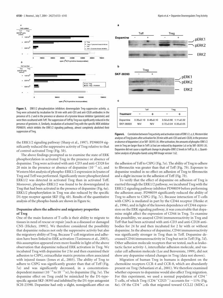
the ERK1/2 signaling pathway (Sharp et al., 1997). PD98059 sig-nificantly reduced the suppressive activity of Treg relative to thatof control-activated Treg (Fig. 5b).
The above findings prompted us to examine the state of ERKphosphorylation in activated Treg in the presence or absence ofdopamine. Treg were activated with anti-CD3 and anti-CD28 for20 min in the presence or absence of dopamine (10�5
M), andWestern blot analysis of phospho-ERK1/2 expression in lysates ofTreg and Teff was performed. Significantly more phosphorylatedERK1/2 was detected in activated Treg than in activated Teff.Moreover, phospho-ERK1/2 was found to be downregulated inTreg that had been activated in the presence of dopamine (Fig. 6a).ERK1/2 phosphorylation in Treg was also reduced by the specificD1-type receptor agonist SKF (Fig. 6b). Results of the quantitativeanalysis of the phospho-bands are shown in Figure 6c.
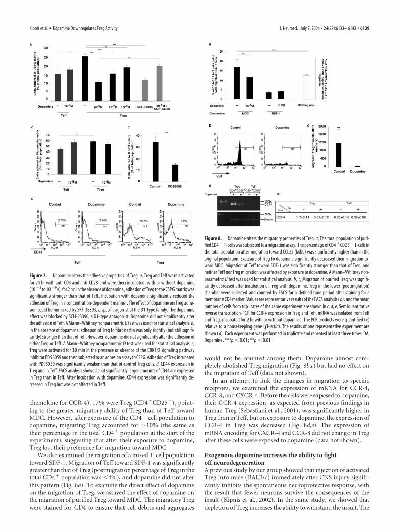
Dopamine alters the adhesive and migratory propertiesof TregOne of the main features of T-cells is their ability to migrate totissues in need of rescue or repair [such as a diseased or damagedCNS (Hickey, 1999)]. We therefore considered the possibilitythat dopamine reduces not only the suppressive activity but alsothe migratory ability of Treg. Because T-cell migration and adhe-sion have been linked to ERK activation (Tanimura et al., 2003),this assumption appeared even more feasible in light of the aboveobservation that dopamine reduced ERK activation in Treg. Weincubated Treg with dopamine for 2 hr and then examined theiradhesion to CSPG, extracellular matrix proteins often associatedwith injured tissues (Jones et al., 2003). The ability of Treg toadhere to CSPG was significantly greater than that of Teff (Fig.7a) and was significantly decreased, in a concentration-dependent manner (10�9 to 10�5
M), by dopamine (Fig. 7a). Thedopamine effect on Treg could be mimicked by the D1-type-specific agonist SKF-38393 and inhibited by the D1-type antagonistSCH-23390. Dopamine had only a slight, nonsignificant effect on
the adhesion of Teff to CSPG (Fig. 7a). The ability of Treg to adhereto fibronectin was greater than that of Teff (Fig. 7b). Exposure todopamine resulted in no effect on adhesion of Treg to fibronectinand a slight increase in the adhesion of Teff (Fig. 7b).
To verify that the effect of dopamine on adhesion of Treg isexerted through the ERK1/2 pathway, we incubated Treg with theERK1/2 signaling pathway inhibitor PD98059 before performingthe adhesion assay. PD98059 significantly reduced the ability ofTreg to adhere to CSPG (Fig. 7c). Because interaction of T-cellswith CSPG is mediated in part by the CD44 receptor (Henke etal., 1996), and in light of the known dependence of CD44 expres-sion on the ERK signaling pathway, it was conceivable that dopa-mine might affect the expression of CD44 in Treg. To examinethis possibility, we assayed CD44 immunoreactivity in Treg andTeff that had been activated with anti-CD3 and anti-CD28 anti-bodies for 24 hr and then incubated for 2 hr with or withoutdopamine. In the absence of dopamine, CD44 immunoreactivitywas significantly stronger in Treg than in Teff. Dopamine de-creased CD44 immunoreactivity in Treg but not in Teff (Fig. 7d).Other adhesion molecule receptors that we tested, such as leuko-tactic factor activity-1, intercellular adhesion molecule, and vas-cular cell adhesion molecule (Lee and Benveniste, 1999), did notshow any dopamine-related changes in Treg (data not shown).
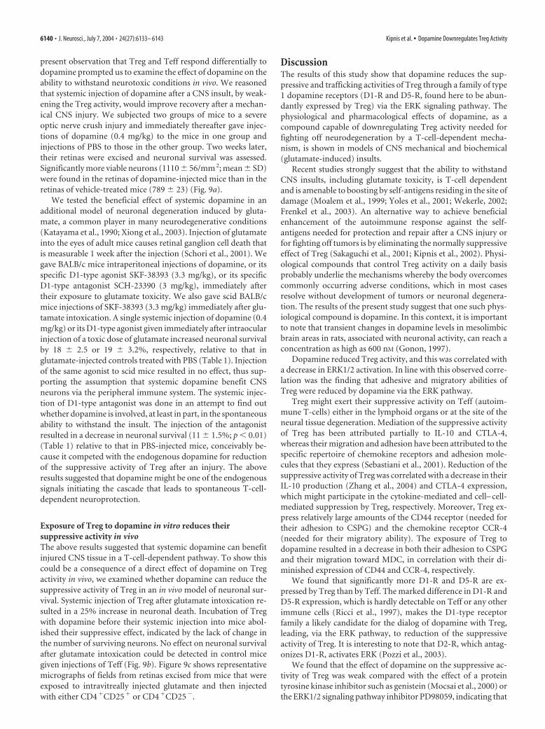
Migration of human Treg in humans is dependent on thechemokine receptors CCR-4 and CCR-8, which are abundantlypresent on Treg (Sebastiani et al., 2001). We therefore examinedwhether exposure to dopamine would also affect Treg migration.For this experiment, we used a normal population of CD4�
T-cells, of which Treg (CD4�CD25�) accounts for �11% (Fig.8a). Of the CD4� cells that migrated toward CCL22 (MDC; a
Figure 5. ERK1/2 phosphorylation inhibitors downregulate Treg-suppressive activity. a,Treg were activated by incubation for 30 min with anti-CD3 and anti-CD28 antibodies in thepresence of IL-2 and in the presence or absence of a tyrosine kinase inhibitor (genistein) andwere then cocultured with Teff. The suppression of Teff by Treg was significantly reduced in thepresence of genistein. b, Similarly, incubation of activated Treg with the specific MEK inhibitorPD98059, which inhibits the ERK1/2 signaling pathway, almost completely abolished theirsuppression of Treg.
Figure 6. Correlation between Treg activity and activation state of ERK1/2. a, b, Western blotanalyses of Treg lysates after activation for 20 min with anti-CD3 and anti-CD28, in the presenceor absence of dopamine ( a) or SKF-38393 ( b). After activation, the amounts of phospho-ERK1/2seen in Treg are larger than in Teff ( a) but are reduced by dopamine ( a) or by SKF-38393 ( b).Dopamine did not cause a significant change in phospho-ERK1/2 levels in Teff (a, b). c, Quanti-tative analysis of phospho-bands using NIH Image version 1.62.
6138 • J. Neurosci., July 7, 2004 • 24(27):6133– 6143 Kipnis et al. • Dopamine Downregulates Treg Activity
chemokine for CCR-4), 17% were Treg (CD4�CD25�), point-ing to the greater migratory ability of Treg than of Teff towardMDC. However, after exposure of the CD4� cell population todopamine, migrating Treg accounted for �10% (the same astheir percentage in the total CD4� population at the start of theexperiment), suggesting that after their exposure to dopamine,Treg lost their preference for migration toward MDC.
We also examined the migration of a mixed T-cell populationtoward SDF-1. Migration of Teff toward SDF-1 was significantlygreater than that of Treg (postmigration percentage of Treg in thetotal CD4� population was �4%), and dopamine did not alterthis pattern (Fig. 8a). To examine the direct effect of dopamineon the migration of Treg, we assayed the effect of dopamine onthe migration of purified Treg toward MDC. The migratory Tregwere stained for CD4 to ensure that cell debris and aggregates
would not be counted among them. Dopamine almost com-pletely abolished Treg migration (Fig. 8b,c) but had no effect onthe migration of Teff (data not shown).
In an attempt to link the changes in migration to specificreceptors, we examined the expression of mRNA for CCR-4,CCR-8, and CXCR-4. Before the cells were exposed to dopamine,their CCR-4 expression, as expected from previous findings inhuman Treg (Sebastiani et al., 2001), was significantly higher inTreg than in Teff, but on exposure to dopamine, the expression ofCCR-4 in Treg was decreased (Fig. 8d,e). The expression ofmRNA encoding for CXCR-4 and CCR-8 did not change in Tregafter these cells were exposed to dopamine (data not shown).
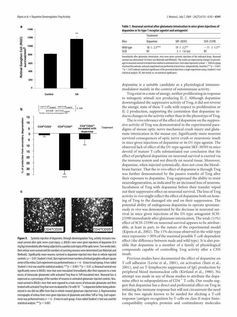
Exogenous dopamine increases the ability to fightoff neurodegenerationA previous study by our group showed that injection of activatedTreg into mice (BALB/c) immediately after CNS injury signifi-cantly inhibits the spontaneous neuroprotective response, withthe result that fewer neurons survive the consequences of theinsult (Kipnis et al., 2002). In the same study, we showed thatdepletion of Treg increases the ability to withstand the insult. The
Figure 7. Dopamine alters the adhesive properties of Treg. a, Treg and Teff were activatedfor 24 hr with anti-CD3 and anti-CD28 and were then incubated, with or without dopamine(10 �5 to 10 �9
M), for 2 hr. In the absence of dopamine, adhesion of Treg to the CSPG matrix wassignificantly stronger than that of Teff. Incubation with dopamine significantly reduced theadhesion of Treg in a concentration-dependent manner. The effect of dopamine on Treg adhe-sion could be mimicked by SKF-38393, a specific agonist of the D1-type family. The dopamineeffect was blocked by SCH-23390, a D1-type antagonist. Dopamine did not significantly alterthe adhesion of Teff. A Mann–Whitney nonparametric U test was used for statistical analysis. b,In the absence of dopamine, adhesion of Treg to fibronectin was only slightly (but still signifi-cantly) stronger than that of Teff. However, dopamine did not significantly alter the adhesion ofeither Treg or Teff. A Mann–Whitney nonparametric U test was used for statistical analysis. c,Treg were activated for 30 min in the presence or absence of the ERK1/2 signaling pathwayinhibitor PD98059 and then subjected to an adhesion assay to CSPG. Adhesion of Treg incubatedwith PD98059 was significantly weaker than that of control Treg cells. d, CD44 expression inTreg and in Teff. FACS analysis showed that significantly larger amounts of CD44 are expressedin Treg than in Teff. After incubation with dopamine, CD44 expression was significantly de-creased in Treg but was not affected in Teff.
Figure 8. Dopamine alters the migratory properties of Treg. a, The total population of puri-fied CD4 � T-cells was subjected to a migration assay. The percentage of CD4 �CD25 � T-cells inthe total population after migration toward CCL22 (MDC) was significantly higher than in theoriginal population. Exposure of Treg to dopamine significantly decreased their migration to-ward MDC. Migration of Teff toward SDF-1 was significantly stronger than that of Treg, andneither Teff nor Treg migration was affected by exposure to dopamine. A Mann–Whitney non-parametric U test was used for statistical analysis. b, c, Migration of purified Treg was signifi-cantly decreased after incubation of Treg with dopamine. Treg in the lower (postmigration)chamber were collected and counted by FACS for a defined time period after staining for amembrane CD4 marker. Values are representative results of the FACS analysis ( b), and the meannumber of cells from triplicates of the same experiment are shown in c. d, e, Semiquantitativereverse transcription-PCR for CCR-4 expression in Treg and Teff. mRNA was isolated from Teffand Treg, incubated for 2 hr with or without dopamine. The PCR products were quantified ( e)relative to a housekeeping gene (�-actin). The results of one representative experiment areshown ( d). Each experiment was performed in triplicate and repeated at least three times. DA,Dopamine. ***p � 0.01; **p � 0.01.
Kipnis et al. • Dopamine Downregulates Treg Activity J. Neurosci., July 7, 2004 • 24(27):6133– 6143 • 6139
present observation that Treg and Teff respond differentially todopamine prompted us to examine the effect of dopamine on theability to withstand neurotoxic conditions in vivo. We reasonedthat systemic injection of dopamine after a CNS insult, by weak-ening the Treg activity, would improve recovery after a mechan-ical CNS injury. We subjected two groups of mice to a severeoptic nerve crush injury and immediately thereafter gave injec-tions of dopamine (0.4 mg/kg) to the mice in one group andinjections of PBS to those in the other group. Two weeks later,their retinas were excised and neuronal survival was assessed.Significantly more viable neurons (1110 � 56/mm 2; mean � SD)were found in the retinas of dopamine-injected mice than in theretinas of vehicle-treated mice (789 � 23) (Fig. 9a).
We tested the beneficial effect of systemic dopamine in anadditional model of neuronal degeneration induced by gluta-mate, a common player in many neurodegenerative conditions(Katayama et al., 1990; Xiong et al., 2003). Injection of glutamateinto the eyes of adult mice causes retinal ganglion cell death thatis measurable 1 week after the injection (Schori et al., 2001). Wegave BALB/c mice intraperitoneal injections of dopamine, or itsspecific D1-type agonist SKF-38393 (3.3 mg/kg), or its specificD1-type antagonist SCH-23390 (3 mg/kg), immediately aftertheir exposure to glutamate toxicity. We also gave scid BALB/cmice injections of SKF-38393 (3.3 mg/kg) immediately after glu-tamate intoxication. A single systemic injection of dopamine (0.4mg/kg) or its D1-type agonist given immediately after intraocularinjection of a toxic dose of glutamate increased neuronal survivalby 18 � 2.5 or 19 � 3.2%, respectively, relative to that inglutamate-injected controls treated with PBS (Table 1). Injectionof the same agonist to scid mice resulted in no effect, thus sup-porting the assumption that systemic dopamine benefit CNSneurons via the peripheral immune system. The systemic injec-tion of D1-type antagonist was done in an attempt to find outwhether dopamine is involved, at least in part, in the spontaneousability to withstand the insult. The injection of the antagonistresulted in a decrease in neuronal survival (11 � 1.5%; p � 0.01)(Table 1) relative to that in PBS-injected mice, conceivably be-cause it competed with the endogenous dopamine for reductionof the suppressive activity of Treg after an injury. The aboveresults suggested that dopamine might be one of the endogenoussignals initiating the cascade that leads to spontaneous T-cell-dependent neuroprotection.
Exposure of Treg to dopamine in vitro reduces theirsuppressive activity in vivoThe above results suggested that systemic dopamine can benefitinjured CNS tissue in a T-cell-dependent pathway. To show thiscould be a consequence of a direct effect of dopamine on Tregactivity in vivo, we examined whether dopamine can reduce thesuppressive activity of Treg in an in vivo model of neuronal sur-vival. Systemic injection of Treg after glutamate intoxication re-sulted in a 25% increase in neuronal death. Incubation of Tregwith dopamine before their systemic injection into mice abol-ished their suppressive effect, indicated by the lack of change inthe number of surviving neurons. No effect on neuronal survivalafter glutamate intoxication could be detected in control micegiven injections of Teff (Fig. 9b). Figure 9c shows representativemicrographs of fields from retinas excised from mice that wereexposed to intravitreally injected glutamate and then injectedwith either CD4�CD25� or CD4�CD25�.
DiscussionThe results of this study show that dopamine reduces the sup-pressive and trafficking activities of Treg through a family of type1 dopamine receptors (D1-R and D5-R, found here to be abun-dantly expressed by Treg) via the ERK signaling pathway. Thephysiological and pharmacological effects of dopamine, as acompound capable of downregulating Treg activity needed forfighting off neurodegeneration by a T-cell-dependent mecha-nism, is shown in models of CNS mechanical and biochemical(glutamate-induced) insults.
Recent studies strongly suggest that the ability to withstandCNS insults, including glutamate toxicity, is T-cell dependentand is amenable to boosting by self-antigens residing in the site ofdamage (Moalem et al., 1999; Yoles et al., 2001; Wekerle, 2002;Frenkel et al., 2003). An alternative way to achieve beneficialenhancement of the autoimmune response against the self-antigens needed for protection and repair after a CNS injury orfor fighting off tumors is by eliminating the normally suppressiveeffect of Treg (Sakaguchi et al., 2001; Kipnis et al., 2002). Physi-ological compounds that control Treg activity on a daily basisprobably underlie the mechanisms whereby the body overcomescommonly occurring adverse conditions, which in most casesresolve without development of tumors or neuronal degenera-tion. The results of the present study suggest that one such phys-iological compound is dopamine. In this context, it is importantto note that transient changes in dopamine levels in mesolimbicbrain areas in rats, associated with neuronal activity, can reach aconcentration as high as 600 nM (Gonon, 1997).
Dopamine reduced Treg activity, and this was correlated witha decrease in ERK1/2 activation. In line with this observed corre-lation was the finding that adhesive and migratory abilities ofTreg were reduced by dopamine via the ERK pathway.
Treg might exert their suppressive activity on Teff (autoim-mune T-cells) either in the lymphoid organs or at the site of theneural tissue degeneration. Mediation of the suppressive activityof Treg has been attributed partially to IL-10 and CTLA-4,whereas their migration and adhesion have been attributed to thespecific repertoire of chemokine receptors and adhesion mole-cules that they express (Sebastiani et al., 2001). Reduction of thesuppressive activity of Treg was correlated with a decrease in theirIL-10 production (Zhang et al., 2004) and CTLA-4 expression,which might participate in the cytokine-mediated and cell– cell-mediated suppression by Treg, respectively. Moreover, Treg ex-press relatively large amounts of the CD44 receptor (needed fortheir adhesion to CSPG) and the chemokine receptor CCR-4(needed for their migratory ability). The exposure of Treg todopamine resulted in a decrease in both their adhesion to CSPGand their migration toward MDC, in correlation with their di-minished expression of CD44 and CCR-4, respectively.
We found that significantly more D1-R and D5-R are ex-pressed by Treg than by Teff. The marked difference in D1-R andD5-R expression, which is hardly detectable on Teff or any otherimmune cells (Ricci et al., 1997), makes the D1-type receptorfamily a likely candidate for the dialog of dopamine with Treg,leading, via the ERK pathway, to reduction of the suppressiveactivity of Treg. It is interesting to note that D2-R, which antag-onizes D1-R, activates ERK (Pozzi et al., 2003).
We found that the effect of dopamine on the suppressive ac-tivity of Treg was weak compared with the effect of a proteintyrosine kinase inhibitor such as genistein (Mocsai et al., 2000) orthe ERK1/2 signaling pathway inhibitor PD98059, indicating that
6140 • J. Neurosci., July 7, 2004 • 24(27):6133– 6143 Kipnis et al. • Dopamine Downregulates Treg Activity
dopamine is a suitable candidate as a physiological immuno-modulator mainly in the context of autoimmune activity.
Treg exist in a state of anergy, neither proliferating in responseto mitogenic stimuli nor producing IL-2. Although dopaminedownregulated the suppressive activity of Treg, it did not reversethe anergic state of these T-cells with respect to proliferation orIL-2 production, supporting the contention that dopamine in-duces changes in the activity rather than in the phenotype of Treg.
The in vivo relevance of the effect of dopamine on the suppres-sive activity of Treg was demonstrated in the experimental para-digms of mouse optic nerve mechanical crush injury and gluta-mate intoxication in the mouse eye. Significantly more neuronssurvived consequences of optic nerve crush or neurotoxic insultin mice given injections of dopamine or its D1-type agonist. Theobserved lack of effect of the D1-type agonist SKF-38393 in micedevoid of mature T-cells substantiated our conclusion that theeffect of peripheral dopamine on neuronal survival is exerted viathe immune system and not directly on neural tissue. Moreover,dopamine, when injected systemically, does not cross the blood–brain barrier. That the in vivo effect of dopamine is through Tregwas further demonstrated by the passive transfer of Treg aftertheir exposure to dopamine. Treg suppressed the ability to resistneurodegeneration, as indicated by an increased loss of neurons.Incubation of Treg with dopamine before their transfer wipedout their suppressive effect on neuronal survival. The loss of Tregactivity in vivo might reflect the effect of dopamine both on hom-ing of Treg to the damaged site and on their suppression. Thepotential ability of endogenous dopamine to operate spontane-ously in vivo was demonstrated by the decrease in neuronal sur-vival in mice given injections of the D1-type antagonist SCH-23390 immediately after glutamate intoxication. The weak (11%)effect of SCH-23390 on neuronal survival appears to be attribut-able, at least in part, to the nature of the experimental model(Kipnis et al., 2002). The 11% decrease observed in the wild-typemice represents �30% of the maximal possible T-cell-dependenteffect (the difference between nude and wild type). It is also pos-sible that dopamine is a member of a family of physiologicalcompounds capable of controlling Treg activity after a CNSinsult.
Previous studies have documented the effect of dopamine onT-cell adhesion (Levite et al., 2001), on activation (Ilani et al.,2001), and on T-lymphocyte suppression of IgG production byperipheral blood mononuclear cells (Kirtland et al., 1980). Noattempt was made in any of those studies to attribute the dopa-mine effect to subpopulations of CD4� T-cells. Our results sug-gest that dopamine has a direct and preferential effect on Treg ininitiating the immune response but will not circumvent the needfor the two signals known to be needed for eliciting a T-cellresponse [antigen recognition by T-cells on class II major histo-compatibility complex proteins and costimulatory molecules
Figure 9. Systemic injection of dopamine, through downregulation Treg, activity increases neu-ronal survival after optic nerve crush injury. a, BALB/c mice were given injections of dopamine (0.4mg/kg) immediately after being subjected to a partial crush injury of the optic nerve. Two weeks later,their retinas were excised and the numbers of surviving neurons were determined (see Materials andMethods). Significantly more neurons survived in dopamine-injected mice than in vehicle-injectedcontrols( p�0.01;Student’sttest).Barsrepresentmeannumbersofretinalganglioncellspersquaremeteroftheretina.Eachexperimentwasperformedtwice;n6 – 8miceineachgroup.Atwo-tailedStudent’s t test was used for statistical analysis; ***p � 0.001; **p � 0.01. a, Neuronal survival wassignificantly worse in BALB/c mice that were inoculated (immediately after their exposure to a toxicexcess of intraocular glutamate) with activated Treg than in Teff-inoculated mice. Neuronal loss isexpressed as a percentage of the number of neurons in untreated glutamate-injected controls. Neu-ronal survival in BALB/c mice that were exposed to a toxic excess of intraocular glutamate and thentreated with activated Treg that were incubated for 2 hr with 10 �5
M dopamine before being admin-istered in vivo did not differ from that in vehicle-treated glutamate-injected mice. b, Representativemicrographs of retinas from mice given injections of glutamate and either Teff or Treg. Each experi-ment was performed twice; n 6 – 8 mice in each group. A two-tailed Student’s t test was used forstatistical analysis; ***p � 0.001.
Table 1. Neuronal survival after glutamate intoxication in mice given injections ofdopamine or its type 1 receptor agonist and antagonist
Mice
Treatment
Dopamine SKF-38393 SCH-23390
Wild type 18 � 2.5*** 19 � 3.2** �11 � 1.5**SCID NT 3 � 1.8 (ns) NT
Immediately after glutamate intoxication, mice were given systemic injections of the indicated drugs. Neuronalsurvival was determined 10 d later (see Materials and Methods). The results are expressed by changes (in percent-age) in neuronal survival in treated mice relative to untreated mice. Each value represents a mean � SEM of a groupof at least five animals, and each experiment was performed at least twice, independently. Asterisks (***p � 0.001;**p � 0.01) indicate statistical significance of the presented data from a single experiment using a Student’s t teststatistical analysis. NT, Not tested; ns, no statistical significance.
Kipnis et al. • Dopamine Downregulates Treg Activity J. Neurosci., July 7, 2004 • 24(27):6133– 6143 • 6141
(Bretscher and Cohn, 1970)]. It was recently suggested that in thepresence of strong immunogens, Teff, with the aid of APCs, canovercome the suppression imposed by Treg (Pasare and Medzhi-tov, 2003). This mechanism is not likely to operate in response toself-antigens, possibly because the self-antigens are neitherpresent in sufficient amounts nor sufficiently potent to inducethe needed response.
In light of the observed effect of dopamine on Treg in thisstudy, the uncontrolled presence of dopamine known to occur inpatients with mental disorders (such as schizophrenia) mightexplain the high incidence of aberrant immune activity in thesepatients (Muller et al., 2000). The dopamine metabolite ho-movanillic acid (HVA) in plasma originates mainly from centraldopamine neurons or from central and peripheral noradrenergic(NA) neurons. The latter give rise to norepinephrine metabolitessuch as 3-methoxy-4-hydroxyphenylglycol (MHPG), in additionto HVA. It has been shown in primates that the association be-tween HVA and MHPG in plasma or urine under varying rates ofNA metabolism can be used to obtain an estimate of the centraldopamine neuronal contribution of HVA to plasma or urine.This estimate is called the central dopaminergic index, which issignificantly upregulated in schizophrenic patients (Chang et al.,1990; Amin et al., 1995). The present results that dopamine, viaits effect on Treg, can lead to an increased ability to mount T-cellresponse to self-antigens, coupled with the known participationof autoimmune T-cells in fighting off cancer (Teunis et al., 2002)and the increased plasma dopamine levels in schizophrenic pa-tients, might explain the relatively low incidence of cancer devel-opment observed in patients with schizophrenia (Dummer et al.,2002).
The observed correlation between the state of ERK activationand the activity of Treg opens the way, via dopamine or its relatedcompounds, to novel therapeutic strategies for fine-tuning Tregactivity and, hence, for fighting off conditions in which Tregactivity needs to be weakened (such as neuronal degenerationand cancer) or strengthened (autoimmune diseases).
ReferencesAmin F, Davidson M, Kahn RS, Schmeidler J, Stern R, Knott PJ, Apter S
(1995) Assessment of the central dopaminergic index of plasma HVA inschizophrenia. Schizophr Bull 21:53– 66.
Ariel A, Yavin EJ, Hershkoviz R, Avron A, Franitza S, Hardan I, Cahalon L,Fridkin M, Lider O (1998) IL-2 induces T cell adherence to extracellularmatrix: inhibition of adherence and migration by IL-2 peptides generatedby leukocyte elastase. J Immunol 161:2465–2472.
Bretscher P, Cohn M (1970) A theory of self-nonself discrimination. Science169:1042–1049.
Chang WH, Chen TY, Lin SK, Lung FW, Lin WL, Hu WH, Yeh EK (1990)Plasma catecholamine metabolites in schizophrenics: evidence for thetwo-subtype concept. Biol Psychiatry 27:510 –518.
Dummer W, Niethammer AG, Baccala R, Lawson BR, Wagner N, ReisfeldRA, Theofilopoulos AN (2002) T cell homeostatic proliferation elicitseffective antitumor autoimmunity. J Clin Invest 110:185–192.
Edgar VA, Cremaschi GA, Sterin-Borda L, Genaro AM (2002) Altered ex-pression of autonomic neurotransmitter receptors and proliferative re-sponses in lymphocytes from a chronic mild stress model of depression:effects of fluoxetine. Brain Behav Immun 16:333–350.
Fisher J, Levkovitch-Verbin H, Schori H, Yoles E, Butovsky O, Kaye JF, Ben-Nun A, Schwartz M (2001) Vaccination for neuroprotection in themouse optic nerve: implications for optic neuropathies. J Neurosci21:136 –142.
Frenkel D, Huang Z, Maron R, Koldzic DN, Hancock WW, Moskowitz MA,Weiner HL (2003) Nasal vaccination with myelin oligodendrocyte gly-coprotein reduces stroke size by inducing IL-10-producing CD4� T cells.J Immunol 171:6549 – 6555.
Gonon F (1997) Prolonged and extrasynaptic excitatory action of dopamine
mediated by D1 receptors in the rat striatum in vivo. J Neurosci17:5972–5978.
Henke CA, Roongta U, Mickelson DJ, Knutson JR, McCarthy JB (1996)CD44-related chondroitin sulfate proteoglycan, a cell surface receptorimplicated with tumor cell invasion, mediates endothelial cell migrationon fibrinogen and invasion into a fibrin matrix. J Clin Invest97:2541–2552.
Hickey WF (1999) Leukocyte traffic in the central nervous system: the par-ticipants and their roles. Semin Immunol 11:125–137.
Ilani T, Ben-Shachar D, Strous RD, Mazor M, Sheinkman A, Kotler M, FuchsS (2001) A peripheral marker for schizophrenia: Increased levels of D3dopamine receptor mRNA in blood lymphocytes. Proc Natl Acad Sci USA98:625– 628.
Im SH, Barchan D, Maiti PK, Fuchs S, Souroujon MC (2001) Blockade ofCD40 ligand suppresses chronic experimental myasthenia gravis bydown-regulation of Th1 differentiation and up-regulation of CTLA-4.J Immunol 166:6893– 6898.
Jones LL, Margolis RU, Tuszynski MH (2003) The chondroitin sulfate pro-teoglycans neurocan, brevican, phosphacan, and versican are differen-tially regulated following spinal cord injury. Exp Neurol 182:399 – 411.
Katayama Y, Becker DP, Tamura T, Hovda DA (1990) Massive increases inextracellular potassium and the indiscriminate release of glutamate fol-lowing concussive brain injury. J Neurosurg 73:889 –900.
Kipnis J, Mizrahi T, Hauben E, Shaked I, Shevach E, Schwartz M (2002)Neuroprotective autoimmunity: naturally occurring CD4�CD25� reg-ulatory T cells suppress the ability to withstand injury to the central ner-vous system. Proc Natl Acad Sci USA 99:15620 –15625.
Kirtland 3rd HH, Mohler DN, Horwitz DA (1980) Methyldopa inhibitionof suppressor-lymphocyte function: a proposed cause of autoimmunehemolytic anemia. N Engl J Med 302:825– 832.
Kohm AP, Carpentier PA, Anger HA, Miller SD (2002) Cutting edge:CD4�CD25� regulatory T cells suppress antigen-specific autoreactiveimmune responses and central nervous system inflammation during ac-tive experimental autoimmune encephalomyelitis. J Immunol169:4712– 4716.
Lee SJ, Benveniste EN (1999) Adhesion molecule expression and regulationon cells of the central nervous system. J Neuroimmunol 98:77– 88.
Lemmer K, Ahnert-Hilger G, Hopfner M, Hoegerle S, Faiss S, Grabowski P,Jockers-Scherubl M, Riecken EO, Zeitz M, Scherubl H (2002) Expres-sion of dopamine receptors and transporter in neuroendocrine gastro-intestinal tumor cells. Life Sci 71:667– 678.
Levite M, Chowers Y, Ganor Y, Besser M, Hershkovits R, Cahalon L (2001)Dopamine interacts directly with its D3 and D2 receptors on normalhuman T cells, and activates beta1 integrin function. Eur J Immunol31:3504 –3512.
Maloy K, Salaun L, Cahill R, Dougan G, Saunders N, Powrie F (2003)CD4�CD25� T(R) cells suppress innate immune pathology throughcytokine-dependent mechanisms. J Exp Med 197:111–119.
McHugh RS, Whitters MJ, Piccirillo CA, Young DA, Shevach EM, Collins M,Byrne MC (2002) CD4(�)CD25(�) immunoregulatory T cells: geneexpression analysis reveals a functional role for the glucocorticoid-induced TNF receptor. Immunity 16:311–323.
Moalem G, Leibowitz-Amit R, Yoles E, Mor F, Cohen IR, Schwartz M (1999)Autoimmune T cells protect neurons from secondary degeneration aftercentral nervous system axotomy. Nat Med 5:49 –55.
Mocsai A, Jakus Z, Vantus T, Berton G, Lowell CA, Ligeti E (2000) Kinasepathways in chemoattractant-induced degranulation of neutrophils: therole of p38 mitogen-activated protein kinase activated by Src family ki-nases. J Immunol 164:4321– 4331.
Muller N, Riedel M, Gruber R, Ackenheil M, Schwarz MJ (2000) The im-mune system and schizophrenia. An integrative view. Ann NY Acad Sci917:456 – 467.
Nakamura K, Kitani A, Strober W (2001) Cell contact-dependent immuno-suppression by CD4(�)CD25(�) regulatory T cells is mediated by cellsurface-bound transforming growth factor beta. J Exp Med 194:629 – 644.
Pasare C, Medzhitov R (2003) Toll pathway-dependent blockade ofCD4�CD25� T cell-mediated suppression by dendritic cells. Science299:1033–1036.
Piccirillo CA, Letterio JJ, Thornton AM, McHugh RS, Mamura M, MizuharaH, Shevach EM (2002) CD4(�)CD25(�) regulatory T cells can mediatesuppressor function in the absence of transforming growth factor beta1production and responsiveness. J Exp Med 196:237–246.
6142 • J. Neurosci., July 7, 2004 • 24(27):6133– 6143 Kipnis et al. • Dopamine Downregulates Treg Activity
Pozzi L, Hakansson K, Usiello A, Borgkvist A, Lindskog M, Greengard P,Fisone G (2003) Opposite regulation by typical and atypical anti-psychotics of ERK1/2, CREB and Elk-1 phosphorylation in mouse dorsalstriatum. J Neurochem 86:451– 459.
Ramsdell F (2003) Foxp3 and natural regulatory T cells: key to a cell lineage?Immunity 19:165–168.
Ricci A, Mariotta S, Greco S, Bisetti A (1997) Expression of dopamine re-ceptors in immune organs and circulating immune cells. Clin Exp Hyper-tens 19:59 –71.
Rothblat DS, Schneider JS (1998) Effects of GM1 ganglioside treatment ondopamine innervation of the striatum of MPTP-treated mice. Ann NYAcad Sci 845:274 –277.
Sakaguchi S, Takahashi T, Yamazaki S, Kuniyasu Y, Itoh M, Sakaguchi N,Shimizu J (2001) Immunologic self tolerance maintained by T-cell-mediated control of self-reactive T cells: implications for autoimmunityand tumor immunity. Microbes Infect 3:911–918.
Schori H, Yoles E, Schwartz M (2001) T-cell-based immunity counteractsthe potential toxicity of glutamate in the central nervous system. J Neu-roimmunol 119:199 –204.
Sebastiani S, Allavena P, Albanesi C, Nasorri F, Bianchi G, Traidl C, Sozzani S,Girolomoni G, Cavani A (2001) Chemokine receptor expression and func-tion in CD4�T lymphocytes with regulatory activity. J Immunol 166:996–1002.
Sharp LL, Schwarz DA, Bott CM, Marshall CJ, Hedrick SM (1997) The in-fluence of the MAPK pathway on T cell lineage commitment. Immunity7:609 – 618.
Shevach EM, McHugh RS, Thornton AM, Piccirillo C, Natarajan K, Margu-lies DH (2001) Control of autoimmunity by regulatory T cells. Adv ExpMed Biol 490:21–32.
Sundstedt A, O’Neill E, Nicolson K, Wraith D (2003) Role for IL-10 in sup-pression mediated by peptide-induced regulatory T cells in vivo. J Immu-nol 170:1240 –1248.
Takeuchi Y, Fukunaga K (2003) Differential regulation of NF-kappaB, SREand CRE by dopamine D1 and D2 receptors in transfected NG108 –15cells. J Neurochem 85:729 –739.
Tanimura S, Asato K, Fujishiro SH, Kohno M (2003) Specific blockade ofthe ERK pathway inhibits the invasiveness of tumor cells: down-regulation of matrix metalloproteinase-3/-9/-14 and CD44. Biochem Bio-phys Res Commun 304:801– 806.
Teunis MA, Kavelaars A, Voest E, Bakker JM, Ellenbroek BA, Cools AR,Heijnen CJ (2002) Reduced tumor growth, experimental metastasis for-mation, and angiogenesis in rats with a hyperreactive dopaminergic sys-tem. FASEB J 16:1465–1467.
Thiffault C, Langston JW, Di Monte DA (2000) Increased striatal dopamineturnover following acute administration of rotenone to mice. Brain Res885:283–288.
Thornton AM, Shevach EM (1998) CD4�CD25� immunoregulatory Tcells suppress polyclonal T cell activation in vitro by inhibiting interleukin2 production. J Exp Med 188:287–296.
Wekerle H (2002) Immune protection of the brain– efficient and delicate.J Infect Dis 186 [Suppl 2]:S140 –S144.
Xiong H, McCabe L, Skifter D, Monaghan DT, Gendelman HE (2003) Ac-tivation of NR1a/NR2B receptors by monocyte-derived macrophage se-cretory products: implications for human immunodeficiency virus typeone-associated dementia. Neurosci Lett 341:246 –250.
Yoles E, Hauben E, Palgi O, Agranov E, Gothilf A, Cohen A, Kuchroo V,Cohen IR, Weiner H, Schwartz M (2001) Protective autoimmunity is aphysiological response to CNS trauma. J Neurosci 21:3740 –3748.
Zhang X, Koldzic DN, Izikson L, Reddy J, Nazareno RF, Sakaguchi S,Kuchroo VK, Weiner HL (2004) IL-10 is involved in the suppression ofexperimental autoimmune encephalomyelitis by CD25(�)CD4(�) reg-ulatory T cells. Int Immunol 16:249 –256.
Kipnis et al. • Dopamine Downregulates Treg Activity J. Neurosci., July 7, 2004 • 24(27):6133– 6143 • 6143


