Embed Size (px)
Citation preview

265
Chapter 12Neurodegeneration and Neuroglia: Emphasis on Astroglia in Alzheimer’s Disease
Alexei Verkhratsky, Vladimir Parpura and José J. Rodríguez
V. Parpura, A. Verkhratsky (eds.), Pathological Potential of Neuroglia, DOI 10.1007/978-1-4939-0974-2_12, © Springer Science+Business Media New York 2014
A. Verkhratsky ()The University of Manchester, Faculty of Life Sciences, Oxford Road, M13 9PT Manchester, UKe-mail: [email protected]
Achucarro Center for Neuroscience, Ikerbasque, Basque Foundation for Science, 48011 Bilbao, Spain
V. ParpuraDepartment of Neurobiology, University of Alabama at Birmingham, 1719 6th Avenue South, CIRC 429, Birmingham, AL 35294, USAe-mail: [email protected]
Department of Biotechnology, University of Rijeka, Radmile Matejčić 2, 51000 Rijeka, Croatiae-mail: [email protected]
J. J. RodríguezDepartment of Neuroscience, Faculty of Medicine and Odontology, University of the Basque Country UPV/EHU and IKERBASQUE (Basque Foundation for Science), Zamudio Technological Park, Laida Bidea Bldg. 205, Floor-1, 48170 Zamudio, Bizkaia, Spaine-mail: [email protected]
Abstract Neurodegenerative diseases, which affect almost exclusively humans, are chronic disorders that ultimately result in atrophy of the brain and profound cognitive deficit. Neurodegenerative process reflects a profound failure of brain homeostasis. Neuroglial cells, being primarily the cells responsible for brain homeo-stasis and defense, naturally contribute to an overall homeostatic failure underlying neurodegeneration. In this chapter we shall deliver a brief on astroglial contribution to common neurodegenerative disorders and then continue with a detailed account on the pathological potential of astroglia in Alzheimer’s disease. Astrocytes undergo complex alterations in Alzheimer’s disease, which are represented by region-specific atrophy and asthenia at the early stages and reactivity at the late stages of the dis-ease. These complex changes can be considered as pathologically relevant because they may define the early cognitive deficits and the later neurotoxicity in Alzheim-er’s disease. Targeting astroglia in neurodegeneration may result in new therapeutic strategies aimed at preventing and delaying the progression of Alzheimer’s disease.
Keywords Astrocytes · Astrogliosis · Alzheimer’s disease · Homeostatic failure neurodegeneration

A. Verkhratsky et al.266
12.1 Neurodegeneration and Neuroglia
Neurodegenerative diseases, which affect almost exclusively humans, are chronic dis-orders that result in a progressive loss of function, structure and number of neural cells, ultimately resulting in atrophy of the brain and profound cognitive deficit. Etiology of neurodegeneration is multifaceted including trauma (caused by physical, chemical or infectious attack), genetic predisposition, metabolic insufficiency or the combination of the above likely with some other, yet unidentified factors. Similarly, cellular and molecular mechanisms of neurodegeneration are many and because of their complex-ity it is often almost impossible to identify the single leading cause. At the cellular level neurodegenerative processes are often associated with aberrant handling of various proteins leading to the accumulation (both intra- and extracellular) of abnormal pro-teins such as β-amyloid, tau or α-synuclein (Jellinger 2008). All in all, however, neu-rodegenerative process reflects a profound failure of brain homeostasis, which results in a functional and structural decline in the connectivity of neural networks, which ultimately destroys information processing. Neurodegeneration begins with a func-tional weakening of synapses and a neurotransmission disbalance, the combination of which affects the flow of information through the neural networks. As a neurodegen-erative disease progresses, the functional abnormalities worsen leading to the disap-pearance of synaptic contacts, alterations of cellular integrity and ultimately to death of subpopulation of the brain cells. These structural-functional changes are reflected by a generalized atrophy of the brain accompanied with profound cognitive deficiency (Terry 2000; Selkoe 2001; Knight and Verkhratsky 2010; Palop and Mucke 2010).
The common and prevailing point of view considers neurons as the main substrates of pathological progression of neurodegeneration, and it is generally assumed that failures in neuronal protein synthesis and/or direct neuronal damage caused by various factors assume the leading role in the pathogenesis of neuro-degenerative disorders. These neuron-centric doctrine has been challenged only recently (Rodriguez et al. 2009; Rossi and Volterra 2009; Salmina 2009; Heneka et al. 2010; Verkhratsky et al. 2010; Rodriguez and Verkhratsky 2011; Verkhratsky et al. 2012, 2013; Brambilla et al. 2013;), when data begun to accumulate indicat-ing a pathogenic potential of neuroglia. Neuroglial cells, being the primary cells responsible for brain homeostasis and defense, naturally contribute to an overall homeostatic failure underlying neurodegeneration.
We shall start this chapter with a brief narration on astroglial contribution to common neurodegenerative disorders and continue with a detailed account on the pathological potential of astroglia in Alzheimer’s disease (AD)
12.2 Astroglial Atrophy and Astrogliosis in Neurodegenerative Diseases
Pathological changes in astroglia that occur in the course of neurodegeneration include astroglial atrophy, both morphological and functional, and astroglio-sis. These pathological reactions are differentially observed at different stages

26712 Neurodegeneration and Neuroglia
of neuropathological progression; often astroglial loss of function is observed at the early stages of the disease, whereas at the advanced stages a development of disease-specific lesions (such as, for example, senile plaques) and neuronal death trigger astrogliotic response. Pathological remodeling of astroglia is accompanied by changes in microglia similarly represented by either microglial loss of func-tion with subsequent fading of neuroprotective capacity, or with the activation of microglia that contributes to neuroinflammation. Neurodegeneration also affects oligodendroglia and NG2 cells leading to a failure of myelination, which further exacerbates the alteration of connectivity in the central nervous system. These com-plex changes in neuroglia are documented for all major types of neurodegenerative disorders.
12.2.1 Neurodegeneration Associated with Toxic Encephalopathies
Astroglial dysfunction lies at the core of acute and chronic neurodegeneration as-sociated with brain poisoning by various toxic agents. The primary mechanism of astroglial-dependent neurotoxicity that results in a profound neuronal death is as-sociated with a failure of astroglial glutamate uptake. Astrocytes selectively express two types of glutamate transporters, the excitatory amino acid transporters 1 and 2 (EAAT1 and 2); with the aid of these transporters astroglial cells remove about 80% of glutamate released during synaptic transmission. The same mechanism is critical for astroglial protection against glutamate excitotoxicity; silencing of astroglial glu-tamate uptake greatly increases neuronal damage following exposure to glutamate (Danbolt 2001). Deficient astroglial glutamate transport is almost invariably present in neurodegeneration and can be considered as one of the common mechanism of this process (Kim et al. 2011).
Toxic poisoning of the brain with metals triggers neuronal death, which causes psychotic symptoms and deteriorates cognition. Astrocytes that express a comple-ment of specific transporters are primary targets of heavy metals. Astrocytes, for ex-ample, accumulate methylmercury, which inhibits glutamate, glutamine and cystine transporters, thus severely compromising glutamate homoeostasis (Yin et al. 2007; Ni et al. 2012). These changes are primary pathogenetic elements of methylmer-cury-induced encephalopathy (or Minamata disease (McAlpine and Araki 1958)) manifested by cognitive decline, impaired vision and hearing, as well as motor symptoms. Astrocytes, which express high capacity manganese transport system, also appear as a main target for manganese toxicity; accumulation of manganese inhibits astroglial glutamate uptake with subsequent excitotoxic neuronal damage (Sidoryk-Wegrzynowicz and Aschner 2013). Astrocytes are primary targets for oth-er heavy metals, such as arsenic, lead and cadmium, which all reduce the expression of glial fibrillary acidic protein (GFAP) and trigger astroglial apoptosis (Rai et al. 2013). Aluminum toxic encephalopathy (which proceeds with cognitive deficits and speech alterations) is mediated through astrocytes, which accumulate aluminum; this metal impairs glutamate transporters and gap junctions and causes astrocytic

A. Verkhratsky et al.268
death (Suarez-Fernandez et al. 1999). Astroglial demise through apoptosis plays a leading role in the encephalotoxic damage caused by cypermethrin, a class II pyre-throid insecticide (Maurya et al. 2012).
12.2.2 Wernicke Encephalopathy
Wernicke encephalopathy, which represents the pathological substrate for Korsa-koff syndrome (ante- and retrograde amnesia, apathy and confabulation (Wernicke 1881–1883; Korsakoff 1889)) is a rapidly progressing thalamo-cortical neurodegen-eration. Failure of astroglial glutamate uptake (resulting from ~ 60–70% decrease in the expression of EAAT1 and EAAT2) is the key pathogenetic element of Wernicke encephalopathy. A decrease in glutamate transporters expression was identified in postmortem samples, as well as in the rat thiamine deficiency model of the disease (Hazell 2009; Hazell et al., 2009); in this model astrocytes showed several atrophic signs including decrease in GFAP profiles, the expression of glutamine synthetase (GS) and GAT-3 γ-aminobutyric acid (GABA) transporter.
12.2.3 Post-infectious Neurodegeneration: Human Immunodeficiency Virus-1 (HIV-1) Associated Dementia
The HIV-associated dementia (HAD) is a primary neuroglial pathology; the HIV-1 infects microglial cells, which sustain virus propagation and through the release of neurotoxic factors precipitate neuronal death (Mattson et al. 2005; Kaul and Lipton 2006). In recent years, despite the overall success in containing HIV infection, the incidence of HAD is on the increase (Kaul 2009). In HAD, astrocytes show signs of both astrodegeneration and reactive astrogliosis. Substantial depletion of astroglial population has been recorded in the basal ganglia, with a correlation between the speed of cognitive impairments and the degree of astroglial death (Thompson et al. 2001). Astrogliotic reactions are most prominent in the entorhinal cortex and in the hippocampus (Vanzani et al. 2006).
12.2.4 Non-AD Dementiae
Astroglial pathology is documented for various forms of non-AD dementiae, such as fronto-temporal lobar degeneration and Pick’s disease. These pathological remodeling include both astroglial atrophy with apoptotic death (Broe et al. 2004) and astrogliosis, the latter being, for example, prominent in the frontal and temporal cortices of patients with fronto-temporal dementia (Kersaitis et al. 2004). In thalam-ic dementia, profound astrogliosis was suggested to represent a key pathogenetic factor (Potts and Leech 2005).

26912 Neurodegeneration and Neuroglia
12.2.5 Amyotrophic Lateral Sclerosis (ALS)
Astrodegeneration seems to be a key factor defining the early stages of experimen-tal ALS; atrophic changes in astroglia and astroglial death in the human superoxide dismutase 1 (hSOD1) G93A mutation transgenic ALS model mice precede neu-ronal death and clinical symptoms (Rossi et al. 2008; Rossi and Volterra 2009). Furthermore, selective silencing of hSOD1 gene in astrocytes delays ALS progres-sion (Yamanaka et al. 2008). Neuronal death, occurring at the later stages of ALS triggers astrogliotic response (Rossi and Volterra 2009).
12.2.6 Parkinson’s Disease
Little is known about the contribution of neuroglia to the pathogenesis of Parkinson’s disease (PD). In recent years, neuroinflammatory component begun to be considered in the context of PD and there are indications of specific role for activated microglia in causing the death of dopaminergic neurons (Depboylu et al. 2012), although there are also data showing that the activation of microglia follows the death of neurons, rather than causing it (Henry et al. 2009). Pharmaco-logical inhibition of microglial activation was found to be neuroprotective against 6-hydroxydopamine (6-OHDA) neurotoxicity (Lazzarini et al. 2013), the treatment with 6-OHDA being employed to generate one of the most common animal model of PD. Astrocytes may also contribute to PD development, being generally protec-tive of dopaminergic neurons at least in vitro (Mena et al. 2002; Mena and Garcia de Yebenes 2008). Similarly, astrocytes in neuronal-glial co-cultures convert L-DOPA, the immediate precursor of dopamine, from neurotoxic to neurotrophic substance, and hence astroglia can be important for L-DOPA substitute therapy (Mena et al. 1996).
12.3 Astrocytes in Alzheimer’s Disease
Alzheimer’s disease (Alzheimer 1907), characterized by specific histopathological lesions represented by senile plaques (extracellular depositions of β-amyloid) and interneuronal tangles resulted from abnormal phosphorylation of tau protein (Braak et al. 1998; Armstrong 2009), is a frequent cause of dementia in aging world popu-lation. The ultimate endpoint of the disease is an atrophic shrinkage of the brain accompanied with severe cognitive decline. The main current hypothesis of AD pathogenesis puts main emphasis on β-amyloid (Gerlai 2001; Hardy and Selkoe 2002; Korczyn 2008; Karran et al. 2011); criticism of which is, however, mounting (Hardy 2006, 2009; Biochemical Society 2011; Reitz 2012). The progression of the disease (according to the spread of β-amyloid load and damage to the grey matter)

A. Verkhratsky et al.270
begins in the transentorhinal cortex and then senile plaques spread to the entorhinal cortex, hippocampus and the temporal, frontal, and parietal lobes (Thompson et al. 2001, 2003).
12.3.1 Note on Astroglia in Aging
Aging is the major risk factor for AD; with the exception of family forms of the disease, which account for an exceedingly small number of cases (< 1%), the incidence of sporadic AD correlates with age. Our knowledge of age-dependent changes in astroglia is rudimentary. There is a general consensus that aging is associated with profuse astrogliosis (Schipper 1996; Unger 1998; Lynch et al. 2010), although this notion is not based on systematic studies of either humans or animals. Majority of histological reports are based on counting GFAP positive astrocytes or on morphometry of GFAP positive profiles. Thus, the findings might be misleading given that in many brain regions healthy astrocytes do not show GFAP immunoreactivity. Consequently, an increase in the number of GFAP posi-tive cells may not reflect actual changes in the quantity of astrocytes. Nonetheless, an increase in the number of astroglial cells was reported for the hippocampus of female C57BL mice (Mouton et al. 2002), for the CA1 area of the hippocampi of old Sprague-Dawley rats (Amenta et al. 1998), and for the parietal cortex and den-tate gyrus of Wistar rats (Pilegaard and Ladefoged 1996; Peinado et al. 1998). At the same time age-dependent changes in the number of astrocytes were observed neither in the primary visual cortex of rhesus monkeys (Peters et al. 2008) nor the number of astrocytes changed in the aged human cortex (Pakkenberg et al. 2003). Increase in GFAP expression, however, was identified in the white matter of old monkeys which may signify axonal damage (Hinman and Abraham 2007). In male SV129/C57BL6 aged mice, systematic study with three astroglial mark-ers GFAP, S100β and GS revealed rather heterogeneous changes in various brain regions (Figs. 12.1, 12.2, 12.3). In the hippocampus, for example, a prominent increase in the surface and volume of GFAP positive profiles in old (24 month) mice was not paralleled with substantial increases in the morphology of GS and or S100β positive profiles; in the entorhinal cortex, aging resulted in a significant reduction of the surface and volume of GFAP-positive profiles with an increase in the expression of S100β-positive astrocytes (Figs. 12.4, 12.5, 12.6) (Rodriguez et al. 2013b).
12.3.2 Astrocytes and β-Amyloid
There are several sporadic reports implicating astrocytes in the accumulation of β-amyloid through either compromised β-amyloid clearance or increased β-amyloid production. Reactive astrocytes, surrounding senile plaques, were suggested to ac-cumulate and degrade β-amyloid (see (Guenette 2003; Nicoll and Weller 2003) for

27112 Neurodegeneration and Neuroglia
details and references). These reactive astrocytes, at least in the transgenic mice expressing mutant amyloid precursor protein (APP), were found to express the amyloid degrading enzyme neprilysin, a zinc metallopeptidase (Apelt et al. 2003). Cultured primary astrocytes, isolated from healthy mice brains, were able to ac-tively accumulate β-amyloid; at the same time, astrocytes obtained from transgenic mice bearing a mutant APP were not capable of taking β-amyloid up (Wyss-Coray et al. 2003), this being another example of functional astroglial asthenia in the con-text of AD. Accumulation of β-amyloid was detected in astroglial cells from the entorhinal cortex of AD patients (Nagele et al. 2003) although β-amyloid was rarely found in astrocytes from the triple transgenic-AD (3xTg-AD) mice (Olabarria et al. 2010), harboring mutated presenilin 1 M146V, APP Swedish mutation (K670N/ M671L) and mutated tau P301L transgenes (Oddo et al. 2003).
Healthy astrocytes do not express the main component of β-amyloidogenic path-way, the β-site APP-cleaving enzyme 1 (BACE 1; generally known as β-secretase), which seems to be exclusively expressed by neurons. Exposure of astrocytes to chronic stress, however, was reported to induce BACE1 expression, thus, po-tentially enabling astrocytes with β-amyloid producing capability; this was, for example, reported for astrocytes activated following immune lesion of cholinergic
Fig. 12.1 Representative confocal 3-dimensional reconstructed images showing GFAP- immunoreactivity (IR) astrocytes in the dentate gyrus (DG), cornus ammonis 1 (CA1) and entorhi-nal cortex (EC) of animals at 3 months (a, e and i), 9 months (b, f and j), 18 months (c, g and k) and 24 months of age (d, h and l), respectively. (Reproduced from Rodriguez et al. (2013b) with permission.)
A. Verkhratsky et al.272
septohippocampal afferents or occlusion of middle cerebral artery (Rossner et al. 2005). Expression of BACE1 was identified in reactive astrocytes in AD mice models (Tg2576, K670N/M671L APP or APP V717I) expressing mutated human amyloid precursor protein (Rossner et al. 2001; Hartlage-Rubsamen et al. 2003; Heneka et al. 2005). Incidentally, increase in APP production was described in a rat model of chronic neocortical astrogliosis, induced by grafting fetal cortical tissue in the midbrain of neonatal animals; these chronically activated astrocytes were im-munopositive for APP, as well as for another AD-related marker apolipoprotein E (Martins et al. 2001). Nonetheless, the role of astroglia in β-amyloid turnover needs further confirmation and investigation.
12.3.3 Astrogliosis in AD
Reactive astrogliosis is generally considered to be a feature of the AD brains, and, indeed, Alois Alzheimer had found association of glia with damaged neurons; he
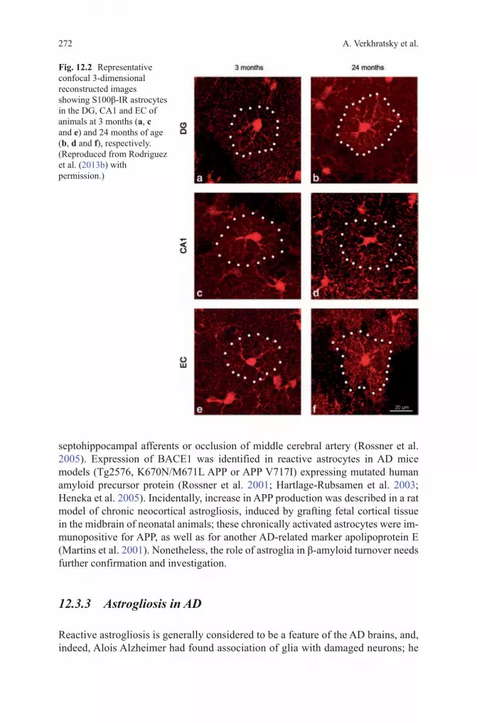
Fig. 12.2 Representative confocal 3-dimensional reconstructed images showing S100β-IR astrocytes in the DG, CA1 and EC of animals at 3 months (a, c and e) and 24 months of age (b, d and f), respectively. ( Reproduced from Rodriguez et al. (2013b) with permission.)

27312 Neurodegeneration and Neuroglia
also observed glial cells abundantly populating senile plaques (Alzheimer 1910). Astrogliotic changes, mainly documented by an increase in the expression of GFAP and astroglial S100 β protein, have been observed in post-mortem tissues from AD patients (Beach and McGeer 1988; Griffin et al. 1989; Meda et al. 2001; Mrak and Griffin 2005; Rodriguez et al. 2009). Some reports claimed a degree of correlation between the astrogliosis (defined as increase in GFAP expression) and the Braak stage of AD, although there was no correlation between astrogliotic changes and β-amyloid load (Simpson et al. 2010). Reactive astrocytes were found to be associated with some senile plaques, but they were also identified in plaque free regions of the grey matter (Simpson et al. 2010). At the same time, no differ-ences in GFAP expression was found between demented and non-demented brains (Wharton et al. 2009). Reactive, hypertrophic astrocytes, associated with senile plaques and perivascular β-amyloid deposits, were also observed in the brains of AD-models mice (Rodriguez et al. 2009; Olabarria et al. 2010; Verkhratsky et al. 2010) (Figs. 12.7 and 12.8).
Fig. 12.3 Light micrographs showing the morphology and cell area of GS positive astrocytes in the DG, CA1 and EC of 3-month-old mice (a, c and e, respectively) and 24-month-old (b, d and e, respectively). (Reproduced from Rodriguez et al. (2013b) with permission.)

A. Verkhratsky et al.274
Changes in GFAP expression reported in the AD tissue may, however, not only reflect the disease-specific changes but also the age-dependent remodeling of astro-cytes, which, as narrated above, remains incompletely characterized. Furthermore, it has to be emphasized that reactive astrogliosis in AD is of a rather mild variety; astrocytes in the grey matter preserve their domain organization and there is no indi-cation of anisomorphic gliosis and the formation of glial scars. Reactive astrocytes in AD animal models show aberrant physiology. These astrocytes, associated with senile plaques, were reported to generate spontaneous Ca2+ oscillations and abnor-mal Ca2+ waves (Kuchibhotla et al. 2009).
Molecular cues initiating astroglial reactivity in AD are multiple and may in-clude extracellular β-amyloid as well as factors released by damaged cells. Soluble
Fig. 12.4 Bar graphs showing the regional comparisons of GFAP surface, volume, and soma vol-ume in the DG (a–c), CA1 (d–f) and EC (g–i) across ages. Bars represent mean± SEM (*p ≤ 0.05; **p ≤ 0.01; ***p ≤ 0.0001 compared with 3 months of age; ◊ p ≤ 0.05; ◊◊ p ≤ 0.01 compared with 9 months of age; in DG, n = 6, 7, 3 and 4 for 3, 9, 12 and 18 months, respectively; in CA1, n = 4, 3, 3, 4 for 3, 9, 12 and 18 months, respectively; in EC, n = 4,5, 3, 3 for 3, 9, 12 and 18 months, respectively). (Reproduced from Rodriguez et al. (2013b) with permission.)

27512 Neurodegeneration and Neuroglia
β-amyloid is reported to trigger reactive changes in astrocytes in vitro (DeWitt et al. 1998). Exposure of cultured astrocytes to β-amyloid also modifies signaling cascades. For example, extracellular β-amyloid triggers abnormal oscillatory Ca2+ fluctuations in cultured primary astrocytes (Abramov et al. 2003, 2004). Incubating primary astrocytes with pathologically relevant concentrations of soluble β-amyloid affects the expression of Ca2+ toolkit components; importantly, this remodeling dif-fers for astrocytes derived from different brain regions (Grolla et al. 2013). Simi-larly, exposure to β-amyloid was claimed to down-regulate glutamate uptake in astroglial cells in vitro (Matos et al. 2008). All in all, however, the precise mecha-nisms of astroglial activation and remodeling of astroglial physiological signaling cascades in AD remains virtually unknown.
12.3.4 Astrodegeneration in AD: Reduction in Astroglial Profiles
Effects of AD pathology on astroglia, however, are not limited with astrogliotic response, to the contrary, astrogliosis most likely occurs in later stages of the dis-ease, and reactive astrocytes are mainly associated with senile plaques (Olabarria et al. 2010). Recent studies of transgenic AD mice models revealed a profound astrodegeneration that occurs at the early stages of AD progression (Olabarria et al. 2010; Yeh et al. 2011; Kulijewicz-Nawrot et al. 2012; Beauquis et al. 2013).
Fig. 12.5 Bar graphs showing the regional comparisons of S100β-IR surface area and volume in the DG (a and b), CA1 (c and d) and EC (e and f) at 2 and 24 months of age. Bars represent mean ± SEM (*p ≤ 0.05 compared with 3 months of age; in DG, n = 3 and 4 for 3 and 24 months, respectively; in CA1, n = 3 for both 3 and 12 months; in EC, n = 4 and 3 for 3 and 12 months, respectively). (Reproduced from Rodriguez et al. (2013b) with permission.)
A. Verkhratsky et al.276
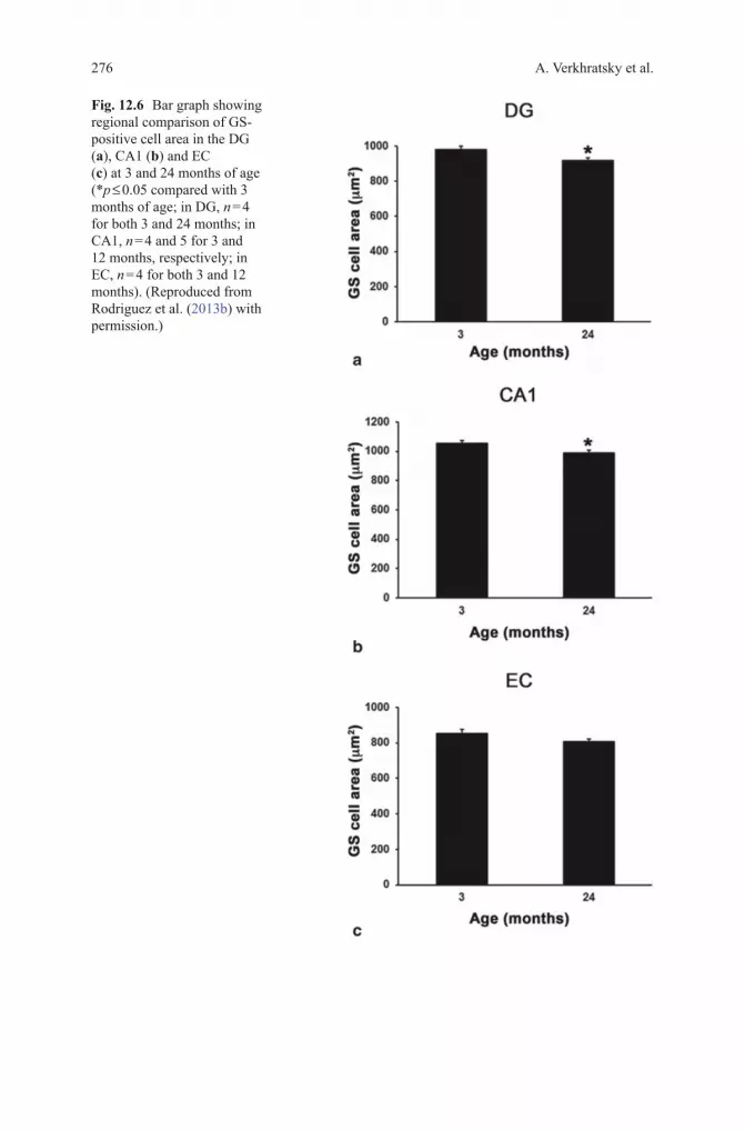
Fig. 12.6 Bar graph showing regional comparison of GS-positive cell area in the DG (a), CA1 (b) and EC (c) at 3 and 24 months of age (*p ≤ 0.05 compared with 3 months of age; in DG, n = 4 for both 3 and 24 months; in CA1, n = 4 and 5 for 3 and 12 months, respectively; in EC, n = 4 for both 3 and 12 months). (Reproduced from Rodriguez et al. (2013b) with permission.)

27712 Neurodegeneration and Neuroglia
Fig. 12.7 Confocal single labeling micrographs of dual labeled immunohistochemistry illustrat-ing the cytoskeleton alterations between astrocytes away (a) and around (b) plaques. Bar graphs showing GFAP positive astrocytic surface (c), volume (d), 2√S/3√V ratio (e) and body volume (f) differences between those astrocytes located around the amyloid plaques (Aβ) and those dis-tant to the plaques in the CA1 of 3xTg-AD animals. 2√S/3√V ratio representation of astrocytic located around the amyloid plaques when compared to non-Tg control mice astrocytes at 12 and 18 months of age (k). Similar astrocytic surface (g), volume (h), 2√S/3√V ratio (i) and body volume (j) differences are observed in the DG at 18 months of age. Bars represent mean ± SEM.* = p < 0.05. (Modified from Olabarria et al. (2010) with permission.)

A. Verkhratsky et al.278
Fig. 12.8 Confocal dual labeling images (GFAP in green and Aβ in red) in 3xTg-AD mice showing the concentration of astrocytes around the Aβ accumula-tions (a–d). Astrocytes surround Aβ plaques (a, b) and Aβ loaded blood vessel (c), undergo astrogliosis including in some cases Aβ intracellular accumula-tion (a, b). Occasionally, some distant astrocytes send reactive processes towards a plaque (d). (Reproduced from Olabarria et al. (2010) with permission.)

27912 Neurodegeneration and Neuroglia
In the above mentioned 3xTg-AD (Oddo et al. 2003), reduction in GFAP-pos-itive profiles have been found in several brain regions (Olabarria et al. 2010; Yeh et al. 2011; Kulijewicz-Nawrot et al. 2012). These atrophic changes were quantified by decreased surface area and volume of GFAP-positive profiles, decreased volume of cell somata, decreased number of primary processes and reduction in the number of primary processes (Fig. 12.9). The total number of GFAP-positive astrocytes, however, remained stable in the hippocampus, entorhinal and prefrontal cortices of AD mice at all ages from birth to senescence (1–24 month of age) (Olabarria et al. 2010; Yeh et al. 2011; Kulijewicz-Nawrot et al. 2012). Similar atrophic changes were observed in hippocampal astrocytes from another AD animal model, the mu-tant APP (PDAPP-J20) mice carrying the Swedish and Indiana APP human muta-tions (Beauquis et al. 2013).
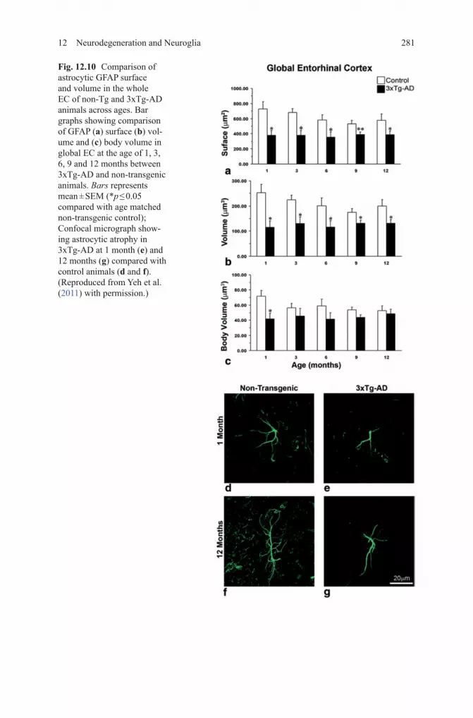
In the 3xTg-AD animals, reduced astroglial profiles appeared very early (at 1 months of age) in the entorhinal cortex, somewhat later in the prefronatal cor-tex (~ 6 months) and substantially later in the hippocampus (~ 9–12 months) (Figs. 12.10, 12.11) (Olabarria et al. 2010; Yeh et al. 2011; Kulijewicz-Nawrot et al. 2012). This atrophy of GFAP-positive profiles preceded β-amyloid deposition and formation of senile plaques. The reduction in GFAP profiles coincided with the re-duced morphological presence of astroglial cells labeled with GS antibodies in the hippocampus and in the prefrontal cortex, but not in the entorhinal cortex (Olabarria et al. 2011; Yeh et al. 2013).
12.3.5 Astrodegeneration in AD: Loss of Homeostatic Support Defines Early Cognitive Impairments
Reduction in astroglial profiles, as evidenced by the morphometry of GFAP- and GS-positive cells, is indicative of a decrease in astroglial territories and hence in reduced astroglial coverage of the grey matter. This atrophy of astrocytes, which occurs early in the disease progression, may represent an important pathological stage in the disease progression. Atrophic astrocytes provide less synaptic coverage with deleterious consequences for synaptic transmission associated with compro-mised ion and neurotransmitter homeostasis and/or reduced local metabolic support (Verkhratsky et al. 2010; Rodriguez and Verkhratsky 2011). Astroglial degenera-tion, furthermore, affects the neuro-vascular unit and lessens neuroprotection. All these changes are likely to weaken synaptic transmission and affect synaptic plas-ticity, and thereby being responsible for initial cognitive deficiency observed during the early stages of AD.
These early cognitive deficits are the very first symptoms of AD, which start to develop years before the occurrence of specific histopathology (Terry 2000; Coleman et al. 2004). Weakening of cognitive abilities reflects in reduced synaptic connectivity due to decreased synaptic function and synaptic loss. Decrease in the number of synapses represents the earliest morphological changes in AD (Terry 2000), while the degree of synaptic loss correlates with the severity of dementia

A. Verkhratsky et al.280
Fig. 12.9 Bar graphs showing a decreased GFAP surface, volume, 2√S/3√V ratio and body volume in both the DG (a, c, i) and the CA1 (b, d, j) of the hippocampus of the 3xTg-AD mice when compared to control animals. Bars represent mean ± SEM.* =p < 0.05. Confocal micrographs illus-trating the astrocytic atrophy in 3xTg-AD mice in the DG (f) and CA1 (h) compared to control animals (e and g, respectively). (Modified from Olabarria et al. (2010) with permission.)
(DeKosky and Scheff 1990; Samuel et al. 1994). Early demise of synapses could be directly related to astrodegeneration and the resulting homeostatic failure. Astro-cytes are critical for synaptogenesis and synaptic maintenance (Ullian et al. 2004; Eroglu and Barres 2010), whereas astroglial plasmalemmal transporters control local concentrations of ions and neurotransmitters, most notably glutamate, which, when not contained, causes local excitotoxicity. Astroglia also supports normal neu-ronal excitability and synaptic function through metabolic support accomplished by lactate shuttle (Magistretti 2006). Astrocytes are also critical for sustaining normal neurotransmission by supplying neurons with glutamine that is indispensable for
28112 Neurodegeneration and Neuroglia
Fig. 12.10 Comparison of astrocytic GFAP surface and volume in the whole EC of non-Tg and 3xTg-AD animals across ages. Bar graphs showing comparison of GFAP (a) surface (b) vol-ume and (c) body volume in global EC at the age of 1, 3, 6, 9 and 12 months between 3xTg-AD and non-transgenic animals. Bars represents mean ± SEM (*p ≤ 0.05 compared with age matched non-transgenic control); Confocal micrograph show-ing astrocytic atrophy in 3xTg-AD at 1 month (e) and 12 months (g) compared with control animals (d and f). (Reproduced from Yeh et al. (2011) with permission.)

A. Verkhratsky et al.282
Fig. 12.11 Confocal images showing the classical morphology of GFAP-positive astrocytes in control non-Tg animals and astrocytic atrophy in the 3xTg-AD animals at 3 months (a and b, respectively) and 18 months (c and d, respectively) in the medial prefrontal cortex (mPFC). Bar graphs showing the decreases in the GFAP-positive surface area and volume throughout the whole extent of the mPFC (e–f) in 3xTg-AD mice when compared with control animals. Bars represent mean ± SEM. (Reproduced from Kulijewicz-Nawrot et al. (2012) with permission.)

28312 Neurodegeneration and Neuroglia
glutamatergic and GABA-ergic pathways. Impairment of these critical functions associated with astrodegeneration can be a primary cause for distorted synaptic con-nectivity and early cognitive deficits in AD (Verkhratsky et al. 2010; Rodriguez and Verkhratsky 2011).
12.3.6 Astrodegeneration in AD: Dysfunctional Neuro-vascular Unit
AD pathology is often (if not always) associated with vascular deficiency. Blood flow is significantly reduced in the brains of patients with AD and especially in the early stages of the disease (see (Zlokovic 2008; Bell and Zlokovic 2009) for comprehensive review). These functional deficits reflect profound remodeling of vascularization in the diseased brains (Farkas and Luiten 2001). Brain microcircula-tion is controlled by both neuronal and astroglial inputs (Zonta et al. 2003; Iadecola and Nedergaard 2007; Attwell et al. 2010). Astrocytes are central elements of the neurovascular units that bridge neurons with local circulation. By releasing vari-ous factors, astrocytes target pericytes, vascular smooth muscle cells and endothelial cells, thus contributing to functional hyperemia and regulating blood-brain barrier (Zonta et al. 2003; Mulligan and MacVicar 2004; Takano et al. 2006). Astroglial atrophy, together with reactive rearrangement of neuro-vascular unit, may occur at both early and late stages of the disease and contribute to cognitive abnormalities and neuronal damage.
12.3.7 Astrodegeneration in AD: Deficits in Metabolic Support
Metabolic failure represents another common feature of AD. Progressive loss of glucose utilization has been observed in functional brain imaging in patients with different stages of AD; importantly, this metabolic stress is present in the early stag-es of the disease thus bearing a diagnostic significance (Mosconi et al. 2008). Ex-periments in vitro, in primary cultured astrocytes, demonstrated that treatment with β-amyloid affects cellular metabolism, although both decrease (Parpura-Gill et al. 1997; Soucek et al. 2003) and increase (Allaman et al. 2010) in glucose utilization were described. Similarly, both decrease (Blass et al. 2000; Liang et al. 2008) and increase (Bigl et al. 1999; Soucek et al. 2003) in the activity of glucose metabolism enzymes were reported in post-mortem AD brains.

A. Verkhratsky et al.284
12.3.8 Astrodegeneration in AD: Paralysis of Astrogliotic Response Defines Susceptibility of Brain Tissue to AD Pathology
Another important consequence of astroglial degeneration in AD is associated with the failure of their defensive function. In the 3xTg mice model, appearance of senile plaques as well as perivascular β-amyloid accumulation triggers astrogliotic response in hippocampal astrocytes, which become hypertrophic and upregulate GFAP expression (Olabarria et al. 2010, 2011). These hypertrophic astrocytes are specifically associated with β-amyloid deposits, whereas astrocytes distant to the plaques are generally atrophic (so in this sense, astroglial atrophy represents the early stage of AD progression and is complimented by astrogliosis at later stages, when specific lesions develop). In entorhinal and prefrontal cortices, however, astrocytic defense response appeared to be compromised because extracellular β-amyloid accumulation does not trigger astrogliotic response (Yeh et al. 2011; Kulijewicz-Nawrot et al. 2012). This protective failure may explain high vulner-ability of entorhinal and prefrontal cortices to AD pathology.
12.4 Astrocytes as Therapeutic Targets in AD
Can astrocytes represent a new and potentially fundamentally important target for therapy in neurodegenerative disorders? Can astroglial atrophy and dysfunction be reversed or delayed, and can this affect the progression of AD or severity of cogni-tive deficits? These questions are of paramount importance for neurogliopathology. Only very recently astroglial cells begin to be considered as objects of treatment. Experiments on transgenic APP and 3xTg-AD mice have shown that chronic expo-sure of these animals to environmental stimulation (physical activity and/or enriched environment) reversed morphological atrophy of astrocytes, increased GFAP expres-sion and normalized the appearance of GFAP-positive profiles (Fig. 12.12); these astroglia-specific changes were paralleled with a decrease in β-amyloid load (Beau-quis et al. 2013; Rodriguez et al. 2013a). Chronic treatment of another AD model, the 5xFAD mice, which co-expresses the mutant forms of human APP (the Swedish mutation: K670N/M671L the Florida mutation: I716V; the London mutation: V717I) and presenilin-1 (M146L/L286V) transgenes (Oakley et al. 2006), with polyunsatu-rated fatty acid 2-hydroxy-docosahexaenoic acid, similarly reverted astroglial atro-phy, restored adult neurogenesis and improved cognitive performance (Fiol-deRoque et al. 2013). Finally, genetic modification of astrocytes in APP/PS1 model of AD, in which astrocytes were virally transfected with a peptide that interferes with the immune/inflammatory calcineurin/nuclear factor of activated T-cells (NFAT) signal-ing cascades, ameliorated cognitive deficits and lowered β-amyloid burden (Furman et al. 2012). These all are of course very preliminary findings and yet they could signal new developments in astroglia-specific therapy of neurodegenerative diseases.

28512 Neurodegeneration and Neuroglia
Fig. 12.12 GFAP-IR of astrocytes in the DG of non-Tg and 3xTg-AD animals housed in different conditions. a High magnification of representative confocal micrographs showing the astrocytic morphology in mice housed in standard conditions (STD;), RUN, and ENR. Scale bars, 10 μm. Note the morphological changes of the astrocytes from both genotypes induced by the different living conditions. b Histograms showing difference of surface area and volume of GFAP-positive astrocytes in the DG of non-Tg and 3xTg-AD mice housed under different housing conditions. c Histograms showing differences in surface area and volume of GFAP-IR astrocytic cell bod-ies and processes detected between non-Tg and 3xTg-AD mice housed under different housing conditions. Bars represent means ± S.E.M., #p < 0.05, ##p < 0.01 compared with non-Tg animals in same housing environment; *p < 0.05, **p < 0.01 compared with non-Tg mice housed under STD; p < 0.01 and p < 0.001 compared with 3xTg-AD mice housed under STD. (Reproduced from Rodriguez et al. (2013a) with permission.)
A. Verkhratsky et al.286
Conclusions
Astrocytes undergo complex alterations in AD, which are represented by atrophy and asthenia at the early stages and reactivity at the late stages of the disease, all these changes being region specific. These complex changes can be considered as pathologically relevant because they may define early cognitive deficits and later neurotoxicity. Targeting astroglia in neurodegeneration may result in new therapeu-tic strategies aimed at preventing and delaying the disease progression.
Acknowledgements We would like to thank Manoj K. Gottipati for comments on a previous ver-sion of the manuscript. Authors research was supported byAlzheimer’s Research Trust (UK) Pro-gramme Grant (ART/PG2004A/1) to AV and JJR; by the National Institutes of Health (The Eunice Kennedy Shriver National Institute of Child Health and Human Development award HD078678) to V.P., by the Grant Agency of the Czech Republic (GACR 309/09/1696) to JJR and (GACR 305/08/1384) to AV.
References
Abramov AY, Canevari L, Duchen MR (2003) Changes in intracellular calcium and glutathione in astrocytes as the primary mechanism of amyloid neurotoxicity. J Neurosci 23:5088–5095
Abramov AY, Canevari L, Duchen MR (2004) β-Amyloid peptides induce mitochondrial dysfunc-tion and oxidative stress in astrocytes and death of neurons through activation of NADPH oxidase. J Neurosci 24:565–575
Allaman I, Gavillet M, Belanger M, Laroche T, Viertl D, Lashuel HA, Magistretti PJ (2010) Amyloid-b aggregates cause alterations of astrocytic metabolic phenotype: impact on neuronal viability. J Neurosci 30:3326–3338
Alzheimer A (1907) Über eine eigenartige Erkrankung der Hirnrinde. Allg Z Psychiatr Psych-Gericht Med 64:146–148
Alzheimer A (1910) Beiträge zur Kenntnis der pathologischen Neuroglia und ihrer Beziehungen zu den Abbauvorgängen im Nervengewebe. In: Nissl F, Alzheimer A (eds) Histologische und histopathologische Arbeiten über die Grosshirnrinde mit besonderer Berücksichtigung der pa-thologischen Anatomie der Geisteskrankheiten. Gustav Fischer, Jena, pp 401–562
Amenta F, Bronzetti E, Sabbatini M, Vega JA (1998) Astrocyte changes in aging cerebral cortex and hippocampus: a quantitative immunohistochemical study. Microsc Res Tech 43:29–33
Apelt J, Ach K, Schliebs R (2003) Aging-related down-regulation of neprilysin, a putative β-amyloid-degrading enzyme, in transgenic Tg2576 Alzheimer-like mouse brain is accom-panied by an astroglial upregulation in the vicinity of β-amyloid plaques. Neurosci Lett 339:183–186
Armstrong RA (2009) The molecular biology of senile plaques and neurofibrillary tangles in Alzheimer’s disease. Folia Neuropathol 47:289–299
Attwell D, Buchan AM, Charpak S, Lauritzen M, Macvicar BA, Newman EA (2010) Glial and neuronal control of brain blood flow. Nature 468:232–243
Beach TG, McGeer EG (1988) Lamina-specific arrangement of astrocytic gliosis and senile plaques in Alzheimer’s disease visual cortex. Brain Res 463:357–361
Beauquis J, Pavia P, Pomilio C, Vinuesa A, Podlutskaya N, Galvan V, Saravia F (2013) Environ-mental enrichment prevents astroglial pathological changes in the hippocampus of APP trans-genic mice, model of Alzheimer’s disease. Exp Neurol 239:28–37
Bell RD, Zlokovic BV (2009) Neurovascular mechanisms and blood-brain barrier disorder in Alzheimer’s disease. Acta Neuropathol 118:103–113

28712 Neurodegeneration and Neuroglia
Bigl M, Bruckner MK, Arendt T, Bigl V, Eschrich K (1999) Activities of key glycolytic enzymes in the brains of patients with Alzheimer’s disease. J Neural Transm 106:499–511
Biochemical Society (2011) The amyloid cascade hypothesis has misled the pharmaceutical industry. Biochem Soc Trans 39:920–923
Blass JP, Sheu RK, Gibson GE (2000) Inherent abnormalities in energy metabolism in Alzheimer disease. Interaction with cerebrovascular compromise. Ann N Y Acad Sci 903:204–221
Braak H, de Vos RA, Jansen EN, Bratzke H, Braak E (1998) Neuropathological hallmarks of Alzheimer’s and Parkinson’s diseases. Prog Brain Res 117:267–285
Brambilla L, Martorana F, Rossi D (2013) Astrocyte signaling and neurodegeneration: new insights into CNS disorders. Prion 7:28–36
Broe M, Kril J, Halliday GM (2004) Astrocytic degeneration relates to the severity of disease in frontotemporal dementia. Brain 127:2214–2220
Coleman P, Federoff H, Kurlan R (2004) A focus on the synapse for neuroprotection in Alzheimer disease and other dementias. Neurology 63:1155–1162
Danbolt NC (2001) Glutamate uptake. Prog Neurobiol 65:1–105DeKosky ST, Scheff SW (1990) Synapse loss in frontal cortex biopsies in Alzheimer’s disease:
correlation with cognitive severity. Ann Neurol 27:457–464Depboylu C, Stricker S, Ghobril JP, Oertel WH, Priller J, Hoglinger GU (2012) Brain-resident
microglia predominate over infiltrating myeloid cells in activation, phagocytosis and in-teraction with T-lymphocytes in the MPTP mouse model of Parkinson disease. Exp Neurol 238:183–191
DeWitt DA, Perry G, Cohen M, Doller C, Silver J (1998) Astrocytes regulate microglial phagocy-tosis of senile plaque cores of Alzheimer’s disease. Exp Neurol 149:329–340
Eroglu C, Barres BA (2010) Regulation of synaptic connectivity by glia. Nature 468:223–231Farkas E, Luiten PG (2001) Cerebral microvascular pathology in aging and Alzheimer’s disease.
Prog Neurobiol 64:575–611Fiol-deRoque MA, Gutierrez-Lanza R, Torres M, Terés S, Barceló P, Rial RV, Verkhratsky A,
Escribá PV, Busquets X, Rodríguez JJ (2013) Cognitive recovery and restoration of cell proliferation in the dentate gyrus in the 5XFAD transgenic mice model of Alzheimer’s disease following 2-hydroxy-DHA treatment. Biogerontology 14:763–765
Furman JL, Sama DM, Gant JC, Beckett TL, Murphy MP, Bachstetter AD, Van Eldik LJ, Norris CM (2012) Targeting astrocytes ameliorates neurologic changes in a mouse model of Alzheim-er’s disease. J Neurosci 32:16129–16140
Gerlai R (2001) Alzheimer’s disease: beta-amyloid hypothesis strengthened! Trends Neurosci 24:199
Griffin WS, Stanley LC, Ling C, White L, MacLeod V, Perrot LJ, White CL, 3rd, Araoz C (1989) Brain interleukin 1 and S-100 immunoreactivity are elevated in Down syndrome and Alzheimer disease. Proc Natl Acad Sci U S A 86:7611–7615
Grolla AA, Sim JA, Lim D, Rodriguez JJ, Genazzani AA, Verkhratsky A (2013) Amyloid-β and Alzheimer’s disease type pathology differentially affects the calcium signaling toolkit in astrocytes from different brain regions. Cell Death Dis 4:e623
Guenette SY (2003) Astrocytes: a cellular player in Ab clearance and degradation. Trends Mol Med 9:279–280
Hardy J (2006) Has the amyloid cascade hypothesis for Alzheimer’s disease been proved? Curr Alzheimer Res 3:71–73
Hardy J (2009) The amyloid hypothesis for Alzheimer’s disease: a critical reappraisal. J Neurochem 110:1129–1134
Hardy J, Selkoe DJ (2002) The amyloid hypothesis of Alzheimer’s disease: progress and problems on the road to therapeutics. Science 297:353–356
Hartlage-Rubsamen M, Zeitschel U, Apelt J, Gartner U, Franke H, Stahl T, Gunther A, Schliebs R, Penkowa M, Bigl V, Rossner S (2003) Astrocytic expression of the Alzheimer’s disease b-secretase (BACE1) is stimulus-dependent. Glia 41:169–179
Hazell AS (2009) Astrocytes are a major target in thiamine deficiency and Wernicke’s encepha-lopathy. Neurochem Int 55:129–135

A. Verkhratsky et al.288
Hazell AS, Sheedy D, Oanea R, Aghourian M, Sun S, Jung JY, Wang D, Wang C (2009) Loss of astrocytic glutamate transporters in Wernicke encephalopathy. Glia 58:148–156
Heneka MT, Sastre M, Dumitrescu-Ozimek L, Dewachter I, Walter J, Klockgether T, Van Leuven F (2005) Focal glial activation coincides with increased BACE1 activation and precedes amy-loid plaque deposition in APPV717I transgenic mice. J Neuroinflam 2:22
Heneka MT, Rodriguez JJ, Verkhratsky A (2010) Neuroglia in neurodegeneration. Brain Res Rev (in press)
Henry V, Paille V, Lelan F, Brachet P, Damier P (2009) Kinetics of microglial activation and degeneration of dopamine-containing neurons in a rat model of Parkinson disease induced by 6-hydroxydopamine. J Neuropathol Exp Neurol 68:1092–1102
Hinman JD, Abraham CR (2007) What’s behind the decline? The role of white matter in brain aging. Neurochem Res 32:2023–2031
Iadecola C, Nedergaard M (2007) Glial regulation of the cerebral microvasculature. Nat Neurosci 10:1369–1376
Jellinger KA (2008) Neuropathological aspects of Alzheimer disease, Parkinson disease and fron-totemporal dementia. Neurodegener Dis 5:118–121
Karran E, Mercken M, De Strooper B (2011) The amyloid cascade hypothesis for Alzheimer’s disease: an appraisal for the development of therapeutics. Nat Rev Drug Discov 10:698–712
Kaul M (2009) HIV-1 associated dementia: update on pathological mechanisms and therapeutic approaches. Curr Opin Neurol 22:315–320
Kaul M, Lipton SA (2006) Mechanisms of neuronal injury and death in HIV-1 associated demen-tia. Curr HIV Res 4:307–318
Kersaitis C, Halliday GM, Kril JJ (2004) Regional and cellular pathology in frontotempo-ral dementia: relationship to stage of disease in cases with and without Pick bodies. Acta Neuropathol 108:515–523
Kim K, Lee SG, Kegelman TP, Su ZZ, Das SK, Dash R, Dasgupta S, Barral PM, Hedvat M, Diaz P, Reed JC, Stebbins JL, Pellecchia M, Sarkar D, Fisher PB (2011) Role of excitatory amino acid transporter-2 (EAAT2) and glutamate in neurodegeneration: opportunities for developing novel therapeutics. J Cell Physiol 226:2484–2493
Knight RA, Verkhratsky A (2010) Neurodegenerative diseases: failures in brain connectivity? Cell Death Differ 17:1069–1070
Korczyn AD (2008) The amyloid cascade hypothesis. Alzheimer’s Demen J Alzheimer’s Assoc 4:176–178
Korsakoff SS (1889) Psychosis polineuritica, s. cerebropathia psychica toxaemica. Med Obozr 32:3–18 [Корсаков СС (1889) Психическое расстройство в сочетании с множественным невритом. Мед обозр 32:3–18]. English translation: Korsakoff SS (1955) Psychic disorder in conjunction with multiple neuritis (trans: Victor M, Yakovlev P). Neurology 5:394–406
Kuchibhotla KV, Lattarulo CR, Hyman BT, Bacskai BJ (2009) Synchronous hyperactivity and intercellular calcium waves in astrocytes in Alzheimer mice. Science 323:1211–1215
Kulijewicz-Nawrot M, Verkhratsky A, Chvatal A, Sykova E, Rodriguez JJ (2012) Astrocytic cy-toskeletal atrophy in the medial prefrontal cortex of a triple transgenic mouse model of Al-zheimer’s disease. J Anat 221:252–262
Lazzarini M, Martin S, Mitkovski M, Vozari RR, Stuhmer W, Bel ED (2013) Doxycycline restrains glia and confers neuroprotection in a 6-OHDA Parkinson model. Glia 61:1084–1100
Liang WS, Reiman EM, Valla J, Dunckley T, Beach TG, Grover A, Niedzielko TL, Schneider LE, Mastroeni D, Caselli R, Kukull W, Morris JC, Hulette CM, Schmechel D, Rogers J, Stephan DA (2008) Alzheimer’s disease is associated with reduced expression of energy metabolism genes in posterior cingulate neurons. Proc Natl Acad Sci U S A 105:4441–4446
Lynch AM, Murphy KJ, Deighan BF, O’Reilly JA, Gun’ko YK, Cowley TR, Gonzalez-Reyes RE, Lynch MA (2010) The impact of glial activation in the aging brain. Aging Dis 1:262–278
Magistretti PJ (2006) Neuron-glia metabolic coupling and plasticity. J Exp Biol 209:2304–2311Martins RN, Taddei K, Kendall C, Evin G, Bates KA, Harvey AR (2001) Altered expression of
apolipoprotein E, amyloid precursor protein and presenilin-1 is associated with chronic reac-tive gliosis in rat cortical tissue. Neuroscience 106:557–569

28912 Neurodegeneration and Neuroglia
Matos M, Augusto E, Oliveira CR, Agostinho P (2008) Amyloid-b peptide decreases glutamate uptake in cultured astrocytes: involvement of oxidative stress and mitogen-activated protein kinase cascades. Neuroscience 156:898–910
Mattson MP, Haughey NJ, Nath A (2005) Cell death in HIV dementia. Cell Death Differ 12(Suppl 1):893–904
Maurya SK, Rai A, Rai NK, Deshpande S, Jain R, Mudiam MK, Prabhakar YS, Bandyopadhyay S (2012) Cypermethrin induces astrocyte apoptosis by the disruption of the autocrine/paracrine mode of epidermal growth factor receptor signaling. Toxicol Sci 125:473–487
McAlpine D, Araki S (1958) Minamata disease: an unusual neurological disorder caused by con-taminated fish. Lancet 2:629–631
Meda L, Baron P, Scarlato G (2001) Glial activation in Alzheimer’s disease: the role of Abeta and its associated proteins. Neurobiol Aging 22:885–893
Mena MA, Garcia de Yebenes J (2008) Glial cells as players in parkinsonism: the “good,” the “bad,” and the “mysterious” glia. Neuroscientist 14:544–560
Mena MA, Casarejos MJ, Carazo A, Paino CL, Garcia de Yebenes J (1996) Glia conditioned medium protects fetal rat midbrain neurons in culture from L-DOPA toxicity. Neuroreport 7:441–445
Mena MA, de Bernardo S, Casarejos MJ, Canals S, Rodriguez-Martin E (2002) The role of astro-glia on the survival of dopamine neurons. Mol Neurobiol 25:245–263
Mosconi L, Pupi A, De Leon MJ (2008) Brain glucose hypometabolism and oxidative stress in preclinical Alzheimer’s disease. Ann N Y Acad Sci 1147:180–195
Mouton PR, Long JM, Lei DL, Howard V, Jucker M, Calhoun ME, Ingram DK (2002) Age and gender effects on microglia and astrocyte numbers in brains of mice. Brain Res 956:30–35
Mrak RE, Griffin WS (2005) Glia and their cytokines in progression of neurodegeneration. Neu-robiol Aging 26:349–354
Mulligan SJ, MacVicar BA (2004) Calcium transients in astrocyte endfeet cause cerebrovascular constrictions. Nature 431:195–199
Nagele RG, D’Andrea MR, Lee H, Venkataraman V, Wang HY (2003) Astrocytes accumulate A b 42 and give rise to astrocytic amyloid plaques in Alzheimer disease brains. Brain Res 971:197–209
Ni M, Li X, Rocha JB, Farina M, Aschner M (2012) Glia and methylmercury neurotoxicity. J Toxicol Environ Health A 75:1091–1101
Nicoll JA, Weller RO (2003) A new role for astrocytes: β-amyloid homeostasis and degradation. Trends Mol Med 9:281–282
Oakley H, Cole SL, Logan S, Maus E, Shao P, Craft J, Guillozet-Bongaarts A, Ohno M, Disterhoft J, Van Eldik L, Berry R, Vassar R (2006) Intraneuronal beta-amyloid aggregates, neurodegen-eration, and neuron loss in transgenic mice with five familial Alzheimer’s disease mutations: potential factors in amyloid plaque formation. J Neurosci 26:10129–10140
Oddo S, Caccamo A, S;hepherd JD, Murphy MP, Golde TE, Kayed R, Metherate R, Mattson MP, Akbari Y, LaFerla FM (2003) Triple-transgenic model of Alzheimer’s disease with plaques and tangles: intracellular Ab and synaptic dysfunction. Neuron 39:409–421
Olabarria M, Noristani HN, Verkhratsky A, Rodriguez JJ (2010) Concomitant astroglial atrophy and astrogliosis in a triple transgenic animal model of Alzheimer’s disease. Glia 58:831–838
Olabarria M, Noristani HN, Verkhratsky A, Rodriguez JJ (2011) Age-dependent decrease in glu-tamine synthetase expression in the hippocampal astroglia of the triple transgenic Alzheimer’s disease mouse model: mechanism for deficient glutamatergic transmission? Mol Neurodegener 6:55
Pakkenberg B, Pelvig D, Marner L, Bundgaard MJ, Gundersen HJ, Nyengaard JR, Regeur L (2003) Aging and the human neocortex. Exp Gerontol 38:95–99
Palop JJ, Mucke L (2010) Amyloid-β-induced neuronal dysfunction in Alzheimer’s disease: from synapses toward neural networks. Nat Neurosci 13:812–818
Parpura-Gill A, Beitz D, Uemura E (1997) The inhibitory effects of beta-amyloid on glutamate and glucose uptakes by cultured astrocytes. Brain Res 754:65–71

A. Verkhratsky et al.290
Peinado MA, Quesada A, Pedrosa JA, Torres MI, Martinez M, Esteban FJ, Del Moral ML, Hernandez R, Rodrigo J, Peinado JM (1998) Quantitative and ultrastructural changes in glia and pericytes in the parietal cortex of the aging rat. Microsc Res Tech 43:34–42
Peters A, Verderosa A, Sethares C (2008) The neuroglial population in the primary visual cortex of the aging rhesus monkey. Glia 56:1151–1161
Pilegaard K, Ladefoged O (1996) Total number of astrocytes in the molecular layer of the dentate gyrus of rats at different ages. Anal Quant Cytol Histol 18:279–285
Potts R, Leech RW (2005) Thalamic dementia: an example of primary astroglial dystrophy of Seitelberger. Clin Neuropathol 24:271–275
Rai A, Maurya SK, Sharma R, Ali S (2013) Down-regulated GFAPα: a major player in heavy metal induced astrocyte damage. Toxicol Mech Meth 23:99–107
Reitz C (2012) Alzheimer’s disease and the amyloid cascade hypothesis: a critical review. Int J Alzheimers Dis 2012:369808
Rodriguez JJ, Verkhratsky A (2011) Neuroglial roots of neurodegenerative diseases? Mol Neuro-biol 43:87–96
Rodriguez JJ, Olabarria M, Chvatal A, Verkhratsky A (2009) Astroglia in dementia and Alzheim-er’s disease. Cell Death Differ 16:378–385
Rodriguez JJ, Terzieva S, Olabarria M, Lanza RG, Verkhratsky A (2013a) Enriched environment and physical activity reverse astrogliodegeneration in the hippocampus of AD transgenic mice. Cell Death Dis 4:e678
Rodriguez JJ, Yeh CY, Terzieva S, Olabarria M, Kulijewicz-Nawrot M, Verkhratsky A (2013b) Complex and region-specific changes in astroglial markers in the aging brain. Neurobiol Ag-ing. doi:10.1016/j.neurobiolaging.2013.07.002
Rossi D, Volterra A (2009) Astrocytic dysfunction: insights on the role in neurodegeneration. Brain Res Bull 80:224–232
Rossi D, Brambilla L, Valori CF, Roncoroni C, Crugnola A, Yokota T, Bredesen DE, Volterra A (2008) Focal degeneration of astrocytes in amyotrophic lateral sclerosis. Cell Death Differ 15:1691–1700
Rossner S, Apelt J, Schliebs R, Perez-Polo JR, Bigl V (2001) Neuronal and glial β-secretase (BACE) protein expression in transgenic Tg2576 mice with amyloid plaque pathology. J Neurosci Res 64:437–446
Rossner S, Lange-Dohna C, Zeitschel U, Perez-Polo JR (2005) Alzheimer’s disease β-secretase BACE1 is not a neuron-specific enzyme. J Neurochem 92:226–234
Salmina AB (2009) Neuron-glia interactions as therapeutic targets in neurodegeneration. J Alzheim-ers Dis 16:485–502
Samuel W, Masliah E, Hill LR, Butters N, Terry R (1994) Hippocampal connectivity and Alzheimer’s dementia: effects of synapse loss and tangle frequency in a two-component model. Neurology 44:2081–2088
Schipper HM (1996) Astrocytes, brain aging, and neurodegeneration. Neurobiol Aging 17:467–480Selkoe DJ (2001) Alzheimer’s disease: genes, proteins, and therapy. Physiol Rev 81:741–766Sidoryk-Wegrzynowicz M, Aschner M (2013) Role of astrocytes in manganese mediated neuro-
toxicity. BMC Pharmacol Toxicol 14:23Simpson JE, Ince PG, Lace G, Forster G, Shaw PJ, Matthews F, Savva G, Brayne C, Wharton
SB (2010) Astrocyte phenotype in relation to Alzheimer-type pathology in the ageing brain. Neurobiol Aging 31:578–590
Soucek T, Cumming R, Dargusch R, Maher P, Schubert D (2003) The regulation of glucose metab-olism by HIF-1 mediates a neuroprotective response to amyloid beta peptide. Neuron 39:43–56
Suarez-Fernandez MB, Soldado AB, Sanz-Medel A, Vega JA, Novelli A, Fernandez-Sanchez MT (1999) Aluminum-induced degeneration of astrocytes occurs via apoptosis and results in neu-ronal death. Brain Res 835:125–136
Takano T, Tian GF, Peng W, Lou N, Libionka W, Han X, Nedergaard M (2006) Astrocyte-mediated control of cerebral blood flow. Nat Neurosci 9:260–267
Terry RD (2000) Cell death or synaptic loss in Alzheimer disease. J Neuropathol Exp Neurol 59:1118–1119

29112 Neurodegeneration and Neuroglia
Thompson KA, McArthur JC, Wesselingh SL (2001) Correlation between neurological progres-sion and astrocyte apoptosis in HIV-associated dementia. Ann Neurol 49:745–752
Thompson PM, Hayashi KM, de Zubicaray G, Janke AL, Rose SE, Semple J, Herman D, Hong MS, Dittmer SS, Doddrell DM, Toga AW (2003) Dynamics of gray matter loss in Alzheimer’s disease. J Neurosci 23:994–1005
Ullian EM, Christopherson KS, Barres BA (2004) Role for glia in synaptogenesis. Glia 47:209–216Unger JW (1998) Glial reaction in aging and Alzheimer’s disease. Microsc Res Tech 43:24–28Vanzani MC, Iacono RF, Caccuri RL, Troncoso AR, Berria MI (2006) Regional differences in
astrocyte activation in HIV-associated dementia. Medicina 66:108–112Verkhratsky A, Olabarria M, Noristani HN, Yeh CY, Rodriguez JJ (2010) Astrocytes in Alzheim-
er’s disease. Neurotherapeutics 7:399–412Verkhratsky A, Sofroniew MV, Messing A, de Lanerolle NC, Rempe D, Rodriguez JJ, Nedergaard
M (2012) Neurological diseases as primary gliopathies: a reassessment of neurocentrism. ASN Neuro 4. doi:10.1042/AN20120010
Verkhratsky A, Rodriguez JJ, Parpura V (2013) Astroglia in neurological diseases. Fut Neurol 8:149–158
Wernicke C (1881–1883) Lehrbuch der Gehirnkrankheiten für Aerzte und Studirende. Theodor Fischer, Kassel und Berlin
Wharton SB, O’Callaghan JP, Savva GM, Nicoll JA, Matthews F, Simpson JE, Forster G, Shaw PJ, Brayne C, Ince PG (2009) Population variation in glial fibrillary acidic protein levels in brain ageing: relationship to Alzheimer-type pathology and dementia. Dement Geriatr Cogn Disord 27:465–473
Wyss-Coray T, Loike JD, Brionne TC, Lu E, Anankov R, Yan F, Silverstein SC, Husemann J (2003) Adult mouse astrocytes degrade amyloid-b in vitro and in situ. Nat Med 9:453–457
Yamanaka K, Chun SJ, Boillee S, Fujimori-Tonou N, Yamashita H, Gutmann DH, Takahashi R, Misawa H, Cleveland DW (2008) Astrocytes as determinants of disease progression in inherited amyotrophic lateral sclerosis. Nat Neurosci 11:251–253
Yeh CY, Vadhwana B, Verkhratsky A, Rodriguez JJ (2011) Early astrocytic atrophy in the entorhi-nal cortex of a triple transgenic animal model of Alzheimer’s disease. ASN Neuro 3:271–279
Yeh CY, Verkhratsky A, Terzieva S, Rodriguez JJ (2013) Glutamine synthetase in astrocytes from entorhinal cortex of the triple transgenic animal model of Alzheimer’s disease is not affected by pathological progression. Biogerontology 14(6):777–787
Yin Z, Milatovic D, Aschner JL, Syversen T, Rocha JB, Souza DO, Sidoryk M, Albrecht J, Aschner M (2007) Methylmercury induces oxidative injury, alterations in permeability and glutamine transport in cultured astrocytes. Brain Res 1131:1–10
Zlokovic BV (2008) The blood-brain barrier in health and chronic neurodegenerative disorders. Neuron 57:178–201
Zonta M, Angulo MC, Gobbo S, Rosengarten B, Hossmann KA, Pozzan T, Carmignoto G (2003) Neuron-to-astrocyte signaling is central to the dynamic control of brain microcirculation. Nat Neurosci 6:43–50


