Embed Size (px)
Citation preview

Advanced Drug Delivery Reviews 63 (2011) 847–864
Contents lists available at ScienceDirect
Advanced Drug Delivery Reviews
j ourna l homepage: www.e lsev ie r.com/ locate /addr
Drug-loaded polyelectrolyte microcapsules for sustained targeting of cancer cells☆
Viviana Vergaro a, Flavia Scarlino a, Claudia Bellomo a, Rosaria Rinaldi a, Daniele Vergara b, Michele Maffia b,Francesca Baldassarre c, Gianluigi Giannelli c, Xingcai Zhangd, Yuri M. Lvovd, Stefano Leporatti a,⁎a CNR-Instituto Nanoscienze, NNL Via Arnesano 16, 73100 Lecce (Italy)b Department of Biological and Environmental Sciences and Technologies (DiSteBA), University of Salento, Ecotekne, SP Lecce - Monteroni 73100 Lecce (Italy)c Department of Internal Medicine, Immunology and Infectious Diseases, Section of Internal Medicine, University of Bari, Medical School, Bari (Italy)d Institute for Micromanufacturing of Louisiana Tech University, 911 Hergot Ave, Ruston, LA 71272, USA
☆ This review is part of the Advanced Drug Delivery Re⁎ Corresponding author. Tel.: +39 0832298241; fax:
E-mail address: [email protected] (S. Le
0169-409X/$ – see front matter © 2011 Elsevier B.V. Adoi:10.1016/j.addr.2011.05.007
a b s t r a c t
a r t i c l e i n f oArticle history:Received 1 January 2011Accepted 7 May 2011Available online 18 May 2011
Keywords:Cancer therapyNanotechnologyDrug deliveryNanocarriersMicrocapsulesLayer-by-layerPolyelectrolyte multilayers
In this review we will overview novel nanotechnological nanocarrier systems for cancer therapy focusing onrecent development in polyelectrolyte capsules for targeted delivery of antineoplastic drugs against cancercells. Biodegradable polyelectrolyte microcapsules (PMCs) are supramolecular assemblies of particularinterest for therapeutic purposes, as they can be enzymatically degraded into viable cells, under physiologicalconditions. Incorporation of small bioactive molecules into nano-to-microscale delivery systemsmay increasedrug's bioavailability and therapeutic efficacy at single cell level giving desirable targeted therapy. Layer-by-layer (LbL) self-assembled PMCs are efficient microcarriers that maximize drug's exposure enhancingantitumor activity of neoplastic drug in cancer cells. They can be envisaged as novel multifunctional carriersfor resistant or relapsed patients or for reducing dose escalation in clinical settings.
views theme issue on "Layer-by-layer Self-Assembled N+39 0832298230.poratti).
ll rights reserved.
© 2011 Elsevier B.V. All rights reserved.
Contents
1. Introduction . . . . . . . . . . . . . . . . . . . . . . . . . . . . . . . . . . . . . . . . . . . . . . . . . . . . . . . . . . . . . 8482. Nanotechnology . . . . . . . . . . . . . . . . . . . . . . . . . . . . . . . . . . . . . . . . . . . . . . . . . . . . . . . . . . . . 848
2.1. Manipulation of individual cells and biomolecules . . . . . . . . . . . . . . . . . . . . . . . . . . . . . . . . . . . . . . . . 8482.2. Protein detection . . . . . . . . . . . . . . . . . . . . . . . . . . . . . . . . . . . . . . . . . . . . . . . . . . . . . . . . 8482.3. Biomarker discovery . . . . . . . . . . . . . . . . . . . . . . . . . . . . . . . . . . . . . . . . . . . . . . . . . . . . . . 8482.4. Molecular imaging . . . . . . . . . . . . . . . . . . . . . . . . . . . . . . . . . . . . . . . . . . . . . . . . . . . . . . . 8482.5. Drug delivery . . . . . . . . . . . . . . . . . . . . . . . . . . . . . . . . . . . . . . . . . . . . . . . . . . . . . . . . . 848
3. Cancer nanotechnology: drug delivery systems . . . . . . . . . . . . . . . . . . . . . . . . . . . . . . . . . . . . . . . . . . . . . 8483.1. Limits of current chemotherapy . . . . . . . . . . . . . . . . . . . . . . . . . . . . . . . . . . . . . . . . . . . . . . . . . 8483.2. Advantages of nanotechnological drug delivery systems . . . . . . . . . . . . . . . . . . . . . . . . . . . . . . . . . . . . . . 849
4. Nanocarriers . . . . . . . . . . . . . . . . . . . . . . . . . . . . . . . . . . . . . . . . . . . . . . . . . . . . . . . . . . . . . 8494.1. Liposomes . . . . . . . . . . . . . . . . . . . . . . . . . . . . . . . . . . . . . . . . . . . . . . . . . . . . . . . . . . . 8494.2. Solid lipid nanoparticles (SLNs) and nanostructured lipid carriers (NLCs) . . . . . . . . . . . . . . . . . . . . . . . . . . . . . . 8494.3. Dendrimers . . . . . . . . . . . . . . . . . . . . . . . . . . . . . . . . . . . . . . . . . . . . . . . . . . . . . . . . . . . 8504.4. Magnetic nanoparticles . . . . . . . . . . . . . . . . . . . . . . . . . . . . . . . . . . . . . . . . . . . . . . . . . . . . . 8504.5. Fullerenes . . . . . . . . . . . . . . . . . . . . . . . . . . . . . . . . . . . . . . . . . . . . . . . . . . . . . . . . . . . 8504.6. Carbon nanotubes . . . . . . . . . . . . . . . . . . . . . . . . . . . . . . . . . . . . . . . . . . . . . . . . . . . . . . . 8504.7. Halloysite nanotubes . . . . . . . . . . . . . . . . . . . . . . . . . . . . . . . . . . . . . . . . . . . . . . . . . . . . . . 850
5. Layer-by-layer assembly of polyelectrolyte multilayers . . . . . . . . . . . . . . . . . . . . . . . . . . . . . . . . . . . . . . . . . . 8505.1. Polyelectrolyte multilayer-coated colloids . . . . . . . . . . . . . . . . . . . . . . . . . . . . . . . . . . . . . . . . . . . . 8515.2. Drug-loaded nanocolloids . . . . . . . . . . . . . . . . . . . . . . . . . . . . . . . . . . . . . . . . . . . . . . . . . . . . 8525.3. Polyelectrolyte microcapsules (PMCs) . . . . . . . . . . . . . . . . . . . . . . . . . . . . . . . . . . . . . . . . . . . . . . 852
anoshells for Drug Delivery".

848 V. Vergaro et al. / Advanced Drug Delivery Reviews 63 (2011) 847–864
5.3.1. PMC encapsulation: pre- and post-loading . . . . . . . . . . . . . . . . . . . . . . . . . . . . . . . . . . . . . . . . 8535.4. PMC interactions with living (cancer) cells . . . . . . . . . . . . . . . . . . . . . . . . . . . . . . . . . . . . . . . . . . . . 856
5.4.1. Drug delivery . . . . . . . . . . . . . . . . . . . . . . . . . . . . . . . . . . . . . . . . . . . . . . . . . . . . . . 8575.4.2. Release . . . . . . . . . . . . . . . . . . . . . . . . . . . . . . . . . . . . . . . . . . . . . . . . . . . . . . . . 8585.4.3. In vivo interactions . . . . . . . . . . . . . . . . . . . . . . . . . . . . . . . . . . . . . . . . . . . . . . . . . . . 860
6. Conclusions and outlook . . . . . . . . . . . . . . . . . . . . . . . . . . . . . . . . . . . . . . . . . . . . . . . . . . . . . . . . 860Acknowledgments . . . . . . . . . . . . . . . . . . . . . . . . . . . . . . . . . . . . . . . . . . . . . . . . . . . . . . . . . . . . . . 860References . . . . . . . . . . . . . . . . . . . . . . . . . . . . . . . . . . . . . . . . . . . . . . . . . . . . . . . . . . . . . . . . . 860
1. Introduction
Drug delivery is a sciencewhich requires the development of tailoredsystems that can deliver defined quantities of a therapeutic payload (eganantineoplastic drug) at a specific target site, at a controlled release (e.g.slow) rate, with or without a specific trigger (e.g. external or internal).However, several drug molecules cannot be formulated or administeredby standard techniques as they exhibit poor water solubility or havelimited stability in thehumanbody. Anovel class of carriers,multilayeredpolyelectrolyte microcapsules (PMCs) have been recently fabricated andengineered to encapsulate various classes of drug molecules, by usingpolymers that are biodegradable or that can respond and release theirpayload in response to well-defined stimuli. In this review we willdescribe recent developments in nanotechnology (e.g. carriers forantineoplastic drug delivery) focusing on PMCs for their multifunction-ality in templating and coating, flexibility in constituents and encapsu-lating species, and tailoring in chemical and physical motifs.
2. Nanotechnology
Nanosciences and nanotechnology have the potential to optimizecurrent medical approaches thus leading to a possible revolution inhealth care and current clinical therapeutic and diagnostic practice.Nanoscience is the rationale for nanotechnology and it describes themanipulation of matter at the atomic, molecular and supramolecularlevels, where properties differ significantly from those at a larger scale.Nanotechnology, instead, refers to the production at the nanometer scale,characterization, analysis and application of devices and bio-nanotech-nological systems [1]. Nanomedicine defines the most rising nanotech-nological application tobiology andmedicine. Present-daynanomedicineranges over nanoscale surgery, tissue engineering, and targeted drugdelivery [2]. Tissue engineering consists in creating nano-sized complexbiocompatible and biodegradable compounds, which can be used asscaffolds to let stem cells grow, thus recreating in vitro functional tissues.
2.1. Manipulation of individual cells and biomolecules
One approach consists in creating multifunctionalized magneticnanoparticles for cell sorting and probing. Functionalizing thesenanostructures with proper ligands – such as ligands for overexpressedtumoral receptors –maybe useful for ex vivo isolation and further easiercharacterization of malignant cells. Additionally, atomic force microsco-py and carbon nanotube offered great opportunities to analyze andmanipulate individual molecules and cells [3]. Recently, a carbon-nanotube-based endoscope has been described where intracellularstructures can be probed in the nm range [4].
2.2. Protein detection
Nanotechnological approaches can be useful for protein of interestisolation among a whole cell lysate using a specific antibody linked tothe nanoparticle surface or for the detection of complex protein–protein interactions. The actual aim is functionalizing nanoparticles,mostly gold nanoprobes, with multiple antibodies or their derived, inorder to improve multiplex detection analysis [5].
2.3. Biomarker discovery
Major works are focused on useful tools for the discovery of novelbiomarkers that provide sensitive and specific detection of early stagedisease. This aim has to face formidable challenges including the broadconcentration range of proteins in biological fluids, especially in plasma,the low abundance of candidate markers and their lability. In thisscenario progress is largely determined by development and implemen-tation of (i) nanotechnological physicochemical procedures for theseparation of multicomponent protein mixtures; (ii) identification andcounting of single molecules by use of molecular detectors [6]. Amagnetic nanosensor technologywith the ability to bematrix insensitiveand to detect a large number of protein species has been recentlydeveloped. This system has demonstrated attomolar sensitivity in avariety of clinically relevantmediawith a lineardynamic rangeof over sixorders of magnitude [7].
2.4. Molecular imaging
In this view nanoparticles are loaded with contrast agents currentlyused in clinical imaging techniques. The nanometer size of this structureas well as their physical/structural properties and a potential propernanoparticle functionalization may contribute to the proper contrastagent biodistribution into target tissues and organs [5].
2.5. Drug delivery
Drug delivery is the most promising nanotechnological applicationin biomedicine, which consists in drug encapsulation (mostlychemotherapeutics) into a nanometer scale complex system (10–1000 nm) in order to improve drug efficacy, drug biodistribution andbioavailability as well as minimize drug toxical effects, linked to theircurrent systemic administration [8] (Fig. 1). Primary goals fornanobiotechnological research in drug delivery include (i) morespecific drug targeting and delivery, (ii) reduction in drug andadjuvant toxicity while maintaining therapeutic effects, (iii) greatersafety and biocompatibility and (iv) faster and lasting development ofmedicines. To reach these purposes is essential and a deepinvestigation about drug incorporation and release – in order tomaximize drug loaded into nanocarriers – as well as biocompatibilityand nanocarriers biodistribution information is also needed [9].
3. Cancer nanotechnology: drug delivery systems
3.1. Limits of current chemotherapy
Chemotherapy is themajor therapeutic approach for the treatment oflocalized and metastasized cancers with the use of chemical antineo-plastic drugs, mostly administered through IV regimens. Althoughchemotherapeutics are widely used in the treatment of cancer, theyshow lots of limits due to:
• Lack of specificity toward neoplastic tissues, causing significantdamageto noncancerous tissues and serious unwanted side effects such as bonemarrow activity suppression (immuno and myelosuppression),

849V. Vergaro et al. / Advanced Drug Delivery Reviews 63 (2011) 847–864
mucositis, nausea, secondary neoplasms, and infertility. Moreover thehighdistributionvolumeof chemotherapeuticsmakes thedrugdeliverynot specific to neoplasms, thus leading to an abnormal concentration ofthe antineoplastic drug also in healthy tissues.
• Lack of selectivity in their mechanism of action, because mostchemotherapeutics don't act on intracellular mechanisms peculiar ofmalignant cells, but on common pathways shared both by neoplastic,both by healthy tissue cells. So that, cytotoxic and/or cytostaticmechanisms induced by these drugs hit also healthy, non-canceroustissues.
• Low molecular weight with high pharmacokinetic volume of distribu-tion both contribute to their cytotoxicity. Moreover, the lowmolecularweight of these chemicals makes them easily excreted, hence a higherconcentration is ultimately required, and consequently a highertoxicity is unavoidable.
• Low therapeutic index of drugs implies that the needed concentrationfor the effective treatment is often high, leading to systemic dose-dependent side effects. Furthermore the adjuvants, which are used inpharmacological practice to improve drug effect and its bioavailability,are toxic themselves, thus increasing the final toxic effect.
• Low solubility and high degradation susceptibility. The low solubilitymakes difficult drug preparation, while the high degradationsusceptibility, mostly at the reticuloendothelial system, avoids theoral drug administration and implies administration regimens notin compliance with the patients, such as IV, transdermic, andintraperitoneal. For these reasons routes of administration, biodis-tribution and elimination of available chemotherapeutic agents canbe modified by drug delivery systems to optimize drug therapy.
• Strong chemoresistance induction. Anticancer drugs can have limitedefficacy because of strong innate or acquired chemoresistancemechanisms: simultaneous cellular resistance to multiple lipophilicdrugs is one of the most important problems in chemotherapy [8].This drug resistance may imply a lack of tumor size reduction ortumoral relapse after, leading to the complete failure of pharmaco-logical treatments. Multidrug resistance is mainly due to over-expression of the plasma membrane P-glycoprotein, which iscapable of extruding out of the cell generally positively chargedxenobiotics including some anticancer drugs as Paclitaxel.
• Difficult drug transport to the neoplasy for physiological and physico-chemical limits. The tumor vasculature often has a disorganizedendothelium, with nanometer sized gaps between the cells [10,11].Macromolecular transport pathways exist across gaps, fenestration,interendothelial junctions and transendothelial but their diameter isinadequate according to high chemotherapeutic steric size anddiameter, hindering their influx to the tumoral masses [8]. Moreover,chemotherapeutic arrival onto the tumor is made difficult by (i)physicochemical properties of the drug (size, surface composition,charge, idrophobicity), (ii) structural physiological barriers, such asBloodBrain Barrier (BBB) and (iii) biophysics events, such as interstitialpressure that opposites the chemotherapeutic extravasation and theirfollowing access to tissues. Further hurdles consist in the bulk structureof the tumors majority of whichinhibit a homogeneous drug deliveryinto the whole tumoral mass, as well as an acidic environment, whicheasily degrades the drugs.
3.2. Advantages of nanotechnological drug delivery systems
Previous limits can be overcame by using nanoparticles (NPs) asdelivery system for chemotherapeutic drugs. These engineered nano-carriers offer numerous advantages toward free drug administration,such as (i) nanometer range, (ii) surface characteristics feasible forspecific targeting, (iii) protective insulation of drugmolecules to enhancetheir stability and minimize their systemic clearance, (iv) possibility ofmultiple drug delivery, in order to define synergistic drug treatmentapproaches (v) combination of drugs with heat tomaximize nanocarrieruptake exploiting thermic effects (synergistic effect) or creating
magnetic nanostructures making delivery easier with the application ofan external magnetic field [10]. Obviously nanocarrier properties drivethe pharmacokinetics, the pharmacodynamics and the biodistribution ofthe entire system in vivo [12] and altering parameters such as size,conformation, forces and surface adsorption can have dramatic effectsand variations on the interaction between the nanocarrier and thebiological environment [13,14]. As an example, their size is critical fortheir renal and liver excretion: kidneys filer particles smaller than 10 nm(about 70 kDa), and the liver can capture particles larger than 100 nm, sothe ideal nanocarrier size must be 10–100 nm [15].
Moreover their surface characteristics also influence their uptakeand clearance in vivo. NP clearance exists in the reticuloendothelialsystem after opsonization and phagocytosis and this event is alsomade by macrophages with a receptor-mediated endocytosis mech-anism [8,16]. A useful mechanism to delay nanoparticles degradationmay be the conjugation with PEG, the so called PEGylation, thatreduces nanoparticle binding to opsonins, avoiding reticuloendothe-lial degradation [17].
Moreover nanoparticles incorporating anticancer agents canovercome such chemoresistance mechanisms to drug action, increas-ing the selectivity of drugs toward cancer cells and reducing theirtoxicity toward normal cells [8]. This is possible by functionalizingnanoparticle surface with specific antibodies or ab-fragments, whichrecognize specific epitopes of TAA (tumor associated antigens) and/orTSA (tumor specific antigens) expressed by malignant cells, thusleading to drug selective targeting on tumoral cells, avoiding healthytissues, which not express TAA and TSA [18].
The delivery of intravenously injectedNPs in cancer tissues relies on apassive diffusion or convection across the hyper-fenestrated tumorvasculature and it is well known that the additional carriers are retainedinto the tumor interstitium, due to the absent lymphatic clearance intumoral areas [19]. This event is called “enhanced permeability andretention effect” (EPR) and leads to an increasing intratumoral drugaccumulation higher than that the systemic one and the retentionexisting in other tissues [16]. Drug release into the tumoral interstitiumcan be controlled by modulating the nanoparticulate structure, e.g.polymerused and the thickness of polymerwall coating thenanoparticle.
4. Nanocarriers
Many different types of drug delivery systems exist and each ofthese shows peculiar structural properties [20]. A brief overview isreported below.
4.1. Liposomes
Liposomes are small lipid vesicles composed of amphipathicphospholipids enclosing an interior aqueous space, within the rangeof 50 to 1000 nm [20]. Phospholipids (phosphatidylcholines, usuallycalled “lecithins”) are the main constituents of liposomes and due totheir amphipathic properties, they readily form concentric bilayers.Themost common laboratory protocol used to create liposomes consistsin sonication, extrusion, reverse-phase evaporation, and solvent injec-tion approaches [19]. Depending on their size and number of bilayers,liposomes can be classified into three categories: multi-lamellar vesicles(MLV), large uni-lamellar vesicles (LUV) and small uni-lamellar vesicles(SUV). Themajor problems associatedwith liposomes are their stability,poor batch to batch reproducibility, difficulty in sterilization, and lowdrug loading capacity.
4.2. Solid lipid nanoparticles (SLNs) and nanostructured lipid carriers(NLCs)
SLN and NLC have been developed very recently and can be easilysynthetized. SLNs are lipid-based drug-delivery carriers with nano- tosub-micron scale size (50–1000 nm) after drug encapsulation;

850 V. Vergaro et al. / Advanced Drug Delivery Reviews 63 (2011) 847–864
moreover, they have a lipidic, biocompatible and biodegradablecomposition and do not require the use of organic solvents for theirassembly. The SLN particle synthesis protocol, which involves high-pressure homogenization techniques, can be performed at a lower costand can be easily scaled up [19]. NLCs, similar to SLNs, are colloidalparticles that typically range in size from 100 to 500 nm. They arecomposed of solid- and liquid-phase lipids, but are generally solid attemperatures above 40 °C. In contrast to the lipid crystal matrix of SLN,the lipid matrix of NLC has an imperfect crystal or amorphous structure,which allows for drug loading in both the molecular form and inclustered aggregates. Both SLN and NLC have been successfully multi-functionalized to target specific cells, and to release drugs in a controlledmanner [19]. SLN and NLC advantages consist in i) controlled drugdelivery and release ii) particularly feasible for synergistic multiple drugencapsulation and iii) increasing blood circulation halftime and exploit-ing EPR retention on tumoral sites. Hydrophobic drugs with shortcirculation half-lives are ideal candidates for delivery via SLN and NLC[19].
4.3. Dendrimers
Dendrimers are repeatedly branched polymeric macromolecules[20,21]. Dendrimers have three components: an initiator core, branches,and terminal functional groups. The core is frequently named (G0) towhich are called first generation monomers (G1), while secondgeneration monomers (G2) are linked to corresponding G1 monomerin a 2:1 ratio and can be properly functionalized coherently with drugdelivery application [20]. Further steps of generations create thedendrimer and its molecular weight doubles with each additionalgeneration. Main advantages are (i) nanoscale sizes, (ii) high numbersof terminal surface groups (Z) suitable for bioconjugation, (iii) aninternal hollow space which can encapsulate small molecule drugs and(iv) Non- or low-immunogenicity due to PEGylation.
4.4. Magnetic nanoparticles
Most common magnetic nanoparticles are iron tetroxide (Fe3O4)NPs of 15–60 nmdiameter [5]). They generally aremade of amagneticcore and than coated with natural or synthetic polymers. Naturalcompounds widely used are carbohydrates, such as dextran, andproteins that are usually cross-linked to avoid their degradation inaqueous solutions. Synthetic coating materials are PEG, PLA and PVA,which have a higher mechanical strength than other naturalchemicals [22]. Other metallic nanoparticles used are also gold shellnanoparticles, which have a dielectric core covered by a thin metallicgold shell. Their properties make them useful mostly for biomedicalimaging and therapeutic applications [5]. Recently, Lee et al.developed magnetism-engineered iron oxide (MEIO) nanoparticlesfor the detection of target biological molecules in vivo [23]. Whenconjugated with an antibody, MnMEIO–Herceptin conjugates dem-onstrated enhanced sensitivity for cancer cell detection as well as forin vivo imaging of small tumors.
4.5. Fullerenes
Fullerenes have a polygonal structure made up of 60 carbon atomsand can be easily functionalized. Their diameter is 0.7 nm, but theyhave a poor solubility in aqueous solvents and are likely to createsupramolecular aggregates, thus they are hardly used in biomedicalapplications. This problem has been solved functionalizing fullerenes.Amphifullerene compounds are functionalized fullerenes, based on a C60core,which contain both hydrophobic (water-insoluble) andhydrophilic(water-soluble) moieties, called AF-1 monomers, and self-assemble toform supramolecular structures referred to as “buckysomes” [24].Buckysomes are self-assembled, water soluble fullerenes used for drugdelivery approaches, such as the Paclitaxel-embedded buckysomes
(PEBs). Currently, in vitro and in vivo preclinical studies are available,since these structures have not been tested in clinical.
4.6. Carbon nanotubes
Carbon nanotubes (CNTs) consist of a single sheet of graphite rolledto form a cylinder [5]. Two types of carbonnanotubes exist:multiwalledcarbon nanotubes (MWNTs) and single-walled carbon nanotubes(SWNTs). MWNTs are defined by several coaxial cylinders, each madeof a single graphene sheet surrounding a hollow core. The outerdiameter of MWNTs ranges from 2 to 100 nm, while the inner diameteris in the rangeof 1–3 nm, and their length is 1 to several nm[25]. SWNTsconsist of a single graphene cylinder and their diameter varies between0.4 and 2 nm. SWNTs are either metallic or semiconducting dependingon their diameter and helicity [25]. CNTs can be used as carriers for thedelivery of drugs, DNA, proteins and other molecular probes into cells[25]. Early experimental studies regarding interactions betweenMWNTs and proteins revealed that both biomacromolecules, bothsynthetic molecules can be adsorbed over the CNT surface [26,27] and/or fill the internal cavity of these cargo-carriers [28]. For drug deliveryapproaches it seems more useful introducing drugs into the interiorcavity of tubes,whoseopen endsmight be capped to generate a nanopill[28,29].
4.7. Halloysite nanotubes
Halloysite clays are two- layered rolled alluminosilicate, chemicallysimilar to kaolin, with hollow tubular structure in the submicrometerrange. The size of halloysite particles varies from 50 to 70 nm inexternal diameter, ca. 15 nm diameter lumen and 1–0.5 μm length[30]. Their preparation can be made with inexpensive materials andsimple protocols of fabrication. Moreover, halloysite nanotubes havedifferent chemistry in the inner and outer surfaces and this propertycan be exploited for different and peculiar modification of inner andouter walls [30].
5. Layer-by-layer assembly of polyelectrolyte multilayers
Polyelectrolyte self-assembly or Layer by Layer (LbL) approachconsists of alternate absorptionof polyanions, suchas PSS ( poly (styrenesulfonate)) and DXS (dextran sulfate), and polycations, like PAH (poly(allylamine hydrochloride)) and PRM (protamine dextran), (see Fig. 2)[31]. As demonstrated first by Decher et al. [31] the technique takesadvantage of attractive electrostatic forces between charged polymersand oppositely charged surfaces, and film growth is achieved stepwiseby the repetitive exposure of substrates to dilute polycation andpolyanion solutions. Hydrophilic and positively charged substrates areimmersed into the solution of polyanion (negatively charged polymer,for example, PSS) for several minutes. As a result, a thin layer (thickness1–2 nm) of the polymer is adsorbed on the surface. Charge overcom-pensation leads to a negative surface re-charging. Then, the substrate iswashed (awashing step is needed to removenot adsorbedmaterial) andplaced into the solutionwith polycation (positively charged polymer, forexample, PAH). The polymer is attached electrostatically to the chargedsurface. The process can be repeated several times to reach a definedmultilayer thickness controlled by layer coating cycling. As depicted inFig. 2, the iterative dipping of a substrate (e.g., a glass microscope slide)into solutions of oppositely charged polyelectrolytes yieldsmultilayeredfilms composed of alternating layers of cationic and anionic polymers.The thicknesses of these films typically range from tens or hundreds ofnanometers to up to several micrometers, depending on the number oflayers deposited and the solution conditions (e.g., pH, ionic strength,etc.) used during fabrication. This polyelectrolyte multilayer coating canbe easily and reproducibly formed on the surface of any chargedsubstrate. By varying the charge density on each polymer or the numberof coating cycles, substrates with a different surface charge and different

Fig. 1. Mechanism of action of drug in nanocarriers toward free drug.From Ref. [8] with permission.
851V. Vergaro et al. / Advanced Drug Delivery Reviews 63 (2011) 847–864
composition of the polymeric coat can be prepared. Layer-by layertechnique of assembly permits the deposition of thin films on a widevariety of macroscopic, microscopic, and nanoscopic objects [32–37].
Fig. 2. Schematic representation of LbL technique.Adapted with permission from Ref. [31].
5.1. Polyelectrolyte multilayer-coated colloids
As shown schematically in Fig. 2, fabrication on the surfaces ofmacroscopic objects can be achieved readily by iteratively dipping thesesubstrates into solutions of oppositely charged polyelectrolytes. Fordeposition onto the surfaces of smaller objects, such asmicroparticles andnanoparticles, however, approaches based on dipping are not functional.As a result, the fabricationoffilm-coatedmicroparticles andnanoparticlesis most often performed by successive cycles of suspension, centrifuga-tion, and resuspension in polyelectrolyte solutions [33,35,37], asillustrated schematically in Fig. 3. Provided that successive layers ofpolyelectrolyte can be deposited without a significant amount of particleaggregation, thismethod allows the coating of particles and also providesan approach to the fabrication of hollowmultilayered film capsules (e.g.,by fabrication onto sacrificial cores that can be removed after fabrication).
Reibetanz et al. [38] used layer-by-layer assembly to depositmultilayered films fabricated from protamine sulfate and dextransulfate on the surfaces of 3 μmsilica particles. Embedding a single layerof plasmid DNA encoding EGFP within these films yielded particlesthat were capable of promoting transgene expression when admin-istered to HEK 293T cells (see Fig. 4). They also show that silica particlescoated with multilayered films containing embedded layers of twodifferent plasmids (one encoding EGFP and one encoding RFP)mediatedthe co-expression of both EGFP and RFP [38]. Levels of transfectionobserved by authors were low, ranging from 3 to 5% of the sub-population of cells that had internalized film-coated particles [38]. Theseresults demonstrate that the embeddedDNAwasmade accessible to cellsin these experiments. It seems that the incorporation of mechanismsdesigned to promote the intracellular disassembly of these films wouldlead to higher levels of transfection. Additional experiments by the samegroup also suggested that the overwhelming majority of multilayer-coated particles remain sequestered in endosomes after internalizationby cells [39]. The results above demonstrate proof-of-concept and thepotential of layer-by-layer methods to contribute to the development ofnew particle-based approaches to DNA, drug or bioactive compound

Fig. 3. Assembly of multilayered polyelectrolyte films on colloidal substrates by sequential exposure to oppositely charged polyelectrolyte solutions.
852 V. Vergaro et al. / Advanced Drug Delivery Reviews 63 (2011) 847–864
delivery. However in the experiments described above, only a smallnumber of cells were reported to internalize film-coated particles [38].The template particles used in this studywere ~3 μmin diameter and aremuch larger than the range of sizes that are generally considered to beoptimal for internalization by endocytosis (e.g., from ~50–200 nm) [40].The extension of this basic approach to the deposition of films onnanoparticles would likely improve transfection efficiencies in cells thatinternalize particles by endocytosis. Clearly, by coupling this generalapproach with biofunctionalization of layer-by-layer capsules [41–43],the conjugation of targeting agents [44,45], and the incorporation ofspecific elements addressing intracellular barriers to cell transfectioncould be used to improve transfection efficiencies and target theseparticles to specific types of cells.
5.2. Drug-loaded nanocolloids
Natural extensionof previously described approach is thedepositionof polyelectrolytefilms on nanoparticles (e.g. drug or inorganic) leadingto coated nanocolloids. In this respect Lvov's group developed recentlydrug-nanocolloids [46]. These novel entities are stable aqueous poly-electrolyte multilayer shells built on drug particles with few nanometerwall thicknesses (up to 100 nm) and made through a layer-by-layerassembly (LbL), which consists in an alternate adsorption of oppositelycharged polyelectrolytes onto solid templates [46]. LbL coating technol-ogy is used to make stable aqueous nanocarriers of poorly soluble drugs
Fig. 4. Scheme of LbL nanocolloidal partiAdapted from Ref. [46].
with a high content of the active drug and controllable drug release rate.To achieve this, aqueous suspensions of poorly soluble drugswithmicronrange particles are subjected to the ultrasonic treatment in order todecrease the size of individual drug particles to the nano level (between100 and 200 nm), andwhile still keeping the nanoparticles formed underthe sonication to prevent their fast agglomeration, stabilize them insolution by applying the LbL coating (alternating addition of polycationsand polyanions to the system) and assembling thin polyelectrolyteshells on their surface (see Fig. 4). In the assembly process, the highlycharged polymeric layer is formed on the drug particle surface after thefirst polymer application, and this layer prevents drug particleaggregation after terminating the sonication. At the end of the process,stable coated nanocolloidal drug dispersions are formedwith high drugcontent in each particle (between 50% and up to 90%) [46]. Moreover, itis also possible to functionalize nanocolloids using a polymer containingreactive groups (such as amino or carboxylic groups) for the last “outer”surface layer, thus allowing the linking of specific ligands, or reportergroups, and other moieties of interest to drug nanoparticles such asmonoclonal antibodies [46].
5.3. Polyelectrolyte microcapsules (PMCs)
Several groups have used templating layer-by-layer assembly tofabricate hollow multilayered capsules by depositing polyelectrolytesonto cores that can be dissolved, degraded, or otherwise removed after
cles formation from insoluble drugs.

853V. Vergaro et al. / Advanced Drug Delivery Reviews 63 (2011) 847–864
film formation. Experiments are reviewed by Refs. [35,37,47–54]. Thisapproach has been used widely to develop approaches to eitherencapsulate or deliver a wide range of macromolecular agents. In fact,packagingof drugs intomicro- or nanocarriers has sparked great intereston biological validation of micro-to-nanoscale delivery systems fortargeted therapy [55,56]. For therapeutic purposes, there is a clear needto fabricate supramolecular assemblies of drug and functional carriermaterials which would be biocompatible and biodegradable underphysiological conditions [57,58]. In this respect, hollow microcapsulesare of particular interest, as they can be fabricated via layer-by layer(LbL) assembly of oppositely charged polyelectrolyte multilayers ofdextran sulfate (DXS), protamine (PRM) or poly-L-arginine (PLA) thatare degraded by intracellular proteases or hydrolytic enzymes, around asacrificial core of calcium carbonate (of few hundred nm to severalmicrometers of diameter) that is dissolved by EDTA after deposition[59,60].Due to theversatility of electrostatic interactions, properties andfunctionalities of the resulting hollow capsules, i.e. their encapsulationor release efficiency, can be finely tuned in the nanometer range byvarying capsule wall thickness and number and composition of thepolymeric layers, hence their permeability in response to changeson thepH, ionic strength or solvent [47]. The intrinsic advantage of LbLfabrication method is unmet by any other technique, as it lies in thepotential of entrapping simultaneously drugs, fluorescent probes orcolloid nanoparticles (e.g. quantum dots or magnetic particles) withtunable functionalities into the biodegradablemultilayers of one uniquehollow capsule (post-loading method) [47,61–63].
Polyelectrolyte microcapsules can be fabricated by LbL techniquepreviously described. After the consecutive assembly of oppositelycharged polymer layers around the CaCO3 core, the core itself is removedto obtain hollow and stable capsules whose inner cavity and polymerwall can be loaded and functionalized, respectively, with a variety ofsubstances suchasmoleculardyes, drugs, andbiomolecules,which retaintheir distinctive properties after the embedding procedure [54]. Theresulting hollow capsules usually have awall thickness of between a fewtens and several hundred nanometers having a diameter ranging fromtens of nanometers to several micrometers, depending on the size of theoriginal core [54], see Fig. 5.
Particularly, the initial stepofnanoparticle formation is the creationofCaCO3 core mixing soluble salts of Ca2+, as CaCl2, and CO3
2− compounds,like Na2CO3. This results initially in an amorphous precipitate, whichsubsequently transforms into aggregated CaCO3 microcrystals with aparticular morphology. The CaCO3 microparticles obtained by thissimple route are uniform and homogenously sized, non-aggregated,high porous spheres. The quality of the resultant microparticles wasfound to be strongly dependent on the experimental conditions such astype of salts used, their concentration, pH values, temperature, rate ofsolution mixing and intensity agitation of the reaction mixture [64,65].After core building, the LbL covering occurs, using an alternate layeringof polyanions and polycations (e.g. DXS-PRM and PSS-PAH). Once amultilayered layer has been created, CaCO3 core dissolution occurs bysaline solutions (e.g. sodium hypoclorite solution) or ethylene diaminetetraacetic acid (EDTA), as in our case. The core dissolution makes anempty cavity, subsequently loaded with drug, like Paclitaxel.
Two fundamental components for capsule fabrication are the coretemplates and the polyelectrolyte pairs. An ideal template has to bestable under the LbL process, soluble in mild conditions and completelyremovable from the inside of the capsules, without affecting themorphology and stability of the multilayer assembled on top of it. Inrecent years numerous materials have been employed as sacrificialtemplates suchaspolystyrene latex,melamine formaldehyde (MF), SiO2,carbonate particles (MnCO3, CaCO3, CdCO3) and biological cells likeerythrocytes [54]. The capsule wall is also crucial for the fabrication offunctional capsules, as their permeability/porosity strongly depends onthe chemical structure and the molecular weight of the employedpolyelectrolyte pairs. The majority of polyelectrolyte capsules describedin literature are composed of pairs of synthetic biocompatible-not-
biodegradable such as anionic poly(sodium) styrene sulfonate andcationic poly(allylamine) hydrochloride, or composed of biocompatibleand biodegradable such as dextran sulfonate and protamine sulfate,which are more prone to therapeutic use. The mechanical/elasticproperties of polyelectrolyte capsules are influenced by several param-eters such as the chemical nature of the polymer used, which can causeweak or strong intermolecular interactions with the multilayer, and themolecular composition of the inner part of the capsules [54].
LbL deposition onto charged polystyrene (PS) particles in solutionwas firstly exploited by Caruso and co-workers [59] to construct hollowpolyelectrolyte shells through the stepwise adsorption of polyelectro-lytes onto a decomposable colloidal template. This template wassubsequently removed after formation of the multilayer shells wasrealized. Instead of PS particles weakly cross-linked melamine formal-dehyde (MF) colloidal particles were used by Donath et al. [60]. Theseparticles decompose in aqueous media at pH values below 1.6. The PSS/PAH polyelectrolyte multilayer film was built up beginning withadsorption of the negatively charged polyelectrolyte onto the positivelycharged MF particles. When these coated MF particles were exposed tolow pH, the core decomposed [60], and the residual MF oligomers wereexpelled from the core, since they could permeate through thepolyelectrolyte layers that form the shell. These MF oligomers wereseparated from the hollow shells by centrifugation. MF particles arewidely used as core templates and have been very well characterized.They are favored above PS particles because of their decomposablecharacter but have several disadvantages, such as their low biocompat-ibility. Furthermore, the oligomers formed after decomposition canpartially remain inside the polymer wall during the dissolution processand there is an increased resistance or difficulty to decomposition upontime. In order to overcome these disadvantages, other biocompatibleand decomposable templates for LbL techniques have been investigated[67]. The two mostly studied template materials are poly-DL-lactic acid(PDLA) and poly(DL-lactic-coglycolic acid) (PLGA). Degradable micro-particles based on these biopolymerswere prepared using the oil/wateremulsion-solvent evaporation technique. PSS and PAH were chosen aspolyelectrolytes to coat onto the biodegradable templates. The next stepwas the removal of the core by dissolution, which was achieved bydissolving the polymers in a mixture of NMP/acetone in a 1:1 volumeratio [67]. Insteadof anMF core,metal carbonate crystalswere also usedby Ma et al. [68]. These cores can be removed easily by EDTA solution.The advantage of using these organometallic polymers for incorporationinto the capsule walls is that they allow changing the permeability ofthesewalls. As will be described in the next section, the CaCO3 templateparticle has found primary use for encapsulation of biologicalcompounds, since it can be dissolved under healthy conditions.
5.3.1. PMC encapsulation: pre- and post-loadingSiO2 particles with a diameter of 25 nm were self-assembled on
polyionic shells, which were fabricated on a template of PS latexparticles with a size of 640 nm by Caruso et al. [69]. The polyionic shellwasmade of PDADMAC,which is anexcellent layer for inducing the self-assembly of SiO2 nanoparticles. Subsequent removal of the templatecore, by exposureof the assemblies to tetrahydrofuran (THF), resulted inhollow inorganic silica spheres and inorganic-hybrid spheres [59].Inorganic/organic nanocomposite hollow microcapsules were laterdeveloped with high mechanical stability and controllable encapsula-tion and release of molecules [70,71]. With a small modification in thepreparation procedure of conventional LbL techniques, low molecularweight fluorophores were incorporated into polyelectrolyte capsules.The deposition of water-insoluble fluorophores was carried out in anonaqueous solution, and the macromolecules were deposited inaqueous solution, temporarily keeping the fluorophores in place beforethey were kinetically trapped by the layer of polyelectrolyte.
Many studies have also been performed on the encapsulation andmembrane permeability of ions in LbL capsules as reviewed bySukhorukov et al. [48]. Inorganic calcium carbonate microparticles were

Fig. 5. Polyelectrolyte multilayers microcapsules are obtained as described in [59,60]. Consecutive adsorption of layer-by-layer assembled polyelectrolyte multilayers (red and blue)onto calcium carbonate microspheres (gray) is followed by dissolution of core templates by EDTA, and successive incubation with anticancer drug (IM, in yellow) in water solutionfor drug loading in the polymeric shells. SEM (left) and AFM (right) images of typically folded and air-dried hollow capsules are shown herein.Adapted from reference [66] with permission.
854 V. Vergaro et al. / Advanced Drug Delivery Reviews 63 (2011) 847–864
used as core forming template by Antipov and co-workers [72]. TheseCaCO3 templates could be dissolved at neutral pH using EDTA, whichallowed proteins to pass through during the encapsulation processwithout forcing them to be exposed to biologically undesirable levels ofpH [61,64,65]. Another approach to encapsulate (bio)macromolecules isby using the electrochemical properties of the polyelectrolytes thatconstitute the capsules. The presence of these polyelectrolytes makesthem sensitive to physicochemical parameters such as ionic strength,solvent condition, temperature, and particularly pH. Much research hasbeen performed to investigate this stimulus-responsive behavior of LbLcapsules [72–84]. ApHchangecan induceelectrostatic repulsionbetweenthe two types of polyelectrolyte, while osmotic pressure can build upwhen ionsdiffuse to the interiorof thevesicles to compensate the charges.The resulting forces form holes in the polyion shells. At pH 4, the bulk ofadsorbed PSS and PAH polymers was not capable of blocking theformation of these types of holes, thus allowingmacromolecules to enterinto the capsule. After the capsules were loaded with macromolecules,they were washed at pH 8 to remove the non-encapsulated macromol-ecules or proteins [85]. At this pH, the pores were sealed, trapping thecompounds that had diffused inside [86]. To control the permeability, thecapsule shell was modified with inorganic nanoparticles, proteins, andlipids [87–89]. Gao et al. have exploited the process of complex formationbetween MF particles remained embedded in the wall after dissolutionandnegatively charged layer (eg PSS), having discovered that it promotesthe driving force for water-soluble molecules to penetrate through thecapsule wall and deposit themselves inside [90]. In this way, it waspossible for many water-soluble substances to be spontaneously
encapsulated [90,91]. A variation on this concept has been exploited toselectively accumulate dyes with a negative charge inside LbL capsulesthat were covered on the inside by positively charged MF rests [92].Mammalian cells have alsobeenexploredasbiological templates [93–95].Donath et al. developed hollow capsules by LbL deposition of PSS andPAH on glutaraldehyde-treated human erythrocytes as templates.Subsequent solubilization of the cytoplasmatic constituents was madeby NaOCl, a deproteinizing agent, which dissolved the core (cell). Theobtained hollow capsule preserved both the size and shape of the cellsandopenedupa route for theproductionof polymeric hollowfilmswithawide range of size and shape, by using a variety of biological templates(e.g. eckinocytes). Polymer shells were found to be permeable to smallmolecules and ions but not tomacromolecules. However, these capsulescould bemadeporous to proteins [93] or nucleic acids [94] by increasingthe ionic strength of the solution. The thickness of the membrane wassmaller than that of regular LbL capsules, as was found after small angleneutron scattering (SANS) investigations [96] Fungal cells [97] andyeastcells [98–100] have also been used as templates for multilayeredpolyelectrolyte capsule.
LbL hollow microcapsules were used as reactors to synthesizeinorganic materials and polymers.[101–104]. In some cases, synthesisof materials was carried out with enzymes inside the polyelectrolytecapsules. For example, urease-containing polyelectrolyte capsules wereused for biomineralization. Urease catalyzes thedecomposition of urea toform carbonate anions. These carbonate anions subsequently interactwith the metal cations present, causing the precipitation of CaCO3 tooccur. Urease was encapsulated inside hollow PAH/PSS polyelectrolyte
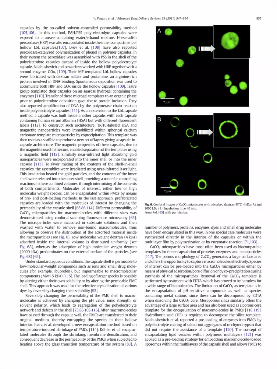
Fig. 6. Confocal images of CaCO3 microcores with adsorbed dextran-FITC, 4 kDa (A) and2000 kDa (B). Incubation time 40 min.From Ref. [65] with permission.
855V. Vergaro et al. / Advanced Drug Delivery Reviews 63 (2011) 847–864
capsules by the so-called solvent-controlled permeability method[105,106]. In this method, PAH/PSS poly-electrolyte capsules wereexposed to a urease-containing water/ethanol mixture. Horseradishperoxidase (HRP)wasalsoencapsulated inside the inner compartmentofhollow LbL capsules.[107]. Lvov et al. [108] have also reportedperoxidase-catalyzed polymerization of phenol in polymer capsules. Intheir system the peroxidase was assembled with PSS in the shell of thepolyelectrolyte capsules instead of inside the hollow polyelectrolytecapsule. Balabushevich and coworkers workedwith HRP together with asecond enzyme, GOx, [109]. Their MF-templated LbL hollow capsuleswere fabricated with dextran sulfate and protamine, an arginine-richprotein involved in DNA-binding. Spontaneous deposition was used toaccumulate both HRP and GOx inside the hollow capsules [109]. Trau'sgroup templated their capsules on an agarose hydrogel containing theenzymes [110]. Transfer of these microgel templates to an organic phaseprior to polyelectrolyte deposition gave rise to protein inclusion. Theyalso reported amplification of DNA by the polymerase chain reactioninside polyelectrolyte capsules [111]. As an extension to the LbL capsulemethod, a capsule was built inside another capsule, with each capsulecontaining human serum albumin (HSA) but with different fluorescentlabels [112]. To construct such architecture, TRITC-labeled HSA andmagnetite nanoparticles were immobilized within spherical calciumcarbonate template microparticles by coprecipitation. This template wasthen used as a scaffold to produce a new set of layers, giving a capsule-in-capsule architecture. The magnetic properties of these capsules, due tothemagnetite used in thecore, enabled separationof the templates usinga magnetic field [112]. Similarly near-infrared light absorbing goldnanoparticles were incorporated into the inner shell or into the innercapsule [113]. To favor mixing of the contents of the shell-in-shellcapsules, the assemblies were irradiated using near-infrared laser light.This irradiation heated the gold particles, and the contents of the innershell were released into the outer shell, providing a route for controllingreactions in these confinedvolumes, through intermixingof thecontentsof both compartments. Molecules of interest, either low or highmolecular weight species, can be encapsulated within PMCs by meansof pre- and post-loading methods. In the last approach, prefabricatedcapsules are loaded with the molecules of interest by changing thepermeability of the capsule shell [65,86,114]. Different permeability ofCaCO3 microparticles for macromolecules with different sizes wasdemonstrated using confocal scanning fluorescence microscopy [65].The microparticles were incubated in substrate solutions and thenwashed with water to remove non-bound macromolecules, thusallowing to observe the distribution of the adsorbed material insidethe microparticles (see Fig. 6). Low molecular weight dextran (4 kDa)adsorbed inside the internal volume is distributed uniformly (seeFig. 6A), whereas the adsorption of high molecular weight dextran(2000 kDa) predominates on the external surface of the particles (seeFig. 6B) [65].
Under standard aqueous conditions, the capsule shell is permeable tolow-molecular-weight compounds such as ions and small drug mole-cules (for example, ibuprofen), but impermeable to macromolecularcomponents (MwN5 kDa) [115]. The loading of larger species is possibleby altering either their own solubility or by altering the permeable PMCshell. This approach was used for the selective crystallization of variousdyes by reversibly changing their solubility [92].
Reversibly changing the permeability of the PMC shell to macro-molecules is achieved by changing the pH value, ionic strength, orsolvent polarity, which leads to segregation of the polyelectrolytenetwork and defects in the shell [73,86,105,116]. After macromoleculeshave passed through the capsule wall, the PMCs are transferred to theiroriginal medium, thereby entrapping the species in their hollowinterior. Ibarz et al. developed a new encapsulation method based ontemperature-induced shrinkage of PMCs [114]. Köhler et al. encapsu-lated molecules through the shrinking, membrane densification, andconsequent decrease in the permeability of the PMCswhen subjected toheating above the glass transition temperature of the system [83]. A
number of polymers, proteins, enzymes, dyes and small drugmoleculeshave been encapsulated in this way. In one special case molecules weresynthesized directly in the interior of the capsules or within themultilayer film by polymerization or by enzymatic reaction [71,103].
CaCO3 microparticles have most often been used as biocompatibletemplates for the encapsulation of proteins, enzymes, and nanoparticles[117]. The porous morphology of CaCO3 generates a large surface areaandoffers theopportunity to capturemacromolecules effectively. Speciesof interest can be pre-loaded into the CaCO3 microparticles either bymeansofphysical adsorption/porediffusionorbyco-precipitationduringsynthesis of the microparticles. Removal of the CaCO3 template isperformed by treatmentwith EDTA,which has proved to be harmless fora wide range of biomolecules. The limitation of CaCO3 as template is inthe encapsulation of pH-sensitive compounds as well as speciescontaining metal cations, since these can be decomposed by EDTAwhen dissolving the CaCO3 core. Mesoporous silica similarly offers theadvantage of a large surface area and has also been used as a porous coretemplate for the encapsulation of macromolecules in PMCs [118,119].Hydrofluoric acid (HF) is required to decompose the silica template.Balabushevitch et al. reported a pre-loading of enzymes into PMCs bypolyelectrolyte coating of salted-out aggregates of α-chymotrypsin thatdid not require the assistance of a template [120]. The concept ofencapsulating lipid vesicles within polymeric multilayers [121] wasapplied as a pre-loading strategy for embedding macromolecule-loadedliposomeswithin themultilayers of the capsule shell and allows PMCs to

856 V. Vergaro et al. / Advanced Drug Delivery Reviews 63 (2011) 847–864
be obtained with a large number of sub-compartments that can bedestroyed by surfactants [122–124].
5.4. PMC interactions with living (cancer) cells
Several drugs have an intracellular target but they have apoor uptakewhen delivered in a soluble form or are lacking in solubility. However,specific targeting by drugmolecules is also often needed to enhance thetherapeutic efficiency or to avoid severe side effects. To evaluate thepotential of PMCs for drug delivery it is important to understand theirinteractions with living cells (e.g. cancer). One of the most importantissues is toxicity. Several research groups have assessed it by performingin vitro cell-viability assays such as the MTT test or Trypan Blue[125,126]. Generally, no severe toxicity was observed at moderatecapsule concentrations even if an outermost polycationic layer seem toincrease their toxicity, giving a pronounced tendency to adhere to thecell surface [127]. Reduced toxicity was observed at elevated capsuleconcentrations: this is often attributed to aggregation of the capsules ontop of the cells as a result of competition for space between capsules andcells. This effect limits themetabolismof the cells, and thus reduces theircytoviability [125,128,129]. Recently Palamà et al. [130] reported onPMCs and coated polymer microcolloid cytotoxicity and uptake bytumor cells. They showed that biosynthetic and biodegradable polyelec-trolyte capsules and microcolloids are readily uptaken by threeneoplastic model cellular lines (rat neuroblastoma, human cervice andbreast cancer). Time-resolved MTT tests demonstrated also the lowmicro-carrier cytotoxicity and good cytocompatibility.
PMCs are phagocytated by cancer cells and immune cells (e.g.macrophages). This was earlier demonstrated by Sukhorukov et al.[37] on a breast cancer cell line. The mechanisms of the uptake ofcapsules by living cells are not yet fully understood and still underinvestigation by several groups [37,131–133]. De Geest et al. [131]produced DEXS/pARG, pSS/p(HPMA-DMAE) and pSS/pAH microcap-sules, loaded with FITC-dextran, and brought them into contact withAfrican green monkey kidney cells (VERO-1 cells). Initially, themicrocapsules adhered to the surface of the cells and during the firsthour of incubation no uptake of capsules by the cells could be observed.However, microcapsules were clearly present in most of the cells 20 hlater, as illustrated in Fig. 7 forDEXS/pARGmicrocapsules. At this point in
Fig. 7. Transmission (A) and confocal (B) image of DXS/pARG capsules filled with FITC-dextrathe presence of intact capsules a fewmicrometer in size in the cells. The red arrows indicatelysosomes in the cytosol red, was used. The green structures are the DXS/pARG capsules filFrom Ref. [131] with permission.
time the capsules seemed to be intact. The transmission image clearlyshows the presence of intact capsules a few micrometer in size in thecells. The red arrows in Fig. 7A indicate the presence of intact capsules.
Studies on in vitro cancer cell lines by De Geest et al. [131] andParak and co-workers [134] indicated that PMCs endup in intracellularacidic vesicles upon cellular uptake. This was demonstrated by red-fluorescent lysosomal staining and observation of colocalization withthe green fluorescence of the capsules. In fact, by staining the nuclei ofthe cellswithDAPI it could be shown that capsules never penetrate thenucleus. In Fig. 8 are shown confocal microscopy images of humanhepatoma cells (HuH7) that have been incubated with capsules.Capsules (red) with dissolved template cores with red fluorescentTRITC in their walls with (PSS/PAH)2 layer sequence were used [134].Kreft et al. confirmed these findings by encapsulating a pH-sensitivedye in PMCs [135]. It was possible to determine that the pH valuesensed by the capsules was typical for a endo/lysosomal environment.De Koker et al. inhibited different endocytotic pathways and actinpolymerization and investigated protrusions of the cellular mem-brane. They proposedmacropinocytosis as themechanismof uptake ofcapsules by bone-marrow-derived dendritic cells [133]. Severalgroups have focused their research on the intracellular fate of thecapsules and have observed a substantial deformation of the capsules,due to the pressure of the surrounding cytoplasm [127,131] Incorpo-ration ofmetal nanoparticles for enhancing themechanical strength ofpolymer films, rendered the capsule resistant to deformation uponcellular uptake [136]. Complete destruction and intracellular degra-dation were demonstrated by De Geest et al. by using degradablepolycations [131]. Co-incubation of cancer cells with PMCs results intheir internalization and gradual disintegration of the capsules over aperiod of 60 h, after which no intact capsules could be observed [131].Similar results were foundwhen bone-marrow-derived dendritic cellswere incubated with dextran sulfate/poly-L-arginine PMCs [133,137].
Proteins andnucleic acids, or cancer therapeutics often have a specifictarget tissue, while delivery to other parts of the body is inefficient oreven problematic, as is the case, for example, for chemotherapeutics.Therefore, functionalization of PMCswith biologicalmolecules (acting asstealthymoieties) plays an important role in shielding the capsules fromunwanted uptake while also enhancing their uptake by the target cells.Internalizing microparticles in the body results in the opsonic proteins
n taken up by VERO-1 cells after 20 h incubation. The transmission image clearly showsthe presence of intact capsules. In the confocal image Lyso-Tracker Red, which stains theled with FITC-dextran present in the cells. Scale bars represent 10 μm.

Fig. 8. Confocal microscopy images (parallel to the substrate) of human hepatoma cells (HuH7) which have been incubated with capsules. Capsules (red) with dissolved template cores(CaCO3 4–6 μmdiameter)with red fluorescent TRITC in theirwalls with (PSS/PAH)2 layer sequencewere used. Before imaging the cells were fixed and their nucleiwere stainedwith bluefluorescent DAPI (blue) and fluorescent antibodies (green) were used against A) early endosomes and B) lysosomes, respectively. The scale bars in images A) and B) represent 50 and20 μm, respectively.From Ref. [134] with permission.
857V. Vergaro et al. / Advanced Drug Delivery Reviews 63 (2011) 847–864
rapidly adsorbing onto the particle surface, thereby causing particleclearance by phagocyting cells. For prolonged circulation times and thetargeted delivery of PMCs, it would be necessary to minimize theadsorption of proteins onto the capsule surface to avoid undesirablephagocytosis [138]. Coating PAH/PSS capsules with a polycationsubstitutedwith a protein-repellent stealth polymer, resulted in a drasticdecrease in protein adsorption. This finding was attributed to thehydrophilicity of the PEG-fully hydrated inwater—which likely preventsprotein adsorption through hydrophobic interactions. Furthermore, thedense PEG brush layer shields electrostatic charges, thus minimizingelectrostatic interactions between the capsules and proteins.Wattendorfet al. investigated the effect of PEGylation on cellular uptake [139]. Thetargeted delivery of PMCs requires functionalization of the capsulesurfacewithmagnetic particles [41], monoclonal antibodies [140,141] orcarbohydrates [142–144].
Cortez et al. described the functionalization of PSS/PAH PMCs withhumanized A33 monoclonal antibodies, which bind to the human A33antigen expressed by 95% of all human colorectal cancer cells. Thisfunctionalization resulted in an enhanced uptake of PMCs by cancer cellscontaining the A33 antigen compared to not functionalized PMCs[140,141]. Surface functionalization of PMCs with galactose moietiescould potentially enhance the binding of PMCs to hepatocytes. In thisrespect, Zhang et al. reported on the synthesis of a galactose-bearingpolycationwhichwasused to construct PMCs in combinationwith PSSorhemoglobin as the polyanion [142,143]. They demonstrated the bio-specificity of this type of PMCs. At present no studies are reported inliterature concerning PMC uptake by hepatocytes. To fill this gap, ourgroup has recently studied PMC cytocompatibility and uptake byHepatocarcinoma(HCC) [145].Detailed investigationsof useof Paclitaxel(PTX)-loadednanocolloids andPMCs inovarian cancer cells (e.g.OVCAR)in order to understand and overcome chemo-resistance are also underway [146]. Magnetic targeting was established by Zebli et al. by usingPSS/PAH capsules functionalized with magnetic metal nanoparticles.They demonstrated that PMCs were preferably internalized by breastcancer cells growing close to the magnetic field [62]. Viruses have alsobeen used to functionalize the surface of PMCs to modulate the cellularuptake. Fischlechner et al. produced virus-modified PMCs by incubatinglipid-coated PMCs with rubella-like particles through lipid fusion of
PMCs and the viral surface. These viral modifications promoted thebinding of PMCs to the cellular surface, stimulated endocytosis, andfavored fusion with the endosomal membrane [42,43,147].
5.4.1. Drug deliveryWater-soluble drug compoundsmust be kept in a non-soluble state,
to allow the LbL coating to be carried out. Numerous drugmicrocrystalslike ibuprofen, dexamethasone, indomethacin, and furosemide havebeen encapsulated in PMCs in this way. The ability of adsorbedpolyelectrolytes to alter the dissolution profile of incorporatedmicrocrystals toward more-prolonged drug release was observed[144,148–153]. Uncharged water-insoluble drug crystals can thus beencapsulated by first providing the crystals with a surface chargethrough adsorption of an ionic surfactant, followed by polyelectrolytecoating [154]. Beyer et al. encapsulated glucose and vitamin C [155] byprecipitation of the drug molecules in an organic phase followed by LbLcoatingwith polyelectrolytes that are soluble in anorganic solvent. Laterthey used hydrogel beads loaded with water-soluble compoundsfollowed by stabilization of the beads through adsorption of colloidsand reversed-phase LbL coating. This approach resulted in anencapsulation efficiency of almost 100%.[110]. Passive drug loadingthrough electrostatic interactions and the use of micelles loaded with ahydrophobic compound have also been reported [156,157]. An activepost-loading approachwasusedbyRadtchenko et al.,whoexploited thechange in the solubilityof poorlywater-solubledrugmolecules betweenthe capsule interior and the external solution, thus allowing precipita-tion of the drug in the capsules void [158]. A strongly hydrophilicpolymer was encapsulated within the capsules, providing a polaritygradient between the interior of the shells and an external water/acetone solution. Soluble drugs in the water/acetone mixture experi-enced a higher partial water content within the capsules and thusprecipitated [159]. It is challenging to entrap water-soluble substanceswithin layers when the capsule wall is made of highly water solublepolyelectrolytes. Theassembly of polyelectrolytes always results in smallpores in the capsule wall, and small molecules can be easily expelled. Toovercome this limitation one might use more hydrophobic polymers inthe capsule shell or by making the LbL shell more hydrophobic afterhollowcapsule formation.A similar effect canbeobtainedalsoby coating
858 V. Vergaro et al. / Advanced Drug Delivery Reviews 63 (2011) 847–864
the capsule surface with a lipid bilayer [160]. In all cases drug releasecould only occur upon mechanical rupture of the capsules.
Anticancer drugs are usually low-molecular-weight drugs. So far,chemotherapeutics such as daunorubicin, doxorubicin and polyphenolshave been successfully encapsulated in PMCs [129,161–164]. Further-more, a higher activity of the encapsulated chemotherapeuticscompared to the free drugs was observed in the cases of doxorubicin-anddaunorubicin-loaded capsules tested in vitroondifferent cancer celllines. Nevertheless experiment onmice demonstrated the effectivenessof encapsulated doxorubicin in reducing the size of tumors [163,165].Other techniques for encapsulating chemotherapeutics have also beenreported, such as the use of emulsion templating or hydrophobicassociation [164,166].Wehave already citednovel formulations by Lvovgroup which developed antineoplastic drug-loaded LbL templatednanocolloids with high payload and slow release [46]. Following thesefindings, our group has exploited these new PXT-nanocolloid nano-formulations on ovarian cancer cells for overcoming PXT drug-resistance [146].
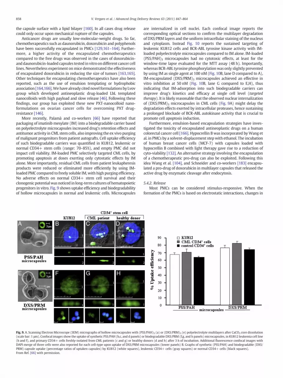
More recently, Palamà and co-workers [66] have reported thatpackaging of imatinib mesylate (IM) into a biodegradable carrier basedon polyelectrolyte microcapsules increased drug's retention effects andantitumor activity in CML stem cells, also improving the ex vivo purgingof malignant progenitors from patient autografts. Cell uptake efficiencyof such biodegradable carriers was quantified in KU812, leukemic ornormal CD34+ stem cells (range: 70–85%), and empty PMC did notimpact cell viability. IM-loaded PMC selectively targeted CML cells, bypromoting apoptosis at doses exerting only cytostatic effects by IMalone. More importantly, residual CML cells from patient leukapheresisproducts were reduced or eliminated more efficiently by using IM-loaded PMC compared to freely soluble IM,with high purging efficiency.No adverse effects on normal CD34+ stem cell survival and theirclonogenic potentialwasnoticed in long-termculturesof hematopoieticprogenitors in vitro. Fig. 9 shows uptake efficiency and biodegradabilityof hollow microcapsules in normal and leukemic cells. Microcapsules
Fig. 9. A. Scanning Electron Microscope (SEM)micrographs of hollowmicrocapsules with (PS(scale bar: 1 μm). Confocal images show the uptake of synthetic PSS/PAH (b,c, and d panels) o(b and f), and primary CD34+ cells freshly-isolated from CML patients (c and g) or healthyDAPI merge of three cells were also reported for each cell-type upon uptake of DXS/PRM mPRM) capsule uptake (percentage ratios of uptaken capsules) by KU812 (white squares), leFrom Ref. [66] with permission.
are internalized in cell nuclei. Each confocal image reports thecorresponding optical sections to confirm the multilayer degradationof DXS/PRM layers and the uniform intracellular staining of the nucleusand cytoplasm. Instead Fig. 10 reports the sustained targeting ofleukemic KU812 cells and BCR-ABL tyrosine kinase activity with IM-loaded polyelectrolyte microcapsules compared to IM alone. IM-loaded(PSS/PAH)3 microcapsules had no cytotoxic effects, at least for thewindow-time lapse evaluated for the MTT assay (48 h). Importantly,whereas BCR-ABL tyrosine phosphorylationwas only slightly preventedby using IM as single agent at 100 nM (Fig. 10B, lane D compared to A),IM-encapsulated (DXS/PRM)3 microcapsules achieved an effective invitro inhibition at 50 nM (Fig. 10B, lane G compared to E,H), thusindicating that IM-adsorption into such biodegradable carriers canimprove drug's kinetics and efficacy at single cell level (targetedtherapy). It is likely reasonable that the observed nuclear internalizationof (DXS/PRM)3 microcapsules in CML cells (Fig. 9A) might delay thedegradation effects exerted by intracellular proteases, hence sustaininga prolonged blockade of BCR-ABL autokinase activity that is crucial topromote cell apoptosis induction.
Furthermore, emulsion-based encapsulation strategies have inves-tigated the toxicity of encapsulated antineoplastic drugs on a humancolorectal cancer cell [166]. Hypocrellin B was incorporated byWang etal. in PMCs by a solvent-displacement stepwith ethanol. The incubationof human breast cancer cells (MCF-7) with capsules loaded withhypocrellin B combined with light therapy gave rise to a reduction ofcyto-viability [132]. An alternative strategy involving the encapsulationof a chemotherapeutic pro-drug can also be exploited. Following thisidea Wang et al. [104], and Schneider and co-workers [183] encapsu-lated a pro-drug of doxorubicin in multilayer capsules that released theactive drug by enzymatic cleavage after endocytosis.
5.4.2. ReleaseMost PMCs can be considered stimulus-responsive. When the
formation of the PMCs is based on electrostatic interactions, changes in
S/PAH)3 (a) or (DXS/PRM)3 (e) polyelectrolyte multilayers after CaCO3 core dissolutionr biodegradable DXS/PRM (f,g, and h panels) microcapsules, in KU812 leukemia cell linedonors (d and h) after 3 h of incubation. Additional fluorescence confocal images withicrocapsules (lower panels) B. Graphs of synthetic (PSS/PAH) and biodegradable (DXS/ukemic CD34+ cells (gray squares) or normal CD34+ cells (black squares).

Fig. 10. A. MTT test for cellular vitality of KU812 cells cultured for 48 h in the absence (−) or in the presence of the indicated concentrations of imatinib (range 10–100 mM) tested assingle agent (no microcapsules), or as released agent by biodegradable microcapsules (DXS/PRM) or by synthetic microcapsules (PSS/PAH). B. Western blotting analysis for theexpression (anti-BCR-ABL) and phospho-tyrosine activation levels (anti-pY BCR-ABL) of the oncoprotein BCR-ABL in KU812 cells cultured in the absence (A) or in the presence of theindicated doses of imatinib tested as single agent (lanes B,C,D, and H) or as released agent by biodegradable DXS/PRM polyelectrolyte microcapsules (lane G). KU812 were alsoincubated with empty (drug-free) DXS/PRM hollow microcapsules as vehicle carrier control (lane F).From Ref. [66] with permission.
859V. Vergaro et al. / Advanced Drug Delivery Reviews 63 (2011) 847–864
the pH value and/or ionic strength are evident triggers that can modifythe interactionsbetween thesuccessive layers, and thusmight beused toinduce the release of encapsulated material [167]. These results havebeen extended to hydrogen-bonded capsules containing a polyioniccomponent [168]. Besides ionic strength and pH value [55,169],temperature [83], solventpolarity [105], glucose [170,171] andoxidation[68] have also been used to modify the permeability of PMCs. However,these parameters are non-physiological triggers, which often hampertheir in-vivo applications. Therefore research has dedicatedmost effortson developing PMCs that are sensitive to more physiologically relevantstimuli, including enzymatic digestion [131,172,173] or the reductiveintracellular environment [174–177]. Enzymatically degradable PMCsbased on oppositely charged polypeptides and/or polysaccharides havenow been generated by several research groups. De Geest and co-workers demonstrated that PMCs consisting of dextran sulfate andpoly-L-arginine could be degraded intracellularly by proteases upon phago-cytosis by in vitro cultured cells [131]. Similar resultswere reported laterby other research groups, who used hyaluronidase and chitinase todecompose capsules containing hyaluronic acid or chitosan as layercomponents [172,173,178]. The transition from an oxidative to areductive environment has also been found to trigger the decompositionof PMCs after cellular uptake. Haynie et al. stabilized PMCs by disulfidebonds through the design of oppositely charged peptides containingcysteine moieties [176,177]. The Caruso research group adopted thisstrategy to develop hydrogen-bonded PMCs by modifying poly(methacrylic acid) with cysteamine. The PMCs were fabricated throughsequential deposition of polyelectrolytes onto sacrificial silica coretemplates followed by oxidative cross-linking of the thiol moieties anddecomposition of the silica cores with HF [174,175]. This approach wasapplied by them later to encapsulate peptides, oligonucleotides, and
low-molecular-weight anticancer drugs [179]. Additionally, release fromPMCs might also be achieved by applying external stimuli to thecapsules. This has mainly been accomplished by incorporating metalnanoparticles or light-responsive dyes into thewalls of the capsules [52].Several research groups have reported on such systems, includingtriggered release from1)dye-functionalized capsules by irradiationwithlight [180], metal nanoparticle embedded by a magnetic field [181],microwaves [182] 2) ultrasound [183–185] and 3) noble metal (silver,gold) embedded capsules by irradiation with a focused laser beam[180,186–191]. Gold nanoparticles (AuNP) exhibit a surface plasmonresonance signal in the visible spectrum around 530 nm [188]. As aresult, AuNP are locally heated when irradiated by laser light of thiswavelength through conversion of photons into thermal energy. Thisheating caused rupture of the capsules and released the encapsulatedpayload. The surface plasmon resonance signal can be tuned bycontrolling the shape and aggregation state of the AuNP on the surfaceof the PMCs, with the signal shifted into infrared region [191].Moreover,further fine-modulating the composition of the PMCs allows the PMCmembrane to be made reversibly permeable by IR irradiation, therebyreleasing only portions of encapsulatedmaterial, without destroying thewhole capsule [190]. This principle was applied to drug delivery bySkirtach and co-workers, who showed that laser-triggered opening canbe performedwithin living cellswithout influencing their viability [189].Later it was shown that capsule breakage also resulted in the rupture ofthe phagosomalmembrane surrounding the capsules, thus releasing theencapsulated material in the cellular cytoplasm [191]. Another class oftriggered-release capsules, is the so-called self-degrading systemswhichare equipped with an internal trigger that causes the release ofencapsulated species. This canbe achievedby co-encapsulatingdigestiveenzymes into thehollow interior of the PMCs [95]. These enzymes digest

860 V. Vergaro et al. / Advanced Drug Delivery Reviews 63 (2011) 847–864
the PMC films itself or process co-encapsulated species into smallerfragments that can be released through the PMC membrane. They havebeen generated by the LbL coating of a degradable microgel core whichswells upon chemical hydrolysis at physiological pH values. When theswellingpressure exceeds the tensile strength of the PMCmembrane, thecapsule ruptures and the encapsulated species are released [192–196].
5.4.3. In vivo interactionsDue to their charged nature, polyelectrolytemicrocapsulesmay cause
significant reactions at tissue level when applied in vivo. De Koker et al.have examined the tissue reaction caused by sub-cutaneous injection ofmicrocapsules composed of dextran sulfate/poly-L-arginine bilayers inmice. This resulted in a fast pro-inflammatory response, characterized bythe recruitment of polymorphonuclear cells and monocytes [125]. Themicrocapsules behaved like an implant, with infiltration starting at theborder and gradually moving toward the center of the injection volume.The inflammation was restricted to the injection site, which rapidlybecame surrounded by several layers of fibroblasts. However no tissuedestruction or ulceration was observed. In this respect, PMCs appear toshowasimilar degreeof inflammationas othermicroparticles of the samesize. De Koker and co-workers exploited further the in vivo fate of RITC-poly-L-arginine-labeled microcapsules following subcutaneous injection.The microcapsules were taken up by phagocytic cells, deformed, andsubsequently degraded. PMCs having a thicker shell (higher number oflayers)weremore resistant to degradation anddeformation [125]. Even ifthere is not much literature in this area, these data have established thefeasibility of using polyelectrolyte microcapsules in vivo. Issues still to besolved are clearly the PMCs' larger size and possible recognition byimmune system which could be by-passed by PEGylation. Moreover,given the close association between inflammation and the induction ofimmune responses, and their capacity to be phagocytated in vivo,polyelectrolyte microcapsules may have possible application in a novelimmunogenic therapy.
6. Conclusions and outlook
In this review we have tried to give an overview of the recentdevelopments in the use of polyelectrolytemultilayer capsules (PMCs) asdrug-delivery vehicles, especially on cancer research application. In thefirst section we have briefly reviewed different approaches used in thefabrication of nano-vehicles such as magnetic nanoparticles, liposomes,carbon nanotubes and dendrimers. Furthermore we have summarizedactual limits and drawbacks of chemotherapy which cannot guaranteeproperties such as specificity, selectivity, high solubility and can havesevere side effects like chemo-resistance or others The use of novelnanotechnological approaches as the use of nanoparticles (NPs) orcapsules is therefore needed. In this respect they have several advantagesdue to their nanometer size, flexibility in surface modification, targeting,and can have protective moieties against degradation upon interactionwith immunosystem. In successive sectionswe have described the layer-by-layer approach for production of LbL templated microcolloids, highpayloaded drug nanocarriers (like antineoplastic drug-nanocolloids).Furthermore we have reported different approaches for synthetizingpolyelectrolytemultilayered capsules (PMCs) andwe reviewed differentapproaches that canbeused to load andunload compounds/molecules ofinterest into and out of PMCs. Preloading strategies, involving porousdrug-loaded templates showed to be rather efficient. Instead of thepostloading approach, which involves a reversible change of the capsulepermeability offers low encapsulation efficacy. Once encapsulated intoPMCs, the drug molecules also have to be released at their target site.Release control of small hydrophilic molecules has revealed to be ratherchallenging with PMCs. Instead successful strategies have been devel-oped for the delivery of macromolecular drugs as well as hydrophobiccompounds. For this aim, PMCs need to be specifically functionalizedallowing to be opened upon triggering. By far, the most promisingsystems are those based on enzymatic degradation of the capsule shell or
on reductive cleavageof adisulfide linkage,which leads todestabilizationof the capsule shell. Recently Ref. [189]also exploited a way to activatecapsules by a laser beam. This new approach can offer perspectives forcontrolled intracellular delivery of drugs. In fact while PMCs arecommonly delivered to endo/lysosomal vesicles, this strategy permitsto escape from such vesicles and the delivery of drug molecules into thecellular cytosol. The delivery of anticancer drugs undoubtedly also haslarge potential and efficacy. Several encapsulation approaches haveshown to be successful in encapsulating anticancer drugs, and thefunctionalization of PMCs by cell-targeting antibodies and stealth PEGcoatings have also been reported. PMCs are ratherflexible and elastic andthese properties could be used to pass through capillary vein. Their largesize, their aggregation during subcutaneous administration and thetargeted release of payload at cancerous tissues remain as the mainchallenges in PMC use as delivery vehicle in vivo. LbL method ofpreparation seems to have a technical limitation to the diameter (usuallybetween 1 and 10 μm, which may explain their relatively limited use fordelivery purposes, i.e., where truly nanoscale small capsules are desired.However, the advantages of LbL capsules, in the sense that capsules withsemipermeable properties can be prepared with relative ease and in abiocompatible manner, render them highly flexible and strongly multi-functional. In this prospective, layer-by layer (LbL) assembly represents aversatile technique for preparing hollow capsules with tunable function-alities in thenano/micrometer range by entrapping simultaneously smallmolecule drugs with synergistic effects for anti-cancer therapy. Partic-ularly, there remains ample room to improve the proposed technology inachieving the coupled delivery of pharmacological and nucleic agents[38,197]. A deeper understanding on how different parameters of LbLassembledhollowcapsules (suchas number or compositionof polymericlayers, pH, ionic strength, solvent) can alter loading/release efficiency ofcoupled-delivery carrier systemsand, subsequently, the in vivo or exvivoeradication of cancer cells will be highly valuable. We therefore expect aconsiderable increase of publications to explore combination of LbLassembly techniqueswithother architectures, suchas liposomes, viruses,and microgels.
Acknowledgments
Financial support of the ItalianMinistry of University and Research(MIUR) through a FIRB project no. RBLA03ER38 is acknowledged. “Con il contributo del Ministero degli Affari Esteri, Direzione Generaleper la promozione e la Cooperazione Sociale ” (Large Scale ITA –USABilateral Project “Nano-trasportatori per la terapia del cancro”).
References
[1] M. Ebbesen, Th.G. Jensen, Nanomedicine: techniques, potential and ethicalimplications, J. Biotechnol. (2006) 1–11 article id. 51516.
[2] O.V. Salata, Applications of nanoparticles in biology and medicine, J. Nanobio-technol. 2 (3) (2004) 1–6.
[3] S. Leporatti, D. Vergara, A. Zacheo, V. Vergaro, G. Maruccio, R. Cingolani, R.Rinaldi, Cytomechanical and topological investigation of MCF-7 cells by scanningforce microscopy, Nanotechnology 20 (5) (2009) 055103.
[4] R. Singhal, Z. Orynbayeva, R.V. Kalyana Sundaram, J.J. Niu, S. Bhattacharyya, E.A.Vitol, M.G. Schrlau, E.S. Papazoglou, G. Friedman, Y. Gogotsi, Multifunctionalcarbon-nanotube cellular endoscopes, Nat. Nanotechnol. 6 (1) (2011) 57–64.
[5] C. Medina, M.J. Santos-Martinez, A. Radomski, O.I. Corrigan, M.W. Radomsk,Nanoparticles: pharmacological and toxicological significance, Br. J. Pharmacol.150 (2007) 552–558.
[6] C.J. Johnson, N. Zhukovsky, A.E. Cass, J.M. Nagy, Proteomics, nanotechnology andmolecular diagnostics, Proteomics 8 (4) (2008) 715–730.
[7] R.S. Gaster, D.A. Hall, C.H. Nielsen, S.J. Osterfeld, H. Yu, K.E. Mach, R.J. Wilson, B.Murmann, J.C. Liao, S.S. Gambhir, S.X. Wang, Matrix-insensitive protein assayspush the limits of biosensors in medicine, Nat. Med. 15 (2009) 1327–1332.
[8] L. Serpe, Conventional chemotherapeutic drug nanoparticles for cancertreatment, in: C.S.S.R. Kumar (Ed.), Nanomaterials for Cancer Therapy, vol. 6,Wiley-VCH, 2006.
[9] W.H. De Jong, P.J.A. Borm, Drug delivery and nanoparticles: applications andhazards, Int. J. Nanomed. 3 (2) (2008) 133–149.
[10] R.K. Jain, Transport of molecules in the tumor interstitium: a review, Cancer Res.47 (1987) 3039–3051.

861V. Vergaro et al. / Advanced Drug Delivery Reviews 63 (2011) 847–864
[11] D.F. Baban, L.W. Seymour, Control of tumor vascular permeability, Adv. DrugDeliv. Rev. 34 (1998) 109–119.
[12] L.S. Jabr-Milane, L.E. van Vlerken, S.Y. Mansoor, M. Amiji, Multifunctionalnanocarriers to overcome tumor drug resistance, Cancer Treat. Rev. 34 (7)(2008) 592–602.
[13] S.M. Moghimi, A.Ch. Hunter, J.C. Murray, Nanomedicine: current status andfuture prospects, FASEB J. 19 (3) (2005) 311–330.
[14] V.V. Pokropivny, V.V. Skorokhod, New dimensionality classifications of nanos-tructures, Physica E 40 (2008) 2521–2525.
[15] F. Alexis, E. Pridgen, L.K. Molnar, O.C. Farokhzad, Factors affecting the clearanceand biodistribution of polymeric nanoparticles, Mol. Pharmaceutics 5 (4) (2008)505–515.
[16] H. Maedaet, The enhanced permeability and retention (EPR) effect in tumorvasculature: the key role of tumor selective macromolecular drug targeting, Adv.Enzyme Regul. 41 (2001) 189–207.
[17] D.E. Owens, N.A. Peppas, Opsonization, biodistribution, and pharmacokinetics ofpolymeric nanoparticles, Int. J. Pharm. 307 (1) (2006) 93–102.
[18] E. Gullotti, Y. Yeo, Extracellulary activated nanocarriers, a new paradigm oftumor targeted drug delivery, Mol. Pharm. 6 (4) (2009) 1041–1051.
[19] M.G. Ozawa, A.J. Zurita, E. Dias-Neto, D.N. Nunes, R.L. Sidman, J.G. Gelovani, W.Arap, R. Pasqualini, Beyond receptor expression levels: the relevance of targetaccessibility in ligand-directed pharmacodelivery systems, Trends Cardiovasc.Med. 18 (4) (2008) 126–133.
[20] D.J. Bharali, M. Khalil, M. Gurbuz, T.M. Simone, S.A. Mousa, Nanoparticles andcancer therapy: a concise review with emphasis on dendrimers, Int. J. Nanomed.4 (2009) 1–7.
[21] A. Puri, K. Loomis, B. Smith, j-H. Lee, A. Yavlovich, E. Heldman, R. Blumenthal,Lipid-based nanoparticles as pharmaceutical drug carriers, Crit. Rev. Ther. DrugCarrier Syst. 26 (6) (2009) 523–580.
[22] S.C. McBain, H.H.P. Yiu, J. Dobson, Magnetic nanoparticles for gene and drugdelivery, Int. J. Nanomed. 4 (2009) 1–7.
[23] J.H. Lee, Y.M. Huh, Y.W. Jun, J.W. Seo, J.T. Jang, H.T. Song, S. Kim, E.J. Cho, H.G.Yoon, J.S. Suh, J. Cheon, Artificially engineered magnetic nanoparticles for ultra-sensitive molecular imaging, Nat. Med. 13 (1) (2007) 95–99.
[24] R. Parthe, J.L. Conyers, Biomedical applications for functionalized fullerene-basednanomaterials, Int. J. Nanomed. 4 (2009) 261–275.
[25] E. Bekyarova, Y. Ni, E.B. Malarkey, V. Montana, J.L. McWilliams, R.C. Haddon, V.Parpura, Applications of carbon nanotubes in biotechnology and biomedicine, J.Biomed. Nanotechnol. 1 (1) (2005) 3–17.
[26] F. Balavoine, P. Schultz, C. Richard, V. Mallouh, Th.W. Ebbesen, C. Mioskowski,Helical crystallization of proteins on carbon nanotubes: a first step towards thedevelopment of new biosensors, Angew. Chem. Int. 38 (1999) 1912–1915.
[27] Z. Guo, P.J. Sadler, S.C. Tsang, Immobilization and visualization of DNA andproteins on carbon nanotubes, Adv. Mater. 10 (1998) 701–703.
[28] J.J. Davis, M.L.H. Green, H.A.O. Hill, Y.C. Leung, P.J. Sadler, J. Sloan, A.V. Xavier, S.C.Tsang, The immobilization of proteins in carbon nanotubes, Inorg. Chem. Acta(1998) 261–266.
[29] R. Gasparac, P. Kohli, M.O. Mota, L. Trofin, Ch.R. Martin, Template synthesis ofnano test tubes, Nano. Lett. 4 (2004) 513–516.
[30] V. Vergaro, E. Abdullayev, Y.M. Lvov, A. Zeitou, R. Cingolani, R. Rinaldi, S.Leporatti, Cytocompatibility of halloysite clay nanotubes, Biomacromolecules 11(2010) 820–826.
[31] G. Decher, Fuzzy nanoassemblies: toward layered polymeric multicomposites,Science 277 (1997) 1232–1237.
[32] P. Bertrand, A. Jonas, A. Laschewsky, R. Legras, Ultrathin polymer coatings bycomplexation of polyelectrolytes at interfaces: suitable materials, structure andproperties, Macromol. Rapid Commun. 21 (2000) 319–348.
[33] S.A. Sukhishvili, Responsive polymer films and capsules via layer-by-layerassembly, Curr. Opin. Colloid Interface Sci. 10 (2005) 37–44.
[34] Z.Y. Tang,Y.Wang, P. Podsiadlo, N.A. Kotov, Biomedical applications of layer-by-layerassembly: from biomimetics to tissue engineering, Adv. Mater. 18 (2006)3203–3224.
[35] B.G. De Geest, N.N. Sanders, G.B. Sukhorukov, J. Demeester, S.C. De Smedt,Release mechanisms for polyelectrolyte capsules, Chem. Soc. Rev. 36 (2007)636–649.
[36] P.T. Hammond, Form and function in multilayer assembly: new applications atthe nanoscale, Adv. Mater. 16 (2004) 1271–1293.
[37] G.B. Sukhorukov, A.L. Rogach,M. Garstka, S. Springer,W.J. Parak, A.Munoz-Javier, O.Kreft, A.G. Skirtach, A.S. Susha, Y. Ramaye, R. Palankar, M. Winterhalter, Multi-functionalized polymer microcapsules: novel tools for biological and pharmaco-logical applications, Small 3 (2007) 944–955.
[38] U. Reibetanz, C. Claus, E. Typlt, J. Hofmann, E. Donath, Defoliation and plasmiddelivery with layer-by-layer coated colloids, Macromol. Biosci. 6 (2006) 153–160.
[39] U. Reibetanz, D. Halozan, M. Brumen, E. Donath, Flow cytometry of HEK 293T cellsinteractingwith polyelectrolytemultilayer capsules containing fluorescein-labeledpoly(acrylic acid) as a pH sensor, Biomacromolecules 8 (2007) 1927–1933.
[40] W. Zauner, M. Ogris, E. Wagner, Polylysine-based transfection systems utilizingreceptor-mediated delivery, Adv. Drug Deliv. Rev. 30 (1998) 97–113.
[41] M. Fischlechner, O. Zschornig, J. Hofmann, E. Donath, Engineering virusfunctionalities on colloidal polyelectrolyte lipid composites, Angew. Chem. Int.Ed. 44 (2005) 2892–2895.
[42] M. Fischlechner, L. Toellner, P. Messner, R. Grabherr, E. Donath, Virus-engineeredcolloidal particles — a surface display system, Angew. Chem. Int. Ed. 45 (2006)784–789.
[43] M. Fischlechner, U. Reibetanz, M. Zaulig, D. Enderlein, J. Romanova, S. Leporatti,S. Moya, E. Donath, Fusion of enveloped virus nanoparticles with polyelectro-
lyte-supported lipid membranes for the design of bio/nonbio interfaces, NanoLett. 7 (11) (2007) 3540–3546.
[44] C. Cortez, E. Tomaskovic-Crook, A.P.R. Johnston, B. Radt, S.H. Cody, A.M. Scott, E.C.Nice, J.K. Heath, F. Caruso, Targeting and uptake of multilayered particles tocolorectal cancer cells, Adv. Mater. 18 (2006) 1998–2003.
[45] A.S. Angelatos, K. Katagiri, F. Caruso, Bioinspired colloidal systems via layer-by-layerassembly, Soft Matter 2 (2006) 18–23.
[46] A. Agarwal, Y. Lvov, R. Sawant, V. Torchilin, Stable nanocolloids of poorly solubledrugs with high content drug prepared using the combination of sonication andlayer-by-layer technology, J. Control. Release 128 (2008) 255–260.
[47] C.S. Peyratout, L. Daehne, Tailor-made polyelectrolyte microcapsules: frommultilayers to smart containers, Angew. Chem. Int. Ed. 43 (2004) 3762–3783.
[48] G.B. Sukhorukov, A. Fery, M. Brumen, H. Mohwald, Physical chemistry ofencapsulation and release, Phys. Chem. Chem. Phys. 6 (2004) 4078–4089.
[49] B.G. De Geest, S. De Koker, G.B. Sukhorukov, O. Kreft, W.J. Parak, A.G. Skirtach, J.Demeester, S.C. De Smedt, W.E. Hennink, Polyelectrolyte microcapsules forbiomedical applications, Soft Matter 5 (2009) 282–291.
[50] L.J. De Cock, S. De Koker, B.G. De Geest, J. Grooten, C. Vervaet, J.P. Remon, G.B.Sukhorukov, M.N. Antipina, Polymeric multilayer capsules in drug delivery,Angew. Chem. Int. Ed. 49 (2010) 2–22.
[51] M. Delcea, A. Yashchenok, K. Videnova, O. Kreft, H. Möhwald, A.G. Skirtach,Multicompartmental micro- and nanocapsules: hierarchy and applications inbiosciences, Macromol. Biosci. 10 (2010) 465–474.
[52] M.F. Bédard, B.G. De Geest, A.G. Skirtach, H. Möhwald, G.B. Sukhorukov,Polymeric microcapsules with light responsive properties for encapsulationand release, Adv. Colloid Interface Sci. 158 (2010) 2–14.
[53] C.E. Mora-Huertasa, H. Fessia, A. Elaissaria, Polymer-based nanocapsules for drugdelivery, Int. J. Pharm. 385 (2010) 113–142.
[54] P. Rivera Gil, L.L. del Mercato, P. del-Pino, A. Muñoz-Javier, W.J. Parak, Nanoparticle-modified polyelectrolyte capsules, Nanotoday 3 (3–4) (2008) 12–21.
[55] T.M. Allen, P.R. Cullis, Drug delivery systems: entering the mainstream, Science303 (2004) 1818–1822.
[56] G.A. Hughes, Nanostructure-mediated drug delivery, Nanomedicine 1 (1) (2005)22–30.
[57] R. Langer, D.A. Tirrell, Designing materials for biology and medicine, Nature 428(6982) (2004) 487–492.
[58] E.M. Pridgen, R. Langer, O.C. Farokhzad, Biodegradable polymeric nanoparticledelivery systems for cancer therapy, Nanomedicine 2 (5) (2007) 669–680.
[59] F. Caruso, R.A. Caruso, H. Mohwald, Nanoengineering of inorganic and hybridhollow spheres by colloidal templating, Science 282 (1998) 1111–1114.
[60] E. Donath, G.B. Sukhorukov, F. Caruso, S.A. Davis, H. Mohwald, Novel hollowpolymer shells by colloid-templated assembly of polyelectrolytes, Angew. Chem.Int. Ed. Engl. 37 (1998) 2201–2205.
[61] D.V. Volodkin, A.I. Petrov, M. Prevot, G.B. Sukhorukov, Matrix polyelectrolytemicrocapsules: new system for macromolecule encapsulation, Langmuir 20 (8)(2004) 3398–3406.
[62] B. Zebli, A.S. Susha, G.B. Sukhorukov, A.L. Rogach, W.J. Parak, Magnetic targeting andcellular uptake of polymer microcapsules simultaneously functionalized withmagnetic and luminescent nanocrystals, Langmuir 21 (10) (2005) 4262–4265.
[63] G.B. Sukhorukov, H. Mohwald, Multifunctional cargo systems for biotechnology,Trends Biotechnol. 25 (3) (2007) 93–98.
[64] G.B. Sukhorukov, D.V. Volodkin, A.M. Gunther, A.I. Petrov, D.B. Shenoy, H.Mohwald, Porous calcium carbonate microparticles as templates for encapsu-lation of bioactive compounds, J. Mater. Chem. 14 (1) (2004) 2073–2081.
[65] D.V. Volodkin, N.I. Larionova, G.B. Sukhorukov, Protein encapsulation via porousCaCO3 microparticles templating, Biomacromolecules 5 (2004) 1962–1972.
[66] I.E. Palamà, S. Leporatti, E. de Luca, N. Di Renzo,M.Maffia, C. Gambacorti-Passerini, R.Rinaldi, G. Gigli, R. Cingolani, A.M.L. Coluccia, Imatinib-loaded polyelectrolytemicrocapsules for sustained targeting of BCR-ABL+ leukemia stem cells, Nanome-dicine 5 (3) (2010) 419–431.
[67] D.B. Shenoy, A.A. Antipov, G.B. Sukhorukov, H. Mohwald, Layer-by-layerengineering of biocompatible, decomposable core–shell structures, Biomacro-molecules 4 (2003) 265–272.
[68] Y.J. Ma, W.F. Dong, M.A. Hempenius, H. Mohwald, G.J. Vancso, Redox-controlledmolecular permeability of composite-wall microcapsules, Nat. Mater. 5 (2006)724–729.
[69] F. Caruso, H. Lichtenfeld, M. Giersig, H. Mohwald, Electrostatic self-assembly ofsilica nanoparticle–polyelectrolyte multilayers on polystyrene latex particles, J.Am. Chem. Soc. 120 (1998) 8523–8524.
[70] D.G. Shchukin, G.B. Sukhorukov, H. Mohwald, Biomimetic fabrication ofnanoengineered hydroxyapatite/polyelectrolyte composite shell, Chem. Mater.15 (2003) 3947–3950.
[71] D.G. Shchukin, G.B. Sukhorukov, H. Mohwald, Smart inorganic/organic nano-composite hollow microcapsules, Angew. Chem. Int. Ed. 42 (2003) 4472–4475.
[72] A.A. Antipov, G.B. Sukhorukov, H. Mohwald, Influence of the ionic strengthon the polyelectrolyte multilayers' permeability, Langmuir 19 (2003)2444–2448.
[73] G. Ibarz, L. Dahne, E. Donath, H. Mohwald, Smart micro and nanocontainers forstorage, transport, and release, Adv. Mater. 13 (2001) 1324–1327.
[74] G.B. Sukhorukov, A. Fery, H. Mohwald, Intelligent micro- and nanocapsule, Prog.Polym. Sci. 30 (2005) 885–897.
[75] K. Kohler, P.M. Biesheuvel, R. Weinkamer, H. Mohwald, G.B. Sukhorukov, Salt-induced swelling-to-shrinking transition in polyelectrolyte multilayer capsules,Phys. Rev. Lett. 97 (2006) 188301.
[76] C. Dejugnat, G.B. Sukhorukov, pH-Responsive properties of hollow polyelectro-lyte microcapsules templated on various cores, Langmuir 20 (2004) 7265–7269.

862 V. Vergaro et al. / Advanced Drug Delivery Reviews 63 (2011) 847–864
[77] D.G. Shchukin, E.A. Ustinovich, G.B. Sukhorukov, H. Mohwald, D.V. Sviridov,Metallized polyelectrolyte microcapsules, Adv. Mater. 17 (2005) 468–472.
[78] T. Mauser, C. Dejugnat, H. Mohwald, G.B. Sukhorukov, Microcapsules made ofweak polyelectrolytes: templating and stimuli-responsive properties, Langmuir2 (2006) 5888–5893.
[79] T. Mauser, C. Dejugnat, G.B. Sukhorukov, Reversible pH-dependent properties ofmultilayer microcapsules made of weak polyelectrolytes, Macromol. RapidCommun. 25 (2004) 1781–1785.
[80] K. Glinel, G.B. Sukhorukov, H. Mohwald, V. Khrenov, K. Tauer, Thermosensitivehollow capsules based on thermoresponsive polyelectrolytes, Macromol. Chem.Phys. 204 (2003) 1784–1790.
[81] A.I. Petrov, A.V. Gavryushkin, G.B. Sukhorukov, Effect of temperature, pH andshell thickness on the rate of Mg2+ and Ox2 — release from multilayeredpolyelectrolyte shells deposited onto microcrystals of magnesium oxalate, J.Phys. Chem. B 107 (2003) 868–875.
[82] K. Kohler, H. Mohwald, G.B. Sukhorukov, Thermal behavior of polyelectrolytemultilayer microcapsules: 2. Insight into molecular mechanisms for thePDADMAC/PSS system, J. Phys. Chem. B 110 (2006) 24002–24010.
[83] K. Kohler, G.B. Sukhorukov, Heat treatment of polyelectrolyte multilayercapsules: a versatile method for encapsulation, Adv. Funct. Mater. 17 (2007)2053–2061.
[84] B.G. De Geest, A.G. Skirtach, T.R.M. De Beer, G.B. Sukhorukov, L. Bracke, W.R.G.Baeyens, J. Demeester, S.C. De Smedt, Stimuli-responsive multilayered hybridnanoparticle/polyelectrolyte capsules, Macromol. Rapid Commun. 28 (2007) 88–95.
[85] O.P. Tiourina, A.A. Antipov, G.B. Sukhorukov, N.L. Larionova, Y. Lvov, H. Mohwald,Entrapment of chymotrypsin into hollow polyelectrolyte microcapsules,Macromol. Biosci. 1 (2001) 209–214.
[86] G.B. Sukhorukov, A.A. Antipov, A. Voigt, E. Donath, H. Mohwald, pH-Controlledmacromolecule encapsulation in and release from polyelectrolyte multilayernanocapsules, Macromol. Rapid Commun. 22 (2001) 44–46.
[87] D.G. Shchukin, T. Shutava, E. Shchukina, G.B. Sukhorukov, Y.M. Lvov, Modifiedpolyelectrolyte microcapsules as smart defense systems, Chem.Mater. 16 (2004)3446–3451.
[88] Z.H. An, H. Mohwald, J.B. Li, pH controlled permeability of lipid/proteinbiomimetic microcapsules, Biomacromolecules 7 (2006) 580–585.
[89] G. Krishna, T. Shutava, Y. Lvov, Lipid modified polyelectrolyte microcapsuleswith controlled diffusion, Chem. Commun. (2005) 2796–2799.
[90] C.Y. Gao, E. Donath, H. Mohwald, J.C. Shen, Spontaneous deposition of water-soluble substances into microcapsules: phenomenon, mechanism, and applica-tion, Angew. Chem. Int. Ed. 41 (2002) 3789–3793.
[91] C.Y. Gao, X.Y. Liu, J.C. Shen, H. Mohwald, Spontaneous deposition of horseradishperoidase into polyelectrolyte multilayer capsules to improve its activity andstability, Chem. Commun. (2002) 1928–1929.
[92] G.B. Sukhorukov, L. Daehne, J. Hartmann, E. Donath, H. Mohwald, Controlledprecipitation of dyes into hollow polyelectrolyte capsules based on colloids andbiocolloids, Adv. Mater. 12 (2000) 112–115.
[93] E. Donath, S. Moya, B. Neu, G.B. Sukhorukov, R. Georgieva, A. Voigt, H. Baumler, H.Kiesewetter, H. Mohwald, Hollow polymer shells from biological templates:fabrication and potential applications, Chem. Eur. J. 8 (2002) 5481–5485.
[94] O. Kreft, R. Georgieva, H. Baumler, M. Steup, B. Muller-Rober, G.B. Sukhorukov, H.Mohwald, Red blood cell templated polyelectrolyte capsules: a novel vehicle forthe stable encapsulation of DNA and proteins, Macromol. Rapid Commun. 27(2006) 435–440.
[95] A. Voigt, N. Buske, G.B. Sukhorukov, A.A. Antipov, S. Leporatti, H. Lichtenfeld, H.Baumler, E. Donath, H. Mohwald, Novel polyelectrolyte multilayer micro- andnanocapsules as magnetic carriers, J. Magn. Magn. Mater. 225 (2001) 59–66.
[96] I. Estrela-Lopis, S. Leporatti, E. Typlt, D. Clemens, E. Donath, Small Angle NeutronScattering investigations (SANS) of polyelectrolyte multilayer capsules tem-plated on human red blood cells, Langmuir 23 (2007) 7209–7215.
[97] L. Liu, X. Duan, H. Liu, S. Wang, Y. Li, Microorganism-based assemblies ofluminescent conjugated polyelectrolytes, Chem. Commun. (2008) 5999–6001.
[98] S. Krol, A. Diaspro, R. Magrassi, P. Ballario, B. Grimaldi, P. Filetici, P. Ornaghi, P.Ramoino, A. Gliozzi, Nanocapsules: coating for living cells, IEEE Trans. Nanobiosci. 3(2004) 32–38.
[99] T. Svaldo Lanero, O. Cavalleri, S. Krol, R. Rolandi, A. Gliozzi, Mechanical propertiesof single living cells encapsulated in polyelectrolyte matrixes, J. Biotechnol. 124(2006) 723–731.
[100] T. Svaldo Lanero, S. Krol, R. Magrassi, A. Diaspro, R. Rolandi, A. Gliozzi, O.Cavalleri, Morphology, mechanical properties and viability of encapsulated cells,Ultramicroscopy 107 (2007) 913–921.
[101] D.G. Shchukin, G.B. Sukhorukov, Nanoparticle synthesis in engineered organicnanoscale reactors, Adv. Mater. 16 (2004) 671–682.
[102] D.G. Shchukin, I.L. Radtchenko, G.B. Sukhorukov, Synthesis of nano-sizedmagnetic ferrite particles inside hollow polyelectrolyte capsules, J. Phys. Chem.B 107 (2003) 86–90.
[103] L. Daehne, S. Leporatti, E. Donath, H. Mohwald, Fabrication of micro reactioncages with tailored properties, J. Am. Chem. Soc. 123 (2001) 5431–5436.
[104] I.L. Radtchenko, M. Giersig, G.B. Sukhorukov, Inorganic particle synthesisin confined micron-sized polyelectrolyte capsules, Langmuir 18 (2002)8204–8208.
[105] Y. Lvov, A.A. Antipov, A. Mamedov, H. Mohwald, G.B. Sukhorukov, Ureaseencapsulation in nanoorganized microshells, Nano Lett. 1 (2001) 125–128.
[106] A.A. Antipov, D. Shchukin, Y. Fedutik, I. Zanaveskina, V. Klechkovskaya, G.Sukhorukov, H. Mohwald, Urease-catalyzed carbonate precipitation inside therestricted volume of polyelectrolyte capsules, Macromol. Rapid Commun. 24(2003) 274–277.
[107] R. Ghan, T. Shutava, A. Patel, V.T. John, Y. Lvov, Enzyme catalyzed polymerizationof phenols within polyelectrolyte microcapsules, Macromolecules 37 (2004)4519–4524.
[108] T. Shutava, Z.G. Zheng, V. John, Y. Lvov, Microcapsule modification with peroxidasecatalyzed phenol polymerization, Biomacromolecules 5 (2004) 914–921.
[109] N.G. Balabushevich, G.B. Sukhorukov, N.I. Larionova, Macromol. polyelectrolytemultilayer microspheres as carriers for bienzyme system: preparation andcharacterization, Rapid Commun. 26 (2005) 1168–1172.
[110] W.C. Mak, J. Bai, X.Y. Chang, D. Trau, Matrix assisted colloidosome reverse-phaselayer-by-layer-encapsulating biomolecules in hydrogel microcapsules withextremely high efficiency and retention stability, Langmuir 25 (2009) 769–775.
[111] W.C. Mak, K.Y. Cheung, D. Trau, Diffusion controlled and temperature stablemicrocapsule reaction compartments for high throughput microcapsule-PCR,Adv. Funct. Mater. 18 (2008) 2930–2937.
[112] O. Kreft, M. Prevot, H. Mohwald, G.B. Sukhorukov, Shell-in-shell microcapsules: anovel tool for integrated, spatially confined enzymatic reactions, Angew. Chem.Int. Ed. 46 (2007) 5605–5608.
[113] O. Kreft, A.G. Skirtach, G.B. Sukhorukov, H. Mohwald, Remote control ofbioreactions in multicompartment capsules, Adv. Mater. 19 (2007) 3142–3145.
[114] G. Ibarz, L. Däne, E. Donath, H. Mohwald, Controlled permeability of polyelectrolytecapsules via defined annealing, Chem. Mater. 14 (2002) 4059–4062.
[115] G.B. Sukhorukov, M. Brumen, E. Donath, H. Möhwald, Hollow polyelectrolyteshells: exclusion of polymers and donnan equilibrium, J. Phys. Chem. B 103(1999) 6434–6440.
[116] T. Shutava, M. Prouty, D. Kommireddy, Y. Lvov, pH Responsible decomposablelayer-by-layer films and capsules on the basis of tannic acid, Macromolecules 38(2005) 2850–2858.
[117] A.I. Petrov, D.V. Volodkin, G.B. Sukhorukov, Protein–calcium carbonate copre-cipitation: a tool for protein encapsulation, Biotechnol. Prog. 21 (2005) 918–925.
[118] Y.J. Wang, A.M. Yu, F. Caruso, Nanoporous polyelectrolyte spheres prepared bysequentially coating sacrificial mesoporous silica spheres, Angew. Chem. Int. Ed.44 (2005) 2888–2892.
[119] L. Dähne, B. Baude, Patent WO 2005 089727 (2005).[120] N.G. Balabushevitch, G.B. Sukhorukov, N.A. Moroz, D.V. Volodkin, N.I. Larionova,
E. Donath, H. Mohwald, Encapsulation of proteins by layer-by-layer adsorptionof polyelectrolytes onto protein aggregates: factors regulating the proteinrelease, Biotechnol. Bioeng. 76 (2001) 207–213.
[121] J.M. Lally, N. Kotov, Patent WO 2003 075888 (2003).[122] R. Chandrawati, B. Stadler, A. Postma, L.A. Connal, S.F. Chong, A.N. Zelikin, F.
Caruso, Cholesterol-mediated anchoring of enzyme-loaded liposomes withindisulfide-stabilized polymer carrier capsules, Biomaterials 30 (2009)5988–5998.
[123] B. Städler, R. Chandrawati, K. Goldie, F. Caruso, Capsosomes: subcompartmenta-lizing polyelectrolyte capsules using liposomes, Langmuir 25 (2009) 6725–6732.
[124] B. Städler, R. Chandrawati, A.D. Price, S.F. Chong, K. Breheney, A. Postma, L.A.Connal, A.N. Zelikin, F. Caruso, A microreactor with thousands of subcompart-ments: enzyme-loaded liposomes within polymer capsules, Angew. Chem. Int.Ed. 48 (2009) 4359–4362.
[125] S. De Koker, B.G. DeGeest, C. Cuvelier, L. Ferdinande,W. Deckers,W.E. Hennink, S. DeSmedt, N. Mertens, In vivo cellular uptake, degradation, and biocompatibility ofpolyelectrolyte microcapsules, Adv. Funct. Mater. 17 (2007) 3754–3763.
[126] Z.H. An, K. Kavanoor, M.L. Choy, L.J. Kaufman, Polyelectrolyte microcapsuleinteractions with cells in two- and three-dimensional culture, Colloids Surf. B 70(2009) 114–123.
[127] A. Munoz-Javier, O. Kreft, A.P. Alberola, C. Kirchner, B. Zebli, A.S. Susha, E. Horn, S.Kempter, A.G. Skirtach,A.L. Rogach, J. Radler,G.B. Sukhorukov,M. Benoit,W.J. Parak,Combined atomic force microscopy and optical microscopy measurements as amethod to investigate particle uptake by cells, Small 2 (2006) 394–400.
[128] C. Kirchner, A.M. Javier, A.S. Susha, A.L. Rogach, O. Kreft, G.B. Sukhorukov, W.J.Parak, Cytotoxicity of nanoparticle-loaded polymer capsules, Talanta 67 (2005)486–491.
[129] X.Y. Liu, C.Y. Gao, J.C. Shen, H. Mohwald, Multilayer microcapsules as anti-cancerdrug delivery vehicle deposition, sustained release, and in vitro bioactivity,Macromol. Biosci. 5 (2005) 1209–1219.
[130] I.E. Palamà, M.A.L. Coluccia, A. della Torre, V. Vergaro, E. Perrone, R. Cingolani, R.Rinaldi, S. Leporatti, Multilayered polyelectrolyte capsules and coated colloids:cytotoxicity and uptake by cancer cells, Sci. Adv. Mater. 2 (2010) 138–150.
[131] B.G. De Geest, R.E. Vandenbroucke, A.M. Guenther, G.B. Sukhorukov, W.E.Hennink, N.N. Sanders, J. Demeester, S.C. De Smedt, Intracellularly degradablepolyelectrolyte microcapsules, Adv. Mater. 18 (8) (2006) 1005–1009.
[132] K.W. Wang, Q. He, X.H. Yan, Y. Cui, W. Qi, L. Duan, J.B. Li, Encapsulatedphotosensitive drugs by biodegradable microcapsules to incapacitate cancercells, J. Mater. Chem. 17 (2007) 4018–4021.
[133] S. De Koker, B.G. De Geest, S.K. Singh, R. De Rycke, T. Naessens, Y. Van Kooyk, J.Demeester, S.C. De Smedt, J. Grooten, Polyelectrolyte microcapsules as antigendelivery vehicles to dendtric cells: uptake, processing, and cross-presentation ofencapsulated antigens, Angew. Chem. Int. Ed. 48 (2009) 8485–8489.
[134] A.M. Javier, O. Kreft, M. Semmling, S. Kempter, A.G. Skirtach, O.T. Bruns, P. delPino, M.F. Bedard, J. Raedler, J. Kaes, C. Plank, G.B. Sukhorukov, W.J. Parak, Uptakeof colloidal polyelectrolyte-coated particles and polyelectrolyte multilayercapsules by living cells, Adv. Mater. 20 (2008) 4281–4287.
[135] O. Kreft, A. Munoz-Javier, G.B. Sukhorukov, W.J. Parak, Polymer microcapsules asmobile local pH-sensors, J. Mater. Chem. 17 (2007) 4471–4476.
[136] M.F. Bèdard, A. Munoz-Javier, R. Mueller, P. del Pino, A. Fery, W.J. Parak, A.G.Skirtach, G.B. Sukhorukov, On the mechanical stability of polymeric micro-containers functionalized with nanoparticles, Soft Matter 5 (2009) 148–155.

863V. Vergaro et al. / Advanced Drug Delivery Reviews 63 (2011) 847–864
[137] B.G. De Geest, M.J. McShane, J. Demeester, S.C. De Smedt, W.E. Hennink,Microcapsules ejecting nanosized species into the environment, J. Am. Chem.Soc. 130 (2008) 14480–14482.
[138] R. Heuberger, G. Sukhorukov, J. Voros, M. Textor, H. Mohwald, Biofunctionalpolyelectrolyte multilayers and microcapsules: control of non-specific and bio-specific protein adsorption, Adv. Funct. Mater. 15 (2005) 357–366.
[139] U. Wattendorf, O. Kreft, M. Textor, G.B. Sukhorukov, H.P. Merkle, Stablestealth function for hollow polyelectrolyte microcapsules through a poly(ethylene glycol) grafted polyelectrolyte adlayer, Biomacromolecules 9(2008) 100–108.
[140] C. Cortez, E. Tomaskovic-Crook, A.P.R. Johnston, A.M. Scott, E.C. Nice, J.K. Heath, F.Caruso, Influence of size, surface, cell line, and kinetic properties on the specificbinding of A33 antigen-targeted multilayered particles and capsules tocolorectal cancer cells, ACS Nano 1 (2) (2007) 93–102.
[141] C. Cortez, E. Tomaskovic-Crook, A.P.R. Johnston, A.M. Scott, E.C. Nice, J.K. Heath, F.Caruso, A general approach for DNA encapsulation in degradable polymermicrocapsules, ACS Nano 1 (2007) 63–69.
[142] F. Zhang, Q. Wu, Z.C. Chen, X. Li, X.M. Jiang, X.F. Lin, Bioactive galactose-branchedpolyelectrolyte multilayers and microcapsules: self-assembly, characterization,and biospecific lectin adsorption, Langmuir 22 (2006) 8458–8464.
[143] F. Zhang, L.J. Liu, Q. Wu, X.F. Lin, Design and in vitro biodegradation of novelhepatocyte-targetable (galactose polycation/hemoglobin) multilayers and mi-crocapsules, Macromol. Chem. Phys. 210 (2009) 1052–1060.
[144] F. Zhang, Q. Wu, Z.C. Chen, M. Zhang, X.F. Lin, Hepatic-targeting microcapsulesconstruction by self-assembly of bioactive galactose-branched polyelectrolytefor controlled drug release system, J. Colloid Interface Sci. 317 (2008)477–484.
[145] F. Scarlino, Sviluppo di Microcarriers nanoingenierizzati per il delivery di farmaciantineoplastici su Cellule di Epatocarcinoma. Master Thesis in Biotechnology,University of Salento (2010).
[146] C. Bellomo, Validation of drug nanocolloids and polyelectrolyte capsules as novelcarriers for controlled drug delivery into ovarian cancer cells. Master Thesis inBiotechnology, University of Bari (2010).
[147] M. Fischlechner, E. Donath, Viruses as building blocks for materials and devices,Angew. Chem. Int. Ed. 46 (2007) 3184–3193.
[148] X.P. Qiu, S. Leporatti, E. Donath, H. Mohwald, Studies on the drug releaseproperties of polysaccharide multilayer encapsulated ibuprofen microparticles,Langmuir 17 (2001) 5375–5380.
[149] Z.F. Dai, A. Heilig, H. Zastrow, E. Donath, H. Mohwald, Novel formulations ofvitamins and insulin by nanoengineering of polyelectrolyte multilayers aroundmicrocrystals, Chem. Eur. J. 10 (2004) 6369–6374.
[150] S.Q. Ye, C.Y. Wang, X.X. Liu, Z. Tong, J. Control. Release 106 (2005) 319–328.[151] A.I. Arida, M.M. Al-Tabakha, Encapsulation of ketoprofen for controlled drug
release, Eur. J. Pharm. Biopharm. 66 (2007) 48–54.[152] N. Pargaonkar, Y.M. Lvov, N. Li, J.H. Steenekamp, M.M. de Villiers, Controlled
release of dexamethasone from microcapsules produced by polyelectrolytelayer-by-layer nanoassembly, Pharm. Res. 22 (2005) 826–835.
[153] H. Ai, S.A. Jones, M.M. de Villiers, Y.M. Lvov, Nanoencapsulation offurosemide microcrystals for controlled drug release, J. Control. Release 86(2003) 59–66.
[154] F. Caruso, W.J. Yang, D. Trau, R. Renneberg, Enzyme encapsulation in layer-by-layerengineered polymer multilayer capsules, Langmuir 16 (2000) 8932–8936.
[155] S. Beyer, W.C. Mak, D. Trau, Reverse-phase LbL-encapsulation of highly watersoluble materials by layer-by-layer polyelectrolyte self-assembly, Langmuir 23(2007) 8827–8832.
[156] Z.W.Mao, L.Ma, C.Y.Gao, J.C. Shen, Preformedmicrocapsules for loadingandsustainedrelease of ciprofloxacin hydrochloride, J. Control.Release 104 (2005) 193–202.
[157] U. Manna, S. Patil, Encapsulation of uncharged water insoluble organic substancein polymeric membrane capsules via layer-by-layer approach, J. Phys. Chem. B112 (2008) 13258–13262.
[158] I.L. Radtchenko, G.B. Sukhorukov, H.Mohwald, 13th International Symposium onMicroencapsulation, Angers, France, 2001, p. 219.
[159] M. Saphiannikova, I. Radtchenko, G. Sukhorukov, D. Shchukin, A. Yakimansky, J.Ilnytskyi, Molecular-dynamics simulations and x-ray analysis of dye precipitatesin the polyelectrolyte microcapsules, J. Chem. Phys. 118 (2003) 9007–9014.
[160] S. Moya, E. Donath, G.B. Sukhorukov, M. Auch, H. Baeumler, H. Lichtenfeld, H.Mohwald, Lipid coating on polyelectrolyte surface modified colloidal particlesand polyelectrolyte capsules, Macromolecules 33 (2000) 4538–4544.
[161] Q.H. Zhao, S.A. Zhang, W.J. Tong, C.Y. Gao, J.C. Shen, Polyelectrolyte microcap-sules templated on poly(styrene sulfonate)-doped CaCO3 particles for loadingand sustained release of daunorubicin and doxorubicin, Eur. Polym. J. 42 (2006)3341–3351.
[162] X. Tao, H. Chen, X.J. Sun, H.F. Chen, W.H. Roa, Formulation and cytotoxicity ofdoxorubicin loaded in self-assembled bio-polyelectrolyte microshells, Int. J.Pharm. 336 (2007) 376–381.
[163] Q.H. Zhao, B.S. Han, Z.H.Wang,C.Y. Gao, C.H. Peng, J.C. Shen,Hollowchitosan-alginatemultilayer microcapsules as drug delivery vehicle: doxorubicin loading and in vitroand in vivo studies, Nanomed. Nanotechnol. Biol. Med. 3 (2007) 63–74.
[164] T.G. Shutava, S.S. Balkundi, P. Vangala, D.P. O'Neal, J.J. Steffan, R.L. Bigelow, J.A.Cardelli, Y.M. Lvov, Layer-by-layer coated gelatin nanoparticles as a vehicle fornatural polyphenols, ACS Nano 3 (2009) 1877–1885.
[165] B.S. Han, B.Y. Shen, Z.H. Wang, M.M. Shi, H.W. Li, C.H. Peng, Q.H. Zhao, Polym.Adv. Technol. 19 (2008) 36–46.
[166] S. Sivakumar, V. Bansal, C. Cortez, S.F. Chong, A.N. Zelikin, F. Caruso, Degradable,surfactant-free, monodisperse polymer-encapsulated emulsions as anticancerdrug carriers, Adv. Mater. 21 (2009) 1820–1824.
[167] A.A. Antipov, G.B. Sukhorukov, S. Leporatti, I.L. Radtchenko, E. Donath, H.Möhwald, Polyelectrolyte multilayer capsule permeability control, Colloids Surf.A 198 (2002) 535–541.
[168] E. Kharlampieva, V. Kozlovskaya, S.A. Sukhishvili, Layer-by-layer hydrogen-bonded polymer films: from fundamentals to applications, Adv. Mater. 21(2009) 3053–3065.
[169] G. Ibarz, L. Dähne, E. Donath, H. Möhwald, Smart micro- and nanocontainers forstorage, transport, and release, Adv. Mater. 13 (2001) 1324–1327.
[170] B.G. De Geest, A.M. Jonas, J. Demeester, S.C. De Smedt, Glucose responsivepolyelectrolyte capsules, Langmuir 22 (2006) 5070–5074.
[171] T. Levy, C. Dejugnat, G.B. Sukhorukov, Polymer microcapsules with carbohy-drate-sensitive properties, Adv. Funct. Mater. 18 (2008) 1586–1594.
[172] T. Borodina, E. Markvicheva, S. Kunizhev, H. Mohwald, G.B. Sukhorukov, O. Kreft,Controlled release of DNA from self-degrading microcapsules, Macromol. RapidCommun. 28 (2007) 1894–1899.
[173] Y. Itoh, M. Matsusaki, T. Kida, M. Akashi, Enzyme-responsive release ofencapsulated proteins from biodegradable hollow capsules, Biomacromolecules7 (2006) 2715–2718.
[174] A.N. Zelikin, A.L. Becker, A.P.R. Johnston, K.L. Wark, F. Turatti, F. Caruso, A generalapproach for DNA encapsulation in degradable polymer microcapsules, ACSNano 1 (2007) 63–69.
[175] A.N. Zelikin, Q. Li, F. Caruso, Degradable polyelectrolyte capsules filled witholigonucleotide sequences, Angew. Chem. Int. Ed. 45 (2006) 7743–7745.
[176] D.T. Haynie, N. Palath, Y. Liu, B.Y. Li, N. Pargaonkar, Biomimetic nanostructuredmaterials: inherent reversible stabilization of polypeptide microcapsules,Langmuir 21 (2005) 1136–1138.
[177] B.Y. Li, D. Haynie, Multilayer biomimetics: reversible covalent stabilization of ananostructured biofilm, Biomacromolecules 5 (2004) 1667–1670.
[178] H. Lee, Y. Jeong,T.G. Park, Shell cross-linkedhyaluronic acid/polylysine layer-by-layerpolyelectrolyte microcapsules prepared by removal of reducible hyaluronic acidmicrogel cores, Biomacromolecules 8 (2007) 3705–3711.
[179] J. Wang, V. Bansal, A.N. Zelikin, F. Caruso, Templated synthesis of single-component polymer capsules and their application in drug delivery, Nano Lett.8 (2008) 1741–1745.
[180] A.G. Skirtach, A.A. Antipov, D.G. Shchukin, G.B. Sukhorukov, Remote activation ofcapsules containing Ag nanoparticles and IR dye by laser light, Langmuir 20(2004) 6988–6992.
[181] Z.H. Lu, M.D. Prouty, Z.H. Guo, V.O. Golub, C. Kumar, Y.M. Lvov, Magnetic switchof permeability for polyelectrolyte microcapsules embedded with Co@Aunanoparticles, Langmuir 21 (2005) 2042–2050.
[182] D.A. Gorin, D.G. Shchukin, A. Mikhailov, K. Kohler, S.A. Sergeev, S.A. Portnov, I.V.Taranov, V.V. Kislov, G.B. Sukhorukov, Effect of microwave radiation on polymermicrocapsules containing inorganic nanoparticles, Tech. Phys. Lett. 32 (2006)70–72.
[183] B.G. De Geest, A.G. Skirtach, A.A. Mamedov, A.A. Antipov, N.A. Kotov, S.C. DeSmedt, G.B. Sukhorukov, Ultrasound-triggered release from multilayeredcapsules, Small 3 (2007) 804–808.
[184] D.G. Shchukin, D.A. Gorin, H. Mohwald, Ultrasonically induced opening ofpolyelectrolyte microcontainers, Langmuir 22 (2006) 7400–7404.
[185] A.G. Skirtach, B.G. De Geest, A. Mamedov, A.A. Antipov, N.A. Kotov, G.B.Sukhorukov, Ultrasound stimulated release and catalysis using polyelectrolytemultilayer capsules, J. Mater. Chem. 17 (2007) 1050–1054.
[186] A.S. Angelatos, B. Radt, F. Caruso, Light-responsive polyelectrolyte/gold nano-particle microcapsules, J. Phys. Chem. B 109 (2005) 3071–3076.
[187] B. Radt, T.A. Smith, F. Caruso, Optically addressable nanostructured capsules forcontrolled delivery, Adv. Mater. 16 (2004) 2184–2198.
[188] A.G. Skirtach, C. Dejugnat, D. Braun, A.S. Susha, A.L. Rogach, W.J. Parak, H.Mohwald, G.B. Sukhorukov, The role of metal nanoparticles in remote release ofencapsulated materials, Nano Lett. 5 (2005) 1371–1377.
[189] A.G. Skirtach, A.M. Javier, O. Kreft, K. Kohler, A.P. Alberola, H. Möhwald, W.J.Parak, G.B. Sukhorukov, Laser-induced release of encapsulated materials insideliving cells, Angew. Chem. Int. Ed. 45 (2006) 4612–4617.
[190] A.G. Skirtach, P. Karageorgiev, M.F. Bedard, G.B. Sukhorukov, H. Mohwald,Reversibly permeable nanomembranes of polymeric microcapsules, J. Am. Chem.Soc. 130 (2008) 11572–11573.
[191] A.G. Skirtach, P. Karageorgiev, B.G. De Geest, N. Pazos-Perez, D. Braun, G.B.Sukhorukov, Nanorods as wavelength-selective absorption centers in the visibleand near-infrared regions of the electromagnetic spectrum, Adv. Mater. 20(2008) 506–510.
[192] B.G. De Geest, S. De Koker, J. Demeester, S.C. De Smedt, W.E. Hennink, Pulsed invitro release and in vivo behavior of exploding microcapsules, J. Control. Release135 (2009) 268–273.
[193] B.G. DeGeest, S. DeKoker, K. Immesoete, J. Demeester, S.C. De Smedt, W.E.Hennink, Self-exploding beads releasing microcarriers, Adv. Mater. 20 (2008)3687–3691.
[194] B.G. De Geest, C. Dejugnat, M. Prevot, G.B. Sukhorukov, J. Demeester, S.C. DeSmedt, Self-rupturing and hollow microcapsules prepared from bio-polyelec-trolyte coated microgels, Adv. Funct. Mater. 17 (2007) 531–537.
[195] B.G. De Geest, C. Dejugnat, G.B. Sukhorukov, K. Braeckmans, S.C. De Smedt, J.Demeester, Self-rupturing microcapsules, Adv. Mater. 17 (2005) 2357–2361.
[196] B.G. De Geest, C. Dejugnat, E. Verhoeven, G.B. Sukhorukov, A.M. Jonas, J. Plain, J.Demeester, S.C. De Smedt, Layer-by-layer coating of degradable microgels forpulsed drug delivery, J. Control. Release 116 (2006) 159–169.
[197] A.P. Johnston, L. Lee, Y. Wang, F. Caruso, Controlled degradation of DNAcapsules with engineered restriction-enzyme cut sites, Small 5 (12) (2009)1418–1421.

864 V. Vergaro et al. / Advanced Drug Delivery Reviews 63 (2011) 847–864
Abbreviations
AuNP: gold nanoparticlesBBB: blood brain barrierCNTs: Carbon nanotubesCML: Chronic myeloid leukemiaDXS: dextran sulfateDNA: deoxyribonucleic acidEDTA: ethylenediaminetetraacetic acidEGFP: enhanced green fluorescent proteinEPR: enhanced permeability and retentionkDa: kilo DaltonFITC: fluorescein isothiocyanateGOx: glucose oxidaseHCC: HepatocarcinomaHF: Hydrofluoric acidHRP: Horseradish peroxidaseIM: ImatinibLbL: layer-by-layerLUV: large unilamellar vesiclesMEIO: magnetism-engineered iron oxideMF: melamine formaldehydeMLV: multilamellar vesiclesMTT: (3-(4,5-dimethylthiazol-2-yl)-2,5-diphenyl-tetrazolium bromidMWNTs: multiwalled carbon nanotubesNLC: nanostructured lipid carriersNPs: nanoparticles
pARG: poly-argininePAH: poly(allylamine hydrochloride)PEBs: Paclitaxel-embedded buckysomesPDLA: poly-DL-lactic acidPDADMAC: poly(diallyldimethylammonium chloride)PGA: poly(ethylene glycol)PLA: poly-L-argininePLGA: poly(DL-lactic-coglycolic acid)PLL: poly-L-lysinePMCs: polyelectrolyte microcapsulesRITC: rhodamine-B-isothiocyanatePRM: protaminePSS: polystyrenesulfonatePS: polystyrenePTX: PaclitaxelPVA: polyvinyl acetateRFP: red fluorescent proteinSANS: small angle neutron scatteringSEM: Scanning Electron MicroscopeSLN: solid lipid nanoparticlesSNARF: seminaphtharhodafluor-1-dyeSUV: small unilamellar vesiclesSWNTs: single-walled carbon nanotubesTAA: tumor associated antigensTHF: TetrahydrofuranTSA: tumor specific antigensTRITC: tetramethylrhodamine isothiocyanate


