Embed Size (px)
Citation preview

1. Introduction
2. Materials and methods
3. Results and discussion
4. Conclusion
Original Research
Dry powder formulation ofsimvastatinAlaa S Tulbah, Hui Xin Ong, Lucy Morgan, Paolo Colombo,
5Paul M Young & Daniela Traini††Sydney University, Woolcock Institute of Medical Research and Discipline of Pharmacology, Sydney
Medical School, Respiratory Technology, Sydney, Australia
Objectives: This study focuses on the development of a dry powder inhaler
(DPI) formulation of simvastatin (SV), a common anti-cholesterol prodrug,
10which could potentially be used for its anti-inflammatory effects and its
ability to reduce mucus production as therapy for respiratory diseases.
Methods: Micronised SV samples were prepared by dry jet-milling. The long-
term chemical stability and physicochemical properties of the formulations
were characterised in terms of particles size, morphology, thermal and mois-
15ture responses. Furthermore, in vitro aerosol depositions were performed.
The formulation was evaluated for cell viability and its effect on cilia beat
activity, using ciliated nasal epithelial cells in vitro. The formulation transport
across an established air interface Calu-3 bronchial epithelial cells and its
ability to reduce mucus secretion was also investigated.
20Results: The particle size of the SV formulation and its aerosol performance
were appropriate for inhalation therapy. Moreover, the formulation was
found to be non-toxic to pulmonary epithelia cells and cilia beat activity up
to a concentration of 10-6 M. Transport studies revealed that SV has the ability
to penetrate into airway epithelial cells and is converted into its active SV
25hydroxy acid metabolite. Single dose of SV DPI also decreased mucus produc-
tion after 4 days of dosing.
Conclusion: This therapy could potentially be used for the local treatment of
diseases like chronic obstructive pulmonary disease or cystic fibrosis, where
hyper mucus production and inflammation are present.
30
Keywords: cilia, dry powder for inhalation, epithelia transport, mucus, simvastatin, stability
Expert Opin. Drug Deliv. (2014) Early Online:1-12
1. Introduction
Lung diseases, including bronchiectasis, asthma, chronic obstructive pulmonary35disease (COPD) and genetic disease such as cystic fibrosis, are characterised by thick
hyper-viscous mucus production. This could lead to subsequent disruption tomucociliary clearance, bronchoconstriction, airflow limitation, inflammation andinfections [1,2]. Current treatments for these diseases (inhaled b-adrenergic agonists,steroids and non-steroidal anti-inflammatories) focus on delaying the onset of
40irreversible damage to the respiratory system and other organs, slowing down theprogression [3]. However, there are still urgent unmet needs in the treatment of thesepatients.
Statins are widely used as an oral anti-cholesterol drug that acts on the 3-hydroxy-3-methyl-glutaryl-Coenzyme A reductase [3-5]. Recent studies have investigated
45other possible effects of statins as immuno-modulatory and anti-inflammatory com-pounds, suggesting a protective mechanism of action for inflammatory diseases [5-8].Specifically, simvastatin (SV) has been found to have anti-inflammatory properties,unrelated to its lipid lowering activity, making it potentially suitable in the manage-ment of the mentioned pulmonary diseases [9]. Clinical studies [10,11] have shown
10.1517/17425247.2015.963054 © 2014 Informa UK, Ltd. ISSN 1742-5247, e-ISSN 1744-7593 1All rights reserved: reproduction in whole or in part not permitted
50 that after 6 weeks of treatment with oral SV for hyper-choles-terolemia, patients showed a decrease in systemic inflamma-tory cytokine levels. Furthermore, a study has also shown adecrease in the number of exacerbations and intubationsoccurring in a group of 185 COPD patients taking statins.
55 Evidence from in vitro studies has also supported that SVhas anti-inflammatory and mucolytic effects [12-15]. However,when investigated as an adjunct therapy to enhance currentasthma therapies, a meta-analysis of randomised controlledtrials showed that statins did not improve lung functions in
60 asthmatic patients [16-18]. It needs to be noted that, in thesestudies, statin was taken via the oral systemic route, ratherthan topically whereby sub-therapeutic levels of statins inthe lung could have contributed to the reduced effectiveness.Generally, inhaled aerosols are the most effective therapeutic
65 treatment for pulmonary diseases since they provide high localpulmonary concentration and decrease systemic side effectsof medications, while avoiding first-pass metabolism [19].Specifically, dry powder inhalers (DPIs) have their advan-
tages in comparison to other inhalable delivery systems like70 metered dose inhalers and nebulisers, including accuracy in
dosing, improved drug stability, breath-actuated deliveryand overall increase in patient compliance [20].This study focuses on developing a stable DPI formulation
of SV as a potential treatment for chronic lung diseases75 characterised by mucus hyper-secretion and inflammation.
The formulation has the potential to be chemically stablefor long periods of time and subsequently, examined in termsof its physicochemical characteristics, in vitro aerosol perfor-mance and long-term chemical stability. Furthermore, the
80 effects of SV DPI formulation have been evaluated on epithe-lia cell viability, ciliary toxicity, mucus inhibition and trans-port across an established air interface Calu-3 bronchial cells.
2. Materials and methods
2.1 Materials85 SV was used as supplied (Jayco Chemical Industries, Thane,
India). SV hydroxy acid (SVA) was manufactured in-houseaccording to the following procedure: 41.8 mg of SV wasdissolved in 1 ml of absolute ethanol; 1.5 ml of NaOH 1Nwas added to the SV ethanol solution. The solution was then
90 incubated at 50!C. After 2 h, the pH of the solution wasadjusted to 7.2 with HCl and subsequently deionised waterwas added to the solution up to 10 ml and the solution wasstored at -20!C [11] until use. Acetonitrile (100%) was pur-chased from Thermo Fisher Scientific Australia Pty Ltd (Score-
95 sby, Vic 3179). Water was purified by reverse osmosis (MilliQ,Millipore, France). All solvents used were of analytical gradeand were supplied by Biolab (Victoria, Australia). Cell culturereagents, including trypsin--EDTA solution (2.5 g/l trypsin,0.5 g/l EDTA), Dulbecco’s modified Eagle’s medium
100 (DMEM), phosphate-buffered saline, L-glutamine solution(200 mm), foetal bovine serum and Hank’s balanced saltsolution (HBSS) were obtained from Invitrogen (Sydney,
Australia). Non-essential amino acids solution, CelLytic! MCell Lysis and protease inhibitor cocktail were purchased
105from Sigma-Aldrich (Sydney, Australia). Transwell cell cultureinserts (0.33 cm2 polyester, 0.4 µm pore size) were purchasedfrom Corning Costar (Lowell, MA, USA), and all other sterileculture plastic wares were from Sarstedt (Adelaide, Australia).
2.2 Particle production110Micronised SV was prepared by air jet milling (Comhas,
Cinisello Balsamo, Italy) at grinding gas pressure of 6 barsand feed pressure of 2.8 bars.
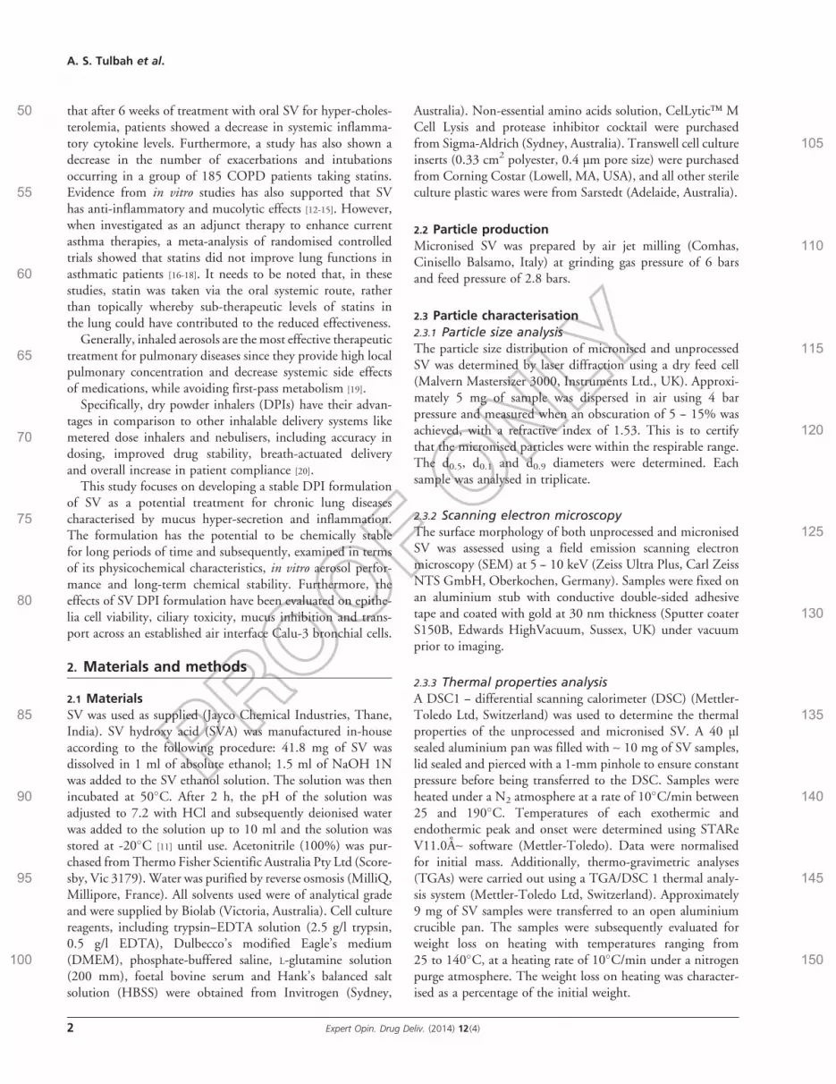
2.3 Particle characterisation2.3.1 Particle size analysis
115The particle size distribution of micronised and unprocessedSV was determined by laser diffraction using a dry feed cell(Malvern Mastersizer 3000, Instruments Ltd., UK). Approxi-mately 5 mg of sample was dispersed in air using 4 barpressure and measured when an obscuration of 5 -- 15% was
120achieved, with a refractive index of 1.53. This is to certifythat the micronised particles were within the respirable range.The d0.5, d0.1 and d0.9 diameters were determined. Eachsample was analysed in triplicate.
2.3.2 Scanning electron microscopy125The surface morphology of both unprocessed and micronised
SV was assessed using a field emission scanning electronmicroscopy (SEM) at 5 -- 10 keV (Zeiss Ultra Plus, Carl ZeissNTS GmbH, Oberkochen, Germany). Samples were fixed onan aluminium stub with conductive double-sided adhesive
130tape and coated with gold at 30 nm thickness (Sputter coaterS150B, Edwards HighVacuum, Sussex, UK) under vacuumprior to imaging.
2.3.3 Thermal properties analysisA DSC1 -- differential scanning calorimeter (DSC) (Mettler-
135Toledo Ltd, Switzerland) was used to determine the thermalproperties of the unprocessed and micronised SV. A 40 µlsealed aluminium pan was filled with ~ 10 mg of SV samples,lid sealed and pierced with a 1-mm pinhole to ensure constantpressure before being transferred to the DSC. Samples were
140heated under a N2 atmosphere at a rate of 10!C/min between25 and 190!C. Temperatures of each exothermic andendothermic peak and onset were determined using STAReV11.0A~ software (Mettler-Toledo). Data were normalisedfor initial mass. Additionally, thermo-gravimetric analyses
145(TGAs) were carried out using a TGA/DSC 1 thermal analy-sis system (Mettler-Toledo Ltd, Switzerland). Approximately9 mg of SV samples were transferred to an open aluminiumcrucible pan. The samples were subsequently evaluated forweight loss on heating with temperatures ranging from
15025 to 140!C, at a heating rate of 10!C/min under a nitrogenpurge atmosphere. The weight loss on heating was character-ised as a percentage of the initial weight.
A. S. Tulbah et al.
2 Expert Opin. Drug Deliv. (2014) 12(4)

2.3.4 Dynamic vapour sorptionDynamic vapour sorption (DVS) (Intrinsic 1) (Mettler-
155 Toledo Ltd, Switzerland) was used to analyse the water uptakeof both unprocessed and micronised SV at different relativehumidity (RH). Both the jet-micronised drug and unpro-cessed SV were dried for 24 h at 0% RH and then exposedfrom 0 to 90% RH for two cycles.
160 2.3.5 Stability study of SV DPIThe chemical stability of micronised and unprocessed SV wasassessed storing the powders in a temperature-controlledcabinet (25!C/60% RH, FR-285C, Thermoline) protectedfrom light up to 9 months. The RH was controlled using
165 2.5 g/ml of sodium bromide solution (Sigma Aldrich,NSW, Australia). Each experiment was tested in triplicatesby weighing 5 mg of sample and dissolving the powder into50 ml of acetonitrile: water (65:35 v/v), which was then ana-lysed on the same day for both SV and its metabolite SVA
170 using a validated HPLC method.
2.3.6 High-pressure liquid chromatographyQuantification of SV and its metabolite SVA was performedusing HPLC. A Shimadzu HPLC system consisting of aLC20AT pump, the SIL20AHT autosampler and an SPD-
175 20A UV--Vis detector (Shimadzu, Sydney, NSW, Australia)was used. Themobile phase comprised amixture of acetonitrile:water (65:35 v/v) and 0.025 M sodium dihydrogen phosphatewith pH adjusted to pH 4.5 with phosphoric acid. The mobilephase was then filtered under vacuum. Analysis of SV was
180 achieved using a reverse phase C-18 column (PhenomenexODS hypersclone) 250 " 4.6 mm, 5-µm particle size. TheHPLC system was set to the following conditions: UV detectorwavelength 238 nm, 100 µl injection volume and flow rate1.5 ml/min. Retention time for SV was 9.1 and for SVA was
185 5.5 min, respectively. Linearity was obtained between 0.01and 50 µg/ml (R2 = 0.99) for both SV and SVA.
2.3.7 In vitro aerosol dispersion characterisationThe in vitro aerosol performance was determined using amulti-stage liquid impinger (MSLI) (Apparatus A, European
190 Pharmacopoeia, Chapter 2.9.18; Copley Scientific, Notting-ham, UK) at days 0, 6 months and 9 months together withan Aerolizer" DPI (Norvartis Surrey, UK). The flow ratethrough the MSLI was set to 60 l/min and was controlledby aAQ3 GAST rotary vein pump and solenoid valve timer set
195 for 4 s (Westech Scientific Instruments, Bedfordshire, UK).20 ml of acetonitrile: water (65:35 v/v) was accurately addedto each collection stage of the MSLI. Micronised SV particles(5 mg) were pre-loaded into size 3 hard gelatin capsules (Cap-sugel, Sydney, Australia) and placed within the chamber of
200 the Aerolizer device. The Aerolizer was fitted into the mouth-piece adapter, which was connected to the MSLI using a USPharmacopoeia throat. Each sample was tested in triplicate.After actuation, the capsule, device, mouthpiece adapter,
throat, all stages and filter were washed separately using aceto-205nitrile: water (65:35 v/v) and analysed using HPLC.
2.4 In vitro bio-characterisation2.4.1 Cell cultureCalu-3 bronchial epithelial cells were purchased from theAmerican Type Culture Collection (Manassas, USA). The cells
210were cultured between passages 37--47 in pre-warmed DMEM---- F12 supplemented with 1% (v/v) L-glutamine solution, 1%(v/v) non-essential amino acid solution and 10% (v/v) foetalbovine serum. Cells were incubated at 37!C in 5% CO2 and95% humidity until confluency was reached. Every 2-- 3 days
215medium was exchanged and cells were passaged weekly accord-ing to ATCC-recommended guidelines [12,21,22].
2.4.2 Epithelial cell and cilia toxicity assayThe cell and cilia toxicity profiles of SV were assessed. Thein vitro pulmonary cytotoxicity experiments were investigated
220using CellTiter 96" Aqueous One Solution Cell proliferationAssay (Promega, USA) on Calu-3, as previously described[22,23], to understand the range of concentrations suitable foruse on pulmonary cells. Briefly, the cells were seeded at a den-sity of 5 " 105 cell/cm2 onto sterile 96-well microtiter plates
225and left overnight to allow for cell attachment. Cells were thentreated with increasing concentrations of SV in completeDMEM medium with a final volume of 200 µl. Stock solu-tion was prepared by dissolving 10 mg of SV in 500 µl ofdimethyl sulfoxide (DMSO) and was added to complete
230medium in a final DMSO concentration of £ 1%. Untreatedcontrols and vehicle controls were included in each experi-ment. Plates were then incubated at 95% humidity and37!C in 5% CO2 for 72 h. Subsequently, 20 µl of AqueousOne Solution was added to each well to analyses the cells
235viability and the plates incubated for 3 h at 37!C in 5%CO2 and 95% humidity. Using a fluorescence plate reader(SpectraMax M2; Molecular devices, USA) the wells weremeasured at 490 nm of absorbance and the concentrationthat produced a 50% decrease in cell viability after 72 h
240(IC50) was calculated following the 72-h treatment period.IC50 values were defined as the drug concentration that pro-duced a decrease of 50% in cell viability compared to theuntreated control. IC50 values were calculated by plotting(%) cell viability against the concentrations (ng/ml) on a log-
245arithmic scale. The cell viability (%) was calculated based onthis formula: (average absorbance of treated cells/averageabsorbance of control cells) " 100. Data were fitted to theHill equation using the General Fit function of GraphPadPrism 6 software (GraphPad Software, Inc. CA 92037
250USA). Each concentration was performed in triplicates.In addition, the acute toxicity of SV on ciliary activity was
investigated using primary nasal epithelial cells from healthyvolunteers (n = 5). Nasal epithelial cells were acquired throughnasal brushings [23,24]. Approximately 5 ml of Medium 199
255(Sigma, Australia) was used to suspend the nasal cells tomaintain cell viability. The cell suspensions were treated with
Dry powder formulation of simvastatin
Expert Opin. Drug Deliv. (2014) 12(4) 3

1 " 10-6 M of SV for 30 min. This concentration was basedon the highest solubility of SV into the culture medium. Thecilia beat frequency (CBF) of nasal cells was analysed under a
260 light microscope (Olympus IX70, Japan) connected to a pho-tomultiplier (Zeiss, West Germany). CBF was then measuredvia electronic signals generated from the photometer (Tektro-nix) that was delivered through an oscilloscope and afterwardfilmed by Mac-Lab (AD Instruments, Mountain View, CA,
265 USA) recording system. Three measurements of CBF weretaken from different regions of ciliated epithelial cells to obtainaccurate frequency values.
2.4.3 Transepithelial drug transport studiesCalu-3 cells were seeded at a density of 5 " 105 cell/cm2 on
270 Transwell polyester inserts (Corning Costar, USA) and
cultured at the air interface where medium was aspiratedfrom the apical chamber 24 h post-seeding. The transportexperiments were performed on the cells between days11 and 14 [21,22]. A modified glass twin stage impinger (TSI;
275Copley Scientific, UK) was used to deposit SV dry powderformulations onto the Calu-3 epithelial cells to simulate thein vivo deposition of aerosol particles as previously described[22,25]. Approximately 0.3 mg of micronised SV DPI wasweighed into size 3 hard gelatine capsules, which was deliv-
280ered from the Aerolizer into the TSI assembly at a flow rateof 60 l/min for 4 s to allow particles £ 6.4 µm aerodynamicdiameter to deposit onto the epithelial cells. Samples wereconsequently taken from the basolateral chamber of thetranswell every 30 min up to 4 h and were replaced with fresh
285transport buffer (Hanks Buffered Salt Solution, Sigma,Australia). After 4 h, SV and SVA remaining on apical surfaceof the cell monolayer and inside the cells were quantitativelyrecovered to obtain the total initially deposited dose.
Transepithelial electrical resistance (TEER) measurements290were performed using an epithelial voltohmmeter (World
Precision Instruments, USA) as previously described to assessthe integrity of the cell monolayer [22,25]. These measurementswere subsequently compared with untreated cells.
2.4.4 Mucus inhibition study295To assess the ability of SV DPI to reduce mucus production, a
single dose of 0.3 mg powder was deposited on Calu-3 at day11 using the modified TSI and mucus analysis was performedon day 14. Mucus glycoproteins were stained with alcian blueas previously described [12,21] to allow the visualisation of
300mucus on the surface of Calu-3 cells after SV depositionand compared with untreated cells (control). First, sampleswere washed with PBS and fixed with 4% paraformaldehyde(PFA) for 20 min. Then, PFA was removed and cells rinsedtwice with PBS and then 100 µl of alcian blue was added to
305the apical chamber and left for 15 min for mucus staining.The inserts were washed with PBS to remove extra stainsand left to dry. Finally, cells were mounted onto glass slideswith mounting medium and sealed. Samples were stored inthe fridge at 4!C prior to analysis. Images of each sample
310were taken using Olympus BX60 microscope (Olympus,Tokyo, Japan) equipped with an Olympus DP71 camera(Wetzlar, Germany). Apple Automator (v 2.0.4 Apple Inc.,Cupertino, California, USA) was used to obtain TIFF imagesfor analysis using ImageJ (v1.42q, NIH) with Colour Profile
315(Dimiter Prodanov; Leiden University Medical Center,Leiden, Netherlands). The ratio of red, green, blue (RGBratio) was calculated by dividing the mean RGB by thesum of the RGB values for each image (RGBR + RGBG +RGBB). The mean RGB was used to quantify mucus produc-
320tion, in both the control and the SV-treated monolayers.Concurrently, TEER measurements, flu-Na permeability
and viability of Calu-3 cells were studied to investigatethe effects of single SV dosing onto the cells and subsequentexposure over the 4 days period. In addition to TEER
Table 1. Micronised and unprocessed simvastatin
material particle size distribution (n = 3 ± S.D.).
Micronised SV Unprocessed SV
d 0.1 (µm) 1.18 ± 0.07 6.98 ± 0.14d 0.5 (µm) 2.2 ± 0.03 17.13 ± 0.21d 0.9 (µm) 4.03 ± 0.2 41.43 ± 1.01
S.D.: Standard deviation; SV: Simvastatin.
A
B
WD = 5.3 mm2 µm
2 µm
EHT = 10.00 kV Mag = 2.00 K × Signal A = SE2
WD = 5.1 mm EHT = 5.00 kV Mag = 3.00 K × Signal A = SE2
COL
OR ONLIN
E•B&W
INPRIN
T•
Figure 1. Scanning electron micrographs of: (A) micronisedSV and (B) unprocessed SV powder.SV: Simvastatin.
A. S. Tulbah et al.
4 Expert Opin. Drug Deliv. (2014) 12(4)

325 measurements, flu-Na (molecular mass, 0.367 kDa) perme-ability was measured to confirm the barrier integrity ofthe Calu-3 epithelial cell after the mucus experiments, aspreviously described [21,25]. Flu-Na was first dissolved inpre-warmed HBSS to a concentration of 2.5 mg/ml. Then,
330 200 µl of this solution was added to the apical surface of thecells to initiate the experiment and 600 µl of HBSS was addedin the basolateral chamber. After that, 100 µl of sample wasdrawn from the chamber every 15 min over a 1-h period whilethe same volume of HBSS was added to replace the sample
335 that was withdrawn. Samples were placed in a black, 96-wellplate, and fluorescence readings were taken using a POLAR-star Optima fluorescence plate reader (BMG Labtech, Offen-burg, Germany) with excitation and emission wavelengthsettings of 485 and 520 nm, respectively.
340 To confirm cell viability, cell counting was performed byharvesting the cells from the transwell insert at day 14. An ali-quot of cells isolated was examined for viability as determinedby trypan blue dye exclusion (0.5% trypan blue). The number
of viable cells on the day 14 was determined by counting the345number of cells that successfully exclude the dye using light
microscopy at 30" magnification.
2.4.5 Statistical analysisAll results are expressed as mean ± standard deviation (S.D.)of at least three separate determinants. To determine signifi-
350cance between groups and control, unpaired two-tailed t-testsand one-way ANOVA were performed (quoted at the level ofp < 0.05).
3. Results and discussion
3.1 Micronised material characterisation
3553.1.1 Particle size distributionGeometric particle size distributions of micronised andunprocessed SV determined by laser diffraction are shown inTable 1. The result suggests micronised SV was suitable forinhalation applications.
3603.1.2 SV powder morphologyThe morphology of the unprocessed and micronised SVparticles by SEM is shown in Figure 1. Both unprocessedand micronised SV had crystalline structures with rectangularshape. As can be seen from Figure 1A, the air jet-milled SV
365particles had an average particle size of ~ 2 µm while theunprocessed SV material exhibited a larger particle size of~ 20 µm Figure 1B. This finding is in good agreement withthe particle size distribution data (Table 1).
3.1.3 Thermal properties analysis370The thermal behaviour of both the micronised and unpro-
cessed SV powders was characterised using DSC and TGA.The DSC thermograph of both the micronised particles andunprocessed material is shown in Figure 2. An endothermicpeak that correlated to the melting point of the micronised
375SV and unprocessed SV was found to be at 140.63!C forboth materials. This is in good correlation with a previousstudy [26] and is characteristic of a crystalline behaviour.Hence, micronised SV powder did not have an effect on thephysical characteristics of the particle, apart from its size. To
380further understand the physicochemical characteristics of themicronised SV powder influence of weight loss versuscontrolled temperature rise (TGA) was also investigated forboth micronised and unprocessed SV and resulting data arepresented in Figure 3. The rapid mass loss observed by TGA
385for micronised and unprocessed SV at ~ 140!C is attributedto the decomposition of the sample, confirming the resultsobserved with the DSC.
3.1.4 Dynamic vapour sorptionTo advance our understanding of the relative stability of SV
390formulations’ solid state, the effect of humidity on moisturesorption was analysed by DVS. The moisture sorptionprofiles (first and second sorption and desorption cycles) for
1.0
1.5
0.5
0
Temperature (°C)
% w
eig
ht
loss
20 4030 50 60 70 80 90 100 110 120 140130 150
Micronized SV
Unprocessed SV
Figure 3. Thermo-gravimetric analyses thermograms ofmicronised and unprocessed simvastatin powder from25 to 140!C.SV: Simvastatin.
Milled SVUnprocessed SV
-10
-20
-30
Temperature (°C)
Heat
flo
w (
mW
)
20 40 60 80 100 120 140 160 180
Figure 2. Differential scanning calorimeter thermograms ofmicronised and unprocessed SV, from 25 to 170!C.SV: Simvastatin.
Dry powder formulation of simvastatin
Expert Opin. Drug Deliv. (2014) 12(4) 5

micronised and unprocessed SV formulations are shown inFigure 4. The presence of hysteresis and the reversibility of
395 the moisture sorption profiles for both formulations suggestthe materials to be crystalline, with no detectable amorphousmaterial present [27], between 0 and 90% RH. Further analysisof the SV formulations isotherm suggested data follow a sig-moidal type (S) curve, suggesting multilayer water sorption
400 onto the crystal surface of the sample [28]. There are four clas-ses of isotherms according to their initial slopes: i) S curves;ii) L curves (Langmuir type); iii) H curves (high affinity);and iv) C curves (constant partition) and the sub-groups ofthese classes are arranged according to their shape [28]. The
405moisture sorption of water onto the micronised sample wasobserved to be slightly greater than the unprocessed SV.Explanations for such variation in the stable crystalline mate-rial could be attributed to difference in the larger surface areaof micronised SV, which in turn affects the surface chemistry
410of the materials. In general, both samples had similar isothermsorption profiles.
3.1.5 Stability studySV is susceptible to hydrolysis, causing the opening of thelactone ring to form SVA metabolite, which could be a
415significant problem associated with formulating SV for
Ch
an
ge in
mass (
%)
RH (%)
Cycle 1 SorpCycle 1 Desorp
Cycle 2 SorpCycle 2 Desorp
0 10 20 30 40 50 60 70 80 90
0.20
0.15
0.10
0.05
-0.05
0
Micronized SVUnprocessed SV
Figure 4. Dynamic vapour sorption isotherms of micronised and unprocessed simvastatin, two cycles from 0 to 90 RH %.RH: Relative humidity; SV: Simvastatin.
% o
f w
eig
hed
do
se o
f S
V
Days
0 50 100 150 200 250 300
80
100
120
140
60
40
20
0
Micronized SV
Unprocessed SV
COL
OR ONLIN
E•B&W
INPRIN
T•
Figure 5. The stability of micronised and unprocessed simvastatin up to 9 months of the storage at 25!C and 60% relativehumidity (n = 3 ± S.D.). Shaded region indicates the ±25% BP pharmacopoeia limit.S.D.: Standard deviation; SV: Simvastatin.
A. S. Tulbah et al.
6 Expert Opin. Drug Deliv. (2014) 12(4)

inhalation [29]. However, although SVA is the active form ofthe drug, hydrolysis prior to absorption will render the activemoiety incapable of being transported into the cell, and thuslimiting its pharmacological actions [12]. Therefore, it is vital
420to ensure that the SV DPI formulation is chemically stable.The chemical stability of micronised SV up to 9 months ofthe storage period at 25!C and 60 RH % [30] is shownin Figure 5. SV DPI showed no chemical degradation to itsactive metabolite SVA up to 9 months, with all values quan-
425tified within pharmacopoeia specification of ±25% (BritishPharmacopoeia 2010) of the nominal dose. Results showedthat the powder had no significant degradation, with an aver-age dose of 5336 ± 411 µg, (target dose = 5000 µg) during thiswhole period. The presence of SVA was also chemically
430analysed and found not to change over the 9 months period(average value 4.48 ± 0.60 µg that is equivalent to 0.55 ±0.98% of the nominal dose).
3.1.6 In vitro aerodynamic assessmentThe in vitro aerosolisation efficiency using an MSLI at 60 l/
435min is showed in Figure 6. Mass deposition of the SV DPIformulation as the percentage of the total drug depositedremaining in the device/capsule, induction port, and eachstage of the MSLI were measured. The fine particle fraction(FPF) was also calculated and presented as the cumulative per-
440centage of drug deposited from stage 3 to the filter, represent-ing particles with aerodynamic diameter of < 6.8 µm. Figure 6shows the percentage drug distribution of micronised SVpowder on each stage of the MSLI at day 0, 6 and 9 months.
At day 0 the total dose delivered per capsules was4454790.83 ± 115.36 µg (target dose = 5000 µg) with an FPF
of 44.62 ± 5.77%. Similarly, at 6 and 9 months, the totaldose of micronised SV delivered per capsules was 4268 ±188 µg and 5118 ± 374 µg while the FPF was 39.60 ±3.79 and 41.17 ± 1.44%, respectively (n = 3, ± S.D.). There
450was no significant difference (p = 0.42) in FPF between day0, 6 and 9 months, confirming that the micronised SV waschemically stable up to 9 months and suitable for deeplung delivery.
3.2 In vitro bio-characterisation
4553.2.1 Epithelia cell and cilia toxicity assayThe toxicity of the SV on bronchial cells was undertaken onCalu-3 cells to understand the range of concentrations suitablefor pulmonary drug delivery, while its effect on ciliary activitywas investigated on ciliated nasal mucosa. Calu-3 cell mono-
460layer was treated for 72 h with increasing drug concentrations,ranging from 0.1 to 250 µm, in order to define the IC50. SVhad an IC50 value of 72.59 µm ± 0.22 as demonstratedin Figure 7A. This was in good agreement with previousstudy [12].
465In addition, the acute toxicity of SV drugs on ciliaryactivity was assessed using primary nasal epithelial cells withfunctioning cilia. These cells were utilised because of theease of sampling collection; furthermore it has been shown
% S
V d
ep
osit
ed
Capsu
le
Device
Adapt
er
Throa
t
Stage
1
Stage
2
Stage
3
Stage
4
Filtter
0
5
10
15
20
25
30Day 06 month9 month
COL
OR ONLIN
E•B&W
INPRIN
T•
Figure 6. In vitro drug deposition of the micronisedsimvastatin by multi-stage liquid impinger using theAerolizer" DPI device at a 60 l/min, (n = 3 ± S.D.).DPI: Inhaled dry powder; S.D.: Standard deviation; SV: Simvastatin.
150A.
B.
50
100
% v
iab
ilit
y
CB
F (
Hz)
Log SV conc (nM)
10
8
6
4
2
0Control SV
0-2-4 2 4
COL
OR ONLIN
E•B&W
INPRIN
T•
Figure 7. Toxicity of simvastatin on: (A) Calu-3 epithelial cellviability measured by CellTiter 96" Aqueous assay followingexposure to increasing concentrations of SV(n = 3 ± S.D.);and (B) CBF of primary nasal epithelial cells after exposure to1 " 10-6 m of SV compared to baseline (control) (n = 5 ± S.D.),CBF: Cilia beat frequency; S.D.: Standard deviation; SV: Simvastatin.
Dry powder formulation of simvastatin
Expert Opin. Drug Deliv. (2014) 12(4) 7

in previous studies [24,31] that nasal epithelial cells can be used470as a substitute or surrogate to bronchial epithelial cells, as
it displayed identical morphologies to lung epitheliumcells, with similar expression of receptors and responses tocytokine stimulation. Analyses of the nasal CBF in responseto 1 " 10-6 M concentration of SV after 30 min are shown
475in Figure 7B. SV was shown not to have any toxicity onCBF (SV: 7.48 ± 1.4 Hz), compared to baseline (control:7.03 ± 1.3 Hz). These data suggest that SV is safe and non-toxic supporting its potential delivery to the lungs as a treat-ment for chronic hyper-secretory mucus diseases.
4803.2.2 Transport studiesOne of the advantages of using the air interface Calu-3 cellmodel coupled with the TSI is the ability to deposit respirableSV particles onto the mucosa surfaces of lung epithelial cells,mimicking bolus deposition of DPI after inhalation [32].
485Transport studies were performed to assess SV ability topenetrate into airway epithelial cells and provide an estimateconversion to the SVA metabolite, where drug amounts are
A.
B.
% o
f S
V D
PI d
ep
osit
ed
10
12
14
8
6
4
2
0
% o
f S
V D
PI d
ep
osit
ed
50
40
30
20
10
0
SV SVA Total
TotalSVSVA
In
OnTransported
0 20 40 60 80 100 120 140 160 180 200 220 240 260
Time (min)COL
OR ONLIN
E•B&W
INPRIN
T•
Figure 8. (A) Transport of simvastatin, simvastatin hydroxy acid and total drug amount (SV + SVA) as a function of time.(B) Cumulative amounts of SV, SVA and total (SV + SVA) recovered in the cells, remaining on the cells and transported acrossthe Calu-3 cells, 4 h after the deposition of SV DPI, (n = 3 ± S.D.).DPI: Inhaled dry powder; S.D.: Standard deviation; SV: Simvastatin; SVA: Simvastatin hydroxy acid.
% T
EE
R
500
600
400
300
200
100
0SVControl
COL
OR ONLIN
E•B&W
INPRIN
T•
Figure 9. Transepithelial electrical resistance over 4 hfollowing deposition of 0.3 mg of SV DPI (n = 3 ± S.D.).DPI: Dry powder inhaler; S.D.: Standard deviation; SV: Simvastatin; TEER:
Transepithelial electrical resistance.
A. S. Tulbah et al.
8 Expert Opin. Drug Deliv. (2014) 12(4)

expressed as the mean cumulative percentage (±S.D.) of thetotal drug recovered. The total drug deposited (SV + SVA)
490 onto the Calu-3 epithelial cell layer was found to be 0.78 ±0.19 µg. Figure 8A displays the cumulative amounts of SVand SVA that were transported across, recovered on andinside the Calu-3 monolayer after micronised SV deposition.On the other hand, Figure 8B shows the transport of SV and
495 SVA across the monolayer as a function of time.Over the 4-h transport experiment, ~ 9.02 ± 1.2% of the
SV and 3.35 ± 0.44% of SVA were transported across the epi-thelial cells. These results demonstrate the ability of SV toenter into and cross the epithelium layer. Interestingly, a large
500 proportion of drug was recovered within the cells and wasfound to be 5.73 ± 1.32% of SV and 40.01 ± 6.15% ofSVA, respectively. The remainder of the drugs was found onthe cells with ~ 19.79 ± 4.89% of SV converted into SVA.This is particularly important as it shows the capability of
505 lung epithelia cells to activate SV into SVA, presumably viacarboxylesterases and cytochrome P450, which is essential ifits muco-inhibiting function is to be realised [12]. Remarkably,conversion of SV to SVA may allow retention of the metabo-lite within the cells, which could be useful in maintaining a
510 high local drug concentration for an extended period oftime. Hence, upon deposition of SV DPI onto the epithe-lium, the dissolution of drug into the surrounding epithelialining fluid creates a high local concentration that drives the
diffusion of SV into the epithelial cells for conversion into515the active SVA counterparts by the enzymes. The hydrophilic
nature of SVA may have contributed to the retention of SVAwithin the cells while the lipophilic SV enables its continuousdiffusion across the cells.
TEER measurements were performed to evaluate the integ-520rity of the Calu-3 monolayer after 4 h of transport studies
(Figure 9). No significant change in resistance was observedcompared with the control (SV-TEER 522 ± 75 Wcm2 andcontrol-TEER 473 ± 72 Wcm2). Therefore, high local con-centrations of SV had no effect on the epithelial cell integrity
525under the conditions and time scale studied.
3.2.3 Mucus inhibition studiesPrevious studies have shown that SV may attenuate acrolein-induced mucin protein synthesis in the airway and airwayinflammation, possibly by blocking AQ4ERK activation mediated
530by Ras protein isoprenylation [31-34]. Furthermore, Marinet al. found that chronic dosing of 1 or 10 µm solution of SVfrom the basolateral chamber (which mimics the deliverythrough the systemic circulation) caused a significant inhibi-tion in mucus production on established air interface Calu-3
535epithelial cell model. This has indicated that SV could beused for treatment of overproduction of airway mucus [12,33-35].
Hence, the ability of SV DPI to inhibit mucus productionvia inhalation needs to be verified. Considering that a
RG
B r
ati
o0
0.1
0.2
0.3
0.4
0.5
SV DPI
*
Untreated control
100 µm 100 µm
A.
B.
(i) (ii)
COL
OR ONLIN
E•B&W
INPRIN
T•
Figure 10. (A) RGB ratio of alcian blue intensity at day 14 of untreated control and after the deposition of DPI SV onto airinterface Calu-3 cells, (n = 3 ± S.D.). (B) Microscopic images of stained monolayers at day 14 of: (i) untreated cells and (ii) afterdeposition of DPI SV.*Significantly different to control (p < 0.05).
DPI: Dry powder inhaler; RGB: Red, green, blue; S.D.: Standard deviation; SV: Simvastatin.
Dry powder formulation of simvastatin
Expert Opin. Drug Deliv. (2014) 12(4) 9

significant amount of SVA was retained within the epithelia540 cells, its ability to reduce mucus production was evaluated
after a single dose of micronised SV deposition onto theepithelia cells. The mucus was analysed at day 14 after SVdeposition and was compared with untreated control by cal-culating the RGB ratio values. This was based on microscopic
545 images taken of the stained mucus for each treatment condi-tion and are shown in Figure 10. Results showed a significantinhibition of mucus production when cells were treated with
SV DPI, compared to the untreated controls at day 14, withan RGB ratio of 0.41 ± 0.02 for the treated cells compared
550with 0.46 ± 0.02 for the untreated cells at day 14 (p > 0.05).In order to confirm cell monolayer integrity during the
course of the mucus inhibition experiments, TEER measure-ments and flu-Na permeability were performed on theCalu-3 cell monolayers treated with SV formulation and was
555compared to untreated cells. However, a significant drop inTEER and loss of monolayer integrity occurred when the cellswere treated with SV after 14 days (Figure 11A) and was con-firmed by the increase in flu-Na permeability (Figure 11B).Hence, cell viability by trypan blue dye exclusion was also per-
560formed in parallel and showed no change in cell viability(Figure 11C; p > 0.05). This indicated that SV could have aneffect on the tight junctions of the epithelium through itsinvolvement in membrane cholesterol synthesis other thancell death. Similar results were found in a study by Lambert
565et al., which showed that cholesterol is the main factor inmaintaining barrier property of epithelial monolayers andappears to stabilise the association of certain proteins withthe tight junctions [36]. They found that the depletion of cho-lesterol by 40 -- 45% using methyl-b-cyclodextrine was
570accompanied by a 80 -- 90% decrease in TEER measurementsand increase in paracellular permeability with intestinal Caco-2 cell line after 4 h. Although our study demonstrated similarfindings in terms of the decrease in membrane integrity afterdepletion of cholesterol, it needs to be noted that the effects
575of SV on the pulmonary tight junctions in this study wereto a lower extent (decreased in TEER by ~ 50%) after a longer4 days SV exposure, where the presence of a protective mucusbarrier on the air interface pulmonary cells could have limitedits action on tight junctions. However, this may actually be
580beneficial in COPD patients, as it was found that there wasan overproduction of 25-hydroxy-cholesterol (25-HC) inthis population of patients. 25-HC is produced from choles-terol and has been associated with neutrophilic inflammationin the airways [37]. This led to a decrease in lung function and
585concomitant elevated levels of IL-8 and neutrophil counts insputum of COPD patients. Therefore, a reduction in choles-terol synthesis [38,39] by SV could potentially be beneficial inthe management of chronic pulmonary inflammatory diseasesas an alternative anti-inflammatory agent. Further investiga-
590tions are needed to confirm this hypothesis.
4. Conclusion
Micronised SV DPI formulation was successfully manufac-tured and characterised in vitro. This formulation was stableup to 9 months at 25!C/60% RH and was proven to be
595able to penetrate across the pulmonary epithelial cells, showedno toxic effect on Calu-3 and on ciliary activity in vitro andmore importantly, demonstrated the ability to significantlydecrease mucus production. Therefore, this formulation hasthe potential to provide a promising dry powder therapy for
600the treatment of hyper-secretory pulmonary diseases. Future
TE
ER
(W
cm
2)
Ap
pare
nt
perm
eab
ilit
y(c
m.s
–1)
Nu
mb
er
of
via
ble
cells (
10
4)
10–4
10–5
10–6
10
8
6
4
2
0
600
500
400
300
200
100
0SV DPIUntreated control
SV DPIUntreated control
SV DPIUntreated control
A.
B.
C.
COL
OR ONLIN
E•B&W
INPRIN
T•
Figure 11. The effects on: (A) TEER, (B) apparent perme-ability of flu-Na across the Calu-3 epithelial cell layer, and (C)cell viability as measured by trypan blue dye exclusionfollowing the deposition of SV DPI for mucus study at day14, (n = 3 ± S.D.).DPI: Dry powder inhaler; S.D.: Standard deviation; SV: Simvastatin; TEER:
Transepithelial electrical resistance.
A. S. Tulbah et al.
10 Expert Opin. Drug Deliv. (2014) 12(4)

studies will assess the mechanisms of SV transport and path-ways of its anti-inflammatory mechanisms.
Declaration of interest
PM Young is the recipient of an AustralianAQ5 Research605 Council Future Fellowship (project number FT110100996).
A/Professor Traini is the recipient of an Australian Research
Council Future Fellowship (project number FT12010063).Alaa Tulban is also grateful to Umm Al-Qura University forthe scholarship and Saudi Government for their financial sup-
610port. The authors have no other relevant affiliations or finan-cial involvement with any organisation or entity with afinancial interest in or financial conflict with the subject matteror materials discussed in the manuscript apart from thosedisclosed.
BibliographyPapers of special note have been highlighted as
either of interest (#) or of considerable interest
(##) to readers.
1. Murray TS, Egan M, Kazmierczak BI.
Pseudomonas aeruginosa chronic
colonization in cystic fibrosis patients.
Curr Opin Pediatr 2007;19(1):83-8
2. Barker AF. Bronchiectasis. N Engl J Med
2002;346(18):1383-93
3. Yang Y, Tsifansky MD, Shin S, et al.
Mannitol-guided delivery of
Ciprofloxacin in artificial cystic fibrosis
mucus model. Biotechnol Bioeng
2011;108(6):1441-9
4. Tobert JA. Lovastatin and beyond: the
history of the HMG-CoA reductase
inhibitors. Nat Rev Drug Discov
2003;2(7):517-26
5. Endo A, Tsujita Y, Kuroda M,
Tanzawa K. Inhibition of cholesterol
synthesis in vitro and in vivo by ML-
236A and ML-236B, competitive
inhibitors of 3-hydroxy-3-methylglutaryl-
coenzyme A reductase. Eur J Biochem
1977;77(1):31-6
6. Vaughan CJ, Murphy MB, Buckley BM.
Statins do more than just lower
cholesterol. Lancet
1996;348(9034):1079-82
7. McAuley DF, O’Kane CM, Craig TR,
et al. Simvastatin decreases the level of
heparin-binding protein in patients with
acute lung injury. BMC Pulmonary Med
2013;13:47
8. Grommes J, Vijayan S, Drechsler M,
et al. Simvastatin reduces endotoxin-
induced acute lung injury by decreasing
neutrophil recruitment and radical
formation. PLoS One 2012;7(6):e38917
9. Hothersall E, McSharry C,
Thomson NC. Potential therapeutic role
for statins in respiratory disease. Thorax
2006;61(8):729-34
10. Rezaie-Majd A, Maca T, Bucek RA,
et al. Simvastatin reduces expression of
cytokines interleukin-6, interleukin-8,
and monocyte chemoattractant protein-1
in circulating monocytes from
hypercholesterolemic patients.
Arterioscler Thromb Vasc Biol
2002;22(7):1194-9.. One of the first few studies to identify
the potential of simvastatin as an
anti-inflammatory agent.
11. Blamoun A, Batty G, DeBari V, et al.
Statins may reduce episodes of
exacerbation and the requirement for
intubation in patients with COPD:
evidence from a retrospective cohort
study. Int J Clin Pract
2008;62(9):1373-8
12. Marin L, Traini D, Bebawy M, et al.
Multiple dosing of simvastatin inhibits
airway mucus production of epithelial
cells: implications in the treatment of
chronic obstructive airway pathologies.
Eur J Pharm Biopharm
2013;84(3):566-72.. One of the first few studies that have
used in vitro Calu-3 model to
investigate the effects of therapeutics
on mucus production.
13. Weitz-Schmidt G. Statins as anti-
inflammatory agents.
Trends Pharmacol Sci 2002;23(10):482-7
14. Marin L, Colombo P, Bebawy M, et al.
Chronic obstructive pulmonary disease:
patho-physiology, current methods of
treatment and the potential for
simvastatin in disease management.
Expert Opin Drug Deliv
2011;8(9):1205-20
15. Shyamsundar M, McKeown ST,
O’Kane CM, et al. Simvastatin decreases
lipopolysaccharide-induced pulmonary
inflammation in healthy volunteers. Am J
Respir Crit Care Med
2009;179(12):1107-14
16. Si X-B, Zhang S, Huo L-Y, et al. Statin
therapy does not improve lung function
in asthma: a meta-analysis of randomized
controlled trials. J Int Med Res
2013;41(2):276-83
17. Cowan DC, Cowan JO, Palmay R, et al.
Simvastatin in the treatment of asthma:
lack of steroid-sparing effect. Thorax
2010;65(10):891-6
18. Menzies D, Nair A, Meldrum KT, et al.
Simvastatin does not exhibit therapeutic
anti-inflammatory effects in asthma.
J Allergy Clin Immunol
2007;119(2):328-35
19. Pilcer G, Amighi K. Formulation strategy
and use of excipients in pulmonary drug
delivery. Int J Pharm 2010;392(1):1-19
20. Crompton GK. Dry powder inhalers:
advantages and limitations.
J Aerosol Med 1991;4(3):151-6
21. Haghi M, Young PM, Traini D, et al.
Time- and passage-dependent
characteristics of a Calu-3 respiratory
epithelial cell model. Drug Dev
Ind Pharm 2010;36(10):1207-14. Development and standardisation of
an immortalised bronchiol epithelia
cell model for the study of pulmonary
drug delivery and tranport.
22. Ong HX, Traini D, Bebawy M,
Young PM. Epithelial profiling of
antibiotic controlled release respiratory
formulations. Pharm Res
2011;28(9):2327-38
23. Ong HX, Traini D, Ballerin G, et al.
Combined inhaled salbutamol and
mannitol therapy for mucus hyper-
secretion in pulmonary diseases. AAPS J
2014;16(2):269-80
24. Rutland J, Cole PJ. Nasal mucociliary
clearance and ciliary beat frequency in
cystic fibrosis compared with sinusitis
and bronchiectasis. Thorax
1981;36(9):654-8
25. Ong HX, Traini D, Salama R, et al. The
effects of mannitol on the transport of
ciprofloxacin across respiratory epithelia.
Mol Pharm 2013;10(8):2915-24
Dry powder formulation of simvastatin
Expert Opin. Drug Deliv. (2014) 12(4) 11

26. Murtaza G. Solubility enhancement of
simvastatin: a review. Acta Pol Pharm
2012;69(4):581-90
27. Young PM, Price R, Tobyn MJ, et al.
Effect of humidity on aerosolization of
micronized drugs. Drug Dev Ind Pharm
2003;29(9):959-66
28. Giles CH, MacEwan T, Nakhwa S,
Smith D. 786. Studies in adsorption.
Part XI. A system of classification of
solution adsorption isotherms, and its use
in diagnosis of adsorption mechanisms
and in measurement of specific surface
areas of solids. J Chem Soc
1960;3973-93. An important paper to understand the
classification of solution adsorption
isotherms.
29. Alvarez-Lueje A, Valenzuela C,
Squella JA, Nunez-Vergara LJ. Stability
study of simvastatin under hydrolytic
conditions assessed by liquid
chromatography. J AOAC Int
2005;88(6):1631-6
30. Communities OfOPotE, Jowitt R,
Wagstaffe PJ, Commission of the
European Communities.
Directorate-General for Science R,
Development, Reference CotECCBo:
Certification of the Water Content of
Microcrystalline Cellulose (Mcc) at
10 Water Activities: Office for Official
Publications of the European
Communities. 1989
31. Andersen I, Camner P, Jensen PL, et al.
A comparison of nasal and
tracheobronchial clearance.
Arch Environ Health 1974;29(5):290-3.. Study investigating the correlation of
nasal cilia function to
mucociliary clearance.
32. Ong HX, Traini D, Young PM.
Pharmaceutical applications of the
Calu-3 lung epithelia cell line.
Expert Opin Drug Deliv
2013;10(9):1287-302. A comprehensive review about
promising human cell lines for study
of drug transport.
33. Chen YJ, Chen P, Wang HX, et al.
Simvastatin attenuates acrolein-induced
mucin production in rats: involvement of
the Ras/extracellular signal-regulated
kinase pathway. Int Immunopharmacol
2010;10(6):685-93
34. Lora JM, Zhang DM, Liao SM, et al.
Tumor necrosis factor-alpha triggers
mucus production in airway epithelium
through an IkappaB kinase beta-
dependent mechanism. J Biol Chem
2005;280(43):36510-17
35. Li D, Gallup M, Fan N, et al. Cloning
of the amino-terminal and 5¢-Flanking
region of the humanMUC5AC mucin
gene and transcriptional up-regulation by
bacterial exoproducts. J Biol Chem
1998;273(12):6812-20
36. Lambert D, O’Neill CA, Padfield PJ.
Depletion of Caco-2 cell cholesterol
disrupts barrier function by altering the
detergent solubility and distribution of
specific tight-junction proteins.
Biochem J 2005;387(Pt 2):553-60.. This paper provides some evidence
that depletion of Caco-2 cell
cholesterol has an effect on
tight-junction proteins.
37. Sugiura H, Koarai A, Ichikawa T, et al.
Increased 25-hydroxycholesterol
concentrations in the lungs of patients
with chronic obstructive pulmonary
disease. Respirology 2012;17(3):533-40
38. McDonald JG, Russell DW. Editorial:
25-Hydroxycholesterol: a new life in
immunology. J Leukoc Biol
2010;88(6):1071-2
39. Yeganeh B, Emilia W, Sudharsana RA,
et al. Targeting the mevalonate cascade as
a new therapeutic approach in heart
disease, cancer and pulmonary disease.
Pharmacol Ther 2014;143(1):87-110
AffiliationAlaa S Tulbah1,2, Hui Xin Ong1,
Lucy Morgan3,4, Paolo Colombo5,
Paul M Young1 & Daniela Traini†1
†Author for correspondence1Sydney University, Woolcock Institute of
Medical Research and Discipline of
Pharmacology, Sydney Medical School,
AQ2Respiratory Technology, Sydney, NSW, 2037,
Australia
E-mail: [email protected] Al Qura University, Faculty of Pharmacy,
Makkah, Saudi Arabia3University of Sydney, Sydney Medical
School-Concord Clinical School, Sydney, NSW,
Australia4Concord Repatriation General Hospital, Sydney,
Australia5University of Parma, Department of Pharmacy,
Parma, Italy
A. S. Tulbah et al.
12 Expert Opin. Drug Deliv. (2014) 12(4)


