Embed Size (px)
Citation preview

The Open Cancer Immunology Journal, 2009, 2, 1-13 1
1876-4010/09 2009 Bentham Open
Ear Pinna: A Privileged DNA Electroporation Site for Inducing Strong Th1 Immune Responses
Jing Ni1, Britta Nolte
1, Vandermeulen Gaëlle
2, Veronique Preat
2, Daniel Scherman
3, Volker
Schirrmacher1 and Philippe Fournier
1*
1 Division of Cellular Immunology, German Cancer Research Center, Heidelberg, Germany
2 Université catholique de Louvain, Department of Pharmaceutics, UCL73.20, 1200 Brussels, Belgium
3 Inserm, U640, Paris, F-75006 France; CNRS, UMR8151, Paris, F-75006 France; Université Paris Descartes, Faculté
de Pharmacie, Chemical and Genetic Pharmacology Laboratory, Paris, F-75270 France; Ecole Nationale Supérieure
de Chimie de Paris, Paris, F-75005 France
Abstract: DNA vaccination appears a very attractive approach for inducing immune responses towards the encoded anti-
gen, but studies in large animals and in humans revealed weaknesses of such responses. In this study, we evaluated a new
approach based on a new device combining DNA vaccination with electroporation (EP) at the ear pinna site. Under opti-
mal EP conditions, the expression of the DNA encoded antigen and the induced immune responses were considerably in-
creased. Very interestingly, DNA vaccination using EP at the ear pinna induced much stronger cellular immune responses
than at the flank skin although antigen expression was similar at both sites. As compared to vaccination at the ear pinna
without EP, IFN- but not IL-4 production by splenocytes from immunized mice was significantly enhanced. In contrast,
IL-4 but not IFN- production was increased by EP at the flank skin. The vaccination site of the ear pinna combined with
EP route even provided therapeutic effects in a mouse tumor model.
In conclusion, this study highlights the ear pinna as a privileged site for the induction of strong Th1 polarized cellular im-
munity against a defined antigen when combining DNA vaccination with EP.
Keywords: Antigen expression, immunization, electroporation, vaccination, tumor therapy.
INTRODUCTION
Genetic immunization using naked DNA [1, 2] offers considerable advantages over conventional vaccines:
(i) high stability of plasmid DNA and relative tem-perature insensitivity making them highly suitable for mass production and easy transportation in both in-dustrialized and developing countries,
(ii) low manufacturing costs,
(iii) lack of infection risks that are associated with attenuated viral vaccines,
(iv) capacity to target multiple antigens (Ags) on one plasmid by inserting several open reading frames from one or more genes and
(v) absence of intrinsic immunogenicity allowing successful boost after DNA vaccination without pro-ducing a heterologous immune response.
It has been shown to be effective and safe for inducing protective immunity in preclinical models of infectious dis-eases [3-6], cancer [7-10] and autoimmune diseases [11-13].
After over a decade of active research, DNA vaccines are now reaching the commercial market with recent approvals of West Nil virus in horses [14], infectious hematopoetic
*Address Correspondence to this author at the German Cancer Research Center (DKFZ), Im Neuenheimer Feld 280, 69120 Heidelberg, Germany; Fax: +49 6221 424962; E-mail: [email protected]
necrosis virus in salmon [15] and melanoma in dogs [16-18]. However, despite more than 200 clinical trials to date [19],
no plasmid DNA products have received approval by FDA
for use in humans. There have been, however, numerous preclinical and clinical studies for most types of cancer [20].
One important problem for all DNA vaccines relates to
the translation of therapy data from small animals to large animals [21] and to humans [22-26]. The efficacy in humans
has been disappointing [24, 26, 27], partly due to the diffi-
culties in scaling up DNA vaccine dose and injection volume for human application [28]. Low vaccine dose results in poor
Ag expression and reduced immunogenicity [29].
In contrast to conventional vaccines, DNA vaccine must
be delivered intracellularly in order to elicit production of the
antigen with minimal toxic effects. Cellular uptake of DNA appears then to be a significant limiting factor for transfec-
tion efficiency in vivo. To overcome this problem, different
delivery approaches have been used to enhance the level of transfection and consequently of antigen expression from
plasmids [30]. Instead of injection of naked DNA plasmids
[31], they can be combined with liposomes [30] or applied by gene guns [32], by microseeding or puncture [33], by
micromechanical disruption methods [34, 35]. Viral vector
systems can also be used [36, 37]. All these methods were capable to improve the induction of Ag specific immunity
[38-42].
In this study, we employed electroporation (EP) because during the past few years great potential of this new technol-

2 The Open Cancer Immunology Journal, 2009, Volume 2 Ni et al.
ogy was demonstrated [43, 44]. It has been evaluated in stud-
ies involving delivery of plasmid DNA in vivo to different
types of tissues. The transfection efficiency was 10-1000 folds greater than that of naked DNA injection with remark-
able reduction of an inter-animal variability [45]. EP is based
on the creation of pores in cell membranes through the appli-cation of electrical pulses for allowing the cellular entry of
macromolecules such as DNA [46]. The exact mechanism
remains elusive. Both active [47-49] and passive mechanism involving simple diffusion of DNA through the membrane
[50] have been proposed. The vaccination efficiency of EP is
also due to an induced inflammation process which leads to the recruitment of DCs, macrophages and lymphocytes to the
injection site [51, 52]. Recent technological developments in
the fields of EP involve devices that are capable of deliver-ing series of pulses controlling the pulse length, the electric
field strength and various other parameters.
Intramuscular (i.m.) [53] and intranasal injections are
commonly used routes of administration for DNA vaccines
[54-57]. But DNA immunization has also been effective in
eliciting an immune response [1, 2] with various other ad-
ministration routes such as intraperitoneal, intravenous, oral, ocular, and transdermal/topical administration [58].
Methods based on electric pulse for increasing DNA de-
livery into cells have mostly been applied to skin. This is an
attractive site for DNA vaccination since this is the most
accessible somatic tissue for gene transfer and it can be
monitored conveniently. More importantly, for vaccination
purposes, it is an active immune surveillance tissue which is
especially rich in specialized cells enhancing immune re-
sponses [59] such as dendritic cells (DCs): epidermal
Langerhan´s cells (LCs) and dermal DCs [60]. By targeting
the skin, DNA immunization attempts to produce an immu-
nologically efficacious response [1, 61]. Upon skin applica-
tion, DNA vaccines allow for protein expression in a variety
of cells, including keratinocytes, LCs, and dermal DCs,
which are located in the two main areas of the skin, the epi-
dermis and the dermis [61]. After maturation, DCs can mi-
grate to local lymph nodes where presentation of antigens to
T cells can occur and initiate a variety of immunological
responses [62, 63]. In mouse models, intradermal (i.d.) injec-
tion is usually applied for vaccination purposes to abdominal
or flank skin. Interestingly, in our previous studies with the
highly metastatic lymphoma ESb tumor, it was shown that
the ear pinna is a privileged site (compared to subcutaneous
(s.c.) tumor inoculation) for the induction of antitumor im-
munity, preventing the outgrowth of an otherwise lethal dose
of tumor cells [64]. Further studies corroborated the superi-
ority of the ear pinna in comparison to i.m. and i.d. immuni-zation sites also for DNA and RNA based vaccines [65, 66].
In this study, we compared the efficiency of EP com-
bined with DNA vaccination at different immunization sites
in term of antigen expression and of immune responses
against the plasmid antigen. After having optimized the con-
ditions of DNA EP, we demonstrate that intradermal DNA
injection in combination with EP at the ear pinna but not at
the flank skin of mice induces Th1 polarized immune re-sponses which exert strong anti-tumor effects.
MATERIALS AND METHODS
Cells. All cell lines were obtained from the tumor cell bank of the German Cancer Research Center (DKFZ, Hei-delberg, Germany). Cell culture media were supplemented with 2 mM L-glutamine, 10 mM HEPES, 100 U/ml penicil-lin and 100 μg/ml streptomycin (all purchased from Gibco Invitrogen, Karlsruhe, Germany). P815, a mastocytoma, and its lacZ-transfected variant P13.1 were cultivated in RPMI-1640 medium with supplements (as indicated above) as well as -mercaptoethanol at a final concentration of 50 μM. The medium for lacZ transfected cells was supplemented with 200 μg/mL G418 to maintain stable lacZ gene expression. DA3, a mammary carcinoma cell line, and DA3-EpCAM (DA3 transfected with human EpCAM gene) were cultivated in RPMI-1640 medium complemented with supplements (as indicated above) and with -mecarptoethanol (50 μM).
Mouse tumor models. Female DBA/2 mice were pur-chased from Charles River WIGA (Sulzfeld, Germany) and used at 6-8 weeks of age. These mice were used for the op-timization of the voltage conditions, the analysis of antigen expression and the ex vivo analysis of the cytotoxic response against the target tumor cells.
Therapy experiments were performed with female Balb/c mice which were also obtained from Charles River WIGA. During tumor challenge, mice were injected s.c. with 1 10
7
DA3-EpCAM or DA3 tumor cells into the flank. For thera-peutic immunization DNA plasmids were injected 1, 2, 3 and 4 weeks after tumor inoculation. All mice were monitored twice a week for tumor development and were killed when the mean tumor diameter reached 20 mm, in accordance with the guidelines of the animal house facility of our institute.
DNA plasmids. The plasmid coding for the luciferase un-der the control of a CMV promoter (CMV-luciferase) was kindly provided by Dr. Daniel Scherman (INSERM, Institut National de la Santé et de la Recherche Médicale, Paris, France). The plasmid pCMV SPORT- gal was purchased from Invitrogen (Karlsruhe, Germany). A plasmid encoding 1.5-kb of the human EpCAM gene (extracellular and trans-membrane domain) was kindly provided by Dr. Frank Mom-burg (DKFZ, German Cancer Research Center, Heidelberg, Germany). To prepare the plasmid (leading to EpCAM ex-pression under control of a CMV promoter), the EpCAM DNA fragment was cloned into pTandem-1 (Novagen, Darmstadt, Germany) to prepare CMV-EpCAM. All plas-mids were grown in Escherichia coli (Top 10) and purified using the Qiagen Endo-free Gega Prep Kit (Qiagen, Hilden, Germany). The DNA stocks having a 260:280 ratio from 1.8 to 2.0, were prepared in Endo-free water (B. Braun Melsun-gen AG, Melsungen, Germany) and stored at -20°C. For in vivo injections, the DNA was diluted and adjusted to 0.5 mg/mL or 1 mg/mL in Endo-free PBS (PromoCell, Heidel-berg, Germany) just before use.
DNA immunizations. Mice were anesthetized by intrape-ritoneal (i.p.) injection of Rompun (4.5mg/kg BW) (Bayer, Leverkusen, Germany) and Ketanest (45 mg/kg BW) (Bayer, Leverkusen, Germany). DNA with the amount of 25 μg/50 μL (for gene expression and immune responses) or 50 μg/50 μL (for the therapeutic tumor model), dissolved in PBS, was injected into ear pinna (i.e.) or shaved flank skin by using a

A New DNA Vaccine Delivery for Optimal Anti-Tumor Immunity The Open Cancer Immunology Journal, 2009, Volume 2 3
BD Insulin syringe (29-gauge, BD Biosciences, Heidelberg, Germany).
Immediately after DNA application, EP was performed using the ELGEN1000 DNA delivery system (Inovio, San Diego, USA) connected to caliper electrodes (as shown in Fig. 1). Various voltages (from 40 to 120 V/cm) and other optimal parameters as timing and sequence of pulses (see Table 1) which are important for effective DNA delivery were applied as suggested by the provider of the device.
Table 1.
Time : 20 ms
Volt : optimized in this study
Number of sequences : 5
Number of trains : 1
Pulse Delay : 100 ms
Train Delay : 100 ms
Current Limit : 1000 mA
In vivo imaging of mice. The IVIS100 imaging system
(Xenogen, Alameda, USA) was used for imaging mice. D-luciferin potassium salt (SYNCHEM, Huddersfield, UK), the firefly luciferase substrate, was diluted to a final concentra-tion of 30 mg/mL in PBS. Imaging of mice was made 5 min after the i.p. injection of 100 μL D-luciferin solution. Biolu-
minescent color images were acquired by Living Image 2.50 software overlay (Xenogen, Alameda, USA) and analyzed by Igor Pro 4.09A software. Bioluminescence signals are ex-pressed in units of photons per second per cubic centimeter per steradian (p/sec/cm
2/sr).
ELISA. Blood samples were collected from the retro-
orbital plexus of mice 2 weeks after DNA immunization.
Plasma were prepared and stored at -20°C. Titers of anti- -
gal antibodies were assessed by ELISA. Briefly, 96-well
plates were coated overnight with purified -gal protein
(Sigma, Taufkirchen, Germany) with PBS, the plates were
then blocked with 2% milk in PBS for 30 min at 37°C.
Plasma samples were serially diluted in this buffer and ap-
plied to the plates for 2 h at room temperature. After further
washing steps with 0.05% Tween-20/PBS, bound antibodies
were detected with a peroxidase-conjugated goat anti-mouse
IgG+M immunoglobulin (1:5000, Dianova, Hamburg, Ger-
many). The plates were then washed and finally developed
with TMB substrate (KPL, Gaithersburg, USA) and optical
density was then read at 450 nm. A monoclonal mouse anti-
-gal antibody (Ab) (Sigma, Munich, Germany) was used as
a positive control. Titers were calculated by the formula
from the Ab curves when the OD at 450 nm was equal to 0.5
OD unit. Levels of IFN- and IL-4 were also measured by
ELISA using the High Sensitivity ELISA Ready-SET-Go
Kits (eBioscience, San Diego, Germany) according to manu-
facturer´s instructions. TGF- was also quantified using the
Duo Set ELISA Development kit mouse plasma TGF- 1 (R&D Systems, Wiesbaden-Nordenstadt, Germany).
Fig. (1). EP device.
The device ELGEN1000 EP-based DNA delivery system (Inovio) was used through all this study. It is composed of two parts: the pulses
generator and the caliper electrodes. A pedal allows an easy control of the pulse generator by a foot. After DNA injection with 50 μL vol-
ume, Ultrasound gel was applied to the local injection site. Caliper electrode was placed at the injection site, with 1 mm distance between the
two electrodes. EP was performed by pressing the pedal followed by a triple beeps which indicated successful EP. Electrodes are connected
to a pulse stimulator which generates the electric signals necessary to enhance the intradermal delivery of the vaccine using the optimal pa-rameters suggested by the Inovio company and indicated in the Table 1.
4 The Open Cancer Immunology Journal, 2009, Volume 2 Ni et al.
In vitro re-stimulation and cytotoxicity assays. Two weeks after lacZ DNA immunization, mice were killed. Splenocytes were prepared and then re-stimulated in vitro for 5 days in RPMI medium containing 10% FCS and 0.5 μg/ml of the synthetic nonamer TPHPARIGL in 5 10
6/mL. This
peptide represents the naturally processed H-2Ld-restricted CD8 T cell epitope of -gal spanning amino acids 876–884. This peptide was synthesized by R Pipkorn (DKFZ, Heidel-berg, Germany). Supernatants of these cultures were col-lected on day 2 or on day 5 for the determination respec-tively of IFN- and IL-4 by ELISA. Re-stimulated spleen cells were used as the effector cells to test their cytotoxic activity in a standard 4 h
51Cr release assay at different effec-
tor : target ratios against 5 103
51Cr-labeled P13.1 (lacZ
+)
and P815 (lacZ-) cells, respectively as described [67, 68].
The amount of 51
Cr released was measured in a gamma counter and the percentage of lysis was calculated from the formula:
((experimental cpm - spontaneous cpm)/(maximum cpm - spontaneous cpm)) 100. Spontaneous release was always below 30%.
STATISTICS
The statistical significance of results from experimental groups in comparison to control groups was determined by the Student’s t test unless otherwise specified. All tests were two-tailed and p<0.05 was considered to be statistically sig-nificant.
RESULTS
Optimization of DNA EP to Skin
All the EPs were performed using the ELGEN1000 DNA delivery system which is represented in Fig. (1) and using
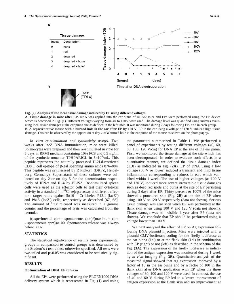
the parameters summarized in Table 1. We performed a panel of experiments by testing different voltages (40, 60, 80, 100, 120 V/cm) for DNA EP at the site of the ear pinna. First, we monitored the tissue damage at the site which has been electroporated. In order to evaluate such effects in a quantitative manner, we defined the tissue damage index (TDI) as indicated in Fig. (2A). EP of DNA using a low voltage (80 V or lower) induced a transient and mild tissue inflammation corresponding to redness in ears which van-ished within 1 week. The use of higher voltages (as 100 V and 120 V) induced more severe irreversible tissue damages such as deep red spots and burns at the site of EP persisting during 3 days after EP. Thirty percent or 100% of the mice showed a punctured skin (Fig. 2B) at the site of EP when using 100 V or 120 V respectively (data not shown). Serious tissue damage was also seen when EP was performed at the flank skin when using 100 V and 120 V (data not shown). Tissue damage was still visible 1 year after EP (data not shown). We conclude that EP should be performed using a voltage lower than 100 V.
We next analyzed the effect of EP on Ag expression fol-lowing DNA plasmid injection. Mice were injected with a plasmid CMV-luciferase coding for the firefly luciferase at the ear pinna (i.e.) or at the flank skin (i.d.) in combination with EP (right) or not (left) as described in the schema of the Fig. (3A). The expression of the firefly luciferase as indica-tion of the antigen expression was monitored during 1 week by in vivo imaging (Fig. 3B). Quantitative analysis of the measured signal showed that Ag expression improved by a factor of 10 in the ear pinna and by a factor of 100 in the flank skin after DNA application with EP when the three voltages of 80, 100 and 120 V were used. In contrast, the use of 40 and 60 V during EP led to a lower improvement of antigen expression at the flank skin and no improvement at
Fig. (2). Analysis of the local tissue damage induced by EP using different voltages.
A. Tissue damage in mice after EP. DNA was applied into the ear pinna of DBA/2 mice and EPs were performed using the EP device
which is described in Fig. (1). Different voltages varying from 40 to 120V were used. The damage level was quantified using indexes evalu-
ating local tissue damages at the ear pinna site as defined in the left table. It was monitored during 7 days following EP. n=3 in each group.
B. A representative mouse with a burned hole in the ear after EP by 120 V. EP in the ear using a voltage of 120 V induced high tissue damage. This can be observed by the apparition at day 7 of a burned hole in the ear pinna of the mouse as shown on the photography.

A New DNA Vaccine Delivery for Optimal Anti-Tumor Immunity The Open Cancer Immunology Journal, 2009, Volume 2 5
the ear pinna. These results showed that DNA EP using a voltage of 80, 100 or 120 V efficiently improved Ag expres-sion in the skin after DNA injection. We decided to use 80 V for all the further EPs.
Long-term Ag Expression after DNA injection and EP
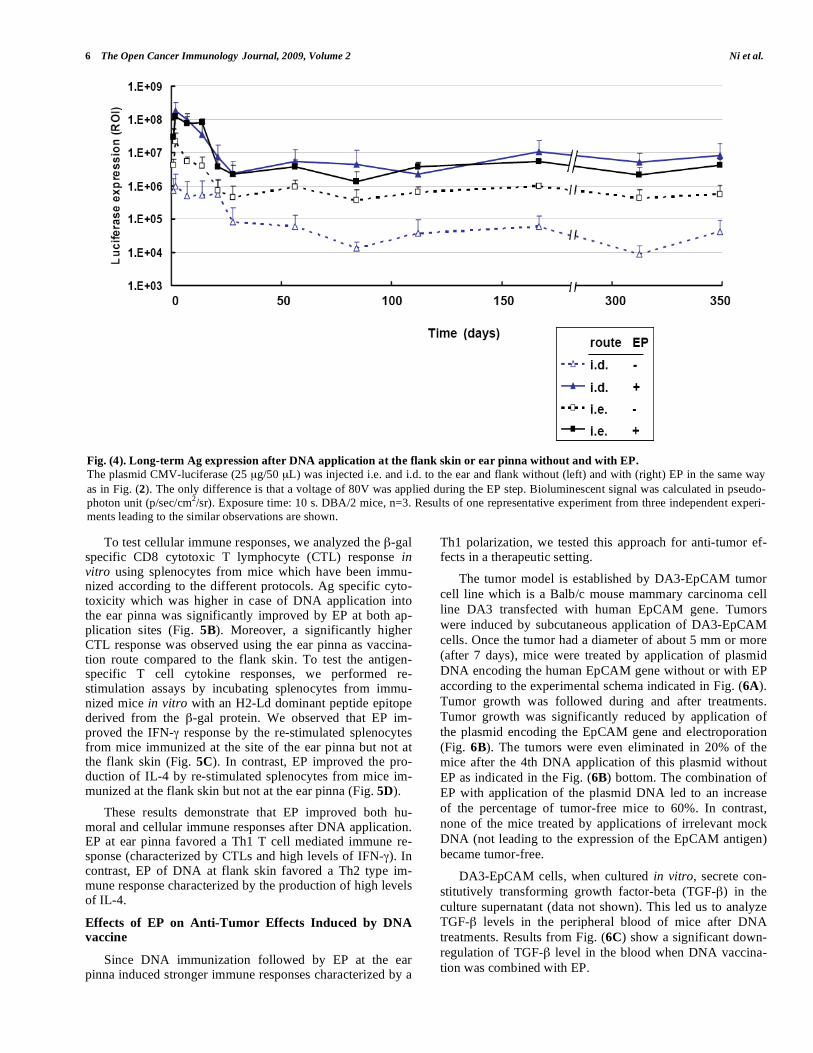
To analyze long-term effects on Ag expression after
DNA injection combined with EP, mice were immunized
with the plasmid CMV-luciferase. Left ear and flank skin
were not electroporated whereas the right ones were (as indi-
cated in Fig. 3A). Luciferase activity was monitored over
350 days after EP. We observed that EP improved the Ag
expression at both application sites. An increase of antigen
expression corresponding to factors between 50 and 500 was
observed when vaccination was performed at the flank site.
In contrast, the antigen expression was only increased by
factors between 5 and 20 when the ear pinna was used as
vaccination site (Fig. 4). Although we observed that the sig-
nals were 5 to 50 times higher at the site of the ear pinna as
compared to the flank skin without EP, similar levels of Ag
expression were obtained for both routes after DNA EP (Fig.
4).
High levels of Ag expression were seen for 2 weeks after DNA EP at both sites. After 4 weeks, Ag expression de-creased by a factor of 10 to 100, but maintained at a low but stable level during 350 days (Fig. 4). These results reveal that EP can be used to increase Ag expression for long terms.
Humoral and Cellular Immune Responses after EP
We then analyzed if EP can improve immune responses
against the target Ag encoded by the plasmid in vivo. For
these investigations, we used a DNA plasmid with the LacZ
gene coding for bacterial beta-galactosidase ( -gal). We ap-
plied this plasmid either to the ear pinna (i.e.) or to the flank
skin (i.d.). Humoral responses were analyzed via ELISA,
testing the levels of antibodies specific for the -gal protein
in the plasma of mice. EP induced an increase of anti- -gal
antibody levels by a factor 46 when immunization was per-
formed at the flank skin. In contrast, the increase of the anti-
body titer towards -gal was only increased by a factor of 5
when the ear pinna was used (Fig. 5A). However, a higher
Ab titer was induced by DNA immunization at the ear pinna
in comparison to the flank skin, no matter if EP was per-formed or not.
Fig. (3). Ag expression after DNA EP using different voltages.
A. DNA transfer into ear and flank skin. The plasmid CMV-luciferase encoding the firefly luciferase gene under the control of the CMV
promoter was injected (25 μg/50 μL) intradermally to the ear pinna and flank skin. And EP was performed (right side of the mouse) or not
(left side).
B. In vivo luciferase expression. Using the experimental design indicated in A, DNA was applied to DBA/2 mice and electroporated using
different voltages comprised between 40 V and 120 V. Bioluminescent signal was monitored over 7 days with the IVIS100 imaging system
using an exposure time of 10 s. One representative mouse of each group (n=3 for each) is shown. Luciferase signal was quantified in pseudo-photon unit (p/sec/cm
2/sr) as described in material and methods. One representative experiment out of three is shown.
6 The Open Cancer Immunology Journal, 2009, Volume 2 Ni et al.
To test cellular immune responses, we analyzed the -gal specific CD8 cytotoxic T lymphocyte (CTL) response in vitro using splenocytes from mice which have been immu-nized according to the different protocols. Ag specific cyto-toxicity which was higher in case of DNA application into the ear pinna was significantly improved by EP at both ap-plication sites (Fig. 5B). Moreover, a significantly higher CTL response was observed using the ear pinna as vaccina-tion route compared to the flank skin. To test the antigen-specific T cell cytokine responses, we performed re-stimulation assays by incubating splenocytes from immu-nized mice in vitro with an H2-Ld dominant peptide epitope derived from the -gal protein. We observed that EP im-proved the IFN- response by the re-stimulated splenocytes from mice immunized at the site of the ear pinna but not at the flank skin (Fig. 5C). In contrast, EP improved the pro-duction of IL-4 by re-stimulated splenocytes from mice im-munized at the flank skin but not at the ear pinna (Fig. 5D).
These results demonstrate that EP improved both hu-moral and cellular immune responses after DNA application. EP at ear pinna favored a Th1 T cell mediated immune re-sponse (characterized by CTLs and high levels of IFN- ). In contrast, EP of DNA at flank skin favored a Th2 type im-mune response characterized by the production of high levels of IL-4.
Effects of EP on Anti-Tumor Effects Induced by DNA vaccine
Since DNA immunization followed by EP at the ear pinna induced stronger immune responses characterized by a
Th1 polarization, we tested this approach for anti-tumor ef-fects in a therapeutic setting.
The tumor model is established by DA3-EpCAM tumor
cell line which is a Balb/c mouse mammary carcinoma cell
line DA3 transfected with human EpCAM gene. Tumors
were induced by subcutaneous application of DA3-EpCAM
cells. Once the tumor had a diameter of about 5 mm or more
(after 7 days), mice were treated by application of plasmid
DNA encoding the human EpCAM gene without or with EP
according to the experimental schema indicated in Fig. (6A).
Tumor growth was followed during and after treatments.
Tumor growth was significantly reduced by application of
the plasmid encoding the EpCAM gene and electroporation
(Fig. 6B). The tumors were even eliminated in 20% of the
mice after the 4th DNA application of this plasmid without
EP as indicated in the Fig. (6B) bottom. The combination of
EP with application of the plasmid DNA led to an increase
of the percentage of tumor-free mice to 60%. In contrast,
none of the mice treated by applications of irrelevant mock
DNA (not leading to the expression of the EpCAM antigen)
became tumor-free.
DA3-EpCAM cells, when cultured in vitro, secrete con-
stitutively transforming growth factor-beta (TGF- ) in the
culture supernatant (data not shown). This led us to analyze
TGF- levels in the peripheral blood of mice after DNA
treatments. Results from Fig. (6C) show a significant down-
regulation of TGF- level in the blood when DNA vaccina-
tion was combined with EP.
Fig. (4). Long-term Ag expression after DNA application at the flank skin or ear pinna without and with EP.
The plasmid CMV-luciferase (25 μg/50 μL) was injected i.e. and i.d. to the ear and flank without (left) and with (right) EP in the same way
as in Fig. (2). The only difference is that a voltage of 80V was applied during the EP step. Bioluminescent signal was calculated in pseudo-
photon unit (p/sec/cm2/sr). Exposure time: 10 s. DBA/2 mice, n=3. Results of one representative experiment from three independent experi-
ments leading to the similar observations are shown.

A New DNA Vaccine Delivery for Optimal Anti-Tumor Immunity The Open Cancer Immunology Journal, 2009, Volume 2 7
Fig. (5). Humoral and cell-mediated immune responses after DNA application at the flank site or at the ear pinna eventually followed
by EP. A. Humoral response. DBA/2 mice (n=3) were immunized i.e. or i.d. with the plasmid pCMV SPORT- gal (25 g/50 L) without
or with EP (80 V). Bloods were collected at day 14 after DNA vaccination. Plasma were then prepared and analyzed by ELISA for anti- -gal
Ab responses (IgG+M). One of 3 independent experiments is shown. B. Cell-mediated immune responses. Mice were sacrificed at day 14
after DNA immunization. Spleens were taken out and re-stimulated for 5 days in vitro with 0.5 μg/mL TPHPARIGL peptide (H2-Ld epitope)
and analyzed for cytotoxicity by a standard 4 h 51
Cr release assay using lacZ+ tumor cells (P13.1) and lacZ
- tumor cells (P815) as target cells.
An effector to target ratio of 100:1 was respected. One of three independent experiments showing similar results is shown. C and D. Cyto-
kine production by splenocytes after in vitro re-stimulation. Supernatants of culture from re-stimulated splenocytes (as described in Fig.
4B) were collected at day 2 for IFN- analysis (C) and at day 5 for IL-4 determination (D). The 2 cytokines were quantified by ELISA. One
representative experiment from three is shown.
To analyze possible immunity against the parental tumor cell line DA3 after DNA treatments to DA3-EpCAM tumors, the DA3-EpCAM tumor bearing mice were re-challenged with DA3 cells at day 59 as shown in the Fig. (7A). We ob-served that DA3 tumor growth was significantly reduced in the group of mice which had received an injection at the ear pinna of the EpCAM encoding plasmid combined with EP in comparison to the group of mice that were immunized with this DNA without EP or with a vector control plasmid. No significant DA3 tumor retardation was observed in the group of mice treated with EpCAM without EP in comparison to mice treated with the mock DNA plasmid.
Taken together, these results provide evidence for the ef-ficiency of therapeutic antitumor treatment when applying xenogeneic DNA vaccination at the ear pinna in combination with EP. Established DA3-EpCAM tumors were shown to regress and, upon re-challenge with DA3 tumor cells, there was growth retardation only in the group of EpCAM DNA EP treated mice. This immunization protocol thus induced immunity not only against the tumor cells with xenogeneic EpCAM expression but also cross-induced immunity against the parental tumor cells, possibly via breaking tolerance.
DISCUSSION
In this study, we combined DNA injection to the skin
with EP. We report that such a vaccination strategy led to
improved cellular immunity toward the antigen encoded by
the plasmid. Of great importance was not only EP but also
the site of immunization. The ear pinna was superior to flank
skin and provided a Th1 polarized response with a strong
anti-tumor immunity.
In this study, we used an EP-based delivery system using
the ELGEN1000 device that has been developed by Inovio
during the MOLEDA project. It is made of a pulse controller
connected to 2 tweezers electrodes. It is designed for human
applications by transferring small molecules including ge-
netic materials and therapeutic drugs to certain tissues.
Clinical trials (phase I/II) have proven safety, tolerability and
immunological reactions in human [69-72]. EP has proven to
be a highly effective technique for the in vivo delivery of
genes to a number of solid tissues. In this study, we demon-
strate for the first time a similar effect for the tissue of ear
pinna.

8 The Open Cancer Immunology Journal, 2009, Volume 2 Ni et al.
To enhance the level of gene expression from plasmids
administrated into tissues, the use of optimal EP parameters
is critical. If conditions are too harsh, severe tissue damage
with extensive cell death can occur. Since gene expression
and protein production from plasmids are dependent on vi-
able cells, it is critical that the majority of the cells being
transfected are not killed. Therefore, the optimal EP condi-
tions are a subtle balance between enhancing permeability
without causing extensive cell death and tissue trauma.
Many studies reported that EP could damage the target tissue
depending on the electric parameter associated with the EP
[69, 73-76]. We defined in this study that EP of DNA at the
ear pinna site of mice is optimally performed using 80 V
with the ELGEN1000 DNA delivery system.
What was remarkable is that, by combining EP and DNA, a relatively long-term stable Ag expression was in-duced in mice. Gene expression was maintained for a long time since the signal could still be detected after 350 days, a time which corresponds to the life-time of a mouse. This might be explained by the long-term existence of the DNA vector in host cells or integration of the plasmid DNA into hosts chromosomal DNA. There are only two published studies examining the potential for integration after EP-mediated DNA delivery [77, 78]. For conventional DNA
delivery, the risk of integration for causing an oncogenic event was observed to be low [79]. The use of a transduced firefly luciferase gene allowed a quick and very sensitive determination of protein expression in vivo since there is no endogenous luciferase enzyme activity in mammalian tis-sues. The level of Ag expression is an important determinant for the magnitude of an immune response to DNA vaccines. Also, however, the induction of an inflammatory response, due to the physical stimulation of skin cells by EP, might be crucial for enhancing immune responses [80]. This unde-fined adjuvant effect might be possibly mediated through local minor tissue damage and release of inflammatory fac-tors [79, 81, 82]. Recent data suggest that in addition to en-hancing gene delivery, EP provides adjuvant-like effects [82, 83]. Several reports have shown that the local effect by EP is responsible for the generation of an inflammatory environ-ment with immune cell infiltration. The migration of these cells seems essential to initiate an adequate immune response to the DNA vaccine [84, 85]. Previous studies using bupiva-caine to enhance immune responses to DNA vaccines [86] suggested that muscle damage / inflammation is important for enhancing immune responses [58, 74, 87]. In another study, Nishijima et al. [88] reported that disruption of the skin barrier results in epidermal Langerhans cell activation as vigorous antigen presenters for T helper cells.
Fig. (6). Anti-tumor effects induced by EP in a therapeutic tumor model.
A. Experimental protocol. Balb/c mice (n=13~15) were inoculated s.c. with 1 107 DA3-EpCAM cells. One week later, DNA encoding the
human EpCAM gene under the CMV promoter was applied 4 times without and with EP respecting 1 week interval (50 g/50 L DNA
between each immunization). The pTandem1 vector was used as a negative control (Mock DNA).
B. Therapeutic effects of DNA treatments. DA3-EpCAM tumor growth was followed and is represented as tumor diameter (top). The
percentage of tumor-bearing mice was calculated for each group during the experiment (bottom). * Compared to the Mock and EpCAM i.e.
groups, p£0.05.
C. Peripheral TGF-b levels. Plasma from DA3-EpCAM tumor bearing mice were taken at day 49 after tumor implantation. Concentrations
of TGF- were defined by ELISA. * Compared to the Mock and EpCAM i.e. groups, p£0.05.

A New DNA Vaccine Delivery for Optimal Anti-Tumor Immunity The Open Cancer Immunology Journal, 2009, Volume 2 9
A recent study [89] reported that EP of skeletal muscles without any application of DNA induces an inflammatory process associated with recruitment and activation of a dan-ger-like pro-inflammatory pathway. These data sustain the idea that the immune responses mounted against the antigen may not only be due to the foreignness of the transfected gene but also to the danger signals associated with the deliv-ery process [90]. Therefore, the efficacy of naked DNA vac-cines delivered by EP resides in the strong stimulation of immunity at the EP site. It has also been observed that EP causes cellular infiltration into the electroporated tissue. This might be very important for generating an immune response following DNA immunization [58] since the transient in-flammation associated with EP may provide an ideal envi-ronment for generation of immune responses to encoded antigens [58, 91]. Thus, the mechanisms by which EP en-hances immune responses to DNA vaccines may be a com-bination of increased gene expression and of generated pro-inflammatory environment.
In vivo gene delivery by EP has been demonstrated to be efficient for introducing DNA also into mouse skin [92, 93]. In most cases of small animal experimentation, i.d. immuni-zation is applied to the abdominal or flank skin. We propose that ear pinna is a more attractive alternative for DNA im-munization. It is a very special site and form of skin. One of the advantages might be the special structure of ear pinna which contains two layers of epidermis and dermis - sepa-rated by a cartilage - rich in professional APCs. In a previous study we provide evidence that CD11c DCs in the ear pinna are essential for the immune responses generated [94]. The
superiority of the ear pinna as a vaccination site is ascribed to its unique immunological features that focus the concen-tration of proceeded antigens in a restricted area, which is connected to a major draining lymph node. It is thought that the concentration of processed antigens results in an en-hanced stimulation of T lymphocytes by antigen-loaded DCs [95]. Intradermal DNA vaccination into the ear pinna route has been reported to produce high levels of neutralizing anti-bodies against rabies virus in dogs [96]. We observed in this study that EP augments immune responses to DNA vaccines. Antibody titers were augmented greatly as compared to in-jection of DNA vaccine without EP. T cell responses were also significantly enhanced with regard to IFN- production and CTL killing activity.
It has been shown that in vivo EP can increase DNA up-take by cells at the site of injection [29,83], leading to in-creased Ag expression [97-99]. Although the exact mecha-nism has yet to be elucidated, the Ag expression allows in-duction of immune responses through MHC-I and/or MHC-II pathways. In either case, DCs and other APCs play a criti-cal role in activating adaptive immunity. They must acquire the antigen to be delivered to the tissue-draining lymph nodes for priming of CD8
+ cytotoxic T-lymphocytes (CTL)
and the activation of CD4+ helper T-lymphocytes. Direct
priming of CTL may be facilitated through DCs endoge-nously expressing antigen. DCs may also acquire antigen exogeneously through MHC I cross-presentation by various means including direct acquisition from dead or dying cells [100, 101]. DCs may also acquire secreted antigen to be dis-played through either MHC class I and/or II pathways [102].
Fig. (7). Cross anti-tumor activity generated by EP.
A. Experimental protocol. DA3-EpCAM tumor bearing mice were therapeutically treated by i.e. immunization with the DNA plasmid en-
coding the human EpCAM gene using EP or not or with the control plasmid as described in Fig. (6). All the animals were challenged at day
59 by a s.c. inoculation of 1´107 DA3 cells.
B. DA3 tumor growth. Tumor growth was monitored by measuring the diameter of the tumor corresponding to the DA3 cell line during 42
days starting from the 2nd tumor challenge. *Compared to the Mock and EpCAM i.e. groups, p<0.05; **Compared to the Mock and EpCAM i.e. groups, p<0.01.

10 The Open Cancer Immunology Journal, 2009, Volume 2 Ni et al.
Although enhancement of immune responses by EP is probably also due to an increase of antigen expression by other cells such as keratinocytes, we believe that DCs play a major role [94] in the induction of a strong immune response at the ear pinna site either by cross-priming or by direct priming. DCs very probably migrate to the draining lymph nodes and trigger the immune response. The role of DCs has been demonstrated during EP-based vaccination. DCs from draining lymph nodes were shown to contain DNA originat-ing from the injection site [103].
Our data show also that the ear pinna route skews the immune response towards Th1 whereas the application of DNA to the skin from the flank skews the immune response towards Th2. Gene gun immunization has been shown to skew immune responses towards Th2. In contrast, EP is more efficient for induction of Th1 immune responses [104-106]. Consequently, the use of EP as a method of antigen delivery and ear pinna as the site of DNA application might explain the Th1 polarization of the immune response. Fur-ther studies need to be performed in order to identify the cells that are involved and understand why the use of this route leads to the generation of such a strong Th1 type cellu-lar immune response.
Mechanisms of tolerance and immune escape are draw-backs to cancer vaccination. TGF- , one of the immunosup-pressive cytokine which is often secreted in large amounts by malignant cells such as the DA3 tumor cell line used in this study. This molecule acts on nontransformed cells pre-sent in the tumor mass as well as on distal cells in the host to suppress antitumor immune responses creating an environ-ment of immune tolerance [107]. A strong immune reactivity is then required to reverse the immunosuppressive effects induced by this cytokine. We observed in this study that EP after DNA injection also down-regulated peripheral TGF- levels in tumor-bearing mice. It is not clear how the down-regulation was achieved. It could be the outcome from the inhibition of tumor growth by DNA EP, because less tumor derived suppressive factors might be secreted by smaller tumors.
The goal of active therapeutic vaccination against cancer is to induce a strong effective immunity against tumor cells and to establish immunological memory that is able to main-tain continuous surveillance against emergent cancer cells. EP was found to improve the kinetics of immune responses by requiring less time than conventional injection to reach a maximal immune response [108, 109] and favoring the in-duction of long-term memory [110] but also the consistency of the immune responses induced when compared with injec-tion without EP. Because these initial results seem promis-ing, vaccination trials based on ear pinna immunization with autologous irradiated tumor cells electroporated with DNA should be considered for the future. Efficacy and tolerability of EP was demonstrated in phase I clinical trials performed in collaboration with some companies [111] where in some studies the EP technology was used for intratumoral delivery of plasmid DNA [112]. The increase of knowledge about immune responses generated from the site of the ear pinna and the potential of DNA EP delivery for generating strong cellular immunity give hope to the field of DNA-based can-cer therapy.
In conclusion, the results presented here show that the ear pinna is a privileged site for DNA EP. Compared to tradi-tional DNA application to the flank skin in mice, the strategy of DNA EP at the ear pinna induced similar Ag expression but led to the induction of very strong cellular immunity to-ward the Ag encoded by the plasmid with a strong Th1 po-larization. By this method, therapeutic anti-tumor effects were also achieved in a mouse tumor model. Such increases in DNA vaccine potency provide encouragement that such an immunization could be tested in large animals.
ACKNOWLEDGEMENTS
This project has been performed within the frame of the project MOLEDA (acronym standing for Molecular Optimi-zation of Laser / Electrotransfer DNA Administration into muscle and skin for gene therapy) sponsored by the Euro-pean Commission during the research program FP6. We are particularly grateful to Annette Arnold for her technical as-sistance. We acknowledge also the DAAD (German Aca-demic Exchange Service) for support of Jing Ni during her PhD thesis.
REFERENCES
[1] Tang DC, DeVit M, Johnston SA. Genetic immunization is a sim-ple method for eliciting an immune response. Nature 1992;
356(6365): 152-4. [2] Ulmer JB, Sadoff JC, Liu MA. DNA vaccines. Curr Opin Immu-
nol. 1996; 8(4): 531-6. [3] Sedegah, M, Hedstrom R, Hobart P, Hoffman SL. Protection
against malaria by immunization with plasmid DNA encoding cir-cumsporozoite protein. Proc Natl Acad Sci USA 1994; 91: 9866-
70. [4] Polack, FP, Lee SH, Permar S, et al. Successful DNA immuniza-
tion against measles: neutralizing antibody against either the he-magglutinin or fusion glycoprotein protects rhesus macaques with-
out evidence of atypical measles. Nat Med 2000; 6: 776-81. [5] Ulmer JB, Donnelly JJ, Parker SE, et al. Heterologous protection
against influenza by injection of DNA encoding a viral protein. Science 1993; 259: 1745-49.
[6] Davis HL, Michel ML, Whalen RG. DNA-based immunization induces continuous secretion of hepatitis B surface antigen and
high levels of circulating antibody. Hum Mol Genet 1993; 2: 1847-51.
[7] Conry, RM, LoBuglio AF, Kantor J, et al. Immune response to a carcinoembryonic antigen polynucleotide vaccine. Cancer Res
1994; 54: 1164-68. [8] Hawkins, RE, Zhu D, Ovecka M, et al. Idiotypic vaccination
against human B-cell lymphoma: rescue of variable region gene sequences from biopsy material for assembly as single-chain Fv
personal vaccines. Blood 1994; 83: 3279-88. [9] Ciernik, IF, Berzofsky JA, Carbone DP. Induction of cytotoxic T
lymphocytes and antitumor immunity with DNA vaccines express-ing single T cell epitopes. J Immunol 1996; 156: 2369-75.
[10] Spellerberg, MB, Zhu D, Thompsett A, King CA, Hamblin TJ, Stevenson FK. DNA vaccines against lymphoma: promotion of
anti-idiotypic antibody responses induced by single chain Fv genes by fusion to tetanus toxin fragment C. J Immunol 1997; 159: 1885-
92. [11] Hsu CH, Chua KY, Tao MH, et al. Immunoprophylaxis of aller-
gen-induced immunoglobulin E synthesis and airway hyperrespon-siveness in vivo by genetic immunization. Nat Med 1996; 2: 540-
44. [12] Wildbaum, G. Nahir MA, Karin N. Beneficial autoimmunity to
proinflammatory mediators restrains the consequences of self-destructive immunity. Immunity 2003; 19: 679-88.
[13] Youssef S, Maor G, Wildbaum G, Grabie N, Gour-Lavie A, Karin N. C-C chemokine-encoding DNA vaccines enhance breakdown of
tolerance to their gene products and treat ongoing adjuvant arthritis. J Clin Invest 2000; 106: 361-71.

A New DNA Vaccine Delivery for Optimal Anti-Tumor Immunity The Open Cancer Immunology Journal, 2009, Volume 2 11
[14] Powell K. DNA vaccines--back in the saddle again? Nat Biotechnol
2004; 22(7): 799-801. [15] Lorenzen N, LaPatra SE. DNA vaccines for aquacultured fish. Rev
Sci Tech 2005; 24(1): 201-13. [16] Bergman PJ, Camps-Palau MA, McKnight JA, et al. Development
of a xenogeneic DNA vaccine program for canine malignant mela-noma at the Animal Medical Center. Vaccine 2006; 24(21): 4582-
5. [17] Bergman PJ, McKnight J, Novosad A, et al. Long-term survival of
dogs with advanced malignant melanoma after DNA vaccination with xenogeneic human tyrosinase: a phase I trial. Clin Cancer Res
2003; 9(4): 1284-90. [18] Liao JC, Gregor P, Wolchok JD, et al. Vaccination with human
tyrosinase DNA induces antibody responses in dogs with advanced melanoma. Cancer Immun 2006; 6: 8.
[19] Therapy Clinical Trials Worldwide – Chart&Table Keyword: Vec-tors (Updated March 2009) available from http:
//www.wiley.co.uk/genmed/clinical/Gene. [20] Bodles-Brakhop AM, Draghia-Akli R. DNA vaccination and gene
therapy: optimization and delivery for cancer therapy. Expert Rev Vac 2008; 7(7): 1085-101.
[21] van Drunen Littel-van den Hurk S, Loehr BI, Babiuk LA. Immuni-zation of livestock with DNA vaccines: current studies and future
prospects. Vaccine 2001; 19(17-19): 2474-9. [22] Gurunathan S, Klinman DM, Seder RA. DNA vaccines: immuno-
logy, application, and optimization. Annu Rev Immunol 2000; 18: 927-74.
[23] Donnelly JJ, Wahren B, Liu MA. DNA vaccines: progress and challenges. J Immunol 2005; 175: 633-9.
[24] Wang R, Doolan DL, Le TP, et al. Induction of antigen-specific cytotoxic T lymphocytes in humans by a malaria DNA vaccine.
Science 1998; 282(5388): 476-80. [25] Calarota S, Bratt G, Nordlund S, et al. Cellular cytotoxic response
induced by DNA vaccination in HIV-1-infected patients. Lancet 1998; 351(9112): 1320-5.
[26] MacGregor RR, Boyer JD, Ugen KE, et al. First human trial of a DNA-based vaccine for treatment of human immunodeficiency vi-
rus type 1 infection: safety and host response. J Infect Dis 1998; 178(1): 92-100.
[27] Wang R, Epstein J, Baraceros FM, et al. Induction of CD4+ T cell-dependent CD8+ type 1 responses in humans by a malaria DNA
vaccine. Proc Natl Acad Sci USA 2001, 98: 10817-22. [28] Stevenson FK, Rice J, Ottensmeier CH, Thirdborough SM, Zhu D.
DNA fusion gene vaccines against cancer: from the laboratory to the clinic. Immunol Rev 2004; 199: 156-80.
[29] Widera G, Austin M, Rabussay D, et al. Increased DNA vaccine delivery and immunogenicity by electroporation in vivo. J Immunol
2000; 164: 4635-40. [30] Patil SD, Rhodes DG, Burgess DJ. DNA-based therapeutics and
DNA delivery systems: a comprehensive review. AAPS J 2005; 7: E61-77.
[31] Hengge UR, Chan EF, Foster RA, Walker PS, Vogel JC. Cytokine gene expression in epidermis with biological effects following in-
jection of naked DNA. Nat Genet 1995; 10: 161-66. [32] Yang NS, Burkholder J, Roberts B, Martinell B, McCabe D. In vivo
and in vitro gene transfer to mammalian somatic cells by particle bombardment. Proc Natl Acad Sci USA 1990; 87: 9568-72.
[33] Ciernik IF, Krayenbühl BH, Carbone DP. Puncture-mediated gene transfer to the skin. Hum Gene Ther 1996; 7: 893-99.
[34] McAllister DV, Allen MG, Prausnitz MR. Microfabricated mi-croneedles for gene and drug delivrey. Annu Rev Biomed Eng
2000; 2: 289-313. [35] Mikszta JA, Alarcon JB, Brittingham JM, Sutter DE, Pettis RJ,
Harvey NG. Improved genetic immunization via micromechanical disruption of skin-barrier function and targeted epidermal delivery.
Nat Med 2002; 8: 415-19. [36] Badiavas E, Mehta PP, Falanga V. Retrovirally mediated gene
transfer in a skin equivalent model of chronic wounds. J Dermatol Sci 1996; 13: 56-62.
[37] Lu B, Federoff H, Wang Y, Goldsmith LA, Scott G. Topical appli-cation of viral vectors for epidermal gene transfer. J Invest Derma-
tol 1997; 108: 803-8. [38] Hirao LA, Wu L, Khan AS, Satishchandran A, Draghia-Akli R,
Weiner DB. Intradermal/subcutaneous immunization by electropo-ration improves plasmid vaccine delivery and potency in pigs and
rhesus macaques. Vaccine 2008; 26: 440-8.
[39] Capone S, Zampaglione I, Vitelli A, et al. Modulation of the im-
mune response induced by gene electrotransfer of a hepatitis C vi-rus DNA vaccine in nonhuman primates. J Immunol 2006; 177:
7462-71. [40] Verstrepen BE, Bins AD, Rollier CS, et al. Improved HIV-1 speci-
fic T-cell responses by short-interval DNA tattooing as compared to intramuscular immunization in non-human primates. Vaccine
2008; 26: 3346-51. [41] Pokorna D, Rubio I, Muller M. DNA-vaccination via tattooing
induces stronger humoral and cellular immune responses than in-tramuscular delivery supported by molecular adjuvants. Genet
Vaccines Ther 2008; 6: 4. [42] Kim D, Hoory T, Monie A, Ting JP, Hung CF, Wu TC. Enhance-
ment of DNA vaccine potency through coadministration of CIITA DNA with DNA vaccines via gene gun. J Immunol 2008; 180:
7019-27. [43] Li S. Electroporation gene therapy: new developments in vivo and
in vitro. Curr Gene Ther 2004; 4(3): 309-16. [44] Wells DJ. Gene therapy progress and prospects: electroporation
and other physical methods. Gene Ther. 2004; 11(18): 1363-9. [45] Andre F, Mir LM. DNA electrotransfer: its principles and an up-
dated review of its therapeutic applications. Gene Ther 2004; 11 (Suppl 1): S33-42.
[46] Rols MP. Mechanism by which electroporation mediates DNA migration and entry into cells and targeted tissues. Methods Mol
Biol 2008; 423: 19-33. [47] Bureau MF, Gehl J, Deleuze V, Mir LM, Scherman D. Importance
of association between permeabilization and electrophoretic forces forintramuscular DNA electrotransfer. Biochim Biophys Acta
2000; 1474(3): 353-59. [48] Satkauskas S, Bureau MF, Puc M, et al. Mechanisms of in vivo
DNA electrotransfer: respective contributions of cell electroperme-abilization and DNA electrophoresis. Mol Ther 2002; 5(2): 133-40.
[49] Satkauskas S, André F, Bureau MF, Scherman D, Miklavcic D, Mir LM. Electrophoretic component of electric pulses determines the
efficacy of in vivo DNA electrotransfer. Hum Gene Ther 2005; 16(10): 1194-201.
[50] Liu F, Heston S, Shollenberger LM, et al. Mechanism of in vivo DNA transport into cells by electroporation: electrophoresis across
the plasma membrane may not be involved. J Gene Med 2006; 8(3): 353-61.
[51] Murtaugh MP, Foss DL. Inflammatory cytokines and antigen pre-senting cell activation. Vet Immunol Immunopathol. 2002; 87(3-4):
109-21. [52] Liu J, Kjeken R, Mathiesen I, Barouch DH. Recruitment of anti-
gen-presenting cells to the site of inoculation and augmentation of human immunodeficiency virus type 1 DNA vaccine immuno-
genicity by in vivo electroporation. J Virol 2008; 82(11): 5643-9. [53] Wolff JA, Malone RW, Williams P, et al. Direct gene transfer into
mouse muscle in vivo. Science 1990; 247: 1465-8. [54] Deck RR, DeWitt CM, Donnelly JJ, Liu MA, Ulmer JB. Charac-
terization of humoral immune responses induced by an influenza hemagglutinin DNA vaccine. Vaccine 1997; 15: 71-78.
[55] Donnelly JJ, Friedman A, Martinez D, et al. Preclinical efficacy of a prototype DNA vaccine: Enhanced protection against antigenic
drift in influenza virus. Nat Med 1995; 1: 583-87. [56] Okuda K, Ihata A, Watabe S, et al. Protective immunity against
influenza A virus induced by immunization with DNA plasmid containing influenza M gene. Vaccine 2001; 19: 3681-91.
[57] Ulmer JB, Deck RR, DeWitt CM, Friedman A, Donnelly JJ, Liu MA. Protective immunity by intramuscular injection of low doses
of influenza virus DNA vaccines. Vaccine 1994; 12: 1541-44. [58] Babiuk S, Baca-Estrada M, Babiuk LA, Ewen C, Foldvari M. Cu-
taneous vaccination: The skin as an immunologically active tissue and the challenge of antigen delivery. J Control Release 2000; 66:
199-214. [59] Puri N, Weyand EH, Abdel-Rahman SM, Sinko PJ. An investiga-
tion of the intradermal route as an effective means of immunization for microparticulate vaccine delivery systems. Vaccine 2000;
18(23): 2600-12. [60] Banchereau J, Steinman RM. Dendritic cells and the control of
immunity. Nature 1998; 392: 245-52. [61] Peachman KK, Rao M, Alving CR. Immunization with DNA
through the skin. Methods 2003; 31: 232-42. [62] Donnelly JJ, Liu MA, Ulmer JB. Antigen presentation and DNA
vaccines. Am J Respir Crit Care Med 2000; 162: S190-3.

12 The Open Cancer Immunology Journal, 2009, Volume 2 Ni et al.
[63] Stoitzner P, Tripp CH, Eberhart A, et al. Langerhans cells cross-
present antigen derived from skin. Proc Natl Acad Sci USA 2006; 103: 7783-8.
[64] Jurianz K, von Hoegen P, Schirrmacher V. Superiority of the ear pinna over a subcutaneous tumour inoculation site for induction of
a Th1-type cytokine response.Cancer Immunol Immunother 1998; 45: 327-33.
[65] Forg P, von Hoegen P, Dalemans W, Schirrmacher V. Superiority of the ear pinna over muscle tissue as site for DNA vaccination.
Gene Ther 1998; 5: 789-97. [66] Schirmacher V, Forg P, Dalemans W, et al. Intra-pinna anti-tumor
vaccination with self-replicating infectious RNA or with DNA en-coding a model tumor antigen and a cytokine. Gene Ther 2000; 7:
1137-47. [67] Kruger A, Schirrmacher V, von Hoegen P. Scattered micrometasta-
ses visualized at the single-cell level: detection and re-isolation of lacZ-labeled metastasized lymphoma cells. Int J Cancer 1994; 58:
275-84. [68] von Hoegen P, Weber E, Schirrmacher V. Modification of tumor
cells by a low dose of Newcastle disease virus. Augmentation of the tumor-specific T cell response in the absence of an antiviral re-
sponse. Eur J Immunol 1988; 18: 1159-66. [69] Gronevik E, von Steyern FV, Kalhovde JM, Tjelle TE, Mathiesen
I. Gene expression and immune response kinetics using electropo-ration-mediated DNA delivery to muscle. J Gene Med 2005; 7:
218-27. [70] Rabussay D. Applicator and electrode design for in vivo DNA
delivery by electroporation. Methods Mol Biol 2008; 423: 35-59. [71] Tjelle TE, Rabussay D, Ottensmeier C, Mathiesen I, Kjeken R.
Taking electroporation-based delivery of DNA vaccination into humans: a generic clinical protocol. Methods Mol Biol 2008; 423:
497-507. [72] Tjelle TE, Salte R, Mathiesen I, Kjeken R. A novel electroporation
device for gene delivery in large animals and humans. Vaccine 2006; 24: 4667-70.
[73] Lefesvre P, Attema J, van Bekkum D. A comparison of efficacy and toxicity between electroporation and adenoviral gene transfer.
BMC Mol Biol 2002; 3: 12. [74] Hartikka J, Sukhu L, Buchner C, et al. Electroporation-facilitated
delivery of plasmid DNA in skeletal muscle: plasmid dependence of muscle damage and effect of poloxamer 188. Mol Ther 2001;
4(5): 407-15. [75] Molnar MJ, Gilbert R, Lu Y, et al. Factors influencing the efficacy,
longevity, and safety of electroporation-assisted plasmid-based gene transfer into mouse muscles. Mol Ther 2004; 10(3): 447-55.
[76] Schertzer JD, Plant DR, Lynch GS. Optimizing plasmid-based gene transfer for investigating skeletal muscle structure and function.
Mol Ther 2006; 13(4): 795-803. [77] Wang Z, Troilo PJ, Wang X, et al. Detection of integration of
plasmid DNA into host genomic DNA following intramuscular in-jection and electroporation. Gene Ther 2004; 11(8): 711-21.
[78] Luckay A, Sidhu MK, Kjeken R, et al. Effect of plasmid DNA vaccine design and in vivo electroporation on the resulting vaccine-
specific immune responses in rhesus macaques. J Virol 2007; 81(10): 5257-69.
[79] Gronevik ES, Tollefsen LI, Sikkeland T, Haug TE, Tjelle I, Mathi-esen. DNA transfection of mononuclear cells in muscle tissue. J
Gene Med 2003; 5: 909-17. [80] Luxembourg A, Hannaman D, Nolan E, et al. Potentiation of an
anthrax DNA vaccine with electroporation. Vaccine 2008; 26(40): 5216-22.
[81] Uno-Furuta S, Tamaki S, Takebe Y, et al. Induction of virus-specific cytotoxic T lymphocytes by in vivo electric administration
of peptides. Vaccine 2001; 19: 2190-96. [82] Dayball K, Millar J, Miller M, Wan YH, Bramson J. Electropora-
tion enables plasmid vaccines to elicit CD8+ T cell responses in the absence of CD4+ T cells. J Immunol 2003; 171: 3379-84.
[83] Ahlén G, Söderholm J, Tjelle T, et al. In vivo electroporation en-hances the immunogenicity of hepatitis C virus nonstructural 3/4A
DNA by increased local DNA uptake, protein expression, inflam-mation, and infiltration of CD3+ T cells. J Immunol 2007; 179(7):
4741-53. [84] Peng B, Zhao Y, Xu L, Xu Y. Electric pulses applied prior to in-
tramuscular DNA vaccination greatly improve the vaccine immu-nogenicity. Vaccine 2007; 25(11): 2064-73.
[85] Scheerlinck JP, Karlis J, Tjelle TE, Presidente PJ, Mathiesen I,
Newton SE. In vivo electroporation improves immune responses to DNA vaccination in sheep. Vaccine 2004; 22(13-14): 1820-5.
[86] Davis HL, Michel ML, Whalen RG. Use of plasmid DNA for direct gene transfer and immunization. Ann N Y Acad Sci 1995; 772: 21-
9. [87] Long YC, Jaichandran S, Ho LP, Tien SL, Tan SY, Kon OL. FVIII
gene delivery by muscle electroporation corrects murine hemo-philia A. J Gene Med 2005; 7(4): 494-505.
[88] Nishijima T, Tokura Y, Imokawa G, Seo N, Furukawa F, Takigawa M. Altered permeability and disordered cutaneous immunoregula-
tory function in mice with acute barrier disruption. J Invest Derma-tol 1997; 109: 175-82.
[89] Chiarella P, Massi E, De Robertis M, Fazio VM, Signori E. Strate-gies for effective naked-DNA vaccination against infectious dis-
eases. Recent Pat Antiinfect Drug Discov 2008; 3(2): 93-101. [90] Brown BD, Lillicrap D. Dangerous liaisons: the role of "danger"
signals in the immune response to gene therapy. Blood 2002; 100(4): 1133-40.
[91] Grønevik E, Mathiesen I, Lømo T. Early events of electroporation-mediated intramuscular DNA vaccination potentiate Th1-directed
immune responses. J Gene Med 2005; 7(9): 1246-54. [92] Titomirov AV, Sukharev S, Kistanova E. In vivo electroporation
and stable transformation of skin cells of newborn mice by plasmid DNA. Biochim Biophys Acta 1991; 1088: 131-34.
[93] Zhang L, Li L, Hoffmann GA, Hoffman RM. Depth-targeted effi-cient gene delivery and expression in the skin by pulsed electric
fields: An approach to gene therapy of skin aging and other dis-eases. Biochem Biophys Res Commun 1996; 220: 633-36.
[94] Ni J, Nolte B, Arnold A, Fournier P, Schirrmacher V. Targeting anti-tumor DNA vaccines to dendritic cells via a short CD11c pro-
moter sequence. Vaccine 2009; 27(40): 5480-7. [95] Akbari O, Panjwani N, Garcia S, Tascon R, Lowrie D, Stockinger
B. DNA vaccination: transfection and activation of dendritic cells as key events for immunity. J Exp Med 1999; 189(1): 169-78.
[96] Perrin P, Jacob Y, Aguilar-Sétien A, et al. Immunization of dogs with a DNA vaccine induces protection against rabies virus. Vac-
cine 1999; 18(5-6): 479-86. [97] Aihara H, Miyazaki J. Gene transfer into muscle by electroporation
in vivo. Nat. Biotechnol 1998; 16: 867-70. [98] Mir LM, Bureau MF, Gehl J, et al. High-efficiency gene transfer
into skeletal muscle mediated by electric pulses. Proc Natl Acad Sci USA 1999; 96: 4262-67.
[99] Mathiesen I. Electropermeabilization of skeletal muscle enhances gene transfer in vivo. Gene Ther 1999; 6: 508-14.
[100] Rubartelli A, Poggi A, Zocchi MR. The selective engulfment of apoptotic bodies by dendritic cells is mediated by the alpha(v)beta3
integrin and requires intracellular and extracellular calcium. Eur J Immunol 1997; 27(8): 1893-900.
[101] Albert ML, Pearce SF, Francisco LM, et al. Immature dendritic cells phagocytose apoptotic cells via alphavbeta5 and CD36, and
cross-present antigens to cytotoxic T lymphocytes. J Exp Med 1998; 188(7): 1359-68.
[102] Steinman RM, Pope M. Exploiting dendritic cells to improve vac-cine efficacy. J Clin Invest 2002; 109(12): 1519-26.
[103] Dupuis M, Denis-Mize K, Woo C, et al. Distribution of DNA vac-cines determines their immunogenicity after intramuscular injection
in mice. J Immunol 2000; 165: 2850-58. [104] Roos AK, Moreno S, Leder C, Pavlenko M, King A, Pisa P. En-
hancement of cellular immune response to a prostate cancer DNA vaccine by intradermal electroporation. Mol Ther 2006; 13(2): 320-
7. [105] Smorlesi A, Papalini F, Amici A, et al. Evaluation of different
plasmid DNA delivery systems for immunization against HER2/neu in a transgenic murine model of mammary carcinoma.
Vaccine 2006; 24(11): 1766-75. [106] Gronevik E, Mathiesen I, Lomo T. Early events of electroporation-
mediated intramuscular DNA vaccination potentiate Th1-directed immune responses. J Gene Med 2005; 7(9): 1246-54.
[107] Teicher BA. Transforming growth factor-beta and the immune response to malignant disease. Clin Cancer Res 2007; 13(21):
6247-51. [108] Kadowaki S, Chen Z, Asanuma H, Aizawa C, Kurata T, Tamura S.
Protection against influenza virus infection in mice immunized by administration of hemagglutinin-expressing DNAs with electropo-
ration. Vaccine 2000; 18(25): 2779-88.

A New DNA Vaccine Delivery for Optimal Anti-Tumor Immunity The Open Cancer Immunology Journal, 2009, Volume 2 13
[109] Otten GR, Schaefer M, Doe B, et al. Enhanced potency of plasmid
DNA microparticle human immunodeficiency virus vaccines in rhesus macaques by using a priming-boosting regimen with recom-
binant proteins. J Virol 2005; 79(13): 8189-200. [110] Tsang C, Babiuk S, van Drunen Littel-van den Hurk S, Babiuk LA,
Griebel P. A single DNA immunization in combination with elec-
troporation prolongs the primary immune response and maintains
immune memory for six months. Vaccine 2007; 25(30): 5485-94. [111] Bodles-Brakhop AM, Heller R, Draghia-Akli R. Electroporation
for the delivery of DNA-based vaccines and immunotherapeutics: current clinical developments. Mol Ther 2009; 17(4): 585-92.
[112] Li S. Delivery of DNA into tumors. Methods Mol Biol 2008; 423: 311-8.
Received: February 18, 2009 Revised: May 15, 2009 Accepted: May 27, 2009
© Ni et al.; Licensee Bentham Open.
This is an open access article licensed under the terms of the Creative Commons Attribution Non-Commercial License
(http://creativecommons.org/licenses/by-nc/3.0/) which permits unrestricted, non-commercial use, distribution and reproduction in any medium, provided the
work is properly cited.


