Embed Size (px)
Citation preview
Early-onset Growth Hormone Deficiency Results in DiastolicDysfunction in Adult-life and is Prevented by Growth HormoneSupplementation
L. Groban1,2, M. Lin1, K.A. Kassik1, R.L. Ingram2, and W.E. Sonntag3
1Department of Anesthesiology Wake Forest University School of Medicine, Winston-Salem,North Carolina2Department of Physiology and Pharmacology, Wake Forest University School of Medicine,Winston-Salem, North Carolina3Donald W. Reynolds Department of Geriatric Medicine, Reynolds Oklahoma Center on Aging,University of Oklahoma Health Sciences Center, Oklahoma City, Oklahoma, USA
AbstractObjective—The primary goal of growth hormone (GH) replacement is to promote linear growthin children with growth hormone deficiency (GHD). GH and insulin-like growth factor-1 (IGF-1)are also known to have roles in cardiac development and as modulators of myocardial structureand function in the adult heart. However, little is known about cardiac diastolic function in youngadults with childhood onset GH deficiency in which GH treatment was discontinued followingpuberty. The aim of the study was to evaluate the effects of long standing GHD and peri-pubertalor continuous GH replacement therapy on diastolic function in the adult dwarf rat.
Design—The dwarf rat, which possesses a mutation in a transcription factor necessary fordevelopment of the somatotroph, does not exhibit the normal peri-pubertal rise in GH around day28 and was used to model childhood or early-onset GHD (EOGHD). In another group of maledwarfs, GH replacement therapy was initiated at 4 weeks of age when GH pulsatility normallybegins. Ten weeks after initiation of injections, GH-treated dwarf rats were divided into 2 groups;continued treatment with GH for 12 weeks (GH-replete) or treatment with saline for 12 weeks.This latter group models GH supplementation during adolescence with GHD beginning inadulthood (adult-onset GHD; AOGHD). Saline-treated heterozygous (HZ) rats were used as age-matched controls. At 26 weeks of age, cardiac function was assessed using invasive ornoninvasive (conventional and tissue Doppler) indices of myocardial contractility and lusitropy.
Results—Systolic function, as determined by echocardiography, was similar among groups.Compared with HZ rats and GH-replete dwarfs, the EOGHD group exhibited significantreductions in myocardial relaxation and increases in left ventricular filling pressure, indicative ofmoderate diastolic dysfunction. This was further associated with a decrease in the cardiac contentof sarcoplasmic reticulum Ca2+ ATPase (SERCA2), one of the important cardiac calciumregulatory proteins. Dwarfs supplemented with GH during the peri-adolescence stage, but not
© 2011 Elsevier Ltd. All rights reserved.Corresponding Author: Leanne Groban, MD Departments of Anesthesiology and Physiology & Pharmacology Wake ForestUniversity School of Medicine Medical Center Boulevard Winston-Salem, NC 27157-1009 [email protected] Phone:336-716-4498 Fax: 336-716-8190.Publisher's Disclaimer: This is a PDF file of an unedited manuscript that has been accepted for publication. As a service to ourcustomers we are providing this early version of the manuscript. The manuscript will undergo copyediting, typesetting, and review ofthe resulting proof before it is published in its final citable form. Please note that during the production process errors may bediscovered which could affect the content, and all legal disclaimers that apply to the journal pertain.
NIH Public AccessAuthor ManuscriptGrowth Horm IGF Res. Author manuscript; available in PMC 2012 April 1.
Published in final edited form as:Growth Horm IGF Res. 2011 April ; 21(2): 81–88. doi:10.1016/j.ghir.2011.01.003.
NIH
-PA Author Manuscript
NIH
-PA Author Manuscript
NIH
-PA Author Manuscript
beyond (AOGHD), exhibited a subtle prolongation in the deceleration time to early filling. Incontrast, continual GH replacement preserved diastolic function such that the cardiac phenotype ofthe GH-replete dwarfs resembled that of their age-matched HZ counterpart.
Discussion—Our data indicate that GHD during adolescence leads to overt diastolic dysfunctionin early adulthood and this is prevented by continual GH replacement therapy. Sincediscontinuation of GH replacement following adolescence only mitigated the lusitropic deficitsthat were observed in untreated dwarfs, GH treatment into adulthood could be beneficial.
Keywordsdwarf; diastolic function; growth hormone; IGF-1, myocardial relaxation; tissue Doppler
INTRODUCTIONAppreciation of the importance of the GH/IGF-1 axis in the development and maintenanceof cardiac structure and function in humans has depended substantially on studies in subjectswith GH-deficiency (GHD). Adults with GHD have reduced cardiac mass, a decrease incontractility, and subtle abnormalities in diastolic function that contribute, in part, toreductions in exercise capacity and quality of life.1-3 Treatment with recombinant humanGH reverses many of the cardiovascular alterations associated with GHD.4-8 However,uncertainty remains regarding the primary actions of GHD in the pathogenesis of diastolicdysfunction in those persons with either long-standing GHD from childhood or adult-onsetGHD. Also, the effect of discontinuation of GH replacement therapy on the subsequentdevelopment of diastolic function abnormalities in young adults who had received optimalGH treatment for somatic growth (mainly height) during puberty is not known.
Approximately 35,000 adults have GHD, and about 6,000 new adult patients are diagnosedannually.9 In addition to a congenital origin, GHD is a frequent consequence of pituitaryradiotherapy in children and adults—for non-functioning adenomas, adrenocorticotropichormone- or prolactin-secreting adenomas.10 Epidemiologic data suggest that adults withGHD have reduced life expectancy as a result of premature coronary heart diseasepresumably because of associated dyslipoproteinemia, hypercholesterolemia, and centraladiposity-related cardiometabolic abnormalities.11 Until now, however, data on theprevalence and corresponding morbidity and mortality related to diastolic heart diseaseamong GHD patients are not known. In the general population, the frequency of LVdiastolic dysfunction increases with age.12,13 In most cases, it remains clinically silent,beginning with relaxation abnormalities accompanying normal systolic function. However,nearly half of patients with isolated diastolic dysfunction develop symptoms of congestiveheart failure (CHF) over a follow-up period of 5 years, and a quarter of these patients requirefrequent hospitalizations for these symptoms.14 Although the link between GHD andincreased incidence of diastolic heart disease remains speculative, studies show that reducedactivity of the GH/IGF-I axis in chronic heart failure (CHF) is associated with poor clinicalstatus and outcome.15-17
The dwarf rodent is a promising model to study specific GH/IGF-1 effects on various organfunctions,18 including the heart, that are independent of age. The dw/dw rat has aspontaneous mutation that results in a decrease in the number of somatotrophs, a decrease inGH secretion from the pituitary and a decline in plasma GH and IGF-1. This model has beenused to mimic childhood or early onset GHD.19 Administering exogenous GH to dwarf rats(dw/dw) starting at postnatal day 28, when the characteristic pulsatile secretion of GHnormally begins, provides adequate GH/IGF-I levels during ongoing periods of tissuedevelopment and somatic growth.20 Discontinuation of GH, following puberty (14 wks of
Groban et al. Page 2
Growth Horm IGF Res. Author manuscript; available in PMC 2012 April 1.
NIH
-PA Author Manuscript
NIH
-PA Author Manuscript
NIH
-PA Author Manuscript
age), generates a cohort of animals with the same developmental history as GH-repletedwarfs but which are GH/I–GF-1-deficient in young adulthood (AOGHD). This isanalogous to prepubertal GH intervention in children for the promotion of linear growth.Continuous GH replacement therapy in the dwarf is used to mimic long-termsupplementation in clinical practice while littermates that are heterozygous (HZ) for theautosomal recessive dwarf trait serve as control animals resembling normal lifetimefluctuations in GH/IGF-1. This model allows for the investigation of the consequences ofGH/IGF-1 deficiency on cardiac morphology and function apart from other physiologicaland pathologic changes known to occur with age and controls for the amount and duration ofGH replacement therapy during the peri-pubertal period.
Accordingly, the aim of the present study was to determine the effect of peri-pubertal GHsupplementation on the cardiac phenotype of adult dwarfs. We hypothesize that peri-pubertal GH supplementation is sufficient to retard the lusitropic abnormalities that occur inthe GHD adult. Moreover, continual GH supplementation, to achieve physiologic levels ofcirculating IGF-1, preserves myocardial relaxation such that the diastolic cardiac phenotypeof the dwarf resembles that of their age-matched counterpart with an intact GH/IGF-1axis.
METHODSAs described previously,19 subjects are male Lewis rats that are heterozygous orhomozygous for the spontaneous autosomal recessive Dw-4 mutation which causes adecrease in GH secretion from the pituitary gland and a subsequent decline in IGF-1production.21 Female heterozygous rats were bred with homozygous male rats to produceheterozygous offspring (HZ) of normal size or homozygous rats possessing a “dwarf”phenotype. Identification of rats was based on body weight at 25-28 days of age, the age atwhich GH pulse amplitude increases in rodents and serum IGF-1 levels.22 Dwarf offspringwere subsequently randomized into one of three treatment groups: 1) early-onset GHD(EOGHD) given saline; 2) adult-onset GHD (AOGHD) administered GH for 10 wkfollowed by saline; or 3) GH-replete with GH administered beginning at 4 weeks of age andcontinued throughout the experimental period of 26 weeks. Saline or GH (200 μgrecombinant porcine GH; Alpharma, Victoria, Australia) was injected subcutaneously twicedaily per animal. Heterozygous animals (normal size and GH/IGF-1 levels) were used ascontrols and given saline injections (Schematic of protocol, Figure 1). This dose andprotocol of GH is based on previous publications and it is the minimal effective dosenecessary to raise IGF-1 levels to that found in heterozygous, wild-type rats.22 Also,porcine GH is approximately 90% homologous to rat GH and its long-term use in rodentmodels has not been associated with the development of antibodies or the loss of aneffective response.22 At 26 weeks of age, cardiac structure and function were studied intwo, independent, experimental cohorts that had undergone identical GH dosing protocolswithin a two-year period (Figure 1). Rodents in cohort A that had been randomized at 4-5weeks of age to HZ (n = 8), GH-replete (n = 8), EOGHD (n = 7), AOGHD (n = 8) groups,underwent non-terminal, echocardiographic examinations. Rodents in cohort B, consistingof HZ (n = 6), GH-replete (n = 6), EOGHD (n = 6), AOGHD (n = 4) groups, wereinstrumented for terminal invasive hemodynamic studies prior to blood and heart retrievalfor serum IGF-1 and SERCA2 immunoblot analyses, respectively. Rats had access to foodand water ad lib and were housed in pairs in the vivarium of the Department of Physiology& Pharmacology at Wake Forest University Medical School. Body weights were measuredat the time of cardiovascular assessment. All procedures were in compliance with the Guidefor the Care and Use of Laboratory Animals, published by the National Institutes of Healthand reviewed and approved by the Wake Forest University School of Medicine's animal careand use committee before commencement of the study.
Groban et al. Page 3
Growth Horm IGF Res. Author manuscript; available in PMC 2012 April 1.
NIH
-PA Author Manuscript
NIH
-PA Author Manuscript
NIH
-PA Author Manuscript
Echocardiography (cohort A)Following 26 weeks of saline or GH treatment, transthoracic echocardiographic (TTE)examinations by an experienced echocardiographer masked to the treatment protocol wereobtained from spontaneously breathing rats that were lightly anesthetized with anintramuscular mixture of ketamine (50 mg/kg) and xylazine (5 mg/kg). As previouslydescribed,23 animals were placed in a shallow left lateral decubitus position withelectrocardiographic adhesive electrodes applied to the paws. The left hemithorax wasshaved and prepped with acoustic coupling gel to increase probe contact. Animals weresecured to the surface of a warming table to maintain normothermia. Using twocommercially available sector scanners equipped with a 12 MHz phased-array transducer(Philips Envisor; Philips Medical Systems, Andover, MA), images were obtained at 100mm/s sweep speed, and recorded on a digital storage optical disc for off-line analysis. Leftventricular M-mode images were obtained in the 2-D short-axis view, close to the papillarymuscles. Diastolic posterior wall thickness (PWTed) and LV end-diastolic and end-systolicdimensions (LVDD, LVSD) were measured. The percentage of LV fractional shortening(%FS), an index of global systolic function, was calculated as ((LVDD-LVSD)/LVDD) ×100. Left ventricular mass was calculated using a standard cube formula, which assumes aspherical LV geometry according to the formula: LV mass (LVmass) = 1.04 × [[LVEDD +PWT + AWT]3 − LVEDD], where 1.04 is the specific gravity of muscle. Relative wallthickness (RWT) was calculated as: 2 × PWT/LVEDD. LV mass and RWT were normalizedto body weight. Mitral inflow measurements of isovolumic relaxation time (IVRT), earlyfilling velocity (Emax), deceleration time of early filling flow velocity (Edectime), and latefilling velocity (Amax) were obtained using pulsed Doppler, with the sample volume placedat the tips of mitral leaflets from an apical four-chamber orientation. The followingmeasurements were made from the septal mitral annular velocity by tissue Doppler imaging(DTI): early diastolic (e′), late diastolic (a′) and E/e′, as a measure of filling pressure.
Invasive Hemodynamics (cohort B)A tracheotomy was performed in rats anesthetized with intraperitoneal ketamine (50 mg/kg)and xylazine (5 mg/kg) and maintained on isoflurane/air-oxygen mixture (1.5%) duringpositive pressure ventilation. Hemodynamic measurements were obtained using a 2F sensortip, pressure catheter (SPR-869, Millar Instruments, Houston, TX), inserted into the carotidartery and then passed into the left ventricle to measure left ventricular systolic (LVSP) andend diastolic pressures (LVDP). Derived systolic and diastolic indices included dP/dt max,dP/dt min, and tau, respectively. At the completion of the study, rats were killed byexsanguination, blood was collected for IGF-1 and hematocrit, and hearts were rapidlyremoved, weighed and stored at −80°C for immunoblots.
Immunoblot AnalysisMembrane proteins from the left ventricular myocardium were isolated from cohort B heartsas described previously.23,24 The frozen hearts were homogenized and centrifuged at 1,000g for 10 min. The supernatants were centrifuged at 100,000 g for 60 min at 4°C. The100,000-g pellets were cellular membrane fractions and were used for immunoblotting ofSERCA2 with rabbit polyclonal antibodies (Abcam, Cambridge, MA) and peroxidase-conjugated secondary anti-rabbit IgG antibodies. Bands were identified bychemiluminescence. To normalize the variability of protein loading, the antibody to β-actin(Sigma Aldrich Chemical Co., St. Louis, MO) was probed onto the SERCA2-strippedmembrane (Recycling kit, Alpha Diagnostic International, Inc., San Antonio, TX). EachSERCA2 was normalized to its own β-actin and expressed in arbitrary units.
Groban et al. Page 4
Growth Horm IGF Res. Author manuscript; available in PMC 2012 April 1.
NIH
-PA Author Manuscript
NIH
-PA Author Manuscript
NIH
-PA Author Manuscript
IGF-1 MeasurementsIGF-1 (Bachem, Torrance, CA) was radiolabeled using the lactoperoxidase, glucose oxidasemethod, and purified on a Sep-Pak silica cartridge (Waters, Milford, MA). Serum wasextracted in acid-ethanol,25 and IGF-1 was measured by radioimmunoassay (RIA).25Results are reported as ng IGF-1 per milliliter plasma.
Statistical AnalysisAll data are presented as mean ± SEM. When significant effects were identified by one-wayanalysis of variance (ANOVA) of the single parameters, the Newman-Keuls multiple-sample comparison test was used to localize differences between groups. If homogeneity ornormality were not satisfactory, a nonparametric one-way ANOVA (Kruskal-Wallis) wasperformed on the ranked measurements (e′, IGF-1, SERCA2) followed by Dunn's multiple-sample comparisons. A P-value of <0.05 was considered significant.
RESULTSPhysical Characteristics
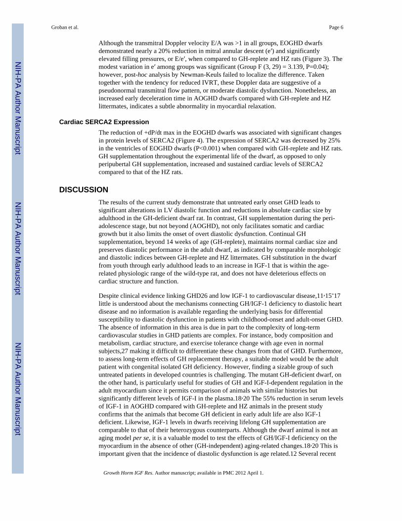
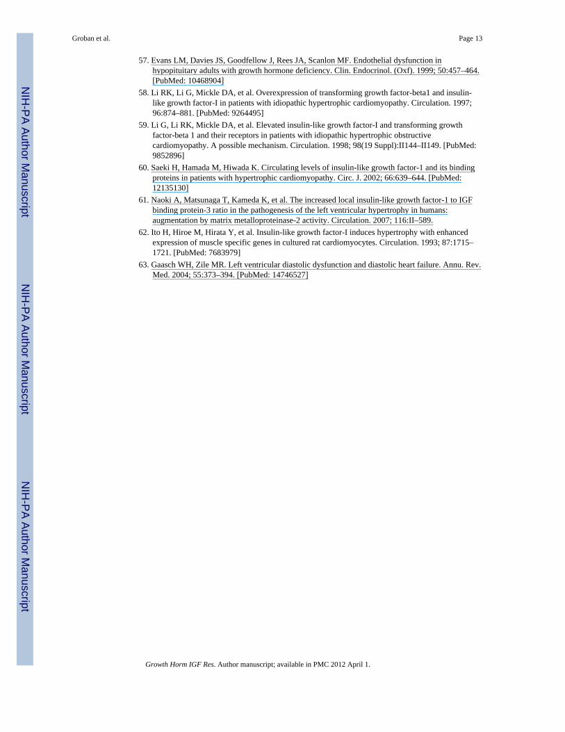
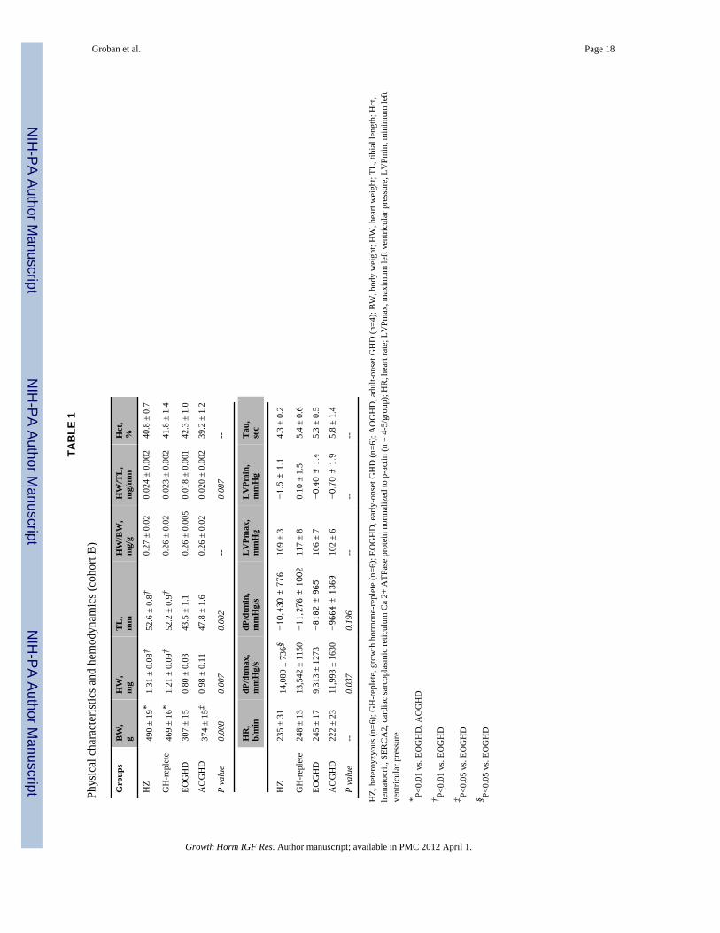
Physical characteristics from animals in cohort B are summarized in Table 1. At 26 weeks ofage, body weight was significantly reduced in EOGHD and AOGHD dwarfs whencompared to GH-replete littermates and HZ rats. Also, adult onset GHD dwarfs weighedmore than EOGHD dwarfs (P<0.05). As expected, tibial length was notably reduced inEOGHD dwarfs versus HZ and GH-replete littermates, and there was no difference in thisindex of somatic growth among HZ, GH-replete and AOGHD rats. Heart weight was lowestin EOGHD dwarfs, but when normalized to body weight or tibial length, no differenceswere observed among groups. Serum IGF-1 levels were 45 to 60% lower in AOGHD andEOGHD dwarfs, respectively, compared to age-matched GH-replete littermates and HZcontrols (Figure 2). Hematocrit did not show differences among the groups.
Invasive Hemodynamic StudiesResting hemodynamic data from 26-week-old rats in cohort B are summarized in Table 1.There were no differences in heart rate, left ventricular pressure, or the time constant ofrelaxation (tau) among groups. However, in the EOGHD dwarfs there was a 33% reductionin maximum rate of pressure rise (+dP/dt) (P<0.05) and more than a 20% reduction inmaximum rate pressure decline (−dP/dt) compared to HZ control rats, substantiating thepresence of cardiac functional deficits from longstanding GHD.
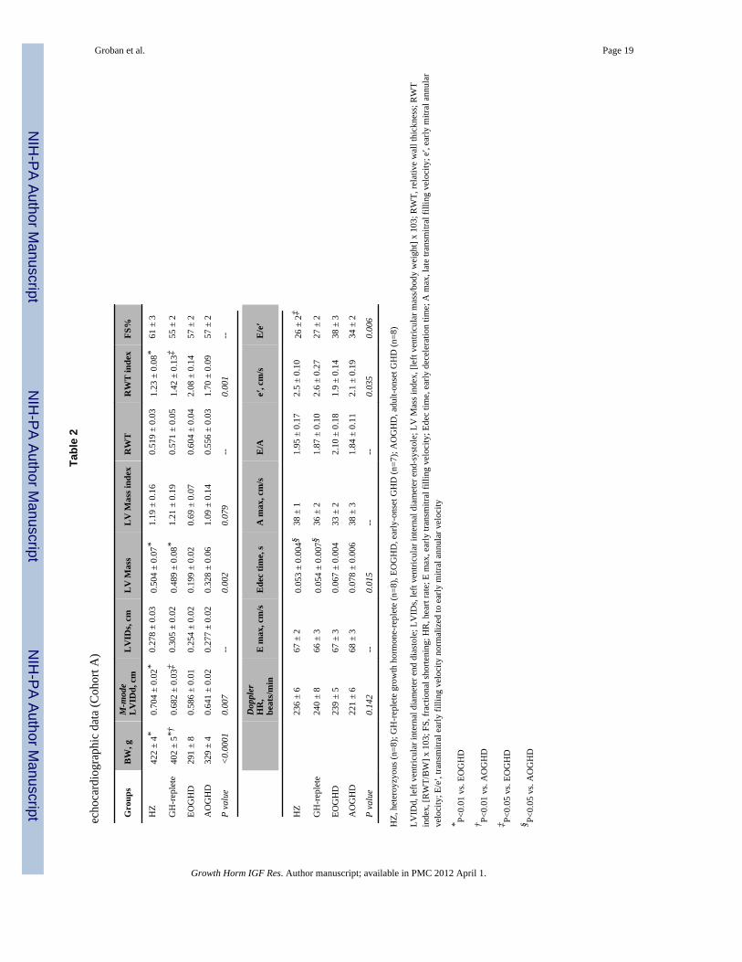
EchocardiographyM-mode and Doppler measurements of left ventricular structure and function obtained at 26weeks of age from animals in cohort A are summarized in Table 2. HZ and GH-replete ratshad a 1.5- and 2.5-fold greater left ventricular mass than EOGHD littermates. In EOGHDdwarfs, LV mass normalized for body weight was only minimally reduced compared to theother groups (P=0.07). The RWT normalized for body weight was significantly greater inEOGHD and AOGHD dwarfs compared to HZ controls and GH-replete dwarfs. Relativewall thickness indexed to body weight was also greater in EOGHD versus AOGHD dwarfs,most likely due to the reduced body weight in EOGHD rather than due to thicker walls inuntreated dwarfs compared to AOGHD dwarfs. Despite significantly reduced LVDD inEOGHD dwarfs compared to HZ and GH-replete dwarfs, the calculated index of systolicfunction, or percent fractional shortening, was similar among groups.
The effect of GH replacement therapy on heart rate and Doppler-derived indices of LVdiastolic function are shown in Table 2. Heart rate was similar among the 4 groups.
Groban et al. Page 5
Growth Horm IGF Res. Author manuscript; available in PMC 2012 April 1.
NIH
-PA Author Manuscript
NIH
-PA Author Manuscript
NIH
-PA Author Manuscript
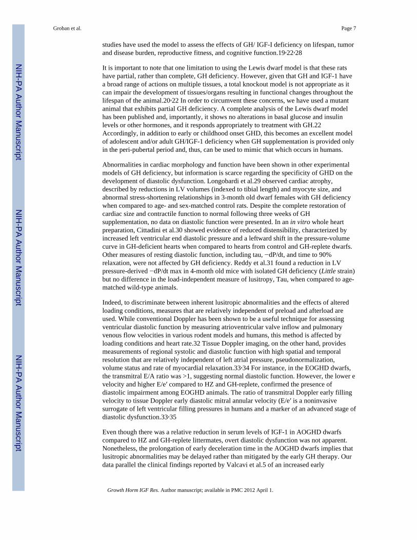
Although the transmitral Doppler velocity E/A was >1 in all groups, EOGHD dwarfsdemonstrated nearly a 20% reduction in mitral annular descent (e′) and significantlyelevated filling pressures, or E/e′, when compared to GH-replete and HZ rats (Figure 3). Themodest variation in e′ among groups was significant (Group F (3, 29) = 3.139, P=0.04);however, post-hoc analysis by Newman-Keuls failed to localize the difference. Takentogether with the tendency for reduced IVRT, these Doppler data are suggestive of apseudonormal transmitral flow pattern, or moderate diastolic dysfunction. Nonetheless, anincreased early deceleration time in AOGHD dwarfs compared with GH-replete and HZlittermates, indicates a subtle abnormality in myocardial relaxation.
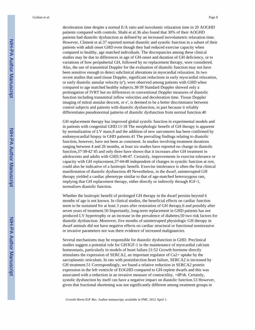
Cardiac SERCA2 ExpressionThe reduction of +dP/dt max in the EOGHD dwarfs was associated with significant changesin protein levels of SERCA2 (Figure 4). The expression of SERCA2 was decreased by 25%in the ventricles of EOGHD dwarfs (P<0.001) when compared with GH-replete and HZ rats.GH supplementation throughout the experimental life of the dwarf, as opposed to onlyperipubertal GH supplementation, increased and sustained cardiac levels of SERCA2compared to that of the HZ rats.
DISCUSSIONThe results of the current study demonstrate that untreated early onset GHD leads tosignificant alterations in LV diastolic function and reductions in absolute cardiac size byadulthood in the GH-deficient dwarf rat. In contrast, GH supplementation during the peri-adolescence stage, but not beyond (AOGHD), not only facilitates somatic and cardiacgrowth but it also limits the onset of overt diastolic dysfunction. Continual GHsupplementation, beyond 14 weeks of age (GH-replete), maintains normal cardiac size andpreserves diastolic performance in the adult dwarf, as indicated by comparable morphologicand diastolic indices between GH-replete and HZ littermates. GH substitution in the dwarffrom youth through early adulthood leads to an increase in IGF-1 that is within the age-related physiologic range of the wild-type rat, and does not have deleterious effects oncardiac structure and function.
Despite clinical evidence linking GHD26 and low IGF-1 to cardiovascular disease,11,15-17little is understood about the mechanisms connecting GH/IGF-1 deficiency to diastolic heartdisease and no information is available regarding the underlying basis for differentialsusceptibility to diastolic dysfunction in patients with childhood-onset and adult-onset GHD.The absence of information in this area is due in part to the complexity of long-termcardiovascular studies in GHD patients are complex. For instance, body composition andmetabolism, cardiac structure, and exercise tolerance change with age even in normalsubjects,27 making it difficult to differentiate these changes from that of GHD. Furthermore,to assess long-term effects of GH replacement therapy, a suitable model would be the adultpatient with congenital isolated GH deficiency. However, finding a sizable group of suchuntreated patients in developed countries is challenging. The mutant GH-deficient dwarf, onthe other hand, is particularly useful for studies of GH and IGF-I-dependent regulation in theadult myocardium since it permits comparison of animals with similar histories butsignificantly different levels of IGF-I in the plasma.18,20 The 55% reduction in serum levelsof IGF-1 in AOGHD compared with GH-replete and HZ animals in the present studyconfirms that the animals that become GH deficient in early adult life are also IGF-1deficient. Likewise, IGF-1 levels in dwarfs receiving lifelong GH supplementation arecomparable to that of their heterozygous counterparts. Although the dwarf animal is not anaging model per se, it is a valuable model to test the effects of GH/IGF-I deficiency on themyocardium in the absence of other (GH-independent) aging-related changes.18,20 This isimportant given that the incidence of diastolic dysfunction is age related.12 Several recent
Groban et al. Page 6
Growth Horm IGF Res. Author manuscript; available in PMC 2012 April 1.
NIH
-PA Author Manuscript
NIH
-PA Author Manuscript
NIH
-PA Author Manuscript
studies have used the model to assess the effects of GH/ IGF-I deficiency on lifespan, tumorand disease burden, reproductive fitness, and cognitive function.19,22,28
It is important to note that one limitation to using the Lewis dwarf model is that these ratshave partial, rather than complete, GH deficiency. However, given that GH and IGF-1 havea broad range of actions on multiple tissues, a total knockout model is not appropriate as itcan impair the development of tissues/organs resulting in functional changes throughout thelifespan of the animal.20,22 In order to circumvent these concerns, we have used a mutantanimal that exhibits partial GH deficiency. A complete analysis of the Lewis dwarf modelhas been published and, importantly, it shows no alterations in basal glucose and insulinlevels or other hormones, and it responds appropriately to treatment with GH.22Accordingly, in addition to early or childhood onset GHD, this becomes an excellent modelof adolescent and/or adult GH/IGF-1 deficiency when GH supplementation is provided onlyin the peri-pubertal period and, thus, can be used to mimic that which occurs in humans.
Abnormalities in cardiac morphology and function have been shown in other experimentalmodels of GH deficiency, but information is scarce regarding the specificity of GHD on thedevelopment of diastolic dysfunction. Longobardi et al.29 observed cardiac atrophy,described by reductions in LV volumes (indexed to tibial length) and myocyte size, andabnormal stress-shortening relationships in 3-month old dwarf females with GH deficiencywhen compared to age- and sex-matched control rats. Despite the complete restoration ofcardiac size and contractile function to normal following three weeks of GHsupplementation, no data on diastolic function were presented. In an in vitro whole heartpreparation, Cittadini et al.30 showed evidence of reduced distensibility, characterized byincreased left ventricular end diastolic pressure and a leftward shift in the pressure-volumecurve in GH-deficient hearts when compared to hearts from control and GH-replete dwarfs.Other measures of resting diastolic function, including tau, −dP/dt, and time to 90%relaxation, were not affected by GH deficiency. Reddy et al.31 found a reduction in LVpressure-derived −dP/dt max in 4-month old mice with isolated GH deficiency (Little strain)but no difference in the load-independent measure of lusitropy, Tau, when compared to age-matched wild-type animals.
Indeed, to discriminate between inherent lusitropic abnormalities and the effects of alteredloading conditions, measures that are relatively independent of preload and afterload areused. While conventional Doppler has been shown to be a useful technique for assessingventricular diastolic function by measuring atrioventricular valve inflow and pulmonaryvenous flow velocities in various rodent models and humans, this method is affected byloading conditions and heart rate.32 Tissue Doppler imaging, on the other hand, providesmeasurements of regional systolic and diastolic function with high spatial and temporalresolution that are relatively independent of left atrial pressure, pseudonormalization,volume status and rate of myocardial relaxation.33,34 For instance, in the EOGHD dwarfs,the transmitral E/A ratio was >1, suggesting normal diastolic function. However, the lower evelocity and higher E/e′ compared to HZ and GH-replete, confirmed the presence ofdiastolic impairment among EOGHD animals. The ratio of transmitral Doppler early fillingvelocity to tissue Doppler early diastolic mitral annular velocity (E/e′ is a noninvasivesurrogate of left ventricular filling pressures in humans and a marker of an advanced stage ofdiastolic dysfunction.33,35
Even though there was a relative reduction in serum levels of IGF-1 in AOGHD dwarfscompared to HZ and GH-replete littermates, overt diastolic dysfunction was not apparent.Nonetheless, the prolongation of early deceleration time in the AOGHD dwarfs implies thatlusitropic abnormalities may be delayed rather than mitigated by the early GH therapy. Ourdata parallel the clinical findings reported by Valcavi et al.5 of an increased early
Groban et al. Page 7
Growth Horm IGF Res. Author manuscript; available in PMC 2012 April 1.
NIH
-PA Author Manuscript
NIH
-PA Author Manuscript
NIH
-PA Author Manuscript
deceleration time despite a normal E/A ratio and isovolumic relaxation time in 20 AOGHDpatients compared with controls. Shahi et al.36 also found that 30% of their AOGHDpatients had diastolic dysfunction as defined by an increased isovolumetric relaxation time.However, Climent et al.37 reported normal diastolic and systolic function in a subset of theirpatients with adult onset GHD even though they had reduced exercise capacity whencompared to healthy, age matched individuals. The discrepancies among these clinicalstudies may be due to differences in age of GH-onset and duration of GH deficiency, or tovariations of how peripubertal GH, followed by no replacement therapy, were considered.Also, the use of transmitral Doppler for the evaluation of diastolic function may not havebeen sensitive enough to detect subclinical alterations in myocardial relaxation. In tworecent studies that used tissue Doppler, significant reductions in early myocardial relaxation,or early diastolic annular velocity (e′), were observed among patients with GHD whencompared to age matched healthy subjects.38,39 Standard Doppler showed only aprolongation of IVRT but no differences in conventional Doppler measures of diastolicfunction including transmitral inflow velocities and deceleration time. Tissue Dopplerimaging of mitral annular descent, or e′, is deemed to be a better discriminator betweencontrol subjects and patients with diastolic dysfunction, in part because it reliablydifferentiates pseudonormal patterns of diastolic dysfunction from normal function.40
GH replacement therapy has improved global systolic function in experimental models andin patients with congenital GHD.11,18 The morphologic benefit of GH therapy is apparentby normalization of LV mass,8 and the addition of new sarcomeres has been confirmed byendomyocardial biopsy in GHD patients.41 The prevailing findings relating to diastolicfunction, however, have not been as consistent. In studies involving treatment durationsranging between 4 and 26 months, at least six studies have reported no change in diastolicfunction,37,38,42-45 and only three have shown that it increases after GH treatment inadolescents and adults with GHD.5,46,47. Certainly, improvements in exercise tolerance orcapacity with GH replacement,37,44,48 independent of changes in systolic function at rest,could also be indicative of a lusitropic benefit. Exercise intolerance is often the first clinicalmanifestation of diastolic dysfunction.49 Nevertheless, in the dwarf, uninterrupted GHtherapy yielded a cardiac phenotype similar to that of age-matched heterozygous rats,implying that GH replacement therapy, either directly or indirectly through IGF-1,normalizes diastolic function.
Whether the lusitropic benefit of prolonged GH therapy in the dwarf persists beyond 6months of age is not known. In clinical studies, the beneficial effects on cardiac functionseem to be sustained for at least 3 years after restoration of GH therapy,6 and possibly afterseven years of treatment.50 Importantly, long-term replacement in GHD patients has notproduced LV hypertrophy or an increase in the prevalence of diabetes;50 two risk factors fordiastolic dysfunction. Moreover, five months of uninterrupted physiologic GH therapy indwarf animals did not have negative effects on cardiac structural or functional noninvasiveor invasive parameters nor was there evidence of increased malignancies.
Several mechanisms may be responsible for diastolic dysfunction in GHD. Preclinicalstudies suggest a potential role for GH/IGF-1 in the maintenance of myocardial calciumhomeostasis, particularly in models of heart failure.51,52 Growth hormone directlystimulates the expression of SERCA2, an important regulator of Ca2+ uptake by thesarcoplasmic reticulum. In rats with postinfarction heart failure, SERCA2 is increased byGH treatment.51 Correspondingly, we found a relative reduction in SERCA2 proteinexpression in the left ventricle of EOGHD compared to GH-replete dwarfs and this wasassociated with a reduction in an invasive measure of contractility, +dP/dt. Certainly,systolic dysfunction by itself can have a negative impact on diastolic function.53 However,given that fractional shortening was not significantly different among treatment groups in
Groban et al. Page 8
Growth Horm IGF Res. Author manuscript; available in PMC 2012 April 1.
NIH
-PA Author Manuscript
NIH
-PA Author Manuscript
NIH
-PA Author Manuscript
the cohort of animals that underwent echocardiograms, implies that systolic dysfunction, perse, was likely not the principle cause for the reduction in myocardial relaxation in GHDdwarfs. Evidence from various experimental and human models indicates that SERCA2adown-regulation might also contribute to diastolic heart disease.54,55 Moreover, we havepreviously reported its relation to the tissue Doppler index of lusitropy, e′, in anexperimental aging model.23,24 Indeed, the negative lusitropic effects of GHD could alsobe the result of GHD-mediated vascular aberrations as recently reported by Ungvari et al.28Moreover, patients with GHD have reduced aortic distensibility and endothelial dysfunctionwhich are potent stimuli for increasing left ventricular load and impairing left ventriculardiastolic relaxation56 and endothelial dysfunction.57 Even though we did not assessvascular morphology or function in the present study, conscious blood pressures measuredby telemetry were similar between dwarf rats and their age-matched heterozygouscounterparts.22 Whether long-term GH deficiency directly influences the myocardialdeterminants of stiffness, including the extracellular matrix, cytoskeleton, or myofilaments,is not clear. All the same, the increase in E/e′ in EOGHD dwarfs suggests an upward shift inthe diastolic pressure-volume relationship.
Besides GH/IGF-1's endocrine effects, the autocrine/paracrine functions of IGF-1 and itsbinding proteins may also contribute to the cardiac phenotype. Several reports have shownthat the expression of the mRNA and protein levels for IGF-1 are elevated in themyocardium58,59 and the circulation60 of hypertrophic cardiomyopathy patients. In cardiacsurgical patients with aortic and mitral valve disease, pericardial total IGF-1/IGFBP-3 ratiowas positively correlated with left ventricular mass index, further suggesting that the localIGF-1 system plays a role in the pathogenesis of LV hypertrophy.61 An interaction betweenIGF-1 and IGFBP-3 in the regulation of hypertrophy has also been implied in anexperimental study. Ito et al.62 showed that that IGF-I has a direct hypertrophic effect oncultured rat cardiomyocytes with concomitant increases in the transcripts of muscle-specificgenes (MLC-2, troponin I, and skeletal a-actin) and that IGFBP-3 has an inhibitory effect onthe IGF-I-induced cardiac hypertrophy in vitro. Although we did not measure local IGFbinding proteins in the present study, Ungvari et al.28 showed that expression of IGFBP1,IGFBP2, and IGFBP4 did not change in the cerebrovascular wall of the dwarf but that therewas a tendency for IGFBP3 to be decreased. Thus, future studies are warranted to determinewhether cardiac tissue expression of components of the IGF-1 system is related to thediastolic dysfunction phenotype of GHD.
Cardiovascular disease is the leading cause of premature death in adults with GHD. Thereasons for this are presumed to be due to an increase in cardiovascular risk factors,including hyperlipidemia, atherosclerosis, endothelial dysfunction, and intimal thickening.11However, since diastolic abnormalities may be the earliest manifestations of heart disease,58it is important to document their occurrence in GHD individuals. Also, since GH treatmentin children with GHD is often discontinued in adolescence after attainment of final height,the clinical concern of premature diastolic dysfunction becomes valid. Using the LewisDwarf model, we have found significant impairments in diastolic functional parameters inEOGHD compared with heterozygous and GH-replete dwarfs by means of tissue Dopplerimaging. Although overt diastolic dysfunction was not observed in AOGHD dwarfs, thesubtle prolongation in early filling implies that the lusitropic effects of peri-pubertal GHmay not have lasting benefits. Given that ventricular diastolic dysfunction, whether fromaging or from underlying disease, is a strong predictor of mortality,35 these experimentaldata support the consideration that GH therapy in individuals with childhood onset GHDshould be continued into adulthood.
Groban et al. Page 9
Growth Horm IGF Res. Author manuscript; available in PMC 2012 April 1.
NIH
-PA Author Manuscript
NIH
-PA Author Manuscript
NIH
-PA Author Manuscript
AcknowledgmentsFUNDING
Funding was provided by the National Institute of Aging K08-AG026764-05 to LG and P01 AG011370 and R01AG26607 to WES.
REFERENCES1. Saccà L, Cittadini A, Fazio S. Growth hormone and the heart. Endocr. Rev. 1994; 15:555–573.
[PubMed: 7843068]2. Merola B, Cittadini A, Colao A, et al. Cardiac structural and functional abnormalities in adult
patients with growth hormone deficiency. J. Clin. Endocrinol. Metab. 1993; 77:1658–1661.[PubMed: 8263155]
3. Longobardi S, Cuocolo A, Merola B, et al. Left ventricular function in young adults with childhoodand adulthood onset growth hormone deficiency. Clin. Endocrinol. (Oxf). 1998; 48:137–143.[PubMed: 9579223]
4. Amato G, Carella C, Fazio S, et al. Body composition, bone metabolism, and heart structure andfunction in growth hormone (GH)-deficient adults before and after GH replacement therapy at lowdoses. J. Clin. Endocrinol. Metab. 1993; 77:1671–1676. [PubMed: 8263158]
5. Valcavi R, Gaddi O, Zini M, Iavicoli M, Mellino U, Portioli I. Cardiac performance and mass inadults with hypopituitarism: effects of one year of growth hormone treatment. J. Clin. Endocrinol.Metab. 1995; 80:659–666. [PubMed: 7852533]
6. Johannsson G, Bengtsson BA, Andersson B, Isgaard J, Caidahl K. Long-term cardiovascular effectsof growth hormone treatment in GH-deficient adults. Preliminary data in a small group of patients.Clin. Endocrinol. (Oxf). 1996; 45:305–314. [PubMed: 8949568]
7. McGauley GA. Quality of life assessment before and after growth hormone treatment in adults withgrowth hormone deficiency. Acta. Paediatr. Scand. Suppl. 1989; 356:70–74. [PubMed: 2816361]
8. Maison P, Chanson P. Cardiac effects of growth hormone in adults with growth hormonedeficiency: a meta-analysis. Circulation. 2003; 108:2648–2652. [PubMed: 14623813]
9. Eledrisi, MS. Growth hormone deficiency. eMedicine: Endocrinology. Available at:http://emedicine.medscape.com/article/120767-overview. Accessed July 8, 2010
10. Molitch ME, Clemmons DR, Malozowski S, Merriam GR, Shalet SM, Vance ML, EndocrineSociety's Clinical Guidelines Subcommittee. Evaluation and treatment of adult growth hormonedeficiency: an Endocrine Society Clinical Practice Guideline. J. Clin. Endocrinol. Metab. 2006;91:1621–1634. [PubMed: 16636129]
11. Colao A. The GH-IGF-I axis and the cardiovascular system: clinical implications. Clin.Endocrinol. (Oxf). 2008; 69:347–358. [PubMed: 18462260]
12. Redfield MM, Jacobsen SJ, Burnett JC Jr, Mahoney DW, Bailey KR, Rodeheffer RJ. Burden ofsystolic and diastolic ventricular dysfunction in the community: appreciating the scope of the heartfailure epidemic. JAMA. 2003; 289:194–202. [PubMed: 12517230]
13. Kitzman DW, Gardin JM, et al. Cardiovascular Health Study Research Group. Importance of heartfailure with preserved systolic function in patients > or = 65 years of age. CHS Research Group.Cardiovascular Health Study. Am. J. Cardiol. 2001; 87:413–419. [PubMed: 11179524]
14. Brogan WC III, Hillis LD, Flores ED, Lange RA. The natural history of isolated left ventriculardiastolic dysfunction. Am. J. Med. 1992; 92:627–630. [PubMed: 1605144]
15. Vasan RS, Sullivan LM, D'Agostino RB, et al. Serum insulin-like growth factor I and risk for heartfailure in elderly individuals without a previous myocardial infarction: the Framingham HeartStudy. Ann. Intern. Med. 2003; 139:642–648. [PubMed: 14568852]
16. Petretta M M, Colao A A, Sardu C, et al. NT-proBNP, IGF-I and survival in patients with chronicheart failure. Growth. Horm. IGF. Res. 2007; 17:288–296. [PubMed: 17383209]
17. Cittadini A, Saldamarco L, Marra AM, et al. Growth hormone deficiency in patients with chronicheart failure and beneficial effects of its correction. J. Clin. Endocrinol. Metab. 2009; 94:3329–3336. [PubMed: 19584187]
Groban et al. Page 10
Growth Horm IGF Res. Author manuscript; available in PMC 2012 April 1.
NIH
-PA Author Manuscript
NIH
-PA Author Manuscript
NIH
-PA Author Manuscript
18. Khan AS, Sane DC, Wannenburg T, Sonntag WE. Growth hormone, insulin-like growth factor-1and the aging cardiovascular system. Cardiovasc. Res. 2002; 54:25–35. [PubMed: 12062358]
19. Nieves-Martinez E, Sonntag WE, Wilson A, et al. Early-onset GH deficiency results in spatialmemory impairment in mid-life and is prevented by GH supplementation. J. Endocrinol. 2010;204:31–6. [PubMed: 19815586]
20. Carter CS, Ramsey MM, Ingram RL, et al. Models of growth hormone and IGF-1 deficiency:applications to studies of aging processes and life-span determination. J. Gerontol. A. Biol. Sci.Med. Sci. 2002; 57:B177–B188. [PubMed: 11983714]
21. Charlton HM, Clark RG, Robinson IC, et al. Growth hormone-deficient dwarfism in the rat: a newmutation. J. Endocrinol. 1988; 119:51–58. [PubMed: 3193048]
22. Sonntag WE, Carter CS, Ikeno Y, et al. Adult-onset growth hormone and insulin-like growth factorI deficiency reduces neoplastic disease, modifies age-related pathology, and increases life span.Endocrinology. 2005; 146:2920–2932. [PubMed: 15790724]
23. Groban L, Pailes NA, Bennett CD, et al. Growth hormone replacement attenuates diastolicdysfunction and cardiac angiotensin II expression in senescent rats. J. Gerontol. A. Biol. Sci. Med.Sci. 2006; 61:28–35. [PubMed: 16456192]
24. Groban L, Jobe H, Lin M, Houle T, Kitzman DA, Sonntag W. Effects of short-term treadmillexercise training or growth hormone supplementation on diastolic function and exercise tolerancein old rats. J. Gerontol. A. Biol. Sci. Med. Sci. 2008; 63:911–920. [PubMed: 18840795]
25. Adams MM, Forbes ME, Linville MC, Riddle DR, Sonntag WE, BrunsoBechtold JK. Stability oflocal brain levels of insulin-like growth factor-I in two well-characterized models of decreasedplasma IGF-I. Growth Factors. 2009; 27:181–188. [PubMed: 19343576]
26. Bengtsson BA. The consequences of growth hormone deficiency in adults. Acta. Endocrinol.(Copenh). 1993; 128(Suppl 2):2–5. [PubMed: 8342387]
27. Toogood AA, Adams JE, O'Neill PA, Shalet SM. Body composition in growth hormone deficientadults over the age of 60 years. Clin. Endocrinol. (Oxf). 1996; 45:399–405. [PubMed: 8959077]
28. Ungvari Z, Gautam T, Koncz P, C J, et al. Vasoprotective effects of life span-extendingperipubertal GH replacement in Lewis dwarf rats. J. Gerontol. A Biol. Sci. Med. Sci. 2010;65:1145–1156. [PubMed: 20713653]
29. Longobardi S, Cittadini A, Strömer H, et al. Echocardiographic assessment of cardiac morphologyand function in mutant dwarf rats. Growth. Horm. IGF. Res. 2000; 10:242–247. [PubMed:11042020]
30. Cittadini A, Stromer H, Katz SE, et al. Differential cardiac effects of growth hormone and insulin-like growth factor-1 in the rat. A combined in vivo and in vitro evaluation. Circulation. 1996;93:800–809. [PubMed: 8641010]
31. Reddy AK, Amador-Noguez D, Darlington GJ, et al. Cardiac function in young and old Littlemice. J. Gerontol. A. Biol. Sci. Med. Sci. 2007; 62:1319–1325. [PubMed: 18166681]
32. Oh JK, Appleton CP, Hatle LK, Nishimura RA, Seward JB, Tajik AJ. The noninvasive assessmentof left ventricular diastolic function with two-dimensional and Doppler echocardiography. J. Am.Soc. Echocardiogr. 1997; 10:246–270. [PubMed: 9109691]
33. Nagueh SF, Middleton KJ, Kopelen HA, Zoghbi WA, Quiñones MA. Doppler tissue imaging: anoninvasive technique for evaluation of left ventricular relaxation and estimation of fillingpressures. J. Am. Coll. Cardiol. 1997; 30:1527–1533. [PubMed: 9362412]
34. Garcia MJ, Thomas JD, Klein AL. New Doppler echocardiographic applications for the study ofdiastolic function. J. Am. Coll. Cardiol. 1998; 32:865–875. [PubMed: 9768704]
35. Yu CM, Sanderson JE, Marwick TH, Oh JK. Tissue Doppler imaging a new prognosticator forcardiovascular diseases. J. Am. Coll. Cardiol. 2007; 49:1903–1914. [PubMed: 17498573]
36. Shahi M, Beshyah SA, Hackett D, Sharp PS, Johnston DG, Foale RA. Myocardial dysfunction intreated adult hypopituitarism: a possible explanation for increased cardiovascular mortality. Br.Heart. J. 1992; 67:92–96. [PubMed: 1739534]
37. Climent VE, Picó A, Sogorb F, Aznar S, Lip GY, Marín F. Growth hormone therapy and the heart.Am. J. Cardiol. 2006; 97:1097–1102. [PubMed: 16563925]
Groban et al. Page 11
Growth Horm IGF Res. Author manuscript; available in PMC 2012 April 1.
NIH
-PA Author Manuscript
NIH
-PA Author Manuscript
NIH
-PA Author Manuscript
38. Ozdogru I, Tanriverdi F, Dogan A, et al. Impaired longitudinal myocardial velocities in patientswith growth hormone deficiency improves after hormone replacement therapy. J. Am. Soc.Echocardiogr. 2007; 20:1093–1099. [PubMed: 17555934]
39. Yurci A, Oflaz H, Meric M, N Ozbey. Mitral and tricuspid annular velocities determined byDoppler tissue imaging in hypopituitary, growth hormone-deficient patients. Horm. Res. 2005;64:107–114. [PubMed: 16166781]
40. Farias CA, Rodriguez L, Garcia MJ, Sun JP, Klein AL, Thomas JD. Assessment of diastolicfunction by tissue Doppler echocardiography: comparison with standard transmitral andpulmonary venous flow. J. Am. Soc. Echocardiogr. 1999; 12:609–617. [PubMed: 10441216]
41. Frustaci A, Perrone GA, Gentiloni N, Russo MA. Reversible dilated cardiomyopathy due togrowth hormone deficiency. Am. J. Clin. Pathol. 1992; 197:503–511. [PubMed: 1553916]
42. Sneppen SB, Steensgaard-Hansen F, Feldt-Rasmussen U. Cardiac effects of low-dose growthhormone replacement therapy in growth hormone-deficient adults. An 18-month randomised,placebo-controlled, double-blind study. Horm. Res. 2002; 58:21–29. [PubMed: 12169777]
43. Ezzat S, Fear S, Gaillard RC, et al. Gender-specific responses of lean body composition and non-gender-specific cardiac function improvement after GH replacement in GH-deficient adults. J.Clin. Endocrinol. Metab. 2002; 87:2725–2733. [PubMed: 12050241]
44. Elgzyri T, Castenfors J, Hägg E, Backman C, Thorén M, Bramnert M. The effects of GHreplacement therapy on cardiac morphology and function, exercise capacity and serum lipids inelderly patients with GH deficiency. Clin. Endocrinol. (Oxf). 2004; 61:113–122. [PubMed:15212653]
45. Minczykowski A, Gryczynska M, Ziemnicka K, Czepczynski R, Sowinski J, Wysocki H. Theinfluence of growth hormone (GH) therapy on cardiac performance in patients with childhoodonset GH deficiency. Growth. Horm. I.G.F. Res. 2005; 15:156–164.
46. Colao A, Di Somma C, Salerno M, Spinelli L, Orio F, Lombardi G. The cardiovascular risk of GH-deficient adolescents. J. Clin. Endocrinol. Metab. 2002; 87:3650–3655. [PubMed: 12161490]
47. Colao A, di Somma C, Pivonello R, et al. The cardiovascular risk of adult GH deficiency (GHD)improved after GH replacement and worsened in untreated GHD: a 12-month prospective study. J.Clin. Endocrinol. Metab. 2002; 87:1088–1093. [PubMed: 11889170]
48. Beshyah SA, Shahi M, Foale R, Johnston DG. Cardiovascular effects of prolonged growthhormone replacement in adults. J. Intern. Med. 1995; 237:35–42. [PubMed: 7830028]
49. Kitzman DW, Groban L. Exercise intolerance. Heart. Fail. Clin. 2008; 4:99–115. [PubMed:18313628]
50. Chrisoulidou A, Beshyah SA, Rutherford O, et al. Effects of 7 years of growth hormonereplacement therapy in hypopituitary adults. J. Clin. Endocrinol. Metab. 2000; 85:3762–3769.[PubMed: 11061536]
51. Tajima M, Weinberg EO, Bartunek J, et al. Treatment with growth hormone enhances contractilereserve and intracellular calcium transients in myocytes from rats with postinfarction heart failure.Circulation. 1999; 99:127–134. [PubMed: 9884389]
52. Ueyama T, Ohkusa T, Yano M, Matsuzaki M. Growth hormone preserves cardiac sarcoplasmicreticulum Ca2+ release channels (ryanodine receptors) and enhances cardiac function incardiomyopathic hamsters. Cardiovasc. Res. 1998; 40:64–73. [PubMed: 9876318]
53. Ouzounian M, Lee DS, Liu PP. Diastolic heart failure: mechanisms and controversies. Nat. Clin.Pract. Cardiovasc. Med. 2008; 5:375–386. [PubMed: 18542106]
54. Hasenfuss G, Schillinger W, Lehnart SE, et al. Relationship between Na+-Ca2+-exchanger proteinlevels and diastolic function of failing human myocardium. Circulation. 1999; 99:641–648.[PubMed: 9950661]
55. Stüdeli R, Jung S, Mohacsi P, et al. Diastolic dysfunction in human cardiac allografts is relatedwith reduced SERCA2a gene expression. Am. J. Transplant. 2006; 6:775–782. [PubMed:16539635]
56. Lehmann ED, Hopkins KD, Weissberger AJ, Gosling RG, Sönksen PH. Aortic distensibility ingrowth hormone deficient adults. Lancet. 1993; 341:309. [PubMed: 8093946]
Groban et al. Page 12
Growth Horm IGF Res. Author manuscript; available in PMC 2012 April 1.
NIH
-PA Author Manuscript
NIH
-PA Author Manuscript
NIH
-PA Author Manuscript
57. Evans LM, Davies JS, Goodfellow J, Rees JA, Scanlon MF. Endothelial dysfunction inhypopituitary adults with growth hormone deficiency. Clin. Endocrinol. (Oxf). 1999; 50:457–464.[PubMed: 10468904]
58. Li RK, Li G, Mickle DA, et al. Overexpression of transforming growth factor-beta1 and insulin-like growth factor-I in patients with idiopathic hypertrophic cardiomyopathy. Circulation. 1997;96:874–881. [PubMed: 9264495]
59. Li G, Li RK, Mickle DA, et al. Elevated insulin-like growth factor-I and transforming growthfactor-beta 1 and their receptors in patients with idiopathic hypertrophic obstructivecardiomyopathy. A possible mechanism. Circulation. 1998; 98(19 Suppl):II144–II149. [PubMed:9852896]
60. Saeki H, Hamada M, Hiwada K. Circulating levels of insulin-like growth factor-1 and its bindingproteins in patients with hypertrophic cardiomyopathy. Circ. J. 2002; 66:639–644. [PubMed:12135130]
61. Naoki A, Matsunaga T, Kameda K, et al. The increased local insulin-like growth factor-1 to IGFbinding protein-3 ratio in the pathogenesis of the left ventricular hypertrophy in humans:augmentation by matrix metalloproteinase-2 activity. Circulation. 2007; 116:II–589.
62. Ito H, Hiroe M, Hirata Y, et al. Insulin-like growth factor-I induces hypertrophy with enhancedexpression of muscle specific genes in cultured rat cardiomyocytes. Circulation. 1993; 87:1715–1721. [PubMed: 7683979]
63. Gaasch WH, Zile MR. Left ventricular diastolic dysfunction and diastolic heart failure. Annu. Rev.Med. 2004; 55:373–394. [PubMed: 14746527]
Groban et al. Page 13
Growth Horm IGF Res. Author manuscript; available in PMC 2012 April 1.
NIH
-PA Author Manuscript
NIH
-PA Author Manuscript
NIH
-PA Author Manuscript

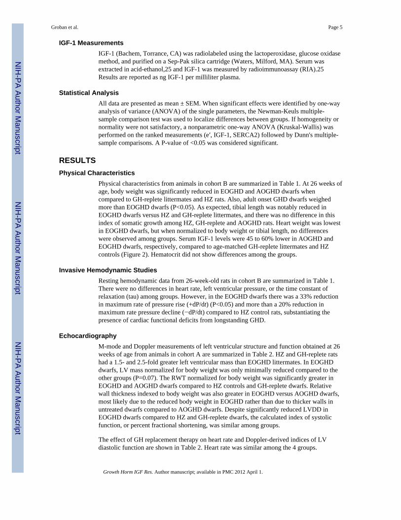
Figure 1.Model representing heterozygous, GH-replete, Early-onset GH deficient, and Adult-onsetGH deficient rats. Normal-sized females heterozygous for the dwarf trait (dw/+) were matedwith homozygous dwarf males to produce heterozygous and homozygous (dw/dw)littermates. Dark areas represent high levels of IGF-1.
Groban et al. Page 14
Growth Horm IGF Res. Author manuscript; available in PMC 2012 April 1.
NIH
-PA Author Manuscript
NIH
-PA Author Manuscript
NIH
-PA Author Manuscript
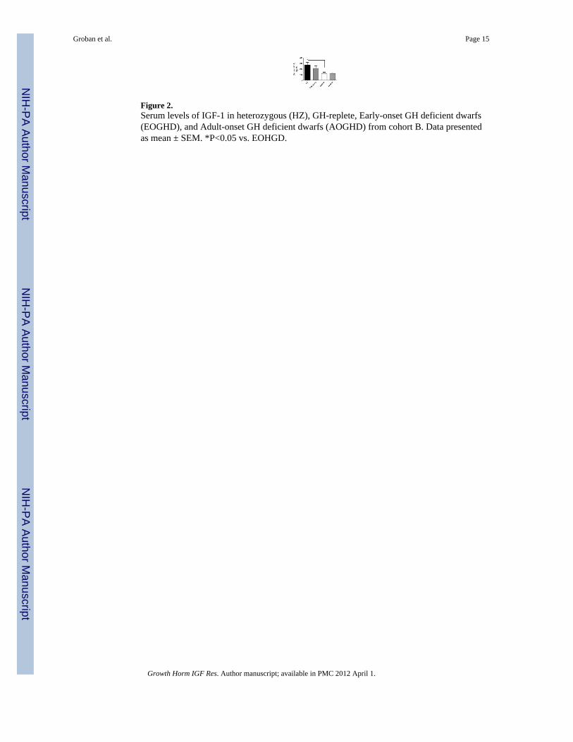
Figure 2.Serum levels of IGF-1 in heterozygous (HZ), GH-replete, Early-onset GH deficient dwarfs(EOGHD), and Adult-onset GH deficient dwarfs (AOGHD) from cohort B. Data presentedas mean ± SEM. *P<0.05 vs. EOHGD.
Groban et al. Page 15
Growth Horm IGF Res. Author manuscript; available in PMC 2012 April 1.
NIH
-PA Author Manuscript
NIH
-PA Author Manuscript
NIH
-PA Author Manuscript
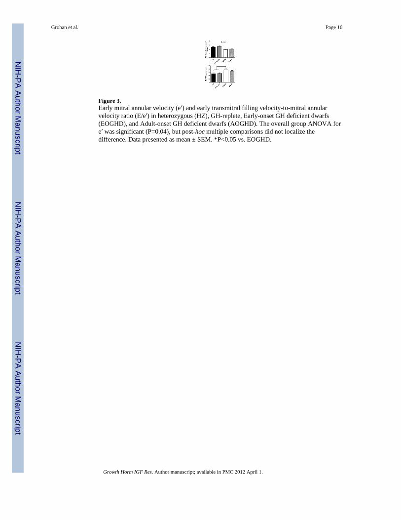
Figure 3.Early mitral annular velocity (e′) and early transmitral filling velocity-to-mitral annularvelocity ratio (E/e′) in heterozygous (HZ), GH-replete, Early-onset GH deficient dwarfs(EOGHD), and Adult-onset GH deficient dwarfs (AOGHD). The overall group ANOVA fore′ was significant (P=0.04), but post-hoc multiple comparisons did not localize thedifference. Data presented as mean ± SEM. *P<0.05 vs. EOGHD.
Groban et al. Page 16
Growth Horm IGF Res. Author manuscript; available in PMC 2012 April 1.
NIH
-PA Author Manuscript
NIH
-PA Author Manuscript
NIH
-PA Author Manuscript
Figure 4.SERCA2 protein normalized to β-actin among GH-replete, Early-onset GH deficient dwarfs(EOGHD), and Adult-onset GH deficient dwarfs (AOGHD) are expressed relative toheterozygous (HZ) band intensity. Data represent mean ± SEM (n= 4-5 hearts/group). *P<0.05 vs. EOGHD. Representative SERCA2 immunoblot from each group is shown below.GH = GH-replete dwarf; EO = Early-onset GH deficient dwarf; AO = Adult-onset GHdeficient dwarf.
Groban et al. Page 17
Growth Horm IGF Res. Author manuscript; available in PMC 2012 April 1.
NIH
-PA Author Manuscript
NIH
-PA Author Manuscript
NIH
-PA Author Manuscript
NIH
-PA Author Manuscript
NIH
-PA Author Manuscript
NIH
-PA Author Manuscript
Groban et al. Page 18
TAB
LE 1
Phys
ical
cha
ract
eris
tics a
nd h
emod
ynam
ics (
coho
rt B
)
Gro
ups
BW
,g
HW
,m
gT
L,
mm
HW
/BW
,m
g/g
HW
/TL
,m
g/m
mH
ct,
%
HZ
490
± 19
*1.
31 ±
0.0
8†52
.6 ±
0.8
†0.
27 ±
0.0
20.
024
± 0.
002
40.8
± 0
.7
GH
-rep
lete
469
± 16
*1.
21 ±
0.0
9†52
.2 ±
0.9
†0.
26 ±
0.0
20.
023
± 0.
002
41.8
± 1
.4
EOG
HD
307
± 15
0.80
± 0
.03
43.5
± 1
.10.
26 ±
0.0
050.
018
± 0.
001
42.3
± 1
.0
AO
GH
D37
4 ±
15‡
0.98
± 0
.11
47.8
± 1
.60.
26 ±
0.0
20.
020
± 0.
002
39.2
± 1
.2
P va
lue
0.00
80.
007
0.00
2--
0.08
7--
HR
,b/
min
dP/d
tmax
,m
mH
g/s
dP/d
tmin
,m
mH
g/s
LV
Pmax
,m
mH
gL
VPm
in,
mm
Hg
Tau
,se
c
HZ
235
± 31
14,0
80 ±
736
§−10
,430
± 7
7610
9 ±
3−1.
5 ±
1.1
4.3
± 0.
2
GH
-rep
lete
248
± 13
13,5
42 ±
115
0−11
,276
± 1
002
117
± 8
0.10
± 1
.55.
4 ±
0.6
EOG
HD
245
± 17
9,31
3 ±
1273
−81
82 ±
965
106
± 7
−0.
40 ±
1.4
5.3
± 0.
5
AO
GH
D22
2 ±
2311
,993
± 1
630
−96
64 ±
136
910
2 ±
6−0.
70 ±
1.9
5.8
± 1.
4
P va
lue
--0.
037
0.19
6--
----
HZ,
het
eroy
zyou
s (n=
6); G
H-r
eple
te, g
row
th h
orm
one-
repl
ete
(n=6
); EO
GH
D, e
arly
-ons
et G
HD
(n=6
); A
OG
HD
, adu
lt-on
set G
HD
(n=4
); B
W, b
ody
wei
ght;
HW
, hea
rt w
eigh
t; TL
, tib
ial l
engt
h; H
ct,
hem
atoc
rit, S
ERC
A2,
car
diac
sarc
opla
smic
retic
ulum
Ca
2+ A
TPas
e pr
otei
n no
rmal
ized
to p
-act
in (n
= 4
-5/g
roup
); H
R, h
eart
rate
; LV
Pmax
, max
imum
left
vent
ricul
ar p
ress
ure,
LV
Pmin
, min
imum
left
vent
ricul
ar p
ress
ure
* P<0.
01 v
s. EO
GH
D, A
OG
HD
† P<0.
01 v
s. EO
GH
D
‡ P<0.
05 v
s. EO
GH
D
§ P<0.
05 v
s. EO
GH
D
Growth Horm IGF Res. Author manuscript; available in PMC 2012 April 1.
NIH
-PA Author Manuscript
NIH
-PA Author Manuscript
NIH
-PA Author Manuscript
Groban et al. Page 19
Tabl
e 2
echo
card
iogr
aphi
c da
ta (C
ohor
t A)
Gro
ups
BW
, gM
-mod
eL
VID
d, c
mL
VID
s, cm
LV
Mas
sL
V M
ass i
ndex
RW
TR
WT
inde
xFS
%
HZ
422
± 4*
0.70
4 ±
0.02
*0.
278
± 0.
030.
504
± 0.
07*
1.19
± 0
.16
0.51
9 ±
0.03
1.23
± 0
.08*
61 ±
3
GH
-rep
lete
402
± 5*
†0.
682
± 0.
03‡
0.30
5 ±
0.02
0.48
9 ±
0.08
*1.
21 ±
0.1
90.
571
± 0.
051.
42 ±
0.1
3‡55
± 2
EOG
HD
291
± 8
0.58
6 ±
0.01
0.25
4 ±
0.02
0.19
9 ±
0.02
0.69
± 0
.07
0.60
4 ±
0.04
2.08
± 0
.14
57 ±
2
AO
GH
D32
9 ±
40.
641
± 0.
020.
277
± 0.
020.
328
± 0.
061.
09 ±
0.1
40.
556
± 0.
031.
70 ±
0.0
957
± 2
P va
lue
<0.
0001
0.00
7--
0.00
20.
079
--0.
001
--
Dop
pler
HR
,be
ats/
min
E m
ax, c
m/s
Ede
c tim
e, s
A m
ax, c
m/s
E/A
e′, c
m/s
E/e′
HZ
236
± 6
67 ±
20.
053
± 0.
004§
38 ±
11.
95 ±
0.1
72.
5 ±
0.10
26 ±
2‡
GH
-rep
lete
240
± 8
66 ±
30.
054
± 0.
007§
36 ±
21.
87 ±
0.1
02.
6 ±
0.27
27 ±
2
EOG
HD
239
± 5
67 ±
30.
067
± 0.
004
33 ±
22.
10 ±
0.1
81.
9 ±
0.14
38 ±
3
AO
GH
D22
1 ±
668
± 3
0.07
8 ±
0.00
638
± 3
1.84
± 0
.11
2.1
± 0.
1934
± 2
P va
lue
0.14
2--
0.01
5--
--0.
035
0.00
6
HZ,
het
eroy
zyou
s (n=
8); G
H-r
eple
te g
row
th h
orm
one-
repl
ete
(n=8
), EO
GH
D, e
arly
-ons
et G
HD
(n=7
); A
OG
HD
, adu
lt-on
set G
HD
(n=8
)
LVID
d, le
ft ve
ntric
ular
inte
rnal
dia
met
er e
nd d
iast
ole;
LV
IDs,
left
vent
ricul
ar in
tern
al d
iam
eter
end
-sys
tole
; LV
Mas
s ind
ex, [
left
vent
ricul
ar m
ass/
body
wei
ght]
x 10
3; R
WT,
rela
tive
wal
l thi
ckne
ss; R
WT
inde
x, [R
WT/
BW
] x 1
03; F
S, fr
actio
nal s
horte
ning
; HR
, hea
rt ra
te; E
max
, ear
ly tr
ansm
itral
filli
ng v
eloc
ity; E
dec
time,
ear
ly d
ecel
erat
ion
time;
A m
ax, l
ate
trans
mitr
al fi
lling
vel
ocity
; e′,
early
mitr
al a
nnul
arve
loci
ty; E
/e′,
trans
mitr
al e
arly
filli
ng v
eloc
ity n
orm
aliz
ed to
ear
ly m
itral
ann
ular
vel
ocity
* P<0.
01 v
s. EO
GH
D
† P<0.
01 v
s. A
OG
HD
‡ P<0.
05 v
s. EO
GH
D
§ P<0.
05 v
s. A
OG
HD
Growth Horm IGF Res. Author manuscript; available in PMC 2012 April 1.

![[Epidemiological evaluation of a rapidly-prevented tularemia outbreak in Canakkale province, Turkey]](https://img.pdfslide.net/doc/110x75/6350e33cb4766da83b03646d/epidemiological-evaluation-of-a-rapidly-prevented-tularemia-outbreak-in-canakkale.jpg)


