Embed Size (px)
Citation preview

JPET #66852 1
Effect of BM-573, a dual thromboxane synthase inhibitor and TP receptor
antagonist, in a porcine model of acute pulmonary embolism.
Alexandre Ghuysen, MD; Bernard Lambermont, MD, PhD; Jean-Michel Dogné, PhD;
Philippe Kolh, MD, PhD; Vincent Tchana-Sato, MD; Philippe Morimont, MD; David
Magis; Julien Hanson; Patrick Segers, PhD; Vincent D’Orio, MD, PhD.
From the Hemodynamics Research Laboratory (HemoLiège) (A.G., B.L., P.K., V.T-
S., P.M., V.D.), the laboratory of Medicinal Chemistry (J-M.D., J.H.) and the
department of Statistics (D.M.), University of Liège, Belgium; the hydraulics
Laboratory, University of Ghent, Belgium (P.S.).
JPET Fast Forward. Published on April 30, 2004 as DOI:10.1124/jpet.104.066852
Copyright 2004 by the American Society for Pharmacology and Experimental Therapeutics.

JPET #66852 2
Running title: Effect of BM-573 in acute pulmonary embolism
Corresponding author: Alexandre Ghuysen
Emergency Care - Department of Medicine
University Hospital of Liege - CHU Sart Tilman B35
4000 Liege - Belgium.
Tel: +3243667729, Fax: +3243667723
E-mail: [email protected]
Number of text pages: 19 (without references)
Number of tables: 1
Number of figures: 6
Number of references: 40
Words in abstract: 244
Words in introduction: 583
Words in discussion: 1320
Abbreviations:
BM-573, N-terbutyl-N’-[2-(4’-methylphenylamino)-5-nitro-benzenesulfonyl]urea;
TP receptor, thromboxane receptor; TXA2, thromboxane A2; TXB2, thromboxane B2;
PAP, pulmonary artery pressure; RV, right ventricular; Ees, end-systolic elastance;
Ea, arterial elastance; R1, pulmonary characteristic resistance; R2, distal pulmonary
vascular resistance; C, total pulmonary compliance; L, inductance; PaO2/FiO2,
arterial oxygen tension to inspired oxygen fraction ratio; PaCO2, arterial carbon
dioxide tension.
Section: cardiovascular

JPET #66852 3
Abstract
The aim of this study was to evaluate the effect of BM-573, a dual thromboxane A2
synthase inhibitor and receptor antagonist, on the hemodynamic response to acute
pulmonary embolism. Six anaesthetised pigs were infused with placebo (Placebo
group) and compared with 6 other pigs receiving a continuous infusion of BM-573
(BM group). Pulmonary embolization with 0,3 g/kg autologous blood clots was carried
out 30 minutes after the start of the infusion. Right ventricular pressure-volume loops
were recorded using a conductance catheter and end-systolic ventricular elastance
was periodically assessed by varying right ventricular preload. Pulmonary vascular
properties were studied by use of a four-element windkessel model. Hemodynamic
data including assessment of right ventricular-arterial coupling were collected at
baseline, and every 30 minutes for 4 hours. Blood samples were collected to assess
gas exchange, thromboxane A2 and prostacyclin plasma levels, and to evaluate
platelet aggregation. Mean pulmonary arterial pressure in the Placebo group
increased significantly more than in the BM group, mainly because of an additional
increase in pulmonary vascular resistance. Arterial and end-systolic ventricular
elastances increased also more in the Placebo group, while right ventricular
efficiency decreased. BM-573 prevented both platelet aggregation induced by U-
46619 or by arachidonic acid, and thromboxane A2 overproduction, while prostacyclin
liberation was preserved. Oxygenation was however not significantly improved.
We conclude that in this animal model of acute pulmonary embolism, infusion of BM-
573 reduced pulmonary vasoconstriction. As a result, right ventricular-vascular
coupling values were maintained at a maximal efficiency level.

JPET #66852 4
Acute pulmonary embolism with hemodynamic instability still carries a high mortality
rate and is associated with sudden death since up to 90% of the non-survivors
succumb within two hours after the onset of symptoms (Stein and Henry, 1995).
Abrupt increase in pulmonary vascular resistance involving mechanical obstruction is
worsened by acute liberation of vasoconstricting mediators, leading to acute right
heart failure and ventricular-vascular uncoupling (Smulders, 2000).
There is growing evidence that thromboxane A2 (TXA2), one of the end products of
arachidonic acid metabolism and a potent proaggregating and vasoconstrictor agent,
may be involved in the early pathogenesis of pulmonary embolism (Klotz et al., 1984;
Oates et al., 1988; Smulders, 2000). TXA2 liberation occurs in the very first minutes
following embolism and its degree of production correlates with the risk of mortality in
experimentally induced pulmonary embolism (Reeves et al., 1983; Utsonomiya et al.,
1982). TXA2 dominates early vasomotor response, accounting for the early
hemodynamic impairment, while prostacyclin is more active in later course (Reeves
et al., 1983; Smulders, 2000). Attempts made in the past to modulate TXA2
production using cyclooxygenase inhibitors showed an attenuation of hemodynamic
response to pulmonary vascular embolization when the drug was used as pre-
treatment (Johnson and Malik, 1985; Perlman et al., 1987; Utsonomiya et al., 1982).
Other results were rather disappointing because prostacyclin production was also
impaired, worsening hypoxemia or increasing pulmonary vascular resistances
(Delcroix et al., 1992; Hofman and Ehrhart, 1987; Johnson and Malik, 1985).
Furthermore, pure TXA2 synthase inhibitors efficacy was limited by an incomplete
blockade at the dosage used and because of the accumulation of endoperoxide H2
(PGH2), a TXA2 precursor, which exerts similar biological effects by occupying

JPET #66852 5
common receptors (FitzGerald, 1991; Fukushima and Kobayashi, 1986; Ishihara et
al., 1986; Lelcuk et al., 1987; Rolin et al., 2001). Therefore, dual compounds acting
as both thromboxane synthase inhibitors and receptor antagonists such as ridogrel
were tested and results suggested a potential interest in many pathophysiological
states (Dogne et al., 2000a). Nevertheless, the effects of such a drug have never
been experimented in an animal model of acute pulmonary embolism.
Torasemide, a loop diuretic, is able to slightly relax the dog coronary artery
precontracted by TXA2 (Uchida et al., 1992). Replacement of its pyridine ring with a
nitrobenzene improves TXA2 antagonism and reveals TXA2 synthase inhibitory
potency (Dogne et al., 2001), while the presence of a tert-butyl chain on the distal
nitrogen of the sulfonylurea function is propitious for a high TXA2 antagonism activity
(Dogne et al., 2000b). These pharmacomodulations led to the development of BM-
573 (N-terbutyl-N’-[2-(4’-methylphenylamino)-5-nitro-benzenesulfonyl]urea), a novel
dual TXA2 synthase inhibitor and receptor antagonist. This product has been shown
to prevent human platelet aggregation and thromboxane synthesis induced by
arachidonic acid and to relax rat aorta artery precontracted with U-46619, a stable
TXA2 agonist (Rolin et al., 2001). In pigs, BM-573 completely antagonised pulmonary
hypertensive effects of U-46619 (Lambermont et al., 2003) and reduced the early
phase of pulmonary hypertension in a model of endotoxic shock (Lambermont et al.,
2004). Finally, BM-573 was able to protect pig from myocardial infarction induced by
coronary thrombosis (Rolin et al., 2003).
Given the role of TXA2 in the pathogenesis of pulmonary vascular abnormalities in
the acute phase of pulmonary embolism, the present study was undertaken to
investigate the effects of this new dual inhibitor in an animal model of acute
pulmonary embolism. Specifically, we estimated the effect of a pretreatment with BM-
JPET #66852 6
573 on pulmonary hemodynamics, on TXA2 and prostacyclin production, on platelet
aggregation and, finally, on gas exchange evolution.

JPET #66852 7
Material and methods
All experimental procedures and protocols used in this investigation were reviewed
and approved by the Ethics Committee of the Medical Faculty of the University of
Liege. They were performed in accordance with the Guide for the Care and Use of
Laboratory Animals as adopted and promulgated by the U.S. National Institutes of
Health.
Surgical preparation
Experiments were performed on 22 healthy pure pietran pigs of either sex weighing
20 to 30 kg. The animals were premedicated with intramuscular administration of
ketamine (20 mg/kg) and diazepam (1 mg/kg). Anesthesia was then induced and
maintained by a continuous infusion of sufentanil (0.5 µg/kg/h) and pentobarbital (5
mg/kg/h). Spontaneous movements were prevented by pancuronium bromide (0.2
mg/kg/h). After endotracheal intubation through a cervical tracheostomy, the pigs
were connected to a volume cycled ventilator (Evita 2, Dräger, Lübeck, Germany) set
to deliver a tidal volume of 10 mL/kg at a respiratory rate of 20 breaths/min. End-tidal
CO2 measurements (Capnomac, Datex, Helsinki, Finland) were used to monitor the
adequacy of ventilation. Respiratory settings were adjusted to maintain end-tidal CO2
between 30 and 35 mmHg while the inspired fraction of oxygen was 40%. The
pulmonary trunk was exposed by means of medial sternotomy. A micromanometer-
tipped catheter (Sentron pressure-measuring catheter, Cordis, Miami, FL) was
inserted into the main pulmonary artery through a stab wound in the right ventricular
outflow tract. A 14 mm diameter perivascular flow probe (Transonic Systems, Ithaca,
NY) was closely adjusted around the main pulmonary artery 2 cm downstream of the
pulmonary valve. The micromanometer-tipped catheter was manipulated so that the

JPET #66852 8
pressure sensor was finally positioned very close to the flow probe. Left atrial
pressure was measured with a micromanometer-tipped catheter inserted into the
cavity through the left atrial appendage. Systemic blood pressure was monitored with
a micromanometer-tipped catheter inserted into the descending thoracic aorta
through the left femoral artery. A 7F, 12 electrodes (8 mm interelectrode distance)
conductance micromanometer tipped catheter (CD Leycom, Zoetermeer, The
Netherlands) was inserted through the right ventricular (RV) infundibulum into the
right ventricle and positioned so that all electrodes were in the RV cavity. A 6F
Fogarty balloon catheter (Baxter Healthcare Corp., Oakland, CA) was advanced into
the inferior vena cava through a right femoral venotomy. Inflation of this balloon
produced a gradual preload reduction.
Experimental protocol
A sample of venous blood was collected before baseline measurements and was
allowed to clot in sampling tubes for 90 minutes, then cut into 3- to 5-mm cubes.
After a 30 min stabilisation period, baseline measurements (Bas) were recorded.
Thereafter, 12 animals were randomly divided in two groups. The first group (BM
group, n = 6), received continuous infusion of BM-573 until the end of the
experiment. BM-573 was synthesised in the laboratory of Medicinal Chemistry of the
University of Liege as previously described (Rolin et al., 2001). It was dissolved in
propylene glycol and water (10 % v/v) to achieve a drug solution of 20 mg/ml. After
sterile filtration, the solution was administrated intravenously (10 mg/kg/h) leading to
a steady state. This dosage has been chosen according to previous
pharmacokinetic studies realized with BM-573 (Rolin et al., 2003; Lambermont et
al., 2004; Dogne et al., 2004). The second group (Placebo group, n = 6) was
perfused with equivalent volume of the same vehicle but without BM-573. After 30

JPET #66852 9
minutes of pre-treatment, measurements were repeated (T0), then embolization of
0,3 g/kg of clots was carried out slowly through the external jugular vein for 5 to 10
minutes.
Besides, to assess the independant effects of BM-573, we measured hemodynamic
parameters in pigs without embolism, before and after BM-573 infusion (BM-wpe
group, n = 5) and in sham-operated control animals (Sham group, n = 5).
Data collection
Hemodynamic data included heart rate, systemic arterial pressure, pulmonary artery
pressure (PAP) wave, pulmonary blood flow wave, left atrial pressure, RV pressure
and volume. RV pressure-volume loops were monitored online throughout the
experiment, recorded every 30 minutes from baseline to T240 during a short apnoeic
phase, and stored for subsequent analysis. All analog signals were continuously
converted to digital form with an appropriate system (Codas, DataQ instruments inc.,
Akron, OH, USA). Left atrial pressure was maintained stable throughout the
experiment by Ringer-lactate infusion as needed. RV pressure-volume loops were
also recorded every 30 minutes from baseline to T240 during a transient occlusion of
the inferior vena cava using the Fogarty balloon. The pressure and flow waves were
sampled at 200 Hz and stored on files. Cardiac cycles were delimited by R wave
detection provided by a permanent recording of a one lead electrocardiogram. Ten
consecutive cycles were recorded during apnoea and numerically averaged to obtain
representative diagrams of pressure and flow waves corresponding to specific
experimental conditions.
Data analysis
Pulmonary circulation

JPET #66852 10
Arterial elastance (Ea), which reflects RV afterload, was calculated using the following
equation (Fourie et al., 1992):
Ea = (R1+R2)/ [Ts + R2C(1-e -R2
C/Td)]
where Ts and Td are the systolic and diastolic time intervals, respectively.
A four-element windkessel model , was used to assess the changes of the pulmonary
vascular bed properties throughout the experimental protocol. In this model, a
resistor (R2) represents the resistive properties of the pulmonary vasculature, which
are considered to reside primarily in the arteriolar system. A capacitor (C), is placed
in parallel with R2 and represents the compliant properties of the pulmonary arterial
tree. A second resistor (R1) is added at the input end of the circuit and reflects the
characteristic impedance of the proximal pulmonary stem and its value depends on
the calliper and compliance of the main pulmonary artery. Finally, an inductance (L)
is added in series to allow positive phases angles between flow and pressure waves
and accounts for the inertial properties of the blood and for the viscous resistive
properties of the vessels wall (Grant and Paradowski, 1987; Lambermont et al.,
1998). The relationship between pressure and flow in standard models is described
by a second order linear differential equation (Lambermont et al., 1998):
a0Q +a1 dQ/dt + a2 d2Q/d2t = b0P + b1 dP/dt + b2 d2P/d2t (Eq.1)
In order to avoid the use of a second derivative, which decreases signal-to noise
ratio, Eq.1 is integrated and becomes:
Q dtt
t
0∫ = k1 P dt
t
t
0∫ + k2 (P(t) - P(t0)) + k3 (Q(t) - Q(t0)) + k4 (dQ/dt - dQ/dt(t0))
(Eq. 2)
where: Q = pulmonary flow, P = pulmonary pressure , t0 = the beginning of the
systole defined as the R wave on the ECG.
k1, k2, k3 and k4 were the following respective functions of L, R1 , C and R2:

JPET #66852 11
k1 = 1
R R1 2+ k2 =
C R
R R
2
1 2+ k3 = -
L + C R R
R R
1 2
1 2+ k4 = -
L C R
R R
2
1 2+ (Eq.3)
The values of the hemodynamic parameters L, R1 , R2 and C were then derived by
solving Eq.3.
Right ventricular function
RV pressure-volume loops were obtained using the conductance catheter method as
previously described (Dickstein et al., 1995). Briefly, a multiple-electrode catheter
placed in the right ventricle is used to set up an electrical field, and adjacent pairs of
electrodes measure the local conductivity of blood, which is proportional to local
blood volume (Dickstein et al., 1995). Structures surrounding the blood-filled
ventricular cavity also contribute to the overall conductance signal. The resulting off-
set, termed parallel conductance, can be estimated by transiently altering the
conductivity of blood with hypertonic saline (Dickstein et al., 1995). In addition, the
conductance signal must be corrected to represent absolute volume. Therefore, to
determine the gain factor (α slope factor), an alternate method of measuring volume
is needed (Dickstein et al., 1995). In this study, we used the value of stroke volume
measured by the pulmonary artery ultrasonic flow probe. Before each measurement,
parallel conductance was determined with the saline method by injecting 3 ml of
NaCl 10% into the inferior vena cava (Dickstein et al., 1995). During a rapid inferior
vena cava occlusion maneuver, end-systolic elastance (Ees) was determined
(Dickstein et al., 1995). End-systolic pressure-volume relationship was determined by
fitting a straight line through the end-systolic pressure-volume points. In the time-
varying elastance model of the ventricle, the total energy generated by each
contraction is represented by the total area under the end-systolic pressure-volume
relation line and the systolic segment of the pressure-volume trajectory and above

JPET #66852 12
the end-diastolic pressure-volume relation curve. This area serves as a reliable
predictor of myocardial oxygen consumption and was designed as pressure-volume
area (PVA) (Fourie et al., 1992). Besides, the area within a pressure-volume
trajectory loop represents external mechanical stroke work (SW). In fact, PVA
consists of SW performed during systole and elastic potential energy (PE) presumed
to be restored in the myocardium at end systole. Efficiency refers to energy
conversion, and is defined as the ratio between useful energy, represented by
mechanical SW to the energy supply to it, represented by PVA (Burkhoff and
Sagawa, 1986; Fourie et al., 1992). Additionally, to assess right ventricular-vascular
coupling, we examined the Ees/Ea ratio. Under normal operating conditions, the right
ventricle operates at a maximum efficiency and at submaximal stroke work (Ees/Ea >
1). The maximal stroke work is obtained when Ees/Ea = 1, while uncoupling occurs
when Ees/Ea is lower than 1 (Burkhoff and Sagawa, 1986; Fourie et al., 1992; Kass
and Kelly, 1992).
Measurements of 6-keto-PGF1α and TXB2 plasma levels
Blood samples were collected using tubes containing 1:9 citrate (final conc. 0.38%).
Platelet poor plasma was obtained by recentrifugation of the supernatant at 1200×g
for 10 min. The production of TXA2 metabolite, thromboxane B2 (TX B2), and of
prostacyclin metabolite (6-keto-PGF1α), was measured by using two competitive
enzyme immunoassay kits (TXB2 EIA kit and 6-keto-PGF1α EIA kit, Cayman
Chemical Company, Ann Harbor, CA, USA) according to previously described
method (Rolin et al., 2001). Blood was sampled at baseline and T0, then every hour
until T240.
JPET #66852 13
Ex vivo platelet aggregation study
The antiplatelet potency of BM-573 was determined according to a previously
described method (Dogne et al., 2000b). Briefly, blood samples were collected using
tubes containing 1:9 citrate (final conc. 0.38%). The platelet-rich plasma (PRP) was
obtained from the supernatant fraction after centrifugation for 20 min at 90 g (25°C).
The remaining blood was centrifuged at 1200 g for 10 min (25°C) and the
supernatant gave the platelet-poor plasma (PPP). The platelet concentration of PRP
was adjusted to 3.108 cells.mL-1 by dilution with PPP. Aggregation tests were
performed according to Born’s turbidimetric method by means of four-channel
aggregometer (bioData Corporation, PAP4) (Born and Cross, 1963). PPP was used
to adjust the photometric measurement to the minimum optical density. PRP (225 µL)
was added in a silanized cuvette and stirred (1100 rev.min-1). Platelet aggregation
was initiated by addition of (5 µL) arachidonic acid (600 µM final) or (1 µL) U-46619
(1 µM final). To evaluate platelet aggregation, the maximum increase in light
transmission (platelet aggregation amplitude) was determined from the aggregation
curve 6 min after addition of the inducer.
Statistical analysis
Global between-group and within-group evolutions of response variables were
investigated by means of linear mixed models. For a response Yijk measured at time
ti on pig j from group k, the model is given by:
Yijk = αk + βk ti + γk ti2 + bj0 + bj1 ti + bj2 ti2 + εijk
where αk, βk and γk are the group-specific fixed effects for the mean structure, bj0, bj1
and bj2 the subject-specific random effects, and εijk the measurement error term.
Estimation and inference about both fixed and random effects were performed using
the SAS software (SAS Institute Inc., Cary, NC, USA). Approximate F-tests (at 5 %

JPET #66852 14
significance level) were used, first to look at potential differences in mean responses
evolutions between the groups (by appropriate hypothesis testing on fixed effect
parameters αk, βk and γk), and next for determining within-group evolution of the
responses along the study time period (by testing statistical significance of time
parameters). Therefore, p-value < 0.05 refers to either an established difference
between groups in mean evolution of parameter values, or a non-constant time
evolution of parameter values within one of the two groups of pigs. Longitudinal data
analysis, although drawn on the whole time period under study, remains valid for any
restricted time window. For practical purpose, a longitudinal data analysis restricted
to the T0 to T30 period of time was added in the mean PAP evolution study.
Evolution of TXB2 and 6-keto-PGF1α plasma levels and platelet aggregation
amplitude were studied at each observation time by means of the Wilcoxon (Mann-
Whitney) test, allowing comparison of population means without assuming a specific
distribution for the variable. Reported p-values indicate whether the true between-
group mean difference is significantly different from zero.

JPET #66852 15
Results
Hemodynamic effects of blood clots embolization (Placebo group)
Analysis of within group parameters evolution in the Placebo group revealed that
blood clots injection was followed by a significant (p < 0.001) increase in heart rate,
while mean blood flow and systemic blood pressure were maintained. Mean PAP
evolution showed a rapid initial increase (p< 0.001), followed by stabilisation above
30 mmHg throughout the experiment (Fig. 1).
As depicted in figure 2, windkessel model analysis pointed out a concomitant
increase in R2 and a decrease in C (p < 0.001), while R1 remained unchanged. As a
consequence, the increase in Ea was significant (p < 0.001) (Fig. 2).
Figure 3 illustrates the global evolution of end-diastolic volume and Ees. Both
parameters significantly increased throughout the experiment (p < 0.001 and p =
0.0025, respectively). However, the increase in Ees was insufficient, because of such
an augmented Ea, to maintain coupling values at their initial levels. Consequently,
Ees/Ea ratio was significantly reduced (p < 0.001). It also achieved values associated
with a significant (p <0.001) reduction in right ventricular mechanical efficiency (Fig.
4).
Comparison between Placebo and BM groups
Comparison between groups showed no significant inter-group differences in heart
rate, mean systemic arterial pressure, mean pulmonary flow, R1 and C courses (Fig.
1-2). In contrast, as compared with the Placebo group, initial increase in mean PAP
and R2 were significantly reduced in the BM group (p = 0.0171 and p = 0.025
respectively), leading to significantly lower Ea values (p = 0.007) (Fig. 1-2). It should
be noticed that BM-573 infusion induced a slight reduction of mean PAP between
baseline and T0. However, further between group evolution was not explained by

JPET #66852 16
this minor effect. Indeed, mean PAP increase between T0 and T30 was significantly
higher in the Placebo group (p=0.02).
Between groups comparison of end-diastolic volume evolution revealed a trend
towards lesser increase in the BM group (p = 0.05), while Ees also increased to
statistically lower levels (p = 0.039) (Fig. 3). Such an evolution of Ees and Ea led to
statistically higher coupling values (p = 0.037) in the BM group (Fig. 4). Finally, inter-
group analysis revealed statistically different evolution in efficiency which remained
higher in the BM group (p = 0.001) (Fig. 4).
Effects of BM-573 on hemodynamic parameters in pigs without embolism:
comparison between the BM-wpe and Sham groups
To assess the independent effects of BM-573 on right ventricular hemodynamics, we
measured the hemodynamic parameters in five pigs without embolism, before and
every 30 minutes after BM-573 infusion until 240 minutes, and in five sham-operated
control animals during the same time interval. In accordance with previous studies by
our group (Lambermont et al., 2004; Rolin et al., 2003), comparison of time course
evolution revealed no effect of BM-573 on mean blood flow, heart rate, aortic blood
pressure, R1, R2, L, Ea, end-diastolic volume, Ees, Ees/Ea ratio and efficiency. The
only difference noticed was that the increase in mean PAP with time was slightly
greater in the sham-operated control animals (from 10.8 ± 1.4 mmHg to 15.5 ± 2.1
mmHg) than in the group receiving BM-573 (from 14.33 ± 1.03 mmHg to 16.49 ± 0.9
mmHg).This mild effect was secondary to a decrease in C (from 4.43 ± 0.33 to 2.99 ±
0.97 ml/mmHg) in the sham-operated control group only.
Effect of BM-573 on platelet aggregation induced by arachidonic acid and U-46619
Platelet aggregation amplitude was evaluated in BM and Placebo groups. Thirty
minutes before BM-573 infusion, ex vivo platelet aggregation induced by arachidonic

JPET #66852 17
acid or U-46619 was complete and irreversible. Intravenous injection (10 mg/kg/h) of
BM-573 resulted in a complete inhibition of platelet aggregation provoked by
arachidonic acid at T0, T120,T180 and T240 (Fig 5). The same antiplatelet effect was
observed when U-46619 was used as inducer. In the Placebo group, platelet
aggregation induced by both inducers remained complete and irreversible throughout
the experiment.
Plasma levels of TXB2 and 6-keto-PGF1α
In the Placebo group, plasma concentration of TXB2 and 6-keto-PGF1α revealed an
early massive release of TXB2, while 6-keto-PGF1α increased linearly (Fig. 6). BM-
573 completely blunted TXB2 secretion following pulmonary embolism (p < 0.01 at
each observation time), but preserved 6-keto-PGF1α liberation which increased
similarly in both groups.
Gas exchange parameters
In the Placebo group, gas exchange parameters values revealed that blood clots
injection caused a significant decrease in pH value and in arterial oxygen tension to
inspired oxygen fraction ratio (PaO2/FiO2) (p < 0.001), whereas arterial carbon
dioxide tension (PaCO2) levels raised significantly (p = 0.0006) (Table 1).
Inter-group comparison revealed a significant difference towards lower levels in pH in
Placebo group (p =0.0354) with a non significant trend (p = 0.058) towards higher
PaCO2 levels. There was no significant inter-group difference in the course of
PaO2/FiO2.
JPET #66852 18
Discussion
Acute embolic pulmonary hypertension not only arises from direct mechanical
obstruction of vessels by blood clots but also in part from active pulmonary
vasoconstriction (Smulders, 2000). This latter accounts for multiple attempts at
reversing induced pulmonary vasoconstriction. In the present study, we focused on
the evaluation of the effects of BM-573 in an experimental model of acute pulmonary
embolism. Our results evidence that BM-573 reduced the vascular load opposed to
the right ventricular ejection during embolic obstruction.
Blood clots injection constantly induced a sharp increase in Ea, resulting from a
prominent rise in R2, an increase in heart rate and a decrease in C, whereas R1,
which reflects the resistive property of the main pulmonary artery was not modified by
insult. BM-573 infusion reduced significantly such an afterload increase due to a
preponderant effect on R2, while heart rate and C remained unaffected by the drug
infusion.
Right ventricular contractility was clearly increased after embolization in both
conditions, as evidenced by the significant Ees augmentation. In the BM group, such
an homeometric adjustment was sufficient to regulate RV output. In contrast, in the
Placebo group further regulation by the Frank-Starling mechanism was necessary to
maintain cardiac output. In other words, increments in Ea was coupled with preload
recruitment to maintain RV performance. This feature is in agreement with previous
studies suggesting the intervention of these two adaptative mechanisms in case of
RV outflow obstruction (de Vroomen et al., 2000; Hon et al., 2001; Lopes Cardozo et
al., 2000; Rose, Jr. et al., 1983).

JPET #66852 19
Animals pretreated with BM-573 experienced no significant alteration in RV
mechanical efficiency. In the Placebo group, mechanical efficiency was significantly
reduced and associated with a shift towards optimal coupling values, which contrast
with hemodynamics findings during baseline where right ventricle operated at
maximum efficiency rather than optimal coupling. Our data evidence that in response
to afterload increases, Ees rise up to a point of optimal coupling but submaximal
efficiency. BM-573 infusion prevented this deleterious effect in such a way that right
ventricular-vascular coupling was maintained at maximal efficiency level. In contrast,
right ventricle turned to a pressure pump operating at maximal coupling and minimal
efficiency in the Placebo group.
Cyclooygenase inhibition impairs not only TXA2 synthesis but also prostaglandins
production. This lack of selectivity may explain the apparent inconsistent effects of
such an inhibition in the sub-acute phase of pulmonary embolism (Delcroix et al.,
1992; Smulders, 2000). Several attempts to antagonise TXA2-mediated embolic
effects were conducted through the use of selective TXA2 synthase inhibitors like
imidazole derivatives dazoxiben (UK 37248) (Garcia Szabo et al., 1983) and ozagrel
(OKY 046) (Fukushima and Kobayashi, 1986; Ishihara et al., 1986; Lelcuk et al.,
1987), which prevent the conversion of PGH2 to TXA2. The main advantage offered
by these compounds is to preserve prostacyclin production. Results were however
disappointing, revealing either transient or only mild effects on hemodynamics
(Fukushima and Kobayashi, 1986; Garcia-Szabo et al., 1988; Ishihara et al., 1986;
Lelcuk et al., 1987; Utsonomiya et al., 1982). These results were explained by the
incomplete TXA2 synthase blockade and the accumulation of PGH2, a TXA2
precursor, acting at common receptors (FitzGerald, 1991; Rolin et al., 2001). Others
compared the effects of inhibition of TXA2 synthase (with dazoxiben) with antagonism

JPET #66852 20
of the TXA2/PGH2 receptor (with L-640,035) in a model of thrombin-induced
pulmonary microembolism (Garcia-Szabo et al., 1988). These authors evidenced a
protective effect on the progressive elevation of pulmonary vascular resistance.
The potential pharmacological interest of BM-573 is based on both inhibition and
blockade of TXA2 receptor. Regarding TXA2 receptor antagonism, we evidenced in a
previous study performed in anesthetized pigs, that BM-573 dose-dependently
blocked pulmonary hypertensive effects induced by intravenous injection of the
stable TXA2 agonist U-46619 (Lambermont et al., 2003). These data were in
agreement with the study of Rolin et al. who showed in vitro that BM-573 was a
strong smooth muscle TXA2 receptor antagonist (Rolin et al., 2001). The present
study evidenced that infusion of BM-573 resulted in a complete prevention of ex vivo
platelet aggregation induced by arachidonic acid, the TXA2 precursor and U-46619,
the stable TXA2 agonist. These results confirm the efficacy of BM-573 as a potent
TXA2 receptor antagonist (Rolin et al., 2003; Dogne et al., 2004).
Regarding the TXA2 / prostacyclin balance, our results demonstrate that BM-573
blocked the TXA2 synthesis but preserved the prostacyclin liberation. This is in
agreement with previous results concluding that BM-573 does not exhibit a
cyclooxygenase inhibition (Dogne et al., 2002). In our experimental model of acute
pulmonary embolism, plasma concentration of TXB2 and 6-keto-PGF1α, the stable
metabolites of thromboxane and prostacyclin respectively, confirmed that massive
release of TXA2 preceded prostacyclin response. Biosynthesis of prostacyclin and
TXA2 are elevated in human syndrome of platelet activation (Oates et al., 1988) and,
as a natural antagonist, endogenous prostacyclin appears to modulate the
cardiovascular action of TXA2 (Cheng et al., 2002). This may be highly relevant in
pulmonary embolism because prostacyclin release reaches its peak level after TXA2,

JPET #66852 21
with therefore a transient situation of increased TXA2 without simultaneous
prostacyclin augmentation. Pulmonary vasoconstriction may be most important in
early steps and antagonists of vasoconstrictive mediators may be more effective
when used in the initial phase of hemodynamic instability whereas blocking
prostacyclin synthesis or action would result in further hemodynamic deterioration
(Smulders, 2000).
Hypoxemia induced by blood clots embolization was however not prevented by BM-
573 pretreatment. Similar results have been found with endothelin-1 antagonist
ZD2574. Endothelin-1 is known to be an activator of the cyclooxygenase pathway,
resulting in enhanced TXA2 formation (Lee et al., 2001). In acute canine autologous
blood clot pulmonary embolism, Delcroix et al. demonstrated that pharmacologic
reduction in pulmonary vascular tone by hydralazine and nitroprusside had no effect
on gas exchange and ventilation to perfusion distribution, although pulmonary
hypertension was reduced (Delcroix et al., 1990). On the contrary, in the same
conditions, cyclooxygenase inhibition was shown to deteriorate gas exchange
probably because of an inhibited production of bronchodilating prostaglandins or
recruitment of previously unperfused embolized areas due to increased pulmonary
artery pressure (Delcroix et al., 1992).
Several limitations of this study must be noted. First, the pharmacological effects of
BM-573 were not specifically compared with a selective TXA2 synthase inhibitor,
receptor antagonist or cyclooxygenase inhibitor. BM-573 is a potent TP receptors
antagonist, able to prevent not only TXA2 but also other mediators such as
prostaglandin D2, 8-iso-PGF2α and endoperoxide prostaglandin H2 from activating
these receptors. Thus, the TXA2 synthase inhibition property of BM-573 may not be
necessary for right heart overload protective effect. Besides, TXA2 synthase inhibition

JPET #66852 22
blunts TXA2 overproduction but maintains prostacyclin liberation, which may, in turn,
acts as a vasodilating and antiaggregating agent by activating its own receptors (IP).
As a result, the effect of pulmonary embolism observed in the BM group may be
dependent on that prostacyclin liberation as well as on the antagonism of the TP
receptors. Thus, dual activity could be unnecessary for BM-573 preventive effect and
further experiments should focus on the evaluation of the specific contributions of
TXA2 receptor antagonism and TXA2 synthase inhibition in pulmonary embolism.
Secondly, clinical extrapolation of the present findings made under conditions of
animal experimentation should be made with caution. Indeed, our experimental
preparation involved use of fresh clots cut in small pieces with a large surface area
leading to more platelet activation compared to what occurs in human clinical setting.
It is therefore likely that patients suffer quantitatively less from humoral mediated
effects than those noted in our study. Only the very acute effects of blood clot
embolization were investigated, which does not allow to forecast subsequent right
ventricular-vascular adaptation. Finally, BM-573 was used as a pretreatment in order
to inhibit the very early TXA2 secretion. Therefore, present results are restricted to
the field of preventive therapy.
In conclusion, this study evidenced that preteatment with BM-573 limited the early
hemodynamic alterations secondary to pulmonary embolism by reducing pulmonary
vasoconstriction. Consequently, ventricular-vascular coupling was maintained at
maximal efficiency level.

JPET #66852 23
References
Born GV and Cross MJ (1963)The aggregation of blood platelets. J Physiol
168:178-195.
Burkhoff D and Sagawa K (1986) Ventricular efficiency predicted by an
analytical model. Am J Physiol 250:R1021-R1027.
Cheng Y, Austin SC, Rocca B, Koller BH, Coffman TM, Grosser T, Lawson JA,
and FitzGerald GA (2002) Role of prostacyclin in the cardiovascular response to
thromboxane A2. Science 296:539-541.
de Vroomen M, Cardozo RH, Steendijk P, van Bel F, and Baan J (2000)
Improved contractile performance of right ventricle in response to increased RV
afterload in newborn lamb. Am J Physiol Heart Circ Physiol 278:H100-H105.
Delcroix M, Melot C, Lejeune P, Leeman M, and Naeije R (1990) Effects of
vasodilators on gas exchange in acute canine embolic pulmonary hypertension.
Anesthesiology 72:77-84.
Delcroix M, Melot C, Lejeune P, Leeman M, and Naeije R (1992)
Cyclooxygenase inhibition aggravates pulmonary hypertension and deteriorates gas
exchange in canine pulmonary embolism. Am Rev Respir Dis 145:806-810.
Dickstein ML, Yano O, Spotnitz HM, and Burkhoff D (1995) Assessment of
right ventricular contractile state with the conductance catheter technique in the pig.
Cardiovasc Res 29:820-826.

JPET #66852 24
Dogne JM, Benoit P, de L, X, Rolin S, Kolh P, Ghuysen A, Tchana-Sato V,
and Masereel B (2002) Antithrombotic properties of BM-573, an original dual
thromboxane receptor antagonist and thromboxane synthase inhibitor. Pathophys
Haemost and Thromb 32S2:60.
Dogne JM, de L, X, Delarge J, David JL, and Masereel B (2000a) New trends
in thromboxane and prostacyclin modulators. Curr Med Chem 7:609-628.
Dogne JM, de L, X, Neven P, Rolin S, Wauters J, David JL, Delarge J, and
Massereel B (2000b) Effects of a novel non-carboxylic thromboxane A2 receptor
antagonist (BM-531) derived from torasemide on platelet function. Prostaglandins
Leukot Essent Fatty Acids 62:311-317.
Dogne JM, Hanson J, de L, X, Tchana-Sato V, Kolh P, De Leval L, Rolin S,
Ghuysen A, Segers P, Lambermont B, Masereel B, and Pirotte B (2004)
Pharmacological characterization of BM-573, a novel thromboxane A2 receptor
antagonist and thromboxane synthase inhibitor in a rat model of arterial thrombosis
and its effects on bleeding time. J Pharmacol Exp Ther.
Dogne JM, Wouters J, Rolin S, Michaux C, Pochet L, Durant F, Delarge J, and
Masereel B (2001) Design, synthesis and biological evaluation of a
sulfonylcyanoguanidine as thromboxane A2 receptor antagonist and thromboxane
synthase inhibitor. J Pharm Pharmacol 53:669-680.
FitzGerald GA (1991) Mechanisms of platelet activation: thromboxane A2 as
an amplifying signal for other agonists. Am J Cardiol 68:11B-15B.
Fourie PR, Coetzee AR, and Bolliger CT (1992) Pulmonary artery compliance:
its role in right ventricular-arterial coupling. Cardiovasc Res 26:839-844.

JPET #66852 25
Fukushima M and Kobayashi T (1986) Effects of thromboxane synthase
inhibition on air emboli lung injury in sheep. J Appl Physiol 60:1828-1833.
Garcia Szabo RR, Minnear FL, Bizios R, Johnson A, and Malik AB (1983)
Role of thromboxane in the pulmonary response to pulmonary microembolization.
Chest 83:76S-78S.
Garcia-Szabo R, Johnson A, and Malik AB (1988) Thromboxane increases
pulmonary vascular resistance and transvascular fluid and protein exchange after
pulmonary microembolism. Prostaglandins 35:707-721.
Grant BJ and Paradowski LJ (1987) Characterization of pulmonary arterial
input impedance with lumped parameter models. Am J Physiol 252:H585-H593.
Hofman WF and Ehrhart IC (1987) Effects of cyclooxygenase inhibition on
pulmonary vascular responses to serotonin. J Appl Physiol 62:1192-1200.
Hon JK, Steendijk P, Khan H, Wong K, and Yacoub M (2001) Acute effects of
pulmonary artery banding in sheep on right ventricle pressure-volume relations:
relevance to the arterial switch operation. Acta Physiol Scand 172:97-106.
Ishihara Y, Uchida Y, and Kitamura S (1986) Effect of thromboxane
synthetase inhibitors (OKY-046, OKY-1580) on experimentally induced air embolism
in anesthetized dogs. Prostaglandins Leukot Med 21:197-206.
Johnson A and Malik AB (1985) Pulmonary transvascular fluid and protein
exchange after thrombin-induced microembolism. Differential effects of
cyclooxygenase inhibitors. Am Rev Respir Dis 132:70-76.
JPET #66852 26
Kass DA and Kelly RP (1992) Ventriculo-arterial coupling: concepts,
assumptions, and applications. Ann Biomed Eng 20:41-62.
Klotz TA, Cohn LS, and Zipser RD (1984) Urinary excretion of thromboxane
B2 in patients with venous thromboembolic disease. Chest 85:329-335.
Lambermont B, D'Orio V, Gerard P, Kolh P, Detry O, and Marcelle R (1998)
Time domain method to identify simultaneously parameters of the windkessel model
applied to the pulmonary circulation. Arch Physiol Biochem 106:245-252.
Lambermont B, Kolh P, Dogne JM, Ghuysen A, Tchana-Sato V, Morimont P,
Benoit P, Gerard P, Masereel B, Limet R, and D'Orio V (2003) Effects of U-46619 on
pulmonary hemodynamics before and after administration of BM-573 a novel
thromboxane A2 inhibitor. Arch Physiol Biochem 111:217-223.
Lambermont B, Kolh P, Ghuysen A, Segers P, Dogne JM, Tchana-Sato V,
Morimont P, Benoit P, Gerard P, Masereel B, and D'Orio V (2004) Effect of a Novel
Thromboxane A2 Inhibitor on Right Ventricular-Arterial Coupling in Endotoxic Shock.
Shock 21:45-51.
Lee JH, Chun YG, Lee IC, Tuder RM, Hong SB, Shim TS, Lim CM, Koh Y,
Kim WS, Kim DS, Kim WD, and Lee SD (2001) Pathogenic role of endothelin 1 in
hemodynamic dysfunction in experimental acute pulmonary thromboembolism. Am J
Respir Crit Care Med 164:1282-1287.
Lelcuk S, Klausner JM, Merhav A, and Rozin RR (1987) Effect of OKY 046, a
thromboxane synthase inhibitor, on lung vascular permeability after pulmonary
embolism in sheep. Thorax 42:676-680.

JPET #66852 27
Lopes Cardozo RH, Steendijk P, Baan J, Brouwers HA, de Vroomen M, and
van Bel F (2000) Right ventricular function in respiratory distress syndrome and
subsequent partial liquid ventilation. Homeometric autoregulation in the right ventricle
of the newborn animal. Am J Respir Crit Care Med 162:374-379.
Oates JA, FitzGerald GA, Branch RA, Jackson EK, Knapp HR, and Roberts LJ
(1988) Clinical implications of prostaglandin and thromboxane A2 formation (1). N
Engl J Med 319:689-698.
Perlman MB, Johnson A, and Malik AB (1987) Ibuprofen prevents thrombin-
induced lung vascular injury: mechanism of effect. Am J Physiol 252:H605-H614.
Reeves WC, Demers LM, Wood MA, Skarlatos S, Copenhaver G, Whitesell L,
and Luderer JR (1983) The release of thromboxane A2 and prostacyclin following
experimental acute pulmonary embolism. Prostaglandins Leukot Med 11:1-10.
Rolin S, Dogne JM, Michaux C, Delarge J, and Masereel B (2001) Activity of a
novel dual thromboxane A(2)receptor antagonist and thromboxane synthase inhibitor
(BM-573) on platelet function and isolated smooth muscles. Prostaglandins Leukot
Essent Fatty Acids 65:67-72.
Rolin S, Petein M, Tchana-Sato V, Dogne JM, Benoit P, Lambermont B,
Ghuysen A, Kolh P, and Masereel B (2003) BM-573, a dual thromboxane synthase
inhibitor and thromboxane receptor antagonist, prevents pig myocardial infarction
induced by coronary thrombosis. J Pharmacol Exp Ther 306:59-65.
Rose CE, Jr., Van Benthuysen K, Jackson JT, Tucker CE, Kaiser DL, Grover
RF, and Weil JV (1983) Right ventricular performance during increased afterload
impaired by hypercapnic acidosis in conscious dogs. Circ Res 52:76-84.

JPET #66852 28
Smulders YM (2000) Pathophysiology and treatment of haemodynamic
instability in acute pulmonary embolism: the pivotal role of pulmonary
vasoconstriction. Cardiovasc Res 48:23-33.
Stein PD and Henry JW (1995) Prevalence of acute pulmonary embolism
among patients in a general hospital and at autopsy. Chest 108:978-981.
Uchida T, Kido H, Yamanaga K, Okita M, and Watanabe M (1992) A novel
loop diuretic, torasemide, inhibits thromboxane A2-induced contraction in the isolated
canine coronary artery. Prostaglandins Leukot Essent Fatty Acids 45:121-124.
Utsonomiya T, Krausz MM, Levine L, Shepro D, and Hechtman HB (1982)
Thromboxane mediation of cardiopulmonary effects of embolism. J Clin Invest
70:361-368.

JPET #66852 29
Footnotes
This work was supported by grants from the FRSM (Fonds de la Recherche
Scientifique Médicale) and the Fondation Léon Frédéricq, Université de Liège. P.Kolh
and V. Tchana-Sato are respectively funded by a post-doctoral and a doctoral grant,
from the FNRS (Fonds National de la Recherche Scientifique, Communauté
Francaise de Belgique), N° 3.4505.01.F. Patrick Segers is the recipient of a post-
doctoral grant from FWO (Fonds voor Wetenschappelijk Onderzoek- Vlaanderen).

JPET #66852 30
Legends for figures:
Figure 1: Time course of conventional hemodynamic parameters in Placebo group
(open square) and in BM group (closed square). Data are presented as mean ±
standard error of the mean. *** indicates significant (p<0.001) change of parameter
value over time in Placebo group. §§§ indicates significant (p<0.001) change of
parameter value over time in BM group. † indicates significant (p<0.05) between
group difference in mean evolution of parameter value.
Figure 2: Time course of pulmonary characteristic resistance (R1), distal resistance
(R2), total pulmonary compliance (C) and arterial elastance (Ea) in the Placebo group
(open square) and BM group (closed square). Data are presented as mean ±
standard error of the mean.
*** indicates significant (p<0.001) change of parameter value over time in Placebo
group. §§ and §§§ indicate significant (p<0.01 and p< 0.001, respectively) change of
parameter value over time in BM group. † and †† indicate significant (p<0.05 and
p<0.01, respectively) between group difference in mean evolution of parameter
value.
Figure 3: Time course of right ventricular end-diastolic volume and end-systolic
elastance (Ees) in the Placebo group (open square) and BM group (closed square).
Data are presented as mean ± standard error of the mean. ** and *** indicate
significant (p<0.01 and p<0.001, respectively) change of parameter value over time
in Placebo group. §§ indicates significant (p<0.01) change of parameter value over
time in BM group. † indicates significant (p<0.05) between group difference in mean
evolution of parameter value.

JPET #66852 31
Figure 4: Time course of right ventricular-vascular coupling (Ees/Ea) and mechanical
efficiency in the Placebo group (open square) and BM group (closed square). Data
are presented as mean ± standard error of the mean. *** indicates significant
(p<0.001) change of parameter value over time in Placebo group. § indicates
significant (p<0.05) change of parameter value over time in BM group. † and ††
indicate significant (p<0.05 and p<0.01, respectively) between group difference in
mean evolution of parameter value.
Figure 5: Time course of platelet aggregation amplitude induced by arachidonic acid
in the Placebo group (empty columns) and BM group (black columns). Data are
presented as mean ± standard error of the mean. *** indicates significant (p<0.001)
between group difference at that observation time (Wilcoxon test).
Figure 6: Time course of TXB2 and 6-keto PGF1α plasma levels in the Placebo group
(empty columns) and BM group (black columns). Data are presented as mean ±
standard error of the mean. ** indicates significant (p<0.01) between group difference
at that observation time (Wilcoxon test).

JPET #66852 32
Gas exchange response after embolization
Time
Baseline T0 T60 T120 T180 T240
Parameter Group
Placebo 7.53±0.02 7.50±0.02 7.33±0.03 7.31±0.05 7.31±0.05 7.29±0.06 *** pH BM 7.52±0.01 7.53±0.01 7.39±0.03 7.36±0.04 7.34±0.04 7.33±0.05 §§§ †
Placebo 531±37 505±30 235±52 207±31 232±45 196±51 *** PaO2/FiO2,
mmHg BM 459±38 467±44 319±69 289±51 213±35 242±32 §§§
Placebo 33.2±0.8 35.2±0.8 56.8±4.9 58.5±6.1 58.7±5.8 64±8.5 *** PaCO2, mmHg BM 33.6±0.4 32.5±1.5 49.1±3.5 47.1±2.9 51.6±3.9 51.6±4.1 §§§
Table 1.Data are presented as means ± SEM. *** indicates significant (p<0.001) change of parameter value over time in Placebo group. §§§ indicates significant (p<0.001) change of parameter value over time in BM group. † indicates significant (p<0.05) between group difference in mean evolution of parameter value.

Figure 1

Figure 2

Figure 3

Figure 4

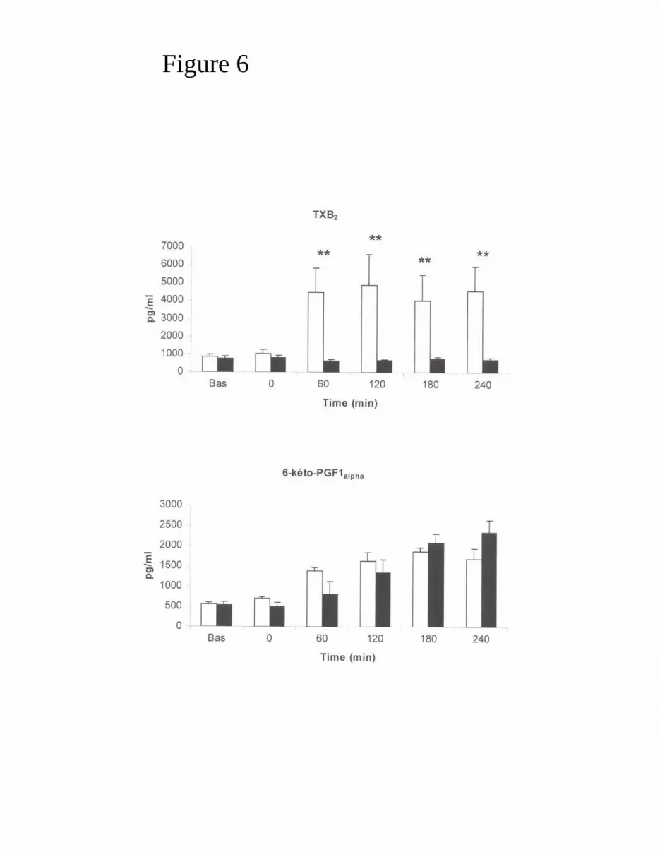
Figure 6


