Embed Size (px)
Citation preview

SLEEP, Vol. 31, No. 6, 2008 868
NARCOLEPSY IS CHARACTERIZED BY EXCESSIVE DAYTIME SLEEPINESS (EDS), A DISRUPTION OF SLEEP-WAKE BEHAVIOR, CATAPLEXY (SUDDEN LOSS of muscle tone provoked by emotional stimuli), and the other REM sleep phenomena, such as sleep paralysis and hypnagogic hallucination.1
Modafinil (a diphenylmethyl-sulfinyl-2-acetamide deriva-tive) is a wake-promoting substance used for the treatment of hypersomnolence, particularly that associated with narco-lepsy.2 Modafinil is an increasingly popular medication due to its safety profile. Most side effects that have been reported are headache (if the modafinil dose was high or increased too quickly) and insomnia or nervousness, which are usually tran-sient and dose dependent.3 Modafinil has multiple modes of actions. It activates α1- and β adrenergic receptors,4 promotes glutamate excitatory transmission in the thalamus, hippocam-pus, and cortex,5 reduces GABA outflow via a monoamine-mediated mechanism,6 and activates serotonergic receptors.7 Although numerous animal studies have been performed, the mechanism whereby modafinil promotes wakefulness is still under investigation. Modafinil was found to increase extra-cellular dopamine levels, as determined by microdialysis in the rat nucleus accumbens and prefrontal cortex, as well as in the caudate of narcoleptic dogs.8 It was revealed that adminis-
tration of modafinil to rats produced wakefulness in associa-tion with activation of tuberomammillary nucleus and orexin neurons, two cell groups implicated in the control of normal wakefulness.9 Although the neurochemistry of this process remains obscure, it may involve histaminergic neurons in the tuberomammillary nucleus and hypocretin neurons in the perifornical area.5,9
Modafinil is effective in maintaining wakefulness in nar-colepsy patients, who have a hypocretin neuron deficit. Other studies have suggested that the cerebral cortex,10 thalamus,5 hippocampus,5 and periaqueductal gray matter11 are also in-volved in the process of promoting wakefulness by modafinil.
The aim of the present study was to investigate regional ce-rebral blood flow (rCBF) changes in human narcolepsy patients who experienced a marked EDS improvement after modafinil administration. To achieve this, we performed 99mTc-ethyl-cysteinate dimer single photon emission computed tomography (SPECT) before and after modafinil or placebo administration in drug-naïve narcoleptics with cataplexy.
SUBJECTS AND METHOD
Subjects
We consecutively recruited 32 narcolepsy patients (M:F = 16:16, 14-47 years) for modafinil administration (modafinil-treated group) and 21 patients (M:F = 11:10, 15-42 years) for placebo administration (placebo-treated group). All patients suffered from narcolepsy with cataplexy and had no drug his-tory for treatment of EDS or cataplexy. A clinical diagnosis of narcolepsy was made by a sleep specialist (SB Hong) based on an EDS >3 months and an unequivocal history of cataplexy, ac-cording to the revised International Classification of Sleep Dis-orders criteria for narcolepsy (ICSD-2).1 The final diagnosis of narcolepsy was confirmed by sleep studies. Other information including the presence of sleep attacks, hypnagogic hallucina-
NArCOlEpSy
Effect of Modafinil on Cerebral Blood Flow in Narcolepsy PatientsEun Yeon Joo, MD*; Dae Won Seo, MD*; Woo Suk Tae, PhD; Seung Bong Hong, MD, PhD
Department of Neurology, Samsung Medical Center, Sungkyunkwan University School of Medicine, Seoul, Korea
Background: To investigate the effects of modafinil on regional cere-bral blood flow (rCBF) in narcolepsy, we performed 99mTc-ethylcysteinate dimer single photon emission computed tomography (SPECT) before and after modafinil or placebo medication.Methods: Brain SPECT was performed twice during the awake state before and after modafinil or placebo administration for 4 weeks in 43 drug-naïve narcoleptics with cataplexy (M/F = 23/20, 29.5 ± 5.8 years). For SPM analysis, all SPECT images were spatially normalized to the standard SPECT template and then smoothed using a 12-mm full width at half-maximum Gaussian kernel. The paired t-test was used to com-pare pre- and post-modafinil or placebo SPECT images.results: The mean modafinil dose used was 207.8 ± 62.3 mg/day. Modafinil significantly reduced Epworth Sleepiness Scale scores from 20.3 ± 2.1 to 5.2 ± 3.1 (P < 0.01), while placebo did not. Compared to
the off-modafinil condition, the on-modafinil condition showed signifi-cantly increased rCBF in the right dorsolateral and bilateral medial pre-frontal cortices. Conversely, after modafinil administration, rCBF was decreased in bilateral precentral gyri, left hippocampus, left fusiform gyrus, bilateral lingual gyri, and cerebellum.There was no significant rCBF change after placebo administration.Conclusion: By a chronic administration of modafinil in narcoleptic patients, rCBF increased in the bilateral prefrontal cortices, whereas it decreased in left mesio/basal, temporal, bilateral occipital areas, and cerebellum.Keywords: Narcolepsy, modafinil, rCBF, SPECT, statistical parametric mappingCitation: Joo EY; Seo DW; Tae WS; Hong SB. Effect of modafinil on ce-rebral blood flow in narcolepsy patients. SLEEP 2008;31(6):868-873.
Disclosure StatementThis was not an industry supported study. The authors have indicated no finan-cial conflicts of interest. *Drs. Joo and Seo contributed equally to this study.
Submitted for publication May, 2006Accepted for publication February, 2008Address correspondence to: Seung Bong Hong, MD, PhD, Department of Neurology, Samsung Medical Center, Sungkyunkwan University School of Medicine, 50 Irwon-dong, Gangnam-gu, Seoul, 135-710, Korea (South); Tel: +82-2-3410-3592; Fax: +82-2-3410-0052; E-mail: [email protected], [email protected]
Modafinil Effect on Cerebral Blood Flow—Joo et al

SLEEP, Vol. 31, No. 6, 2008 869
tions, sleep paralysis, and a positive family history of narco-lepsy were obtained from patients and their families.
Study Design
Before modafinil or placebo ingestion, all subjects under-went a physical examination, a routine blood test (hematology and chemistry panel including liver function tests), brain MRI, and baseline brain SPECT.
Each patient in modafinil group received modafinil 200 mg per day for 4 weeks. If a patient complained of an intolerable adverse event due to modafinil medication, (for example, head-ache, nervousness, or a hand tremor), the modafinil dose was reduced to 100-150 mg/day or divided into 2 dosages (100 mg in the morning and 100 mg at noon) to reduce symptoms. When a patient did not feel any EDS improvement, the modafinil daily dosage was increased to 300-400 mg/day.
Each patient in placebo group received one tablet a day of similar size and shape with modafinil, in the morning for 4 consecutive weeks. The placebo tablets were prepared indepen-dently by a hospital pharmacist.
Subjective EDS improvements were determined by Epworth Sleepiness Scale (ESS) percent decreases, defined as ([pre-modafinil ESS – post-modafinil ESS] or [pre-placebo ESS – post-placebo ESS]/24) × 100%. During the drug administration period, patients were examined by a physician every 2 weeks; blood testing for hematology and liver function was performed twice before medication was administered and again at the end of study period.
Overnight polysomnography and Multiple Sleep latency Test (MSlT)
The day before polysomnography, patients were asked not to drink alcohol or caffeinated beverages. Overnight polysomnog-raphy was recorded by Alice-3 system (Healthdyne, USA) and Compumedics S-series (Compumedics, Australia). Standard polysomnography consists of the 4-channel electroencephalo-gram (EEG, C3/A2; C4/A1; O1/A2; O2/A1), 4-channel elec-trooculogram (EOG), electromyogram (EMG) of submental, intercostal, and anterior tibialis muscles, and electrocardiogram with surface electrodes. Thermistor (for monitoring orona-sal airflow), nasal air pressure monitor, oximetry (for oxygen saturation), piezoelectric bands (for thoracic and abdominal wall motion), and a body position sensor were also attached to the patients. Patients were recorded on videotape, using an infrared video camera, and were continuously observed by a polysomnography technician. Patients went to bed at 23:00 and were awakened at 07:00. Sleep architecture was scored in 30-sec epochs, and sleep staging was interpreted according to the standard criteria.11 MSLT was performed using 4-channel EEG, 4-channel EOG, and single-channel EMG recordings by Al-ice-3 system (Healthdyne, USA) in the day following overnight polysomnography. MSLT consisted of 5 naps scheduled at 2-h intervals starting at 09:00. Patients were invited to lie down on a bed in a dark sound-attenuated room and instructed to try to fall asleep. Sleep latency was defined as the time elapsed from the start of the test (lights out) to the first 30-s epoch scored as sleep. Each sleep latency test was ended 20 min after the onset
of sleep or after 30 min of wakefulness. A sleep onset REM period (SOREMP) was defined as one or more epochs of REM sleep occurring within 15 min of the first 30-s epoch scored as sleep.
Final diagnosis of narcolepsy was determined based on clini-cal symptoms, overnight polysomnography, and MSLT by ap-plying the criteria of narcolepsy.1
99mtc-ethylcysteinate Dimer (99mtc-ECD) Brain SpECT
99mTc-ECD was injected intravenously for the SPECT stud-ies. A brain SPECT scan was performed within 30 to 60 min of radiotracer injection (25mCi) using a 3-headed Triad XLT system equipped with low-energy and high-resolution collima-tors (Trionix Research Laboratory, Twinsburg, OH, USA). The transaxial system resolution of this camera was 6.9 mm full width at half maximum. Images were reconstructed by filtered back-projection using a Butterworth filter. Attenuation correc-tion was performed using Chang’s method (attenuation coef-ficient = 0.12 cm-1).12 The SPECT voxel dimension was 3.56 × 3.56 × 3.56 mm (x, y, z). SPECT studies were performed before narcolepsy treatment commenced. All participants were asked to refrain from caffeinated beverages but were allowed to drink water from 07:00 until the end of the SPECT study. Be-fore radiotracer administration, patients had a nap (15-20 min) if sleepy, but were instructed not to fall asleep after the ECD injection. Wakefulness after the ECD injection was monitored using 4-channel EEG, 2-channel EOG, and single-channel EMG recordings. Informed consent was obtained from all pa-tients after the study protocol, SPECT study procedure, and po-tential hazards associated with radioisotope injection had been explained. The Institutional Review Board at Samsung Medical Center authorized the informed consent form and the study pro-tocol, which included the administration of a radioactive sub-stance and SPECT scanning.
SpM Analyses of Brain SpECT Images Obtained Before Versus After Modafinil or placebo Administration, and on-Modafinil Versus on-placebo Conditions
Pre- and post-modafinil or placebo SPECT images were ma-nipulated using MATLAB 6.3 (The MathWorks, Natick, MA) incorporated into SPM 2 software (Wellcome Department of Cognitive Neurology, Institute of Neurology, University of London, UK). Raw SPECT images (interfile 3.0 format) were converted to Analyze format. Before the spatial normalization of pre- and post-modafinil or placebo SPECT images using a standard SPECT template, post-modafinil or placebo SPECT images were realigned to pre-modafinil or placebo SPECT im-ages for each subject, and realigned matrixes and subject mean images were saved. Using these realignment processes, all post-modafinil or placebo SPECT images were correctly registered to pre-modafinil or placebo SPECT images. Mean images were spatially normalized into a standard SPECT template using a 12-parameter affine transformation and a nonlinear transforma-tion, and the normalization parameters of each subject mean image were then adjusted to realigned pre- and post-modafinil or placebo SPECT images of same subjects. The accuracy of the spatial normalization was checked using a cross-registra-
Modafinil Effect on Cerebral Blood Flow—Joo et al

SLEEP, Vol. 31, No. 6, 2008 870
tion function. Spatially normalized images were then smoothed by convolution using an isotopic Gaussian kernel with a 14-mm full width at half maximum to increase the signal to noise ratio.13,14 Scaling of counts was based on normalization of white matter counts across the study conditions.
Normalization that includes white and gray matter voxels would be problematic. Because white matter is known to be less affected by the experimental effect or disease condition,15 the level of rCBF in whole brain was normalized with the mean count of white matter. Moreover, no experimental data were demonstrated that white matter is mainly affected by modafinil. After normalization, post hoc paired comparisons were per-formed between on- and off-modafinil or placebo administra-tion. The group comparisons between off-modafinil and off-placebo and on-modafinil and on-placebo administrations were performed using unpaired t-test. To test the prior hypothesis that modafinil improves the prefrontal function,16 a small vol-ume correction (SVC) was made in these areas. We applied the corrections on volumes of interest centered on the coordinates and the results of increased rCBF with small volume correction (sphere with 50 mm radius, center point: x, y, z = 0, 36, 16). The height threshold was set at an uncorrected P < 0.001 and the extended threshold to kE > 50.17 The results were superim-posed on the 2-D planes of a single patient’s MRI template after spatial normalization.
rESUlTS
patient Characteristics (Table 1)
All 53 patients (modafinil-treated group, N = 32, placebo-treated group, N = 21) had EDS, sleep attacks, and cataplexy. The mean ESS of patients and their mean Stanford Sleepiness Scale (SSS) suggest moderate to severe EDS.
Detailed clinical information as well as overnight polysom-nography and MSLT findings are listed in Table 1.
Wakefulness Monitoring During SpECT Study
EEG, EOG, and EMG were monitored during SPECT studies to measure the duration of wakefulness after ECD injection.
Before modafinil or placebo administration, mean sleep la-tency after an ECD injection for SPECT study was 6.24 ± 3.4 min (range 3–10.5). After modafinil or placebo administration, the mean sleep latency was 10.7 ± 2.8 min (range 6–15) in modafinil-treated group (paired t-test, P <0.001) and 5.9 ± 3.2 (range 3.5–9.0) in placebo-treated group (P = 0.212).
Modafinil or placebo Effect on Excessive Daytime Sleepiness
Modafinil administration decreased ESS from 20.3 ± 2.1 to 5.2 ± 3.1 (P < 0.01), which suggests that modafinil markedly improves EDS. After 2 weeks of modafinil at 200 mg/day, 6 pa-tients (18.7%, 6/32) did not feel any subjective EDS improve-ment (ESS decrease, 0% to 25%). Thus, the daily modafinil dosage was increased to 250–400 mg/day in those patients until they felt satisfactory improvement of EDS. However, ESS and SSS were not changed significantly after 4 weeks placebo ad-ministration (18.4 ± 3.9 → 16.3 ± 2.7 in ESS; 4.4 ± 2.1 → 4.5 ± 1.9 in SSS, Wilcoxon signed rank test, P > 0.05).
Summarizing, most patients experienced the disappearance or a significant subjective improvement of EDS (mean ESS de-crease, 69.9% ± 12.5%) on modafinil (207.8 ± 62.3 mg/day).
Modafinil Dosage and Adverse Effects
After receiving modafinil medication, 10 patients (31.2%, 10/32) experienced a mild headache for several days (mean 4.2 ± 2.2 days, range 1–9) but this disappeared despite continuous medication at the same dosage. Five patients (15.6%, 5/32) suf-fered hand tremors or nervousness, and dosage was reduced in these patients to 100 or 150 mg. No serious adverse event oc-curred with modafinil. Patients receiving placebo had no ad-verse events.
Table 1—Patients’ Characteristics
Modafinil treated group (n = 32) Placebo treated group (n = 21) PM : F 16 : 16 10 : 12 0.469Mean age, yr 31.4 ± 12.3 29.1 ± 9.9 0.490EDS onset age, yr 17.9 ± 4.9 17.6 ± 5.1 0.814Cataplexy onset age, yr 20.5 ± 5.5 19.6 ± 5.1 0.533Mean ESS 17.2 ± 3.5 16.0 ± 3.9 0.252Mean SSS 4.3 ± 1.9 4.4 ± 2.0 0.762HH/SP, number 23/19 17/15 0.754/0.842Overnight polysomnography Mean sleep latency, min 7.6 ± 3.8 6.4 ± 3.5 0.264Mean REM sleep latency, min 50.0 ± 46.8 60.7 ± 44.1 0.408Mean AHI, per hour 1.7 ± 1.6 1.6 ± 1.7 0.882Mean arousal index, per hour 13.7 ± 5.5 11.5 ± 4.5 0.114Multiple Sleep Latency Test Mean sleep latency, min 3.8 ± 2.9 4.6± 2.8 0.299Sleep-onset REM periods, number 3.7 ± 1.3 3.7 ±1.2 0.975Mean REM sleep latency, min 2.6 ± 2.3 2.9 ± 2.6 0.675
ESS, Epworth Sleepiness Scale; SSS, Stanford Sleepiness Scale; HH, hypnagogic hallucination; SP, sleep paralysis; REM, rapid eye move-ment; Independent t-test, P < 0.05, #Chi-square test, only in case of M:F and HH/SP, number, P < 0.05.
Modafinil Effect on Cerebral Blood Flow—Joo et al
SLEEP, Vol. 31, No. 6, 2008 871
4) On-modafinil (N = 32) Vs. On-placebo Group (N = 21)
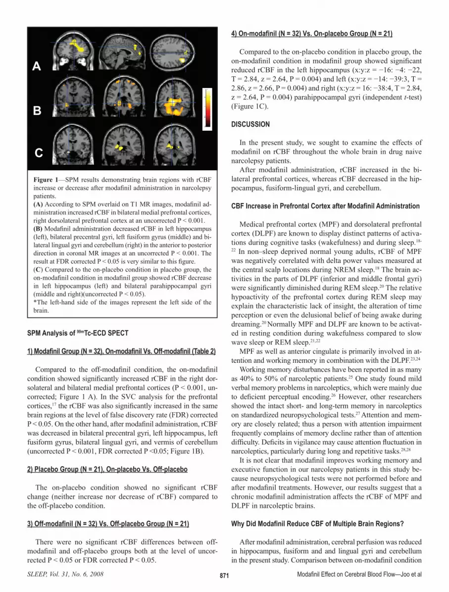
Compared to the on-placebo condition in placebo group, the on-modafinil condition in modafinil group showed significant reduced rCBF in the left hippocampus (x:y:z = −16: −4: −22, T = 2.84, z = 2.64, P = 0.004) and left (x:y:z = −14: −39:3, T = 2.86, z = 2.66, P = 0.004) and right (x:y:z = 16: −38:4, T = 2.84, z = 2.64, P = 0.004) parahippocampal gyri (independent t-test) (Figure 1C).
DISCUSSION
In the present study, we sought to examine the effects of modafinil on rCBF throughout the whole brain in drug naive narcolepsy patients.
After modafinil administration, rCBF increased in the bi-lateral prefrontal cortices, whereas rCBF decreased in the hip-pocampus, fusiform-lingual gyri, and cerebellum.
CBF Increase in prefrontal Cortex after Modafinil Administration
Medical prefrontal cortex (MPF) and dorsolateral prefrontal cortex (DLPF) are known to display distinct patterns of activa-tions during cognitive tasks (wakefulness) and during sleep.18-
22 In non–sleep deprived normal young adults, rCBF of MPF was negatively correlated with delta power values measured at the central scalp locations during NREM sleep.18 The brain ac-tivities in the parts of DLPF (inferior and middle frontal gyri) were significantly diminished during REM sleep.20 The relative hypoactivity of the prefrontal cortex during REM sleep may explain the characteristic lack of insight, the alteration of time perception or even the delusional belief of being awake during dreaming.20 Normally MPF and DLPF are known to be activat-ed in resting condition during wakefulness compared to slow wave sleep or REM sleep.21,22
MPF as well as anterior cingulate is primarily involved in at-tention and working memory in combination with the DLPF.23,24
Working memory disturbances have been reported in as many as 40% to 50% of narcoleptic patients.25 One study found mild verbal memory problems in narcoleptics, which were mainly due to deficient perceptual encoding.26 However, other researchers showed the intact short- and long-term memory in narcoleptics on standardized neuropsychological tests.27 Attention and mem-ory are closely related; thus a person with attention impairment frequently complains of memory decline rather than of attention difficulty. Deficits in vigilance may cause attention fluctuation in narcoleptics, particularly during long and repetitive tasks.28,28
It is not clear that modafinil improves working memory and executive function in our narcolepsy patients in this study be-cause neuropsychological tests were not performed before and after modafinil treatments. However, our results suggest that a chronic modafinil administration affects the rCBF of MPF and DLPF in narcoleptic brains.
Why Did Modafinil reduce CBF of Multiple Brain regions?
After modafinil administration, cerebral perfusion was reduced in hippocampus, fusiform and and lingual gyri and cerebellum in the present study. Comparison between on-modafinil condition
SpM Analysis of 99mTc-ECD SpECT
1) Modafinil Group (N = 32), On-modafinil Vs. Off-modafinil (Table 2)
Compared to the off-modafinil condition, the on-modafinil condition showed significantly increased rCBF in the right dor-solateral and bilateral medial prefrontal cortices (P < 0.001, un-corrected; Figure 1 A). In the SVC analysis for the prefrontal cortices,17 the rCBF was also significantly increased in the same brain regions at the level of false discovery rate (FDR) corrected P < 0.05. On the other hand, after modafinil administration, rCBF was decreased in bilateral precentral gyri, left hippocampus, left fusiform gyrus, bilateral lingual gyri, and vermis of cerebellum (uncorrected P < 0.001, FDR corrected P <0.05; Figure 1B).
2) placebo Group (N = 21), On-placebo Vs. Off-placebo
The on-placebo condition showed no significant rCBF change (neither increase nor decrease of rCBF) compared to the off-placebo condition.
3) Off-modafinil (N = 32) Vs. Off-placebo Group (N = 21)
There were no significant rCBF differences between off-modafinil and off-placebo groups both at the level of uncor-rected P < 0.05 or FDR corrected P < 0.05.
A
B
C
Figure 1—SPM results demonstrating brain regions with rCBF increase or decrease after modafinil administration in narcolepsy patients.(A) According to SPM overlaid on T1 MR images, modafinil ad-ministration increased rCBF in bilateral medial prefrontal cortices, right dorsolateral prefrontal cortex at an uncorrected P < 0.001.(B) Modafinil administration decreased rCBF in left hippocampus (left), bilateral precentral gyri, left fusiform gyrus (middle) and bi-lateral lingual gyri and cerebellum (right) in the anterior to posterior direction in coronal MR images at an uncorrected P < 0.001. The result at FDR corrected P < 0.05 is very similar to this figure.(C) Compared to the on-placebo condition in placebo group, the on-modafinil condition in modafinil group showed rCBF decrease in left hippocampus (left) and bilateral parahippocampal gyri (middle and right)(uncorrected P < 0.05).*The left-hand side of the images represent the left side of the brain.
Modafinil Effect on Cerebral Blood Flow—Joo et al

SLEEP, Vol. 31, No. 6, 2008 872
We recently performed a SPECT study to investigate the ef-fects of modafinil on rCBF in normal subjects (in press). This study showed that a single dose of modafinil (400 mg at once) increased rCBF mainly in thalamus and pons as well as prefron-tal and cingulate cortices in healthy volunteers. There were no brain areas showing reduced rCBF by modafinil as well as place-bo administration. In the present study, we observed that chronic modafinil treatment for 4 weeks produced both rCBF increase (in right dorsolateral and anterior cingulate cortices) and rCBF decrease (in bilateral fronto-temporal cortices and cerebellum).
To our knowledge, there has been no SPECT study of rCBF changes in long-term administrations of modafinil in either nar-colepsy patients or healthy volunteers. The applicability of the short-term effects to chronic drug therapy is very limited because there are known changes in the rate of metabolism, drug-induced changes in receptor density or efficiency of receptor coupling, and sensitization in chronic drug therapy. Thus short-term effect of modafinil administration may be different from the effect of a long-term administration.
Modafinil modulated the blood flow of the thalamus and pons in our previous work, but we could not find rCBF changes in those areas in this study. Moreover, we administered single dose of modafinil 400 mg at once to normal subjects in our pre-vious study whereas the average modafinil dose was 207.8 mg a day administered to narcolepsy patients in the present study. Only modafinil-treated patients showed the improvement of EDS by ESS and prolonged sleep latency during SPECT stud-ies. It seems that long-term modafinil administration produces clinically beneficial effects on narcolepsy patients through the top down control circuitry involved in attention and executive functions, although a task-activation study was not performed before and after modafinil administration in our patients.
Our previous FDG-PET and SPECT studies showed definite hypometabolism or hypoperfusion of the hypothalamus and thalamus in drug-naïve narcoleptics.13,14 Thus, a small increase in rCBF by modafinil in the hypothalamus may have been missed by SPECT, due to its lack of sensitivity.
and on-placebo condition showed that modafinil treated group showed the CBF decrease in bilateral parahippocampal gyri ad-jacent to hippocampus. As presented, the brain regions showing CBF decrease were slightly different between the findings of off-and on-modafinil group and that of on-modafinil and on-placebo groups. Different statistical methods (paired t-test vs. independent t-test) and individual variations of baseline CBF29 in 2 different subject groups (modafinil-treated vs. placebo-treated group) may produce similar not the same compared to the paired comparison between the same subjects before and after drug administration.
Various receptors of GABA, an inhibitory neurotransmitter, exist in temporal lobe including hippocampus and extratempo-ral neocortex.30 The posterior lobe of cerebellum is involved in the fine motor coordination via the inhibition of involuntary movement using GABA. Modafinil showed the neuroprotective effect in the movement-related behavior, locomotor activity, hand-eye coordination, and small fast movements in an animal model of Parkinson disease.31 It was reported that the wakeful-ness produced by modafinil was related to a reduction in GABA release in several brain regions.32
Although there was no evidence that mesio-basal temporal areas and cerebellum showing reduced rCBF play the role in the wake-promotion, modafinil significantly reduced rCBF in the GABA-related temporal lobe and cerebellum as well as im-proved the vigilance of our narcoleptic patients (ESS, 20.3 ± 2.1 → 5.2 ± 3.1) in the present study. Nevertheless, the reason why modafinil reduces rCBF in several regions of narcoleptic brains remains unclear.
Modafinil Effect on rCBF in Narcolepsy patients Versus Healthy Volunteers
There are quite a few conflicting reports concerning the lo-cus of modafinil effect. In previous animal study, administra-tion of modafinil induced marked Fos labeling in neurons of the anterior hypothalamic nucleus and adjacent areas that were considered to be wake-promoting areas.33
Table 2—SPM Results Showing MNI Coordinates and Significance Levels of Brain Regions with rCBF Increase or Decrease after Modafinil Administration in Narcolepsy Patients
Coordinates (mm)Location Side x y z Z T FDR Uncorrected P corrected PrCBF increaseMedial prefrontal cortex L -4 58 8 3.7 4.46 0.024* 0.00011 R 4 40 44 3.2 3.75 0.024* 0.00009Dorsolateral prefrontal cortex R 36 30 36 4.2 5.48 0.024* 0.00001rCBF decreasePrecentral gyrus L -40 -18 64 3.6 4.35 0.026 0.00014 R 18 -34 76 3.6 4.32 0.027 0.00015Hippocampus L -30 -4 -20 4 4.92 0.013 0.000036Fusiform gyrus L -20 -34 -16 4.4 5.64 0.006 0.00025Lingual gyrus L -8 -54 -6 5.2 7.55 0.003 0.0000001 R 24 -46 4 5 7 0.003 0.0000003Cerebellum (vermis/uvula) B 0 -64 -30 4.7 6.38 0.004 0.0000013
L:left, R:right, B:both, FDR: false discovery rate, *MNI (Montreal Neurological Institute) coordinate. *: Small volume cor-rected (SVC) FDR P (sphere with 50mm radius, center point: x, y, z = 0, 36, 16).
Modafinil Effect on Cerebral Blood Flow—Joo et al

SLEEP, Vol. 31, No. 6, 2008 873
13. Joo EY, Tae WS, Kim JH, Kim BT, Hong SB. Glucose hypome-tabolism of hypothalamus and thalamus in narcolepsy. Ann Neu-rol 2004;56:437-40.
14. Joo EY, Hong SB, Tae WS, et al. Cerebral perfusion abnormality in narcolepsy with cataplexy. Neuroimage 2005;28:410-6.
15. Spencer JS, Carmack PS, Gunst RF, Schucany WR, Woodward WA, Haley RW. Using a white matter reference to remove the de-pendency of global signal on experimental conditions in SPECT analyses. Neuroimage 2006;32:49-53.
16. Hunter MD, Ganesan V, Wilkinson ID, Spence SA. Impact of modafinil on prefrontal executive function in schizophrenia. Am J Psychiatry 2006;163:2184-6.
17. Friston KJ, Holmes A, Poline JB, Price CJ, Frith CD. Detecting activations in PET and fMRI: levels of inference and power. Neu-roimage 1996;4:223-35.
18. Dang-Vu TT, Desseilles M, Laureys S, et al. Cerebral correlates of delta waves during non-REM sleep revisited. Neuroimage 2005; 28:14-21.
19. Maquet P, Ruby P, Maudoux A, et al. Human cognition during REM sleep and the activity profile within frontal and parietal cor-tices: a reappraisal of functional neuroimaging data. Prog Brain Res 2005;150:219-27.
20. Hobson JA, Pace-Schott EF, Stickgold R, Kahn D. To dream or not to dream? Relevant data from new neuroimaging and electro-physiological studies. Curr Opin Neurobiol 1998;8:239-44.
21. Maquet P, Degueldre C, Delfiore G, et al. Functional neuroanato-my of human slow wave sleep. J Neurosci 1997;17:2807-12.
22. Maquet P. Functional neuroimaging of normal human sleep by positron emission tomography. J Sleep Res 2000;9:207-31.
23. Peterson BS, Skudlarski P, Gatenby JC, Zhang H, Anderson AW, Gore JC. An fMRI study of Stroop word-color interference: evi-dence for cingulate subregions subserving multiple distributed at-tentional systems. Biol Psychiatry 1999;45:1237-58.
24. Ungerleider LG, Courtney SM, Haxby JV. A neural system for human visual working memory. Proc Natl Acad Sci U S A 1998;95:883-90.
25. Broughton R, Ghanam Q, Hishikawa Y, Sugita Y, Nevsimalova S, Roth B. Life effects of narcolepsy in 180 patients from North America, Asia and Europe compared to matched controls. Can J Neurol Sci 1981;8:199-204.
26. Henry GK, Satz P, Heilbronner RL. Evidence of perceptual en-coding deficit in narcolepsy. Sleep 1993;16:123-7.
27. Aguirre M, Broughton R, Stuss D. Does memory impairment exist in narcolepsy-cataplexy. J Clin Exp Neuropsychol 1985;7:14-24.
28. Fulda S, Schulz H. Cognitive dysfunction in sleep disorders. Sleep Med Rev 2001;5: 423-45.
29. Porter BE, Zhang G, Celix J, et al. Heterogeneous GABAA recep-tor subunit expression in pediatric epilepsy patients. Neurobiol Dis 2005;18:484-91.
30. Van Laere K, Versijpt J, Audenaert K, et al. 99mTc-ECD brain perfusion SPET: variability, asymmetry and effects of age and gender in healthy adults. Eur J Nucl Med 2001;28:873-87.
31. Van Vliet SA, Vanwersch RA, Jongsma MJ, van der Gugten J, Olivier B, Philippens IH. Neuroprotective effects of modafinil in a marmoset Parkinson model: behavioral and neurochemical as-pects. Behav Pharmacol 2006;17:435-62.
32. Fuxe K, Rambert FA, Ferraro L, et al. Preclinical studies with modafinil-evidence for vigilance enhancement and neuroprotec-tion. Drugs Today 1996;32:313-26.
33. Lin JS, Hou Y, Jouvet M. Potential brain neuronal targets for amphetamine methylphenidate-, and modafinil-induced wakeful-ness, evidenced by c-fos immunocytochemistry in the cat. Proc Natl Acad Sci U S A 1996;93:14128-33.
CONClUSION
This study is the first to investigate the effects of modafinil on rCBF in drug-naive narcoleptics with cataplexy. The present study demonstrates that the dose of modafinil that induces a satisfactory wakefulness-promoting response in human narco-leptics also causes regional increase in cerebral blood flow in the bilateral prefrontal cortices.
ACKNOWlEDGMENTS
This study was supported by a grant (M103KV010017-07-K2201-01710) from Brain Research Center of the 21st Century Frontier Research Program funded by the Ministry of Science and Technology of the Republic of Korea and by a grant (no. A050462) of the Good Health R&D Project, Ministry of Health & Welfare, Republic of Korea.
rEFErENCES
1. American Academy of Sleep Medicine. The international clas-sification of sleep disorders: diagnostic & coding manual, 2nd ed. Westchester, IL: American Academy of Sleep Medicine; 2005.
2. Bastuji H, Jouvet M. Successful treatment of idiopathic hyper-somnia and narcolepsy with modafinil. Prog Neuropsychophar-macol Biol Psychiatry 1988;12:695-700.
3. U.S. Modafinil in narcolepsy multicenter study group: Random-ized trial of modafinil for the treatment of pathological somno-lence in narcolepsy. Ann Neurol 1998;43:88-97.
4. Duteil J, Rambert FA, Pessonnier J, Hermant JF, Gombert R, As-sous E. Central alpha 1-adrenergic stimulation in relation to the behaviour stimulating effect of modafinil; studies with experi-mental animals. Eur J Pharmacol 1990;180:49-58.
5. Ferraro L, Tanganelli S, O’Connor WT, Antonelli T, Rambert F, Fuxe K. The vigilance promoting drug modafinil decreases GABA release in the medial preoptic area and in the posterior hypothalamus of the awake rat: possible involvement of the sero-tonergic 5-HT3 receptor. Neurosci Lett 1996;220:5-8.
6. Tanganelli S, Ferraro L, Bianchi C, Fuxe K. 6-hydroxy-dopamine treatment counteracts the reduction of cortical GABA release pro-duced by the vigilance promoting drug modafinil in the awake freely moving guinea-pig. Neurosci Lett 1994;171:201-4.
7. Tanganelli S, Perez de la Mora M, Ferraro L, et al. Modafinil and cortical gamma-aminobutyric acid outflow. Modulation by 5-hy-droxytryptamine neurotoxins. Eur J Pharmacol 1995;273:63-71.
8. Wisor JP, Nishino S, Sora I, Uhl GH, Mignot E, Edgar DM. Do-paminergic role in stimulant-induced wakefulness. J Neurosci 2001;21:1787-94.
9. Scammell TE, Estabrooke IV, McCarthy MT, et al. Hypothalamic arousal regions are activated during modafinil-induced wakeful-ness. J Neurosci 2000;22:8620-8.
10. Tanganelli S, Fuxe K, Ferraro L, Janson AM, Bianchi C. Inhibi-tory effects of the psychoactive drug modafinil on gamma-amin-obutyric acid outflow from the cerebral cortex of the awake freely moving guinea-pig. Possible involvement of 5-hydroxytryptam-ine mechanisms. Naunyn Schmiedeberg Arch Pharmacol 1992;345:461-5.
11. Rechtshaffen A, Kales A. eds. A manual of standardized terminol-ogy, techniques, and scoring system for sleep stages of human subjects. UCLA, Brain Information Service/Brain Research Insti-tute, Los Angeles; 1968.
12. Chang L. A method for attenuation correction in computed to-mography. IEEE Trans Nucl Sci 1987;NS-25:638-43.
Modafinil Effect on Cerebral Blood Flow—Joo et al


![[Brazilian guidelines for the treatment of narcolepsy]](https://img.pdfslide.net/doc/110x75/6336e04536d54cc94b0f7097/brazilian-guidelines-for-the-treatment-of-narcolepsy.jpg)


