Embed Size (px)
Citation preview

Page 1 of 9 (Page number not for citation purposes)
Research Article
Effect of the Montivipera bornmuelleri snake venom on human blood: coagulation disorders and hemolytic activities Claudine Accary1,£, Mohamad Rima1,£, Achraf Kouzayha1,2, Walid Hleihel3, Riyad Sadek4, Jean Claude Desfontis5, Ziad Fajloun1,2,**, Souad Hraoui-Bloquet6,* 1
Laboratory of Applied Biotechnology, Azm Center for the Research in Biotechnology and its Application, Doctoral School for Sciences and Technology, Tripoli, Lebanon 2 Faculty of Sciences, Biochemistry Department , Section III, Lebanese University, Tripoli, Lebanon
3 Faculty of Medical Science, USEK, Kaslik-Jounieh, Lebanon
4 American University, Biology Department, Beirut, Lebanon
5 LUNAM, Oniris, Unit of Animal Pathophysiology and Functional Pharmacology, Nantes, France
6 Faculty of Sciences II (Fanar), Lebanese University, B.P. 90656 Jdeidet El Maten, Lebanon
£ Both authors contributed equally to this work
Corresponding Author & Address:
Souad Hraoui-Bloquet* Faculty of Sciences II (Fanar), Lebanese University, B.P. 90656 Jdeidet El Maten, Lebanon; Phone: +961 1 68 69 83; Email: [email protected]
Ziad Fajloun** Laboratory of Applied Biotechnology, Azm Center for the Research in Biotechnology and its Applications, Doctoral School for Sciences and Technology, Lebanese University, El Mittein Street, Tripoli, Lebanon; Phone: + 961 6 213 255, Fax: + 961 6 213 383, Email: [email protected]
Published: 9th June, 2014 Accepted: 9th June, 2014 Received: 16th February, 2014 Open Journal of Hematology, 2014, 5-4 © Hraoui-Bloquet and Fajloun et al.; licensee Ross Science Publishers ROSS Open Access articles will be distributed under the terms of the Creative Commons Attribution License (http://creativecommons.org/licenses/by/3.0), which permits unrestricted use, distribution, and reproduction in any medium, provided that the original work will always be cited properly. Keywords: M. bornmuelleri, blood clotting, cytotoxicity, hemolysis, phospholipases A2
ABSTRACT
Viper’s venom is a source of biopharmaceutical compounds, hence the need to assess the effect of this animal extract on human blood. Here, we studied the blood coagulation disorders and hemolytic activities of the venom of M. bornmulleri viper. The pro-coagulant and anticoagulant effects are analyzed with venom concentrations ranging from 0.4 to 0.0031 mg/mL. Thus, the PT is way above the normal value indicating an anticoagulant activity whereas for the aPTT, the high concentration of the venom showed an anticoagulant activity, but a pro-coagulant effect was occurred when the venom concentration decreases to 0.05 and/or 0.025 mg/mL. Hemolytic tests, performed in suspension (30% RBCs) and on blood agar plate (5% RBCs), show that an increased concentration of the venom going until 1.6 mg cannot produce a hemolytic effect, even in the presence of Ca
2+ (hemolysis < 0.5%). Also, on the blood agar plate no hemolytic area appeared even with 0.04
mg of the lyophilized venom. Otherwise, the venom was able to induce a low hemolytic activity (hemolysis = 1.3 %) by acting on L-α-PC used as substrate. In this case, the destruction of erythrocytes increased proportionally to the added amount of phospholipids which are hydrolyzed to fatty acids and lysophospholipids (two toxic substances for RBCs), probably due to the presence of PLA2 in the venom and which are known by their ability to hydrolyze lecithin
Open Journal of Hematology
OPEN ACCESS

Open Journal of Hematology, 2014, 5-4 Cytotoxicity testing on human blood of the Montivipera bornmuelleri venom
Page 2 of 9 (Page number not for citation purposes)
INTRODUCTION
Snake’s venom is a complex mixture of proteins, nucleotides and inorganic ions. This combination gives an array of toxicity to the venom, where the peptides and polypeptides are responsible for a variety of toxic properties [1]. The snake crude venom has a wide spectrum of biological activities, such as neurotoxic, cardiotoxic, cytotoxic and antibacterial activity [1-3]. Furthermore, some viper venom components show antitumor and antiangiogenic activities [4]. Due to this broad range of biological functions, the snake venom and its bioactive components have been the subject of many scientific articles in different research fields, including biochemistry, biophysics, toxicology, pharmacology and medicine [1, 5-7].
According to their ability to shorten or prolong the blood clotting process, snake venom proteins that affect blood coagulation can functionally be classified as pro-coagulant or anticoagulant proteins. Pro-coagulant proteins are either serine proteinases or metalloproteinases. They induce blood coagulation either by specifically activating zymogen, one of the blood coagulation factors, or by directly converting soluble fibrinogen into an insoluble fibrin clot [8]. However, those which prolong the blood coagulation process are proteins or glycoproteins. Some of these anticoagulant proteins exhibit enzymatic activities, such as PLA2 and proteinase, whereas others do not exhibit any enzymatic activity. The mechanism of anticoagulant activity of only a few of these proteins is well understood [9]. Thus, venoms of Crotalidae and Viperidae snakes were considered as complex mixtures of numerous molecules that can possess both pro-coagulant and anticoagulant properties [10-12]. Pro-coagulant molecules, especially from the Viperidae, have been used in medical applications and diagnosis such as RVV-factor X activator (RVVX) a well-known pro-coagulant from Russell’s viper venom (RVV) useful for measuring a lupus anticoagulant [13].
Moreover, the assessment of the blood effect of snake venoms concerning the coagulopathy and hemolytic activity is not a recent concept. In fact, since 1961, Rosenfeld and collaborators had conducted a study on the hemolytic effects of 29 snake venoms [14]. All
these venoms have shown indirect hemolytic activities except those of M. frontalis, B. cotiara, B. jararaca, N. naja, B. caeruleus and various other Indian and South American viper venoms which have revealed a more potent hemolytic effect. In 1979, a study was performed on 24 other snakes’ venoms for their ability to hemolysis washed human RBCs in the presence of albumin and Ca2+ [15]. Except for Crotalus h. horridus, all venoms hemolysed the RBCs to a varying degree. The majority of the venoms hydrolyzed membrane phospholipids in the presence or absence of albumin, as long as Ca2+ was present. A correlation was found between fatty acid cleaved and hemolytic activities. These results suggested that membrane damage as evidenced by membrane phospholipid splitting is a prerequisite to hemolysis in this system [16].
M. bornmuelleri is one of venomous viper species found in Lebanon. Very little is known about the biology of this endemique and rare species. M. bornmuelleri venom has been the subject of some previous studies conducted by our team of researchers [17, 18]. Our earlier results about the characterization of the M. bornmuelleri venom demonstrated that it is a complex mixture of proteins and enzymes and, has considerable biological effects such as pro-inflammatory and antibacterial activities. Henceforth, the impact evaluation of M. bornmuelleri venom on human blood concerning the coagulation disruption and hemolytic activities, designing the principal aim of this work, seems essential to assess the important exploration of the interest biomolecules presented in this natural extract.
MATERIALS AND METHODS
A. Materials
Venom was manually milked from M. bornmuelleri vipers, freeze-dried into crystal shape, and stored at -20°C in a dry, cold and light-free place. In our study, the lyophilized venom was dissolved in PBS to obtain a solution of 20 mg/mL. L-α- PC and Triton X-100 were from Sigma-Aldrich. The calcium chloride was from Riedel-de Haën. The nutrient agar was purchased from Bio-Rad. Pacific Hemostasis Thromboplastin-DS, Pacific Hemostasis aPTT XL, Pacific Hemostasis Calcium Chloride reagents were obtained from
Open Journal of Hematology, 2014, 5-4 Cytotoxicity testing on human blood of the Montivipera bornmuelleri venom
Page 3 of 9 (Page number not for citation purposes)
Fisher Diagnostics Company. The products: Collagen/EPI and Collagen ADP cartridges were purchased from Siemens Healthcare Diagnoatics.
B. Methods 1. Human plasma coagulation effect
PT and aPTT tests were performed in a certificated medical laboratory using Behring coagulation system according to the manufacturer's instructions. Freshly aspirated blood from healthy volunteer who had not taken any drugs for at least 2 weeks prior to sampling was collected in 3.2% sodium citrate tube and used directly. Samples were centrifuged for 10 min at 4000 rpm, and the plasma was recuperated. Venom was mixed to the plasma to get a range of concentrations of 0.5, 0.25, 0.125, 0.05, 0.025 mg/mL. PT test was performed on plasma 100µL incubated for 1min at 37°C. Coagulation is initiated by addition of 200 µL of Pacific Hemostasis Thromboplastin-DS reagent. Results are expressed as the time needed for clot formation. Positive and negative controls were used to evaluate the results obtained in the presence of the venom. For the realization of aPTT test, 100 µL of plasma were mixed to 100 µL of Pacific Hemostasis aPTT-XL reagent and incubated 2 min at 37°C. Coagulation is initiated by adding 100 µL of calcium chloride. The time needed for clot formation is called activated partial thromboplastin time.
2. Hemolytic activity 2.1. Direct hemolytic activity: The toxicity of the M. bornmuelleri venom on RBCs was evaluated using fresh human blood. Blood was collected aseptically from healthy volunteers into tubes treated with EDTA and centrifuged at 3000 rpm for 5 min. The supernatant was discarded and the pellet containing RBCs were washed thrice with PBS by repeated centrifugation at 3000 rpm for 5 min. Thereafter, thirty percent RBCs suspension was prepared and a volume of 300 µL was taken in each tube and treated with different amounts of a solution of venom (20 mg/ml). The tube considered as positive control contains 200 µL of distilled
water and 100 µL of RBCs, whereas the negative control contains 200 µl of PBS and 100 µL of RBC. All tubes were then incubated at 4°C for 30 min and centrifuged at 3000 rpm for 5 min. Absorbance of the supernatants were measured at 540 nm and the values obtained with the positive control represented a 100% hemolysis. The percentage of hemolysis was then calculated using the following formula:
[
]
2.2. Hemolytic activity on human blood agar plate: Human blood agar plates were prepared by adding 5 mL of RBCs to 95 mL of sterile nutrient agar aseptically and the solution was poured immediately into the Petri dishes. After solidification, the wells are cut into the agar (6 mm diameter) then loaded with 20 µL of test solutions with different concentrations of M. bornmuelleri venom. The plates were observed for hemolysis after 24 h of incubation at room temperature.
2.3. Hemolytic activity in the presence of divalent ions (Ca2+): In this experiment, calcium ions (20 mM) were incubated with the test solutions of M. bornmuelleri venom for evaluating their effects on erythrocyte lysis. 2.9 mg of CaCl2.2H2O were dissolved in 1 mL of PBS, the resulting solution were used for the preparation of the RBCs suspension (30% V/V). After treatment with different amounts of venom test solutions, the tubes were incubated and centrifuged as described earlier, and the degree of hemolysis was calculated after measuring the absorbance of the released hemoglobin into the supernatant at 540 nm. In this case, the tube considered as positive control contains 200 µL of distilled water and 100 µL of RBCs, whereas the negative control contains 200 µL of PBS and 100 µL of RBCs in the presence of CaCl2.
3. Indirect hemolytic activity
Indirect hemolysis involves the conversion of lecithin to lysolecithin and

Open Journal of Hematology, 2014, 5-4 Cytotoxicity testing on human blood of the Montivipera bornmuelleri venom
Page 4 of 9 (Page number not for citation purposes)
fatty acids, which are known to induce hemolysis of erythrocytes. Erythrocyte suspensions were incubated with different amounts of venom samples in the presence of 30 µg/mL lecithin and 1.25 mM CaCl2. The tubes were then incubated and centrifuged as described earlier, and the degree of hemolysis was calculated after measuring the absorbance of the supernatant at 540 nm.
Table 1: Prothrombin time (PT) and activated partial thromboplastin time (aPTT) performed using Behring coagulation system.
M. bornmuelleri venom concentrations [mg/mL]
PT in seconds
aPTT in seconds
Standard 13 25-40
Control 12.5 33.4
0.4 > 200 > 150
0.2 > 200 > 150
0.1 > 200 > 150
0.05 > 200 16.9
0.025 51.5 18.6
0.0125 31.3 22.6
0.00625 31.3 26.8
0.003125 26.5 34.2
RESULTS AND DISCUSSION
1. Human plasma coagulation effects
PT and aPTT tests were performed to evaluate the effect of M. bornmuelleri venom on the blood coagulation cascade. Results obtained for PT and aPTT tests shown in Table 1 are compared to the standard used for system calibration. For venom concentration ≥ 0.05 mg/mL, there was a total anticoagulation activity in PT test with time exceeding 200 seconds. This effect decreases with a lower concentration of the crude venom, even a concentration of 0.0031 mg/mL still exhibit an anti-coagulant activity. In aPTT test, the venom has shown an anti-coagulant action with high concentration ≥ 0.1 mg/mL but when concentration decreases to 0.05 and 0.025 mg/mL, the clotting time went down the normal value pointing to a pro-coagulant activity of the crude venom of M. bornmuelleri. Below these concentrations, the crude venom has no action on blood coagulation and aPTT value returns to normal. The crude venom of M. bornmuelleri showed both pro-coagulant and anticoagulant activities with different concentrations; PT and aPTT are both
disrupted suggesting that the defect lies in multiple pathways or in the common pathway of the coagulation cascade.
In general, Viperidae venoms mainly cause hemorrhaging and coagulation disorders. Over the years, many toxins affecting blood circulation have been isolated and characterized from various viper venoms [19]. Some affect platelet aggregation, whereas others affect blood coagulation cascade. Venom proteins that affect blood coagulation can be classified as pro-coagulant or anticoagulant proteins based on their ability to shorten or prolong the blood-clotting process [11, 12]. Pro-coagulant proteins are either serine-proteases or metalloproteases [8]. These factors inhibit blood coagulation by different mechanisms. Some of these anti-coagulant proteins exhibit enzymatic activities, such as PLA2 and proteinase, whereas others do not exhibit any enzymatic activity [20, 21]. Moreover, Dambisya et al. (1994) discussed previously the presence of numerous molecules in the compound venomous of the Viperidae family that can possess both pro-coagulant and anti-coagulant properties [22]. In fact, these authors reported that the different concentrations of C. hodostoma (Malayan pit viper) venom showed the dual effects (pro and anti-coagulant). These observations are relevant to our results obtained of the M. bornmuelleri venom on the coagulation cascade. In future work, confirmation tests should be done for better analysis and in order to localize the action of the crude venom to certain activation step(s) in the cascade.
2. Hemolytic activity 2.1. Direct hemolytic activity
To evaluate the potential of M. bornmuelleri venom to induce hemolysis, 30% suspensions of human RBCs were treated with different amounts of M. bornmuelleri venom (1, 2, 4, 6, 8, 10, 15, 20, 40 and 80 µL from initial solution at 20 mg/mL). After incubation at 4°C for 30 min, the tubes were centrifuged and the absorbance of the supernatants was measured at 540 nm. These values were used to determine the percentage of hemolysis in each tube depending on the

Open Journal of Hematology, 2014, 5-4 Cytotoxicity testing on human blood of the Montivipera bornmuelleri venom
Page 5 of 9 (Page number not for citation purposes)
amount of venom used. The results, shown in figure 1, prove that the venom has no direct hemolytic activity on a 30% suspension of RBCs. Indeed, we noticed that even when 80 µL of the venom solution were added (equivalent to 1.6 mg of crude venom) the supernatant is clear,
its absorbance is negligible and the percentage of hemolysis is always lower than 1%. These results suggest that the components of the venom cannot act in a direct way on RBCs membrane and therefore induce its hemolysis.
Figure 1: Direct hemolytic activity of the M. bornmuelleri venom. Hemolysis was determined by an absorbance reading at 540 nm after treating the RBC suspension with 1, 2, 3, 4, 6, 8, 10, 15, 20, 30, 40 and 80 µL of the venom solution (concentrated at 20 mg/mL) and compared to hemolysis achieved with water (reference for 100% hemolysis). The data are means of triplicate experiments.
Since our principal aim is to study the M. bornmuelleri venom for the development of new biopharmaceutical compounds, it is necessary that this natural extract don’t affect human cells, particularly the RBCs. Indeed, it is now important to mention that our observations, demonstrating -in vitro- the absence of the direct hemolytic activity, could be considered as sensitive results to express the degree of cytotoxicity of any substance derived from the M. bornmuelleri venom. This idea is supported by some previous studies showing that the neutralization of the hemolytic power of the animal venom tested on rabbit, sheep, or human erythrocytes seems to correlate with the neutralization of venom lethality in mice [23, 24].
2.2. Hemolytic activity on human blood agar plate
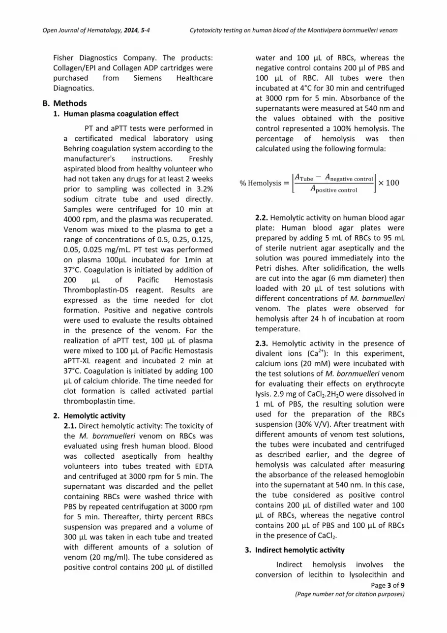
Another test was conducted to
evaluate the hemolytic activity using Petri dishes containing nutrient agar supplemented by RBCs (5%). However, it allows the assessment of hemolysis that appears as an easily visible light area on the agar. 20 µL of the crude venom (from initial concentration at 20 mg/ mL), 20 µL of Triton X-100 (positive control) and 20 µL of PBS have been introduced into the wells dug in the agar. Results represented in figure 2 were observed after 24h of incubation at room temperature. They show an absence of enlightenment around the well containing the venom. Therefore, the absence of hemolytic activity was further confirmed also on blood agar plate, while 40 μg of M. bornmuelleri venom didn’t show any hemolytic halo. This proves that this venom under these conditions, does not exercise any hemolytic effect, which isn’t the case of the positive control which shows a clearly enlightened area.
Open Journal of Hematology, 2014, 5-4 Cytotoxicity testing on human blood of the Montivipera bornmuelleri venom
Page 6 of 9 (Page number not for citation purposes)
Figure 2: Hemolytic activity of the M. bornmuelleri venom on blood agar plate. E: control (PBS buffer, pH 7.4), T: Triton X-100, V: crude venom
2.3. Hemolytic activity in the presence of divalent ions (Ca2+)
Most snake venoms contain phospholipases that hydrolyze free or membrane phospholipids into fatty acids and lysophospholipids. Some of these PLA2 require Ca2+ for their activity [4], that’s why the hemolytic activity of M. bornmuelleri venom was tested in the presence of Ca2+ at a concentration of 20 mM. Results obtained didn’t differ from the previous one demonstrating that even in the presence of Ca2+, the hemolysis was similar or lower the negative control (data not shown). It’s well known that these divalent ions play an important role in hemolysis by increasing the activity of several hemolytic toxins [25], that’s why the absence of hemolysis even in presence of Ca2+ justify our previous results saying that M. bornumelleri venom is unable to induce direct hemolysis.
3. Indirect hemolytic activity
Hemolytic activities observed with animal venoms can be categorized into two groups: those which lyse erythrocytes directly or indirectly (i.e., requiring the presence of lecithin or free fatty acids) in order to induce hemolysis [26]. In this case, phospholipases cannot act on membrane phospholipids but
induce hemolysis by lysophospholipids known by their surfactant power.
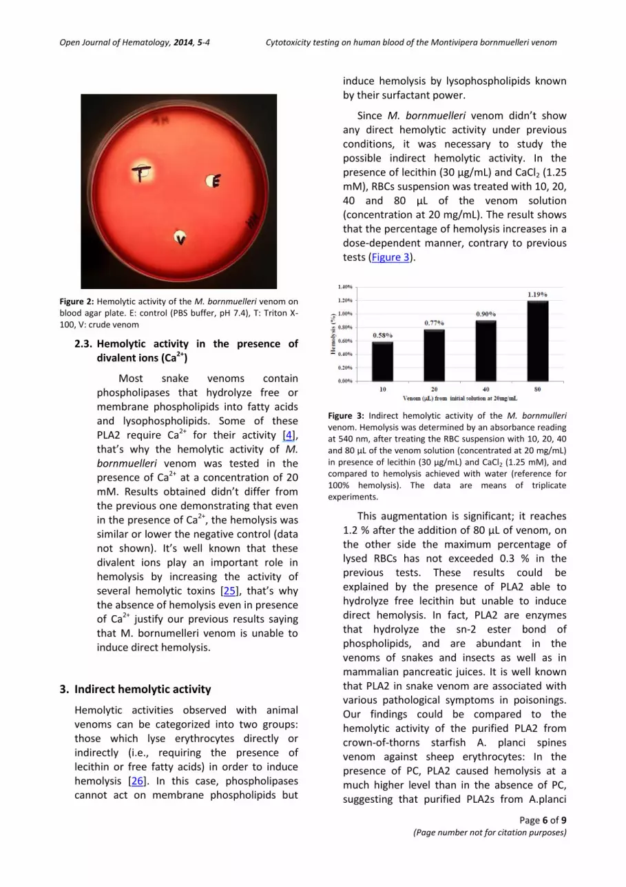
Since M. bornmuelleri venom didn’t show any direct hemolytic activity under previous conditions, it was necessary to study the possible indirect hemolytic activity. In the presence of lecithin (30 µg/mL) and CaCl2 (1.25 mM), RBCs suspension was treated with 10, 20, 40 and 80 µL of the venom solution (concentration at 20 mg/mL). The result shows that the percentage of hemolysis increases in a dose-dependent manner, contrary to previous tests (Figure 3).
Figure 3: Indirect hemolytic activity of the M. bornmulleri venom. Hemolysis was determined by an absorbance reading at 540 nm, after treating the RBC suspension with 10, 20, 40 and 80 µL of the venom solution (concentrated at 20 mg/mL) in presence of lecithin (30 µg/mL) and CaCl2 (1.25 mM), and compared to hemolysis achieved with water (reference for 100% hemolysis). The data are means of triplicate experiments.
This augmentation is significant; it reaches 1.2 % after the addition of 80 µL of venom, on the other side the maximum percentage of lysed RBCs has not exceeded 0.3 % in the previous tests. These results could be explained by the presence of PLA2 able to hydrolyze free lecithin but unable to induce direct hemolysis. In fact, PLA2 are enzymes that hydrolyze the sn-2 ester bond of phospholipids, and are abundant in the venoms of snakes and insects as well as in mammalian pancreatic juices. It is well known that PLA2 in snake venom are associated with various pathological symptoms in poisonings. Our findings could be compared to the hemolytic activity of the purified PLA2 from crown-of-thorns starfish A. planci spines venom against sheep erythrocytes: In the presence of PC, PLA2 caused hemolysis at a much higher level than in the absence of PC, suggesting that purified PLA2s from A.planci

Open Journal of Hematology, 2014, 5-4 Cytotoxicity testing on human blood of the Montivipera bornmuelleri venom
Page 7 of 9 (Page number not for citation purposes)
venom were very weak as to direct hemolysis, but exhibited indirect hemolytic activity [27].
Compared to other vipers, V. palestinae and V. russellii venoms showed the presence of PLA2, which is inefficient in inducing neither lysis nor substantial phospholipid splitting of washed erythrocytes, unless it was fortified with the direct lytic factor fraction derived from Cobra venom. Actually the synergy between cytotoxin and phospholipase is noticed and their cross-contamination induced a significant increase in the cytolytic power of one or the other [23, 28].
Also, PLA2 fractions (phosphatide acyl-hydrolase, EC 3.1.14) isolated from the Cobra venoms were devoid of hemolytic activity and caused no significant breakdown of phospholipids in the intact erythrocytes. Another protein component derived from the same venoms (the direct lytic factor) was weakly hemolytic but showed no phospholipase activity. The two fractions, when combined, produced strong hemolysis and concomitant hydrolysis of erythrocyte phospholipids to lysophospholipids [28].
CONCLUSION
In this study, we demonstrated the dual effect of M. bornmuelleri venom on human blood coagulation. Thus, the venom showed -pro- or anti-coagulant activities at different concentrations; extrinsic and intrinsic pathways are both disrupted suggesting that the defect lies in multiple pathways or in the common pathway of the coagulation cascade.
We also showed that M. bornmuelleri venom does not induce direct hemolysis of a 30% suspension of RBCs even in the presence of 1.6 mg of lyophilized venom. Contrariwise, hemolytic
activity is recorded in case where the lecithin is added to the suspension. This activity increases proportionally to the added amount of venom it refers to the presence of PLA2 known by their ability to hydrolyze lecithin. On the other side, the absence of cytotoxins or other components known as direct lytic factor could explain the lack of direct hemolytic activity. Finally, M. bornmuelleri venom is still not extensively explored and further investigated on its component may be a useful tool for a better understanding of its action mechanism and may lead to a new range of molecules with pharmaceutical interest.
ABBREVIATIONS
PT – Prothrombine Time
aPTT – activated Partial Thromboplastine Time
PLA2 – Phospholipase A2
L-α-PC – L-α-Phosphatidylcholine
RBCs – Red Blood Cells
PBS – Phosphate Buffered Saline
EDTA – Ethylene Diamine Tetraacetic Acid
EC 3.1.14 – Enzyme Commission number
CONFLICT OF INTEREST
No conflict of interest about this work
ACKNOWLEDGEMENT
We would like to thank Dr. Wissam Mansour and Mrs Jiana Al Masri for their technical supports. We are deeply appreciative to the National Council for Scientific Research (CNRS-Lebanon) and Francophone University Agency (AUF) for the financial support and the funding of our work.
REFERENCES
[1] Koh DCI, Armugam A, Jeyaseelan K. Snake venom components and their applications in biomedicine. Cell Mol Life Sci. 2006; 63: 3030-41. http://dx.doi.org/10.1007/s00018-006-6315-0
[2] Doley R, Kini RM. Protein complexes in snake venom. Cell Mol Life Sci. 2009; 66: 2851-71. http://dx.doi.org/10.1007/s00018-009-0050-2
[3] Perumal Samy R, Gopalakrishnakone P, Thwin MM, Chow TKV, Bow H, Yap EH, Thong TWJ.

Open Journal of Hematology, 2014, 5-4 Cytotoxicity testing on human blood of the Montivipera bornmuelleri venom
Page 8 of 9 (Page number not for citation purposes)
Antibacterial activity of snake, scorpion and bee venoms: a comparison with purified venom phospholipase A2 enzymes. J Appl Microbiol. 2007; 102: 650-9. http://dx.doi.org/10.1111/j.1365-2672.2006.03161.x
[4] Zouari-Kessentini R, Srairi-Abid N, Bazaa A, El Ayeb M, Luis J, Marrakchi N. Antitumoral Potential of Tunisian Snake Venoms Secreted Phospholipases A2. Biomed Res Int. 2013; 2013: 391389. http://dx.doi.org/10.1155/2013/391389
[5] El-Refael MF, Sarkar NH. Snake venom inhibits the growth of mouse mammary tumor cells in vitro and invivo. Toxicon. 2009; 54: 33-41. http://dx.doi.org/10.1016/j.toxicon.2009.03.017
[6] Guimarães-Gomes V, Oliveira-Carvalho AL, Junqueira-de-Azevedo IL, S Dutra DL, Pujol-Luz M, Castro HC, Ho PL, Zingali RB. Cloning, characterization, and structural analysis of a C-type lectin from Bothrops insularis (BiL) venom. Arch Biochem Biophys. 2004; 432: 1-11. http://dx.doi.org/10.1016/j.abb.2004.08.018
[7] Stocker K. Use of snake venom proteins in medicine. Schweiz Med Wochenschr. 1999; 129: 205.
[8] Kini RM, Rao VS, Joseph JS. Pro-coagulant proteins from snake venoms. Haemostasis. 2001; 31: 218-24.
[9] Kornalik F. The influence of snake venom proteins on blood coagulation. In: Harvey AL, editor. Snake Toxins. New York: Pergamon Press. 1991; pp. 323-383.
[10] Braud S, Bon C, Wisner A. Snake venom proteins acting on hemostasis. Biochimie. 2000; 82: 851-9. http://dx.doi.org/10.1016/S0300-9084(00)01178-0
[11] Matsui T, Fujimura Y, Titani K. Snake venom proteases affecting hemostasis and thrombosis. Biochim Biophys Acta. 2000; 1477: 146-56. http://dx.doi.org/10.1016/S0167-4838(99)00268-X
[12] White J. Snake venoms and coagulopathy. Toxicon. 2005; 45: 951-67. http://dx.doi.org/10.1016/j.toxicon.2005.02.030
[13] Derksen RH, de Groot PG. Tests for lupus anticoagulant revisited. Thromb Res. 2004; 114: 521-6. http://dx.doi.org/10.1016/j.thromres.2004.06.009
[14] Rosenfeld G, Kelen EMA, Nudel F. Hemolytic Activity of Animal Venoms. I. Classification in Different Types and Activities. Memorias do Instituto Butantan. 1960; 30: 117-32.
[15] Condrea E. Hemolytic effects of snake venoms. In: Snake venoms. Handbook of Experimental Pharmacology. 1979; 52: 448-79.
[16] Tu AT, Homma M, Hong BS. Hemorrhagic, myonecrotic, thrombotic and proteolytic activities of viper venoms. Toxicon. 1969; 6: 175-8.
http://dx.doi.org/10.1016/0041-0101(69)90117-2 [17] Hraoui-Bloquet S, Sadek R, Accary C, Hleihel W,
Fajloun Z. An ecological study of the montane Montivipera bornmuelleri (Werner,1898) viper including preliminary biochemical characterization of venom. Lebanese Science Journal. 2012; 13: 89-101.
[18] Accary C, Souad Hraoui-Bloquet S, Hamze M, Sadek R, Hleihel W, Desfontis JC, Fajloun Z. Preliminary proteomic analysis and biological characterization of the crude venom of Montivipera bornmuelleri; a viper from Lebanon. Frontiers in Toxinology. 2013; In press.
[19] Suntravat M, Nuchprayoon I, Pérez JC. Comparative study of anti-coagulant and pro-coagulant properties of 28 snake venoms from families Elapidae, Viperidae, and purified Russell’s viper venom-factor X activator(RVV-X). Toxicon. 2010; 56: 544-53. http://dx.doi.org/10.1016/j.toxicon.2010.05.012
[20] Meier J, Stocker K. Effects of snake venoms on hemostasis. Crit Rev Toxicol. 1991; 21: 171-82. http://dx.doi.org/10.3109/10408449109089878
[21] Kornalik F. The influence of snake venom proteins on blood coagulation. In: Harvey AL. (ed.) Snake Toxins. 1991; Pergamon Press, New York, pp 323-383.
[22] Dambisya YM, Lee TL, Gopalakrishnakone P. Action of Calloselas marhodostoma (Malayan pit viper) venom on human blood coagulation and fibrinolysis using computerized thromboelastography (CTEG). Toxicon. 1994; 32: 1619-26. http://dx.doi.org/10.1016/0041-0101(94)90320-4
[23] Chippaux JP. Venins de serpent et envenimations. IRD Orstom. 2002.
[24] Maček P, Belmonte G, Pederzolli C, Menestrina G. Mechanism of action of equinatoxin II, a cytolysin from the sea anemone Actinia equine L. belonging to the family of actinoporins. Toxicology. 1994; 87: 205-27. http://dx.doi.org/10.1016/0300-483X(94)90252-6
[25] Celedón G1, González G, Lissi E, Cerda T, Martinez D, Soto C, Pupo M, Pazos F, Lanio ME, Alvarez C. Effect of calcium on the hemolytic activity of Stichodactyla helianthus toxin sticholysin II on human erythrocytes. Toxicon. 2009; 54: 845-50. http://dx.doi.org/10.1016/j.toxicon.2009.06.017
[26] Rosenfeld G, Kelen EMA, Nudel F. Hemolytic Activity of Animal Venoms. I. Classification in Different Types and Activities. Memórias do Instituto Butantan. 1960; 30: 117-32.
[27] Lee CC, Tsai WS, Hsieh HJ, Hwang DF. Hemolytic activity of venom from crown-of-thorns starfish Acanthaster planci spines. J Venom Anim Toxins Incl Trop Dis. 2013; 19: 22. http://dx.doi.org/10.1186/1678-9199-19-22

Open Journal of Hematology, 2014, 5-4 Cytotoxicity testing on human blood of the Montivipera bornmuelleri venom
Page 9 of 9 (Page number not for citation purposes)
[28] Condrea E, De Vries A, Mager J. Hemolysis and splitting of human erythrocyte phospholipids by
snake venoms. Biochim Biophys Acta. 1964; 84: 60-73.


