Embed Size (px)
Citation preview

Effect on DNA relaxation of the single Thr718Alamutation in human topoisomerase I: a functionaland molecular dynamics studyGiovanni Chillemi1, Paola Fiorani2, Silvia Castelli2, Alessandro Bruselles1,2,
Piero Benedetti3 and Alessandro Desideri2,*
1CASPUR Interuniversities Consortium for Supercomputing Applications, Via dei Tizii 6b, Rome 00185, Italy,2Department of Biology, National Institute for the Physics of Matter, University of Rome Tor Vergata,Via Della Ricerca Scientifica, Rome 00133, Italy and 3Department of Biology, University of Padua,Via Ugo Bassi 58/B, Padua 35131, Italy
Received February 15, 2005; Revised and Accepted May 20, 2005
ABSTRACT
The functional and dynamical properties of the humantopoisomerase I Thr718Ala mutant have been com-pared to that of the wild-type enzyme using functionalassays and molecular dynamics (MD) simulations. Atphysiological ionic strength, the cleavage and religa-tion rates, evaluated on oligonucleotides containingthe preferred topoisomerase I DNA sequence, arealmost identical for the wild-type and the mutatedenzymes, as is the cleavage/religation equilibrium.On the other hand, the Thr718Ala mutant shows adecreased efficiency in a DNA plasmid relaxationassay. The MD simulation, carried out on the enzymecomplexed with its preferredDNA substrate, indicatesthat the mutant has a different dynamic behaviorcompared to the wild-type enzyme. Interestingly,no changes are observed in the proximity of the muta-tion site, whilst a different flexibility is detected inregions contacting the DNA scissile strand, such asthe linker and the V-shaped a helices. Taken together,the functional and simulation results indicate a directcommunication between the mutation site andregions located relatively far away, such as the linkerdomain, that with their altered flexibility confer areduced DNA relaxation efficiency. These results pro-vide evidence that the comprehension of the topo-isomerase I dynamical properties are an importantelement in the understanding of its complex catalyticcycle.
INTRODUCTION
Eukaryotic topoisomerase I (Top1) is a monomeric enzymethat catalyzes the relaxation of supercoiled DNA duringimportant processes including DNA replication, transcription,recombination and chromosome condensation (1–3). Humantopoisomerase I (hTop1) is composed of 765 aminoacids, andthe crystal structure of the N-terminal truncated protein(topo70) together with proteolytic experiments have shownthat the enzyme is composed of four different domains:N-terminal domain (residues 1–214), core domain (residues215–635), linker domain (residues 636–712) and C-terminaldomain (residues 713–765) (4–6).
The catalytic cycle of the enzyme involves a nucleophilicattack of the active site tyrosine (Tyr-723) on the DNA back-bone resulting in a breakage of one DNA strand, with theenzyme covalently attached to the 30-phosphate at the nick.According to the ‘rotation model’, the enzyme changes thelinking number of DNA allowing the free 50-DNA substrate torotate around the intact strand. It has been proposed that thisrotation could be partially controlled by the linker domain andthe V-shaped a helices (a6 from core subdomain I and a5from core subdomain II, Figure 1). A second nucleophilicattack, driven now by the 50-hydroxyl DNA end, restores intactDNA and frees enzyme (6).
Human topoisomerase I is of significant medical interestbeing the only target of the antitumor drug camptothecin(CPT). CPT reversibly binds to the covalent intermediateDNA–enzyme, stabilizing the cleavable complex and reducingthe rate of religation. The stalled topoisomerase I collideswith the progression of the replication fork producing lethaldouble-strand DNA breaks and cell death (1,7). Recently, an
*To whom correspondence should be addressed. Tel: +39 06 72594376; Fax: +39 06 2022798; Email: [email protected]
� The Author 2005. Published by Oxford University Press. All rights reserved.
The online version of this article has been published under an open access model. Users are entitled to use, reproduce, disseminate, or display the open accessversion of this article for non-commercial purposes provided that: the original authorship is properly and fully attributed; the Journal and Oxford University Pressare attributed as the original place of publication with the correct citation details given; if an article is subsequently reproduced or disseminated not in its entirety butonly in part or as a derivative work this must be clearly indicated. For commercial re-use, please contact [email protected]
Nucleic Acids Research, 2005, Vol. 33, No. 10 3339–3350doi:10.1093/nar/gki642
Published online June 8, 2005 by guest on O
ctober 15, 2015http://nar.oxfordjournals.org/
Dow
nloaded from

important contribution toward the understanding of theinteraction of CPT with topoisomerase I and DNA has beenprovided by the crystal 3D structure of the ternary complexbetween topo70 covalently linked to DNA and the CPT deriv-ative Topotecan (8). The structure shows that the drug inter-calates into the DNA duplex and moves the 50-hydroxyl endof the DNA away from the scissile phosphate. This misalign-ment of the two ends probably slows down the religation step.Besides the effects on the religation reaction, CPT binding alsoreduces the linker domain mobility. This long-range effecthas been highlighted by the fact that the linker shows a definedelectron density in the structure of the Topotecan–DNA–topo70 ternary complex, but not in that of the DNA–topo70binary complex (8). Moreover, the presence of the linkerdomain is required for a full CPT inhibition (9) and recentlywe have demonstrated that a single mutation in the linkerdomain confers CPT resistance (10). Finally, it has beenalso proposed that the drug effect is not limited to the slowingof the religation, but also includes the hindering of DNArotation (11).
A CPT-like behavior has been proposed for the threonine722 to alanine mutant, first isolated in yeast by Megonigal andcoauthors (12). This mutant, when expressed in Saccharomycescerevisiae exhibits a dramatic reduction in cell viability,enhancing the stability of the cleavable complex, with a mech-anism similar to the action of CPT (12). The same mutation
introduced in the corresponding residue 718 of the humanenzyme (htop1Thr718Ala) shows a similar phenotype (13).
To gain further information on the CPT inhibition mech-anism, we have compared wild-type and htop1Thr718Alaenzymes measuring the cleavage, relaxation and religationsteps, while the intra and inter-domain communications havebeen analyzed through molecular dynamics (MD) simulation.The data indicate that the htop1Thr718Ala mutant slows downDNA relaxation, probably because of an altered dynamics atthe level of the linker domain and the V-shaped a helices.
MATERIALS AND METHODS
Yeast strains and plasmids
ANTI-FLAG M2 affinity gel Freezer-Safer, FLAG peptideand M2 monoclonal antibody were purchased from Sigma.S.cerevisiae strain EKY3 (ura3-52, his3D200, leu2D1,trp1D63, top1::TRP1, MATa was described previously(14,15). Plasmid YCpGAL1-hTop1 in which the hTop1 isexpressed under the galactose-inducible promoter in a singlecopy plasmid, has been described (15,16). htop1Thr718Alawas generated by oligonucleotide-directed mutagenesis ofthe hTop1 gene, and then cloned into BamHI–SalI-cutpBM126 to yield YCpGAL1-htop1Thr718Ala. The epitope-tagged construct YCpGAL1-heTOP1 contains the N-terminalsequence DYKDDDY recognized by the M2 monoclonalantibody. The epitope-tag was subcloned into YCpGAL1-htop1Thr718Ala to produce YCpGAL1-hetop1Thr718Ala(13). The oligos used for the religation experiment were kindlyprovided by Mary-Ann Bjornsti, St Jude Children’s ResearchHospital, Memphis, TN.
Purification of DNA topoisomerase I
To purify heTOP1 and heTop1Thr718Ala EKY3, cells weretransformed with YCpGAL1-heTOP1 and YCpGAL1-hetop1Thr718Ala, grown on SC-uracil plus 2% dextroseand diluted 1:100 in SC-uracil plus 2% raffinose. At an opticaldensity of A595 = 1.0, the cells were induced with 2% galac-tose for 6 h. Cells were then harvested by centrifugation,washed with cold water and resuspended in 2 ml buffer/gcells using a buffer containing 50 mM Tris, pH 7.4, 1 mMEDTA, 1 mM ethylene glycol bis(b-aminoethyl ether)-N, N, N0, N0-tetraacetic acid, 10% (v/v) glycerol completedwith protease inhibitors cocktail (Roche 1836153), and sup-plemented with 0.1 mg/ml sodium bisulfite and 0.8 mg/mlsodium fluoride. After addition of 0.5 vol of 425–600 mmdiameter glass beads, the cells were disrupted by vortexingfor 30 s alternating with 30 s on ice. The lysate were centri-fuged and KCl 0.15 M final concentration was added to thesample prior to loading onto 2 ml ANTI-FLAG M2 affinity gelcolumn equilibrated as described in the technical bulletin(Sigma). The column was washed with 20 column volumesof Tris-buffered saline (TBS) (50 mM Tris–HCl and 150 mMKCl, pH 7.4). Elution of FLAG-fusion-heTop1 was performedby competition with five column volumes of a solutioncontaining 100 mg/ml FLAG peptide in TBS. Fractions of500 ml were collected and glycerol 40% final concentrationwas added; all preparations were stored at �20�C. The frac-tions were resolved by SDS–PAGE; protein concentrationand integrity were measured through immunoblot assay,
Figure 1. Topo70 hTop1 structure in complex with duplex DNA. Core Sub-domains I, II and III are rendered in yellow, blue and red, respectively. Linkerand C-terminal domains are rendered in green and cyan, respectively. Theregion containing the catalytic Tyr-723 and the mutated Thr-718 is highlightedin the insert.
3340 Nucleic Acids Research, 2005, Vol. 33, No. 10
by guest on October 15, 2015
http://nar.oxfordjournals.org/D
ownloaded from

using the epitope-specific monoclonal antibody M2. Afternormalization to the same amount of protein, the activity ofthe wild-type and mutant DNA topoisomerase I, as assayedby relaxation of supercoiled DNA at 150 mM KCl, has beenfound almost identical. In all the biochemical experiments, thesame amount of wild-type and mutated protein has been used.
DNA Top1 activity in vitro
Top1 activity was assayed with a DNA relaxation assay(14–17). Top1 preparations were incubated in 30 ml reactionvolume containing 0.5 mg (DNA excess) or 0.05 mg (enzymeexcess) of negatively supercoiled plasmid pHC624 and reac-tion buffer (20 mM Tris–HCl, 0.1 mM Na2EDTA, 10 mMMgCl2, 50 mg/ml acetylated BSA and 150 mM KCl, pH 7.5).Reactions were stopped with a final concentration of 0.5%SDS after 1 h at 37�C. The supercoiled pHC624 DNA, that ispresent in both monomeric and dimeric forms, has been usedas substrate. The presence of these two forms permits to followthe relaxation efficiency of the enzyme with two differentsubstrates.
To assess the effects of ionic strength on enzyme activity,KCl was added in the DNA relaxation assay. The reactionwas stopped with a final concentration of 0.5% SDS, andelectrophoresis of the samples was carried out in a 1% agarosegel. DNA was visualized by staining of the gel with ethidiumbromide and the gel image was analyzed using the Image-Quant software. The percentage of relaxation, measured asthe amount of supercoiled plasmid converted to the relaxedform, is plotted as a function of salt concentration.
Kinetics of religation using oligonucleotide substrate
Oligonucleotide substrate CL14 (50-GAAAAAAGACTTAG-30) was radiolabeled with [g-32P]ATP at its 50 end. The CP25complementary strand (50-TAAAAATTTTTCTAAGTCTTT-TTTC-30) was phosphorylated at its 50 end with unlabeledATP. The two strands were annealed at a 2-fold molar excessof CP25 over CL14 as described (10). CL14/CP25 (20 nM)was incubated with an excess of enzyme for 60 min at 23�Cfollowed by 30 min at 37�C in 20 mM Tris (pH 7.5), 0.1 mMNa2EDTA, 10 mM MgCl2, 50 mg/ml acetylated BSA and150 mM KCl. Once cleavage had occurred, KCl was addedto a final concentration of 0.5 M in order to inhibit furthercleavage by the enzyme. Religation reactions were initiatedby adding a 200-fold molar excess of R11 oligonucleotide(50-AGAAAAATTTT-30) over the duplex CL14/CP25. Forvarious time points at 37�C, 5 ml aliquots were removedand the reaction was stopped with 0.5% SDS. After ethanolprecipitation, samples were resuspended in 5 ml of 1 mg/mltrypsin and incubated at 37�C for 30 min. Samples wereanalyzed by denaturing urea/PAGE. The percentage of theremaining covalent complex (cleavage 1) was determined byPhosphorImager and ImageQuant software, and normalized onthe total amount of radioactivity in each lane. The religationrate (kr) was determined by fitting the data to the equationln(% remaining cleavable complex) = 4.605 � krt (9).
DNA religation was also assessed using alternative methodsdescribed by Colley et al. (18), which permits in a singleexperiment to detect both cleaved and religated oligonuc-leotides. Briefly, 20 nM CL14/CP25 was incubated with a10-fold excess of the complementary R11 religation strand
(50-AGAAAAATTTT-30) in 20 mM Tris–HCl, pH 7.5, 10 mMMgCl2, 0.1 mM EDTA, 50 mg/ml acetylated BSA at 50 or150 mM KCl. An identical concentration of native or mutatedenzyme was added at 25�C. Aliquots (5 ml) were taken, asfunction of time, and the reaction was stopped adding 0.5%SDS and heating up to 75�C. Reaction products were resolvedin 20% acrylamide/7 M urea gels and visualized with aPhosphorImager. This procedure permits to well separatethe enzyme-bound cleaved oligonucleotide from the unboundreligated oligonucleotide product.
Kinetics of cleavage using oligonucleotide substrate
The duplex (CL14/CP25) substrate was generated as describedabove. The suicide cleavage reactions were carried out byincubating 20 nM of the duplex with an excess of enzymein 20 mM Tris (pH 7.5), 0.1 mM Na2EDTA, 10 mM MgCl2,50 mg/ml acetylated BSA and 150 mM KCl at 23�C in a finalvolume of 50 ml as described by Yang and Champoux (19).A 5 ml sample of the reaction mixture before the addition ofthe protein was removed and used as the zero time point. Atvarious time points, 5 ml aliquots were removed and the reac-tion stopped with 0.5% SDS. After ethanol precipitation,samples were resuspended in 5 ml of 1 mg/ml trypsin andincubated at 37�C for 30 min. Samples were analyzed bydenaturing urea/PAGE. The percentage of cleavage 1 wasdetermined by PhosphorImager and ImageQuant softwareand normalized on the total amount of radioactivity in eachlane. The kinetics of cleavage was also followed through theenzyme-bound cleaved oligonucleotide product as describedin the previous section.
Cleavage/religation equilibrium
Oligonucleotides CL25 (50-GAAAAAAGACTTAGAAAAA-TTTTTA-30) was radiolabeled with [g-32P]ATP at its 50 end.The CP25 complementary strand (50-TAAAAATTTTTCTA-AGTCTTTTTTC-30) was phosphorylated at its 50 end withunlabeled ATP. The two strands were annealed at a 2-foldmolar excess of CP25 over CL25. Duplex CL25/CP25 (10 nM)was incubated with an excess of enzyme at 25�C in 20 mMTris, pH 7.5, 0.1 mM Na2EDTA, 10 mM MgCl2, 50 mg/mlacetylated BSA and either 50 or 150 mM KCl. After 30 min,the reaction was stopped adding 0.5% SDS and either heatingup to 75�C or precipitating with ethanol and digesting withtrypsin. Reaction products were resolved in 20% acrylamide/7 M urea gels and the percentage of cleavage (%Cl) wasdetermined using a PhosphorImager and ImageQuant soft-ware. The value of the cleavage/religation equilibriumconstant (Kcr) has then been calculated from the equationKcr = %Cl/(100 � %Cl).
MD simulations
The topo70–DNA covalent complex was modeled obtainingthe starting position for residues 215–633 and 641–765 fromthe crystal structure 1a36 (6), and those for residues 203–214from the crystal structure 1ej9 (5). The seven residues con-stituting the loop region that connects the linker to the coredomain [residues 634–640, which are lacking in the 1a36Protein Data Bank (PDB) structure because of thermalfluctuation] and the covalent bond between Tyr-723 and theDNA-1 base were added to the system by molecular modeling
Nucleic Acids Research, 2005, Vol. 33, No. 10 3341
by guest on October 15, 2015
http://nar.oxfordjournals.org/D
ownloaded from

using the SYBYL program (Tripos Inc., St Louis, MO). Thesame procedure has been followed to reconstruct the partiallymissing sidechains of the residues not fully detected in theX-ray diffraction structure. The spatial environment of eachnew residue was checked for close contact or overlap withneighboring residues, and stereochemical regularization of thestructures was obtained by the Powell minimization methodimplemented in the SYBYL program. Residue 718 has beenchanged from threonine to alanine to generate the mutanttopo70–DNA complex.
The systems were modeled with the AMBER95 all atomforce-field (20), and placed in a rectangular box (127 ·84 · 108 s
3) filled with TIP3P water molecules (21). Na+
counterions were added to make the system electro neutral.The resulting total systems contained 9417 protein atoms,1401 DNA atoms, 21 Na+ counterions and 28 313 watermolecules for topo70, giving a total of 95 778 atoms; and9413 protein atoms, 1401 DNA atoms, 21 Na+ counterionsand 28 314 water molecules for the Thr718Ala mutant, givinga total of 95 777 atoms. All the system atoms have been sub-jected to an unconstrained simulation that allows the move-ment of both protein and DNA atoms. Note that this largenumber of atoms require efficient parallel computers to besimulated. Simulation of one of the two systems for 3250 psrequired 408 h of wall clock time on eight CPUs of the IBMsp4 (Power4 at 1.3 GHz). The systems were simulated inperiodic boundary conditions, using a cutoff radius of 9 s
for the non-bonded interactions, and updating the neighborpair list every 10 steps. The electrostatic interactions werecalculated with the Particle Mesh Ewald method (22,23).The SHAKE algorithm (24) was used to constrain all bondlengths involving hydrogen atoms. Optimization and relaxa-tion of solvent and ions were initially performed keeping thesolute atoms constrained to their initial position with decreas-ing force constants of 500, 25, 15 and 5 kcal/(mol A).The systems have been simulated for 3250 ps at a constanttemperature of 300 K using Berendsen’s method (25) and at aconstant pressure of one bar with a 2 fs time step. Pressure andtemperature coupling constants were 0.5 ps. The analysesreported in this manuscript refer to the last 3000 ps of thetrajectory (i.e. from 250 to 3250 ps), since the trend of the rootmean square deviations (RMSDs) indicates that the systemsare well stabilized after 250 ps.
The principal component analysis (26,27), the direct hydro-gen bonds, the RMSDs and root mean square fluctuations(RMSF) have been calculated using the GROMACS MDpackage version 3.1.4 (28). The water-mediated hydrogenbond calculation has been carried out using an in-house writtencode, based on the GROMACS g_hbond code.
RESULTS
Relaxation activities of hTop1 and hTop1Thr718Ala
In order to analyze the different ionic strength requirement forthe hTop1Thr718Ala and the wild-type protein, a relaxationplasmid assay was performed using a DNA substrate con-stituted by a dimeric (DSC) and monomeric (MSC) form(Figure 2A). The data plotted in Figure 2B indicate that,with both substrates, the two enzymes have their maximumrelaxation efficiency in the 100–200 mM range whereas at low
salt concentration (50 mM), the mutant is more efficient thanthe wild-type. The different salt profile shown by the wild-typeand the mutated enzyme suggests a different electrostaticbehavior. However, since the single mutation does not concerna charged residue, this effect is probably due to differentstructural–dynamical properties that modulate the interactionwith DNA. At physiological salt concentration (150 mM KCl),the two enzymes show a maximum and comparable relaxationactivity, therefore all the results presented in this paper havebeen carried out at this salt concentration.
The catalytic process of topoisomerase I is composed ofdifferent steps such as DNA association, cleavage, strand rota-tion, ligation and dissociation. In order to test the role playedby the association and dissociation rates, the relaxation activ-ities of the native and mutated enzyme have been assayedeither with excess of supercoiled DNA relative to enzyme(in a time range from 0.5 to 60 min, Figure 3A), or with excessof enzyme compared to DNA (in a time range from 4 to 480 s,Figure 3B). In both experimental conditions, the nativeenzyme relaxes supercoiled DNA three to four times fasterthan the mutant. The absence of any effect of the enzyme/DNAratio on the relaxation experiments indicates that the lowerrelaxation efficiency of the mutant cannot be ascribed to variedassociation/dissociation rates, but must be found in thecleavage/strand-rotation/religation steps of catalysis (29).
Cleavage and religation rate of hTop1 andhTop1Thr718Ala
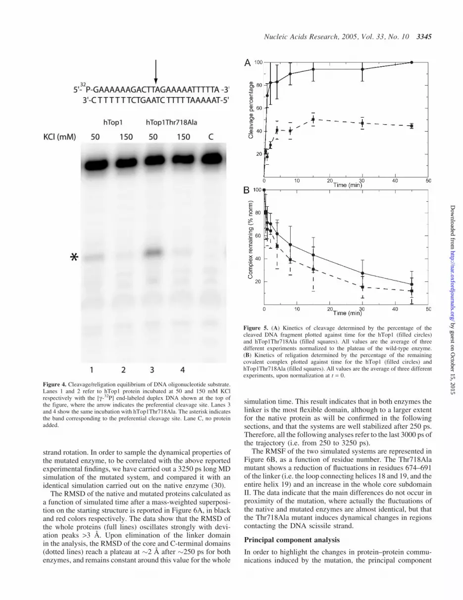
In order to understand the cleavage religation property of themutated enzyme, we have carried out a cleavage/religationequilibrium experiment on a 25mer full duplex oligonuc-leotide substrate CL25 (50-GAAAAAGACTTAGAGAAAA-ATTTT-30)/CP25 at medium (150 mM KCl) and low (50 mMKCl) ionic strength (Figure 4). At 150 mM KCl, the intensityof the band due to the cleavable complex is identical forboth enzymes indicating that in this condition the enzymesare characterized by identical cleavage/religation equilibriumconstant. In detail, evaluation of the cleavage percentageof the labeled scissile strand using the PhosphorImager andImageQuant software indicates that Kcr is 2 · 10�4 for bothenzymes.
On the other hand at 50 mM KCl, the equilibrium of thehTop1Thr718Ala is shifted toward the cleavable complexwhen compared to the wild-type enzyme. Quantitative evalu-ation of Kcr gives a value of 3 · 10�4 and 9 · 10�4 for thewild-type and the Thr718Ala mutant, respectively. A similarbehavior has been observed at 50 mM KCl for a 900 bp DNAfragment where the Thr718Ala mutation shifted the equilib-rium toward cleavage, as detected by the presence of severalcovalent enzyme–DNA intermediates (13).
Since the cleavage/religation equilibrium constant, for alinear DNA substrate can be defined as Kcr = kcl/kr, the experi-ment reported in Figure 4 suggests that at physiological ionicstrength, the native and mutated enzymes are characterized byidentical cleavage (kcl) and religation (kr) rates. In order toverify that the identical equilibrium constant is not due to anidentical variation of the cleavage and religation rate, thecleavage chemical step has been investigated using a 50 endradiolabeled suicide substrate CL14 (50-GAAAAAAGACT-T#AG-30), containing the preferred Top1 sequence (marked
3342 Nucleic Acids Research, 2005, Vol. 33, No. 10
by guest on October 15, 2015
http://nar.oxfordjournals.org/D
ownloaded from

by the arrow), annealed to the CP25 (50-TAAAAATTTTTC-TAAGTCTTTTTTC-30) complementary strand, to producea duplex with an 11-base 50 single-strand extension. Withthis substrate, the religation step is excluded because theshort oligonucleotide (AG-30) generated during cleavagecannot be religated, leaving the enzyme covalently attachedto the 12 oligonucleotide 30 end (9). The suicide cleavagesubstrate has been incubated with an excess of hTop1 orhTop1Thr718Ala in a time course experiment. The amountof cleaved fragment, normalized to the plateau value of thehTop1, has been plotted as a function of time in Figure 5A.Data show that both enzymes have an almost identical cleav-age rate (kcl), reaching in a comparable time a plateau that,however, has a lower value for the hTop1Thr718Ala mutant.
Any quantitative evaluation of kcl from the slope of the firstpart of the curve has been unsuccessful due to the paucity ofexperimental points present in this region.
DNA religation step has then been studied by testing theability of both enzymes to religate the oligonucleotide R11(50-AGAAAAATTTT-30) added to the cleaved suicide sub-strate. The first step of the assay consists in the incubation ofan excess of hTop1 or hTop1Thr718Ala with the suicide sub-strate for 60 min in order to generate the cleaved complex withthe enzyme covalently attached to the 30 end. Once cleavagehas occurred, the salt concentration is raised to 0.5 M KCl toprevent further cleavage activity by the enzyme (19) and theR11 oligonucleotide is then added to the mixture to initiatethe ligation process. Aliquots have been removed at different
Figure 2. (A) Relaxation of negative supercoiled plasmid DNA of purified hTop1 (lanes 1–8) and hTop1Thr718Ala (lanes 9–16) at KCl concentrations of 0, 50, 100,150, 200, 250, 300 and 350 mM. Reaction products were resolved in an agarose gel and visualized by ethidium bromide staining. The dimeric and monomeric forms ofthe supercoiled plasmid DNA are indicated by ‘DSC’ and ‘MSC’, respectively. Lane C, no protein added. (B) Percentage of relaxation measured as the amount ofmonomeric (squares) and dimeric (circles) supercoiled plasmid converted to the relaxed form by hTop1 (full line) and hTop1Thr718Ala (dotted line), plotted as afunction of salt concentration.
Nucleic Acids Research, 2005, Vol. 33, No. 10 3343
by guest on October 15, 2015
http://nar.oxfordjournals.org/D
ownloaded from

times, the reaction stopped by addition of SDS and the pro-ducts analyzed by PAGE. The percentage of the remainingcleaved complex, determined as described under Materials andMethods, plotted as a function of time in Figure 5B indicatesthat hTop1 and hTop1Thr718Ala have very similar religationrates (kr), their values, evaluated from the decay of the curve,being equal to 0.14 and 0.19 min�1, respectively. The identityof the religation rate, kr, and of the cleavage/religation equi-librium, Kcr, indicates that the chemistry of the enzyme at150 mM KCl is identical for both the wild-type and themutated enzyme and that these steps of the enzymatic reactioncannot be the origin of the reduced relaxation efficiency ofthe mutant.
The cleavage and religation efficiency of the wild-type andmutated enzyme has been also tested in a single experimentusing the procedure described by Colley et al. (18) at twodifferent ionic strengths, namely at 50 and 150 mM KCl.At 150 mM KCl, hTop1 and hTop1Thr718Ala display asimilar behavior, in agreement with the previously described
experiments (Figure 5) characterized by an almost equalcleavage and religation rate. On the other hand at low ionicstrength (50 mM KCl), the mutated enzyme is characterized byhigher cleavage efficiency and by lower religation efficiencywhen compared to the wild-type enzyme (data not shown). Inline, a recent work has shown that the yeast topoisomeraseI mutant Thr722Ala (corresponding to the Thr718Ala in thehuman enzyme) displays a decreased religation rate comparedto the wild-type protein, when measured at 50 mM KCl on a 36oligonucleotide DNA substrate (18).
Root mean square deviations and fluctuations
The experiments described above indicate that upon the singleThr718Ala mutation, the chemistry of the enzyme is unalteredand the relaxation efficiency is reduced. This suggests that themain effect caused by the mutation as detected by experimentscarried out at 150 mM KCl ionic strength is a perturbationin the dynamical behavior that alters the control of the DNA
Figure 3. Relaxation kinetics for hTop1 and hTop1Thr718Ala at their salt optima concentrations. (A) Purified hTop1 (lanes 1–8) and hTop1Thr718Ala (lanes 9–16)were incubated with 0.5 mg/ml supercoiled plasmid DNA for 0.5, 1, 2, 4, 8, 15, 30 and 60 min at 37�C at 150 mM KCl. (B) Purified hTop1 (lanes 1–8) andhTop1Thr718Ala (lanes 9–16) were incubated with 0.05mg/ml supercoiled plasmid DNA for 4, 8, 15, 30, 60, 120, 240, 480 s at 37�C at 150 mM KCl. The dimeric andmonomeric forms of the supercoiled plasmid DNA are indicated by ‘DSC’ and ‘MSC’, respectively. Lane C, no protein added.
3344 Nucleic Acids Research, 2005, Vol. 33, No. 10
by guest on October 15, 2015
http://nar.oxfordjournals.org/D
ownloaded from
strand rotation. In order to sample the dynamical properties ofthe mutated enzyme, to be correlated with the above reportedexperimental findings, we have carried out a 3250 ps long MDsimulation of the mutated system, and compared it with anidentical simulation carried out on the native enzyme (30).
The RMSD of the native and mutated proteins calculated asa function of simulated time after a mass-weighted superposi-tion on the starting structure is reported in Figure 6A, in blackand red colors respectively. The data show that the RMSD ofthe whole proteins (full lines) oscillates strongly with devi-ation peaks >3 s. Upon elimination of the linker domainin the analysis, the RMSD of the core and C-terminal domains(dotted lines) reach a plateau at �2 s after �250 ps for bothenzymes, and remains constant around this value for the whole
simulation time. This result indicates that in both enzymes thelinker is the most flexible domain, although to a larger extentfor the native protein as will be confirmed in the followingsections, and that the systems are well stabilized after 250 ps.Therefore, all the following analyses refer to the last 3000 ps ofthe trajectory (i.e. from 250 to 3250 ps).
The RMSF of the two simulated systems are represented inFigure 6B, as a function of residue number. The Thr718Alamutant shows a reduction of fluctuations in residues 674–691of the linker (i.e. the loop connecting helices 18 and 19, and theentire helix 19) and an increase in the whole core subdomainII. The data indicate that the main differences do not occur inproximity of the mutation, where actually the fluctuations ofthe native and mutated enzymes are almost identical, but thatthe Thr718Ala mutant induces dynamical changes in regionscontacting the DNA scissile strand.
Principal component analysis
In order to highlight the changes in protein–protein commu-nications induced by the mutation, the principal component
Figure 4. Cleavage/religation equilibrium of DNA oligonucleotide substrate.Lanes 1 and 2 refer to hTop1 protein incubated at 50 and 150 mM KClrespectively with the [g-32P] end-labeled duplex DNA shown at the top ofthe figure, where the arrow indicates the preferential cleavage site. Lanes 3and 4 show the same incubation with hTop1Thr718Ala. The asterisk indicatesthe band corresponding to the preferential cleavage site. Lane C, no proteinadded.
Figure 5. (A) Kinetics of cleavage determined by the percentage of thecleaved DNA fragment plotted against time for the hTop1 (filled circles)and hTop1Thr718Ala (filled squares). All values are the average of threedifferent experiments normalized to the plateau of the wild-type enzyme.(B) Kinetics of religation determined by the percentage of the remainingcovalent complex plotted against time for the hTop1 (filled circles) andhTop1Thr718Ala (filled squares). All values are the average of three differentexperiments, upon normalization at t = 0.
Nucleic Acids Research, 2005, Vol. 33, No. 10 3345
by guest on October 15, 2015
http://nar.oxfordjournals.org/D
ownloaded from

Figure 6. Root mean square deviations (RMSD) and fluctuations (RMSF). (A) Protein RMSD from the starting structure are represented as a function of simulationtime in black and red full lines for the wild-type and Thr718Ala mutant, respectively. The core and C-terminal domain RMSD are represented in black and red dottedlines for the wild-type and Thr718Ala mutant, respectively. (B) The per residue RMSF are represented as a function of the residue number in black and red lines for thewild-type and Thr718Ala mutant, respectively.
3346 Nucleic Acids Research, 2005, Vol. 33, No. 10
by guest on October 15, 2015
http://nar.oxfordjournals.org/D
ownloaded from

analysis has been applied to both the mutated and the nativeenzyme trajectories to identify the main 3N directions alongwhich the majority of the protein motion is defined (26,27).This analysis is based on the diagonalization of the covariancematrix built from the atomic fluctuations after the removalof the translational and rotational movement, and it hasbeen carried out on the 563 Ca atoms of the protein. The
displacement of each Ca along the first eigenvector havingthe largest eigenvalue shown in Figure 7A, indicates that thelinker domain is the protein region with the maximum dis-placement along this direction for both systems, but with anabsolute value larger for the wild-type than for the mutatedenzyme. The larger linker fluctuations in the wild-type canbe better appreciated looking at the projections of the MD
Figure 7. (A) Displacements of each Ca atom along the eigenvector with the largest eigenvalue (first eigenvector) are represented in black and red lines for the wild-type and Thr718Ala mutant, respectively. Representation of two extreme projections of the MD motions along the first eigenvector for the wild-type protein (B) andmutant proteins (C). Black arrows indicate the amplitude and the direction of the motion.
Nucleic Acids Research, 2005, Vol. 33, No. 10 3347
by guest on October 15, 2015
http://nar.oxfordjournals.org/D
ownloaded from

motions along the first eigenvector for the wild-type andmutant proteins shown in Figure 7B and C, respectively.Note that the projections of the protein motions have beenrepresented together with the average protein–DNA structurescalculated from the fully unconstrained simulations. Figure 7also shows an increase in the mutant Ca displacement in thewhole core subdomain II and the N-terminal portion of coresubdomain I (Figure 7A). The two extreme projections ofthe MD motions along the first eigenvector illustrate howthe motions of the V-shaped a6 from core subdomain I anda5 from core subdomain II are strongly increased upon theThr718Ala mutation (Figure 7B and C). The principal com-ponent analysis indicates that the regions more altered in theirdynamics as a result of the single mutation are those contactingthe DNA scissile strand, which probably play a role during theDNA relaxation process. A dynamic visualization of the MDprojections along the first three eigenvectors are available atNAR Online.
Hydrogen bonds in the mutation site region
As a final analysis, we have calculated the network ofthe protein–DNA and protein–protein hydrogen bonds, in theproximity of the mutation site. A MD conformation ofthe Thr718Ala mutant in the proximity of the mutation site,representing the hydrogen bond network formed by theenzyme during the simulation, is shown in Figure 8. Thepicture shows the involvement of Ala-718 in direct andwater-mediated hydrogen bonds with the protein and DNA,respectively.
A hydrogen bond between the side chain of Thr-718 and theG+2 phosphate group of the scissile strand has been detectedin several crystallographic structures of topo70 (PDB id 1a36,1k4s, 1k4t) (6–8). This bond has been proposed to play animportant role in the orientation of the +1 50-OH group duringthe nucleophilic attack in the religation step (8). The topo70–DNA complex simulation confirms the importance of theThr-718–G+2 bond, being present during the whole simulationtime (data not shown). This hydrogen bond is obviously absentin the Thr718Ala mutant because of the lack of the threonineside chain and analysis of the trajectory indicates that thebackbone of alanine forms a direct hydrogen bond with theG+2 phosphate group for only 3% of the simulation time.However, the absence of a direct bond between the Ala-718and the G+2 base is compensated by a highly stable water-mediated hydrogen bond, present for 95% of the simulationtime (Figure 8). In the wild-type enzyme, a similar bond islacking and a water-mediated hydrogen bond between the sidechain of Thr-718 and the G+2 base is present only for 18% ofthe simulation time.
Deletion of the side chain of Thr-718 also induces changesin the local network of protein–protein hydrogen bonds. In thewild-type enzyme, Thr-718 forms direct hydrogen bondsthrough its side chain with His-632 and Ser-517, for 43 and41% of the simulation time respectively, and through itsbackbone with Leu-721 and Asn-722, for 19 and 23% of thesimulation time, respectively (data not shown). In the mutatedenzyme, the backbone of Ala-718 forms direct hydrogenbonds with Leu-721 and Asn-722, for 66 and 99% of thesimulation time respectively, and with the catalytic Tyr-723for 18% of the simulation time.
CONCLUSIONS
The results presented here show that at 150 mM KCl theThr718Ala mutant has a DNA relaxation rate lower thanthe wild-type enzyme (Figure 3) even though the cleavage/religation equilibrium (Figure 4), the religation rate(Figure 5B) and then the cleavage rate (Figure 5A) are com-parable to those of the wild-type enzyme when using as sub-strate a short linear oligonucleotide containing the preferredcleavage site. A direct comparison between the relaxation rate(Figure 3) and the cleavage and religation rates (Figure 5) ismade impossible by the requirement of different substratesin the two sets of experiment, i.e. a supercoiled DNA substratemade by thousands of base pairs in the relaxation experi-ment and a DNA linear substrate containing 25 bp in thecleavage and religation experiments. On the other hand, thecleavage and religation experiments (Figure 5) can be moredirectly compared with the equilibrium experiment (Figure 4)since a linear substrate is used in both cases. At physiologicalionic strength, the wild-type and mutated enzyme have anidentical equilibrium (Figure 4), as well as an identical religa-tion rate (Figure 5B) indicating that at this ionic strength,the reduced relaxation rate of the Thr718Ala mutant is notdue to a decreased religation rate but to a lower DNA binding
Figure 8. Percentage of hydrogen bonds in the mutation site region evaluatedfrom the MD trajectory. Protein residues and bases are highlighted in cyan andpurple, respectively. The oxygen and nitrogen atoms involved in the hydrogenbonds are highlighted with red and blue spheres, respectively. Direct and water-mediated hydrogen bonds are represented in black full and dotted lines,respectively, together with the percentage of their lifetime in the simulationtrajectory.
3348 Nucleic Acids Research, 2005, Vol. 33, No. 10
by guest on October 15, 2015
http://nar.oxfordjournals.org/D
ownloaded from

or to a decreased strand rotation efficiency. The relaxationexperiments reported in Figure 3 show a decreased relaxationefficiency of the mutant when compared to the wild-type,independent of the DNA/enzyme ratio. This result excludesthat the difference in DNA relaxation between the two pro-teins can be ascribed to the enzyme substrate association/dissociation rate, implying that the main effect of the singleThr718Ala mutation is associated to the rotation step of thecatalysis (29). The in vitro biochemical results then provideunambiguous evidences that at physiological ionic strength,the main functional difference between the native and theThr718Ala mutated Top1 is confined to their strand rotationthat is lower for the mutated enzyme.
The MD simulation furnishes an explanation for such anexperimental behavior providing structural/dynamical datathat permit to interpret the identical religation rate and thedifference in the strand rotation. The identical religationrate of the wild-type and the mutated enzyme is actually anunexpected result since it has been suggested that Thr-718plays a role in the right positioning of the 50 end through ahydrogen bond between the aminoacidic hydroxyl group andthe G+2 phosphate group of the scissile strand (8). The MDtrajectory confirms the importance of this hydrogen bond sinceit is present in the native enzyme for the entire duration of thesimulation (30). However, the simulation also shows that uponmutation of the threonine in alanine the lack of the directhydrogen bond with the G+2 phosphate group is compensatedby a water-mediated hydrogen bond to the backbone ofAla-718 (Figure 8). The lack of a similar bond in the wild-type trajectory suggests either that in the mutant, the water-mediated bond plays a main role in the right positioningof the 50 end, or that Thr-718 does not play any role in thereligation rate.
The MD simulation also permits to explain how the muta-tion of a residue such as Thr-718, which is relatively close tothe catalytic site, may have a large effect on the strand rotationstep of the catalysis. The plot of the RMSF in fact, indicatesthat in proximity of the mutation site the two enzymes haveidentical fluctuations, and that the main differences are local-ized in the linker domain and in core subdomain II that aresupposed to be involved in the DNA strand rotation (6)(Figure 6B). The principal component analysis confirms theRMSF results, showing that, in the mutant, the linker domainand the V-shaped helices are the regions with the major dis-placement along the first eigenvector (Figure 7).
DNA strand rotation is actually strongly dependent on theenzyme conformation and dynamics. It has been reported thatthe clamp of the enzyme around DNA crosslinking residueHis-367 and Ala-499 still permits strand rotation, but this is nolonger possible when the crosslink is made between Gly-365and Ser-534 (31,32). The fundamental role of the linkerdomain in modulating the strand rotation has been demon-strated by Champoux and co-workers (9), showing thatthis domain is required for an efficient CPT inhibition. Theincreased mobility of the linker has been shown to be the mostlikely explanation for the CPT resistance displayed by themutant in which alanine at 653 is substituted with a proline(10). Multiple mutations in the linker domain render theenzyme hypersensitive to the drug, indicating the occurrenceof protein communications between this domain and thedrug binding pocket (33). Finally, a direct correlation between
inhibitor and linker mobility has been shown upon acomparison of the electron density maps of the enzymecrystals in the presence or absence of Topotecan (8).
Here, we show the existence of a direct communicationbetween two regions located far away, i.e. the region closeto the active site and the linker domain. In fact, the Thr718Alamutation induces different flexibility in the linker and in theV-shaped a helices (Figures 6B and 7), confirming the inter-esting correlation between linker mobility and DNA relaxationefficiency. Moreover, the experimental data show that strandrotation is an important step in the catalytic process and thatis directly controlled by the enzyme. In line, a recent reportmeasuring real-time single-molecule DNA relaxation hasshown that Top1 releases DNA supercoils by a swivel mech-anism that involves friction between the rotating DNA and theenzyme, underlining the importance of the enzyme dynamicproperties (34).
Taken together, these results indicate that for topoisomeraseI the description of the structural/dynamical properties are animportant achievement to fully understand its function andmust be taken into account for the design of new drugswith improved efficiency.
SUPPLEMENTARY MATERIAL
Supplementary Material is available at NAR Online.
ACKNOWLEDGEMENTS
We thank Mary-Ann Bjornsti for helpful discussion during thepreparation of the manuscript and S.Z. Pedersen for his carefulreview of this article. This work was partly supported by grantsfrom MURST COFIN2003, from Ministero della Salute, fromFIRB project on Bioinformatics for Genomics and Proteomicsand FIRB project for Functional Genomics. Funding to pay theOpen Access publication charges for this article was providedby MURST COFIN2003.
Conflict of interest statement. None declared.
REFERENCES
1. Chen,A. and Liu,L.F. (1994) DNA topoisomerases: essential enzymes andlethal targets. Annu. Rev. Pharmacol. Toxicol., 34, 191–218.
2. Nitiss,J. (1998) Investigating the biological functions of DNAtopoisomerases in eukaryotic cells. Biochim. Biophys. Acta, 1400, 63–82.
3. Wang,J.C. (1996) DNA topoisomerases. Annu. Rev. Biochem., 65,635–692.
4. Stewart,L., Ireton,G.C. and Champoux,J.J. (1996) The domainorganization of human topoisomerase I. J. Biol. Chem., 271, 7602–7608.
5. Redinbo,M.R., Stewart,L., Kuhn,P., Champoux,J.J. and Hol,W.G.J.(1998) Crystal structures of human topoisomerase I in covalent andnoncovalent complexes with DNA. Science, 279, 1504–1513.
6. Stewart,L., Redinbo,M.R., Qiu,X., Hol,W.G.J. and Champoux,J.J. (1998)A model for the mechanism of human topoisomerase I. Science,279, 1534–1541.
7. Pommier,Y., Pourquier,P., Fan,Y. and Strumberg,D. (1998) Mechanismof action of eukaryotic DNA topoisomerase I and drugs targeted to theenzyme. Biochim. Biophys. Acta, 1400, 83–105.
8. Staker,B.L., Hjerrild,K., Feese,M.D., Behnke,C.A., Burgin,A.B.,Jrand Stewart,L. (2002) The mechanism of topoisomerase I poisoning
by a camptothecin analog. Proc. Natl Acad. Sci. USA, 99,15387–15392.
Nucleic Acids Research, 2005, Vol. 33, No. 10 3349
by guest on October 15, 2015
http://nar.oxfordjournals.org/D
ownloaded from

9. Stewart,L., Ireton,G.C. and Champoux,J.J. (1999) A functionallinker in human topoisomerase I is required for maximum sensitivityto camptothecin in a DNA relaxation assay. J. Biol. Chem., 274,32950–32960.
10. Fiorani,P., Bruselles,A., Falconi,M., Chillemi,G., Desideri,A. andBenedetti,P. (2003) Single mutation in the linker domain confers proteinflexibility and camptothecin resistance to human topoisomerase I.J. Biol. Chem., 278, 43268–43275.
11. Champoux,J.J. (2000) Structure-based analysis of the effects ofcamptothecin on the activities of human topoisomerase I. Ann. N. Y. Acad.Sci., 922, 56–64.
12. Megonigal,M.D., Fertala,J. and Bjornsti,M.-A. (1997) Alterations in thecatalytic activity of yeast DNA topoisomerase I result in cell cyclearrest and cell death. J. Biol. Chem., 272, 12801–12808.
13. Fiorani,P., Amatruda,J.F., Silvestri,A., Butler,R.H., Bjornsti,M.-A. andBenedetti,P. (1999) Domain interactions affecting human DNAtopoisomerase I catalysis and camptothecin sensitivity. Mol.Pharmacol., 56, 1105–1115.
14. Bjornsti,M.-A., Benedetti,P., Viglianti,G.A. and Wang,J.C. (1989)Expression of human DNA topoisomerase I in yeast cells lacking yeastDNA topoisomerase I: restoration of sensitivity of the cells to theantitumor drug camptothecin. Cancer Res., 49, 6318–6323.
15. Kauh,E.A. and Bjornsti,M.-A. (1995) SCT1 mutants suppress thecamptothecin sensitivity of yeast cells expressing wild-type DNAtopoisomerase I. Proc. Natl Acad. Sci. USA, 92, 6299–6303.
16. Bjornsti,M.-A. and Wang,J.C. (1987) Expression of yeast DNAtopoisomerase I can complement a conditional-lethal DNAtopoisomerase I mutation in Esherichia coli. Proc. Natl Acad.Sci. USA, 84, 8971–8975.
17. Benedetti,P., Fiorani,P., Capuani,L. and Wang,J.C. (1993) Camptothecinresistance from a single mutation changing glycine 363 of human DNAtopoisomerase I to cysteine. Cancer Res., 53, 4343–4348.
18. Colley,W.C., Van Der Merwe,M., Vance,J.R., Burgin,A.B.,Jr andBjornsti,M.-A. (2004)Substitutionof conserved residues within the activesite alters the cleavage religation equilibrium of DNA topoisomerase I.J. Biol. Chem., 279, 54069–54078.
19. Yang,Z. and Champoux,J.J. (2002) Reconstitution of enzymatic activityby the association of the cap and catalytic domains of humantopoisomerase I. J. Biol. Chem., 277, 30815–30823.
20. Cornell,W.D., Cieplak,P., Bayly,C.I., Gould,I.R., Kenneth,M., Merz,J.,Ferguson,D.M., Spellmeyer,D.C., Fox,T., Caldwell,J.W. andKolman,P.A. (1995) A second generation force field for the simulation ofproteins, nucleic acids, and organic molecules. J. Am. Chem. Soc.,117, 5179–5197.
21. Jorgensen,W.L., Chandrasekhar,J., Madura,J.D., Impey,R.W. andKlein,M.L. (1983) Comparison of simple potential functions forsimulating liquid water. J. Chem. Phys., 79, 926–935.
22. Darden,T., York,D. and Pedersen,L. (1993) Particle mesh Ewald:an N.log(N) method for Ewald sums in large systems. J. Chem. Phys.,98, 10089–10092.
23. Cheatham,T.E., Miller,J.L., Fox,T., Darden,T.A. and Kollman,P.A.(1995) Molecular dynamics simulation on solvated biomolecularsystems: the particle mesh Ewald method leads to stable trajectories ofDNA, RNA, and proteins. J. Am. Chem. Soc., 117, 4193–4194.
24. Ryckaert,J.-P., Ciccotti,G. and Berendsen,H.J.C. (1977) Numericalintegration of the Cartesian equations of motion of a system withconstraints: molecular dynamics of n-alkanes. J. Comput. Phys.,23, 327–341.
25. Berendsen,H.J.C., Postma,J.P.M., van Gusteren,W.F., Di Nola,A. andHaak,J.R. (1984) Molecular dynamics with coupling to an external bath.J. Comput. Phys., 81, 3684–3690.
26. Garcia,A.E. (1992) Large-amplitude nonlinear motions in proteins.Phys. Rev. Lett., 68, 2696–2699.
27. Amadei,A., Linssen,A.B. and Berendsen,H.J. (1993) Essential dynamicsof proteins. Proteins, 17, 412–425.
28. Berendsen,H.J.C., van der Spoel,D. and van Drunen,R. (1995)GROMACS: a message-passing parallel molecular dynamicsimplementation. Comp. Phys. Commun., 95, 43–56.
29. Frohlich,R.F., Andersen,F.F., Westergaard,O., Andersen,A.H. andKnudsen,B.R. (2004) Regions within the N-terminal domain of humantopoisomerase I exert important functions during strand rotationand DNA binding. J. Mol. Biol., 336, 93–103.
30. Chillemi,G., Redinbo,M., Bruselles,A. and Desideri,A. (2004) Role of thelinker domain and the 203–214 N-terminal residues in the humantopoisomerase I DNA complex dynamics. Biophys J., 87, 4087–4097.
31. Woo,M.H., Losasso,C., Guo,H., Pattarello,L., Benedetti,P. andBjornsti,M.-A. (2003) Locking the DNA topoisomerase I protein clampinhibits DNA rotation and induces cell lethality. Proc. Natl Acad. Sci.USA, 100, 13767–13772.
32. Carey,J.F.,Schultx,S.J.,Sisson,L.,Fazzio,T.G. andChampoux,J.J. (2003)DNA relaxation by human topoisomerase I occurs in the closed clampconformation of the protein. Proc. Natl Acad. Sci. USA, 100, 5640–5645.
33. Scaldaferro,S., Tinelli,S., Borgnetto,M.E., Azzini,A. and Capranico,G.(2001) Directed evolution to increase camptothecin sensitivity ofhuman DNA topoisomerase I. Chem. Biol., 8, 871–881.
34. Koster,D.A., Croquette,V., Dekker,C., Shuman,S. and Dekker,N.H.(2005) Friction and torque govern the relaxation of DNA supercoils byeukaryotic topoisomerase IB. Nature, 434, 671–674.
3350 Nucleic Acids Research, 2005, Vol. 33, No. 10
by guest on October 15, 2015
http://nar.oxfordjournals.org/D
ownloaded from


