Embed Size (px)
Citation preview

129
available at www.jstage.jst.go.jp/browse/islsm ORIGINAL ARTICLES
Introduction
As women age, relaxing of the vaginal wall can lead tovaginal relaxation syndrome (VRS), which is exacerbat-ed by childbirth, especially multiple pregnancies and
deliveries, and the vaginal atrophy associated withmenopause-related hormonal changes. VRS can lead toa number of problems, both physical and psychologi-cal, a major one of which is lessening of sexual satis-faction for both the female and her partner, oftenreferred to as “loose vagina”. Urinary incontinence (UI)is another problem associated with VRS, either of thestress or urge type, and can be mildly irritating or total-ly debilitating. The quality of life of affected women
Treatment of Vaginal Relaxation Syndrome with anErbium:YAG Laser Using 90° and 360° Scanning Scopes:
A Pilot Study & Short-term Results
Min Seok Lee
Sophie-Marceau Women’s Clinic, Daegu, South Korea
Background and Aims: Vaginal relaxation syndrome (VRS) is both a physical and psychologicalproblem for women and often their partners. Recently the 2940 nm Er:YAG laser has attractedattention for VRS treatment. The current study evaluated the clinical efficacy of this nonsurgicallaser procedure.Subjects and Methods: Thirty postpartum females with VRS or vaginal atrophy, ages from 33 – 56yr (mean 41.7 yr) were divided randomly into two groups, Group A and Group B. Both groupswere treated for 4 sessions at 1~2-weekly intervals with a 2940 nm Er:YAG via 90° and 360° scan-ning scopes. In Group A the first 2 sessions were performed with the 360° scope and the final 2with the 90° scope in multiple micropulse mode, 1.7 J delivered per shot, 3 multishots, 3 passesper session. Group B underwent multiple micropulse mode treatment with the 90° scope in all 4sessions (same parameters as Group A) then during the final 2 sessions an additional 2 passes/ses-sion were delivered with the 360° scope, long-pulsed mode, 3.7 J delivered per shot. Perineometerassessments were performed at baseline and at 2 months post-treatment for vaginal tightness.Histological specimens were taken at baseline and at 2 months post-procedure. Subjective satisfac-tion with vaginal tightening was assessed together with improvement in sexual satisfaction. Resultswere tested for statistical significance with the paired Student’s t-test.Results: All subjects successfully completed the study with no adverse events. Significant improve-ment in vaginal wall relaxation was seen in all subjects at 2 months post-procedure based on theperineometer values, on the partners’ input for vaginal tightening (76.6%) and for sexual satisfac-tion as assessed by the subjects themselves (70.0%). The histological findings suggested better elas-ticity of the vaginal wall with tightening and firming.Conclusions: Both regimens of Er:YAG laser treatment for VRS produced significant improvementin vaginal relaxation. With multishots delivered in the multiple micropulse mode via scanningscopes, nonsurgical Er:YAG laser treatment was pain-free, safe, side effect free, easily tolerated andeffective.
Key words: Multishot micropulse mode • vaginal tightening • 360° scanning scope • 90° scanningscope • perineometer • sexual satisfaction • elastinogenesis • collagenesis • tissueremodeling
Received date: May 7th, 2014Accepted date: June 4th, 2014
Laser Therapy 23.2: 129-138©2014 JMLL, Tokyo, Japan
Addressee for Correspondence:Min Seok Lee MD.,PhD.706-817 Sophie-Marceau Women’s Clinic,42-2, Beomyeo 3-Dong, Suseong-GuDaegu Metropolitan, South KoreaTel: 82-53-756-3006, 4006(O) Fax: 82-53-756-5077E mail: [email protected]

130
available at www.jstage.jst.go.jp/browse/islsmORIGINAL ARTICLES
MS Lee
can decline dramatically as they become afraid to goout socially or even to work because of the humiliationof an unpredictable and involuntary loss of urinarycontrol. According to the National Institute of Diabetesand Digestive and Kidney Diseases (NIDDK) of theNational Institutes of Health (NIH) in the USA, UIoccurs in women at least twice as often as in men. Thecombination of loss of sexual satisfaction for both part-ners and the psychological and social problems associ-ated with UI mandate that a real, lasting and consistentsolution for the underlying condition of VRS isrequired. A variety of VRS treatment options exists span-ning the spectrum from noninvasive approaches tofrankly invasive surgical procedures. For the noninva-sive approaches, behavioral training such as Kegelexercises can tighten up relaxed musculature in thepelvic floor and to a lesser extent in the vaginal wall;“tightening creams”, hormonal creams, sprays andother pharmacological approaches are also available.However, although these are noninvasive and innocu-ous, the effectiveness is somewhat limited and thelatency period temporary, requiring continuous appli-cation in the case of the pharmacotherapies. Surgicalprocedures, in which vaginal and associated tissues areincised and rearranged, 1) can offer a much better andlonger lasting final result. The results of surgicalvaginoplasties, however, have to be balanced againstthe much greater risks involved in any surgery per-formed on the extremely sensitive vaginal tissues.Downtime for recovery is longer, and there are recog-nized risks associated with scar formation or nervedamage leading to dysesthesia. 2,3)
The laser has recently been added to the tradi-tional armamentarium associated with surgicalapproaches for VRS because of its precision and theability to limit damage depth with particular wave-lengths having high water absorption, such as the CO2(10600 nm) and particularly the Er:YAG (2940 nm).This approach was called laser-assisted vaginoplasty.Taking the bulk laser beam and splitting it into multi-ple microbeams has provided even more control of thedepth of the microablative columns (MACs), resultingin better efficacy with less downtime for the subject,and has become popularized as so-called “vaginaltightening”. Compared with a vaginoplasty, thisapproach encompasses non-reconstructive strategiesaimed at restoring the muscle tone of the vagina bytightening the supportive structures of the vulvovaginalcomplex, in an effort either to at least reduce theeffects of aging and childbirth. Fractional Er:YAG systems with a dedicated gyne-
cological delivery system recently became commercial-ly available offering a nonsurgical approach fallingunder the concept of vaginal tightening. In the systemused in the present study, special scanning scopes, oneallowing 360° beam delivery and the other a 90° side-firing scope, were designed to deliver selective andprecise ablation with controlled coagulative damage tothe entire vaginal wall for the treatment of all symp-toms of VRS, or to the wall of the anterior vaginalcanal for the treatment of UI, or both. The presentstudy was designed to assess the efficacy of the multi-ple micropulse mode Er:YAG laser fitted with suchscanning scopes in the treatment of VRS, and com-pared two different treatment protocols.
Subjects and Methods
Subjects
The study subjects comprised 30 postpartum femaleswith VRS or vaginal atrophy, ages from 33 – 56 yr(mean 41.7 yr) who were divided randomly into twogroups, Group A and Group B. Five subjects had givenbirth once, 20 twice, and 5 had undergone 3 deliveries.There was no significant difference between thegroups as regards mean age (Group A, 42.93 yr; GroupB, 40.53 yr), body mass index (BMI) (Group A 22.3;Group B, 22.6) or parous status. Regarding the men-strual status, in Group A, pre-, peri- and post-menopausal females accounted for 9, 1 and 5 subjects,respectively; in group B, 14, 1 and 0. Group A wasthus at a more advanced menopausal status than groupB. One subject in Group A had undergone a cesareansection, compared with 5 subjects in Group B. Thesubject demographics and relevant histories are givenin Table 1. Regarding the degree of VRS, 3, 9 and 3subjects were graded mild, moderate and severe,respectively, in Group A, compared with 7, 3 and 5 inGroup B (Table 2). Levator ani muscle (LAM) poweras assessed with a digital examination was respectivelygood, moderate, poor and very poor in 5, 2, 7 and 1subjects in Group A, compared with 4, 7, 3 and 1 sub-jects, respectively, in Group B (Table 2). A perineome-ter (ExTT-101, APIMEDS Inc, South Korea) was usedfor objective measurement of the strength of voluntarycontractions of the pelvic floor muscles at baseline andafter treatment. Table 2 also shows the baseline peri-neometer readings for the maximum (Pm) pressuresand average (Pa) pressures (in mmHg) and the time forwhich pressure was maintained (Pt in seconds).

Er:YAG laser for VRS 131
available at www.jstage.jst.go.jp/browse/islsm ORIGINAL ARTICLES
Table 1: Patient characteristics
Serial No Age BMI(%) Parous status Delivery type Menstrual status Remarks
Group A (n = 15)
P-1 36 21.9 2 NSVD premen
P-2 38 23.2 2 C-sec premen
P-3 35 22.6 2 NSVD premen previous p-repair Hx(+)
P-4 40 20.1 2 NSVD premen Previous sexual assault Hx(+)
P-5 41 24.1 3 NSVD premen
P-6 53 27.1 2 NSVD postmen atrophic vaginitis case
P-7 56 25 2 NSVD postmen atrophic vaginitis case
P-8 35 19.7 1 NSVD premen previous LEEP Hx(+)
P-9 35 19.7 1 NSVD premen
P-10 56 24.4 3 NSVD postmen
P-11 51 24.3 2 NSVD perimen
P-12 52 22.4 3 NSVD postmen atrophic vaginitis case
P-13 43 20.4 2 NSVD premen
P-14 36 22 2 NSVD premen
P-15 37 20.6 2 NSVD premen
Group B (n=15)
P-16 49 20 3 NSVD premen
P-17 42 24.3 1 C-sec premen
P-18 42 25 2 NSVD premen
P-19 33 22 2 C-sec premen
P-20 38 19.1 2 C-sec premen
P-21 40 20.6 1 NSVD premen
P-22 34 32.2 2 C-sec premen
P-23 42 23.2 2 NSVD premen
P-24 44 24.3 2 C-sec premen
P-25 41 20.8 1 NSVD premen previous TOT op(+)
P-26 33 17.6 3 NSVD premen
P-27 33 18.4 2 NSVD premen
P-28 52 26.7 2 NSVD perimen previous TOT op(+) / HRT(+)
P-29 38 22.8 2 NSVD premen
P-30 47 21.4 2 NSVD premen
KEY: BMI, body mass index; NSVD, normal spontaneous vaginal delivery; C-sec, cesarean section; Premen, pre-menstrual; perimen, perimenstrual; postmen, postmenstrual; p-repair, posterior vaginal repair; LEEP, loop electro-surgical excision procedure; TOT, transobturator tape surgery; HRT, hormone replacement therapy
132
available at www.jstage.jst.go.jp/browse/islsmORIGINAL ARTICLES
MS Lee
Table 2: Vaginal wall relaxation status at baseline as assessed by digital examination, at baselineand after treatment tested with a perineometer, postprocedural partners’ assessment ofvaginal tightening and patients’ assessment of postprocedural sexual satisfaction.
Pat.No
Degree ofVWRS
LAM power(digital exam)
Perineometer test values
Vaginaltightening(Partner)
Sexualsatisfaction(Patient)
B/L Post-2 months
Pm(mmHg)
Pa(mmHg)
Pt(s)
Pi(mmHg)
Pa(mmHg)
Pt(s)
Group A
P-1 mild good 39 34 10 41 35 17 3 3
P-2 mild good 48 38 9 61 44 10 2 3
P-3 moderate poor 13 6 20 17 6 36 0 0
P-4 moderate good 28 21 6 49 42 8 0 3
P-5 moderate poor 12 7 6 22 16 19 2 2
P-6 moderate good 28 19 20 50 40 21 3 3
P-7 mild poor 7 3 21 13 7 19 0 3
P-8 moderate good 11 6 23 32 24 23 1 1
P-9 moderate poor 8 6 7 15 11 25 3 3
P-10 severe moderate 19 14 2 27 20 14 1 0
P-11 severe poor 13 8 2 19 9 6 1 1
P-12 moderate moderate 8 4 10 31 25 16 3 3
P-13 moderate poor 8 4 16 14 8 26 2 2
P-14 moderate poor 20 10 49 22 10 53 3 3
P-15 severe very poor 0 0 0 15 10 8 1 1
Group B
P-16 severe moderate 21 15 21 20 14 23 1 1
P-17 moderate moderate 8 5 11 10 6 10 1 0
P-18 mild good 19 9 46 15 8 40 0 0
P-19 mild moderate 15 5 37 24 16 25 0 0
P-20 mild good 20 12 8 28 19 8 0 0
P-21 mild good 37 30 13 38 24 17 1 1
P-22 mild poor 26 19 14 34 26 15 3 3
P-23 moderate very poor 0 0 0 20 12 14 3 3
P-24 mild moderate 17 14 22 22 18 56 2 2
P-25 moderate good 22 15 36 32 24 17 1 1
P-26 severe moderate 31 24 5 34 28 5 0 0
P-27 severe poor 18 12 25 19 12 33 0 0
P-28 severe moderate 27 21 4 65 54 8 2 2
P-29 mild poor 10 7 24 14 10 58 1 1
P-30 severe moderate 22 18 30 15 9 56 2 2
Pat. No, patient reference No; VWRS, vaginal wall relaxation status; LAM, levator ani muscle; BL, baseline values;Post-2 months, values 2 months after the final treatment; Pm, maximum pressure; Pa, average pressure; Pt, time forwhich pressure was maintained; Vaginal tightening (Partner), assessment by the patient’s partner of the degree ofimprovement in vaginal tightness post-treatment using the following scale: little or no improvement, 0; someimprovement, 1; good improvement, 2; excellent improvement, 3.Patient sexual satisfaction was self-rated using the following scale: dissatisfied, 0; somewhat satisfied, 1; satisfied, 2;extremely satisfied, 3.

133
available at www.jstage.jst.go.jp/browse/islsm ORIGINAL ARTICLES
Laser system
The laser system used was the ACTION II™ Er:YAG(Lutronic, Goyang, South Korea) delivering a wave-length of 2940 nm. When fitted with the dedicatedvaginal scanning scopes (Petit Lady™ system, Lutronic)the laser can be operated in the multiple micropulsemode (pulse width of 250 µs, selectable number ofmultipulses), or in the long-pulsed mode (1000 ms, sin-gle pulse). There were two scopes comprising the ded-icated vaginal scanning scope system (Figure 1): onescope delivered a 360° ring-shaped beam with anapproximate beam width at target of 2-3 mm (Petit360), and the other delivered around-shaped 90° beamwith an approximate beam width at target of 5 mm(Petit 90), both scopes being supported in the vaginaby a specially designed guide (Figure 2). In general,for the 360° scope the supporting guide is inserted,then the scope is fully inserted into the guide. Thebody of the scope is marked with 2.5 mm gradations.The laser parameters are set including the multiplepulse option, the laser is fired, the probe is withdrawnby 1 gradation (2.5 mm) marked on the probe body,and the process repeated for the entire length of thevaginal canal. Multiple passes may be made. For the90° scope in the treatment of UI, following insertion ofthe supporting cage the probe is inserted with theactive part of the scope at the 12 o’clock position. Thelaser is fired, and the scope turned to the 2 o’clockposition, fired again, then turned to the 10 o’clockposition and fired again. The scope is withdrawn by 2gradations (5 mm), returned to the 12 o’clock position,and the process is repeated for the required number ofpasses. To treat the entire vaginal wall with the 90°scope, however, for each set of shots the active part ofthe scope is set at the 12 o’clock, then 2 o’clock, 4o’clock, 6 o’clock, 8 o’clock and 10 o’clock positions,the scope is withdrawn by 2 gradations, and theprocess repeated.
Treatment protocol by group (N=15 for both groups)
Group A: The protocol called for four treatment ses-sions, 1-2 weeks apart. In sessions 1 and 2, treatmentwas applied with the 360° scope as described aboveusing the multiple micropulse mode. The multishotoption was set to 3 multishots and 1.7 J/shot wasselected by manually setting the pulse energy at 15 mJon the screen display in Fractional Mode to convert thedisplay screen settings to the gynecological scope set-tings. Three passes were delivered along the entirelength of the vaginal canal in each treatment session.
In sessions 3 and 4, the 90° scope was used at thesame parameters as for sessions 1 and 2. For each setof shots the active part of the scope was set at the 12o’clock, then 2 o’clock, 4 o’clock, 6 o’clock, 8 o’clockand 10 o’clock positions to treat the entire circumfer-ence of the vaginal wall, as described above.Group B: Four treatment sessions were delivered, 1-2weeks apart. Sessions 1-4 called for the 90° scope usedas in Group A, multiple micropulse mode, 3 multishotsetting (pulse width of 250 µs), and 1.7 J/shot. Threepasses were delivered for each treatment session. Insessions 3 and 4 an additional 2 passes per sessionwere delivered with the 360° scope in long-pulsedmode, pulse width of 1000 ms (1 s), 3.7 J/shot (manu-ally set as 11 J/cm² on the display screen with a spotsize of 6.4 mm in Long Pulsed mode).
Assessments
Punch biopsies were taken at baseline and at 2 monthsafter the final session, formalin fixed and routinely pre-pared for light microscopy with hematoxylin and eosinand elastica van Giesen staining. Perineometer readingwere repeated at the same time point and comparedwith the baseline values. Subjective assessments at thesame time point included the subject’s partner’s evalua-tion of the degree of vaginal tightening, and the sub-ject’s own evaluation of the degree of improvement insexual satisfaction. Subjective scoring was on 4-pointscales as follows: For vaginal tightening, little or noimprovement, 0; some improvement, 1; good improve-ment, 2; excellent improvement, 3. For patient sexualsatisfaction, dissatisfied, 0; somewhat satisfied, 1; satis-fied, 2; extremely satisfied, 3.
Statistical analysis
Perineometer readings of all subjects were compared atbaseline and 2 months after the final session with thepaired Student’s t-test. The same test was used to com-pare between Groups A and B for perineometer read-ings, partner’s assessment of vaginal tightening andsubject’s assessment of improvement in sexual satisfac-tion. A P value equal to or less than 0.05 was consid-ered statistically significant, with marginal significanceat P equal to or less than 0.1. The statistical softwareused was STATISTICA V10, StatSoft, OK, USA.
Results (Table 2)
All subjects completed the treatment and the 2-monthassessment. All patients were aware of some heating inthe vagina during treatment, and a very few patients(3: 1 from Group A and 2 from Group B) felt mild
Er:YAG laser for VRS
134
available at www.jstage.jst.go.jp/browse/islsmORIGINAL ARTICLES
MS Lee
Table 3: Statistical analyses of the data comparing the baseline and post-procedureresults for the entire patient population, and between Groups A and B
Fig. 3: Hematoxylin and eosin stained specimens of the vaginal wall from patient 1 (Left) at baseline, and(right) 2 months after treatment with the system used in the present study showing improved mucos-al architecture in both the epithelium and lamina propria. Scale units are as shown on the bars.
GroupsPm (mmHg)
S-Sig(p)
Pa (mmHg)S-Sig(p)
Pt (s)S-Sig(p)
VT SSBL(SEM)
Post(SEM)
BL(SEM)
Post(SEM)
BL(SEM)
Post(SEM)
Total18.50(2.04)
27.27(2.60)
*< 0.01(=0.0052)
12.87(1.76)
19.57](2.3)
*< 0.05(=0.013)
16.57(2.4)
22.87(2.9)
< 0.1(=0.097) � �
Grp A*vs
Grp B**
17.47(3.4)
28.53(3.9) *<0.1
(= 0.091)
12.00(2.92)
20.47(3.55) NS
(=0.330)
13.4(3.19)
20.07(3.13) NS
(=0.539)NS
(=0.101)*< 0.05(=0.011)13.73
(2.05)18.67(2.09)
13.73(2.06)
18.67(3.09)
19.73(3.49)
25.67(4.81)
BL, baseline; Pm, maximum pressure; Pa, average pressure; Pt, time for which pressure was maintained, in seconds; SEM,standard error of means; Post, 2 months post-treatment; S-Sig, statistical significance; (p), P value (paired Student’s t-test); VT,vaginal tightening (assessed by patients’ partners); SS, sexual satisfaction (patients’ subjective assessment); NS, no significance
Fig. 1: The 360° scanning scope (left) andthe 90° scanning scope (right).
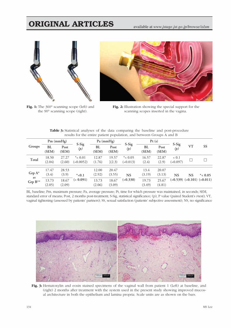
Fig. 2: Illustration showing the special support for thescanning scopes inserted in the vagina.

135
available at www.jstage.jst.go.jp/browse/islsm ORIGINAL ARTICLES
vaginal ecchymosis with a mild burning sensationwhich lasted for 24-48 hr and evolved spontaneously.No subject reported any major or lasting adverse sideeffects after any of the treatments. Improvement wasseen in all perineometer scores, in the majority of sub-jects (76.6%) for the partner’s assessment of vaginaltightening and in the subjects’ own assessment ofimproved sexual satisfaction (70.0%).
Statistical assessments (Table 3)
Overall values in the vaginal wall relaxation status forthe total subject population showed a statistically sig-nificant improvement between the baseline and find-ings at 2 months after treatment for the maximum peri-neometer pressure (p <0.01), and the average pressure(p < 0.05), with marginal significance noted in the timefor which the pressure was maintained (p < 0.1).
Er:YAG laser for VRS
Fig. 4: In these elastica van Giesen stainedspecimens from the vaginal wall ofpatient 2 at a higher magnification thanin Fig 3, the findings can be seen com-paring the baseline (left) with the situa-tion 2 months post-treatment (right)where a thicker epithelium and denserlamina propria are observed. Scale unitsare as shown on the bars.
Fig. 5: Hematoxylin and eosin-stained section of theanatomy of the vaginal wall, with the variouszones labeled.
Fig. 6: Schematic representation of the dual-mode technique.In the first stage (left panel) the 1st shot of the 250 µsmultipulse delivers controlled and minimal ablationopening a window in the epidermal tissue, allowingthe thermal effect of subsequent shots to penetrateinto the lamina propria. In the second stage, the sub-ablative single shot in the 1000 ms long-pulsed modedelivers thermal damage deep into the lamina propriathrough the existing epidermal window.

136
available at www.jstage.jst.go.jp/browse/islsmORIGINAL ARTICLES
MS Lee
In the comparison of results between groups,Group A had slightly better results than Group B withmarginal significance in the maximum pressure (p<0.1) and an increased trend without significant differ-ence seen in the average perineometer pressure or thetime for which pressure was maintained. As for vaginaltightening as assessed by the subject’s partner, GroupA showed an increasing trend without significancecompared with Group B, but with a statistically signifi-cant difference seen for the increase in sexual satisfac-tion assessed by the subjects themselves (p <0.05).
Histological assessment
The histological findings in general showed evidenceof a thicker and more cellular epithelium and a morecompact lamina propria with a denser arrangement ofconnective tissue. Figures 3 (Patient 1) and 4(Patient 2) show representative specimens at baselineand two months after the final treatment under hema-toxylin and eosin and elastica van Giesen staining,respectively. Taken together, the results of the histo-logical analysis suggest tightening and firming of thevaginal wall. Both specimens came from comparativelyyoung patients, but even in these young patientsimprovements in and rejuvenation of the vaginal struc-tures can be seen 2 months after the final Petit Ladytreatment.
Subject assessments
As seen in Table 2, in the case of the subjects’ part-ners, 23 out of 30 (76.6%) noted improvement in vagi-nal tightening as follows: some improvement, 9; goodimprovement, 6; and excellent improvement, 7. Therewas no statistical significance in these results betweengroups A and B. As for the patient sexual satisfactioncompared with the preoperative condition, 21 out ofthe 30 patients were satisfied (70.0%), made up as fol-lows: somewhat satisfied, 7; satisfied, 5; and extremelysatisfied, 9. Group A subjects were significantly moresatisfied than subjects in group B (p < 0.05)
Discussion
The important fact from the present study, based onthe results of the nonsurgical multiple micropulsemode Er:YAG treatment for vaginal relaxation syn-drome, was that all patients completed the study,improvement was seen in both vaginal tightening forall subjects and increased sexual satisfaction wasreported for the majority of the total patient populationand their partners. The choice of the Er:YAG with its2940 nm wavelength for this gynecological scanning
probe-based system was predicated on the absorptionpeak of water at that wavelength. As human tissue,especially mucous membranes, has a very high per-centage of water, it is a good target for this wave-length. Because of the extremely high absorption inwater, the incident photon energy is almost totallyquenched in the first few micrometers of tissue, pro-ducing at appropriate parameters a very controlled col-umn of ablation with an extremely narrow band of sec-ondary coagulation, known as residual thermal damage(RTD). 4) This translates into shorter downtime withquicker healing, and was the cornerstone of the use ofthe Er:YAG in full-face ablative laser resurfacing com-pared with the CO2 laser, which had a much largerRTD zone. 5) For many clinicians practicing laser resur-facing, however, this larger zone of RTD actually madethe CO2 laser superior to the Er:YAG in terms of thelatency and degree of the result, although use of theCO2 laser dramatically increased the downtime. In thecase of the very delicate vaginal canal, however, thedepth control associated with the Er:YAG wavelengthoffers major benefits. Splitting an Er:YAG laser beam into multiplemicropulses further controls the depth of the ablativecolumns (referred to as microablative columns [MACs]),which allows the deposition of adequate RTD toensure a healthy and swift wound healing processwhile leaving undamaged normal tissue between theMACs and thereby speeds up tissue recovery evenmore because of the participation in the process of thenormal tissue. In the delicate tissues of the vaginalcanal, it is imperative that the ablative damage depth isminimized, but that enough RTD is created in the tar-get tissue to induce actively the wound healingprocess, plumping up and rejuvenating the squamousepithelial component of the mucosa, while firming upthe connective tissue of the lamina propria (Figure 4)and thus causing tightening of the supportive struc-tures of the vulvovaginal complex. In the case of the approach in the present study,there were two scanning scopes to deliver the multiplemicropulses: the 360° and the 90° scope. The 360°scope delivered a ring-shaped circular beam withmicropulse beam width at the target of 2-3 mm, where-as the 90° scope delivered a round beam with amicropulse beam width of around 5 mm at the tissue.For both scopes, the first set of multiple micropulsescleanly created a few µm-thick epidermal window inthe vaginal epithelium with minimal RTD, and subse-quent micropulses created a pulse-stacking effect with-out further ablation, but with thermal build-up downinto the lamina propria, increasing the RTD zone and

137
available at www.jstage.jst.go.jp/browse/islsm ORIGINAL ARTICLES
ensuring a good wound healing response (Figure 5,left panel). Therefore, taking the respective beamdiameters into consideration, to ensure even coverageof the target tissue the 360° scope was withdrawn by 1increment marked on the scope body after each set ofshots, and the 90° scope was withdrawn by 2 incre-ments for each set of shots, each increment represent-ing a distance of 2.5 mm. In the present study, to attempt even better tight-ening of the vaginal canal through induced enhancedtissue remodeling, it was considered that a more “CO2-like” effect should be delivered to the tissue, i.e. moreresidual thermal damage with minimal controlled abla-tion. It was shown as early as 2001 by Trelles and col-leagues that the Er:YAG at appropriate parameterscould deliver almost as good a coagulative effect as theCO2 through an ablated epidermal window, 6) so forthe Group B subjects the 1000 ms long-pulsed modewas added for the last 2 sessions as an adjunct to the250 µs multiple micropulse mode treatment to delivermore of a thermal effect (Figure 5, right panel). This protocol actually represents a potential limi-tation of the present study, because the protocol forgroup A was not exactly repeated in Group B with theaddition of the long-pulsed mode, and this makes itmore difficult to compare the effect accurately betweenthe groups with and without the long-pulsed mode.Groups A and B both received 4 sessions, 1-2 weeksapart. However in Group A the first 2 sessions weredelivered with the 360° scope, and sessions 3 and 4with the 90° scope, multiple micropulse mode in allcases. For group B, on the other hand, all 4 sessionswere delivered with the 90° scope in multiplemicropulse mode, but in the last 2 sessions patientswere additionally treated with the 360° scope in thelong-pulsed mode. Another limitation was the fact thatno specific attention was paid to the urinary inconti-nence (UI) status of any individual patients, so no con-clusions can be drawn as to the efficacy of thisapproach for UI. However, as UI is one of the majorsymptoms associated with VRS, improvement in theVRS status could be correlated with improvement ofthe UI: future studies should address this separately. When the results in the group of 30 patients arelooked at as a whole, significantly better results wereseen in the perineometer readings, vaginal tighteningand sexual satisfaction. However, the author expectedto see better results in Group B because of the addi-tional long-pulsed mode treatment, but as the datashow, it was Group A subjects who had the statisticallysignificantly better results, both from the perineometerreadings and the subjects’ own assessment of sexual
satisfaction. This was not a little surprising, especiallyas Group A was at a more advanced menopausal statethan Group B, and so might have had more vaginalatrophy. In addition, subjects in Group A had experi-enced more natural childbirths than in Group B, with 1C-section in the former compared to 5 C-sections in thelatter: A C-section obviously puts much less strain onthe birth canal, especially the vulvovaginal complex. Inshort, a better result was achieved in Group A whosevaginal canal was in potentially worse physiologicalcondition than Group B. Overall, however, the objective perineometryreadings clearly showed high statistical significancebetween the baseline and the 2-month post-proceduralreadings for the entire patient population, as did thepatient and partner assessments of sexual satisfactionand vaginal tightening, respectively, so both treatmentprotocols were efficacious and safe. It is an acceptedfact that a fluence delivered by a larger spot size pene-trates better and deeper than the same fluence with asmall spot size. Why was the result superior in GroupA when the pulse energy for the multiple micropulsemode was identical at 15 mJ? The answer is uncertain.Perhaps the order of treatment was important, with the2-3 mm spot size of the 360° scope for the first 2 ses-sions, followed by the 90° scope with its 5 mm beamdiameter in the Group A protocol. Future studies needto be designed to take these considerations intoaccount, with the addition if possible of histologicalassessment immediately after treatment to help com-pare the morphology at baseline with changes in thevaginal architecture following Er:YAG laser treatmentwith the scanning scope system, and further relatedwith the follow-up results.
Conclusions
Both regimens of Er:YAG laser treatment for VRS andvaginal atrophy with the multiple micropulse mode(Group A) and dual mode (multiple micropulse modewith 2 additional sessions of long-pulse mode, GroupB) produced significant improvement in vaginal relax-ation with tightening in both the objective perineome-ter readings, from the results of the partners’ assess-ments of improved vaginal tightness (76.6%), and fromthe subjects’ own improvements in perceived sexualsatisfaction (70.0%).This nonsurgical treatment involv-ing multiple micropulse and dual Er:YAG modes withthe Er:YAG laser and gynecological probes used in thepresent study was safe, easily tolerated, pain- and sideeffect free and effective in the treatment of VRS.
Er:YAG laser for VRS

138
available at www.jstage.jst.go.jp/browse/islsmORIGINAL ARTICLES
MS Lee
References
1: Goodman MP. Female cosmetic genital surgery.Obstet Gynecol, 2009;113:154-159.
2: Davies MC, Creighton SM, Woodhouse CR. Thepitfalls of vaginal construction. BJU Int,2005;95:1293-1298
3: American College of Obstetricians andGynecologists (ACOG). Vaginal “Rejuvenation” andCosmetic Vaginal Procedures (in PDF) (2007)
4: Price CR, Carniol PJ, Glaser DA. Skin resurfacingwith the erbium:YAG laser. Facial Plast Surg Clin
North Am. 2001; 9: 291-302. 5: Newman JB, Lord JL, Ash K, McDaniel DH.
Variable pulse erbium:YAG laser skin resurfacingof perioral rhytides and side-by-side comparisonwith carbon dioxide laser. Lasers Surg Med, 2000;26:208-214.
6: Trelles MA, Mordon S, Benítez V, Levy JL. Er:YAGlaser resurfacing using combined ablation andcoagulation modes. Dermatol Surg, 2001; 27:727-34.