Embed Size (px)
Citation preview

Effects of Steroid Hormones on Human
Polymorphonuclear Leukocyte Lysosomes
ROBERTH. PERsELLIN and LEIGHroN C. Ku
From the Rheumatology Division, Department of Medicine, University of TexasHealth Science Center, San Antonio, Texas 78284
A B S T R A C T Lysosomal membrane stabilization hasbeen proposed as a mechanism for the anti-inflammatoryaction of corticosteroid hormones. This hypothesis wasbased on studies with liver organelles. We studied theaction of steroids on intact lysosomes isolated from hu-man peripheral blood polymorphonuclear (PMN) leuko-cytes. Both androstenedione and progesterone, 10 -10'M, caused leakage of acid hydrolase markers from theseorganelles, thus resembling their effects on liver ly-sosomes. But none of the anti-inflammatory steroidstested protected organelle membranes from either deter-gent lysis (Triton X-100) or heat incubation (37GC,90 min). Hydrocortisone (HC), HC sodium succinate,HC acetate, HC hemisuccinate, prednisone, and dexa-methasone were without detectable stabilizing activityat concentrations of 10'-5 X 10' M. Release of the ly-sosomal marker, P-glucuronidase, was not retarded byany of the compounds studied. In addition, PMNleuko-cyte lysosomes isolated from human volunteers receiv-ing prednisolone were not more stable than control or-ganelles, nor did serum from steroid-treated humansprotect intact lysosomes from detergent lysis.
Variations in cholesterol and phospholipid contentsof liver and PMNleukocyte lysosome membranes couldpossibly account for the different reactivity to corti-costeroids observed. Webelieve that the anti-inflamma-tory activity of adrenal corticosteroids can best be ex-plained by their inhibitory effects on cellular metabolismrather than by their direct interaction with lysosomalmembranes.
INTRODUCTIONThe anti-inflammatory action of adrenal corticosteroidhormones is believed to be mediated in part by their
This work was presented in part at the annual meetingof the American Rheumatism Association, June 1973, LosAngeles, Calif.
Received for publication 13 March 1974 and in revisedform 5 June 1974.
effects on lysosomal membranes. Early studies by deDuve, Wattiaux, and Wibo (1) and by Weissmann andThomas (2) showed that hydrocortisone retarded therelease of acid hydrolases from isolated lysosomes. Sincethe contents of these subcellular organelles can induceinflammation, inhibition of their release would modifyand diminish the inflammatory process (3). These ob-servations, however, were based on studies with liverlysosomes, subcellular organelles not usually involvedwith extrahepatic inflammation. Recent studies usingphagocytic cells and bactericidal systems have failedto document stabilization by hydrocortisone of the ly-sosomes in these cells (4-6). Although Wright andMalawista (4) found less extracellular release of granu-lar enzymes in 5 X 10' M cortisol, neither altered de-granulation nor impaired intracellular digestion by ly-sosomal hydrolases was detected by Mandell, Rubin,and Hook (5) or by Wiener, Marmary, and Curelaru(6). And no effects were observed by these authors us-ing hydrocortisone in the usual physiologic and pharma-cologic concentrations. Since the polymorphonuclear(PMN)1 leukocyte is the dominant cell in acute inflam-matory reactions-reactions that can be suppressed bycorticosteroids-we have studied the effect of adrenocorti-cal hormones on lysosomes isolated from these cells.
METHODSIntact leukocyte lysosomes were isolated from normal hu-man venous blood PMNleukocytes by the method of Cho-dirker, Bock, and Vaughan (7). Briefly, after hypotoniclysis of red cells in 0.2% saline and washing in 0.34 Msucrose, leukocytes (75-90% PMN) were disrupted byrapid pipetting in 0.2 Msucrose containing aqueous heparin,50 U/ml. Intact large granules composed mainly of lyso-somes were isolated by differential centrifugation (the or-ganelles sedimenting between 800 g for 15 min and 25,000 gfor 10 min), washed, and suspended in 0.3 M sucrose con-taining heparin so that the absorbance 520 nm of thesuspension was 0.6-0.8. Approximately 30 ml of a uni-
1Abbreviation used in this paper: DMSO, dimethyl sul-foxide; PMN, polymorphonuclear.
The Journal of Clinical Investigation Volume 54 October 1974 919-925 919

TABLE I
Effect of Incubatton at 370C for 90 min on Release of Enzymes from HumanPMNLeukocyte Lysosomes*
I3-Glucuronidaset Acid phosphatase§
Percent PercentLysosomal fraction assayed Asb0 nm of total A420 nm of total
A. Activity in supernate before incubation 0.076 15±1.1 0.223 28±1.3
B. Activity in superinate after 370C incubation 0.172 35±2.7 0.379 47±3.1
C. Activity released by incubation (B - A) 0.096 20±2.9 0.156 19±3.6
D. Activity recovered in pellet after incubationl )0.321 65±4.3 0.427 53±6.7
E. Total enzyme content (B + D) 0.493 100±6.3 0.806 100±8.9
* Intact granulocyte lysosomes were stressed by 370C incubation and enzyme activities assayed in clearsupernates. See text for details.I Presented are means±41 SEMof 16 experiments, each with three to six replicates. All values have beencorrected for substrate blanks.§ Presented are means± 1 SEMof four experiments, each with four to six replicates. All values have beencorrected for substrate blanks.
11 After 370C-90 min incubation and centrifugation at 25,000 g-10 min, the pelleted intact organelles wereresuspended to original volume, disrupted by freezing and thawing seven times, and the liberated enzymeactivities assayed in the clear supernates after centrifugation to remove disrupted membranes.
form lysosome suspension could be obtained from 50 ml ofvenous blood. The lysosomes, isolated and maintained at20C until use, were stable without release of their enzymecontents for periods in excess of 48 h. All experimentspresented were performed on organelles within 16 h ofisolation.
Lysosome stress procedure. The intact lysosome suspen-sion, 0.6 ml, was mixed with 6 ,ul of either the steroid tobe tested solubilized in dimethyl sulfoxide (DMSO) orwith DMSOalone. The final concentration of DMSOwas1%o in all tubes. After mixing, the lysosome suspensionswere allowed to stand for 20 min at room temp and thenwere incubated for 90 min at 370C with gentle mixing.After incubation, intact lysosomes were removed by cen-trifugation at 25,000 g for 10 min and the clear supernateswere assayed for enzyme activities. In some experimentsthe centrifuged intact lysosomes were resuspended to origi-nal volume in 0.3 M sucrose, frozen and thawed seventimes to disrupt remaining intact organelles and to releasetheir soluble contents, and centrifuged 25,000 g for 10 min.The resulting supernates were assayed for enzyme activi-ties. In some studies, lysosomes were suspended in 0.15 Mphosphate-buffered saline, pH 7.4, and in other studies in0.3 M sucrose. Identical results were obtained with eachmedium.
A second lysosome stress procedure was employed insome experiments. PMN leukocyte lysosomes suspended in0.3 M sucrose containing 30 U aqueous heparin/ml weremixed with solubilized steroid or with solvent alone at roomtemp. The absorbance 520 nm of the suspension of intactlysosomes was stable until the addition of Triton X-100(Rohm and Haas Co., Philadelphia, Pa.). This nonionicdetergent disrupts organelle membranes, releasing theirsoluble contents, resulting in a rapid decrease in absorbanceof the lysosomal suspension (8). Volumes used were 0.8ml of lysosome suspension (Am nm = 0.6) and 0.1 ml ste-roid followed by 0.1 ml detergent, final concentration 0.01%o
(vol/vol). Absorbance was measured before the additionof detergent and at intervals of 60 s after its addition withrapid mixing.
To study the effect of in vivo adrenocorticosteroids, nor-mal adult males received prednisolone by mouth, 60 mg/dayin divided doses. After 3 days, venous blood PMN leuko-cyte lysosomes isolated from three steroid-treated individ-uals were pooled and stressed with heat incubation, and theliberated enzyme activity was quantitated as a percent oftotal enzyme content of the suspension. The results werecontrasted with a lysosome suspension obtained from un-treated controls. Six control subjects were needed to pro-vide sufficient numbers of PMNleukocytes for these stressexperiments. The absorbance of the organelle suspensionsfrom both treated and control subjects was made identicalbefore testing. In addition, sera obtained daily for 3 daysbefore and during steroid administration were studied fortheir effects on detergent-stressed normal lysosomes. 0.1ml of serum was mixed with 0.8 ml of the intact lysosomesuspension before the addition of 0.1 ml of 0.1%o TritonX-100 and the absorbance change measured. This methodfor the detection of serum membrane reactants that modifyorganelle permeability has been previously described (9-10). A lysosomal stabilizer in the serum will retard therate of absorbance change followed addition of the mem-branolytic detergent.
Activity of the lysosomal enzyme, 6-glucuronidase, wasmeasured by the method of Fishman, Springer, and Bru-netti (11) with phenolphthalein glucuronide as substrate.The time of 370C incubation of 0.2-ml samples at pH 4.5was 6 h. Absorbance 550 nm was determined after additionof glycine buffer, pH 10.4. Activity as determined by usinga beef liver 8-glucuronidase standard (Nutritional Bio-chemicals Corporation, Cleveland, Ohio) showed one Fish-man U= 0.0025 ODm.n/h at 370C. Acid phosphatase wasdetermined by the method of Andersch and Szczpinskiusing p-nitrophenylphosphate as substrate (12). The time
920 R. H. Persellin and L. C. Ku

of incubation at 370C, pH 4.8, of 0.2-ml samples was 5 h.Absorbance 420 nm was measured after addition of 0.1 NNaOH. 1 U of enzyme activity as standardized againstwheat germ acid phosphatase (Sigma Chemical Co., St.Louis, Mo.) was 0.340 ODnonm/30 min at 370C.
Steroid hormones studied were 4-pregnene-11 8,17a,21-triol-3,20-dione (hydrocortisone) and 1,4-pregnadiene-17a,21-diol-3,11,20-trione (prednisone) from Sigma Chemical Co., hy-drocortisone sodium succinate from Upjohn Co., Kalama-zoo, Mich., 9ac-fluoro-16a-methylprednisolone (dexametha-sone) from Merck & Co., Inc., Rahway, N. J., and hydro-cortisone acetate, hydrocortisone hemisuccinate, 4-andros-tene-3,17-dione (androstenedione), and A4-pregnene-3,20-di-one (progesterone) from Mann Research Labs Inc., NewYork. After solubilization in DMSO, all steroids were di-luted in lysosomal buffers for use in lysosome stress tests.All compounds were studied at multiple dilutions for pos-sible inhibitory effects on lysosome enzyme activities.
RESULTSIncubation at 370C exerts a significant and reproduciblestress on intact isolated human leukocyte lysosomes(Table I). In the absence of steroid hormones, incuba-tion for 90 min induced a release of 20% of the totalP-glucuronidase and 19% of the total acid phosphatasecontents of the lysosome suspension. Total content wasdetermined by summing the activities present in the0.3 M sucrose suspension after incubation and the en-zyme content released by subsequent freezing and thaw-ing of the remaining intact lysosomes. Cellular acidphosphatase from other than lysosomal sites was believedto account for the differences observed between the twoenzymes assayed (13). Because 8-glucuronidase is morelocalized to the large granule (lysosomal) fraction ofPMNleukocytes (14), this enzyme was considered the
U) 120
w-JO
J 100---- -----
w
N>-80-zw-Jo 60-
0o 40
0~
W 350
~3O0w>- 250-Nz-j 200-
1500
PROGESTERONE ANDROSTENEDIONE
FIGURE 1 Effect of steroids on jB-glucuronidase releasefrom human PMN leukocyte lysosomes. Intact lysosomeswere incubated at 370C for 90 min in varying molar con-centrations of steroid solubilized in DMSO. Enzyme activi-ties liberated from lysosomes in DMSOalone served ascontrols, were corrected for 8-glucuronidase activity insupernates of lysosomes kept at 20C, and set as 100%orelease. Shown are means±1 SD of four experiments, eachwith two to six replicates.
most important marker in subsequent studies on lysosomemembrane permeability. Some experiments were per-formed with 0.15 M phosphate-saline buffer, pH 7.4,rather than sucrose as the suspending medium. Resultsobtained were within the same ranges as those seen insucrose and data were combined.
HYDROCORTISONE DEXAMETHASONE HYDROCORTISONE PREDNISONESODIUMSUCCINATE
FIGuRE 2 Effect of anti-inflammatory steroids on 8-glucuronidase release from human PMNleukocyte lysosomes. Intact lysosomes were incubated at 370C for 90 min in varying molarconcentrations of steroid solubilized in DMSO. Enzyme activity liberated from lysosomes inDMSOalone served as a control and represented 100% release. Shown are means± 1 SD offour experiments, each with four replicates for all steroids except prednisone, which wasstudied three times, each with four replicates.
Steroid Effects on PMNLeukocyte Lysosomes 921

E
0In
MI'r-
NzLU
I0
w w w w
I IIIIzFIOuUE3 Effect of preincubation with 5 X 10' M dexa-methasone on labilizing activity of 5 X 10- M androstene-dione. p-Glucuronidase released from lysosomes incubatedat 37°C for 90 min was corrected for enzyme activitymeasured in supernates of organelle suspensions maintainedat 2°C. After centrifugation, 0.2-ml samples were incubatedwith phenolphthalein glucuronide and change in absorb-ance 550 nm was determined. See text for details. Shownare meansl SD of two experiments, each with three rep-licates.
The effects of progesterone and of androstenedione,10_-10v M, are shown in Fig. 1. These steroids, neitheroxygenated nor hydroxylated at the C-1l position, havepreviously been shown to induce membrane rupture andallow leakage of enzymes from liver lysosomes (15). Itcan be seen that these steroids also labilize lysosomesfrom human PMNleukocytes. Almost the total lysosomecontent of P-glucuronidase was released after incubationwith 10 Mprogesterone. The membranolytic effect wasdose related both for progesterone and androstenedione.Neither steroid inhibited the enzyme measured.
The anti-inflammatory steroids previously shown tostabilize liver organelles, however, were not capable ofretarding release of enzyme from PMNlysosomes (Fig.2). Hydrocortisone, dexamethasone, prednisone, andhydrocortisone sodium succinate did not prevent therelease of P-glucuronidase from lysosomes stressed byheat incubation. These steroids were studied at variousconcentrations including the usual physiologic and phar-macologic ranges. Less of the lysosomal enzyme assayedwas found in the supernate after incubation of the or-ganelles with either hydrocortisone hemisuccinate orwith hydrocortisone acetate, in concentrations rangingfrom 10 to 10' M. The activities of both of the solublegranulocyte lysosomal enzymes studied were inhibited by
the addition of these two compounds, this effect beingdose related. Thus, the finding of less enzyme in thesupernate was due to enzyme inhibition by these corti-costeroid preparations rather than to lysosomal mem-brane stabilization. In no case could any of the anti-in-flammatory steroids studied be found to stabilize PMNleukocyte lysosomes to a statistically significant degree.
Lysosomes preincubated for 20 min at 230C with dex-amethasone, 5 X 10' M, were then challenged with thelabilizer, androstenedione, 5 X 10' M (Fig. 3). After370C incubation for 90 min, lysosomes released as muchenzyme as when incubated in androstenedione alone.P-Glucuronidase release in dexamethasone alone did notdiffer from the control.
PMNleukocyte lysosomal membrane stabilization byan anti-inflammatory steroid was also investigated by adifferent stress technique (Fig. 4). Hydrocortisonehemisuccinate at either 5 X 10 or 5 X 10-' M couldnot protect isolated organelles from the lytic effects ofa nonionic detergent. A stabilizing agent would be ex-pected to retard the change in absorbance of the lysoso-mal suspension after the addition of the membranolyticTriton X-100. In no case was the Amwmchange of thesteroid-treated lysosomes statistically different fromcontrols.
Studies with an anti-inflammatory steroid (dexametha-sone) and with a membrane labilizer (progesterone)
0.5 -
Ec 04-0v
wz
cr 03-
0.2-
0
0-0--
CONTROLSUCROSEDMSO0.1%HC5X10-4 IN SUCOSEHC5X -3 IN SJCROSEHC5X 10-4 IN DMSO
2 3
MINUTES
FIGURE 4 Effect of hydrocortisone hemisuccinate (HC)on lysis of PMN leukocyte lysosomes by Triton X-100(0.01%). Steroid was solubilized either in ethanol and di-luted in 0.3 M sucrose or in 1%o DMSO, and preincubatedwith intact organelles, and change in absorbance 520 nmwas measured after addition of nonionic detergent. Sol-vents without HC were also studied. Control shows Aaw nmchange without Triton X-100. Presented are the means of8-12 replicates for each point.
922 R. H. PerseUin and L. C. Ku
were performed with normal rat liver as the lysosomesource (Fig. 5). Liver organelles contained in the sub-cellular fraction sedimenting between 1,000 g for 10 minand 25,000 g for 10 min were stressed with 370C incu-bation for 45 min (16). This incubation period causedthe liver organelles to leak 16.5±1.5% of their total en-zyme content in the absence of steroid. Enzyme releasewas augmented by the PMNleukocyte lysosome labilizer,progesterone, at several concentrations. Unlike its actionon PMNleukocyte lysosomes, however, dexamethasoneprevented the release of P-glucuronidase from heat-stressed liver organelles. Significantly less enzyme wasdetected at both 5 X 10' and 1 X 10' M concentrations,and dexamethasone did not inhibit the liver lysosomalenzyme studied.
Lysosomes isolated from normal human volunteersreceiving prednisolone were stressed by heat incubationand the f-glucuronidase activity released was assayed. Asimultaneously studied organelle suspension obtainedfrom control subjects released 38.8±1.9% (SEM) of thetotal granule content of P-glucuronidase after incuba-tion at 370C for 90 min. The lysosomes from steroid-treated volunteers released 37.9±2.9%. These valuesrepresent data obtained from five control and six steroidstudies, each with three replicates. The plasma cortisolconcentrations studied at the time PMNleukocytes wereobtained averaged 14.7 ttg/100 ml in control subjectsand 0.8 Ag/100 ml in volunteers receiving prednisolone.Thus in vivo anti-inflammatory steroids did not render
icI
.
Nz
-J
80zIda. DEXAMETHASONE PROGESTERONE
FIGURE 5 Steroid effects on 8-glucuronidase release fromrat liver lysosomes. Liver organelles in 0.25 M sucrose-0.02M Tris, pH 7.1, (A.0 nm of 1: 10 dilution = 1.000) werepreincubated for 20 min at 2°C with steroid hormone or itssolvent and then heat-stressed at 37°C for 45 min. Enzymeliberated in solvent alone was set as 100% release. Shownare means±1 SD of six determinations. P values (Student'st test) versus the control for 5 X 101 and 1 X 10' dexa-methasone and for 1 X 10-' M progesterone are <0.02,and for 1 X 10-' M progesterone <0.01. Differences ob-served at the other steroid concentrations were not sig-nificant.
100-
080-
et6
84oW6o-
-i
n0
+-
I_I .. I .-- ,IDAY I 2 3 4 5 6
NOSTEROID PREDNISOLONE60 mg/day
30
15
0-
8
0F8r
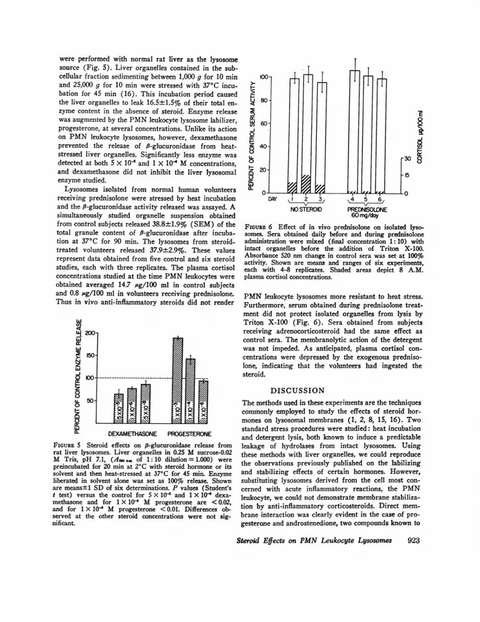
FIGuRE 6 Effect of in vivo prednisolone on isolated lyso-somes. Sera obtained daily before and during prednisoloneadministration were mixed (final concentration 1:10) withintact organelles before the addition of Triton X-100.Absorbance 520 nm change in control sera was set at 100%activity. Shown are means and ranges of six experiments,each with 4-8 replicates. Shaded areas depict 8 A.M.plasma cortisol concentrations.
PMNleukocyte lysosomes more resistant to heat stress.Furthermore, serum obtained during prednisolone treat-ment did not protect isolated organelles from lysis byTriton X-100 (Fig. 6). Sera obtained from subjectsreceiving adrenocorticosteroid had the same effect ascontrol sera. The membranolytic action of the detergentwas not impeded. As anticipated, plasma cortisol con-centrations were depressed by the exogenous predniso-lone, indicating that the volunteers had ingested thesteroid.
DISCUSSION
The methods used in these experiments are the techniquescommonly employed to study the effects of steroid hor-mones on lysosomal membranes (1, 2, 8, 15, 16). Twostandard stress procedures were studied: heat incubationand detergent lysis, both known to induce a predictableleakage of hydrolases from intact lysosomes. Usingthese methods with liver organelles, we could reproducethe observations previously published on the labilizingand stabilizing effects of certain hormones. However,substituting lysosomes derived from the cell most con-cerned with acute inflammatory reactions, the PMNleukocyte, we could not demonstrate membrane stabiliza-tion by anti-inflammatory corticosteroids. Direct mem-brane interaction was clearly evident in the case of pro-gesterone and androstenedione, two compounds known to
Steroid Effects on PMNLeukocyte Lysosomes 923

induce leakage of liver organelle membranes (15). Butthe anti-inflammatory steroid hormones we tested ex-erted no stabilizing activity on PMN leukocyte lyso-somes. And the agents were studied at various concen-trations including both physiologic and pharmacologicranges. With only two steroids, hydrocortisone hemisuc-cinate and hydrocortisone acetate, could we find lessenzyme activity in the supernates after stressing ly-sosomes by heat incubation. But in both cases this wasdue to enzyme inhibition by these hormone preparationsrather than to lysosomal membrane stabilization. de Duveet al. (1) observed less free acid phosphatase activityreleased from liver organelles in cortisone acetate andcautioned about the possible misinterpretation of ly-sosome membrane stabilization by what in fact were en-zyme inhibitors. And finally, the administration of pred-nisolone to human volunteers not only failed to modifythe integrity of their PMN leukocyte lysosomes, butalso did not impart a stabilizing activity to their serawhen tested with normal leukocyte granules.
Numerous publications have dealt with the effectsof corticosteroids on liver lysosomal membrane integrity.Not all studies (17, 18) have confirmed the stabilizationhypothesis. Either no effect or mild stabilization has beennoted with anti-inflammatory steroids, depending uponexperimental conditions. These diverse observations havealso been found using PMNleukocyte lysosomes. Thus,Weissmann, Becher, and Thomas (8) showed that 10"M hydrocortisone would not protect rabbit peritonealPMN leukocyte lysosomes from streptolysin 0 lysis.And Willis, Davison, Ramwell, Brocklehurst, and Smith(19) could not alter the release of P-glucuronidase fromisolated lysosomes by hydrocortisone even at concentra-tions up to 1 mg/ml. Both hydrocortisone and para-methasone were found to be without effect at 10-8-104 Mon rat blood leukocyte lysosomes but capable of stabiliz-ing rabbit and guinea pig peritoneal exudate PMNor-ganelles using hypotonic incubation media (20, 21).The variables found by these authors to alter lysosomalmembrane permeability have included species and or-gan sources of organelles, tonicity and pH of media, andthe types of stress procedures employed. In addition,lysosomes from both liver (22) and PMN leukocytes(23, 24) are heterogeneous in composition and form.These variables could account for some of the publisheddiscrepancies on how drugs affect membrane stability.We used experimental conditions more physiologic forour studies with lysosomes. Whereas both liver andPMN leukocyte lysosomes can be labilized by proges-terone and androstenedione, only liver organelles werestabilized by the anti-inflammatory steroids, and then toonly a mild degree.
The reasons for the different reactivity of liver andPMN leukocyte lysosomes to anti-inflammatory corti-
costeroids are unclear; our observations could be due tovariations in membrane structure. Although the studiesperformed thus far (25, 26) are in agreement concern-ing protein content (both liver and leukocyte organellemembranes containing approximately 50% protein/wetwt) differences have been found in the lipids of thesemembranes. Rat liver lysosomal membranes containedcholesterol and phospholipid at a ratio of 1: 3.4 (25),and a cell membrane fraction from rabbit PMNleuko-cyte granules had a ratio of 1: 1.4 (26). If steroids alterpermeability by their action at the lipid-water interfaceof membranes, these differences in the composition ofthe membrane lipids could explain our results.
Using viable cell preparations, numerous authors, in-cluding Mandell, Rubin, and Hook (5) and Wiener et al.(6), have shown that hydrocortisone prevents the re-lease of lysosomal hydrolases during phagocytosis. Thisassociation with anti-inflammatory corticosteroids hasalso been observed with in vivo models of inflammation(27, 28) and lysosome rupture (29). But direct mem-brane interaction by these compounds and resultingstabilization need not be invoked to explain their anti-phlogistic actions. Corticosteroids have a wide rangeof biological activities that could account for their sup-pressive effects on inflammation (30-34). Interferencewith glucose transport (33) and inhibition of ATP gen-eration (34), or suppression of NADHoxidase ac-tivity (5) are several actions that could lead to de-pressed cell function. After phagocytosis, these effects ofhydrocortisone would prevent the intracellular metabolicactivity necessary for merger of organelle membraneswith the endocytic vacuole, the subsequent degranulation,and the extrusion of lysosome contents from the cell. Inlight of our experimental observations presented above,these are more plausible explanations for the mechanismof anti-inflammatory steroid action than the hypothesis ofdirect membrane stabilization.
ACKNOWLEDGMENTSSharon E. Vance and June K Rushing provided experttechnical assistance.
This study was supported in part by grants from TheRobert A. Welch Foundation, Houston, Tex., and TheSouth Central Texas Chapter of the Arthritis Foundation.
REFERENCES1. de Duve, C., R. Wattiaux, and M. Wibo. 1962. Effects
of fat-soluble compounds on lysosomes in vitro. Bio-chem. Pharmacol. 9: 97-116.
2. Weissmann, G., and L. Thomas. 1963. Studies on lyso-somes: II. The effect of cortisone on the release ofacid hydrolases from a large granule fraction of rabbitliver induced by an excess of vitamin A. J. Clin. In-vest. 42: 661-669.
3. Weissmann, G. 1967. The role of lysosomes in inflam-mation and disease. Annu. Rev. Med. 18: 97-112.
924 R. H. PerseUin and L. C. Ku

4. Wright, D. G., and S. E. Malayaista. 1973. Mobiliza-tion and extracellular release of granular enzymes fromhuman leukocytes during phagocytosis: inhibition bycolchicine and cortisol but not by salicylate. ArthritisRheum. 16: 749-758.
5. Mandell, G. L., W. Rubin, and E. W. Hook. 1970.The effect of an NADHoxidase inhibitor (hydrocorti-sone) on polymorphonuclear leukocyte bacterial activity.J. Clin. Invest. 49: 1381-1388.
6. Wiener, E., Y. Marmary, and Z. Curelaru. 1972. The invitro effect of hydrocortisone on the uptake and intra-cellular digestion of particulate matter by macrophagesin culture. Lab. Invest. 26: 220-226.
7. Chodirker, W. B., G. N. Bock, and J. H. Vaughan.1968. Isolation of human PMNleukocytes and granules:observations on early blood dilution and on heparin.J. Lab. Clin. Med. 71: 9-19.
8. Weissmann, G., B. Becher, and L. Thomas. 1964. Stud-ies on lysosomes. V. The effects of streptolysins andother hemolytic agents on isolated leucocyte granules.J. Cell Biol. 22: 115-126.
9. Persellin, R. H. 1969. Lysosome stabilization by leuko-cyte granule membrane antiserum. J. Immunol. 103: 39-44.
10. Hempel, K. H., L. A. Fernandez, and R. H. Per-sellin. 1970. Effect of pregnancy sera on isolated lyso-somes. Nature (Lond.). 225: 955-956.
11. Fishman, W. H., B. Springer, and R. Brunetti. 1948.Application of an improved glucuronidase assay methodto the study of human blood 8-glucuronidase. J. Biol.Chem. 173: 449-456.
12. Andersch, M. A., and A. J. Szczypinski. 1947. Use ofp-nitrophenylphosphate as the substrate in determina-tion of serum acid phosphatase. Am. J. Clin. Pathol. 17:571-574.
13. Farquhar, M. G., D. F. Bainton, M. Baggiolini, and C.de Duve. 1972. Cytochemical localization of acid phos-phatase activity in granule fractions from rabbit poly-morphonuclear leukocytes. J. Cell Biol. 54: 141-156.
14. Weissmann, G., R. B. Zurier, and S. Hoffstein. 1972.Leukocytic proteases and the immunologic release oflysosomal enzymes. Am. J. Pathol. 68: 539-559.
15. Weissmann, G. 1965. Studies of lysosomes-VI. Theeffect of neutral steroids and bile acids on lysosomesin vitro. Biochent. Pharmacol. 14: 525-535.
16. Persellin, R. H. 1972. Lysosome stabilization by adju-vant arthritis serum. Arthritis Rheum. 15: 144-152.
17. Ignarro, L. J. 1971. Effects of anti-inflammatory drugson the stability of rat liver lysosomes in vitro. Biochem.Pharmacol. 20: 2847-2860.
18. Brown, J. H., and N. L. Schwartz. 1969. Interaction oflysosomes and anti-inflammatory drugs (33938). Proc.Soc. Exp. Biol. Med. 131: 614-620.
19. Willis, A. L., P. Davison, P. W. Ramwell, W. E.Brocklehurst, and B. Smith. 1972. Release and actionsof prostaglandins in inflammation and fever: inhibitionby anti-inflammatory and and antipyretic drugs. InProstaglandins in Cellular Biology. P. W. Ramwell and
B. B. Pharriss, editors. Plenum Publishing Corporation,New York. 227-268.
20. Ignarro, L. J. 1971. Dissimilar effects of anti-inflamma-tory drugs on stability of lysosomes from peritonealand circulating leukocytes and liver. Biochem. Pharmna-col. 20: 2861-2870.
21. Ignarro, L. J., and C. Colombo. 1972. Enzyme releasefrom guinea-pig polymorphonuclear leucocyte lysosomesinhibited in vitro by anti-inflammatory drugs. Nat. NewBiol. 239: 155-157.
22. Tanaka, K., and Y. lizuka. 1968. Suppression of enzymerelease from isolated rat liver lysosomes by non-steroidalanti-inflammatory drugs. Biochem. Pharmacol. 17: 2023-2032.
23. Baggiolini, M., J. G. Hirsch, and C. de Duve. 1969.Resolution of granules from rabbit heterophil leukocytesinto distinct populations by zonal sedimentation. J.Cell Biol. 40: 529-541.
24. Welsh, I. R. H., and J. K. Spitznagel. 1971. Distribu-tion of lysosomal enzymes, cationic proteins, and bac-tericidal substances in subcellular fractions of humanpolymorphonuclear leukocytes. Infect. Immun. 4: 97-102.
25. Thines-Sempoux, D. 1967. Chemical similarities be-tween the lysosome and plasma membranes. Biochem. J.105: 20-21P.
26. Woodin, A. M., and A. A. Wieneke. 1966. Composi-tion and properties of a cell-membrane fraction from thepolymorphonuclear leukocyte. Biochem. J. 99: 493-500.
27. Arrigoni-Martelli, E., and A. Restelli. 1972. Release oflysosomal enzymes in experimental inflammations: ef-fects of anti-inflammatory drugs. Eur. J. Pharnmacol.19: 191-198.
28. Andersen, A. J. 1970. Lysosomal enzyme activity in ratswith adjuvant-induced arthritis. Annl. Rheum. Dis. 29:307-313.
29. Szego, C. M. 1972. Lysosomal membrane stabilizationand antiestrogen action in specific hormonal targetcells. Gynecol. Invest. 3: 63-95.
30. Weissmann, G., and L. Thomas. 1964. The effects ofcorticosteroids upon connective tissue and lysosomes.Recent Prog. Horm. Res. 20: 215-245.
31. Cline, M. J., and K. L. Melmon. 1966. Plasma kininsand cortisol: a possible explanation of the anti-inflamma-tory action of cortisol. Science (Wash. D. C.). 153:1135-1137.
32. Cooper, M. R., L. R. DeChatelet, and C. E. McCall.1972. The in vitro effect of steroids on polymorpho-nuclear leukocyte metabolism (36916). Proc. Soc. Exp.Biol. Med. 141: 986-990.
33. Gemsa, D., C. H. Woo, H. H. Fudenberg, and R.Schmid. 1973. Erythrocyte catabolism by macrophagesin vitro. The effect of hydrocortisone on erythrophago-cytosis and on the induction of heme oxygenase. J.Clin. Invest. 52: 812-822.
34. Young, D. A. 1969. Glucocorticoid action on rat thymuscells: interrelationships between carbohydrate protein,and adenine nucleotide metabolism and cortisol effectson these functions in vitro. J. Biol. Chem. 244: 2210-2217.
Steroid Effects on PMNLeukocyte Lysosomes 925


![[Gastrointestinal hormones in food intake control]](https://img.pdfslide.net/doc/110x75/635ee880dcf4a1629e036ce6/gastrointestinal-hormones-in-food-intake-control.jpg)


