Embed Size (px)
Citation preview
EFFICIENT ISOLATION AND PROPAGATION OFHUMAN IMMUNODEFICIENCY VIRUS ON RECOMBINANTCOLONY-STIMULATING FACTOR I-TREATED MONOCYTES
BY HOWARD E. GENDELMAN,* JAN M. ORENSTEIN,° MALCOLM A. MARTIN,*CAROL FERRUA,* RITA MITRA,4 TERRI PHIPPS," LARRY A. WAHL,"H. CLIFFORD LANE,f ANTHONY S . FAUCI,f DONALD S. BURKE,T
DONALD SKILLMAN," AND MONTE S. MELTZERl
From the *Laboratory of Molecular Microbiology, National Institute of Allergy and InfectiousDiseases; the LLaboratory of Microbiology and Immunology, National Institute of Dental Research;
and the 'Laboratory of Immunoregulation, NIAID, National Institutes of Health, Bethesda,Maryland 20892; the °Department ofPathology, George Washington University Medical Center,Washington, DC 20036; and the $Walter Reed Army Institute to Research, Walter Reed Army
Medical Center, Washington, DC 20307
Infection with the human immunodeficiency virus (HIV) (1-3) often resultsin clinically apparent disease only after intervals of months to years. During thislatent or subclinical phase of infection, HIV continues to replicate at low levelsdespite an often vigorous but apparently ineffective host immune response (4) .Mechanisms that contribute to this persistent, low-level infection, as well as thecellular reservoirs for HIV during this latent period, are not fully understood .Several lines of evidence now document cells of the monocyte/macrophage lin-eage as major targets for persistent HIV in vivo (5-12) . In this respect, HIV issimilar to several ruminant lentiviruses that show strong tropism for macro-phages during both viral latency and active replication (13-15) . If macrophagesalso serve as a viral reservoir during HIV infection, then analysis of theseinfected cells may explain mechanisms of viral persistence, dissemination, andultimately clinical disease.
In this report, we describe an in vitro system that allows replication of HIVin blood-derived monocyte/macrophages from normal donors . Purified mono-cytes were cultured for intervals > 3 mo in medium supplemented with humanrCSF-1 (16, 17) . These cultures provided susceptible target cells for HIV infec-tion . Cocultivation of PBMC from patients with AIDS or AIDS-related complex(ARC)' and rCSF-1-treated monocytes from normal donors resulted in isolationof progeny HIV virions in the majority of patients tested .
Materials and MethodsIsolation and Culture ofPeripheral Blood Monocytes.
Populations of monocytes were iso-lated by countercurrent centrifugal elutriation of mononuclear leukocyte-rich fractions
H. E. Gendelman is a Carter-Wallace Fellow of Columbia University College of Physicians and Sur-geons, New York, NY. Address correspondence to Dr. M. S. Meltzer, Department of Immunology,Walter Reed Army Institute of Research, Washington, DC 20307.
1 Abbreviations used in this paper:
ARC, AIDS-related complex; UA, uranyl acetate.
1428
J. Exp. MED. © The Rockefeller University Press - 0022-1007/88/04/1428/14 $2 .00Volume 167 April 1988 1428-1441
on October 9, 2013
jem.rupress.org
Dow
nloaded from
Published April 1, 1988 on O
ctober 9, 2013jem
.rupress.orgD
ownloaded from
Published April 1, 1988
on October 9, 2013
jem.rupress.org
Dow
nloaded from
Published April 1, 1988 on O
ctober 9, 2013jem
.rupress.orgD
ownloaded from
Published April 1, 1988
on October 9, 2013
jem.rupress.org
Dow
nloaded from
Published April 1, 1988 on O
ctober 9, 2013jem
.rupress.orgD
ownloaded from
Published April 1, 1988
on October 9, 2013
jem.rupress.org
Dow
nloaded from
Published April 1, 1988 on O
ctober 9, 2013jem
.rupress.orgD
ownloaded from
Published April 1, 1988
on October 9, 2013
jem.rupress.org
Dow
nloaded from
Published April 1, 1988 on O
ctober 9, 2013jem
.rupress.orgD
ownloaded from
Published April 1, 1988
on October 9, 2013
jem.rupress.org
Dow
nloaded from
Published April 1, 1988 on O
ctober 9, 2013jem
.rupress.orgD
ownloaded from
Published April 1, 1988
on October 9, 2013
jem.rupress.org
Dow
nloaded from
Published April 1, 1988 on O
ctober 9, 2013jem
.rupress.orgD
ownloaded from
Published April 1, 1988
GENDELMAN ET AL .
1429
of blood cells from normal donors undergoing leukopheresis (18) . Cell suspensions were> 96% monocytes by the criteria of cell morphology on Wright-stained cytosmears (96± 2%, mean ± SEM for six determinations), by granular peroxidase (95 ± 3%), and bynonspecific esterase (98 ± 2%) . Elutriated monocytes were cultured as adherent cellmonolayers in DMEM (formula 780176AJ, Gibco, Grand Island, NY) supplemented with10% freshly obtained, heat-inactivated, normal human serum, 50 Ag/ml gentamicin, and1,000 U/ml rCSF-1 (Cetus Corp., Emeryville, CA) (16, 17) .
Isolation and Culture of PHA-stimulated PBMC (Lymphoblasts) .
PBMC isolated fromwhole blood by Ficoll-diatrizoate density grandient centrifugation were cryopreservedand stored in liquid nitrogen. 3 d before use for virus isolation, cells were quickly thawedand stimulated with the T cell mitogen, PHA (1-3) .
Virus Isolation by Monocyte or Lymphoblast Cocultivation.
Monocytes treated with rCSF-1 and maintained in culture for at least 7 d were used for cocultivation experiments withfreshly isolated PBMC from seropositive HIV-infected individuals . Aliquots of Ficoll-dia-trizoate-separated PBMC (5 X 10⁵ cells/culture well) were admixed with equal numbersof adherent rCSF-1-treated monocytes in 16-mm-diameter culture wells (Cluster24 ;Costar Data Packaging Corp., Cambridge, MA) or with suspensions of PHA-stimulatedlymphoblasts (1-3) . Fluids from all cultures were sampled daily and assayed by ELISA(Cellular Products, Inc ., Buffalo, NY) for presence of HIV-specific antigens and/orreverse transcriptase activity for at least 40 d . Reverse transcriptase assays were per-formed with [ 2P] deoxythymidinetriphosphate in a protocol modified from thatdescribed by Goff et al . (19, 20) .
Immunofuorescence Analysis by Flow Cytometry .
Uninfected (10 d) and HIV-infected (40d) rCSF-1-treated monocytes were cultured and recovered from Teflon-coated tissue cul-ture flasks (Cole-Parmer Instrument Co., Chicago, IL) . For all experiments, 106 cellswere incubated with 1 :100 dilution of mAb anti-HLe-1 (CD45, Becton Dickinson & Co.,Mountain View, CA), Leu-M3 (CD14, Becton Dickinson & Co.) B4 (CD 19), J5 (CD10),T4 (CD4), T6 (CD 1), T8 (CD8), and TI 1 (CD2 ; all from Coulter Immunology, Hialeah,FL) or 1 :50 dilution of pooled AIDS patients' sera from HIV-1- and HIV-2-infectedindividuals. After the initial antibody incubation, cells were washed after centrifugationand resuspended in 1 :100 dilution of fluorescein-conjugated horse anti-mouse or goatanti-human IgG. Immunofluorescence of individual cells previously fixed in 1 % para-formaldehyde were analyzed by FACS flow cytometry .
Detection ofHIV-specific Polypeptides by Radioimmunoprecipitation .
Adherent monolayersof rCSF-1-treated monocytes chronically infected (30 d) with HIV-1 (patient Ada, secondpassage) were washed twice and cultured in methionine-free DMEM with 2% dialyzedFCS for 2 h . Cells were labeled with ["S]methionine (100 ,ACi/ml) for 8 h . Radiolabeledcell lysates were mixed with AIDS patients' sera for 12 h at 4°C and the immune com-plexes were recovered on protein A-Sepharose (Pharmacia Fine Chemicals, Piscataway,NJ) . Eluted immune complexes were subjected to SDS-PAGE as described (21) .
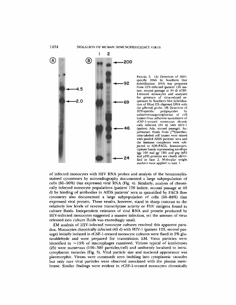
Detection of HIV-specific DNA by Southern Blot Hybridization.
DNA was prepared fromHIV-infected (patient 120 isolate, second passage at 30 d) rCSF-l-treated monocytes andanalyzed for presence of virus-related sequences by Southern blot hybridization of HindIII-digested cellular DNA with the pBenn6 gag-pol-env probe (22) .
In Situ Hybridization with HIVRNA Probes.
Subgenomic viral DNA fragments presentin pBI (23), pBenn6 (22), pBl l (23), and a recombinant plasmid (pRG-B) that containsa 1 .35-kb Hind III fragment mapping between 8.25 and 9.6 kb on the proviral DNAwere subcloned into SP6/T7 vectors (Promega Biotec, Madison, WI), and the pooledDNAs were transcribed using 3'S-UTP (Amersham Corp., Arlington Heights, IL) . Thelabeled RNAs were incubated with 40 mM NaHCO3/60 AM Na2COs , pH 10.2, beforehybridization to facilitate their entry into cells . Cytosmears of cultured monocytes wereprepared onto polylysine-coated glass slides, fixed in periodate/lysine paraformaldehyde/glutaraldehyde, and pretreated with proteinase K, triethanolamine, and HCl . Specimenswere prehybridized in 10 mM Tris (pH 7.4), 2X SSC (1X SSC is 0.15 M NaCl, 0.015 Msodium citrate, pH 7.4), 1X Denhardt's solution (0.02% polyvinylpyrrolidone, 0 .02%Ficoll, 0.02% BSA), and 200 Ag/ml yeast tRNA at 45°C for 2 h, and hybridized in this
1430
ISOLATION OF HUMAN IMMUNODEFICIENCY VIRUS
solution with 10% dextran sulfate, 5 uM dithiolthreitol and 106 cpm 35S-labeled HIVRNA. Slides were serially washed in solutions with RNase to reduce binding of nonhy-bridized probe. Autoradiography was performed in absolute darkness (6).To control for the specificity of in situ hybridization, probes synthesized in the sense
orientation (same polarity as viral mRNA) were incubated with replicate cell prepara-tions. Additionally, uninfected cells were hybridized with antisense probes (i .e ., comple-mentary to viral mRNA) .EM Examination of Monocyte Cultures .
HIV-infected or uninfected rCSF-1-treatedmonocytes were grown on plastic dishes or recovered from Teflon flasks . Cells were har-vested at 10 and 40 d, washed in PBS, and immediately fixed with 2% glutaraldehyde in0.1 M cacodylate buffer (pH 7.4) overnight at 4°C. Fixed cells were gently transferredto 1.5-ml microfuge tubes using a large-bore Pasteur pipette and were pelleted aftercentrifugation . The cell pellet was further processed through 1% Os04, blocked in uranylacetate (UA), stained for 1 h in saturated UA in 50% ethanol, dehydrated in gradedethanol and propylene oxide, and embedded in epon . Thin sections were stained withUA and lead citrate and examined in a Zeiss EM IOAR EM operating at 60 kV.
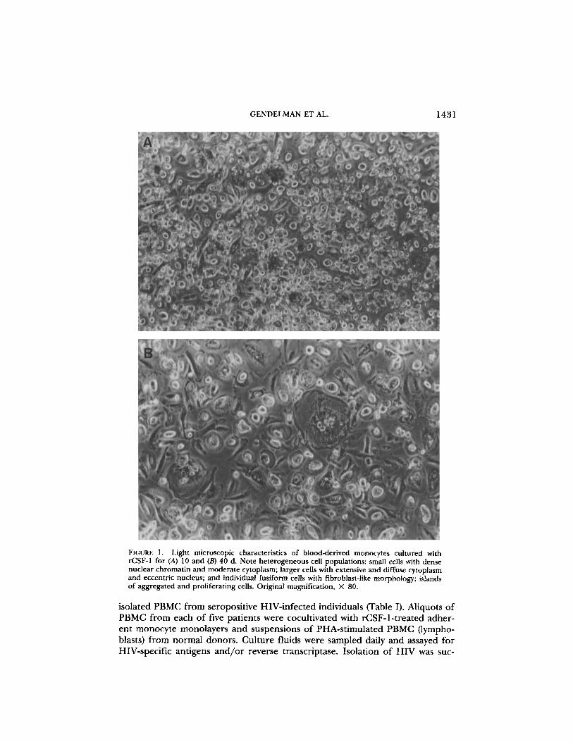
ResultsCulture of rCSF-1-treated Blood Monocytes. Relatively pure populations of
monocytes were obtained by countercurrent centrifugal elutriation of mononu-clear leukocyte-rich fractions of blood cells from normal donors undergoing leu-kopheresis (18) . Such cell suspensions were > 96% monocytes by criteria of cellmorphology on Wright-stained cytosmears, by granular peroxidase, and by non-specific esterase . Purified monocytes were cultured in medium supplementedwith 1,000 U/ml rCSF-1 . After 5-7 d of culture, clusters of rounded, looselyadherent, proliferating monocytes were observed scattered throughout a mono-layer of adherent fusiform cells (Fig . 1) . Low levels of cell division were con-firmed by [3H]thymidine incorporation and the presence of mitotic figures in1-5% of the cells . In coincident experiments, monocytes in aliquots of the samecell suspension cultured without rCSF-1 for 7 d appeared spread, vacuolated,and granular. No proliferating cell clusters were observed and the absolute cellnumber was < 20% of the initial inoculum. In contrast, the number of cells inrCSF-1-treated monocyte cultures at 7-10 d ranged from 90-150% of the initialinoculum . EM examination of 100 individual cells after 10 d in culture showedthat all cells had ultrastructural characteristics typical of macrophages: irregularoutlines, abundant lysosomes, prominent perinuclear Golgi, and eccentricnuclei . Cell surface antigens in these monocyte cultures were also characterizedat 10 d by mAbs and analyzed by FACS flow cytometry. More than 98% of cellswere positive for HLe-1 (CD 45) and Leu-M3 (CD 14); binding of anti-B4 (CD19), J5 (CD10), T4 (CD 4), T6 (CD1), T8 (CD 8) or T11 (CD2) were each belowlevels of detection. Thus by antigenic, histochemical, morphologic, and ultra-structural analysis, virtually all of the cells in these 10-d suspensions were iden-tified as monocytes/macrophages.
Isolation of HIV from PBMC of Seropositive Individuals onto rCSF-1-treatedMonocytes of Normal Donors . Repeated attempts to propagate established labo-ratory strains of HIV in monocytes were uniformly negative over a time intervalof >6 mo (data not shown) . These attempts were repeated with the rCSF-1-treated monocyte culture technique described above. Monocytes treated withrCSF-1 for at least 7-10 d were used for cocultivation experiments with freshly
GENDELMAN ET AL .
1431
FIGURE 1. Light microscopic characteristics of blood-derived monocytes cultured withrCSF-1 for (A) 10 and (B) 40 d. Note heterogeneous cell populations : small cells with densenuclear chromatin and moderate cytoplasm; larger cells with extensive and diffuse cytoplasmand eccentric nucleus; and individual fusiform cells with fibroblast-like morphology ; islandsof aggregated and proliferating cells . Original magnification, X 80 .
isolated PBMC from seropositive HIV-infected individuals (Table I) . Aliquots ofPBMC from each of five patients were cocultivated with rCSF-1-treated adher-ent monocyte monolayers and suspensions of PHA-stimulated PBMC (lympho-blasts) from normal donors. Culture fluids were sampled daily and assayed forHIV-specific antigens and/or reverse transcriptase . Isolation of HIV was suc-
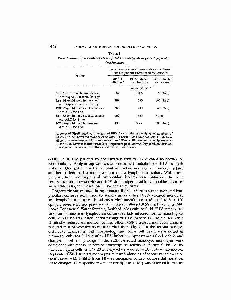
1432 ISOLATION OF HUMAN IMMUNODEFICIENCY VIRUS
TABLE IVirus Isolation from PBMC ofHIV-infected Patients by Monocyte or Lymphoblast
Cocultivation
Patient
HIV reverse transcriptase activity in culturefluids of patient PBMC cocultivated with :
Aliquots of Ficoll-diatrizoate-separated PBMC were admixed with equal numbers ofadherent rCSF-l-treated monocytes or with PHA-stimulated lymphoblasts . Fluids fromall cultures were sampled daily and assayed for HIV-specific reverse transcriptase activ-ity for 40 d. Reverse transcriptase levels represent peak activity . Day at which virus wasfirst detected in monocyte cultures is shown in parentheses .
cessful in all five patients by cocultivation with rCSF-1-treated monocytes orlymphoblasts . Antigen-capture assays confirmed isolation of HIV in eachinstance . One patient had a lymphoblast isolate and not a monocyte isolate ;another patient had a monocyte but not a lymphoblast isolate . With threepatients, both monocyte and lymphoblast isolates were obtained; the peakreverse transcriptase activity and HIV viral antigen level in lymphoblast cultureswere 10-fold higher than those in monocyte cultures .
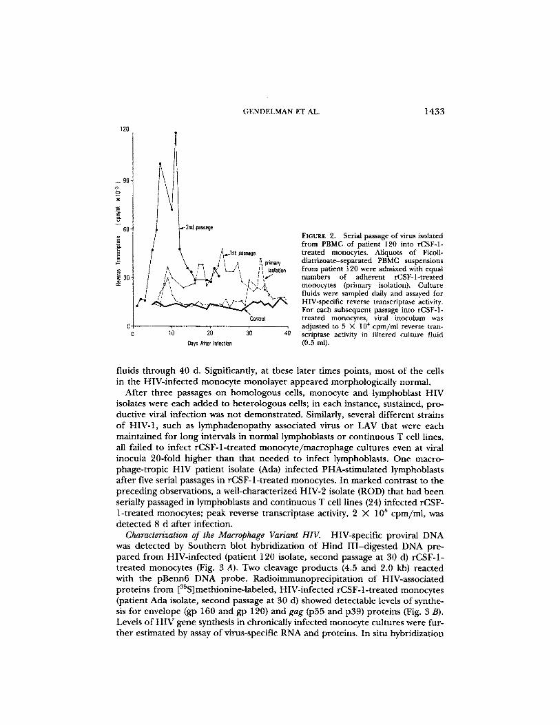
Progeny virions released in supernatant fluids of infected monocyte and lym-phoblast cultures were used to serially infect other rCSF-1-treated monocyteand lymphoblast cultures . In all cases, viral inoculum was adjusted to 5 X 10'cpm/ml reverse transcriptase activity in 0.5-ml filtered (0 .22-Am filter units ; Mil-lipore Continental Water Systems, Bedford, MA) culture fluid . HIV initially iso-lated on monocyte or lymphoblast cultures serially infected normal homologouscells with all isolates tested . Serial passage of HIV (patient 120 isolate, see Table1) initially isolated on monocytes into other rCSF-1-treated monocyte culturesresulted in a progressive increase in viral titer (Fig . 2) . In the second passage,distinctive changes in cell morphology and some cell death were noted inmonocyte cultures 8-14 d after HIV infection . Appearance of cell debris andchanges in cell morphology in the rCSF-1-treated monocyte monolayer werecoincident with peaks of reverse transcriptase activity in culture fluids . Multi-nucleated giant cells with > 20 nuclei/cell were noted in 10-20% of monocytes .Replicate rCSF-1-treated monocytes cultured alone as adherent monolayers orcocultivated with PBMC from HIV seronegative control donors did not showthese changes . HIV-specific reverse transcriptase activity was detected in culture
CD4+ Tcells/mms
PHA-inducedlymphoblasts
cpm/ml X 10-3
rCSF-1-treatedmonocytes
Ada: 36-yr-old male homosexual 252 1,000 70 (35 d)with Kaposi's sarcoma for 4 yr
Ree: 44-yr-old male homosexual 208 900 100 (22 d)with Kaposi's sarcoma for I yr
120: 27-yr-old male i .v. drug abuser 360 200 40 (25 d)with ARC for 1 yr
121 : 32-yr-old male i .v . drug abuser 582 500 Nonewith ARC for 9 mo
167: 24-yr-old male homosexual 433 None 160 (36 d)with ARCfor 1 yr
X
_ff
if
120
GENDELMAN ET AL.
1433
FIGURE 2.
Serial passage of virus isolatedfrom PBMC of patient 120 into rCSF-1-treated monocytes. Aliquots of Ficoll-
Ih, primary
diatrizoate-separated PBMC suspensionsisolation
from patient 120 were admixed with equalnumbers of adherent rCSF-1-treatedI ;I . , {primry}monocytesa isolation . Culturefluids were sampled daily and assayed forHIV-specific reverse transcriptase activity.For each subsequent passage into rCSF-1-
Control
treated monocytes, viral inoculum wasadjusted to 5 X 10' cpm/mi reverse tran-
20
30
4
scriptase activity in filtered culture fluidOat's MW Nation
MZ in]).
fluids through 40 d . Significantly, at these later t0__
in the HIV-infected monocyte monolayer appeared morphologically normal .After three passages on homologous cells, monocyte and lymphoblast HIV
isolates were each added to heterologous cells ; in each instance, sustained, pro-ductive viral infection was not demonstrated. Similarly, several different strainsof HIV-1, such as lymphadenopathy associated virus or LAV that were eachmaintained for long intervals in normal lymphoblasts or continuous T cell lines,all failed to infect rCSF-1-treated monocyte/macrophage cultures even at viralinocula 20-fold higher than that needed to infect lymphoblasts . One macro-phage-tropic HIV patient isolate (Ado) infected PHA-stimulated lymphoblastsafter five serial passages in rCSF-1-treated monocytes . In marked contrast to thepreceding observations, a well-characterized HIV-2 isolate (ROD) that had beenserially passaged in lymphoblasts and continuous T cell lines (24) infected rCSF-1-treated monocytes ; peak reverse transcriptase activity, 2 X 10⁵ cpm/ml, wasdetected 8 d after infection .
Characterization of the Macrophage Variant HIV.
HIV-specific proviralwas detected by Southern blot hybridization of Hind III-digested DNA pre-pared from HIV-infected (patient 120 isolate, second passage at 30 d) rCSF-1-treated monocytes (Fig . 3 A) . Two cleavage products (4.5 and 2.0 kb) reactedwith the pBenn6 DNA probe . Radioimmunoprecipitation of HIV-associatedproteins from [3lSjmethionine-labeled, HIV-infected rCSF-1-treated monocytes(patient Ada isolate, second passage at 30 d) showed detectable levels of synthe-sis for envelope Q 160 and gp 12Q and gag (p55 and p39) proteins (Fig . 3 B) .Levels of HIV gene synthesis in chronically infected monocyte cultures were fur-
ed by assay of virus-specific RNA and proteins . In situ hybridizati
1434
ISOLATION OF HUMAN IMMUNODEFICIENCY VIRUS
FIGURE 3. (A) Detection of HIV-specific DNA by Southern blothybridization . DNA was preparedfrom HIV-infected (patient 120 iso-late, second passage at 30 d) rCSF-1-treated monocytes and analyzedfor presence of virus-related se-quences by Southern blot hybridiza-tion of Hind III-digested DNA withthe pBenn6 probe. (B) Detection ofHIV-specific polypeptides byradioimmunoprecipitation of celllysates from adherent monolayers ofrCSF-1-treated monocytes chroni-cally infected (30 d) with HIV-1(patient Ada, second passage) . Su-pernatant fluids from [s5S]methio-nine-labeled cell lysates were mixedwith pooled AIDS patients' sera andthe immune complexes were sub-jected to SDS-PAGE . Immunopre-cipitate bands representing envelope(gp 160 and gp 120) and gag (p55and p39) proteins are clearly identi-fied in lane 2. Molecular weightmarkers were applied to lane 1 .
of infected monocytes with HIV RNA probes and analysis of the hematoxylin-stained cytosmears by autoradiography documented a large subpopulation ofcells (60-90%) that expressed viral RNA (Fig . 4) . Similarly, analysis of chroni-cally infected monocyte populations (patient 120 isolate, second passage at 40d) by binding of antibodies in AIDS patients' sera as quantified by FACS flowcytometry also documented a large subpopulation of cells (60-88%) thatexpressed viral protein. These results, however, stand in sharp contrast to therelatively low levels of reverse transcriptase activity or HIV antigens found inculture fluids . Independent estimates of viral RNA and protein produced byHIV-infected monocytes suggested a massive infection, yet the amount of virusreleased into culture fluids was exceedingly small.EM analysis of HIV-infected monocyte cultures resolved this apparent para-
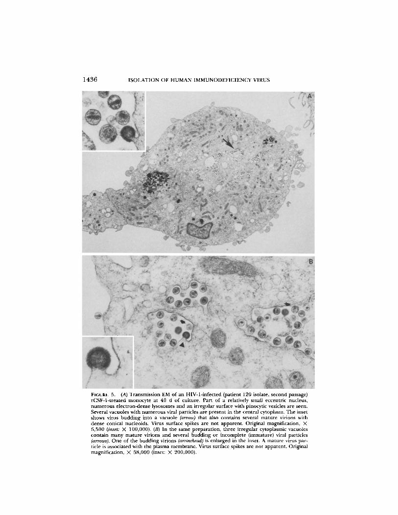
dox. Monocytes chronically infected (40 d) with HIV-1 (patient 120, second pas-sage) initially isolated in rCSF-I-treated monocyte cultures were fixed in 2% glu-taraldehyde and were prepared for transmission EM. Virus particles wereidentified in ^-15% of macrophages examined . Virions typical of lentiviruses(25) were numerous (100-300 particles/cell) and uniformly localized to intra-cytoplasmic vacuoles (Fig. 5) . Viral particle size and nucleoid appearance waspleomorphic. Virons were commonly seen budding into cytoplasmic vacuolesbut only rare viral particles were observed associated with the plasma mem-brane. Similar findings were evident in rCSF-1-treated monocytes chronically

GENDELMAN ET AL .
1435
FIGURE 4 .
In situ hybridization of HIV-infected (patient Ada isolate, third passage) rCSF-1-treated monocytes . Silver grains (HIV-specific RNA) overlie infected cells.
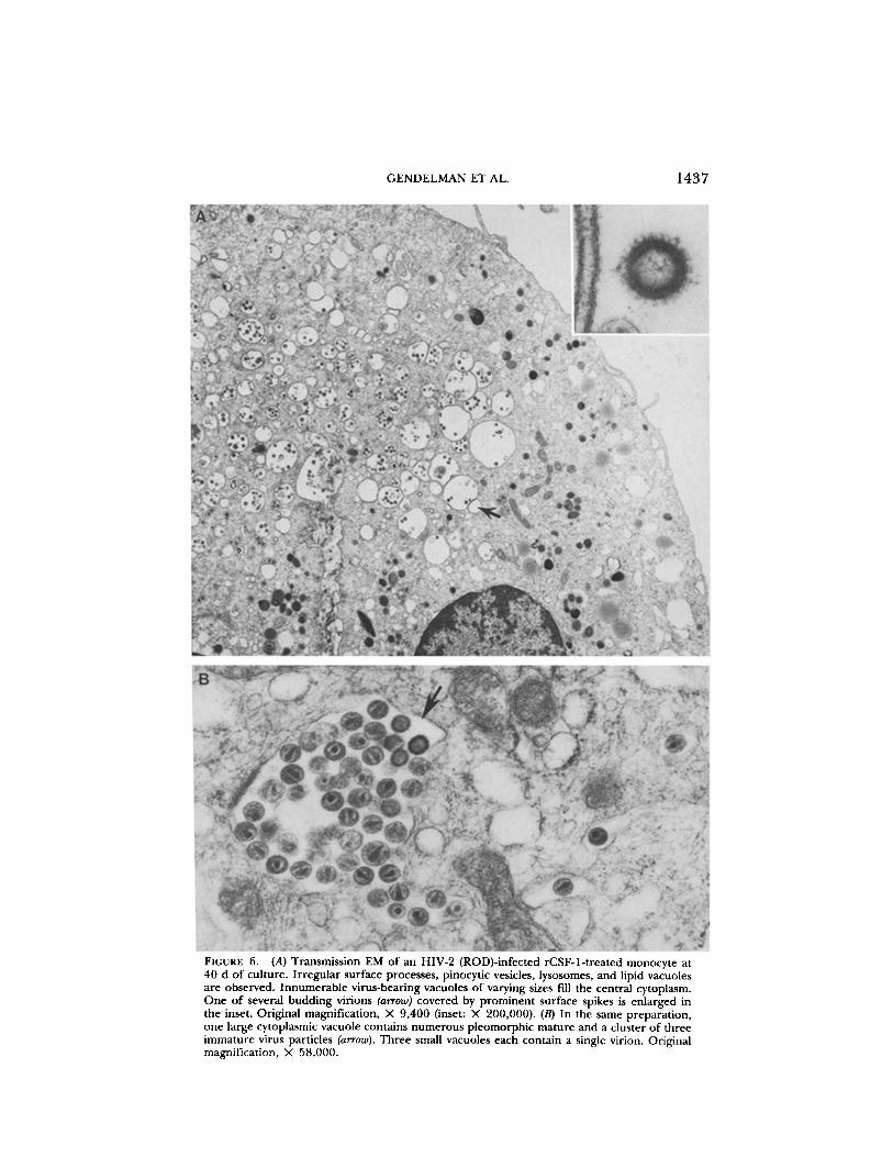
infected (40 d) with the HIV-2 (ROD) isolate (Fig. 6) . With HIV-1, plasma mem-brane budding was observed at low levels during the acute phase of infection(10-14 d), but again intracellular accumulation of virus particles within cyto-plasmic vacuoles was the predominant finding .The pattern of HIV replication in monocytes and T cells is thus very different .
HIV-infected monocytes accumulate large numbers of budded virus in intracy-toplasmic vacuoles during both acute and chronic infections ; release of virusfrom the plasma membrane is infrequent and at relatively low levels (0-10 par-ticles/cell section) . This pattern of viral replication in rCSF-1-treated monocyteswas observed with three different patient isolates of HIV (patients Ada and120), and HIV-2 (ROD); viral particles were identified in -15% of monocytesexamined at 4-6 wk. In contrast, the HIV-infected T cell releases large numbersof viral particles from the plasma membrane (often hundreds of virions/cell sec-tion) ; the number of virions that reside in cytoplasmic vacuoles in these cells isexceedingly small . The concept that HIV virions produced in macrophages accu-mulate intracellularly and are only inefficiently transported out of the cell wasconfirmed by comparison of fluid-phase reverse transcriptase levels in monocytecultures before and after three successive freeze-thaw cycles . The amount ofreverse transcriptase activity detected in monocyte cultures after the freeze-thawcycles was 10-20 times higher (9 X 10⁵ cpm/ml) than that of control levels .Virus released into culture fluids by freeze-thaw cycles was fully infectious forrCSF-1-treated monocytes .
1436
ISOLATION OF HUMAN IMMUNODEFICIENCY VIRUS
FIGURE 5.
(A) Transmission EM of an HIV-1-infected (patient 120 isolate, second passage)rCSF-1-treated monocyte at 40 d of culture. Part of a relatively small eccentric nucleus,numerous electron-dense lysosomes and an irregular surface with pinocytic vesicles are seen .Several vacuoles with numerous viral particles are present in the central cytoplasm. The insetshows virus budding into a vacuole (arrow) that also contains several mature virions withdense conical nucleoids. Virus surface spikes are not apparent . Original magnification, X6,500 (inset : X 100,000) . (B) In the same preparation, three irregular cytoplasmic vacuolescontain many mature virions and several budding or incomplete (immature) viral particles(arrows) . One of the budding virions (arrowhead) is enlarged in the inset . A mature virus par-ticle is associated with the plasma membrane. Virus surface spikes are not apparent . Originalmagnification, X 58,000 (inset : X 200,000) .
GENDELMAN ET AL .
FIGURE 6.
(A) Transmission EM of an HIV-2 (ROD)-infected rCSF-1-treated monocyte at40 d of culture . Irregular surface processes, pinocytic vesicles, lysosomes, and lipid vacuolesare observed. Innumerable virus-bearing vacuoles of varying sizes fill the central cytoplasm.One of several budding virions (arrow) covered by prominent surface spikes is enlarged inthe inset . Original magnification, X 9,400 (inset : X 200,000) . (B) In the same preparation,one large cytoplasmic vacuole contains numerous pleomorphic mature and a cluster of threeimmature virus particles (arrow) . Three small vacuoles each contain a single virion. Originalmagnification, X 58,000 .
1437
1438 ISOLATION OF HUMAN IMMUNODEFICIENCY VIRUS
DiscussionThe preceding observations document recovery of HIV tropic for macro-
phages in a majority of patients tested . It is not clear at this point whether theefficient isolation of HIV from patients' leukocytes into rCSF-1-treated mono-cytes represents a change in target cell susceptibility to virus or to increasedmonocyte viability in culture over extended time intervals . The role of CSF-1, amacrophage growth factor, in HIV infection of macrophages may not be anal-ogous to that of the T cell growth factor, IL-2, for T cells (1-3) . We were ableto document only a small subpopulation of proliferating monocytes (^-1-5%)during culture with rCSF-1, yet the percentage of HIV-infected cells detectedby in situ hybridization with HIV RNA probes or by immunofluorescence withAIDS patients' sera exceeded 60-90%. By whatever mechanism, the findingspresented in these studies, as well as those in previous reports that documentbiologically distinct HIV in brain and lung tissue, implicate variant HIV as majorparticipants in disease pathogenesis (5) . The evidence in toto strongly suggeststhe macrophage variant HIV as a major virus reservoir in early and late disease .This concept must now be included in future drug testing and vaccine devel-opment strategies . Moreover, the intracellular sequestration of virions in chron-ically infected macrophages suggests new models for viral persistence and thedissemination of disease . Indeed, accumulation of HIV within cytoplasmic vac-uoles of macrophage-derived, multinucleated giant cells in the brain has beenrecently described (26) . These observations in brain tissue from AIDS patientsclosely parallel the ultrastructural findings in HIV-infected macrophagesreported here . Retention of virus within macrophages is not novel for retrovi-ruses. Other lentiviruses, such as caprine arthritis encephalitis and ovine pro-gressive pneumonia virus also bud into and accumulate in cytoplasmic vacuoles(27, 28) . These viruses have strong tropism for blood monocytes or tissue mac-rophages, yet viral replication is restricted and entirely dependent upon host cell(macrophage) differentiation . The visna virus-infected macrophages act as true"Trojan horses" (13) . Infected, immature blood monocytes restrict virus repli-cation to minimal levels . After these cells enter tissue and differentiate intomature macrophages, however, visna virus replication increases more than sev-eral-thousand-fold (15) . Whether these same mechanisms apply to HIV-infectedhuman macrophages remains to be determined.Although patient-derived HIV efficiently infects rCSF-1-treated monocyte tar-
get cells, low numbers of progeny virus are released into culture fluids . Duringchronic infection, these cells appear morphologically unaffected by the infec-tion, yet EM analysis documents large factories of virions in cytoplasmic vacu-oles . In tissues of AIDS patients, HIV-infected mononuclear phagocytes aredetected at high frequency in the brain, lymph node, and skin (5-7, 10-12) . Dothese hidden virus factories explain how HIV escapes a competent host immunesurveillance response? It is interesting to further speculate that macrophagevariant HIV are the forms responsible for virus latency and dissemination . Atsome time during disease, the macrophage variant HIV acquires T cell tropism .Perhaps viral envelope glycoprotein undergoes successive mutations to acquireaffinity for the CD4 determinant and T cell tropism . This acquired change instructure, and thus function, of HIV would represent a second stage of virus
GENDELMAN ET AL.
1439
infection heralded by T4 helper cell depletion and followed by the inevitabledevelopment of opportunistic infection and death. The full biologic conse-quence of distinct T cell and macrophage tropic viruses in AIDS awaits furtherinquiries . The system for in vitro maintenance of viable, HIV-susceptiblemonocyte/macrophages described in this report can facilitate this search.
SummaryMonocytes were maintained in tissue culture for > 3 mo in media supple-
mented with rCSF-1 . These cultures provided susceptible target cells for isola-tion and propagation of virus from PBMC of HIV-infected patients. HIV iso-lated into monocytes readily infected other rCSF-1-treated monocytes but onlyinefficiently infected PHA-stimulated lymphoblasts . Similarly, laboratory HIVstrains passaged in T cell lines or virus isolated from patients' leukocytes intoPHA-stimulated lymphoblasts inefficiently infected rCSF-1-treated monocytes .Persistent, low-level virion production was detected in macrophage culturefluids by reverse transcriptase activity or HIV antigen capture through 6-7 wk.Marked changes in cell morphology with cell death, syncytia, and giant cell for-mation were observed in monocyte cultures 2 wk after infection, but at 4-6 wk,all cells appeared morphologically normal . However, the frequency of infectedcells in these cultures at 6 wk was 60-90% as quantified by in situ hybridizationwith HIV RNA probes or by immunofluorescence with AIDS patients' sera .Ultrastructural analysis by EM also showed a high frequency of infected cells ;virtually all HIV budded into and accumulated within cytoplasmic vacuoles andvirus particles were only infrequently associated with the plasma membrane.Retention of virus within macrophages and the macrophage tropism of HIVvariants may explain mechanisms of both virus persistence and disseminationduring disease .
We thank Francois Clavel for generously supplying the HIV-2 (ROD) isolate ; SundarajanVenkatesan, Klaus Strebel, Samuel Silverstein, and Barry Bloom for helpful discussions ;Peter Ralph for his generous and continuing support ; Julie McCliffe for clinical materi-als ; and Carol Cronin for excellent editorial assistance.
Received for publication 14 October 1987 and in revisedform 21 December 1987.
References1 . Barre-Sinoussi, F ., J-C . Chermann, F. Rey, M . T . Nugeyre, S . Chamaret, J . Gruest,C . Dauguet, C. Axler-Blin, F . Vezinet-Brun, C. Rouzioux, W. Rozenbaum, and L.Montagnier . 1983 . Isolation of a T-lymphotropic retrovirus from a patient at riskfor acquired immune deficiency syndrome (AIDS) . Science (Wash. DC). 220:868 .
2 . Gallo, R . C ., S . Z . Salahuddin, M . Popovic, G . M . Shearer, M . Kaplan, B . F . Haynes,T. J . Palker, R. Redfield, J . Oleske, B . Safai, G. White, P . Foster, and P . D . Mark-ham . 1984 . Frequent detection and isolation of cytopathic retroviruses (HTLV-III)from patients with AIDS and at risk for AIDS . Science (Wash. DC). 224:500 .
3 . Levy, J . A ., A . D . Hoffman, S . M . Kramer, J . A . Lanois, J . M . Shimbukuro, and L.S . Oskiro . 1984 . Isolatio n of lymphocytopathic retroviruses from San Franciscopatients with AIDS . Science (Wash. DC). 225:840 .
4 . A . S . Fauci . 1986 . Current issues in developing a strategy for dealing with theacquired immunodeficiency syndrome . Proc. Natl . Acad . Sci . USA. 83:9278
1440
ISOLATION OF HUMAN IMMUNODEFICIENCY VIRUS
5 . Gartner, S ., P . Markovits, D . M . Markovitz, M . H . Kaplan, R . C . Gallo, and M.Popovic . 1986 . The role of mononuclear phagocytes in HTLVIII/LAV infection .Science (Wash. DC). 223 :215 .
6 . Koenig, S ., H. E . Gendelman, J . M. Orenstein, M. C . Dal Canto, G . H . Pezeshpour,M. Yungbluth, F . Janotta, A. Aksamit, M . A . Martin, and A . S . Fauci . 1986 . Detec-tion of AIDS virus in macrophages in brain tissue from AIDS patients with enceph-alopathy . Science (Wash. DC) . 233:1089 .
7 . Wiley, C . A ., R . D . Schrier, J . A. Nelson, P . W . Lampert, and M. B . A . Oldstone .1986 . Cellular localization of human immunodeficiency virus infection within thebrains of acquired immune deficiency patients . Proc. Natl . Acad. Sci . USA . 83:7089 .
8 . Nicholson, J . K. A ., G . D . Cross, C . S . Callaway, and J . S . McDougal . 1986 . In vitroinfection of human monocytes with human T lymphotropic virus type III/lymph-adenopathy-associated virus (HTLV-III/LAV) . J. Immunol . 137:323 .
9 . Ho, D . D ., T. R. Rota, and M. S. Hirsch . 1986 . Infection of monocyte/macrophagesby human T lymphotropic virus type III . J. Clin . Invest. 77 :1712 .
10 . Tschachler, E ., V . Groh, M. Popovic, D. L . Mann, K. Konrad, B . Safai, L . Eron, F .diMarzo Veronese, K . Wolff, and G. Stingl . 1987 . Epidermal Langerhans cells . Atarget for HTLV-III/LAV infection . J. Invest. Dermatol. 88:233 .
11 . Tenner-Racz, K., P . Racz, M . Dietrich, and P . Kern . 1985 . Altered follicular den-dritic cells and virus-like particles in AIDS and AIDS-related lymphadenopathy . Lan-cet . 1 :105 .
12 . Armstrong, J . A . and R . Horne . 1984 . Follicular dendritic cells and virus-like parti-cles in AIDS-related lymphadenopathy . Lancet . II:370 .
13 . Haase, A . T. 1986 . Pathogenesis of lentivirus infections . Nature (Lond .) . 322:130 .14 . Narayan, O., and L . C . Cork . 1985 . Lentivira l disease of sheep and goats : chronic
pneumonia leukoencephalomyelitis and arthritis . Rev. Infect . Dis . 7:89 .15 . Gendelman, H. E ., O . Narayan, S. Molineaux, J . E . Clements, and Z . Ghotbi. 1985 .
Slow persistent replication of lentiviruses : role of tissue macrophages and macro-phage precursors in bone marrow . Proc. Natl. Acad. Sci . USA . 82:7086 .
16 . Clark, S . C ., and R . Kamen . 1987 . The human hematopoietic colony-stimulating fac-tors . Science (Wash. DC). 236 :1229 .
17 . Kawasaki, E . S ., M . B . Ladner, A. M . Wang, J . V. Arsdell, M . K . Warren, M. Y .Coyne, V . L . Schwenkart, M-T . Lee, K. J . Wilson, A . Boosman, E . R . Stanley, P .Ralph, and D. F . Mark. 1985 . Molecula r cloning of a complementary DNA encodinghuman macrophage-specific colony-stimulating factor (CSF-1) . Science (Wash . DC) .230:291 .
18 . Wahl, L . M., I . M . Katona, R. L . Wilder, C . C . Winter, B . Haraoui, I . Scher, and S .Wahl. 1984 . Isolation of human mononuclear cell subsets by counter flow centrif-ugal elutriation (CCE) . I . Characterization of B-lymphocyte, T-lymphocytes andmonocyte-enriched fractions by flow cytometric analysis . Cell. Immunol. 85:373 .
19 . Goff, S ., P . Traktman, and D . Baltimore . 1981 . Isolation and properties of Moloneymurine leukemia virus mutants : use of a rapid assay for release of virion reversetranscriptase . J. Virol . 38:239 .
20 . Willey, R. L ., D . H . Smith, L . A. Lasky, T . S . Theodore, P . L . Earl, B . Moss, D .Capon, and M. A. Martin . 1988 . In vitro mutagenesis identifies a region within theenvelope gene of the human immunodeficiency virus that is critical for infectivity . J.Virol . 66:139 .
21 . Lightfoote, M . M., J . E . Coligan, T . M . Folks, A . S . Fauci, M. A . Martin, and S .Venkatesan . 1986 . Structural characterization of reverse transcriptase and endonu-clease polypeptides of the acquired immunodeficiency syndrome retrovirus . J. Virol .60:771 .
GENDELMAN ET AL .
1441
22 . Folks, T . M., S . Benn, A . Rabson, T . Theodore, M. D. Hoggan, M. A . Martin, M.Lightfoote, and K . W. Sell . 1985 . Characterization of a continuous T-cell line sus-ceptible to the cytopathic effects of the acquired immunodeficiency syndrome(AIDS)-associated retrovirus . Proc. Natl. Acad. Sci . USA. 82:4539 .
23 . Benn, S ., J . Rutledge, T . Folks, J. Gold, L . Baker, J . McCormick, P . Feorino, P . Piot,T . Quinn, and M. A. Martin . 1985 . Genomic heterogeneity of AIDS retroviral iso-lates from North America and Zaire . Science (Wash. DC). 230:949 .
24 . Clavel, F ., F . Guetard, F . Vezinet-Brun, S . Chamaret, M. A . Rey, M . O . Santos-Fer-reira, A. G . Laurent, C . Dauguet, C . Katlama, C . Rouzioux, D. Klatzmann, J . L .Champalimaud, and L . Montagnier. 1986 . Isolation of a new human retrovirus fromWest African patients with AIDS . Science (Wash. DC). 233:343.
25 . Munn, R . J ., P. A. Marx, J . K. Yamamoto, and M. B . Gardner . 1985 . Ultrastructuralcomparison of the retroviruses associated with human and simian acquired immu-nodeficiency syndromes . Lab. Invest. 53:194 .
26 . Meyenhofer, M. F ., L . G . Epstein, E-C Cho, and L. R. Sharer . 1987 . Ultrastructuralmorphology and intracellular production of human immunodeficiency virus (HIV)in brain . J. Neuropath. Exp. Neurol. 46:474 .
27 . Dahlberg, J . E ., J . M. Gaskin, and K. Perk . 1981 . Morphological and immunologicalcomparison of caprine arthritis encephalitis and ovine progressive pneumoniaviruses . J. Virol. 39:914 .
28 . Lairmore, M. D., G . Y . Akita, H . 1 . Russell, and J . C. DeMartini . 1987 . Replicationand cytopathic effects of ovine lentivirus strains in alveolar macrophages correlatewith in vivo pathogenicity . J. Virol . 61 :4038 .


