Embed Size (px)
Citation preview

Electromyographic Analysis of the RotatorCuff and Deltoid Musculature DuringCommon Shoulder External RotationExercisesMichael M. Reinold, DPT, ATC1
Kevin E. Wilk, PT2
Glenn S. Fleisig, PhD3
Nigel Zheng, PhD4
Steven W. Barrentine, MS5
Terri Chmielewski, PT, PhD6
Rayden C. Cody, MD7
Gene G. Jameson, MA5
James R. Andrews, MD8
Study Design: Prospective single-group repeated-measures design.Objectives: To quantify electromyographic (EMG) muscle activity of the infraspinatus, teres minor,supraspinatus, posterior deltoid, and middle deltoid during exercises commonly used to strengthenthe shoulder external rotators.Background: Exercises to strengthen the external rotators are commonly prescribed in rehabilita-tion, but the amount of EMG activity of the infraspinatus, teres minor, supraspinatus, and deltoidduring these exercises has not been thoroughly studied to determine which exercises would bemost effective to achieve strength gains.Methods and Measures: EMG measured using intramuscular electrodes were analyzed in 10healthy subjects during 7 shoulder exercises: prone horizontal abduction at 100° of abduction andfull external rotation (ER), prone ER at 90° of abduction, standing ER at 90° of abduction, standingER in the scapular plane (45° abduction, 30° horizontal adduction), standing ER at 0° ofabduction, standing ER at 0° of abduction with a towel roll, and sidelying ER at 0° of abduction.The peak percentage of maximal voluntary isometric contraction (MVIC) for each muscle
1 Coordinator of Rehabilitative Research and Clinical Education, Healthsouth Rehabilitation, AmericanSports Medicine Institute, Birmingham, AL.2 National Director of Research and Education, Healthsouth Rehabilitation, American Sports MedicineInstitute, Birmingham, AL.3 Smith and Nephew Chair of Research, American Sports Medicine Institute, Birmingham, AL.4 Coordinator of Joint Biomechanics Research, American Sports Medicine Institute, Birmingham, AL.5 Biomechanist, American Sports Medicine Institute, Birmingham, AL.6 Assistant Research Professor, Department of Physical Therapy, University of Florida, Gainesville, FL.7 Assistant Professor, Department of Orthopedics and Rehabilitation, University of Vermont, Burlington,VT.8 Medical Director, American Sports Medicine Institute, Alabama Sports Medicine and Orthopedic Center,Birmingham, AL.This study was approved by the American Sports Medicine Institute Institutional Review Board,Birmingham, AL.Address correspondence to Michael M. Reinold, Coordinator of Rehabilitative Research and ClinicalEducation, Healthsouth Sports Medicine and Rehabilitation Center, American Sports Medicine Institute,1201 11th Avenue South, Suite 100, Birmingham, AL 35205. E-mail: [email protected]
was compared among exercises using a 1-wayrepeated-measures analysis of variance(P�.05).Results: EMG activity varied significantlyamong the 7 exercises. Sidelying ER producedthe greatest amount of EMG activity for theinfraspinatus (62% MVIC) and teres minor(67% MVIC). The greatest amount of activity ofthe supraspinatus (82% MVIC), middle deltoid(87% MVIC), and posterior deltoid (88% MVIC)was observed during prone horizontal abduc-tion at 100° with full ER.Conclusions: Results from this study provideinitial information to develop rehabilitationprograms. It also provides information helpfulfor the design and conduct of future studies.J Orthop Sports Phys Ther 2004;34:385-394.
Key Words: dynamic stabilization,infraspinatus, supraspinatus, teres mi-nor
The glenohumeral jointexhibits the greatestamount of motion ofany articulation in thehuman body, conse-
quently little inherent stability isprovided by its osseous configura-tion.42 Functional stability of the
Journal of Orthopaedic & Sports Physical Therapy 385
RE
SE
AR
CH
RE
PO
RT

shoulder is accomplished through the integratedfunctions of the joint capsule, ligaments, and glenoidlabrum, as well as the dynamic stabilization of thesurrounding musculature, particularly the rotator cuffmuscles.2,8,15,37 The rotator cuff musculature main-tains stability by compressing the humeral head intothe concave glenoid fossa during upper extremitymotion.42
Thus, the rotator cuff muscles play a vital role innormal arthrokinematics and asymptomatic shoulderfunction. The overhead athlete requires the rotatorcuff to maintain an adequate amount ofglenohumeral joint congruency for asymptomaticfunction.41 Sufficient strength of the external rotators(infraspinatus and teres minor), in particular, isintegral during the overhead throwing motion todevelop an approximation force on the upper arm atthe shoulder equal to body weight to prevent jointdistraction.11
Andrews and Angelo1 found that overhead throw-ers most often present with rotator cuff tears locatedfrom the midsupraspinatus and extending posteriorlyto the midinfraspinatus area, which they believe to bea result of the force produced to resist distraction,horizontal adduction, and internal rotation at theshoulder during arm deceleration. Furthermore,Walch et al39 documented the undersurface frayingof the infraspinatus with internal impingement. Thus,the external rotators are muscles that appear to beinvolved in the pathomechanics leading to, or result-ing from, different shoulder pathologies such asinternal impingement,17,39 joint laxity,8,11,17,36,37 labrallesions,11,17,36 and rotator cuff lesions,11,29,36 particu-larly in overhead athletes.40,44
Consequently, many authors5,10,38,40,41,44-46 have ad-vocated emphasis on shoulder external rotationstrengthening during rehabilitation or athletic condi-tioning programs to enhance muscular strength andendurance in overhead athletes. Rehabilitation pro-grams for specific pathologies, such as rotator cuffimpingement or repair surgery, also emphasizestrengthening of the shoulder external rotators. Fur-thermore, rotator cuff strengthening exercises areoften incorporated in athletic conditioning programsfor injury prevention and performance enhancement.The balance between external and internal rotationstrength is important to normal glenohumeral jointfunction, especially during athletic activities.42 Anadequate external-internal rotator muscle strengthratio has been emphasized in the literature.7,10,40,43
Several studies documented the electromyographic(EMG) activity of the glenohumeral musculatureduring specific shoulder exercises.3,5,6,13,22,25,26,28,38,47
Townsend et al38 determined that the best exercisefor the infraspinatus muscle was to perform proneshoulder horizontal abduction with external rotation(ER), producing 88% of maximal voluntary isometriccontraction (MVIC), while the most effective exercise
for the teres minor muscle was sidelying ER, produc-ing 80% MVIC. In a different study comparing ER inthe scapular and frontal planes during isokinetictesting, Greenfield et al13 suggested that the highestamount of muscle activity for the external rotatorsoccur when the shoulder is in the scapular plane.
Ballantyne et al3 compared sidelying ER with ER inthe prone position at 90° of shoulder abduction. Theauthors report no significant difference between exer-cises with approximately 50% EMG activity (meanEMG was normalized as a percent of the highestmean value of repetitions) for both the infraspinatusand teres minor. Conversely, Blackburn et al5 simi-larly compared the sidelying ER and prone ERexercises and noted greater activity during prone ERfor the infraspinatus (prone, 80% EMG; sidelying,30% EMG) and teres minor (prone, 88% EMG; side,45% EMG). EMG values were normalized in relationto the maximum amount of EMG activity producedby each muscle across the test series. Blackburn et al5
also reported high levels of EMG activity of theinfraspinatus (80% EMG) and teres minor (70%EMG) when performing a prone shoulder horizontalabduction movement at 90° and 100° of abductionwith full external rotation.
Current trends in rehabilitation have focused moreclosely on functional rehabilitation through sport-specific exercises designed to strengthen the externalrotators in a position that replicates the capsularstrain and muscular length-tension relationships ob-served during athletic competition, such as externalrotation in the 90° abducted position for overheadathletes.44-46 In addition, many clinicians have advo-cated the use of a towel roll placed between the armand side of the body while performing externalrotation strengthening to enhance stability and toincrease posterior cuff muscular activation.46 Thus, itappears that there is controversy regarding the opti-mal exercises for the external rotators and that manyexercises are being utilized and advocated withoutjustification based on EMG analysis.
Several authors have analyzed the EMG activity ofthe supraspinatus musculature and deltoid muscles todetermine exercises that produce the mostsupraspinatus activity with the least deltoid involve-ment.20,25,35,38,47 This has been theorized to avoidpotential deleterious superior humeral head migra-tion associated with high deltoid activity.20,35,47 Ana-tomically, based on the fiber orientation posterior tothe longitudinal axis of rotation, the supraspinatusand posterior deltoid may be active during theexternal rotation movement.21 However, there is alack of data regarding the contribution of thesupraspinatus and deltoid musculature during exter-nal rotation exercises.
The purpose of this investigation was to measurethe normalized EMG activity of the infraspinatus,teres minor, supraspinatus, and deltoid muscles dur-
386 J Orthop Sports Phys Ther • Volume 34 • Number 7 • July 2004
ing a variety of commonly prescribed rehabilitationexercises to strengthen the shoulder external rota-tors.
METHODS
Subjects
The participants for this investigation were 5 malesand 5 females (mean age, 28.1 years; age range, 22-38years). Each volunteered for the study and reportedno history of shoulder injury or instability, nor didany subjects report a history of shoulder pain. Eachsubject signed an informed consent and the rights ofeach subject were protected. The research protocolwas approved by the Institutional Review Board of theAmerican Sports Medicine Institute.
Electrode Insertion
The dominant shoulder of each subject was utilizedfor testing. The skin near the electrode insertion siteswas shaved and cleaned with alcohol for testing.Paired hook wires (44-gauge stainless steel × 100 mm)were inserted into the supraspinatus, infraspinatus,teres minor, posterior deltoid, and middle deltoidusing a 27-gauge, 30-mm needle as a cannula (NicoletBiomedical, Madison, WI). Ethyl Chloride wassprayed on the skin at each electrode insertion pointto help minimize the discomfort associated withelectrode insertion. One of the investigators (R.C.C.)experienced with the use of intramuscular electrodesand their placement inserted all electrodes. Tech-nique and location of insertion for all muscles wereperformed according to Basmajian and DeLuca,4 andPerotto.32 Electrode placement was assisted by palpa-tion and visual localization of the muscle. Electrodeswere inserted into the supraspinatus 1.5 cm superiorto the midpoint of the spine of the scapula.32
Electrodes were placed into the infraspinatus 2.5 cminferior to the midpoint of the spine of the scapula.32
Electrodes for the teres minor were inserted at apoint one third of the way between the acromion andinferior angle of the scapula along the lateral bor-der.32 Electrode placement for the rotator cuff mus-culature involved needle insertion until contact wasmade with the base of the scapula to assure thatadequate depth was achieved. Electrodes were in-serted into the posterior deltoid 2.5 cm inferior tothe posterior margin of the acromion.32 Electrodesfor the middle deltoid were inserted at a point halfway between the tip of the acromion and the deltoidtubercle.32
The inserted wires were then attached to the leadsof the EMG system (Noraxon USA, Scottsdale, AZ).The subject was then passively moved through theiravailable range of motion and instructed to activelymove the shoulder joint in internal and external
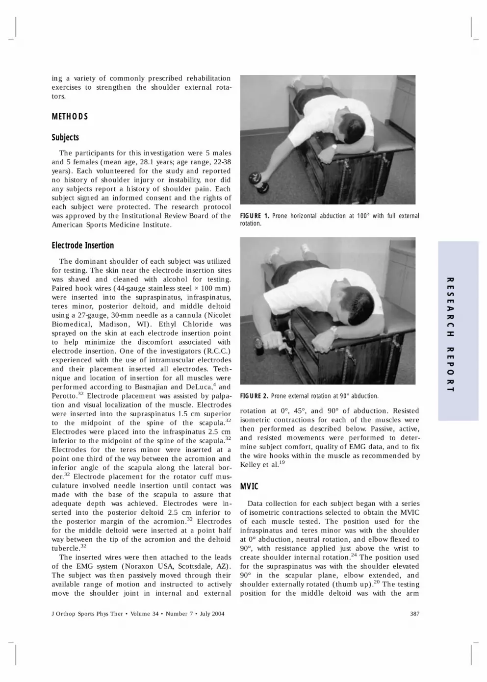
FIGURE 1. Prone horizontal abduction at 100° with full externalrotation.
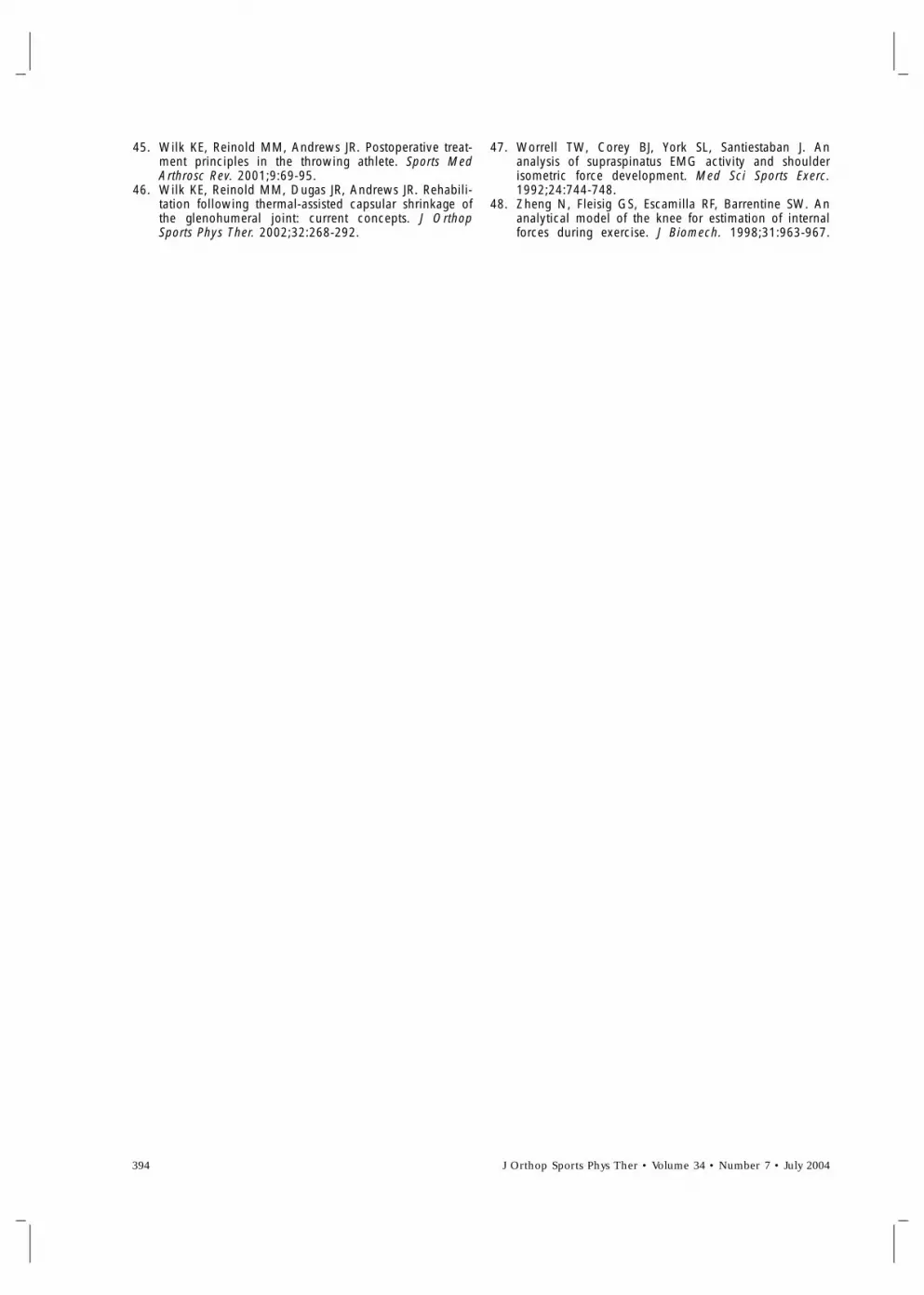
FIGURE 2. Prone external rotation at 90° abduction.
rotation at 0°, 45°, and 90° of abduction. Resistedisometric contractions for each of the muscles werethen performed as described below. Passive, active,and resisted movements were performed to deter-mine subject comfort, quality of EMG data, and to fixthe wire hooks within the muscle as recommended byKelley et al.19
MVIC
Data collection for each subject began with a seriesof isometric contractions selected to obtain the MVICof each muscle tested. The position used for theinfraspinatus and teres minor was with the shoulderat 0° abduction, neutral rotation, and elbow flexed to90°, with resistance applied just above the wrist tocreate shoulder internal rotation.24 The position usedfor the supraspinatus was with the shoulder elevated90° in the scapular plane, elbow extended, andshoulder externally rotated (thumb up).20 The testingposition for the middle deltoid was with the arm
J Orthop Sports Phys Ther • Volume 34 • Number 7 • July 2004 387
RE
SE
AR
CH
RE
PO
RT

FIGURE 3. Standing external rotation at 90° abduction.
FIGURE 4. Standing external rotation in the scapular plane.
abducted to 90° with neutral rotation (palm down)with resistance applied just proximal to the elbow inan inferior direction.21 The testing position for theposterior deltoid was with the arm abducted 90° withneutral rotation (palm down) with resistance appliedjust proximal to the elbow in the anterior direction.21
Exercises
The subjects were then tested for 7 shoulderrehabilitation exercises performed in random order.The exercises performed were: prone horizontal ab-duction at 100° with full ER (Figure 1), prone ER at90° abduction (Figure 2), standing ER at 90° ofabduction (Figure 3), standing ER in the scapularplane (ie, 45° abduction, 30° horizontal adduction)24
(Figure 4), standing ER at 0° abduction (Figure 5),standing ER at 0° abduction with a towel roll placed
between the trunk and elbow (Figure 6), and sidely-ing ER at 0° of abduction (Figure 7). Each subjectperformed 10 repetitions of each exercise. The speedof the repetitions was regulated by a metronome setto 60 beats per minute, where each concentric andeccentric phase was performed during 1 beat. Thesubject performed each exercise holding a dumbbellselected by a physical therapist (T.C.). Dumbbell massdetermination was made based on the maximummass that the subject could use while maintainingproper form for 10 repetitions and maintaining aproper cadence (10-repetition maximum). This deter-mination was performed in a testing session con-ducted prior to the EMG experiment.
Mean (±SD) mass used for each exercise were 2.2 ±0.8 kg for prone horizontal abduction at 100° withfull ER, 1.9 ± 1.0 kg for prone ER at 90° ofabduction, 3.6 ± 0.9 kg for standing ER at 90° ofabduction, 3.6 ± 1.4 kg for standing ER in thescapular plane, 7.1 ± 2.7 kg for standing ER at 0°abduction (same weight was used with and withouttowel), and 4.0 ± 1.9 kg for sidelying ER at 0°abduction.
Data Collection and Processing
For the MVIC trials, the subject was instructed to‘‘ramp up’’ to maximum effort. The investigatorverbally encouraged the subject to reach and main-tain maximum effort while EMG data were collectedfor 5 seconds. Once data collection was completedthe subject was instructed to relax. EMG data duringthe first and last second of each MVIC trial was
FIGURE 5. Standing external rotation at 0° abduction.
388 J Orthop Sports Phys Ther • Volume 34 • Number 7 • July 2004

FIGURE 6. Standing external rotation at 0° abduction with a towelplaced between elbow and body.
FIGURE 7. Sidelying external rotation.
discarded and the remaining 3 seconds of data wereused. The data were processed with a 25-Hz high-passfilter, then rectified, and finally integrated using a100-millisecond moving average window. Peak valuewas then identified as MVIC.48 Each muscle wastested once.
For the exercise trials, EMG data were collected at1000 Hz for 10 seconds. Data collection was initiatedafter the third repetition. Because the metronomepaced each 10-repetition exercise set to be about 20seconds, the 10-second data collection captured the 4middle repetitions (repetitions 4-7). The data werethen normalized by expressing the peak EMG valuefor each muscle for each trial as a percentage of theMVIC of the corresponding muscle. Maximum EMGexpressed in percent MVIC values for each musclewere averaged for the subject’s 4 repetitions.
Statistical Analysis
Statistical analysis was performed using SPSS 10.0statistical software (SPSS, Inc., Chicago, IL). An intra-class correlation coefficient (ICC3,1) was used todetect same-day test-retest reliability of EMG datausing the values of the 4 trials for analysis. EMGdifferences among the exercises were tested forstatistical significance using a separate 1-way repeated-measures analysis of variance for each muscle. Statisti-cal significance was set at P�.05. The Tukey test wasused for post hoc analysis to compare specific pairs ofexercises, using P�.05 for the level of significance.
RESULTS
The EMG activity of each muscle (percent MVIC)during each exercise, the corresponding ICCs, andthe statistically significant findings are listed in Tables1 through 5. The statistical analysis revealed a statisti-cally significant difference between some of theexercises for each muscle tested. Most ICCs werehigh, ranging from 0.71 to 0.99, with a mean of 0.92.The exception was for prone ER at 90° of abductionfor the middle deltoid, with an ICC of 0.45.
DISCUSSION
For all 5 muscles, statistically significant differenceswere noted in the amount of EMG activity across the7 exercises tested.
TABLE 1. Mean (±SD) electromyographic (EMG) activation ofthe infraspinatus expressed as a percentage of maximum volun-tary isometric contraction (MVIC) for 7 shoulder exercises.Intraclass correlation coefficients (ICC3,1) are also provided.
Exercise* % MVIC ICC
1. Sidelying external rotation at 0° ofabduction
62 ± 13† 0.81
2. Standing external rotation in thescapular plane (45° abduction, 30°horizontal adduction)
53 ± 25 0.87
3. Prone external rotation at 90° of ab-duction
50 ± 23 0.92
4. Standing external rotation at 90° ofabduction
50 ± 25 0.97
5. Standing external rotation at 0° ab-duction with a towel roll
50 ± 14 0.76
6. Standing external rotation at 0° ab-duction without a towel roll
40 ± 14 0.86
7. Prone horizontal abduction at 100°with full external rotation
39 ± 17 0.73
* The 1-way repeated-measures ANOVA indicated a significant maineffect across exercises (F = 3.288, P = .008).† Exercise 1 is significantly different than exercise 6 (P = .011) andexercise 7 (P = .008).
J Orthop Sports Phys Ther • Volume 34 • Number 7 • July 2004 389
RE
SE
AR
CH
RE
PO
RT

TABLE 2. Mean (±SD) electromyographic (EMG) activation ofthe teres minor expressed as a percentage of maximum volun-tary isometric contraction (MVIC) for 7 shoulder exercises.Intraclass correlation coefficients (ICC3,1) are also provided.
Exercise* % MVIC ICC
1. Sidelying external rotation at 0° ofabduction
67 ± 34† 0.87
2. Standing external rotation in thescapular plane (45° abduction, 30°horizontal adduction)
55 ± 30 0.79
3. Prone external rotation at 90° of ab-duction
48 ± 27 0.97
4. Standing external rotation at 0° ab-duction with a towel roll
46 ± 21 0.90
5. Prone horizontal abduction at 100°with full external rotation
44 ± 25 0.97
6. Standing external rotation at 90° ofabduction
39 ± 13 0.97
7. Standing external rotation at 0° ab-duction without a towel roll
34 ± 13 0.90
* The 1-way repeated-measures ANOVA indicated a significant maineffect across exercises (F = 3.726, P = .004).† Exercise 1 is significantly different than exercise 6 (P = .014) andexercise 7 (P = .003).
TABLE 3. Mean (±SD) electromyographic (EMG) activation ofthe supraspinatus expressed as a percentage of maximum volun-tary isometric contraction (MVIC) for 7 shoulder exercises.Intraclass correlation coefficients (ICC3,1) are also provided.
Exercise* % MVIC ICC
1. Prone horizontal abduction at 100°with full external rotation
82 ± 37† 0.97
2. Prone external rotation at 90° of ab-duction
68 ± 33‡ 0.97
3. Standing external rotation at 90° ofabduction
57 ± 32 0.94
4. Sidelying external rotation at 0° ofabduction
51 ± 47 0.89
5. Standing external rotation at 0° ab-duction with a towel roll
41 ± 37 0.71
6. Standing external rotation at 0° ab-duction without a towel roll
41 ± 38 0.94
7. Standing external rotation in thescapular plane (45° abduction, 30°horizontal adduction)
32 ± 24 0.93
* The 1-way repeated-measures ANOVA indicated a significant maineffect across exercises (F = 8.802, P�.001).† Exercise 1 is significantly different than exercises 4 (P = .008), 5, 6,and 7 (P�.001).‡ Exercise 2 is significantly different than exercises 5, 6 (P = .005),and 7 (P = .002).
Infraspinatus and Teres Minor
EMG activity across the 7 exercises varied from62% MVIC to 39% MVIC for the infraspinatus andfrom 67% MVIC to 34% MVIC for the teres minor.Based on the statistical analysis, a greater percentMVIC was generated in the infraspinatus while per-forming sidelying ER at 0° abduction, as compared tostanding ER at 0° abduction without a towel roll (P =.011) and prone horizontal abduction at 100° with
full ER (P = .008). Similarly, a statistically greaterpercent MVIC was generated for the teres minorwhile performing sidelying ER at 0° abduction, ascompared to standing ER at 90° abduction (P = .014)and standing ER at 0° without a towel roll (P = .003).Therefore, these data would support the hypothesisthat to specifically strengthen both muscles, sidelyingER at 0° abduction should be favored over the other3 exercises.
Despite the differences in EMG values measuredfor the first 5 exercises in Tables 1 and 2, varyingfrom 62% to 50% for the infraspinatus and 67% to44% for the teres minor, the statistical analysisshowed no significant differences among these exer-cises, which suggests that all 5 exercises may havesimilar strengthening effects. However, this lack ofstatistical significance needs to be interpreted withcaution because it is likely due to this study’s limitednumber of subjects. Consequently, further studies areneeded to establish if statistically or clinically signifi-cant differences exist across these exercises.
Activity of the infraspinatus (39%) and teres minor(44%) were low to moderate during prone horizontalabduction with external rotation. This was differentthan the results of Townsend et al,38 which showedhigh activity of the infraspinatus (88%) during thisexercise. The difference between studies is most likelydue to methodological differences. AlthoughTownsend et al38 used methods similar to those ofthe current study with indwelling EMG, they did notreport the positioning or procedure for obtainingtheir MVIC, making comparison between the studiesdifficult.
Supraspinatus and Deltoid
The greatest activity for the supraspinatus (82%)and deltoid muscles (posterior, 88% MVIC; middle,82% MVIC) were observed during prone horizontalabduction at 100° of abduction and full ER. This wasconsistent with the data from Worrell et al,47 Malangaet al,25 and Blackburn et al,5 who also showed highactivity level of these muscles during this exercise.
The supraspinatus and deltoid muscles showedactivity throughout each external rotation exercise.High activity of the supraspinatus, middle, and poste-rior deltoid were observed during prone and stand-ing external rotation at 90° of abduction. The highactivity during the exercises at 90° abduction may bein part due to the need to stabilize the upperextremity in the 90° abduction position while per-forming the external rotation movement. The findingof supraspinatus activity in our study is consistent withKronberg et al,22 who noted approximately 50%activity of the supraspinatus during the externalrotation movement.
390 J Orthop Sports Phys Ther • Volume 34 • Number 7 • July 2004

Clinical Implications
Exercises designed to strengthen the rotator cuffwith minimal deltoid involvement are often desired tominimize the amount of superior humeral headmigration, thus reducing the chance of subacromialimpingement.20,25,35,47 In this study, the high amountof middle (82%) and posterior deltoid (88%) EMGactivity during prone horizontal abduction at 100°may make this exercise disadvantageous to patientswho have pathology affecting the rotator cuff’s abilityto provide dynamic stabilization or who suffer fromor are at risk of subacromial impingement.
Furthermore, in general, exercises in the scapularplane or at 0° abduction showed significantly lessmiddle and posterior deltoid activity than those at90° abduction. It is our hypothesis that duringexternal rotation at 90° abduction, the high amountof middle deltoid activity may provide an assistivecompressive force rather than superior humeral headmigration due to the muscle tissue alignment andresultant force vectors.33 Poppen and Walker33 haveshown that at 90° abduction, the resultant forcevector of the deltoid and rotator cuff muscles pro-duces a centralized compressive force of the humeralhead within the glenoid fossa, rather than purely asuperior-oriented vector. Similarly, we could postulatethat the slightly higher EMG activity of the posteriordeltoid during ER at 90° may function to provide acompressive force as well as assist in external rotation.
The moderate activity of all muscles tested duringER at 90° of abduction (both prone and standing)supports the use of these exercises to simulate theposition of athletic competition when a sport-specificposition is desired in the rehabilitation of overheadathletes. Exercise performed in the 90°-abductedposition, either standing or prone, replicates theshoulder position, capsular strain, and muscle fiberlength-tension relationships observed in sporting ac-tivities, making strength gains in this position advan-tageous.11,12,41,44 In this position, the glenohumeraland scapulothoracic musculature function to providejoint movement and dynamic stabilization simulta-neously.
While standing external rotation at 90° abductionmay have a functional advantage over 0° of abductionand in the scapular plane due to the close replicationof this position in sporting activities, the combinationof abduction and external rotation places strain onthe shoulder’s capsule, particularly the anterior bandof the inferior glenohumeral ligament.30,42 When thearm is not in an abducted position, ER places lessstrain on this portion of the joint capsule. Therefore,although muscle activity was low to moderate duringER at 0° of abduction, this rehabilitation exercise maybe worthwhile when strain of the inferiorglenohumeral ligament complex is of concern.
TABLE 4. Mean (±SD) electromyographic (EMG) activation ofthe middle deltoid expressed as a percentage of maximum vol-untary isometric contraction (MVIC) for 7 shoulder exercises.Intraclass correlation coefficients (ICC3,1) are also provided.
Exercise* % MVIC ICC
1. Prone horizontal abduction at 100°with full external rotation
82 ± 32† 0.95
2. Standing external rotation at 90° ofabduction
55 ± 23‡ 0.99
3. Prone external rotation at 90° of ab-duction
49 ± 15§ 0.45
4. Standing external rotation in thescapular plane (45° abduction, 30°horizontal adduction)
38 ± 19 0.81
5. Sidelying external rotation at 0° ofabduction
36 ± 23 0.97
6. Standing external rotation at 0° ab-duction with a towel roll
11 ± 6 0.96
7. Standing external rotation at 0° ab-duction without a towel roll
11 ± 7 0.94
* The 1-way repeated-measures ANOVA indicated a significant maineffect across exercises (F = 13.444, P�.001)† Exercise 1 is significantly different than exercises 2 (P = .046), 3 (P= .043), 4, 5, 6, and 7 (P�.001).‡ Exercise 2 is significantly different than exercises 6 (P = .007) and 7(P�.001).§ Exercise 3 is significantly different than exercises 6 (P = .017) and 7(P = .002).
TABLE 5. Mean (±SD) electromyographic (EMG) activation ofthe posterior deltoid expressed as a percentage of maximumvoluntary isometric contraction (MVIC) for 7 shoulder exercises.Intraclass correlation coefficients (ICC3,1) are also provided.
Exercise* % MVIC ICC
1. Prone horizontal abduction at 100°with full external rotation
88 ± 33† 0.90
2. Prone external rotation at 90° of ab-duction
79 ± 31‡ 0.96
3. Standing external rotation at 90° ofabduction
59 ± 33§ 0.95
4. Sidelying external rotation at 0° ofabduction
52 ± 42� 0.84
5. Standing external rotation in thescapular plane (45° abduction, 30°horizontal adduction)
43 ± 30 0.98
6. Standing external rotation at 0° ab-duction with a towel roll
31 ± 27 0.92
7. Standing external rotation at 0° ab-duction without a towel roll
27 ± 27 0.93
* The 1-way repeated-measures ANOVA indicated a significant maineffect across exercises (F = 12.433, P�.001).† Exercise 1 is significantly different than exercises 3 (P = .046), 4(P = .005), 5, 6, and 7 (P�.001).‡ Exercise 2 is significantly different than exercises 6 and 7 (P�.001).§ Exercise 3 is significantly different than exercises 6 (P = .007) and 7(P = .005).� Exercise 4 is significantly different than exercises 6 (P = .038) and 7(P = .047).
Sidelying ER at 0° abduction may be a betterchoice than standing ER with the arm at the side;while both include ER with 0° of abduction, all 5muscles demonstrated higher activity during sidelying
J Orthop Sports Phys Ther • Volume 34 • Number 7 • July 2004 391
RE
SE
AR
CH
RE
PO
RT

ER. This could possibly be due to a greater effect ofgravity observed when performing ER in the sidelyingposition. This was similar to the results of Townsendet al38 and Ballantyne et al,3 but differed from theresults of Blackburn et al,5 who reported greateractivity of the infraspinatus and teres minor duringprone ER. No significant differences between sidely-ing ER and prone ER were observed in the presentstudy. Initially, the sidelying position may be a betterchoice than the prone position when protection ofthe glenohumeral ligaments is warranted, progressingto incorporate prone and standing ER at 90° abduc-tion when protection of the capsuloligamentous tis-sue is not warranted and strengthening and dynamicstabilization with the shoulder in a sport-specificposition is desired.
Theoretically, ER in the scapular plane at 45° ofabduction and 30° of horizontal adduction may be abeneficial shoulder ER exercise, as it offers a compro-mise between the functional benefit of ER at 90° ofabduction and the reduced capsular strain of ER at0° of abduction.42 The scapular plane positions themuscles of the glenohumeral joint in their optimalalignment and length-tension relationship to providedynamic stabilization. Furthermore, because the longaxis of the humerus intersects near the center of theglenoid fossa of the scapula when the arm is in thisposition, humeral head translation during ER may beminimized.42 The infraspinatus (53% MVIC) andteres minor (55% MVIC) were particularly activeduring ER in the scapular plane, consistent with theresults of Greenfield.13
Theoretically, ER at 0° of abduction with a towelroll provides both low capsular strain and low activityof the muscles that adduct the arm to hold the towel.Our clinical experience has shown that adding atowel roll to the external rotation exercise providesassistance to the patient by keeping the elbow to theside, ensuring that proper technique is observedwithout shoulder abduction and muscle substitutionof the deltoid. In this study, while the use of a towelroll resulted in greater activation of the infraspinatusand teres minor, these differences were not statisti-cally significant. Also, activity of the middle andposterior deltoid was similar between the 2 exercises.Despite the lack of significant findings, we haveclinically incorporated the use of a towel roll duringexternal rotation at 0° of abduction.
LimitationsThere are several limitations to EMG studies, in-
cluding reliability and validity concerns. This studyrepresents a common application of EMG duringrehabilitation exercises and was as reliable and validas possible using this mode of objective research. Theresults of our study may be influenced by improperelectrode placement and submaximal effort by thepatient. Our electrode placement was similar to that
of previous studies.3-5,19,20,25,32,38,47 During data col-lection each subject was working at an intensity levelconsistent with a 10-repetition maximum trial.
While we found statistically significant differencesacross the 7 exercises for all 5 muscles, the lack ofadditional differences between exercises, in particularfor the infraspinatus and teres minor, may have beendue to the low number of subjects and large standarddeviations. Accordingly, this lack of statistical differ-ence between exercises should be interpreted withcaution in light of the likely low statistical power.Therefore, for some of our comparisons, our findingsof no statistical difference may be related to lowpower rather than an actual lack of difference be-tween exercises.14,23 The descriptive results (meanand standard deviation) of this study may be astarting point when designing rehabilitation pro-grams. Future research should consider using thesedata to estimate appropriate sampling size.
Furthermore, the current study analyzed the EMGresponse of these exercises in young asymptomaticpatients, using dumbbells. Future studies involvinggroups of asymptomatic and symptomatic subjects,with varied ages and pathologies such as instability orsubacromial impingement, and with different types ofresistance (ie, resistive elastic bands) should be per-formed.
CONCLUSION
Muscle activity patterns from this study can helpclinicians choose the exercises that best fit theirobjectives. Based on the results of this study, EMGactivity varied based on the arm position duringexercise. The exercise that produced the greatestamount of EMG activity of the infraspinatus and teresminor was sidelying ER. Prone horizontal abductionat 100° with full ER produced maximum muscleactivity of the supraspinatus, middle deltoid, andposterior deltoid muscles. Considerations when select-ing external rotation exercises may be made based onthe amount of infraspinatus and teres minor activityas well as the amount of desired concomitant activityof the supraspinatus and deltoid musculature.
REFERENCES1. Andrews JR, Angelo RL. Shoulder arthroscopy for the
throwing athlete. Tech Orthop. 1988;3:75.2. Apreleva M, Hasselman CT, Debski RE, Fu FH, Woo SL,
Warner JJ. A dynamic analysis of glenohumeral motionafter simulated capsulolabral injury. A cadaver model.J Bone Joint Surg Am. 1998;80:474-480.
3. Ballantyne BT, O’Hare SJ, Paschall JL, et al.Electromyographic activity of selected shoulder musclesin commonly used therapeutic exercises. Phys Ther.1993;73:668-677; discussion 677-682.
392 J Orthop Sports Phys Ther • Volume 34 • Number 7 • July 2004

4. Basmajian JV, Deluca CJ. Muscles Alive: Their Func-tions Revealed. 5th ed. Balitmore, MD: Williams &Wilkins; 1985.
5. Blackburn TA, McLeod WD, White B, Wofford L. EMGanalysis of posterior rotator cuff exercises. Athl Train.1990;25:40-45.
6. Bradley JP, Tibone JE. Electromyographic analysis ofmuscle action about the shoulder. Clin Sports Med.1991;10:789-805.
7. Brown LP, Niehues SL, Harrah A, Yavorsky P, HirshmanHP. Upper extremity range of motion and isokineticstrength of the internal and external shoulder rotators inmajor league baseball players. Am J Sports Med.1988;16:577-585.
8. Cain PR, Mutschler TA, Fu FH, Lee SK. Anterior stabilityof the glenohumeral joint. A dynamic model. Am JSports Med. 1987;15:144-148.
9. DiGiovine NM, Jobe FW, Pink M, Perry J. Anelectromyographic analysis of the upper extremity inpitching. J Shoulder Elbow Surg. 1992;1:15-25.
10. Ellenbecker TS, Mattalino AJ. Concentric isokineticshoulder internal and external rotation strength inprofessional baseball pitchers. J Orthop Sports PhysTher. 1997;25:323-328.
11. Fleisig GS, Barrentine SW, Escamilla RF, Andrews JR.Biomechanics of overhand throwing with implicationsfor injuries. Sports Med. 1996;21:421-437.
12. Fleisig GS, Escamilla RF, Andrews JR, Matsuo T, Sat-terwhite Y, Barrentine SW. Kinematic and kinetic com-parison between baseball pitching and football passing.J Appl Biomech. 1996;12:207-224.
13. Greenfield BH, Donatelli R, Wooden MJ, Wilkes J.Isokinetic evaluation of shoulder rotational strengthbetween the plane of scapula and the frontal plane.Am J Sports Med. 1990;18:124-128.
14. Greenfield ML, Kuhn JE, Wojtys EM. A statistics primer.Power analysis and sample size determination. Am JSports Med. 1997;25:138-140.
15. Harryman DT, 2nd, Sidles JA, Clark JM, McQuade KJ,Gibb TD, Matsen FA, 3rd. Translation of the humeralhead on the glenoid with passive glenohumeral motion.J Bone Joint Surg Am. 1990;72:1334-1343.
16. Hislop HJ, Montgomery J. Muscle Testing: Techniquesof Manual Examination. Philadelphia, PA: W.B.Saunders; 1995.
17. Jobe FW, Kvitne RS, Giangarra CE. Shoulder pain in theoverhand or throwing athlete. The relationship of ante-rior instability and rotator cuff impingement. OrthopRev. 1989;18:963-975.
18. Jobe FW, Tibone JE, Perry J, Moynes D. An EMGanalysis of the shoulder in throwing and pitching. Apreliminary report. Am J Sports Med. 1983;11:3-5.
19. Kelly BT, Cooper LW, Kirkendall DT, Speer KP. Techni-cal considerations for electromyographic research onthe shoulder. Clin Orthop. 1997;140-151.
20. Kelly BT, Kadrmas WR, Speer KP. The manual muscleexamination for rotator cuff strength. Anelectromyographic investigation. Am J Sports Med.1996;24:581-588.
21. Kendall FP, McCreary EK, Provance PG. Muscles: Test-ing and Function. Baltimore, MD: Williams & Wilkins;1993.
22. Kronberg M, Nemeth G, Brostrom LA. Muscle activityand coordination in the normal shoulder. Anelectromyographic study. Clin Orthop. 1990;76-85.
23. Lieber RL. Statistical significance and statistical powerin hypothesis testing. J Orthop Res. 1990;8:304-309.
24. Magee DJ. Orthopedic Physical Assessment. Philadel-phia, PA: W.B. Saunders; 1997.
25. Malanga GA, Jenp YN, Growney ES, An KN. EMGanalysis of shoulder positioning in testing and strength-ening the supraspinatus. Med Sci Sports Exerc.1996;28:661-664.
26. McCann PD, Wootten ME, Kadaba MP, Bigliani LU. Akinematic and electromyographic study of shoulderrehabilitation exercises. Clin Orthop. 1993;179-188.
27. Moseley JB, Jr., Jobe FW, Pink M, Perry J, Tibone J.EMG analysis of the scapular muscles during a shoulderrehabilitation program. Am J Sports Med. 1992;20:128-134.
28. Moynes DR, Perry J, Antonelli DJ, Jobe FW.Electromyography and motion analysis of the upperextremity in sports. Phys Ther. 1986;66:1905-1911.
29. Neer CS, 2nd, Craig EV, Fukuda H. Cuff-teararthropathy. J Bone Joint Surg Am. 1983;65:1232-1244.
30. O’Brien SJ, Neves MC, Arnoczky SP, et al. The anatomyand histology of the inferior glenohumeral ligamentcomplex of the shoulder. Am J Sports Med.1990;18:449-456.
31. Pappas AM, Zawacki RM, McCarthy CF. Rehabilitationof the pitching shoulder. Am J Sports Med.1985;13:223-235.
32. Perotto AO. Anatomical Guide for theElectromyographer: The Limbs and Trunk. Springfield,IL: Charles C. Thomas; 1994.
33. Poppen NK, Walker PS. Forces at the glenohumeraljoint in abduction. Clin Orthop. 1978;165-170.
34. Portney LG, Watkins MP. Foundations of Clinical Re-search: Applications to Practice. Norwalk, CT: Appleton& Lange; 1993.
35. Reinold MM, Ellerbusch MT, Barrentine SW, et al.Electromyographic analysis of the supraspinatus anddeltoid muscles during rehabilitation exercises [ab-stract]. J Orthop Sports Phys Ther. 2002;32:A43.
36. Rockwood CA, Matsen FA. The Shoulder. Philadelphia,PA: W.B. Saunders; 1998.
37. Saha AK. Dynamic stability of the glenohumeral joint.Acta Orthop Scand. 1971;42:491-505.
38. Townsend H, Jobe FW, Pink M, Perry J.Electromyographic analysis of the glenohumeralmuscles during a baseball rehabilitation program. Am JSports Med. 1991;19:264-272.
39. Walch G, Boileau P, Noel E, Donell ST. Impingement ofthe deep surface ofthe infraspinatus tendon on theposterior glenoid rim. J Shoulder Elbow Surg.1992;1:239-245.
40. Wilk KE, Andrews JR, Arrigo CA, Keirns MA, Erber DJ.The strength characteristics of internal and externalrotator muscles in professional baseball pitchers. Am JSports Med. 1993;21:61-66.
41. Wilk KE, Arrigo C. Current concepts in the rehabilita-tion of the athletic shoulder. J Orthop Sports Phys Ther.1993;18:365-378.
42. Wilk KE, Arrigo CA, Andrews JR. Current concepts: thestabilizing structures of the glenohumeral joint. J OrthopSports Phys Ther. 1997;25:364-379.
43. Wilk KE, Arrigo CA, Andrews JR. Isokinetic testing ofthe shoulder abductors and adductors: windowed ver-sus nonwindowed data collection. J Orthop Sports PhysTher. 1992;15:107-112.
44. Wilk KE, Meister K, Andrews JR. Current concepts inthe rehabilitation of the overhead throwing athlete.Am J Sports Med. 2002;30:136-151.
J Orthop Sports Phys Ther • Volume 34 • Number 7 • July 2004 393
RE
SE
AR
CH
RE
PO
RT
45. Wilk KE, Reinold MM, Andrews JR. Postoperative treat-ment principles in the throwing athlete. Sports MedArthrosc Rev. 2001;9:69-95.
46. Wilk KE, Reinold MM, Dugas JR, Andrews JR. Rehabili-tation following thermal-assisted capsular shrinkage ofthe glenohumeral joint: current concepts. J OrthopSports Phys Ther. 2002;32:268-292.
47. Worrell TW, Corey BJ, York SL, Santiestaban J. Ananalysis of supraspinatus EMG activity and shoulderisometric force development. Med Sci Sports Exerc.1992;24:744-748.
48. Zheng N, Fleisig GS, Escamilla RF, Barrentine SW. Ananalytical model of the knee for estimation of internalforces during exercise. J Biomech. 1998;31:963-967.
394 J Orthop Sports Phys Ther • Volume 34 • Number 7 • July 2004


