Embed Size (px)
Citation preview

FEATURE ARTICLE
Electronic Properties of Functional Biomolecules at Metal/Aqueous Solution Interfaces
J. Zhang,† Q. Chi,† A. M. Kuznetsov,‡ A. G. Hansen,† H. Wackerbarth,† H. E. M. Christensen,†J. E. T. Andersen,† and J. Ulstrup* ,†
Building 207, Department of Chemistry, Technical UniVersity of Denmark, DK-2800 Lyngby, Denmark, andThe A.N. Frumkin Institute of Electrochemistry of the Russian Academy of Sciences, Leninskij Prospect 31,117071 Moscow, Russia
ReceiVed: August 2, 2001; In Final Form: NoVember 5, 2001
Monolayers of molecules, which retain their function in the adsorbed state on solid surfaces, are importantin materials science, analytical detection, and other technology approaching the nanoscale. Molecularmonolayers, including layers of functional biological macromolecules, offer new insight in electronic propertiesand stochastic single-molecule features and can be probed by new methods which approach the single-moleculelevel. One of these is in situ scanning tunneling microscopy (STM) in which single-molecule electronicproperties directly in aqueous solution are probed. In situ STM combined with physical electrochemistry,single-crystal electrodes, and spectroscopic methods is now a new dimension in interfacial bioelectrochemistry.We overview first some approaches to spectroscopic single-molecule imaging, including fluorescencespectroscopy, chemical reaction dynamics, atomic force microscopy, and electrochemical single-electrontransfer. We then focus on in situ STM. In addition to high structural resolution, in situ STM offers a single-molecule spectroscopic perspective. This emerges most clearly when adsorbate molecules contain accessibleredox levels, and the tunneling current decomposes into successive single-molecule interfacial electron transfer(ET) steps. Theories of electrochemical ET and in situ STM of redox molecules as well as specific cases areaddressed. Two-step in situ STM represents different molecularmechanismsand even new ETphenomena,related to coherent many-electron transfer. A number of systems are noted to accord with these views. Thediscussion is concluded by attention to one of the still very few redox proteins addressed by in situ STM, theblue copper proteinPseudomonas aeruginosaazurin. Use of comprehensive electrochemical techniques hasascertained that well-defined protein monolayers in two opposite orientations can be formed and interfacialtunneling patterns disclosed.P. aeruginosaazurin emerges as by far the most convincing case where in situSTM of functional metalloproteins to single-molecule resolution has been achieved. This comprehensiveapproach holds promise for broader use of in situ STM as a single-molecule spectroscopy of metalloproteinsand illuminates prerequisites and limitations of in situ STM of biological macromolecules.
1. IntroductionTwo-dimensional films of proteins, DNA and DNA frag-
ments, and other biomolecules at solid surfaces in contact with
aqueous electrolyte solution are broadly important. Recognizedtechnological perspectives, relate to (a) chromatography,1 (b)protein-membrane interactions and pharmaceutical drug de-livery,2,3 (c) biologically induced corrosion,4 (d) biocompatibilityof metallic implantates,5 (e) support for attached tissue cultures6
(“biofilms”), (f) analytical chemistry involving immobilizedenzymes or antibodies and their substrates7,8 (glucose, dioxygen,
* To whom correspondence should be addressed. Phone:+45 45252359.Fax: +45 45883136. E-mail: [email protected].
† Technical University of Denmark.‡ The A.N. Frumkin Institute of Electrochemistry of the Russian Academy
of Sciences.
© Copyright 2002 by the American Chemical Society VOLUME 106, NUMBER 6, FEBRUARY 14, 2002
10.1021/jp0129941 CCC: $22.00 © 2002 American Chemical SocietyPublished on Web 01/17/2002

etc.) or antigens9 (e.g., fluorescein), (g) DNA hybridization andscreening,10,11 and (h) other areas where, broadly, biologicalliquids are in contact with solid surfaces.12,13 Protein filmtechnology has been framed broadly by macroscopic conceptsand statistically averaged observables, such as adsorptionisotherms,12-14 spectroscopy15-18 (UV/vis, ellipsometry, internalreflection absorption and fluorescence, IR and Raman spec-troscopy), microcantilever technology,19,20quartz crystal micro-balance21 (QCM), and electrochemistry.22-24
In situ scanning probe microscopies (SPM) such as in situscanning tunneling (STM)25-27 and atomic force microscopy(AFM)25,28and single-molecule electromagnetic spectroscopies(SMS)29-31 now offer structural and dynamic detail of individual(bio)molecules. In situ single-molecule notions have beenextended to electrochemical32-34 and conductivity behavior34-36
in metallic nanosize structures. The notion in situ here and inthe following refers to the adsorbed molecular films directly incontact with aqueous electrolyte solution as the natural reactionmedium (Figure 1). In situ SPM systems hold perspectives intwo-dimensional protein and other biomolecular film structureand function. They hold a clue tostructural mapping and
visualization of adsorbed molecules in situ with unprecedentedresolution but, also, new approaches to single-moleculefunctionand spectroscopy. These are represented by AFM force-distance relations and STM tunnel current/bias Voltage oroVerVoltagerelations. In situ nanogap conductance is a relatedspectroscopic property.34-36 Such correlations reflect single-molecule electronic properties analogous to single-moleculeoptical absorption and emission, with crucial information aboutenergetics and electronic-vibrational coupling not otherwiseoffered.
The complementarity of in situ SPMs to macroscopic methodsoffers another dimension of single-molecule characterization butalso discloses limitations. Alimitation is that analyticaliden-tification at the molecular level is, broadly, elusive. In situ STM/AFM resolution of soft biological macromolecules is also atbest molecular, far from the close to atomic resolution for smallor intermediate-size molecules, and the number of well char-acterized in situ two-dimensional biomolecular systems is stillquite small.Combinationof in situ SPM with other techniquessuch as single-crystal voltammetry, X-ray photoelectron spec-troscopy (XPS), and electrochemical impedance spectroscopyis therefore essential. An additionaldimension, intermediatebetween macroscopic and molecular scale is in the context ofsupramolecularassemblies. Visualization and control of bio-molecular function, say of immobilized enzymes, and correlationwith macroscopic enzyme kinetics are here one perspective.Two-dimensional assemblies with electronic rectification andother molecular functions are another one. These areas now offerworking examples and are much closer to realization than justa few years ago.37-39
In this paper, we address recent theoretical and experimentaladvances of in situ SPM. Focus is on in situ STM of largemolecules and biomolecules, combined with interfacial electro-chemical electron transfer (ET), viewed as a single-moleculespectroscopy. In section 2, we overview some approaches tosingle-molecule spectroscopic properties. These include single-molecule excitation and emission, chemical dynamics, Coulombblockade at ultra-small electrodes, and AFM. A brief overviewof theoretical notions of interfacial ET is given in section 3 asa reference. In section 4, we discuss the in situ STM process,as a class of single-molecule spectroscopic phenomena and asa new case for nanoscale electrochemical ET. In situ STM is,thus, a special long-range ET process with clear analogies anddifferences compared with other chemical and biological long-range ET processes.40-44 Current-voltage relations offer spec-troscopic information regarding energetics and electronic levelbroadening and clues to the STM tunnelingmechanisms. Thisis followed by a discussion of recent data which illustrate insitu STM and single-molecule function. Section 4 is concludedby a discussion of a redox metalloprotein, i.e., the blue single-copper proteinPseudomonas aeruginosaazurin immobilized onAu(111) surfaces.45-47 This is the first case for structural single-molecule resolution of a documented functional redox proteinunder potential control. It is shown, particularly, that high-qualitysingle-crystal electrode surfaces andcomplementaryelectro-chemical and spectroscopic techniques must be combined within situ STM. Some perspectives are discussed in Section 5.
2. Some Examples of Single-molecule Spectroscopy
To put in situ STM in perspective, we consider first someother single-molecule spectroscopies developed over the pastdecade.
2.1. Single-Molecule Fluorescence Excitation and Emissionin Condensed Phases.Single-molecule fluorescence spectra and
Figure 1. A. Upper figure: Schematic view of ex situ STMconfiguration. The tip-sample distance is controlled by the bias voltageVbias and the tunneling current. Lower figure: Schematic view ofelectrochemical configuration. The electrode-solution potential dif-ference is controlled relative to a reference electrode. B. In situ STMconfiguration. Both the sample-solution and the tip-sample potentialdifferences are controlled by combining the configurations in A. Thetip is insulated except at the very end.
1132 J. Phys. Chem. B, Vol. 106, No. 6, 2002
excited state lifetimes have provided detailed information aboutlocal molecular environments of large organic molecules at highdilution in optically transparent matrices both at cryogenic androom temperatures.30,31,48-51 Optical excitation by laser beamsof micrometer lateral extension are focused on resonance withwings of inhomogeneously broadened absorption bands wherefew molecules are represented.Spatialresolution is thus in thesubmicrometer range, butoptical resolution is at the single-molecule level. Important data are (a) bursts of fluorescenceafter laser illumination, (b) emission spectra with single-molecule photon distributions, (c) single-molecule fluorescencelifetimes, and (d) fluorescence tracking to disclose structuraland timefluctuationscaused by the local (nanoscale) environ-ment. Single-molecule spectroscopy can resolve molecularcomponents in inhomogeneously broadened spectra, caused bylocal environmentdistributionsin space and time. Distributionsof fluorescence lifetimes can be disentangled, and correlationsbetween single-molecule fluorescence lifetimes and peak fre-quencies can be constructed. Notable is stochastic time evolutionof single-molecule features, spanning time ranges from sub-seconds to thousands of seconds, caused by configurationalfluctuations or by molecular hopping between different environ-ments. This perspective carries over to in situ STM at the solid-liquid interface.
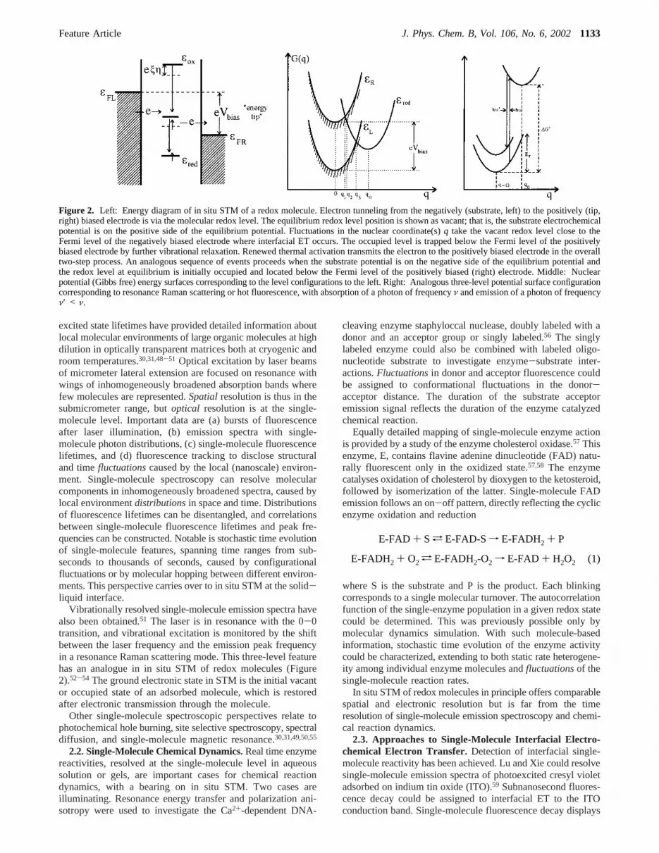
Vibrationally resolved single-molecule emission spectra havealso been obtained.51 The laser is in resonance with the 0-0transition, and vibrational excitation is monitored by the shiftbetween the laser frequency and the emission peak frequencyin a resonance Raman scattering mode. This three-level featurehas an analogue in in situ STM of redox molecules (Figure2).52-54 The ground electronic state in STM is the initial vacantor occupied state of an adsorbed molecule, which is restoredafter electronic transmission through the molecule.
Other single-molecule spectroscopic perspectives relate tophotochemical hole burning, site selective spectroscopy, spectraldiffusion, and single-molecule magnetic resonance.30,31,49,50,55
2.2. Single-Molecule Chemical Dynamics.Real time enzymereactivities, resolved at the single-molecule level in aqueoussolution or gels, are important cases for chemical reactiondynamics, with a bearing on in situ STM. Two cases areilluminating. Resonance energy transfer and polarization ani-sotropy were used to investigate the Ca2+-dependent DNA-
cleaving enzyme staphyloccal nuclease, doubly labeled with adonor and an acceptor group or singly labeled.56 The singlylabeled enzyme could also be combined with labeled oligo-nucleotide substrate to investigate enzyme-substrate inter-actions.Fluctuationsin donor and acceptor fluorescence couldbe assigned to conformational fluctuations in the donor-acceptor distance. The duration of the substrate acceptoremission signal reflects the duration of the enzyme catalyzedchemical reaction.
Equally detailed mapping of single-molecule enzyme actionis provided by a study of the enzyme cholesterol oxidase.57 Thisenzyme, E, contains flavine adenine dinucleotide (FAD) natu-rally fluorescent only in the oxidized state.57,58 The enzymecatalyses oxidation of cholesterol by dioxygen to the ketosteroid,followed by isomerization of the latter. Single-molecule FADemission follows an on-off pattern, directly reflecting the cyclicenzyme oxidation and reduction
where S is the substrate and P is the product. Each blinkingcorresponds to a single molecular turnover. The autocorrelationfunction of the single-enzyme population in a given redox statecould be determined. This was previously possible only bymolecular dynamics simulation. With such molecule-basedinformation, stochastic time evolution of the enzyme activitycould be characterized, extending to both static rate heterogene-ity among individual enzyme molecules andfluctuationsof thesingle-molecule reaction rates.
In situ STM of redox molecules in principle offers comparablespatial and electronic resolution but is far from the timeresolution of single-molecule emission spectroscopy and chemi-cal reaction dynamics.
2.3. Approaches to Single-Molecule Interfacial Electro-chemical Electron Transfer. Detection of interfacial single-molecule reactivity has been achieved. Lu and Xie could resolvesingle-molecule emission spectra of photoexcited cresyl violetadsorbed on indium tin oxide (ITO).59 Subnanosecond fluores-cence decay could be assigned to interfacial ET to the ITOconduction band. Single-molecule fluorescence decay displays
Figure 2. Left: Energy diagram of in situ STM of a redox molecule. Electron tunneling from the negatively (substrate, left) to the positively (tip,right) biased electrode is via the molecular redox level. The equilibrium redox level position is shown as vacant; that is, the substrate electrochemicalpotential is on the positive side of the equilibrium potential. Fluctuations in the nuclear coordinate(s)q take the vacant redox level close to theFermi level of the negatively biased electrode where interfacial ET occurs. The occupied level is trapped below the Fermi level of the positivelybiased electrode by further vibrational relaxation. Renewed thermal activation transmits the electron to the positively biased electrode in the overalltwo-step process. An analogous sequence of events proceeds when the substrate potential is on the negative side of the equilibrium potential andthe redox level at equilibrium is initially occupied and located below the Fermi level of the positively biased (right) electrode. Middle: Nuclearpotential (Gibbs free) energy surfaces corresponding to the level configurations to the left. Right: Analogous three-level potential surface configurationcorresponding to resonance Raman scattering or hot fluorescence, with absorption of a photon of frequencyν and emission of a photon of frequencyν′ < ν.
E-FAD + S a E-FAD-Sf E-FADH2 + P
E-FADH2 + O2 a E-FADH2-O2 f E-FAD + H2O2 (1)
Feature Article J. Phys. Chem. B, Vol. 106, No. 6, 20021133
exponential kinetics but 40 different molecules spanned a 7-foldlifetime variation, disclosing molecular stochastic effects. Single-molecule electrochemical detection has also been based on thescanning electrochemical microscope.60,61 A few molecules(ultimately a single molecule) are trapped in a 10-20 nm pocketunder a SPM tip squeezed against a substrate electrode. Single-molecule currents cannot be directly detected, but if electro-chemical conversion of a molecule at the tip is followed byreconversion at the substrate, the current is amplified by manyorders of magnitude. Oxidation of a ferrocene derivative(trimethylammonium-methyl-ferrocene) at a Pt-Ir tip followedby reconversion to ferrocene at ITO substrate has illustratedthis. Current-time records over several hundred seconds, andstatistical analysis could, further, identify 0.2 pA individualmolecule fluctuations in the ultramicroscopic solution spaceunder the tip.
Other interfacial electrochemical ET systems have shownsingle-electron tunneling (SET) and Coulomb blockade effects.62
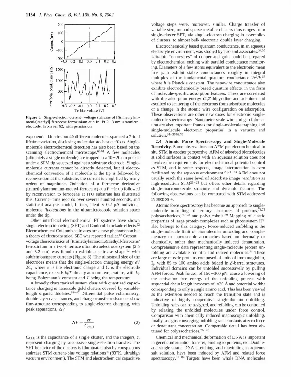
Electrochemical Coulomb staircases are a new phenomenon buta theory of electrochemical SET was reported earlier.63 Current-voltage characteristics of [(trimethylammonio)methyl]-ferrocene/ferrocinium in a two-interface ultramicroelectrode system (2.5and 3.2 nm) was found to exhibit a staircase shape,62 withsubfemtoampere currents (Figure 3). The ultrasmall size of theelectrodes means that the single-electron charging energye2/2C, wheree is the electronic charge andC is the electrodecapacitance, exceedskBT already at room temperature, withkB
being Boltzmann’s constant andT being the temperature.A broadly characterized system class with quantized capaci-
tance charging is nanoscale gold clusters covered by variable-length organic thiolates.64-67 Differential pulse voltammetry,double layer capacitances, and charge-transfer resistances showfine-structure corresponding to single-electron charging, withpeak separations,∆V
CCLU is the capacitance of a single cluster, and the integers,z,represent charging by successive single-electron transfer. TheSET behavior of the clusters is illuminated also by conspicuousstaircase STM current-bias voltage relations66 (83°K, ultrahighvacuum environment). The STM and electrochemical capacitive
voltage steps were, moreover, similar. Charge transfer ofvariable-size, monodisperse metallic clusters thus ranges fromsingle-cluster SET, via single-electron charging in assembliesof clusters, to almost bulk electronic double layer charging.
Electrochemically based quantum conductance, in an aqueouselectrolyte environment, was studied by Tao and associates.34,35
Ultrathin “nanowires” of copper and gold could be preparedby electrochemical etching with parallel conductance monitor-ing. Diameters of a few atoms equivalent to the electronic meanfree path exhibit stable conductances roughly in integralmultiples of the fundamental quantum conductance 2e2/h,68
whereh is Planck’s constant. The nanowire conductance alsoexhibits electrochemically based quantum effects, in the formof molecule-specific adsorption features. These are correlatedwith the adsorption energy (2,2′-bipyridine and adenine) andascribed to scattering of the electrons from adsorbate moleculesor a change in the atomic wire configuration on adsorption.These observations are other new cases for electronic single-molecule spectroscopy. Nanometer-scale wire and gap fabrica-tion are also important frames for single-molecule trapping andsingle-molecule electronic properties in a vacuum andsolution.34-36,69,70
2.4. Atomic Force Spectroscopy and Single-MoleculeReactivity. Some observations on AFM put electrochemical insitu STM in another perspective. AFM of adsorbed biomoleculesat solid surfaces in contact with an aqueous solution does notinvolve the requirements for electrochemical potential controlas STM, and in some respects, image interpretation is evenfacilitated by the aqueous environment.26,71-73 AFM does notusually reach the same level of adsorbate imageresolutionashigh-resolution STM26-28 but offers other details regardingsingle-macromolecule structure and dynamic features. Thefollowing observations can be compared with STM addressedin section 4.
Atomic force spectroscopy has become an approach to single-molecule unfolding of tertiary structures of proteins,74,75
polysaccharides,76-78 and polyalcohols.79 Mapping of elasticproperties of large protein complexes such as photosystem II80
also belongs to this category. Force-induced unfolding is thesingle-molecule limit of biomolecular unfolding and comple-mentary to macroscopic approaches based on thermally orchemically, rather than mechanically induced denaturation.Comprehensive data representing single-molecule protein un-folding are available for titin and related molecules.74 Theseare large muscle proteins composed of units of immunoglobin,Ig, with 89 to 100 amino acids folded inâ-barrel structures.Individual domains can be unfolded successively by pullingAFM forces. Peak forces, of 150-300 pN, cause a lowering ofthe activation free energy of the unfolding process withsequential chain length increases of≈30 Å and potential widthscorresponding to only a single amino acid. This has been viewedas the extension needed to reach the transition state and isindicative of highly cooperative single-domain unfolding.Unfolding ratescan be assigned, and refolding can be controlledby relaxing the unfolded molecules under force control.Comparison with chemically induced macroscopic unfolding,finally, assigns converging unfolding rate constants at zero forceor denaturant concentration. Comparable detail has been ob-tained for polysaccharides.76-78
Chemical and mechanical deformation of DNA is importantin genetic information transfer, binding to proteins, etc. Double-and single-strand DNA stretching, and unwinding in aqueoussalt solution, have been induced by AFM and related forcespectroscopy.81-84 Targets have been whole DNA molecules
Figure 3. Single-electron current-voltage staircase of [(trimethylam-monio)methyl]-ferrocene-ferrocinium at a Ir-Pt 2-3 nm ultramicro-electrode. From ref 62, with permission.
∆V ) zeCCLU
(2)
1134 J. Phys. Chem. B, Vol. 106, No. 6, 2002

with mesoscopic contour lengths, 15-20 µm or≈5 × 10⁴ basepairs,81-83 and 10-30 base pair oligonucleotides.84 DNA canbe stretched by elastic forces to 1.7 times its contour length ata force of 70 pN.81,82Longer stretching involves conformationalchanges in the carbohydrate units. Elastic DNA stretching iscontrolled also by hydration, ionic strength, and chemical cross-linking, framed by multistate models, with ladder conformationsand partial melting as intermediate states. AFM of 10-30 basepair oligonucleotide duplexes have illuminated unwinding tosingle strands84 by covalent attachment of complementarystrands to the tip and substrate. “Zipper-like” unwinding occursin the 20-50 pN force range, correlated with the number ofbase pairs, temperature and ionic strength, and macroscopicthermal dissociation rates.84
2.5. Force-Spectroscopy of Ligand-Receptor Interactions.Formation and dissociation of biological ligand-receptorcomplexes are cases of single-molecule biologicalreactiVity,quantified by in situ AFM. Avidin-biotin (protein-smallligand)85,86 and antigen-antibody (protein-protein)87-89 as-sociation and dissociation have been characterized. Mappingof the forces between biotin (receptor) covalently immobilizedon polymer beads and avidins immobilized by tip adsorptionprovides distributions of single protein-receptor pair inter-actions.85,86 These correlate with the unbinding enthalpy andbinding potential widths in the structural range of the protein-binding pocket. Single-molecule pair interactions also emergefrom AFM of antigen-antibody complexes between fluorescein(antigen) and wildtype and mutant Ig fragments (antibody) ongold-plated substrates. The pair interaction force,F, correlateswith the macroscopic dissociation rate constant,koff, as
where koff0 is a constant andxâ represents the difference
between the protein-ligand distance in the bound and transitionstates.
AFM has, finally, been brought to approach single-macro-molecule chemical reactivity, but this notion has been assignedto different levels of resolution. Transcription of DNA to RNAby E. coli RNA polymerase on mica, followed by in situ AFM,is close to molecular resolution, and processing of individualDNA molecules through the enzyme could be imaged in realspace and time.90 Single-molecule enzyme activity and inhibitionof lysozyme on mica91 could be monitored in AFM as reversibleheight fluctuations in the presence of substrate and inhibitormolecules, respectively. Hydrolysis of immobilized phospholipidcatalyzed by phospholipase A2 in solution92 is, finally, a casefor AFM of enzyme activity, where the substrate is immobilizedand the solute enzyme is freely mobile. Lateral resolution hereis, however, in micrometer rather than nanometer ranges.
3. Notions of Interfacial Electrochemical ElectronTransfer
3.1. Some General Observations.In situ STM of molecularadsorbates resembles in crucial respects electrochemical inter-facial ET, and some theoretical notions on physical electro-chemistry constitute part of our discussion of in situ STM. Itwas noted that chemical and biological ET in homogeneoussolution has reached high levels of theoretical and experimentalcharacterization.40-44 Similar detail in ET between molecularredox centers and metal, semiconductor, or semimetal electrodeshas been more elusive. Fundamental similarities and differencesfrom ET in solution were recognized early.43,44,93-96 Among
the former, the nature of the ET process as a quantummechanical transition between two electronic-vibrational states,free energy relations, the notion of “work terms” and “doublelayer effects”, and the view of ET as a tunneling process96,97
are the most important. Particularly, the Brønsted relationbetween the rate constant and the reaction free energy ofelementary charge transfer (proton transfer) processes in solu-tion98 is equivalent to the Butler-Volmer relation between theelectrochemical current,j(η) and the overvoltageη:99
This is the simplest electrochemical free energy relation andcarries over to in situ STM.j0 is the exchange current. Theactivation Gibbs free energy,G0q, incorporates all of the nuclearactivation features.R () R(η)) is the electrochemical transfercoefficient
A conspicuousdifferenceis that the electrode incorporates acontinuousmanifold of electronic states, whereas only a singlepair of states is involved in homogeneous ET. Consequencesof this, such as the absence of an inverted Gibbs free energyregion, are recognized.43,44,100A second difference relates to thenature of the electronic wave functions. These are spatiallylocalized for ET in solution but two-dimensionally delocalizedin electrochemical ET. The electrochemical tunneling factor istherefore conveniently approached by invoking electronicdensi-ties rather than individual level wave functions.101,102
Theoretical and experimental detail of electrochemical EThas been fraught with the complexity of the nonuniforminterfacial environment. New well characterized systems are,however, becoming available, with variable-length pure andfunctionalized alkanethiolates and related molecules self-as-sembled on electronically “soft” metals, mostly gold infocus.103-112 Interfacial ET of small redox molecules such asferrocene exhibits clear features of electron tunneling acrossthe alkylthiolate films. This warrants new theoretical approachesbased on electronic density functional theory for the metalsurface and statistical mechanical frames for the reactingmolecules and the solvent. Combined with in situ SPM,molecular-based views of the electrochemical interface aretherefore approaching a new level.
Electrochemical ET of adsorbed biological macromolecules,redox metalloproteins in particular,113-115 was long regardedseparately from recent achievements of physical electrochemistrysuch as single-crystal electrode procedures, surface spec-troscopies, etc. “Soft” macromolecules such as single- andmulticenter redox proteins are, thus, vulnerable to strong electricfields, environmental anisotropy, etc. Focus in interfacialbioelectrochemistry has therefore been different and directedtoward conditions for robust voltammetry. Diffusive reversibleET patterns are broadly observed for cytochromes, blue copperproteins, and iron-sulfur proteins, whereas peroxidases, blueoxidases, iron-sulfur enzymes, and flavoenzymes are examplesof macroscopically mapped monolayer enzyme electro-catalysis.113-116
Well-defined monolayers of redox proteins at pure ormodified electrode surfaces have opened options for methodsother than cyclic voltammetry, such as electrochemicalimpedance,47,117-120 electroreflectance spectroscopy,119,121-123
and surface enhanced resonance Raman spectroscopy,124-126
enabling characterization of both adsorption and interfacial ET.
koff(F) ) koff0 exp(Fxâ
kBT) (3)
j(η) ) j0 exp(- ReηkBT); j0 ∝ exp(- G0q
kBT) (4)
R ) -kBT[d ln j(η)/d(eη)] (5)
Feature Article J. Phys. Chem. B, Vol. 106, No. 6, 20021135

Introduction of theoretical notions based on the overpotentialdependence of the rate constants, electron tunneling, etc. aretherefore warranted. Bowden et al.127 addressed, for example,monolayers of horse heart cytochromec electrostatically ad-sorbed on self-assembledω-mercaptoalkanoic acids on poly-crystalline gold. Niki et al.122,123reported rates for ET betweencyt c and gold electrodes across variable-lengthω-mercaptoal-kanoic acids by electro-reflectance spectroscopy. The electro-static nature of the surface interactions was substantiated byionic strength and pH effects. Tunneling rates displayedexponential distance decay for carbon chain lengths longer thaneight CH2 groups. Similar observations based on potential jumptechniques and surface enhanced resonance Raman spectroscopyhave been reported.126 A comprehensive study based on severalinterfacial techniques has disentangled adsorption and interfacialET features ofPseudomonas aeruginosaazurin at single-crystalgold electrodes covered by variable-length alkylthiolates.47,118
The protein is here adsorbedhydrophobically. This will be infocus in section 4. Multicenter redox metalloenzyme voltam-metry has, finally, been brought to a level where intramolecularET and othermechanisticfeatures can be distinguished. Studiesby Armstrong and associates have, for example, identifiedelectrochemical analogues of peroxidase catalytic cycles anddisclosed interfacial ET routes through the ET centers offumarate reductase in the electrocatalytic reduction of fuma-rate.23,116 Observations such as these warrant stronger effortstoward introducing new physical electrochemical methodologyand new theoretical frames, also in interfacial bioelectrochem-istry.
3.2. Some Theoretical Frames of Electrochemical andBioelectrochemical ET.Figure 4 illustrates the fundamentalelectrochemical (cathodic) ET process.43,44,100,128The current iscomposed of contributions from all electronic levels of the (heremetal) electrode. The redox level is strongly coupled to theenvironment, with the initially vacant level in equilibrium wellabove the Fermi level of the electrode,εF. Configurationalfluctuations lower the level to match populated levels in theelectrode where ET proceeds, with subsequent electron trappingon the molecule. In the diabatic limit of weak electrode-molecule interaction, the current density is, broadly indepen-dently on the system properties:
Cox(z*) is the concentration of oxidized molecule at the distancez* from the surface, and∆z is a narrowz range aroundz*. f(ε)
is the Fermi function, andF(ε) is the electronic level density atthe energyε:
whereme is the mass of the electron,εb is the bottom of theconduction band, 2πp is Planck’s constant, andW(ε;η) is therate constant (s-1) for ET from the levelε. Equations 5-7 area generalbasis for a range ofspecificsystems. These extend tometal, semiconductor, semimetal, and superconductor electrodesand to harmonic and anharmonic electronic-vibrational interac-tions, nuclear tunneling, etc.43,44,129,130 The following formapplies broadly:
whereER is the nuclear reorganization Gibbs free energy, andωeff is the effective vibrational frequency of all the nuclearmodes which contribute toER. κel(ε;η) is the electronictransmission coefficient, the most important feature of whichis the electron exchange factor,TεA(ε;η), which couples the levelε with the molecular redox level.
Levels around the Fermi level dominate in broad overpotentialranges at metal electrodes, wherej(η) takes the quadratic form
and Cred is the concentration of the reduced molecule. Analternative form is101,102
where δε is a narrow energy range aroundεF, V(z*) is thephysical interaction between the electrode and the molecule,andM(η) is the square of the electronicdensity oVerlap. Thisform is suited to incorporate interactions between the metalelectrode and the interfacial environment. An approximatesimple form ofM(η) is
wherea (≈z*) is the distance of the molecular center from theelectrode surface andzj ) zj(η) is the front of the metallic electrondensity.âmet and âmol (Å-1) are the distance decay factors ofthe electronic densities of the metal and the molecule, respec-tively, andA andB are weakly distance and electrode potentialdependent quantities. Equation 11 discloses lability of theelectronic factor induced by variable electrode charge,101,102butâmet often significantly exceedsâmol, and tunneling is stilldominated by the molecular term in eq 11.
Figure 4. Electronic energies (left) and potential (Gibbs free) energysurfaces for interfacial electrochemical ET. Cathodic process.
j(η) ) ∫dε F(ε)f(ε)i(ε;η); i(ε;η) ) eCox(z*)∆zW(ε;η) (6)
f(ε) ) {1 + exp[(ε - εF)/kBT]}-1;
F(ε) ) (Vme3/2/p3π3)x2(ε - εb) (7)
W(ε;η) ) κel(ε;η)ωeff
2πexp{-
[ER + eη - (ε - εF)]2
4ERkBT }κel(ε;η) ) [TεA(ε;η)]2x 4π3
ERkBTp2ωeff2
, 1 (8)
j(η) ≈ j0 exp[- eη2kBT
-(eη)2
4ERkBT]j0 ) j(η ) 0) ) [πeF(εF)/p]x2CoxCredkBT/ER ×
∆z[TεA(ε ≈ εF;η ) 0)]2 exp(-ER
4kBT) (9)
j(η) ) (πe/2p)δε∆zCox[V(z*)] 2M(η) exp[-(ER + eη)2
4ERkBT ](10)
M(η) ≈ A exp[-âmet(a - zj)] + B exp[-âmol(a - zj)] (11)
1136 J. Phys. Chem. B, Vol. 106, No. 6, 2002

The formalism above refers to thediabatic limit. The validityconditions for this limit are different from ET in homogeneoussolution because of the manifold of electronic levels43,131
The opposite, adiabatic limit is equally appropriate to bothelectrochemical ET and in situ STM. There are two approachesto this limit. In one approach, the focus is on multiple transitionsbetween two manifolds of electronic levels representing thereactants’ (R) and products’ (P) states.131 The potential surfaces,spanned by the nuclear coordinate(s)q, are
ε andε′ are energies for different electron distributions, countedfrom the energy corresponding to the Fermi distribution.Transitions are described by master equations, with simplesolutions in the two limits given by eq 12 and the oppositeinequality. The adiabatic current is
The activation free energy is determined by the maximum ofthe potential free energy surface
The quadratic form in eq 8 or 9 is, however, often appropriate.The electronic factor,κF, is thus absent in the adiabatic limit.
The other approach was initiated by Hush,132,133who recastthe electronic manifold into a single effective orbital. Thisresembles work much later based on chemisorption theory.134
Chemisorption-based formalism applies both to electrochemi-cal135 and electrocatalytic ET processes between a reactingmolecule and a chemisorbed atom electronically broadened bythe metal.136,137 Such views have also been central to STM.Recent work on adiabatic electrochemical ET based on chemi-sorption theory was initiated by Schmickler135 and followed byothers.138-141 Common to these approaches is the constructionof potential surfaces such as eq 16 based on the width of theredox level,∆. The following form139 is illuminating:
and shows that the electronic coupling,∆, reduces the diabaticactivation barrier (∆ < ER) but recovers the quadratic forms atvery small∆. Electronic diabatic effects appear as electron-hole pair excitations which arouse electronic “friction”. Thetransmission coefficientresemblesthe transmission coefficientassociated with multiple single-level transitions, but the twoviews are different. The temperature-dependent number ofcontributing metallic levels is, for example, not inherent inelectron-hole pair excitation.
Views of adiabatic electrochemical ET carry over to in situSTM. The most important observation is that features of biasand overvoltage spectroscopy are dominated by thenucleardynamics, but an apparent tunneling factor still appears as thenumberof contributing levels.
4. In Situ STM As an Interfacial Single-MoleculeSpectroscopy
In situ STM involves interfacial ET of adsorbed moleculesand can be regarded both as a single-molecule spectroscopy
and a class of nanoscale electrochemical processes. We providesome theoretical notions and note cases of in situ STM of redoxadsorbate molecules. Emphasis is on tunnel mechanisms whenlow-lying molecular levels approach the Fermi levels of thesubstrate and tip and thermally activated ET channels open.
4.1. Electronic Specificity and Spectral Features of SurfaceAdsorbates.4.1.1. Electron Tunneling through Liquid WaterLayers.In situ STM has been established as a high-resolutiontechnique for topography and dynamics of the electrochemicalinterface. The resolution of surface reconstruction,142-144 metalunderpotential deposition,142,143adsorption,145-156 phase transi-tions,144,154,155and adsorbed biological macromolecules45-47,157-161
ranges from atomic to mesoscopic. This qualifies in situ STMas a single-molecule spectroscopy. Transfer from ultrahighvacuum (UHV) or air (ex situ STM) to aqueous environmentinvolves more profound technological and fundamental changesthan for AFM. The central external parameter of ex situ STMis the bias voltage between the substrate and tip. Electrochemicalin situ STM requires independent control of the substrate andtip electrochemical potentials (Figure 1). An additional externalparameter is therefore the voltage difference between substrateand solution, expressed by the overvoltage of the substrateelectrode.26,162-164 The second major difference is the static andfluctuational effects of an assembly of water molecules in thetunnel gap. Static effects arise from electronic interactions withthe electronic solvent polarization and pseudopotential forces.Other effects are associated with inertial polarizationfluctua-tions. Both effectsenhanceelectron tunneling compared tovacuum.164 The following enhancement factor for randomlyfluctuating barriers of widtha and average heightUav(z) wasderived early:165
wherel is the correlation length of the barrier height fluctuations,me is the mass of the electron, andV0 is the mean deviationfrom Uav(z).The ratio between the tunneling amplitudes throughthe fluctuating barrier,Γ, and the average barrier height,Γ0,always exceeds unity and is one reason for common observationof lower STM barriers in solution than in a vacuum.164 Suchviews have been used as a frame for noise analysis.166,167Inertialpolarization fluctuations superimposed on a rectangular barrierof heightU, for example, recast eq 17 in the form167
where the temperature and polarization correlation length,λ,appear explicitly.
Solvent structural fluctuations have been advanced in studiesof bias potential variations across the potential of zero charge(pzc).168 Tunneling through layers of water molecules has beenaddressed theoretically by schemes where the time dependentSchrodinger equation was solved in the fields of the watermolecular assemblies preconfigured by molecular dynamicscomputation.169-171 The electron-water interaction holds abalance between repulsive interactions with the oxygen atomsand attractive interactions with the hydrogen atoms and theelectronic polarizability. This leads the electronic density to
κel(ε)F(ε)kBT , 1 (12)
UR(ε;q) ) UR(q) + ε; UP(ε′;q;η) ) UP(q;η) + ε′ (13)
jad(η) ) eCox∆zWad(η); Wad(η) )ωeff
2πexp[-
Gadq (η)
kBT ] (14)
Uad(q) ) -kBT ln{exp[-UR(q)
kBT ] + exp[-UP(q;η)
kBT ]} (15)
Gadq )
(ER + eη)2
4ER+ ∆
2πln[ ∆2
∆2 + (ER + eη)2] (16)
ΓΓ0
) exp(2me2V0
2l
p2 ∫0
a dz
x2me[Uav(z) - ε]);V0 ) x⟨[U(z) - Uav(z)]
2⟩ (17)
ΓΓ0
) exp(πmekBTcλ
p2U ); c ) ε∞-1 - εs
-1 (18)
Feature Article J. Phys. Chem. B, Vol. 106, No. 6, 20021137

expand three-dimensionally, analogous to electron tunneling inredox metalloproteins and other long-range ET in homogeneoussolution. Instantaneous water orientations are crucial and induceboth orders of magnitude variations and resonance-like featuresin individual tunneling routes.
4.1.2. Tunneling through Atomic and Molecular Adsorbates.Theoretical approaches to STM of atomic and molecularadsorbates include the adsorbate-substrate electronic structureand the tunneling current. The former rests on jellium172 or bandstructure calculations173 and molecular electronic structurecalculations. Most approaches to the current rest on perturbationtheories, following the formalism of Bardeen174 and Tersoff andHamann175 and of adsorption by Newns and Anderson134 andLang.172 This led early to the recognition that the electronicstructure rather than molecular shape is imaged.172-177Tunnelingcurrents are mediated byorbitals which may take significantvalues alsobetweenadsorbate atoms. The currents are alsodetermined by the imagingconditions, and different contrastsemerge as the bias voltage bring different HOMOs and LUMOsclose to the two Fermi levels.
One illustration of the electronic features is the adsorptionof porphyrin on Au(111) modified by preadsorbed iodide insolution.178 Porphyrin is imaged at low currents, but iodide isimaged underneathat high currents. The porphyrin layerreappears on current reversal, showing that the difference isrooted in electronic effects and not in desorption or otherirreversibility. Investigations of adenine and functionalized long-chain alkanes on highly oriented pyrolytic graphite (HOPG) ina vacuum have provided theoretical frames for such observationsand formal conditions for STM contrast behavior,179-181 on thebasis of tight-binding and extended Hu¨ckel theory. The substrate-adsorbate wave functions at the energyε, æsyst(ε), was recastas a linear combination of the adsorbate,æads, and substratecontinuum wave functions,ψ(ε′)
whereaads(ε) andb(ε′) are mixing coefficients. The current isobtained by coupling the tip wave functions,ætip(ε′′) perturba-tively to the broadened adsorbate states
whereεads is the adsorbate energy,Λads,substr is the adsorbateenergy shift, andTε′,ads and Tads,ε are the electron exchangefactors for adsorbate coupling to the tip and substrate, respec-tively.
Equation 20 reflects the superexchange nature of the STMprocess. Bright contrasts are expected as adsorbate energiesapproach resonance with the Fermi energy,εadsf 0. The sameatoms can therefore give different contrasts. For example, N6,N7, and N9 in adenine in given orientations on HOPG do notappear in STM, whereas N1, N3, and all of the carbons do. Thiscan be assigned to the atomic LUMO coefficients and the energydenominators. Sulfur, nitrogen, alkene and alkyne multiplebonds, and iodine give brighter contrasts than the methylenebackbone, oxygen, and the other halogens darker contrasts.Functional group contrasts can be related to their ionizationpotentials, indicative of hole transfer. These studies have notaddressed the solvent polarization, which would modify theadsorbate charges and energetics, but approaches to the latterare available,136,137offering comparison of molecular adsorbatesex situ and in situ.
4.2.In Situ STM of Redox Molecules as a Single-MoleculeSpectroscopy.4.2.1. Mechanisms and ET Scenarios of RedoxAdsorbate in Situ STM.We address next some theoreticalexpectations when adsorbed redox molecules are probed by insitu STM52-54,182-185 and approaches to in situ STM spectros-copy and single-molecule electrochemical ET are offered.Combination with in situ AFM of the same potential controlledtarget molecules would provide complete mapping of bothelectronic and topographic adsorbate properties. The presenceof two electrodes, moreover, discloses important differencesfrom single-surface electrochemical ET, even to the extent ofnew interfacial ET phenomena. Real system data, whichilluminate these expectations in varying degrees of detail, willbe addressed in sections 4.3 and 4.4, but their number is small.The notions could, therefore, also be a heuristic frame in newsystem design. Figure 2 shows schematically the in situ STM/electrochemical system.52-54,183-185 A molecule with a discreteelectronic redox level is located in the gap between substrateand tip, both represented by continuous distributions of elec-tronic levels. These are populated up to the Fermi levels,εFL
(substrate) andεFR (tip), which are mutually shifted by the biasvoltageVbias. All energies are referred toεFL. Other representa-tions of the finite-size tip are also available.53,175
The redox level is electroactive and can exist in an oxidized,εox, and a reduced,εred, valence state. The localized electronicstates are strongly coupled to the molecular and solventenvironment, similar to electrochemical ET. The environmentalreorganization Gibbs free energy in the tunnel gap is, however,significantly smaller than in the semiinfinite space in electro-chemical ET. The activationless and barrierless overpotentialregions are therefore much more important for in situ STM thanfor electrochemical ET. The reorganization free energy can, forexample, easily be expected to approach energies correspondingto the bias voltage. This has strong implications for the natureof the in situ STM process. A consequence of the electronic-vibrational interaction is that the redox level energy dependsstrongly on the level population. As in electrochemical ET, thevacant state energy is above the Fermi energies of the electrodes,cf. Figure 4. The occupied or reduced level energy is lowbecause of excess electronic-vibrational interaction and locatedbelow the substrate Fermi level, but because the in situ STMreorganization Gibbs free energy is small, the occupied levelmay be trappedaboVe the Fermi level of the positively biasedelectrode (tip). This expands the in situ STM current/voltagepatterns.185 The physical interaction between the redox leveland the metallic substrate and tip can, further, be either weakor strong. These limits accord with differentmechanismsof theSTM process.
As in electrochemical ET, the in situ STM process iscontrolled by nuclear configurationalfluctuations. These takean initially vacant level above the Fermi level of the negativelybiased electrode or an initially occupied level below the Fermilevel of the positively biased electrode to levels close to theappropriate Fermi level. The following scenarios can then beenvisaged:
(a) The redox level can remain above (or below) both Fermilevels. ET or hole transfer between substrate and tip is thenmediated by superexchange via the lowered, and fluctuating,off-resonance redox level. This is equivalent to an “indentation”in the tunneling barrier.
(b) The vacant (occupied) redox level can be taken acrossthe Fermi level of the negatively (positively) biased electrodewhere ET from occupied levels in the negatively biasedelectrode to the vacant redox level (or from the occupied redox
æsyst(ε) ) aads(ε)æads+ ∫ dε′ b(ε′) ψ(ε′) (19)
i tunn ∝ ∫ dε′ ∫ dε′′[Tε′,ads]
2[Tads,ε′′]
(εads+ Λads,substr)2 + [Tε′,ads]
2(20)
1138 J. Phys. Chem. B, Vol. 106, No. 6, 2002

level to vacant levels in the positively biased electrode) occurs.The originally vacant (occupied), now temporarily occupied(vacant), level initiates vibrational relaxation, but the secondET step proceeds while the level is still in the “energy tip region”between the two Fermi levels. This can be called vibrationallycoherenttwo-step ET.53,183 The overall process is representedby the three-level potential surface diagrams in Figure 2 wherethe initial and final states are vertically displaced by the biasvoltage, eVbias.52 Relations between this pattern and otherelectronic-vibrational three-level processes such as resonanceRaman scattering and hot fluorescence have been noted.44,52-54,186
(c) Coherent two-step ET requires strong adsorbate interactionwith both substrate and tip. If the interaction is weak, the redoxlevel relaxes in the intermediate state, and the process becomesa sequence of two equilibrated ET steps.53
(d) A new tunnelingphenomenon arises when the adsorbateinteractions with both substrate and tip is strong. Vibrationalrelaxation is again initiated after the first ET step, but a multitudeof individual electron exchange events proceeds underway tointermediate state equilibrium, before the level is trapped belowor above the appropriate Fermi level.Many electrons are thustransferred in a single in situ STM event.184 This could be onereason for the high apparent current densityper moleculecommonly encountered in STM.
(e) Figures 5 and 6 represent thetwo single-moleculespectroscopic current-voltage relations, based on variation ofthe bias Voltage, at fixed substrate potential, and the substrateoverpotential, at fixed bias voltage. The latter entails parallelvariation of substrate and tip potential relative to a commonreference electrode. Bias voltage variation induces vertical tipFermi level displacements. As the redox level is exposed topart of the potential drop, features in the current-bias voltagerelation are expected when the bias voltage takes the redox levelacross the substrate Fermi level. Overpotential variation at fixedbias potential is equivalent to a parallel vertical shift of bothFermi levels. The current then first rises as the redox levelapproaches one of the Fermi levels. Further increase leads thecurrent to decrease, as vacant levels of the positively biasedelectrode become increasingly thermally inaccessible.
(f) The expectations in e apply wheneVbias is small comparedwith the single-electron transfer reorganization Gibbs freeenergy,eVbias e ER. When eVbias exceedsER, a new featureappears.185,187After the first ET step, say from the negativelybiased electrode to the vacant redox level, the occupied levelrelaxes to a positionaboVe the Fermi level of the positivelybiased electrode. Electrons are then transmitted to the latter byactivationless interfacial ET (Figure 7), with nobias Voltagedependence.OVerpotentialdependence is confined to the region
up toeη f ER with a constant tunneling current aboveER. Stillfurther increase eventually takes the relaxed occupied redox levelbelowboth Fermi levels where the current drops. The spectro-scopic features are thus strongly convoluted with the large biasvoltage. AsER in the tunnel gap is small, this could be acommon expectation.
4.2.2. Tunneling Current and Two-Step ET Mechanisms ofin Situ STM.We provide next analytical formalism for some ofthe cases listed above, appropriate for reported cases of in situSTM of large redox molecules, considered in sections 4.3 and4.4. We focus on two-step ET where the redox state populationis temporarily changed. Independently of the specific nucleardynamics,insidethe energy tip regiontwo characteristic times,τL
st and τRst, characterize electron exchange between the redox
level and the negatively (“left”) and positively (“right”) biasedelectrode
Figure 5. Dependence of normalized in situ STM tunneling current,i tunnad /i tunn
ad,max, on the overvoltage at fixed bias voltage. Fully adiabatic limit.eêηin units ofkBT. Vbias ) 0.1 V. The four correlations in each frame represent, from top to bottom, increasing values of the reorganization Gibbs freeenergyEr. In units ofkBT, Er ) 8, 12, 16, and 20. Left:γ ) 0.5. Middle: γ ) 0.75. Right: γ ) 0.25.
Figure 6. Dependence of the normalized in situ STM tunneling current(Figure 5) on the bias voltage. Energy quantities in units ofkBT. γ )0.5. Er ) 12. Top: the current. Bottom: the derivative current.
τLst ≈ p
∆L; τR
st ≈ p∆R
(21)
Feature Article J. Phys. Chem. B, Vol. 106, No. 6, 20021139

The energy broadenings,∆L and∆R, are given by
whereVkL andVkR are the electron exchange integrals couplingthe redox level with the left and right metal, respectively.FL
and FR are the corresponding metallic electronic densities ofstate.
The steady-state tunneling current through the redox levelproceeds in the tunneling time
In the limit of weak coupling in both molecule-metal contacts,τL
st and τRst are long anditunn is small. The equilibrium redox
levels are, however, strongly off-resonance with metallic energylevels in the energy tip region wheneVbias < ER on which wefocus first. Electron tunneling is thus controlled by nuclearconfigurational fluctuations, which bring the redox levelintothe tip region. Resonance between the redox level and accessiblelevels in the two metals is thus confined to a certain vibrationalrelaxation time,τrel. In thediabatic limit of weak coupling withthe metals, this time it is small compared to bothτL
st andτRst
so that only a single ET step can be accommodated within thetime τrel before vibrational relaxation takes the redox level outof the energy tip region.Dynamicsof the vibrational system isthus crucial. The nuclear environmental effects cannot bereduced to static inhomogeneous broadening, and the processis in general a stepwise two-electron transfer process.53
A rate constant isomorphous with those for electrochemicalinterfacial ET can be assigned to each of the ET steps. The rateconstant for ET from the “left” metal (negatively biased) to theoxidized redox level,εox, is denotedkBo/r. ET from the “right”metal (positively biased) is assigned the rate constantkAo/r. Thesecond step of the process involves ET from the reduced redoxlevel, εred, to either the “left” or the “right” metal, assigned the
rate constantkBr/o and kAr/o, respectively. Thediabatic rateconstants are
RL and RR are the respective electrochemical transfer coef-ficients,ê is the fraction of the substrate-solution potential drop,andγ is the fraction of the bias potential, at the molecular redoxcenter.
The steady-state tunneling current is, in the totally diabaticlimit
Equation 26 reduces to simpler forms at finite bias,eVbias >kBT ≈ 25 mV. For example,kAo/rcan be disregarded at positivebias, giving
Equations 26 and 27 show that thermally activated stepwiseET is indeed competitive with superexchange in the fullydiabatic limit. itunn
diab thus follows the second, and superexchangethe fourth, power of the (small) tunneling factor. The lattermechanism can therefore be disregarded when coupling betweenthe redox level and both metals is weak.
Equations 24-27 and the scenario discussed apply to the fullydiabatic limit of weak interaction at both metal electrodes. Theopposite,adiabatic, limit is represented by a different formalismand even a new ET phenomenon. The adiabatic limit accordswith, cf. section 3:
The electronic energy range over which ET proceeds is thereforesmall, i.e.,, kBT:
and so are the corresponding relaxation times through∆εL and∆εR, i.e., τ∆L andτ∆R:
whereUi andUf are diabatic potential surfaces spanned by thenuclear coordinate(s)q andV is the velocity for thermal motionalongq. Equations 28-30 disclose the following nontraditionalnature of the fully adiabatic in situ STM process:
Figure 7. Oxidized and reduced equilibrium energy levels of the redoxmolecule in the in situ STM configuration when the bias voltage islarge and exceeds the reorganization Gibbs free energyEr. The currentis large and activationless above a threshold value of the overvoltageapproximately corresponding toEr and largely independent of both thebias voltage and overvoltage above this value. Large overvoltages,≈2Er,eventually make the current drop again.
∆L ≈ π∑k
(VkL)2δ(ε - εkL) ) π(VkL)2FL
∆R ≈ π∑k
(VkR)2δ(ε - εkR) ) π(VkR)2FR (22)
τst ) τLst + τR
st, i tunn ) eτst
(23)
τrel , τLst andτR
st (24)
kBo/r ) κLFL
ωeff
2π (2kBT
RL) exp[-
(ER - eêη - eγVbias)2
4ERkBT ]kBr/o ) κRFR
ωeff
2π (2kBT
RR) ×
exp[-(ER - eVbias+ eêη + eγVbias)
2
4ERkBT ]kAr/o ) kBo/r exp(-
eêη + eγVbias
kBT );kAo/r ) kBr/o exp(eêη + eγVbias- eVbias
kBT ) (25)
i tunndiab ) e
kBo/rkBr/o - kAo/rkAr/o
kBo/r + kBr/o + kAo/r + kAr/o(26)
i tunndiab ) e
kBo/rkBr/o
kBo/r + kBr/o(27)
κLFLkBT > 1; κRFRkBT > 1 (28)
∆εL ≈ 1κLFL
; ∆εR ≈ 1κRFR
; ∆εL,∆εR , kBT (29)
τ∆S)
∆εS
V|∂(Ui - Uf)/∂q|; τS , τrel; S) L and R (30)
1140 J. Phys. Chem. B, Vol. 106, No. 6, 2002

(a) Thermal fluctuations again take the oxidized (reduced)redox level across the Fermi level of the negatively (positively)biased electrode. As the redox level crosses into the energy tipregion, the average level population,⟨n⟩, changes from close tozero aboveεFL, or close to unity belowεFR, to the followingvaluebetweenzero and unity
(b) The population isdynamic. The first ET step leading tolevelpopulationis followed bydepopulationprior to vibrationalrelaxation. This is a consequence of the adiabatic limit of stronginteraction and the short electron exchange time compared withthe vibrational relaxation time:
which follows from eqs 28-30. The single-electron transfercycle is followed by a sequence of subsequent cycles transferringa numberof electrons,no/r, in a single STM event before theoccupied or vacant level is trapped.no/r is of the order
This number can be large, up to several orders of magnitude.The fully adiabatic limit is thus physically notably different fromthe more conventional behavior of the fully diabatic limit bytransferring coherently a large number of electrons in the single-molecule STM process.
The steady-state adiabatic tunneling current is
Go/rq (η,Vbias) and Gr/o
q (η,Vbias) are the activation Gibbs freeenergies determined by the barrier height along the coordinateq, cf. section 3.2. The following forms are mostly appropriate:184,185
More generally, the activation free energies are given by theeffective adiabatic potential alongq, i.e., U(q), determined bythe average redox level occupation number,⟨n(q)⟩188
Equations 34-37 can be given the following simple andbroadly valid forms when the bias voltage is small in the sensenoted and the redox level contacts with the two metalssymmetric, i.e.,κLFL ) κRFR ) κF. By eq 33, we can converteq 34 to
The electronic properties of the metals thus appear explicitlyeven though the rate constants areadiabaticand independentof these properties, cf. eq 35, quite differently from electro-chemical ET at a single surface whereκF is only apparent inthe diabatic limit. This is becauseκF in eqs 34 and 39 nowdeterminesthe numberof electrons transferred.
Equation 39 can be given the following explicit form, usingeqs 35-37:
which represents the dependence of the tunneling current onboth the overvoltage and bias voltage when|eVbias| < ER. Themost important implication is a currentmaximum, itunn
ad,maxat
located at the equilibrium potential,ηmax ) 0, when the redoxsite is exposed to half the bias potential drop,γ ) 1/2. This isdue to the two-step nature of the process and the confinementof contributing metallic levels to the energy tip. It isnot due toonset of the inverted overpotential region. A maximum alsoprevails for resonance and coherent ET but is shifted to theoverpotential|eη| ≈ ER.182,183The maximum for adiabatic two-step ET is shifted to positive and negative overpotentials whenthe redox center is closer to the substrate and tip, respectively,184
(Figures 5 and 6). Equations 27 and 39-41, along with eqs 26and 35-37, represent the “spectroscopic” band shape featuresin the tunneling current dependence of the overvoltage and biasvoltage, determined by level energetics and electronic-vibrationalcoupling.
4.2.3. Tunneling Spectroscopic Band ShapessA Generaliza-tion. The notions above illuminate expectations of in situ STMcurrent-voltage spectroscopy. Inclusion of vibrational finestructure caused by local high-frequency modes is straightfor-ward.189 The following other cases, which we consider briefly,are important: (i) the partially adiabatic limit where onemolecule-metallic contact accords with the adiabatic limit andthe other one accords with the diabatic limit;187 (ii) large biasvoltages whereeVbiasexceeds the reorganization free energy;187
and (iii) functional molecules with more than one redoxcenter.39,190
4.2.3.1. Partially Adiabatic in Situ STM Processes.This limitapplies when the redox center is significantly closer to one ofthe electrodes than to the other one.184 It could be important,for example, in efforts toward in situ STM mapping of redoxmetalloproteins where the redox centers are commonly asym-metrically located in the protein structure. Electronic equilibriumis then established at the contact of strong interaction. Thecurrent-voltage relations resemble those for the fully adiabaticlimit with the current maximum shifted towardnegatiVe and
i tunnad ) eκF(eVbias)
ko/rkr/o
ko/r + kr/o(39)
i tunnad ) 1
2eκF(eVbias)
ωeff
2πexp(-
ER + eVbias
4kBT ) ×
cosh-1[(1/2 - γ)eVbias- eêη2kBT ] (40)
η ) ηmax) 1ê(12 - γ)Vbias (41)
⟨n⟩ ≈ ∆L
∆L + ∆R(31)
τrel . τL ≈ p∆εL
; τrel . τR ≈ p∆εR
(32)
no/r ≈ eVbias
∆ε; ∆ε ) 1
κLFL+ 1
κRFR, kBT (33)
i tunnad ) 2eno/r
ko/rkr/o
ko/r + kr/o(34)
ko/r )ωeff
2πexp[-
Go/rq (η,Vbias)
kBT ];kr/o )
ωeff
2πexp[-
Gr/oq (η,Vbias)
kBT ] (35)
Go/rq )
(ER - eêη - eγVbias)2
4ER;
Gr/oq )
(ER - eVbias+ eêη + eγVbias)2
4ER(36)
kr/o ) ko/r exp[-eêη + eγVbias
kBT ] (37)
U(q) ) 12pωq2 - ∫0
qdq ⟨n(q)⟩ x2ERpω (38)
Feature Article J. Phys. Chem. B, Vol. 106, No. 6, 20021141

positiVe potentials when the electronic contacts are strongest atthe positiVely andnegatiVely biased electrode, respectively.
4.2.3.2. In Situ STM Currents at Large Bias Voltages.It wasnoted (Figure 7) that a different pattern emerges when the biasvoltage exceeds the reorganization Gibbs free energy,|eVbias|> Er. An initially vacant redox level above the Fermi level ofthe negatively biased electrode is then trapped below this levelbut above the Fermi level of the positively biased electrode afterredox level reduction, rather than below this level such as forsmall bias voltages,|eVbias| < Er. Similarly, an initially occupiedredox level below the Fermi level of the positively biasedelectrode is trapped above this level but below the Fermi levelof the negatively biased electrode after oxidation of the redoxlevel.
This pattern, denoted as “nonreactive mode”,185 imposes newfeatures on the current/voltage relationships. Increasing thepositivebiasVoltageat a given overvoltage above the equilib-rium potential first takes the vacant redox level to resonancewith the Fermi level of the negatively biased electrode. Thecurrent strongly increases around this value of the bias voltage(≈2Er) and then remains independent ofeVbias and the temper-ature, taking the form
where∆L and∆R are given by eq 22. The current/bias voltagerelation thus displays the character of a rectifying “switch”device of in situ STM. Increasing negativeoVerVoltage, at givenlarge positive bias voltage,|eVbias| > Er, also first increases thecurrent in a nonreactive mode. When|eη| f Er, anη range of≈Vbiasof constant tunneling current follows, again given by eq42. A current drop follows this when|eη| has reached such highvalues that the reduced redox level is trapped below the Fermilevel of the positively biased electrode. The current/overvoltagerelation therefore resembles a rectangular “pulse” instead of aGaussian such as for small bias voltage. Similar patterns emergewhen the equilibrated redox level is initially in the reduced statebelow the Fermi level of the positively biased electrode.
Work on these cases is in progress.185,187In section 4.4, weshall use the notions to a specific system.
4.2.3.3. Rectification and Molecular Electronic Function ofMulticenter Redox Molecules.Notions and formalism in mul-tiphonon in situ STM have perspectives in the context ofmulticenter redox molecules. Electronic delocalization in metal-lic cluster compounds or clusters in metalloproteins would, forexample, hold promise for improved electronic overlap andenhanced electron tunneling. Molecular adsorbates may alsopossess distinct redox sites such as in donor-acceptor moleculesof the form D-S-A, where D is a donor group, A isan acceptor, and S is a molecular spacer. D-S-A moleculeshave long been a focus as prototype molecular scale electronicelements with rectification, threshold, and switchfeatures.37-39,190,191This perspective has recently strengthenedas actual molecular monolayer rectification192,193 and logicalcircuits based on molecular redox switches194 coming close toqualify as “molecular devices” have been reported. In situ STMis here central and enjoys the unique merit compared with othersingle-molecule spectroscopies of possessing intrinsically themetallic interfaces needed in contacting any functional moleculardevice with outer electronic circuits.
Figure 8 illustrates molecular D-S-A rectification andswitching. The D-S-A monolayer is inserted between twometallic conductors. The donor is closest to the substrate, and
the acceptor is closest to the tip. In the simplest case, the donorlevel (occupied) must be below and the acceptor level (vacant)above both Fermi levels at zero bias voltage. On application ofapositiVebias voltage, both levels are lowered, but the acceptorlevel moves faster than the donor level. As the levels cross,electron flow from the negatively to the positively biasedelectrode, via intramolecular ET, is induced. Application of anegatiVe bias voltage increases the gap between the occupieddonor and the vacant acceptor level, enhancing the ET barrier.The scheme in Figure 8 has been incorporated in a theoreticalframe on the basis of electron flow as a sequence of electronic-vibrational transitions through the D-S-A molecules similarto in situ STM of redox molecules with a single center.195Figure9 is representative of computed current-bias voltage relationsand illuminates rectification and switch effects, determined bylevel energies and electronic-vibrational couplings. Theseobservations are a frame for monolayer rectification to bediscussed below.
4.3. In Situ STM of Electronic Structures and Reactivityof Large (Bio)Molecules.Mapping of topography, electronicstructure, and reactivity of organic aromatics,196-200 sulfurcompounds,145-147,201 metalloporphyrins202-206 and -phthalo-cyanins,207-209hetero-poly-tungstates,210,211functional nanoscaleassemblies,39,193,194,212and metalloproteins45-47,157-159,213-216areexciting achievements of in situ STM.217 Some of these systemscan be controlled by the STM bias and overvoltage, disclosinga single-molecule spectroscopic function. The systems interfacewith broader single-molecule classes such as discussed in section2. We now bridge observed electronic function in some of thesesystems with the theoretical frames in Section 4.2.
i tunnnon-react) e
p
∆L∆R
∆L + ∆R(42)
Figure 8. Rectification and threshold effects in ET between two metalssuch as an STM substrate and tip, via intramolecular ET in a donor-acceptor molecule. Left: Zero bias voltage when the donor level,εD
red,is occupied and the acceptor level,εA
ox, is vacant. Middle: A positivethreshold bias voltage,Vbias, brings the two levels to resonance whereET occurs. Right: The oxidized donor level,εD
ox, and the reducedacceptor level,εD
ox, relax to positions opposite to those in the initialstate. Negative bias voltages would increase the donor-acceptor energygap with insignificant current flow in the opposite direction.
Figure 9. Normalized current-bias voltage relations for the donor-acceptor system in Figure 8. The two curves reflect different potentialdistributions in the tunneling gap as represented by the parametersγD
and the acceptorγA. Solid line: γD ) 0.25,γA ) 0.75. Dashed line:γD ) 0.1, γA ) 0.5. Other details in ref 195.
1142 J. Phys. Chem. B, Vol. 106, No. 6, 2002

4.3.1. Orbital-Mediated Electron Tunneling Spectroscopy ofMetal Phthalocyanins.STM of several metal phthalocyanins(MPc) on Au(111) in UHV illustrate the nature of STM as anelectronic rather than topographic single-molecule map-ping.207,208Themolecularstructures of CoPc, FePc, CuPc, andNiPc are identical, but CoPc (d7) and FePc (d6) show a brightcontrast at the metal center, whereas CuPc (d9) and NiPc (d8)show a “hole”. The drastic difference is caused by CoPc andFePc d orbitals close to resonance with the substrate Fermi level,whereas those of CuPc and NiPc are strongly off-resonance.The CoPc and FePc d orbitals but not those of CuPc and NiPcare therefore favorable superexchange mediators, cf. eq 20.
These observations might carry over to in situ STM. Thishas not been achieved, but inelastic tunneling spectroscopy ofthe MPc’s in Al-Al2O3-MPc-Pb tunnel junctions points totunneling via low-lying redox levels, temporarily oxidized orreduced in appropriate bias voltage ranges.209 Observationssupporting this view are (a) spectral peaks at positive andnegative bias in the temperature range 4-200 °K and (b)correlations between the peaks and the MPc room temperatureoxidation potentials. Room temperature redox processes, how-ever, invariably involve strong electronic-vibrational couplingto an environmental low-frequency nuclear continuum. Orbital-mediated transitions in the solid-state junctions are, strikingly,independent of temperature in the whole range. Electronic levelbroadening must therefore involve distributions of high-frequency modes or temperature-dependent reorganization ordriving force parameters.
4.3.2. Current-Voltage Spectroscopy of Organic RedoxMolecules.Electrochemical conversion of large organic mol-ecules has been mapped to single-molecule resolution. Tao etal. could follow electrochemical oxidation of xanthine197 andguanine198 on graphite by combining in situ AFM and STM.Significant differences in the two imaging modes of xanthineoxidation to uric acid were observed, reflecting the topographicand electronic properties, respectively. Such differences havebeen found for other aromatic molecules such as 2,2′-bipyri-dine.154 Oxidation could be followed by both AFM and STMas reaction zones expanding from surface defects, leaving anordered uric acid monolayer distinguishable from the reactantmonolayer. In combined in situ STM and IR studies, theelectrooxidation of adsorbed phenoxide on Au(111) to monomer,dimer, and trimer products could also be followed.200 Thereactant molecules were found to be adsorbed end-on throughthe oxygen atom, whereas the products were oriented parallelto the electrode.
Molecular scale imaging of surface monolayer phase transi-tions, for example, of 2,2′-bipyridin and DNA bases have beenaddressed by single-crystal voltammetry and in situ STM.154,155
These are other cases for dynamic phenomena, at levelscomplementary to in situ AFM. Taniguchi et al. could followchanges in high-resolution STM of self-assembled monolayersof bis(2-anthraquinyl)disulfide on Au(100) induced by electro-chemical reduction to the hydroquinone.196New single-moleculefeatures were assigned to different molecular orientations in theoxidized and reduced states but can also be ascribed to differentmediating orbitals in the two potential ranges. Snyder and Whitefound symmetric ex situ STM current-bias voltage relations ofFe-protoporphyrin IX (FePP) multilayers on HOPG.205The datawere interpreted as tunneling via FePP. The current could beconverted to a rate constant which exhibited a bias voltagedependence resembling the quadratic form expected from ETtheory, but the rate constants must be composite in view of themultilayer nature of the adsorbate.
Data by Lindsay, Tao, and their associates offer more directaccordance with in situ single-molecule spectroscopy and STMas sequential two-step ET.199,203,204Current-bias voltage relationsfor several metalloporphyrins covalently linked to Au(111) inmesitylene solution illuminate metal specific behavior anddifferent STM contrasts for redox active and inactive metal-loporphyrins.204 The itunn/Vbias relations for the former arestrongly asymmetric, with broad derivative current/bias voltagepeaks at large negative bias voltage, cf. Figures 5 and 6.Different single molecules displayeddistributions of peakpotentials and widths, reflecting possibly single-moleculestochastic effects. The formalism in section 4.2 offers optionsfor the two-step mechanism. If the formal potential is close tozero and the fraction of the bias voltage drop at the metallopor-phyrin site,γ ≈ 0.4, then reorganization Gibbs free energies of0.2-0.3 eV and coinciding tunneling factors give the observedpeak position and width. Partially or totally diabatic behavior,with the substrate-adsorbate coupling weakest, also shifts thepeak derivative potential to negative values withγ values largerthan 0.5. The second view accords best with the theoreticalconclusions in ref 204. Thiolated carotene embedded in a self-assembled 1-docosanethiol monolayer (straight-chain alkanethiolwith 22 carbon atoms) on Au(111) in toluene is another casefor single-molecule conductivity.199 Carotene has the samelength as the embedding alkanethiol. A conducting AFM tipwas used so that both topography and conductivity could beimaged. A new AFM force/bias voltage spectroscopy wasestablished, and the electrostatic nature of the tip-surfaceinteraction was identified. The alkanethiol is, moreover, elec-tronically intransparent, so that both molecules are imaged inAFM, but only carotene is imaged in STM. Carotene is, finally,oxidized at fairly low potentials (+0.53 V, SCE) and thereforea case for two-step ET. Current-voltage relations were sym-metric over more than one V, indicative ofγ-values close to0.5, but these correlations did not exhibit enough fine-structureto determine spectroscopic band shape parameters or formechanistic discrimination.
The opening of ET channels by low-lying redox levels inthe in situ STM mode is most strikingly illustrated by Tao’sstudies of iron protoporphyrin IX (FePP) coadsorbed with redoxinactive H2PP on HOPG.203,217In addition to molecular resolu-tion for both molecules the redox active FePP showed a strongresonance in the current/oVerVoltagerelation across the formalequilibrium potential. Structurally similar but electronicallydifferent adsorbates can thus be clearly distinguished at thesingle-molecule level. It also holds clues to themechanismofthe STM process. Resonance tunneling218 and coherent two-step ET183 are formally in keeping with the observed maximumbut in both mechanisms the maximum is expected at a potentialshifted from the equilibrium potential by the reorganizationGibbs free energy,ER, cf. Section 4.2.1. As noted,ER is smallin the tunnel gap,183and reservations as to the reference potentialhave been expressed.218A maximum at the equilibrium potentialis, however, fully in keeping with sequential two-step ET andγ ≈ 0.5.184 This illuminates the diagnostic value of thetheoretical frames but also the need for considering more thana single tunneling mechanism.
4.3.3. STM of Functional Nanosize Molecular Assemblies.4.3.3.1. Bridge-Assisted ET Systems Resembling NanoscaleDeVices.STM of large molecules with electron or hole statesclose to the Fermi levels of the enclosing electrodes offerperspectives in expanding areas of chemical or electrochemicalnanoscale electronic systems with “device-like” functions.37-39
This notion applies when the electronic properties, ultimately
Feature Article J. Phys. Chem. B, Vol. 106, No. 6, 20021143

of single molecules or supramolecular assemblies can be broughtto display rectification, switch, amplification, single-electrontunneling, or other electronic function analogous to micro- andmacroscopic electronics. Target molecules must be large in orderto accommodate tunable electronic function. For use in single-molecule technology, they must also be robust. Metalloproteinsmay thus have much to offer regardingfundamentalstructure-function relationships in nanoscale electronics, but focus shouldbe on smaller rigid molecular entities in actual devices. Suchunits could be phthalocyanins, porphyrins, aromatic ringsystems, catenanes and rotaxanes, and other robust intermediate-size redox molecules.
Attempts to construct single-molecule wires, rectifiers, switches,storage elements, photodiodes and -switches, amplifiers, etc.have rested on broad chemical system varieties: (i) self-assembled condensed porphyrins;219,220 (ii) organic donor-acceptor molecules;190-193,221-223 (iii) carbon nanotubes;224 (iv)supramolecular ladders, grids, and other transition metal com-plex arrays;225-227 (v) polynuclear transition metal complexes.The metal ions can be structurally close to each other andinteract strongly, creating a generic storage element,228,229 orspatially separate, with weak electronic interaction, constitutingthe basis for rectification and switching;37-39,230,231(vi) single-molecule conduction of molecules trapped in metallic nano-gaps;34-36,39,70 (vii) differential resistance patterns in self-assembled heteropolytungstates210,211 and organic moleculararrays;232,233 (viii) molecular D-S-A photodiodes composedof porphyrin, ferrocene, and quinone units in Langmuir-Blodgett films;230 and (ix) catenane- and rotaxane-basedswitches.194 These can be held “open” or “closed” by keepingthe redox center in the oxidized and reduced state, respectively,by external potential control.
4.3.3.2. Two Case Studies.Monolayers of hexadecylquino-linium tricyanodimethanide between electrodes of gold, alumi-num, or graphite and deposited aluminum or magnesium havebeen reported as the first case for single-molecule currentrectification by intramolecular ET.192,193 The ground state isT-D+-S-A- T T-D-S-A, where T is the hexadecylsubstituent required for Langmuir-Blodgett films, D+ is quino-linium, A- is cyanodimethanide, and S is an aromatic spacer.Electron flow on metallic substrates is from A- to D+. Thereis a low-lying charge transfer state 2.2 eV above the groundstate,234 with a spectral bandwidth corresponding to a nuclearreorganization Gibbs free energy of 0.90 eV. The current-voltage relation is strongly asymmetric, with much highercurrents at positive bias with the A--end toward the negativelybiased electrode. The threshold bias voltage for current onsetis +1.5 V. This value is little affected in the temperature range105-298°K, but the transition from blocked to rectified currentis sharper, and the current at a given bias voltage is smaller thelower the temperature. The view of rectification as intramo-lecular ET controlled by an electric field (section 4.2.3) accordsroughlywith the data.195 The ground state HOMO is below andthe excited state LUMO above the Fermi levels of the electrodesat zero bias voltage. The threshold voltage accords roughly withthe optical excitation energy if the LUMO energy variationfollows the bias voltage. The reorganization free energydetermined from the optical transition is notably smaller thanthe threshold bias voltage energy (cf. Figures 8 and 9). Thiscould imply that rectification proceeds coherently, via a dynami-cally populated intermediate state where the donor is oxidizedand the acceptor reduced, cf. section 4.2.3 and Figure 7.
Reference 212 reported STM of a 6 nm gold nanoparticlelinked to a gold substrate by a 20 methylene unit ofR,ω-
polymethylenedithiol with a pyridinium group (bipy) in thecenter. The redox state of the latter determines the behavior ofthis junction. The views in section 4.2 accord with the STMpattern, but the system is more composite by the presence ofthe gold nanoparticle. Current-distance relations in aqueoussolution under full electrochemical control could be determinedfor given substrate potentials at a fixed bias potential. Thetunneling decay length,â-1, was much shorter at a high positivepotential (+0.492 vs NHE), i.e., 0.6 Å, than at the low potential-0.21 V (NHE), withâ-1 ) 1.5 Å. In the former case, bipy isfully oxidized. The potential in the latter case is close to theequilibrium potential of bipy2+/•+. The absolute energy of theredox orbital is 4.29 eV, and the absolute value of the goldsubstrate is 4.99 eV at+0.49 V (NHE) and 4.29 eV at-208mV. The fast current-distance decay at high potential is inkeeping with superexchange where the redox level is coupledto the electrodes purely electronically. Electrons tunnel throughbipy in a barrier corresponding to the off-resonance vacant redoxorbital, into the gold nanoparticle, and further to the tip. Theredox level is thermally accessible at the low potential, and STMhere is likely to involve two-step ET. If the two electroniccoupling factors are approximately equal, this is the potentialof maximum current. bipy•+ is oxidized by ET to the tip andthen reduced by ET from the substrate, or bipy2+ is reduced byET from the substrate and reoxidized by ET to the tip. In eithercase, the hopping mechanism reduces the tunneling distancesignificantly. The environmental reorganization Gibbs freeenergy from the main 290 nm optical absorption band of bipy2+
is about 0.31 eV. With a bias voltage of+0.2 V, this indicatesthat the transition involves sequential two-step ET rather thana dynamically populated bipy2+ state.
The current-bias voltage could be recorded in a two-electrodeconfiguration over a broad range where the substrate potentialwas varied from 0 to-2.0 V relative to the tip potential. Astrong 0.2 V wide resonance at-1.75 V was apparent in thederivative current/bias voltage relation. This accords again withopening of an ET channel via bipy2+/•+. The discrepancybetween the resonance voltage and the difference between theFermi energy of the gold substrate and the redox level energycould be caused by a parallel but smaller upward energy shiftof the redox level (γ ≈ 0.6) when the bias potential is scannednegative. This could be illuminated by current/voltage scansunder full potentiostatic control. Several equidistant smallerresonances (0.3-0.35 V) between 0 and-1.5 V also appear.The spacing excludes coupling to local high-frequency modes,and the gold particles are probably too large for single-electroncharging of the gold nanoparticle. These effects are presentlyelusive, but this system emerges as a potential target formultifarious properties of nanoscale systems approaching devicebehavior.
4.3.4. STM of Redox and Other Metalloproteins.Imaging ofsingle biological macromolecules such as DNA and proteinsearly became a vision in STM. Phophorylase kinase andphosphorylase b,235 serum albumin,236 immunoglobin G,237
vicilin,238cytochromes,239hemoglobin,240catalase,241,242glucoseoxidase,243 lysozyme,244 the multicenter metalloprotein nitro-genase,245 and photosynthetic reaction centers246 are cases forclaimed molecular resolution. This was, however, for air ambientand often with high bias voltage or in aqueous solution(lysozyme) but with no potential control. Imaging of metallo-proteins by in situ STM in aqueous buffer with full potentiostaticcontrol dates from the mid-1990s. Imaging of functional proteinsis fraught with recognized technological and fundamentaldifficulties. Immobilization is the most important among the
1144 J. Phys. Chem. B, Vol. 106, No. 6, 2002
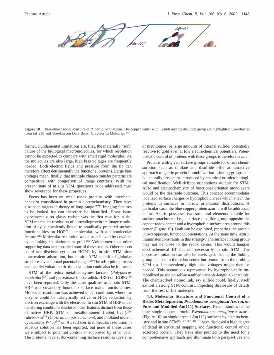
former. Fundamental limitations are, first, the materially “soft”nature of the biological macromolecules, for which resolutioncannot be expected to compare with small rigid molecules. Asthe molecules are also large, high bias voltages are frequentlyneeded. Both electric fields and pressure from the tip cantherefore affect detrimentally the functional proteins. Large biasvoltages mean, finally, that multiple charge-transfer patterns arecompetitive, with congestion of image contrasts. With thepresent state of in situ STM, questions to be addressed mustshow reverence for these properties.
Focus has been on small redox proteins with interfacialbehavior consolidated in protein electrochemistry. They havealso been targets in theory of long-range ET. Imaging featuresto be looked for can therefore be identified. Horse heartcytochromec on glassy carbon was the first case for in situSTM molecular resolution of a metalloprotein.157 Image resolu-tion of cyt c covalently linked to anodically prepared surfacefunctionalities on HOPG is molecular, with a submolecularfeature.215 Molecular resolution was also achieved by covalentcyt c linking to platinum or gold.159 Voltammetric or othersupporting data accompanied none of these studies. Other reportscould not disclose cytc on HOPG by in situ STM afternoncovalent adsorption, but in situ AFM identified globularstructures over a broad potential range.160The adsorption processand parallel voltammetric time evolution could also be followed.
STM of the redox metalloenzymes laccase (PolypherusVersicolor)247 and peroxidase (horseradish, HRP) on HOPG158
have been reported. Only the latter qualifies as in situ STM.HRP was covalently bound to surface oxide functionalities.Molecular resolution was achieved under conditions where theenzyme could be catalytically active in H2O2 reduction byelectron exchange with the electrode. In situ STM of HRP underdenaturing conditions disclosed other features distinct from thoseof native HRP. STM of metallothionein (rabbit liver),214
rubredoxin248 (Clostridium pasteurianum), and thiolated mutantcytochrome P-450249 on Au surfaces to molecular resolution inaqueous solution has been reported, but none of these caseswere subject to potential control or supported by other data.The proteins have sulfur-containing surface residues (cysteine
or methionine) or large amounts of internal sulfide, potentiallyreactive to gold even at low electrochemical potentials. Poten-tiostatic control of proteins with these groups is therefore crucial.
Proteins with given surface groups suitable for direct chemi-sorption such as thiolate and disulfide offer an attractiveapproach to gentle protein immobilization. Linking groups canbe naturally present or introduced by chemical or microbiologi-cal modification. Well-defined orientations suitable for STM/AFM and electrochemistry of functional oriented monolayerswould be the desirable outcome. This concept accommodateslocalized surface charges or hydrophobic areas which attach theproteins to surfaces in narrow orientation distributions. Aparticular case, the blue copper protein azurin, will be addressedbelow. Azurin possesses two structural elements suitable forsurface attachment, i.e., a surface disulfide group opposite thecopper redox center and a hydrophobic surface area around thiscenter (Figure 10). Both can be exploited, preparing the proteinin two opposite, functional orientations. At the same time, azurinilluminates constraints in this strategy. The surface linking groupmay not be close to the redox center. This would hamperelectrochemical ET but not necessarily in situ STM. Theopposite limitation can also be envisaged; that is, the linkinggroup is close to the redox center but remote from the probingSTM tip. Inconveniently high bias voltages might then beneeded. This scenario is represented by hydrophobically im-mobilized azurin on self-assembled variable-length alkanethiols.The chemisorbed atomic link, say sulfide could, finally, itselfexhibit a strong STM contrast, impeding disclosure of detailsfrom the rest of the molecule.
4.4. Molecular Structure and Functional Control of aRedox Metalloprotein, Pseudomonas aeruginosaAzurin, onPure and Modified Au(111) Surfaces.Recent studies of theblue single-copper proteinPseudomonas aeruginosaazurin(Figure 10) on single-crystal Au(111) surfaces by electrochem-istry and in situ STM45-47,117,118,187have disclosed a high degreeof detail in structural mapping and functional control of theadsorbed protein. They have also pointed to the need for acomprehensive approach and illuminate both perspectives and
Figure 10. Three-dimensional structure ofP. aeruginosaazurin. The copper center with ligands and the disulfide group are highlighted. Coordinatesfrom ref 250 and Brookhaven Data Bank. Graphics in Molscript.251
Feature Article J. Phys. Chem. B, Vol. 106, No. 6, 20021145

limitations of in situ STM approaches to electronic propertiesof redox proteins.
4.4.1. Adsorption and ET Function of P. aeruginosa Azurinat Bare Au(111) Electrodes.The molecule is immobilized intwo opposite orientations, at bare and modified single-crystalAu(111)-surfaces (Figure 11). These studies are so far theonlycase for both orientational control and structural mapping of afunctional redox protein under electrochemical potential controlapproachingthe molecular level.Twofeatures are crucial. Oneis the copper atom coordinated to two His, Cys, Met, and Glyclose to one end of the molecule, and the other one is the surfacedisulfide group opposite the copper center. The location of thecopper center enables facile electron exchange with solutereaction partners or electrode surfaces. The two functional unitsare linked byâ strands facilitating electron relay also along thisroute. Prerequisites have been atomically planar electrodesurfaces, a variety of electrochemical techniques combined within situ STM, and reference molecules. Electrochemical andspectroscopic data support in situ STM as a case for mappingof a truly functional redox protein on the solid surface.
Azurin voltammetry at edge-plane pyrolytic graphite isreversible and diffusion-controlled.252 Single-crystal Au(111)-electrode surfaces are suitable for adsorption via the disulfide.ET functionthroughthe protein in the adsorbed state is retained,because of bonding of the sulfur atoms to the gold surface, assupported by several observations:45-47,117
(1) Reductive desorption in the potential range-0.8 to-1.1V (SCE) is clearly apparent in differential pulse voltammetry.This feature is a fingerprint of Au-thiolate bond formation.The equivalent charge comprises 70-80% of a monolayer (7× 10-12 mol cm-2), cf. point 3.
(2) X-ray photoelectron spectroscopy discloses the same twoS2p fingerprint peaks (163.3 and 162.2 eV) for adsorbed azurinas for adsorbed butanethiol and the amino acid cystine. A strongN1s signal (401 eV) is also seen. A sulfur peak at 164.1 eVreflects, interestingly, the two sulfur donor atoms at the coppercenter.
(3) In situ STM shows about 70% monolayer coverage(Figure 12A), cf. point 1, that is stable for weeks. The molecularstructures have the same lateral extension as azurin (3.7( 0.4nm).
These observations offer a coherent view of azurin adsorptionvia surface disulfide. The following other observations testifythat ET function is retained in the adsorbed state:
(4) There are symmetric cathodic and anodic differential pulsesignals in the Cu2+/+-potential region (0.10 V, SCE). The chargeaccords with that for reductive desorption. Such signals appearneither for cysteine and cystine nor for Zn-substituted azurin.
(5) The interfacial ET rate constant at the equilibrium potentialis 30 s-1 determined by impedance spectroscopy. This value issmaller by an order of magnitude than that for azurin adsorbedin the opposite orientation on alkanethiol-modified Au(111)surfaces.
These points show that a stable functional azurin monolayerdirectly bound via surface disulfide has formed on the Au(111)surface and that single-molecule in situ STM of functional azurinhas been achieved.
4.4.2. Adsorption and Interfacial ET of Azurin on Self-Assembled Alkanethiol Monolayers.Azurin adsorption on vari-able-length alkanethiol monolayers on Au(111) by hydrophobicinteraction between the terminal alkanethiol methyl group andthe hydrophobic protein surface around the copper center is analternative adsorption mode.118 The molecular orientation isopposite to adsorption on bare Au(111), with the copper centerfacing the electrode. The most striking difference from adsorp-tion on bare Au(111) is that stable, almost ideal azurinmonolayer voltammetry emerges. The rate constants for electrontunneling between the electrode and the copper center, acrossthe alkanethiols, are significantly higher than for tunnelingthrough the protein in the direct adsorption mode, for similartunneling distances.
The following data disclose a coherent view of adsorptionand ET of fully functional azurin:
(I) Azurin adsorbs in stable submonolayers. Figure 12B showsa representative STM image of the azurin/decanethiol surface.
Figure 11. Two molecular orientations of adsorbed azurin on Au-(111). A. Direct adsorption on bare Au(111) via the disulfide group,with the copper center opposite to the Au(111) surface. B. Adsorptionon an alkanethiol monolayer self-assembled on Au(111) by hydrophobicinteractions with the surface region around the copper center whichfaces the Au(111) surface. From ref 118, with permission.
Figure 12. A. In situ STM image ofP. aeruginosaazurin on bareAu(111). 50 mM ammonium acetate, pH 4.6. Substrate potential-0.1V (SCE),Vbias ) 0.20 V. Tunneling current 0.80 nA. Scan area: 110× 110 nm. B. Ex situ STM image ofP. aeruginosaazurin ondecanethiol self-assembled on Au(111) prepared from 50 mM NH4Ac,pH 4.6. Bias voltage 0.81 V. Tunneling current 0.16 nA. Scan area:100 × 100 nm.
1146 J. Phys. Chem. B, Vol. 106, No. 6, 2002

The high order of the alkanethiol and the uniform dimensionsand distribution of the protein structures over the surface arenotable.
(II) Azurin monolayer voltammetry is reversible, with sym-metric cathodic and anodic peaks and linear peak heightdependence on the scan rate (Figure 13). The peaks separate athigher scan rates as kinetic control takes over.
(III) Azurin monolayer coverage is stable and sensitive. Thecurrent decay on alkanethiols with more than nine carbon atomsis only a few percent after several hundred scans, and clearvoltammetric peaks appear for less than 2% coverage.118
Saturation approaches 70% of a monolayer atµM concentrationsfor alkanethiols between 10 and 14 carbon atoms but is smallerfor shorter chains.
I-III show that azurin adsorbs in a fully functional state withnarrow orientational distribution. A suitable combination of bareand pretreated Au(111) surfaces has thus enabled proteinadsorption in two opposite orientations. Interfacial ET is muchmore facile in the alkanethiol than in the disulfide adsorption
mode. Alkanethiols thus provide a more efficient medium forlong-range ET than adsorbed protein.
Double layer capacitance and reductive desorption exhibitpatterns which both depend characteristically on the alkylthiolatechain length and are affected in subtle ways by azurin adsorp-tion. This is discussed in ref 118. The following other featuresshow that adsorbed azurin monolayers are stable and uniformenough to accord with notions from ET theory:
(IV) The interfacial standard ET rate constants over a broadrange of thiol lengths can be obtained from voltammetric peakseparation and electrochemical impedance spectroscopy.118
Figure 14A shows the dependence of the rate constants on thenumber of carbon atoms. As for capacitance data, a biphasiccorrelation is noted. The rate constants are independent of thethiol length for shorter thiols but follow an exponentialdependence with the decay factor,â ≈ 1.0 per CH2 unit forlonger thiols. This accords with patterns for covalently linkedferrocene on polycrystalline gold106 and electrostatically boundcyt c.123,126The physical cause of the weak dependence at theshorter alkanethiols is elusive. Preorganization123 or penetrationof azurin into the shorter-chain structures118 can be suggested,but such steps would still depend on the overpotential. Theywould be rate determining for shorter, electron tunneling forlonger chains.
(V) The overvoltage dependence of the rate constants isavailable from chronoamperometry.118 Figure 14B shows datafor a 13 CH2 unit alkanethiol. The cathodic and anodic branchesare symmetric and follow a quadratic dependence, with alimiting transfer coefficientR(η ) 0) ) 0.5 and the reorganiza-tion Gibbs free energy of 0.3 eV. There is no sign of electronic
Figure 13. A. Monolayer voltammograms ofP. aeruginosaazurin ondodecanethiol monolayer self-assembled on Au(111). Different scanrates. B. Anodic (Ipa) and cathodic (Ipc) voltammetric current peak heightdependence on the scan rate for the data in A. 50 mM NH4Ac, pH 4.6.
Figure 14. A. Dependence of interfacial ET rate constants at zeroovervoltage on alkane chain length forP. aeruginosaazurin adsorbedon variable-length alkanethiols self-assembled on Au(111). B. Depen-dence of interfacial ET rate constant on the overvoltage forP.aeruginosaazurin adsorbed on tetradecanethiol self-assembled on Au-(111). 50 mM NH4Ac, pH 4.6.
Feature Article J. Phys. Chem. B, Vol. 106, No. 6, 20021147

lability of the metallic electron density caused by varying excesselectrode charges.101,102
These data propose azurin as a “prototype” redox protein inmolecular protein assemblies of high order, stability, andsensitivity. The orientation of the assembled protein moleculescan be controlled, ET function is retained, and overvoltage anddistance dependence can be characterized in detail and framedby electrochemical ET theory.
4.4.3. In Situ STM Tunneling Contrast Variation of P.aeruginosa Azurin.In situ STM spectroscopy (sections 4.2 and4.3) of redox metalloproteins would hold exciting perspectivesfor mapping single-biomolecule electronic properties, but tech-nological and fundamental issues have posed barriers for a realspectroscopic dimension. A “technological” issue is the limitedpotential ranges for ordered layer stability. A broader issue isthat high bias voltages, i.e., a significant fraction of a volt, arefrequently needed. Tunneling through the proteins, assisted bythe redox centers, can be identified but still involves tunnelingsteps over more than 10 Å through metal-free protein matter.Tunneling must therefore be assisted by high driving forces,i.e., bias or overvoltages. This raises, first, the risk of deforma-tion or disassembly of the protein layers by mechanical contactor electric field effects in the tunnel gap. The bias voltages can,second, easily exceed the small solvent reorganization Gibbsfree energy, taking STM spectroscopy into the range where thecurrent variation is weak, cf. section 4.3.2. The following datafor azurin substantiate the importance of these observations.187
Figure 15 shows in situ STM images in the substrate potentialrange from-0.35 to 0 V (SCE), at a fixed bias voltage of+0.2 V. With the copper center close to the tip, this correspondsto -(0.25-0.30) to 0-0.1 V at the copper center, relative tothe equilibrium potential, i.e., a broad potential range on thecathodic side of the equilibrium potential. A dense (≈70%)monolayer is clearly seen in most of the range, with molecularresolution and little contrast variation. The images becomeblurred, and the apparent surface coverage decreases at boththe lower and upper limits of the stability potential range, buta switch back to potentials inside the range identifies the samekind of dense monolayer as before the potential excursions.
This image pattern promptstwo observations. The sequencein Figure 15 parts A and Bresembles, first, the behaviorexpected if a redox process is involved. The upper potentiallimit of the stability range is close to the equilibrium potentialof the copper center and would correspond to the energy leveldiagram in Figure 7. At the positive potential limit, the redoxlevel is vacant and above the Fermi energy of the substrate.Thermal activation of the two-step ET process is then required,and the tunneling current is low. The redox level becomesoccupied at substrate potentials across the equilibrium potential,but because of the relatively high bias voltage, the reduced levelonly relaxes to a position above or close to the Fermi level ofthe tip. An overvoltage range of about the bias voltage, withactivationless high tunneling current weakly dependent on theovervoltage, therefore follows. The current only drops againwhen the overvoltage has assumed a higher negative value wherethe reduced level is trapped well below the Fermi level of thepositively biased tip. This would correspond to the lowerpotential limit of monolayer stability in Figure 15.
The broad potential range of robust high contrast and weakercontrasts on either side could reflect a redox spectroscopicfeature convoluted with the bias voltage. However, althoughattractive, this is almost certainly not theonly cause of thepattern in Figure 15, as indicated by the followingsecondobservation. Figure 15C shows a comparison between the area
scanned during the potential excursion beyond the lowerpotential limit of monolayer stability and a different area notscanned. A similar comparison applies to the positive stabilitypotential limit. Both areas shown are imaged within the stabilitypotential range butafter the potential excursion. The adsorbatepopulation is clearly lower in the area scanned during the wholepotential sweep than in the area which was not scanned. Theprotein monolayer is thus robust to potential variations acrossthe equilibrium potential and to changes in the redox state, butinterference by tip-assisted desorption is apparent in the multiplyscanned area. Such issues must be recognized but are not
Figure 15. Sequences of in situ STM images ofP. aeruginosaazurinadsorbed on bare Au(111) for a range of Au(111) potentials at a fixedbias potential,Vbias ) 0.2 V. 50 mM NH4Ac, pH 4.6. A. The sequencea-f shows images for increasing negative potentials with the values(SCE) -0.15, -0.20, -0.20, -0.25, -0.25, -0.30, and-0.35 V.Azurin is in the reduced state throughout. Groups of azurin molecularstructures are framed. The group of five molecules in the triangularframe in the upper part of e only contains four azurin molecules in f.B. The sequence g-j shows the in situ STM pattern of azurin forincreasing positive potentials. The values (SCE) are-0.15, -0.10,-0.05, and 0.0 V. i and j are around the equilibrium potential ofazurin and show lower apparent coverage and weaker contrasts than atthe lower potentials. C. Comparison of the apparent coverage andcontrast pattern at-0.15 V after potential excursion to the positivelimit. k is the scan area under the tip, and l is a different area of thesame sample.
1148 J. Phys. Chem. B, Vol. 106, No. 6, 2002

prohibitive for in situ imaging of metalloprotein adsorbateelectronic structure changes.
5. Conclusions and Some Perspectives
The discussion above has focused on mechanisms of thefundamental in situ STM process of adsorbate molecules withaccessible redox levels, which can be brought to resonance withthe Fermi levels of the substrate and tip by thermal fluctuationsin the environmental nuclear configurations. The first observa-tion is that different thermally assisted ET channels emerge, inaddition to inherent superexchange via the low-lying fluctuatingredox level. The most important ET channels accord withnotions of coherent and sequential two-step ET and fully diabaticand partially and fully adiabatic two-step ET. Analogues of theseare known in interfacial electrochemical ET, long-range solutionchemical and biological ET, and optical processes such asresonance Raman scattering and hot fluorescence. The in situSTM processes accord with analytical theoretical formalism withfeatures in common with that for these other processes. Pathwaystowards a “unified” theory to cover both the multifarious insitu STM mechanisms and analogous chemical and optical three-level processes are, moreover, visible. The second observationis that the in situ STM configuration with two electrochemicalinterfaces is also an environment for new ETphenomenawithno direct parallel in other ET processes. The most importantnovel observation is apparent in the fully adiabatic limit wherethe strong interactions at both metallic surfaces transmit a largenumber of electrons in a single in situ STM event. This couldresolve issues related to the high observed current densities permolecule mostly observed in STM. The multielectron transfernature of the in situ STM process in the fully adiabatic limit isrooted in the truly vibrationally coherent transmission of theelectrons, whereas the intermediate occupied or vacant redoxlevel relaxes toward vibrational equilibrium. The coherenttransmission of a large number of electrons holds a perspectivein the context of single-molecule device construction, whereswitching and other intended features would be stronglyamplified.
The observations regarding molecular in situ STM mecha-nisms of redox molecules offers novel single-molecule spec-troscopic perspectives, with analogues in other recent single-molecule spectroscopies. This is the third observation. Theovervoltage and the bias voltage are the two external parameters,and the current-voltage band shapes offer detailed insight intothe level energetics and electronic-vibrational coupling. Theenvironmental reorganization (Gibbs free) energy in the tun-neling gap is, however, small compared to this central quantityin electrochemical ET and ET processes in homogeneoussolution. Convolution of the intrinsic spectral band shape withthe bias voltage is therefore important.
Comparison of in situ STM of redox molecules as a single-molecule spectroscopy with other single-molecule spectroscopiesis the fourth observation. In situ STM offers probably the higheststructural resolution and in principle electronic or functionalresolution of small and intermediate-size molecules. Structuralresolution of biological macromolecules is competitive withother single-molecule spectroscopies and complementary byoffering both electronic and topographic resolution. Functionalresolution of single-molecule electronic properties for biologicalmacromolecules must, however, be approached with adequatereverence for the molecular properties. Relatively high biasvoltages are, for example, needed for electronic transmissionthrough the electronically insulating molecular matter. Thisposes problems as to the behavior of the soft molecules under
such drastic conditions and calls for appropriate conduct in theuse of the theoretical frames.
The most important perspective of in situ STM as a single-molecule spectroscopy compared with other single-moleculespectroscopies is that this spectroscopy could offer visualizationand control of the electronic properties of single molecules andbiological macromoleculesin action. Redox metalloenzyme-substrate complex formation must, for example, be accompaniedby significant electronic structural changes compared to the freeenzyme. This is apparent from the widely different interfacialelectrochemical behavior of the enzyme and enzyme-substratecomplex. Such changes could be reflected conspicuously in thein situ STM images. Such imaging and control would referdirectly to the natural aqueous environment of the moleculesbut with reservations as to electrical field effects and spatialconfinements. Mapping and control of single-molecule electronicreactivity would provide exciting insight into single-moleculefunctional individuality (stochastic features) and biomolecularelectronic features in resting and active states. Biomoleculesmay not be suitable as components in working molecular andhybrid-molecular devices, but their multifarious behavior insingle-molecule in situ STM is likely to represent all of thefeatures needed for real device construction and offers guidancein these respects. The most direct device perspective of in situSTM of biological macromolecules is instead that structure andfunctional elements of enzyme electrodes, biosensors, multi-electrode arrays, and other functional biotechnology toward thesingle-molecule level could be disclosed by this spectroscopy.This could again be important for multiple screening ofbiological liquids and in other contexts where such media arein contact with metallic surfaces.
Reported cases of in situ STM spectroscopy are presentlysmall in number. The final, and fifth observation, has been tooverview the properties of some important cases and to showhow their STM behavior can be interfaced with notions of insitu STM theory. Intermediate-size molecules such as FePP andR,ω-polymethylenedithiol-linked bipyridinium are robust enoughto form stable monolayers and exhibit current-voltage spec-troscopy. They are, further, large enough to undertake single-molecule electronic function and still be targets for high-resolution imaging. In situ STM of molecules such as thesetherefore promise to be a spectroscopic tool for single-moleculeproperties on metallic surfaces. Single-molecule behavior ofredox metalloproteins is most accurately and comprehensivelyillustrated byP. aeruginosaazurin. The properties of this classof molecules have pointed to ways of controlled orientation ofwell-defined monolayers of fully functional protein molecules,comprehensive characterization of the electronic properties ofthe immobilized molecules, and single-molecule in situ STM,which approachesa spectroscopic dimension. Azurin on bareand modified Au(111)-surfaces, thus, at the same time, offersbroader use of in situ STM of redox proteins and illuminatesthe prerequisites and limitations of in situ STM as a single-molecule spectroscopy for biological macromolecules.
Acknowledgment. We would like to thank Dr. Esben P.Friis for helpful discussions and for the preparation of Figure1. Financial support from the Danish Technical ScienceResearch Council and the EU Program INTAS (99-1093) isacknowledged.
References and Notes
(1) Janson, J.-C.; Ryde´n, L. Protein Purification, Principles, High-Resolution Methods and Applications; Wiley VCH: Weinheim, Germany,1998.
Feature Article J. Phys. Chem. B, Vol. 106, No. 6, 20021149

(2) Lasic, D. D.Liposomes: From Physics to Applications, 2nd ed.;Elsevier: Amsterdam, The Netherlands, 1995.
(3) Storm, G.; Crommelin, D. J. A.Pharm. Science Technol. Today1998, 1, 19-31.
(4) Telegdi, J.; Keresztes, Z.; Pa´linkas, G.; Kalman, E.; Sand, W.Appl.Phys. AsMater. Sci. Pro.1998, 66, S639-S642.
(5) Walavaara, B.; Askendal, A.; Lundstro¨m, I.; Tengvall, P. J.Biomater. Sci. Polym. Ed.1996, 8, 41-48.
(6) Telegdi, J.; Shaban, A.; Beczner, J.; Keresztes, Z.; Ka´lman, E.Mater. Sci. Forum1998, 289, 77-82.
(7) Losche, M.Curr. Opin. Solid State Mater. Sci.1997, 2, 546-556.(8) Proteins at Interfaces II. Fundamentals and Applications; Horbett,
T. A., Brash, J. L., Eds.; ACS Symp. Ser. 602; American ChemicalSociety: Washington, DC, 1995.
(9) Huetz, P.; Schaaf, P.; Voegel, J.-C.; Mann, E. K.; Miras, B.; Ball,V.; Freund, M.; Cazenave, J.-P.;Proteins at Interfaces II. Fundamentalsand Applications; Horbett, T. A., Brash, J. L., Eds.; ACS Symp. Ser. 602;American Chemical Society: Washington, DC, 1995; pp 334-349.
(10) Service, R. P.Science1998, 282, 396-399.(11) Marshall, A.; Hodgson, J.Nature Biotechnol.1998, 16, 27-31.(12) Protein Array: An Alternative Biomolecular System. Nagayama,
K., Ed.; AdV. Biophys.1997, 34.(13) Willner, I.; Katz, E.Angew. Chem., Int. Ed.2000, 39, 1180-1218.(14) Tengvall, P.; Lundstro¨m, I. Biomaterials1998, 19, 407-422.(15) Lee, J. E.; Saavedra, S. S.Langmuir1996, 12, 4025-4032.(16) Chapters in ref 8.(17) Seibert, M.; Picorel, R.; Kim, J. H.; Cotton, T. M.Methods Enzymol.
1992, 213, 31-42.(18) Dong, S. J.; Niu, J. J.; Cotton, T. M.Methods Enzymol. 1995, 246,
701-732.(19) Fritz, J.; Baller, M. K.; Lang, H. P.; Rothuizen, H.; Vettiger, F.;
Meyer, E.; Gu¨ntherodt, H.- J.; Gerber, C.; Gimzewski, J. K.Science2000,288, 316-318.
(20) Moulin, A. M.; O’Shea, S. J.; Welland, M. E.Ultramicroscopy2000, 82, 23-31.
(21) Tudos, A. J.; Vanderberg, P. J.; Johnson, D. C.Anal. Chem.1995,67, 552-556.
(22) Ryan, M. D.; Bowden, E. F.; Chambers, J. O.Anal. Chem.1994,66, 360R-427R.
(23) Armstrong, F. A.; Heering, H. A.; Hirst,J. Chem. Soc. ReV. 1997,26, 169-179.
(24) Bioelectrochem.; Faraday Discuss.2000, 116.(25) Magonov, S. M.; Whangbo, M.-H.Surface Analysis with STM and
AFM; Verlag Chemie: Weinheim, Germany, 1996.(26) Nanoscale Probes of the Solid/Liquid Interface; Gewirth, A. A.,
Siegenthaler, H., Eds.; Kluwer: Dordrecht, The Netherlands, 1995.(27) Gewirth, A. A.; Niece, B. K.Chem. ReV. 1997, 97, 1129-1162.(28) Electrochemical Nanotechnology; Lorenz, W. J., Plieth, W., Eds.;
Wiley-VCH: Weinheim, Germany, 1998.(29) Moerner, W. E.Science1994, 265, 46-53.(30) Single Molecule Optical Detection, Imaging, and Spectroscopy;
Basche´; Moerner, W. E., Orrit, M., Wild, U. P., Eds.; Verlag Chemie:Munich, 1996.
(31) Tamarat, P.; Maali, A.; Lournis, B.; Orrit, M.J. Phys. Chem. A2000, 104, 1-15.
(32) Ullman, R.; Will, T.; Kolb, D. M.Ber. Bunsen-Ges. Phys. Chem.1995, 99, 1414-1420.
(33) Xie, Z.-X.; Kolb, D. M. J. Electroanal. Chem.2000, 481, 177-182.
(34) Li, C. Z.; Sha, H.; Tao, N. J.Phys. ReV. B 1998, 58, 6775-6778.(35) Li, C. Z.; He, H. X.; Bogozi, A.; Bunch, J. S.; Tao, N. J.Appl.
Phys. Lett.2000, 76, 1333-1335.(36) Mopurgo, A. F.; Marcus, C. M.; Robinson, D. B.Appl. Phys. Lett.
1999, 74, 2084-2086.(37) Molecular Electronics; Mahler, G., May, V., Schreiber, M., Eds.;
M. Dekker: New York, 1996.(38) Molecular Electronics; Jortner, J., Ratner, M., Eds.; Blackwell:
Oxford, U.K., 1997.(39) Molecular Electronics. Science and Technology. Aviram, A., Ratner,
M., Eds.;Ann. N.Y. Acad. Sci.1998, 852.(40) Metal Ions in Biological Systems; Sigel, H., Sigel, A.; Eds.; M.
Dekker: New York, 1991; Vol. 27.(41) Protein Electron Transfer; Bendall, D. S., Ed.; BIOS: Oxford, U.K.,
1996.(42) Electron Transfer from Isolated Molecules to Biomolecules. Jortner,
J., Bixon, M., Eds.;AdVances in Chemical Physics 106 and 107, parts Iand II.; Wiley: New York, 1998.
(43) Kuznetsov, A. M.Charge Transfer in Physics, Chemistry, andBiology; Gordon & Breach: Amsterdam, 1995.
(44) Kuznetsov, A. M.; Ulstrup, J.Electron Transfer in Chemistry andBiology. An Introduction to the Theory; Wiley: Chichester, U.K., 1999.
(45) Friis, E. P.; Andersen, J. E. T.; Madsen, L. L.; Møller, P.; Ulstrup,J. J. Electroanal. Chem.1997, 431, 35-38.
(46) Friis, E. P.; Andersen, J. E. T.; Kharkats, Yu. I.; Kuznetsov, A.M.; Nichols, R. J.; Zhang, J.-D.; Ulstrup, J.Proc. Natl. Acad. Sci. U.S.A.1999, 96, 1379-1384.
(47) Chi, Q.; Zhang, J.; Nielsen, J. U.; Friis, E. P.; Chorckendorff, I.;Canters, G. W.; Andersen, J. E. T.; Ulstrup, J.J. Am. Chem. Soc.2000,122, 4047-4055.
(48) Orrit, M.; Bernard, J.; Personov, R. I.J. Phys. Chem.1993, 97,10290-10298.
(49) Xie, X. S.; Dunn, R. C.Science1994, 265, 361-367.(50) Lu, H. P.; Xie, S.Nature1997, 385, 143-146.(51) Stip, B. C.; Rezaei, M. A.; Ho, W.Phys. ReV. Lett.1999, 82, 1724-
1727.(52) Kuznetsov, A. M.; Ulstrup, J.Chem. Phys.1991, 157, 25-33.(53) Kuznetsov, A. M.; Sommer-Larsen, P.; Ulstrup, J.Surf. Sci.1992,
275, 52-64.(54) Kharkats, Yu. I.; Kuznetsov, A. M.; Ulstrup, J.J. Phys. Chem.1995,
99, 13545-13554.(55) Ha, T.; Glass, J.; Enderle, T.; Chemla, D. S.; Weiss, S.Phys. ReV.
Lett. 1998, 80, 2093-2096.(56) Ha, T.; Ting, A. Y.; Liang, J.; Caldwell, W. B.; Deniz, A. A.;
Chemla, D. S.; Schultz, P. G.; Weiss, S.Proc. Natl. Acad. Sci. U.S.A.1999,96, 893-898.
(57) Lu, H. P.; Lun, L.; Xie, X. S.Science1998, 282, 1877-1882.(58) Vrielink, A.; Lloyd, L. F.; Blow, D. M. J. Mol. Biol. 1991, 219,
533-554.(59) Lu, H. P.; Xie, X. S.J. Phys. Chem. B1997, 101, 2753-2757.(60) Bard, A. J.; Fan, F.-R. F.Acc. Chem. Res.1996, 29, 572-578.(61) Mirkin, M. V.; Horrocks, B. R.Anal. Chim. Acta2000, 406, 119-
146.(62) Fan, F. F.; Bard, A. J.Science1997, 277, 1791-1793.(63) Kuznetsov, A. M.; Ulstrup, J.J. Electroanal. Chem.1993, 362,
149-152.(64) Ingram, R. S.; Hostetler, M. J.; Murray, R. W.; Schaaf, T. G.;
Khoury, J. T.; Whetten, R. L.; Bigioni, T. P.; Guthrie, D. K.; First, P. N.J. Am. Chem. Soc.1997, 119, 9289-9290.
(65) Chen, S.; Murray, R. W.; Feldberg, S. W.J. Phys. Chem. B1998,102, 9998-9907.
(66) Chen, S.; Murray, R. W.J. Phys. Chem. B1999, 103, 9996-10000.(67) Templeton, A. C.; Wuelfing, W. P.; Murray, R. W.Acc. Chem.
Res.2000, 33, 27-36.(68) Datta, S.Electronic Transport in Mesoscopic Systems; Cambridge
University Press: Cambridge, U.K., 1997.(69) Bezryadin, A.; Dekker, C.; Schmid, G.Appl. Phys. Lett.1997, 71,
1273-1275.(70) Porath, D.; Bezryadin, A.; de Vries, S.; Dekker, C.Nature2000,
403, 635-638.(71) Binnig, G.; Quate, C. F.; Gerber, C.Phys. ReV. Lett. 1986, 56,
930-936.(72) Drake, B.; Prater, C. B.; Weisenhorn, A. L.; Gould, S. A.; Albrecht,
T. A.; Quate, C. F.; C Cennell, D. S.; Hansma, H. G.; Hansma, P. K.Science1989, 243, 1586-1589.
(73) Hansma, H. G.; Hoh, J. H.Annu. ReV. Biophys. Biophys. Chem.1994, 23, 115-139.
(74) Rief, M.; Gautel, M.; Oesterhelt, F.; Fernandez, J. M.; Gaub, H. E.Science1997, 276, 1109-1112.
(75) Carrion-Vazques, M.; Oberhauser, A. F.; Fowler, S. B.; Marszalek,P. E.; Broedel, S. E.; Clarke, J.; Fernandez, M.Proc. Natl. Acad. Sci. U.S.A.1999, 96, 3694-3699.
(76) Rief, M.; Oesterhelt, F.; Heymann, B.; Gaub, H. E.Science1997,275, 1295-1297.
(77) Li, H.; Rief, M.; Oesterhelt, F.; Gaub, H. E.AdV. Mater.1998, 3,316-319.
(78) Li, H.; Rief, M.; Oesterhelt, F.; Gaub, H. E.; Zhang, X.; Shen, J.Chem. Phys. Lett.1999, 305, 197-201.
(79) Li, H.; Zhang, W.; Shen, J.; Liu, B.; Gao, C.; Zou, G.Macromol.Rapid Comm.1998, 19, 609-611.
(80) Shao, L.; Tao, N. J.; Leblanc, R. M.Chem. Phys. Lett.1997, 273,37-41.
(81) Cluzel, P.; Lebrun, A.; Heller, C.; Lavery, R.; Viovy, J.-L.;Chatenay, D.; Caron, F.Science1996, 271, 792-794.
(82) Smith, S.; Cui, Y.; Bustamente, C.Science1996, 271, 795-799and references therein.
(83) Lee, G. U.; Chrisey, L. A.; Colton, R. J.Science1994, 266, 771-773.
(84) Strunz, T.; Oroszlan, K.; Scha¨fer, R.; Gunterodt, H.-J.Proc. Natl.Acad. Sci. U.S.A.1999, 96, 1277-11282.
(85) Lee, G. U.; Kidwell, D. A.; Colton, R. J.Langmuir1994, 10, 354-357.
(86) Moy, V. T.; Florin, E.-L.; Gaub, H. E.Science1994, 266, 257-259.
(87) Hinterdorfer, P.; Baumgartner, W.; Gruber, H. J.; Schilcher, K.;Schindler, H.Proc. Natl. Acad. Sci. U.S.A.1996, 93, 3477-3481.
1150 J. Phys. Chem. B, Vol. 106, No. 6, 2002

(88) Ros, R.; Schwesinger, F.; Anselmetti, D.; Kubon, M.; Scha¨fer, R.;Pluckthun, A.; Tiefenauer, L.Proc. Natl. Acad. Sci. U.S.A.1998, 95, 7402-7405.
(89) Schwesinger, F.; Ros, R.; Strunz, T.; Anselmetti, D.; Gu¨nterodt,H.-J.; Honegger, A.; Jermutus, L.; Tiefenauer, L.; Plu¨ckthun, A.Proc. Natl.Acad. Sci. U.S.A.2000, 97, 9972-9977.
(90) Kasas, S.; Thomson, N. H.; Smith, B. L.; Hansma, H. G.; Zhu, X.;Guthold, M. B.; Bustamente, C.; Kool, E. T.; Kashlev, M.; Hansma, P. K.Biochemistry1997, 36, 461-468.
(91) Radmacher, M.; Fritz, M.; Hansma, H. G.; Hansma, P. K.Science1994, 265, 1577-1579.
(92) Balashev, K.; Nielsen, L. K.; Callisen, T. H.; Svendsen, A.;Bjørnholm, T.Probe Micros.2001, 2, 177-185.
(93) Dogonadze, R. R.; Chizmadzhev, Yu. I.Dokl. Akad. Nauk SSSR,Ser. Fiz. Nauk1962, 144, 1077-1080.
(94) Dogonadze, R. R.; Chizmadzhev, Yu. I.Dokl. Akad. Nauk SSSR,Ser. Fiz. Khim. 1962, 145, 848-851.
(95) Marcus, R. A.Can. J. Chem.1958, 37, 155-162.(96) Gerischer, H.Z. Phys. Chem. NF1960, 26, 223-247; Z. Phys.
Chem. NF1960, 26, 325-338.(97) Gurney, R. W.Proc. R. Soc. London A1931, 134, 137-154.(98) Brønsted, J. N.; Pedersen, K. J.Z. Phys. Chem.1924, 108, 185-
235.(99) Frumkin, A. N.Z. Phys. Chem. A1932, 116-118.(100) Dogonadze, R. R. InReactions of Molecules at Electrodes; Hush,
N. S., Ed.; Wiley: New York, 1971; pp 135-227.(101) Kornyshev, A. A.; Kuznetsov, A. M.; Ulstrup, J.J. Phys. Chem.
1994, 98, 3832-3837. Erratum:J. Phys. Chem.1994, 98, 6664.(102) Kornyshev, A. A.; Kuznetsov, A. M.; Nielsen, J. U.; Ulstrup, J.
Phys. Chem. Chem. Phys.2000, 2, 141-144.(103) Finklea, H. A. In Electroanalytical Chemistry; Bard, A. J.,
Rubinshtein, I., Eds.; M. Dekker: New York, 1996; pp 109-335.(104) Miller, C.; Gratzel, M. J. Phys. Chem. 1991, 95, 5225-5233.(105) Chidsey, C. E. D.Science1991, 251, 919-922.(106) Smalley, J. F.; Feldberg, S. W.; Chidsey, C. E. D.; Lindford, M.
R.; Newton, M. D.; Liu, Y.-P.J. Phys. Chem.1995, 99, 13141-13149.(107) Cheng, J.; Saghi-Szabo, Tossell, J. A.; Miller, C. J.J. Am. Chem.
Soc.1996, 118, 680-684.(108) Sek, S.; Misicka, A.; Bilewicz, R.J. Phys. Chem. B2000, 104,
5399-5402.(109) Weber, K.; Hockett, L.; Creager, S. E.J. Phys. Chem. B1997,
101, 8286-8291.(110) Weber, K.; Creager, S. E.J. Electroanal. Chem.1998, 458, 17-
22.(111) Khanova, L. A.; Krishtalik, L. I.Elektrokhimiya1998, 34, 5-13.(112) Finklea, H. O.; Yoon, K.; Chamberlain, E.; Allen, J.; Haddox, R.
J. Phys. Chem. B2001, 105, 3088-3092.(113) Guo, L. H.; Hill, H. A. O.AdV. Inorg. Chem.1991, 36, 341-
370.(114) Hill, H. A. O.; Hunt, N. I.Methods Enzymol. 1993, 227, 501-
522.(115) Bogdanovskaya, V. A.; Tarasevich, M. P.SoV. Sci. ReV. B Chem.
1992, 17, 1-101.(116) Armstrong, F. A.; Wilson, G. S.Electrochim. Acta2000, 45,
2623-2645.(117) Chi, Q.; Zhang, J.-D.; Friis, E. P.; Andersen, J. E. T.; Ulstrup, J.
Electrochem. Comm.1999, 1, 91-96.(118) (a) Chi, Q.; Zhang, J.; Andersen, J. E. T.; Ulstrup, J.J. Phys.
Chem. B2001, 105, 4669-4679. (b) Fristrup, P.; Grubb, M.; Zhang, J.;Christensen, H. E. M.; Hansen, A. M.; Ulstrup, J.J. Electroanal. Chem.2001, 511, 128-133.
(119) Gaigalas, A. K.; Niaura, G.J. Colloid Interface Sci.1997, 193,60-70.
(120) Gaigalas, A. K.; Ruzgas, T.J. Electroanal. Chem.1999, 465, 96-101.
(121) Feng, Z.-Q.; Sagara, T.; Niki, K.Anal. Chem.1995, 67, 3564-3569.
(122) Feng, Z.-Q.; Imabayashi, S.; Kakiuchi, T.; Niki, K.J. Chem. Soc.,Faraday Trans.1997, 93, 1367-1370.
(123) Avila, A.; Gregory, B. W.; Niki, K.; Cotton, T. M.J. Phys. Chem.B 2000, 104, 2759-2766.
(124) Hobara, D.; Niki, K.; Zhou, C. G.; Chumanov, G.; Cotton, T. M.Colloids Surf. AsPhys. Chem. Eng. Asp.1994, 93, 241-250.
(125) Lecomte, S.; Wackerbarth, H.; Soulimane, T.; Buse, G.; Hilde-brandt, P.J. Am. Chem. Soc.1998, 120, 7381-7382.
(126) Murgida, D. H.; Hildebrandt, P.J. Am. Chem. Soc.2001, 123,4062-4068.
(127) Song, S.; Clark, R. A.; Bowden, E. F.; Tarlov, M. J.J. Phys. Chem.1993, 97, 6564-6572.
(128) Dogonadze, R. R.; Kuznetsov, A. M.Prog. Surf. Sci.1975, 6,1-42.
(129) Kuznetsov, A. M.; Ulstrup, J.Phys. Chem. Chem. Phys.1999, 1,5587-5592.
(130) Kuznetsov, A. M.; Ulstrup, J.Electrochim. Acta2001, 46, 3325-3333.
(131) (a) Dogonadze, R. R.; Kuznetsov, A. M.; Vorotyntsev, M. A.J.Electroanal. Chem.1970, 25, App. 17-19. (b) Dogonadze, R. R.;Kuznetsov, A. M.; Vorotyntsev, M. A.Croat. Chim. Acta1972, 44, 257-273.
(132) Hush, N. S.Trans. Faraday Soc.1961, 57, 557-580.(133) Hush, N. S.J. Electroanal. Chem.1999, 470, 170-195.(134) Muscat, J. P.; Newns, D. M.Prog. Surf. Sci. 1978, 9, 1-43.(135) Schmickler, W.J. Electroanal. Chem.1986, 204, 31-43.(136) (a) Schmickler, W.J. Electroanal. Chem.1980, 113, 159-170.
(b) Schmickler, W.J. Electroanal. Chem.1982, 137, 189-198.(137) Kuznetsov, A. M.; Ulstrup, J.J. Electroanal. Chem.1985, 195,
1-19.(138) Sebastian, K. L.J. Chem. Phys.1989, 90, 5056-5067.(139) Smith, B. B.; Hynes, J. T.J. Chem. Phys.1993, 99, 6517-6530.(140) (a) Boroda, Yu. G.; Voth, G. A.J. Chem. Phys.1996, 104, 6168-
6183. (b) Boroda, Yu. G.; Voth, G. A.J. Electroanal. Chem.1998, 450,95-107.
(141) (a) Kuznetsov, A. M.; Medvedev, I. G.J. Electroanal. Chem.2001,502, 15-35. (b) Kuznetsov, A. M.; Medvedev, I. G.Russ. J. Electrochem.2001, 37, 335-340. (c) Kuznetsov, A. M.; Medvedev, I. G.Russ. J.Electrochem.2001, 37, 341-352. (d) Kuznetsov, A. M.; Medvedev, I. G.Russ. J. Electrochem.2001, 37, 567-577.
(142) Kolb, D. M. InStructure of Electrified Interfaces; Lipkowski, J.,Ross, P. N., Eds.; Verlag Chemie: Weinheim, Germany, 1993; pp 65-102.
(143) Magnussen, O. M.; Mo¨ller, F.; Vogt, R. J.; Behm, R. J. InElectrochemical Nanotechnology; Lorenz, W. J., Plieth, W., Eds.; VerlagChemie: Weinheim, Germany, 1998; pp 159-169.
(144) Kornyshev, A. A.; Vilfan, I.Electrochim. Acta1995, 40, 109-127.
(145) Bunge, E.; Port, S. N.; Roelfs, B.; Meyer, H.; Baumga¨rtel, H.;Schiffrin, D. J.; Nichols, R. J.Langmuir1997, 13, 85-90.
(146) Poirier, G. E.Chem. ReV. 1997, 97, 1117-1127.(147) Arce, F. T.; Vela, M. E.; Salvarezza, R. C.; Arvia, A. J.J. Chem.
Phys.1998, 109, 5703-5706.(148) Nielsen, J. U.; Esplandiu, M. J.; Kolb, D. M.Langmuir2001, 17,
3454-3459.(149) Tao, N. J.; DeRose, J. A.; Lindsay, S. M.J. Phys. Chem.1993,
97, 910-919.(150) Itaya, K.Prog. Surf. Sci.1998, 58, 121-248.(151) Zhang, J.; Wan, L.-J.; Itaya, K.J. Electroanal. Chem. 2001, 500,
156-162.(152) Wandlowski, T.; Lampner, D.; Lindsay, S. M.J. Electroanal.
Chem.1996, 404, 215-226.(153) Dretschkow, T.; Wandlowski, T.J. Electroanal. Chem.1999, 467,
407-416.(154) Cunha, F.; Tao, N. J.; Wang, X. W.; Jin, Q.; Duong, B.; D’Agnese,
J. Langmuir1996, 12, 6410-6418.(155) Wandlowski, T.J. Electroanal. Chem.1995, 395, 83-89.(156) Lindsay, S. M.; Tao, N. J.; DeRose, J. A.; Orlen, P. I.; Lyubshenko,
Y. L.; Harrington, R. E.; Shlakhtenko, L.Biophys. J.1992, 61, 1570-1584.
(157) Andersen, J. E. T.; Møller, P.; Pedersen, M. V.; Ulstrup, J.Surf.Sci.1995, 325, 193-205.
(158) Zhang, J.; Chi, Q.; Dong, S.; Wang, E.Bioelectrochem. Bioenerg.1996, 39, 267-274.
(159) Andersen, J. E. T.; Olesen, K. G.; Danilov, A. I.; Foverskov, C.E.; Møller, P.; Ulsrup, J.Bioelectrochem. Bioenerg.1997, 44, 57-63.
(160) Boussaad, S.; Tao, N. J.; Arechabaleta, R.Chem. Phys. Lett.1997,280, 397-403.
(161) Boussaad, S.; Tao, N. J.J. Am. Chem. Soc.1999, 121, 4510-4515.
(162) Danilov, A. I.Russ. Chem. ReV. 1995, 64, 767-781.(163) Gewirth, A. A.; Niece, B. K.Chem. ReV. 1997, 97, 1129-1162.(164) Schmickler, W. InImaging of Surfaces and Interfaces; Lipkowski,
J., Ross, P. N., Eds.; Wiley-VCH: New York, 1999; pp 305-338.(165) Gurevich, Yu. Ya.; Kuznetsov, A. M.SoV. Phys. Solid State1975,
17, 2076-2078.(166) Sumetskij, M.; Kornyshev, A. A.Phys. ReV. B 1993, 48, 17493-
17506.(167) Kornyshev, A. A.J. Electroanal. Chem.1994, 376, 9-14.(168) Pan, J.; Jing, T. W.; Lindsay, S. M.J. Phys. Chem. 1994, 98,
4205-4208.(169) Schmickler, W.Surf. Sci.1995, 335, 416-421.(170) Mosyak, A.; Nitzan, A.; Kosloff, R.J. Chem. Phys.1996, 104,
1549-1559.(171) Benjamin, I.; Evans, D.; Nitzan, A.J. Chem. Phys.1997, 106,
6647-6654.(172) Lang, N. D.Solid State Phys. 1973, 28, 225-299.(173) Sautet, P.Chem. ReV. 1997, 97, 1097-1116.(174) Bardeen, J.Phys. ReV. Lett. 1961, 6, 57-59.
Feature Article J. Phys. Chem. B, Vol. 106, No. 6, 20021151

(175) (a) Tersoff, J.; Hamann, D. R.Phys. ReV. Lett.1983, 50, 1998-2001. (b) Tersoff, J.; Hamann, D. R.Phys. ReV. B 1985, 31, 805-813.
(176) (a) Lang, N.Phys. ReV. Lett. 1985, 55, 230-233. (b) Lang, N.Phys. ReV. Lett. 1986, 56, 1164-1167.
(177) (a) Lang, N.Phys. ReV. B 1986, 34, 5947-5950. (b) Lang, N.Phys. ReV. B 1987, 36, 8173-8176.
(178) Kunitake, M.; Batina, N.; Itaya, K.Langmuir1995, 11, 2337-2340.
(179) Ou-Yang, H.; Marcus, R. A.; Ka¨llebring, B.J. Chem. Phys.1994,100, 7814-7824.
(180) Claypool, C. L.; Fagliani, F.; Goddard, W. A., III.; Gray, H. B.;Lewis, N. S.; Marcus, R. A.J. Phys. Chem. B1997, 101, 5978-5995.
(181) Fagliani, F.; Claypool, C. L.; Lewis, N. S.; Goddard, W. A., III.J. Phys. Chem. B1997, 101, 5996-6020.
(182) Schmickler, W.; Widrig, C.J. Electroanal. Chem.1992, 336, 213-221.
(183) Friis, E. P.; Kharkats, Yu. I.; Kuznetsov, A. M.; Ulstrup, J.J.Phys. Chem. A1998, 102, 7851-7859.
(184) Kuznetsov, A. M.; Ulstrup, J.J. Phys. Chem. A2000, 104, 11531-11540.
(185) Kuznetsov, A. M.; Ulstrup, J.Probe Micros.2001, 2, 187-200.(186) Friis, E. P.; Andersen, J. E. T.; Madsen, L. L.; Møller, P.; Nichols,
R. J.; Olesen, K. G.; Ulstrup, J.Electrochim. Acta1998, 43, 2889-2897.(187) Zhang, J.; Kuznetsov, A. M.; Ulstrup, J. In preparation.(188) Kuznetsov, A. M.; Schmickler, W.Chem. Phys. Lett.2000, 327,
314-318.(189) Sumi, H.J. Phys. Chem. B1998, 102, 1833-1844.(190) Nanostructures Based on Molecular Materials; Gopel, W., Ziegler,
C., Eds.; Verlag Chemie: Weinheim, Germany, 1992.(191) Molecular Electronics- Science and Technology; Aviram, A.,
Ed.; Engineering Foundation: New York, 1989.(192) Metzger, R. M.; Chen, B.; Ho¨pfner, U.; Lakshmikanthan, M. V.;
Vuillaume, D.; Kawai, T.; Wu, X.; Tachibana, H.; Hughes, T. V.; Sakurai,H.; Baldwin, J. W.; Hosch, C.; Cava, M. P.; Brehmer, L.; Ashwell, G. J.J.Am. Chem. Soc.1997, 119, 10455-19466.
(193) Metzger, R. M.Acc. Chem. Res.1999, 32, 950-957.(194) Collier, C. P.; Wong, E. W.; Belohradsky´, M.; Raymo, F. M.;
Stoddart, J. F.; Kuekes, P. J.; Willams, R. S.; Heath, J. R.Science1999,285, 391-394.
(195) Kuznetsov, A. M.; Ulstrup, J.J. Chem. Phys.2002, in press.(196) Yoshimoto, S.; Hirakawa, N.; Nishiyama, K.; Taniguchi, I.
Langmuir2000, 16, 4399-4404.(197) Tao, N. J.; Shi, Z.Surf. Sci.1994, 321, L149-L156.(198) Tao, N. J.; Shi, Z.J. Phys. Chem.1994, 98, 7422-7426.(199) Leatherman, G.; Durantini, E. N.; Gust, D.; Moore, T. A.; Moore,
A. L.; Stone, S.; Zhou, Z.; Rez, P.; Liu, Y. Z.; Lindsay, S. M. J. Phys.Chem. B1999, 103, 4006-4010.
(200) Richard, K. M.; Gewirth, A. A.J. Phys. Chem.1995, 99, 12288-12293.
(201) Zhang, J.; Chi, Q.; Nielsen, J. U.; Friis, E. P.; Andersen, J. E. T.;Ulstrup, J.Langmuir2000, 16, 7229-7237.
(202) Tao, N. J.; Cardenas, G.; Cunha, F.; Shi, Z.Langmuir1995, 11,4445-4448.
(203) Tao, N. J.Phys. ReV. Lett. 1996, 76, 4066-4070.(204) Han, W.; Durantini, E. N.; Moore, T. A.; Moore, A. L.; Gust, D.;
Leatherman, G.; Deely, G. R.; Tao, N.; Lindsay, S. M.J. Phys. Chem.1997, 101, 10719-10725.
(205) Snyder, S. R.; White, H. S.J. Electroanal. Chem.1995, 394, 177-185.
(206) Gimzewski, J. K.; Joachim, C.; Schlittler, R. R.; Langlais, V.;Tang, H.; Johannsen, I.Science1998, 281, 531-533.
(207) Mazur, U.; Hipps, K. W.J. Phys. Chem.1995, 99, 6684-6688.(208) Hipps, K. W.; Lu, X.; Wang, X. D.; Mazur, U.J. Phys. Chem.
1996, 100, 11207-11220.(209) Mazur, U.; Hipps, K. W.J. Phys. Chem. B1999, 103, 9721-
9727.(210) Kaba, M. S.; Song, I. K.; Barteau, M. A.J. Phys. Chem.1996,
100, 19577-19581.(211) Kaba, M. S.; Song, I. K.; Barteau, M. A.J. Vac. Sci. Technol. A
1997, 15, 1299-1304.(212) Gittins, D. I.; Bethell, D.; Schiffrin, D. J.; Nichols, R. J.Nature
2000, 40, 67-69.(213) Davis, J. J.; Halliwell, C. M.; Hill, H. A. O.; Canters, G. W.; van
Amsterdam, M. C.; Verbeet, M. P.New J. Chem.1998, 1119-1123.(214) Davis, J. J.; Hill, H. A. O.; Kurz, A.; Jacob, C.; Maret, W.; Vallee,
B. L. Phys. Chem. Comm.1998, Article 2.
(215) Zhang, B.; Wang, E.Nanobiology1996, 4, 85-91.(216) Khomutov, G. B.; Belovolova, L. V.; Gubin, S. P.; Khanin, V.
V.; Obydenov, A. Yu.; Soldatov, E. S.; Trifonov, A. S.Bioelectrochem.Bioeng.2001, in press.
(217) Tao, N. J.; Li, C. Z.; He, H. X.J. Electroanal. Chem.2000, 492,81-93.
(218) Schmickler, W.; Tao, N. J.Electrochim. Acta1997, 42, 2809-2815.
(219) Lu, T. X.; Reimers, J. R.; Crossley, M. J.; Hush, N. S.J. Phys.Chem. 1994, 98, 11878-11884.
(220) Reimers, J. R.; Hall, L. E.; Crossley, M. J.; Hush, N. S.J. Phys.Chem. A1999, 103, 4385-4397.
(221) Martin, A. S.; Sambles, J. R.; Ashwell, G. J.Phys. ReV. Lett.1993,70, 218-221.
(222) Dhirani, A.; Lin, P.-H.; Guyot-Sionnest, P.; Zehner, R. W.; Sita,L. R. J. Chem. Phys.1997, 106, 5249-5253.
(223) Datta, S.; Tian, W.; Hong, S.; Reifenberger, R.; Henderson, J. I.;Kubiak, C. P.Phys. ReV. Lett. 1997, 79, 2530-2533.
(224) Collins, P. G.; Zettl, A.; Bando, H.; Thess, A.; Smalley, R. E.Science1997, 278, 100-103.
(225) Semenov, A.; Spatz, J. P.; Møller, M.; Lehn, J.-M.; Sell, B.;Schubert, W.; Schubert, U. S.Angew. Chem., Int. Ed.1999, 38, 2547-2550.
(226) Baxter, P. N. W.; Lehn, J.-M.; Baum, G.; Fenske, D.Chem.-Eur. 1999, 5, 102-112.
(227) Lehn, J.-M.Supramolecular Chemistry; Verlag Chemie: Wein-heim, Germany, 1995.
(228) Balzani, V.; Campagna, S.; Denti, G.; Juris, A.; Serroni, S.;Venturi, M. Acc. Chem. Res.1998, 31, 26-34.
(229) Sessoli, R.; Gatteschi, D.; Caneschi, A.; Novak, M. A.Nature1993, 365, 141-143.
(230) Fujihira, M.Ann. N.Y. Acad. Sci.1998, 852, 306-329.(231) Burrell, A. K.; Wasielewski, M. R.J. Porph. Phthaloc. 2000, 4,
401-406 and references therein.(232) Reed, M. A.; Zhou, C.; Muller, C. J.; Burgin, T. P.; Tour, J. M.
Science1997, 278, 252-254.(233) Chen, J.; Reed, M. A.; Rawlett, A. M.; Tour, J. M.Science1999,
286, 1550-1552.(234) Ashwell, G. J.Thin Solid Films1990, 186, 155-165.(235) Lee, G.; Evans, D. F.; Elings, V.; Edstrom, R. D.J. Vac. Sci.
Technol. B1991, 9, 1236-1241.(236) Feng, L.; Hu, C. Z.; Andrade, J. D.J. Colloid Interface Sci.1988,
126, 650-653.(237) Leatherbarrow, R. J.; Stedman, M.; Wells, T. N. C.J. Mol. Biol.
1991, 221, 361-365.(238) Welland, M. E.; Miles, M. J.; Lambert, N.; Morris, V. J.; Coombs,
J. H.; Pethica, J. B.Int. J. Biol. Macromol.1989, 11, 29-32.(239) Nawaz, Z.; Cataldi, T. R. I.; Knall, J.; Somekh, R.; Pethica, J. B.
Surf. Sci.1992, 265, 139-155.(240) Zhang, J.-D.; Chi, Q.; Dong, S.-J.; Wang, E.J. Electroanal. Chem.
1994, 379, 535-539.(241) Leggett, G. J.; Davies, M. C.; Jackson, D. E.; Roberts, C. J.;
Tendler, S. J. B.; Williams, P. M.J. Phys. Chem.1993, 97, 8852-8854.(242) Zhang, J.-D.; Chi, Q.-J.; Dong, S.-J.; Wang, E.-K.Electroanalysis
1998, 10, 738-746.(243) Chi, Q.; Zhang, J.; Dong, S.; Wang, E.J. Chem. Soc., Faraday
Trans.1994, 90, 2057-2060.(244) Haggerty, L.; Lenhoff, A. M.Biophys. J.1993, 64, 886-895.(245) Diemann, E.; Branding, A.; Mu¨ller, A. Naturwissenschaften1992,
79, 267-268.(246) Lee, I.; Lee, R. J.; Warmack, R. J.; Allison, D. P.; Greenbaum,
E. Proc. Natl. Acad. Sci. U.S.A.1995, 92, 1965-1969.(247) Andersen, J. E. T.; Møller, P.; Thuesen, M. H.; Ulstrup, J.
Electrochim. Acta1996, 41, 2005-2010.(248) Mukhopadhyay, R.; Davis, J. J.; Kyritsis, P.; Hill, H. A. O.; Meyer,
J. J. Inorg. Biochem.2000, 78, 251-254.(249) Davis, J. J.; Djuricic, D.; Lo, K. K. W.; Wallace, E. N. K.; Wong,
L. L.; Hill, H. A. O. Faraday Dicuss.2000, 116, 15-22.(250) Nar, H.; Messerschmidt, A.; Huber, R.J. Mol. Biol. 1991, 221,
765-772.(251) Kraulis, P.J. Appl. Crystallogr. 1991, 24, 946-950.(252) Farver, O.; Zhang, J.; Chi, Q.; Pecht, I.; Ulstrup, J.Proc. Natl.
Acad. Sci. U.S.A.2001, 98, 4426-4430.
1152 J. Phys. Chem. B, Vol. 106, No. 6, 2002


