Embed Size (px)
Citation preview
Emergency Medicine:Avoiding the Pitfalls andImproving the Outcomes
Edited by
Amal Mattu, MDProgram Director, Emergency Medicine ResidencyCo-Program Director, Emergency Medicine/InternalMedicine Combined ResidencyAssociate Professor of Emergency MedicineUniversity of Maryland School of MedicineBaltimore, Maryland
and
Deepi Goyal, MDAssociate Program DirectorMayo Emergency Medicine ResidencyAssistant Professor of Emergency MedicineMayo clinic College of MedicineRochester, Minnesota


Emergency Medicine: Avoiding the Pitfalls andImproving the Outcomes

This text is dedicated to the residents and faculty in
Emergency Medicine at the University of Maryland Medical
Center for providing the inspiration for this work; to my col-
league Deepi Goyal, a true friend, scholar, and role model; to
Mary Banks and Blackwell Publishing for supporting this
work; to my children Nikhil, Eleena, and Kamran for provid-
ing the greatest inspiration in my life; and to my wife Sejal
for her incredible support of all that I do, and without whom
none of this would be possible.
Amal Mattu, MD
This text is dedicated to all those whose support and inspira-
tion brighten my every day: to the Emergency Medicine res-
idents, faculty, and nurses at Mayo Medical Center whose
curiosity, patience, and passion benefits all those they touch;
to Amal Mattu, a colleague, teacher, and mentor who I feel
truly fortunate to call a friend; and most importantly to
my wife Bhargavi and my children Kiran and Seeta whose
unfailing support and understanding have made this all
possible.
Deepi Goyal, MD

Emergency Medicine:Avoiding the Pitfalls andImproving the Outcomes
Edited by
Amal Mattu, MDProgram Director, Emergency Medicine ResidencyCo-Program Director, Emergency Medicine/InternalMedicine Combined ResidencyAssociate Professor of Emergency MedicineUniversity of Maryland School of MedicineBaltimore, Maryland
and
Deepi Goyal, MDAssociate Program DirectorMayo Emergency Medicine ResidencyAssistant Professor of Emergency MedicineMayo clinic College of MedicineRochester, Minnesota

© 2007 by Blackwell Publishing
BMJ Books is an imprint of the BMJ Publishing Group Limited, used under
licence
Blackwell Publishing, Inc., 350 Main Street, Malden, Massachusetts
02148-5020, USA
Blackwell Publishing Ltd, 9600 Garsington Road, Oxford OX4 2DQ, UK
Blackwell Publishing Asia Pty Ltd, 550 Swanston Street, Carlton, Victoria 3053,
Australia
The right of the Author to be identified as the Author of this Work has been
asserted in accordance with the Copyright, Designs and Patents Act 1988.
All rights reserved. No part of this publication may be reproduced, stored in a
retrieval system, or transmitted, in any form or by any means, electronic,
mechanical, photocopying, recording or otherwise, except as permitted by the
UK Copyright, Designs and Patents Act 1988, without the prior permission of
the publisher.
First published 2007
1 2007
Library of Congress Cataloging-in-Publication Data
Emergency medicine: avoiding the pitfalls and improving the outcomes/edited
by Amal Mattu and Deepi Goyal.
p.; cm.
Includes bibliographical references.
ISBN-13: 978-1-4051-4166-6 (pbk.:alk. paper)
1. Emergency medicine. I. Mattu, Amal. II. Goyal, Deepi.
[DNLM: 1. Emergencies. 2. Emergency Medicine. WB 105 E541 2007]
RC86.7.E4444 2007
616.02’5–dc22
2006027486
ISBN: 978-1-4051-4166-6
A catalogue record for this title is available from the British Library
Set in 9/12 pt Meridien Roman by Charon Tec Ltd (A Macmillan Company),
Chennai, India
www.charontec.com
Printed and bound in Singapore by Markono Print Media Pte Ltd
Commissioning Editor: Mary Banks
Editorial Assistant: Victoria Pittman
Development Editor: Lauren Brindley
Production Controller: Rachel Edwards
For further information on Blackwell Publishing, visit our website:
http://www.blackwellpublishing.com
The publisher’s policy is to use permanent paper from mills that operate a
sustainable forestry policy, and which has been manufactured from pulp
processed using acid-free and elementary chlorine-free practices. Furthermore,
the publisher ensures that the text paper and cover board used have met
acceptable environmental accreditation standards.
Blackwell Publishing makes no representation, express or implied, that the drug
dosages in this book are correct. Readers must therefore always check that any
product mentioned in this publication is used in accordance with the prescribing
information prepared by the manufacturers. The author and the publishers do
not accept responsibility or legal liability for any errors in the text or for the
misuse or misapplication of material in this book.

Contributors vii
Preface ix
1. Evaluation and Management of Patients with
Chest Syndromes 1
Richard A. Harrigan & Michael A. DeAngelis
2. Management of the Dyspneic Patient in
the ED 17
Jairo I. Santanilla & Peter M.C. DeBlieux
3. Evaluation and Management of the Patient with
Abdominal Pain 25
Joseph P. Martinez
4. Management of Patients with Acute Back Pain in
the ED 33
Michael E. Winters
5. Headache Management 39
Stephen Schenkel
6. Evaluation and Management of the Patient with
Neck Pain 46
Joshua Broder & Anita L’Italien
7. Trauma Management in the ED 55
David E. Manthey & Bret A. Nicks
8. Management of Infectious Diseases 63
David J. Karras, Wayne A. Satz & Jeffrey Barrett
9. Wound Care in Emergency Medicine 72
Siamak Moayedi & Mercedes Torres
10. Management of the Pregnant Patient in
the ED 79
Kristine Thompson
11. Pediatric Care in the ED 85
Ghazala Sharieff
12. Management of Hematology/Oncology Patients
in the ED 93
Robert L. Rogers
13. Management of Intoxicated/Violent
Patients 99
Yesha Patel & Gus M. Garmel
Index 109
Contents
v


Jeffrey Barrett, MD
Assistant Professor
Department of Emergency Medicine
Temple University School of Medicine
Philadelphia, PA
Joshua Broder, MD, FACEP
Associate Residency Program Director
Assistant Clinical Professor
Division of Emergency Medicine
Department of Surgery
Duke University Medical Center
Durham, NC
Michael A. DeAngelis, MD
Assistant Professor of Emergency Medicine
Department of Emergency Medicine
Temple University School of Medicine
Philadelphia, PA
Peter M. C. DeBlieux, MD
Professor of Clinical Medicine
Louisiana State University School of Medicine in
New Orleans
New Orleans, LA
Gus M. Garmel, MD, FACEP, FAAEM
Co-Program Director, Stanford/Kaiser EM Residency
Clinical Associate Professor
Emergency Medicine (Surgery)
Stanford University School of Medicine
and
Senior Emergency Physician
The Permanente Medical Group
Kaiser Santa Clara, CA
Richard A. Harrigan, MD
Associate Professor of Emergency Medicine
Department of Emergency Medicine
Temple University Hospital and School of Medicine
Philadelphia, PA
David J. Karras, MD, FACEP, FAAEM
Professor of Emergency Medicine
Associate Chair for Academic Affairs
Department of Emergency Medicine
Temple University School of Medicine
Philadelphia, PA
Anita L’Italien, MD
Chief Resident
Department of Emergency Medicine
University of North Carolina at Chapel Hill
Chapel Hill, NC
David E. Manthey, MD
Director, Undergraduate Medical Education
Associate Professor
Wake Forest University School of Medicine
Winston-Salem, NC
Joseph P. Martinez, MD
Assistant Professor of Emergency Medicine
Assistant Dean for Student Affairs
University of Maryland School of Medicine
Baltimore, MD
Siamak Moayedi, MD
Assistant Professor
Department of Emergency Medicine
University of Maryland School of Medicine
Baltimore, MD
Bret A. Nicks, MD
Assistant Medical Director
Wake Forest University School of Medicine
Winston-Salem, NC
Yesha Patel, MD
Assistant Professor, Emergency Medicine
Tufts University School of Medicine
Tufts-New England Medical Center
Boston, MA
Contributors
vii

Robert L. Rogers, MD, FAAEM, FACEP, FACP
Assistant Professor of Emergency Medicine
Director of Undergraduate Medical Education
The University of Maryland School of Medicine
Baltimore, MD
Jairo I. Santanilla, MDChief Resident
Louisiana State University School of Medicine in New Orleans
New Orleans, LA
Wayne A. Satz, MDAssociate Professor
Department of Emergency Medicine
Temple University
Philadelphia, PA
Stephen Schenkel, MD, MPPAssistant Professor
University of Maryland School of Medicine
Baltimore, MD
Ghazala Sharieff, MDDirector of Pediatric Emergency Medicine
Palomar-Pomerado Health System/California Emergency
Physicians
and
Associate Clinical Professor
Children’s Hospital and Health Center
University of California
San Diego, CA
Mercedes Torres, MDChief Resident
Department of Emergency Medicine
University of Maryland School of Medicine
Baltimore, MD
Kristine Thompson, MDClinical Instructor, Emergency Medicine
Department for Emergency Medicine
Mayo Clinic
Jacksonville, FL
Michael E. Winters, MDAssistant Professor of Emergency Medicine and Medicine
Program Director, Combined Emergency Medicine/Internal
Medicine Training Program
University of Maryland School of Medicine
Baltimore, MD
viii | Contributors

Emergency Medicine is a high-risk specialty. The seasoned
practitioner is well aware that that even the most mundane
of patients may, at any moment, be on the brink of a cata-
strophic outcome. The emergency physician must, therefore,
be ever wary of these “disasters in waiting.” It seems that
most pitfalls in emergency medicine, many of which result in
medicolegal consequences, occur not purely due to a lack of
knowledge but rather to simply “letting one’s guard down.”
This text was created in order to focus the attention of emer-
gency physicians on these common pitfalls. The text is not
comprehensive in scope, but rather it focuses the readers’
attention on an assortment of chief complaints and patient
groups that are frequently encountered in Emergency
Medicine. The authors of each chapter were chosen for their
expertise in the respective topics, and they have focused their
text on potential pitfalls in everyday clinical practice that
represent high risk for patient morbidity, mortality, and litiga-
tion. At the end of each chapter, they have provided impor-
tant pearls for improving patient outcomes. Although the text
is primarily intended for use by the seasoned practitioner,
physicians-in-training should find many teaching points that
will assist their education as well.
Finally, we hope that the reader will not relegate this text to
the bookshelf alongside other voluminous, dusty reference
books. Rather, we hope that the reader finds the text of
appropriate size and practicality to read cover-to-cover and to
use frequently during everyday practice in the Emergency
Department and other acute-care settings.
Amal Mattu, MD
Deepi Goyal, MD
Preface
ix

Introduction
Chest pain is a common emergency department (ED) com-
plaint with a well-known differential diagnosis. Yet compared
to the abdomen, the chest contains relatively few structures
(e.g., the heart, the lungs, the great vessels, the esophagus) to
consider as the source of the complaint when evaluating a
patient with chest pain. In these few structures, however,
there exists the potential for several life-threatening maladies,
some of which unfortunately occur rather commonly. In
patients with chest pain, initial attention is often devoted to
establishing the presence or absence of acute coronary syn-
drome (ACS), but indeed there are several other syndromes of
critical importance and clinical relevance to consider. In this
chapter, we consider six pitfalls related to ACS, followed by a
variety of pitfalls related to other diseases of the chest: aortic
dissection (AD), pulmonary embolism (PE), pericarditis, pneu-
mothorax, esophageal rupture, and finally, herpes zoster.
Pitfall | Over-reliance on the classic presenceof chest pain for the diagnosis of acutemyocardial infarction (MI)
Although chest pain has long been considered the hallmark
clinical feature of acute myocardial infarction (MI), it is impor-
tant to recognize that the absence of chest pain in no way
excludes the diagnosis. In a large observational study, Canto et
al. examined the presenting complaints of nearly 435,000
patients with confirmed MI enrolled in the National Registry of
Myocardial Infarction 2 (NRMI-2) database and found that
one-third of the patients presented to the hospital without
chest pain [1]. Other studies have reported similar findings. In
one study, over 20% of 2096 patients diagnosed with acute MI
presented with symptoms other than chest pain [2]. In another
smaller study, nearly half (47%) of 721 patients hospitalized for
acute MI presented to the ED without chest pain [3]. Risk fac-
tors associated with the absence of chest pain included age,
female gender, non-white race, diabetes mellitus, and a prior
history of congestive heart failure or stroke (see Table 1.1) [1].
In the elderly population, chest pain is reported less fre-
quently according to the NRMI-2 database, patients experi-
encing an acute MI without chest pain are, on average, 7
years older (74 versus 67 years) [1]. Uretsky et al. reported a
mean age of 69.1 years in those patients without chest pain
as compared to 58.7 years in those with chest pain [4].
Under the age of 85, chest pain is still present in the major-
ity of patients but other non-pain symptoms (referred to as
“anginal equivalents”) such as shortness of breath, syncope,
weakness, and confusion are common. Over the age of 85,
60–70% of patients with acute MI present without chest
pain; shortness of breath is the most frequent anginal equiv-
alent in this population [5].
Women are more likely than men to experience acute MI
without chest pain [1–3, 6]. In one study, women over the
age of 65 were the most prevalent group to experience acute
MI without chest pain [6]. In another study of 515 women
surveyed after experiencing an acute MI, only 57% reported
chest pain at the time of their MI. The most frequent anginal
equivalents reported were shortness of breath (58%), weak-
ness (55%), unusual fatigue (43%), cold sweats (39%), and
dizziness (39%) [7].
Patients with diabetes mellitus are at increased risk for
acute MI and are more likely to present without chest pain
[1, 8]. Medically unrecognized acute MI has been noted in
up to 40% of patients with diabetes as compared to 25% of
the non-diabetic population [8]. Although the NRMI-2
database noted that diabetics were more likely to experience
acute MI without chest pain (32.6% versus 25.4%), two-
thirds of those who experienced acute MI without chest
pain were still non-diabetics [1].
KEY FACT | Over the age of 85, 60–70% of patients
with acute MI present without chest pain.
Chapter 1 | Evaluation and Management of Patients withChest Syndromes
Richard A. Harrigan & Michael A. DeAngelis
1
Table 1.1 Risk factors for painless acute MI [1].
Risk Factors % Without Chest Pain
Prior heart failure 51
Prior stroke 47
Age � 75 years 45
Diabetes mellitus 38
Non-white 34
Women 39

Patients experiencing an acute MI without chest pain are
more likely to suffer delays in their care. Analysis of the
NRMI-2 database revealed that these patients were less
likely to receive aspirin, heparin, or beta-adrenergic blockers
in the initial 24 h and were much less likely to receive fibri-
nolysis or primary angioplasty (25.3% versus 74.0%) [1].
They were also more likely to die in the hospital compared
to patients who presented with chest pain (23.3% versus
9.3%) [1]. Uretsky et al. reported a nearly 50% mortality
rate in patients hospitalized with acute MI who presented
without chest pain compared to an 18% mortality rate in
those presenting with chest pain [4]. The 30- and 365-day
mortality rates have also been noted to be higher in this
group [2]. Clearly, populations other than diabetics are at
risk to present without chest pain while having an acute MI;
women and the elderly are among those groups identified to
be at particular risk.
Pitfall | Exclusion of cardiac ischemia based on reproducible chest walltenderness
ED visits for chest pain comprise 5–8% of all ED cases [9].
The etiologies of chest pain range from benign to life threat-
ening. The goal of the emergency physicians (EP) is to iden-
tify the life-threatening causes, including acute MI. Ruling
out acute MI in the clinically stable patient presenting with
chest pain and a non-diagnostic ECG represents a particular
challenge to the EP.
Certain chest pain characteristics have been shown to
decrease the likelihood of acute MI. Lee et al. examined
multiple chest pain characteristics to identify patients at low
risk for acute MI. The combination of three variables – sharp
or stabbing pain, no history of angina or acute MI, and pain
that was pleuritic, positional, or reproducible – defined a
very low-risk group [10]. Other studies have concluded that
positional chest pain suggests a non-ACS etiology [11, 12].
Chest pain localized to a small area of the chest is often
thought to suggest a musculoskeletal etiology. In one study,
however, 27 of 403 patients (7%) with acute MI localized
their pain to an area as small as a coin [13].
Chest wall tenderness, or reproducible chest pain, is a
clinical feature that may persuade the EP to make a diagno-
sis of musculoskeletal pain. On examining the patient, the
EP should be careful in determining if the pain induced by
chest palpation is the same pain as the presenting pain.
If there is no defined injury or event that could have led to a
soft tissue injury, the EP should be reluctant to render a
diagnosis of musculoskeletal pain.
Several studies have shown that chest wall tenderness can
be misleading. In two separate studies, as many as 15% of
patients diagnosed with acute MI had some degree of chest
wall tenderness on examination [4, 14]. In another study,
17/247 (7%) of patients with acute MI or unstable angina
had their pain partially or fully reproduced on chest wall
palpation [10]. More recently, Disla et al. noted that 6% of
patients with chest wall tenderness on their initial examina-
tion were ultimately diagnosed with acute MI [15].
Several other studies have demonstrated that chest wall
tenderness “suggests” a non-ACS etiology of chest pain. In one
prospective observational study, the presence of chest wall
tenderness reduced the probability of acute MI (LR, 0.2; 95%
CI, 0.1–1.0) [16]. Panju et al. and Chun and McGee con-
cluded after separate meta-analyses that chest wall tenderness
decreased the likelihood of acute MI (LR, 0.2–0.4; LR, 0.3
respectively) [17, 18]. However, considering the pre-test prob-
ability of acute MI noted in both meta-analyses (12.5–17.4%),
the post-test probability of acute MI was still 4.3–6.3%.
Although certain chest pain characteristics decrease the
likelihood of acute MI, none is powerful enough to support
discharging at-risk patients without additional testing. In
patients with chest pain, chest wall tenderness may suggest
that acute MI is less likely but it does not effectively rule
out the diagnosis. Given the potential implications of missing
the diagnosis of acute MI, using chest wall tenderness as an
independent rule out strategy is not recommended in patients
at risk for ACS.
Pitfall | Assumption that acute MI cannot be diagnosed with a 12-lead ECG in thepresence of pre-existing left bundle branchblock or ventricular paced rhythm
The 12-lead ECG is an invaluable tool in the diagnosis of
acute MI; in fact, it is the defining test of an ST-segment ele-
vation MI (STEMI). There is a tendency to proffer diagnostic
surrender when confronted with a patient presenting with
signs and symptoms of ACS and an ECG that demonstrates
either left bundle branch block (LBBB) or ventricular paced
rhythm (VPR); the decision may be made to “wait for the car-
diac enzymes” to establish a diagnosis. In fact, whereas these
two electrocardiographic entities may confound or obscure
the diagnosis of STEMI, there are published criteria that offer
fairly specific (if not sensitive) evidence of STEMI in the face
of LBBB and VPR.
LBBB
Delayed depolarization of ventricular myocardium in patients
with LBBB results in the following characteristic findings:
1. QRS complex width � 0.12 s;
2. broad QS or rS pattern in the right precordial leads (leads
V1, V2, and sometimes V3);
KEY FACT | 7% of patients with acute MI or unstable
angina had their pain partially or fully reproduced on
chest wall palpation.
2 | Chapter 1

3. monophasic R-wave in the lateral leads (some, if not all, of
leads I, aVL, V5, and V6); the absence of a q-wave in lateral
leads.
Characteristically, in LBBB the affected leads also feature
discordance of the ST-segment/T-wave complex: when the
major QRS vector is directed downward (as in the right pre-
cordial leads) the ST-segment will be elevated and the T-wave
will be prominently positive. Similarly, if the major QRS
vector is directed upward (as in the lateral leads), the ST-
segment will be depressed and the T-wave will be inverted
(see Figure 1.1). Loss of this characteristic pattern, often
referred to as the “rule of appropriate discordance,” is an
electrocardiographic clue to acute MI in patients with LBBB.
Using the GUSTO-1 database, Sgarbossa and colleagues devel-
oped electrocardiographic criteria for STEMI in the face of
pre-existing LBBB [19]. These criteria, listed in Table 1.2, can
be committed to memory, but are perhaps better recalled after
examining a tracing that demonstrates the criteria (see Figure
1.2) and comparing it to the appearance of LBBB without
ischemia (see Figure 1.1). Meeting the threshold criterion
score of �3 points (see Table 1.2) established the diagnosis of
acute MI with 90% specificity. Others have reported problems
with sensitivity and inter-rater reliability using the Sgarbossa
criteria for acute MI in the presence of LBBB [20–22]. Smith
and Whitwam argue that the sensitivity of the ECG for acute
MI (as defined by CPK-MB elevation) without LBBB mirrors
that of the ECG with LBBB – approximately 45% [23]. The
important point to remember here is that the acute MI can be
diagnosed on an ECG with LBBB, but that the ECG is more
useful in ruling-in the diagnosis than in excluding it – just as
is the case in patients with symptoms of ACS and no LBBB
(i.e., normal conduction) on their presenting ECG.
VPR
Returning to the GUSTO-1 database, Sgarbossa and colleagues
generated electrocardiographic criteria for acute MI in the
presence of a VPR [24]. Notably, these criteria were derived
from an extremely small subject pool – 17 patients (as opposed
to the 131 who had served a parallel role in the data set for
LBBB and STEMI discussed above). The criteria that per-
formed best were not surprisingly the same ones that were
published for acute MI and LBBB [19]. However, the most
useful criterion for acute MI in the presence of VPR was that
which performed least well in the LBBB data set –
STE � 5 mm discordant to the QRS complex. Perhaps this is
due to the fact that most ECGs with VPR feature very few prin-
cipally positive QRS complexes; the vector generated by a ven-
tricular pacing spike emanating from the right ventricular apex
(where the pacing wire typically sits) results in predominantly
negative QRS complexes in most if not all precordial leads and
often in the inferior leads as well (see Figure 1.3). Thus, there
is more “opportunity” to witness out-of-proportion discordant
ST-segment elevation than there is to feature concordant ST-
segment elevation or concordant ST-segment depression.
However, both may be evident in acute MI in the presence of
VPR (see Figure 1.4). And so, as with acute MI and LBBB, the
ECG in the presence of VPR is more likely to rule in the diag-
nosis of acute MI than it is to rule it out.
KEY FACT | … STEMI can be diagnosed on an ECG with
LBBB … the ECG is more useful in ruling in the diagnosis
than in excluding it.
Evaluation and Management of Patients with Chest Syndromes | 3
I
II
III aVF
aVL
aVR V1
V2
V3
V4
V5
V6
Figure 1.1 LBBB:
This tracing demonstrates an uncomplicated LBBB. Note the widened QRS complex (�0.12 s), the monophasic notched R-wave in the lateral leads
(best seen in leads I and V5 here), and the absence of a Q-wave in lateral leads (I, aVL, V5, V6). There is discordance between the major vector of the
QRS complex and the major vector of the ST-segment /T-wave complex that follows. Contrast these morphologies to those seen in Figure 1.2.
Table 1.2 Sgarbossa’s criteria for STEMI in the presence of LBBB [19].
ST-segment elevation �1 mm concordant with QRS complex (score 5)
ST-segment depression �1 mm in leads V1, V2, or V3 (score 3)
ST-segment elevation �5 mm discordant to the QRS complex (score 2)
Score �3 means patient is likely experiencing a STEMI; score of �3
means ECG is indeterminate and more information is needed.

4 | Chapter 1
Figure 1.2 Acute MI in the presence of LBBB.
[Reproduced with permission from Elsevier; Brady WJ, Pollack ML. Acute myocardial infarction: confounding patterns. In: Chan TC, Brady WJ, Harrigan RA,
et al. (eds). ECG in Emergency Medicine and Acute Care. Philadelphia: Elsevier Mosby, 2005, p. 183, Fig. 34-4.]. The ECG demonstrates concordant
ST-segment elevation in leads I, aVL, V5, and V6 as well as concordant ST-segment depression in leads V1 to V3, violating the rule of appropriate
discordance.
I
II
III aVF
aVL
aVR V1
V2
V3
V4
V5
V6
Figure 1.3 VPR:
This tracing shows a functioning ventricular pacemaker set at 60 bpm. Small-amplitude pacemaker spikes can be seen before the widened QRS
complexes (these are best seen in leads II and V1 here). Note the predominance of negatively deflected QRS complexes – since 9 of 12 leads have
negative QRS complexes, there is less opportunity for concordant ST-segment elevation – the criterion that functioned best in the study defining
criteria for detection of acute MI with coexistent LBBB [19]. There is ample opportunity for the detection of discordant ST-segment elevation � 5 mm,
however; this is the criterion that performed best in the study which defined criteria for detection of acute MI with coexistent VPR [24]. There is no
evidence of acute MI on this tracing, however.

Pitfall | Use of a “GI cocktail” to distinguishbetween cardiac versus non-cardiac chest pain
Distinguishing gastroesophageal pain from ischemic chest
pain can be difficult. Both may share similar characteristics
such as dyspepsia and response to nitrates; however, one is
an emergency and the other is not. A “GI cocktail” is some-
times used in the ED in an attempt to make this differentia-
tion. Compositions vary, but a GI cocktail usually consists of
a mixture of a liquid antacid, viscous lidocaine, and a liquid
anticholinergic/barbiturate compound [25].
In one small study from the 1970s, Schwartz noted that
the administration of a GI cocktail was highly reliable in dif-
ferentiating ischemic chest pain from gastroesophageal pain.
Sixty patients presenting with chest pain, epigastric pain, or
both were treated with 20 ml of viscous lidocaine. None of
the patients who obtained significant pain relief from the GI
cocktail (37/60) were found to have myocardial ischemia.
Among those who did not respond to the GI cocktail (23/60),
myocardial ischemia or acute MI was diagnosed in more than
half (13/23) [26].
More recently, Wrenn et al. performed a retrospective
review of ED charts to determine the practice patterns
regarding the administration of GI cocktails. During a
3-month period, 97 patients received a GI cocktail for various
presenting complaints including abdominal pain (49), chest
pain (40), and dyspnea (4). Over two-thirds of the patients
(66/97) also received at least one other medication and the
median time of administration of the other drug was 9 min
before the GI cocktail. The most common medications given
included opiates (56), nitroglycerin (22), and aspirin (10).
Of the patients admitted for possible myocardial ischemia,
8/11 (73%) were noted to have some degree of relief after
administration of a GI cocktail [27].
Beyond the research of Schwartz and Wrenn, the litera-
ture on the use of GI cocktails in the evaluation of chest pain
is sparse. In one small case series, three patients diagnosed
with acute MI had complete relief of their pain after admin-
istration of a GI cocktail [28]. One patient, however, did
receive nitroglycerin in parallel with the GI cocktail. In
another slightly larger case series, 7% of patients with ischemic
chest pain got relief of their symptoms after receiving a GI
cocktail [29].
Research on the use of the GI cocktail as a diagnostic test
in the evaluation of chest pain is clearly limited. In addition,
the interpretation of this test remains difficult because the
GI cocktail is often administered soon before or after the
administration of other potential pain relievers. One thing is
clear: there is not enough evidence to suggest that the response
of a patient with chest pain to a GI cocktail should in any
way direct the disposition decision.
Pitfall | Assumption that a normal ECG rulesout cardiac ischemia
When working through the differential diagnosis of chest
pain, it is often said that the patient cannot be having an MI
if ECG is normal. This is not true; in fact, no historical com-
plaint, physical finding, or ECG pattern has a negative pre-
dictive value of 100% for MI. The patient may be less likely
to be experiencing an MI if the ECG is normal, but more is
needed than a normal ECG to discard the diagnosis. Further-
more, when considering the ECG and the literature behind
this topic, a “normal” ECG must be strictly defined; that is,
Evaluation and Management of Patients with Chest Syndromes | 5
I
II
III
VI
aVF
aVL
aVR V1
V2
V3
V4
V5
V6
Figure 1.4 Acute MI in the presence of VPR.
[Reproduced with permission from Elsevier; Brady WJ, Pollack ML. Acute myocardial infarction: confounding patterns. In: Chan TC, Brady WJ, Harrigan
RA, et al. (eds). ECG in Emergency Medicine and Acute Care. Philadelphia, Elsevier Mosby, 2005, p. 187, Fig. 34-10.]. The electrocardiogram
demonstrates evidence of concordant ST-segment elevation in leads II, III, and aVF; and reciprocal ST-segment depression in leads I and aVL.

the negative predictive value of a normal ECG differs from
that of an ECG with non-specific changes.
Data from the Acute Cardiac Ischemia-Time Insensitive
Predictive Instrument (ACI-TIPI) trial highlights this issue.
In that study, 889/10,689 patients were diagnosed with acute
MI (by creatine kinase (CK)); 19 of those 889 were mistak-
enly discharged to home. Seventeen of those 19 (90%) had
either a normal (2) or a non-ischemic (15) ECG. Four risks
for inappropriate discharge were culled from that data;
women �55 years old, non-white race, dyspnea as a chief
complaint, and a normal ECG [30]. Combining data from
two large studies totaling nearly 12,000 patients, of which
nearly 2000 had an acute MI (again defined by CK criteria),
Smith and colleagues [31] describe a concerning incidence
of acute MI in patients with non-specific, and even normal,
ECGs. Four hundred forty-two patients had a non-specific ECG
yet had an acute MI – meaning 22.5% of all patients with
acute MI had a non-specific ECG, and 8.6% of all patients
with a non-specific ECG ended up having an acute MI. The
normal ECG lessened the likelihood of acute MI, but the
numbers here were still impressive: 125 patients had a nor-
mal ECG yet had an acute MI – translating to 6.4% of all
patients with acute MI had a normal ECG, and 3.4% of all
patients with a normal ECG had an acute MI. Smith et al.
stress several important issues with these studies. They were
performed in the pre-troponin era; it is unclear if only initial
ECGs were included; and these studies did not differentiate
ongoing chest pain from a history of recent chest pain [31].
Singer and associates showed that the negative predictive
value of a “normal” ECG for acute MI does not improve as
time passes from symptom onset – which seems counterin-
tuitive. Analyzing data from 526 patients, 104 (20%) of
whom had acute MI, they restricted their study to the initial
ECG, yet did not report if the ischemic symptoms were
ongoing. They found that the ECG maintained a 93% nega-
tive predictive value for acute MI at 0–12 h after the onset of
symptoms [32]. Here, a “normal” ECG included those with
non-specific ST-segment/T-wave changes as well as isolated
fascicular blocks, illustrating again that this literature is at
time confusing, in that the serum biomarker used to define
MI varies, as does the definition of a “normal” ECG. What is
clear is that the EP must not regard a non-specifically abnor-
mal, or even a normal, ECG as proof-positive that a given
patient is not presenting with symptoms of acute MI.
Furthermore, a discussion of this issue is notably without
reference to the predictive ability of the ECG in excluding
unstable angina. The literature discussed above does not
include this entity, and due to a lack of a clear gold standard
definition of unstable angina, this remains a murky area of
concern.
Pitfall | Discharge of patients after a singleset of negative cardiac enzymes
In recent years, the role of cardiac markers in the diagnosis
and treatment of patients with chest pain and suspected ACS
has evolved considerably. A recent consensus guideline of
the European Society of Cardiology (ESC) and the American
College of Cardiology (ACC) redefined acute MI and high-
lighted the central importance of cardiac markers [33].
Cardiac markers provide a non-invasive means of determining
whether myocardial damage has occurred. When ischemia
gives way to infarction, the myocardial cell membrane is dis-
rupted and various chemical markers are released into the
systemic circulation. The timing of the rise of each cardiac
marker is variable. Myoglobin is elevated within 2–4 h after
acute MI and rapidly returns to baseline. CK-MB begin ris-
ing in the 3–6 h range and falls below the acute MI range at
about 2 days. Troponin also begins rising at about 3–6 h
post-infarction and gradually returns to baseline over
approximately 1 week. In the past, elevation of the CK-MB
fraction (CK-MB) was considered the gold standard in diag-
nosing acute MI. More recently, the cardiac troponins (I or
T) have become the preferred cardiac markers for identify-
ing myocardial damage. Regardless of which cardiac enzyme
is used, however, it is important to remember that these
tests can only detect myocardial cell death but not ischemia.
Cardiac troponins are highly sensitive for the detection of
myocardial injury. A single troponin measurement at the
time of presentation, however, appears to have limited util-
ity in ruling out acute MI. The sensitivity of a single isolated
troponin has been reported to be anywhere from 4% to
100% [34]. Variation in test sensitivity is explained by the
timing of the troponin testing. Longer symptom duration
yields higher sensitivity. Serial testing, especially when per-
formed at least 6 h after symptom onset, markedly improves
the sensitivity of troponins for acute MI. The evidence sup-
porting the use of cardiac troponins in the diagnosis of non-
MI ACS is limited. In up to 33% of patients diagnosed with
classic unstable angina, cardiac troponins may be slightly
elevated [35]. Current thought is now that these “enzyme
leaks” are likely caused by micro-infarcts. In patients with
ACS, increased troponin levels appear to be an indicator of
increased risk for acute MI and death [36].
Once considered the gold standard, CK-MB is outper-
formed by cardiac troponins in terms of both sensitivity and
specificity for acute MI. The sensitivity of a single CK-MB
determination in diagnosing acute MI is also dependent on
the elapsed time from symptom onset. The overall sensitivity
of a single isolated CK-MB has been reported to be any-
where from 14% to 100% [34]. If testing occurs within 3 h
KEY FACT | [Cardiac enzymes] can only detect
myocardial cell death but not ischemia.
KEY FACT | 6.4% of all patients with acute MI had
a normal ECG.
6 | Chapter 1

of symptom onset, the sensitivity of CK-MB is only 25–50%.
After 3 h, the sensitivity is increased, ranging from 40% to
100%. Because CK-MB rises relatively quickly, serial test-
ing, even over a relatively short time period, has been
shown to increase the sensitivity considerably. In one study,
a change in a 2-h CK-MB level had a sensitivity of 93.2% for
acute MI [37].
Myoglobin is found in both skeletal and cardiac muscle,
thereby limiting its specificity. Because myoglobin is rapidly
released after myocardial injury, it has been identified as a
potential early indicator of acute MI. The sensitivity of a sin-
gle myoglobin at the time of presentation, however, has
been noted to be as low as 21% [34]. Serial testing signifi-
cantly improves the diagnostic utility of myoglobin. In one
study, doubling of the level 1–2 h after the initial measure-
ment was nearly 100% sensitive for the diagnosis of acute
MI [38].
More recent studies have looked into the use of serial
measurements of multiple markers. McCord et al. noted that
when myoglobin and troponin were drawn at presentation
and at 90 min, the sensitivity for acute MI was 96.9% and
the negative predictive value was 99.6% [39]. Ng et al.
reported similar results utilizing a three-marker approach
and a 90-min accelerated pathway, reporting nearly 100%
sensitivity and 100% negative predicative value for acute MI
[40]. It is critical to remember, however, that cardiac
enzymes will not be reliably elevated in the setting of car-
diac ischemia.
Ultimately, determining the disposition of patients with sus-
pected ACS requires the EP to gather and interpret many
pieces of information. The combined data from the history,
physical, ECG, and cardiac markers should guide the EP in
managing a patient with chest pain or suspected ACS. Single
determination of cardiac markers at the time of presentation
appears to be inadequate to exclude the diagnosis of acute
MI and provides no information about the possibility of car-
diac ischemia.
Pitfall | Over-reliance on a “classic”presentation for diagnosis of AD
Acute dissection of the thoracic aorta is, unfortunately, both
challenging to diagnose and potentially lethal if the diagno-
sis is missed. Furthermore, misattributing the chest pain of
acute AD to ACS can lead to disastrous results as anticoagu-
lant and fibrinolytic therapy are staples of the treatment of
the latter [41, 42]. Classically, the patient with AD has a his-
tory of hypertension and experiences the sudden onset of
profound ripping or tearing chest pain that radiates to the
back (interscapular region – perhaps migrating to the low
back) [43]. It is important to note, however, that the
absence of this history in no way excludes the diagnosis;
symptoms may be atypical – or may even be absent. Indeed,
one report [43] looking at pooled data from 16 studies,
found a history of any pain to be only 90% sensitive for the
diagnosis of acute AD (CI 85–94%) (see Table 1.3), with
more precise and classic pain descriptions faring less well.
Data reported from the International Registry of Acute
Aortic Dissection (IRAD) [44] included 464 patients from 12
referral centers; some type of pain was reported in 94% of
Type A dissections and 98% of Type B dissections; it was chest
pain in 79% and 63%, respectively. The pain was abrupt in
onset in roughly 85% of all dissections, and it was character-
ized as severe or the “worst ever” in 90% of both groups.
Interestingly, it was classified as “sharp” (64%) more often
than “ripping or tearing” (51%) [44]. Since the classic descrip-
tion has been well-documented to be less than universal,
knowledge of atypical presentations of AD together with an
awareness of risk factors enhances diagnostic capability.
KEY FACT | Single determinations of cardiac markers at
the time of presentation appear to be inadequate to
exclude the diagnosis of acute MI and provide no
information about the possibility of cardiac ischemia.
Evaluation and Management of Patients with Chest Syndromes | 7
Table 1.3 Sensitivity of clinical history of pain in acute thoracic AD [43].
Confidence
Pain Description Sensitivity (%) Intervals (%)
Any pain 90 85–94
Chest pain 67 56–77
Anterior chest pain 57 48–66
Posterior chest pain 32 24–40
Back pain 32 19–47
Abdominal pain 23 16–31
Sudden-onset pain 84 80–89
Severe pain 90 88–92
Ripping/tearing pain 39 14–69
Risk factors for AD [43].
• Hypertension
• Bicuspid aortic valve
• Previous cardiac surgery, particularly aortic valve
replacement
• Coarctation of the aorta
• Marfan syndrome
• Ehlers–Danlos syndrome
• Turner syndrome
• Giant cell arteritis
• Third-trimester pregnancy
• Cocaine abuse
• Trauma

So how do patients with acute AD present, if not with
chest pain, or indeed any pain? Syncope was reported in 13%
of Type A AD in IRAD; 2% of those patients did not have
any pain or neurological findings (only 4% of Type B dissec-
tions presented with syncope) [44]. Others have reported
syncope (at times painless) in acute AD as well [42, 45–47].
Another common diagnosis associated with acute AD is acute
stroke, this being mediated by flap occlusion of a carotid
artery in Type A dissection. IRAD data found 17/289 (6%)
to present with acute stroke symptoms [44]; the more
broadly defined finding of a new focal neurologic deficit was
reported in 17% of pooled studies [43]. The neurologic
deficit may be peripheral rather than central, due to the site
of occlusion; motor and sensory findings in a lower extrem-
ity have been reported with acute AD in the absence of pain
[48]. AD may also present as an acutely painful ischemic leg
or as acute chest pain radiating to the back with simultaneous
incontinence and bilateral lower extremity paralysis. Other
atypical presentations of acute AD include abdominal or flank
pain, hoarseness (recurrent laryngeal nerve compression),
swelling and bruising of the neck, cough (mainstem bronchus
compression), dysphagia (esophageal compression), Horner’s
syndrome (sympathetic chain compression), pulsatile stern-
oclavicular joint, superior vena cava syndrome, and testicular/
groin pain [43–46, 49–51].
Pitfall | Use of the chest X-ray to excludethe diagnosis of AD
AD is the most common fatal condition involving the aorta
[45]. Left untreated, about 75% of patients with AD involving
the ascending aorta will die within 2 weeks. If diagnosed early
and treated successfully, the 5-year survival rate approaches
75% [49]. Because early diagnosis is so important, the EP
must maintain a high level of suspicion for AD. In the setting
of chronic hypertension, AD should be considered in any
patient with sudden and severe chest or back pain.
When AD is being considered, a chest X-ray should be
obtained and examined for abnormalities of the aortic sil-
houette. This is best accomplished with a standing pos-
teroanterior (PA) view. Portable anteroposterior (AP) views
may falsely enlarge the cardiomediastinal silhouette and lat-
eral chest X-rays rarely show evidence of AD [52]. Many
radiographic findings have been noted in AD but unfortu-
nately the majority of these findings are subjective and not
well defined. Although the chest X-ray may suggest the
diagnosis, it is rarely definitive.
Radiographic findings in AD may include widening of
the mediastinum, abnormalities of the aortic knob and aortic
contour, increased aortic diameter, left-sided pleural effusion,
tracheal deviation, and esophageal deviation [49, 53]. The
double density sign is observed when the false lumen is less
radiopaque than the true lumen [49]. The calcium sign, con-
sisting of the displacement of the aorta’s intimal calcification
from the aortic knob by 1 cm or more, is highly suggestive of
AD but is only present in a minority of cases [43, 49].
Widened mediastinum, defined as a measurement � 8 cm
at the level of the aortic knob, is considered by many to be
the most sensitive radiographic finding. According to one
study, widening of the mediastinum and widening of the
aortic knob were the only two radiographic features of sig-
nificance in predicting dissection [54]. A tortuous aorta,
common in hypertensive patients, may widen the medi-
astinum and be hard to distinguish from AD. Other causes of
mediastinal widening include adenopathy, lymphoma, and
an enlarged thyroid.
The IRAD, consisting of 12 international referral centers,
published data on 464 patients diagnosed with AD. A
widened mediastinum was noted in only 62% of all patients
and an abnormal aortic contour was noted in only 50% of
all patients (see Table 1.4). However, 21.3% of the patients
were noted to have an absence of both a widened medi-
astinum and an abnormal aortic contour and 12.4% did not
have any abnormalities noted on their chest X-rays [44].
In a meta-analysis of 13 studies, which included 1337
radiographs of patients diagnosed with AD, the sensitivity of
plain chest X-rays was noted to be 90%. Absence of a widened
mediastinum and abnormal aortic contour, in particular,
decreased the probability of disease (negative LR, 0.3; 95%
CI, 0.2–0.4). However, no specific radiographic abnormality
was dependably present and therefore the absence of any
one particular finding could not be used to rule out AD [43].
When asked to evaluate the presenting chest X-ray of
patients with and without AD in a blinded manner, physi-
cians from various specialties read 84% of the normal films
as “not suspicious” for AD and only 73% of the AD films as
“suspicious” for AD [55]. The most frequent finding identi-
fied on the AD chest X-rays was a widened mediastinum
(38%). In another study, EP read 32% of AD chest X-rays as
KEY FACT | A widened mediastinum was noted in only
62% of all patients and an abnormal aortic contour was
noted in only 50% of all patients.
8 | Chapter 1
Table 1.4 Chest X-ray findings in AD (Types A and B) [44].
Radiographic Finding % Present
No abnormalities 12
Absence of widened mediastinum or abnormal
aortic contour 21
Widened mediastinum 62
Abnormal aortic contour 50
Abnormal cardiac contour 26
Pleural effusion 19
Displacement/calcification of aorta 14

“normal” and noted a widened mediastinum only 10% of
the time [45].
Although an apparently normal chest X-ray may decrease
the likelihood of AD, it cannot be used exclusively to rule
out the diagnosis of AD. If the clinical history and/or physi-
cal examination raise the suspicion for AD, further imaging
should always be pursued.
Pitfall | Over-reliance on the presence ofclassic pleuritic chest pain and dyspnea inthe evaluation of PE
PE remains a common cause of morbidity and mortality.
Because so many cases of PE go undiagnosed, the actual
incidence of PE remains unknown. Most cases of fatal PE are
not actually diagnosed until autopsy. Despite advances in
diagnostic methods and treatment over the last several
decades, mortality rates have changed very little [56].
When promptly diagnosed and treated, PE rarely causes
death. In fact, less than 10% of deaths caused by PE occur in
those patients in which treatment is initiated. The majority
of deaths (90%) occur in patients who are never treated
because the diagnosis is never made [57].
The clinical presentation of PE is often subtle and many
patients may actually be asymptomatic. The true rate of
asymptomatic PE in the general population is unknown. In
one study, of 387 patients diagnosed with PE, 34% of the
patients were asymptomatic [56]. Atypical presentations may
also occur. Patients may present with non-pleuritic chest pain,
abdominal pain, back pain, fever, cough, wheezing, palpita-
tions, and syncope [56, 58].
The classic triad of pleuritic chest pain, dyspnea, and
hemoptysis is not only non-specific but it is also not sensi-
tive. At most it is present 20% of the time [58]. The combi-
nation of chest pain, dyspnea, and tachypnea has been
noted be as high as 97% sensitive for PE in various studies.
However, the patients enrolled in these studies had symp-
toms suggestive of PE. Thus, patients with atypical features
and asymptomatic patients were excluded [56].
Pleuritic chest pain has long been considered one of the clas-
sic symptoms of PE. However, its presence is neither sensi-
tive nor specific for PE. In one study, for example, pleuritic
chest pain occurred in only 44% of patients with PE versus
30% of patients without PE. In fact, the pain was described
instead as substernal chest pressure, typical of cardiac
ischemia, in 16% [59]. Chest pain is more common if pul-
monary infarction has occurred because of pleural irritation.
Pulmonary infarction is more likely to occur in older
patients with underlying cardiopulmonary disease [58]. In
one study, nearly three quarters of patients with proven PE
had pulmonary infarction [56].
Patients with PE are more likely to report dyspnea. In one
study, sudden onset of dyspnea was by far the most frequent
symptom in patients with PE, occurring in nearly 80% of
the patients diagnosed with PE [59]. In another study, as
many as 92% of patients diagnosed with PE reported dysp-
nea [60]. However, the severity of dyspnea is not always
related to the degree of obstruction within the pulmonary
vasculature. It has been suggested that many patients can be
asymptomatic with as much as a 50% obstruction [61].
The signs and symptoms of PE are relatively non-specific
and therefore the clinical recognition of PE is difficult.
Although pleuritic chest pain and dyspnea make the diagno-
sis of PE more likely, the absence of these symptoms should
not rule out the diagnosis.
Pitfall | The use of ECG findings to rule in orrule out PE
Patients with PE typically present with some combination of
dyspnea, chest pain, tachypnea, and tachycardia – yet as
with most illnesses, there is no combination of findings on
the history and physical examination that either clinches or
excludes the diagnosis. Thus, the EP looks to other easily
obtainable tests (e.g., ECG, chest X-ray, D-dimer assay)
when confronted with a patient with these signs and symp-
toms. The S1Q3T3 pattern on ECG has long been linked with
the diagnosis of PE, yet the literature suggests it is neither
sensitive nor specific for PE.
Roughly 70 years ago, the S1Q3T3 pattern was first reported
in a series of seven patients with acute right heart strain sec-
ondary to PE, and was defined as such: an S-wave in lead I and
a Q-wave in lead III with an amplitude of at least 0.15 mV
(1.5 mm), and an associated inverted T-wave in lead III [62].
Others have avoided a strict criterion amplitude for these
findings, or have used a variation of this finding, when look-
ing at the ECG in PE [59, 63, 64]. Combining their data with
those of three other studies, Ferrari and colleagues found the
incidence of S1Q3T3 to range from 12% to 50% in patients
with confirmed PE [65]. Others stress the importance of look-
ing at the incidence of a finding (such as S1Q3T3) in patients
with suspected PE (which generalizes more readily to our situ-
ation in the ED, where we are seeking a diagnosis) – where it
has been found with equivalent frequency (approximately
12%) in patients with and without PE [66]. In either patient
population, the S1Q3T3 pattern is clearly not sensitive or
specific for PE.
KEY FACT | Sinus tachycardia was found in only 8–69%
of patients (with PE).
KEY FACT | The presence of pleurisy is neither sensitive
nor specific for PE. In one study … pleuritic chest pain
occurred in only 44% of patients with PE versus 30% of
patients without PE.
Evaluation and Management of Patients with Chest Syndromes | 9

Another ECG finding that is classically linked with PE –
sinus tachycardia – should be recognized as less than univer-
sal; sinus tachycardia was found in only 8–69% of patients
over six studies [67]. Other electrocardiographic findings
occur at relatively low rates as well, including right atrial
strain (2–31%) and right bundle branch block (6–67%)
[67]. There are scattered reports of other entities, including
atrial fibrillation and flutter, new changes in frontal plane
QRS axis (especially rightward shift), clockwise shift in the
precordial transition zone (i.e., toward the left precordial
leads), low QRS voltage, ST-segment depression, and S1S2S3
[63, 67, 68].
So what ECG finding, if any, should be linked with PE?
Ferrari [65] found precordial T-wave inversion was the most
common finding in their series of 80 patients, occurring in
68% of patients with confirmed PE. The frequency of this
finding exceeded those of sinus tachycardia (26%) and
S1Q3T3 (50%) in their series.
Two points should be emphasized with regard to this
topic. First, the literature on the incidence of any ECG find-
ing in PE comes principally from populations where the
people are known to have the disease – thus they may have
more obvious disease (i.e., large PEs) since they entered the
subject pool when someone made the diagnosis. Second, ECG
changes that resemble cardiac ischemia, especially T-wave
inversions, can occur in patients with PE. Physicians should
never rule in or rule out PE simply based on ECG finding.
Pitfall | Failure to differentiate pericarditisfrom other chest syndromes
On the surface, pericarditis seems as though it would be easy
to recognize. Classically, pericarditis features the rather sudden
onset of progressive, central, pleuritic chest pain that is worse
with lying supine and improved with sitting up and leaning
forward. On physical examination, a mono-, di-, or tri-phasic
pericardial friction rub will be heard best when the patient
is sitting up and leaning forward. The ECG shows diffuse
ST-segment elevation, usually with PR-segment depression,
while lead aVR (due to its opposite vector polarity) often
demonstrates PR-segment elevation with ST-segment depres-
sion [69]. In actuality, however, acute pericarditis may be
the great masquerader, in that historical features may vary,
the elusive rub may be difficult to capture with the stetho-
scope, and the ECG bears some similarity to other syndromes,
most notably ACS and benign early repolarization (BER).
Confusingly, pericarditis shares historical characteristics
with other diseases such as pleurisy, PE, pneumothorax,
pneumonia, acute MI, AD, and chest wall pain. All may fea-
ture pleuritic chest pain; the location of the pleural irritation
may localize the pain away from the heart, moving other
diagnoses up on the differential hierarchy. Proximal AD may
be complicated by the development of a pericardial effusion,
as might pericarditis – thus sudden onset of chest pain plus
pericardial effusion on bedside ultrasound does not neces-
sarily equal either disease. Like acute MI, the chest pain in
pericarditis may radiate to the neck or shoulder area; how-
ever, radiation to the trapezius ridge(s) suggests pericarditis,
because both phrenic nerves course through the anterior
pericardium and innervate each trapezius ridge [69, 70].
Thus, the history is important but often insufficient in dis-
tinguishing the cause of the pain.
How often a pericardial rub is detectable in acute pericardi-
tis is really not known; rubs are notoriously transient and
unpredictable, although if present, they are virtually pathog-
nomonic for pericarditis [69, 71, 72]. Rubs vary in description
(e.g., rasping, creaking, scraping, grating, scratching, squeak-
ing) and seem to overlie normal heart sounds. Typically best
heard along the mid-to-lower left sternal border, rubs are best
accentuated by positioning that brings the heart closer to the
anterior chest wall – sitting up and leaning forward, or exam-
ination of the patient on all fours [70–72]. Experts differ on
which phase of respiration optimizes auscultation of the rub –
end-expiration [70, 72] or inspiration (if there is increased
pericardial fluid) [71]. Pleural rubs are best distinguished from
pericardial ones by location and phasic variation – the former
varies with breathing, the latter with the heart cycle [70, 72].
If three phases occur with a pericardial rub, they are attrib-
uted to atrial systole, ventricular systole, and early diastolic
filling [70, 71]. Signs of pericardial tamponade – hypotension,
tachycardia, elevated jugular venous distension, muffled heart
sounds, pulsus paradoxus – should be sought; however, these
will be absent in pericarditis without sufficient effusion to
cause or approach tamponade physiology. Tamponade may be
expected in approximately 15% of cases of idiopathic origin,
and as many as 60% of cases due to neoplastic, purulent, or
tuberculous causes [70].
It is important to realize that laboratory diagnosis offers
another juncture for confusion in this disease. Elevation of the
peripheral leukocyte count, sedimentation rate, and C-reactive
protein are neither sensitive nor specific. Disturbingly, serum
troponin levels are elevated in 35–50% of patients – due to
either epicardial inflammation or, more rarely, myocardial
involvement in the form of myocarditis. Serum troponin
elevation varies directly with the magnitude of ST-segment
elevation on the ECG [70, 73]. Thus, three hallmarks of acute
MI – chest pain radiating to the neck and shoulder, elevation
of serum troponin, and ST-segment elevation on the ECG –
may be seen with pericarditis. With that being said, the sub-
tleties of the ST-segment elevation are usually helpful in dis-
tinguishing the two diseases.
ST-segment elevation, at times subtle and at times pro-
nounced, can be seen in both acute MI and acute pericarditis.
KEY FACT | Precordial T-wave inversion was the most
common finding, occurring in 68% of patients with
confirmed PE.
10 | Chapter 1

Morphologically, the ST-segments of acute pericarditis are
classically concave upward, whereas the ST-segments in acute
MI can be concave upward, straight, or convex upward (see
Figure 1.5) [74]. One morphologic feature of the ST-segment
that distinguishes acute MI from pericarditis is reciprocal
ST-segment depression; with the former, this dramatically
increases the specificity of the ECG. Reciprocal ST-segment
depression may logically appear on the ECG in the area rep-
resenting the opposing electrical view from that of the
infarcted territory; for example, in inferior (leads II, III, and
aVF) STEMI, lead aVL (which is directed 150° opposite to
lead III in the frontal plane) may demonstrate ST-segment
depression (which also may be seen, to a lesser extent, in lead
I) [75]. Save for lead aVR and at times lead V1, the presence
of ST-segment depression on the ECG in acute pericarditis is
extremely rare [71, 75, 76]. This emphasizes another key
distinction on the ECG between acute MI and pericarditis –
the former features regional abnormalities that reflect infarct
territory of the affected coronary artery, whereas most cases
of acute pericarditis demonstrate diffuse ST-segment elevation.
Similarly, regional development of Q-waves in the company
of ST-segment elevation favors acute MI [72, 75].
PR-segment depression is another distinguishing feature
of the ECG in acute pericarditis. As with ST-segment changes,
diffuse changes suggest pericarditis; focal, regional changes do
not. PR-segment depression is itself of undetermined speci-
ficity, and can be seen in atrial infarction. Leads II, V5, and
V6 often feature the most obvious PR-segment depression;
lead aVR may again behave oppositely, revealing PR-segment
elevation in acute pericarditis. PR-segment depression may
coincide with or even precede ST-segment elevation in peri-
carditis [69, 71, 72, 76]. PR-segment depression is most specific
for acute pericarditis when it occurs in multiple leads; how-
ever, the finding is transient and is therefore not universally
present in all patients with pericarditis.
Like the PR- and ST-segments, the T-wave behaves differ-
ently in acute MI and pericarditis. While both diseases can
feature T-wave inversions following ST-segment elevation,
there is an important distinction: T-waves do not invert in
pericarditis until the resolution of ST-segment elevation
phase, whereas in acute MI, they many invert while the
ST-segments remain elevated [70, 77]. This characteristic
serves to emphasize the value of serial ECG sampling; regional
ST-segment evolution, and the timing of dynamic T-wave
changes will aid in securing a diagnosis.
Stepping away from the electrocardiographic similarities
and differences of acute MI and pericarditis, some attention
must be given to differentiating acute pericarditis from BER
on the ECG – should a patient with baseline BER on the ECG
present with chest pain consistent with pericarditis. Both
pericarditis and BER feature diffuse ST-segment elevation
with concave upward morphology. Marked PR depression
may occur in pericarditis, whereas mild PR depression, as a
function of the natural process of atrial depolarization [75],
may be seen on any ECG, including those with BER. One
useful distinguishing factor is that the ST-segment elevation
of BER is stable over time (i.e., should be present on old trac-
ings), whereas the ST-segment elevation of acute pericarditis,
though not given to minute-to-minute change (unlike ACS),
should be absent on old ECGs, should they be available for
comparison [78]. Another useful distinguishing characteristic
focuses on the relative amplitudes of the J point and the
T-wave. If the height of the J point in lead V6 measures more
than 25% of the amplitude of the corresponding T-wave
peak, the ECG diagnosis is likely pericarditis, rather than
BER. The end of the PR-segment should be used as a baseline
when making this comparison [79, 80]. Restated, pericarditis
yields more ST-segment elevation per T-wave amplitude (in
lead V6) than does BER (see Figure 1.6).
Pericarditis can be difficult to diagnose – with similarities to
other diseases in history, laboratory test results, and electrocar-
diographic appearance. The evidence must be weighed in total
in difficult cases. Furthermore, a case can be made for urgent
echocardiography as a diagnostic adjunct. Since any form of
pericardial inflammation can lead to pericardial effusion,
echocardiography is recommended when making the diagno-
sis of pericarditis [69]. Thus, the degree of effusion can be
assessed, which has important implications for treatment and
disposition. Moreover, regional wall motion abnormalities
may be seen in acute MI, whereas they would not be expected
(unless pre-existing) in pericarditis or BER. Echocardiography
is also the best test to detect ventricular aneurysm, which also
may cause ST-segment elevation on the ECG [81].
KEY FACT | T-waves do not invert in pericarditis until
the resolution of the ST-segment elevation phase,
whereas in acute MI, they may invert while the
ST-segments remain elevated.
Evaluation and Management of Patients with Chest Syndromes | 11
(a) (b)
Figure 1.5 ST-segments in acute pericarditis and acute MI.
[Reproduced with permission from Elsevier; Chan, TC. Myopericarditis.
In: Chan TC, Brady WJ, Harrigan RA, et al. (eds). ECG in Emergency
Medicine and Acute Care. Philadelphia: Elsevier Mosby, 2005, p. 203,
Fig. 37-7.]. (a) demonstrates concave ST-segment elevation typical of
acute pericarditis. Acute MI may also demonstrate this same type of ST-
segment morphology. However, the presence of concurrent PR-segment
depression confirms the diagnosis of acute pericarditis; (b) demonstrates
convex ST-segment elevation highly specific for acute MI.

Pitfall | Assumption that the standard chestX-ray completely rules out pneumothorax
Pneumothorax is a common condition affecting all age groups.
It may occur spontaneously or as the result of trauma. Primary
spontaneous pneumothoraces most often occur in tall, young
males without any underlying parenchymal lung disease. A
history of smoking is very common (90%). Secondary spon-
taneous pneumothoraces occur in older patients with known
lung disease, primarily chronic obstructive pulmonary disease
(COPD). Depending on the severity of the underlying disease
and the size of the pneumothorax, secondary spontaneous
pneumothoraces can be life threatening. In fact, COPD
patients have a 3.5 fold increase in mortality when sponta-
neous pneumothoraces occur. Tension pneumothorax in the
absence of trauma is relatively rare, and is associated with
spontaneous pneumothorax in only 1–3% of cases [82].
Traumatic pneumothoraces occur as the result of blunt or
penetrating trauma or as complication of a medical proce-
dure. Tension pneumothoraces are much more likely to
develop in the setting of trauma.
The classic symptoms of pneumothorax include pleuritic
chest pain and shortness of breath. However, nearly one-
third of patients (30%) may be asymptomatic or present
only with minor complaints [82]. On physical examination,
there may be decreased chest wall movement, hyperreso-
nance to percussion, and decreased or absent breath sounds
on the affected side.
The chest X-ray is the primary diagnostic modality used to
screen for pneumothorax. The overall sensitivity of chest
X-rays in detecting pneumothorax has been reported to be
as high as 80%. Diagnosis is typically made by identifying a
visceral pleural line on an upright, inspiratory chest X-ray.
This line is seen initially at the apex of the lung and along
the lateral pleural margin. The absence of lung markings
peripheral to the pleural line may also be noted. With small
pneumothoraces, an overlying rib may obscure the pleural
line. Skin folds, the inner borders of the scapula, large bul-
lae, and indwelling lines may all be mistaken for a pneu-
mothorax. In most cases, an upright, inspiratory chest X-ray
is the only study required to make the diagnosis.
If pneumothorax is strongly suspected and a pleural line is
not visualized, an expiratory chest X-ray can be obtained.
In full expiration the lung density is increased while the
volume of air in the pleural space remains constant, in theory
making it easier to detect a pneumothorax. A recent random-
ized controlled trial, however, revealed no difference in the
ability of radiologists to detect pneumothoraces on inspiratory
and expiratory films [83]. A lateral decubitus film can also be
obtained. Although a lateral decubitus chest X-ray may be diag-
nostic, when clinically feasible, an upright chest X-ray is the
procedure of choice for suspected pneumothoraces [84, 85].
Although the standard chest X-ray is usually sufficient to
diagnose a pneumothorax, the literature demonstrates that
missed pneumothoraces are still relatively common [85]. In
200 intensive care unit patients, 47 patients (23.5%) had
missed pneumothoraces on routine chest X-rays [86]. In one
study of 90 trauma patients, the initial supine chest X-ray
failed to detect pneumothorax in 35 patients (39%) [87]. In
another study of 103 severely injured patients with blunt
12 | Chapter 1
(a)
T
ST
(b)
T
ST
Figure 1.6 Pericarditis versus BER.
Acute pericarditis and BER can be differentiated on the ECG by determining the ratio of the ST segment and T wave amplitudes. The terminal portion
of the PR segment should be used as the baseline for the purpose of performing this calculation. Typically lead V6 is evaluated. A ratio (ST segment/T
wave) of �0.25 favors a diagnosis of pericarditis (a), whereas BER (b) is more likely if the ratio is �0.25. [79]

trauma, 27 (26%) had pneumothoraces missed on their ini-
tial chest X-ray only to be picked up on thoracic CT [88]. In
yet another study, one-third of all traumatic pneumotho-
races were missed on the initial chest X-ray and diagnosed on
abdominal CT [89]. If the initial chest X-ray is inconclusive
and there is a significant suspicion of pneumothorax, CT
imaging should be pursued in any high-risk patient group
(COPD, trauma, mechanically ventilated). As the diagnostic
sensitivity of a test (chest CT) increases, the issue of clinical
relevance emerges. Clearly some trivial pneumothoraces
found only on chest CT need no treatment.
Pitfall | Excluding the diagnosis ofBoerhaave’s syndrome due to an absenceof antecedent retching or vomiting
First described by the Dutch physician Herman Boerhaave
in 1724, Boerhaave’s syndrome refers to rupture of the
esophagus – and is associated with high morbidity and mor-
tality. At times referred to as spontaneous rupture of the
esophagus, Boerhaave’s syndrome is probably best thought
of as rupture due to the development of a tear after a rise in
the intraluminal pressure of this structure. The classic triad
for this syndrome includes forceful emesis, chest pain, and
subcutaneous emphysema. Patients usually appear very ill,
prefer to sit up and lean forward, and may have lateralizing
pulmonary findings on examination (rales, wheezing,
decreased breath sounds) in addition to the subcutaneous
emphysema, if it is present. Chest X-ray abnormalities
include atelectasis, infiltrates, and pleural effusion, usually
on the left because 90% of cases are due to a tear in the left
posterolateral wall of the lower third of the esophagus,
which communicates with the left pleural cavity in 80% of
cases. Pneumomediastinum and hydropneumothorax may
be apparent on the chest X-ray as well. Definitive diagnosis
is usually made by computed tomographic scan of the tho-
rax or by esophagram, although false negative studies may
occur with either [90–93].
In one literature review [90] antecedent retching or vomit-
ing was absent in 21% of cases of Boerhaave’s syndrome.
Thus, it should be emphasized that the diagnosis should not
be excluded in the absence of this historical feature. Indeed,
Boerhaave’s syndrome has been reported after a variety of
events, some less dramatic than others. Belching [94],
simply swallowing a sandwich [95], violent cough [96],
defecation, childbirth, weight lifting, asthma attacks, seizures,
and blunt abdominal trauma [97, 98] have all been reported
as precipitant events for Boerhaave’s syndrome. It has been
reported to complicate the vomiting associated with acute
MI [99]. It should especially be considered in patients with
chest pain after a recent esophageal endoscopic procedure.
Notably, it is also seen in children [97, 98], and may present
with a right-sided esophageal tear – leading to findings on
physical examination and chest X-ray on the right side rather
than the classic occurrence on the left [91, 98]. Thus, as with
most diseases, atypical isolated features of the history and
physical examination, and even negative initial diagnostic
tests, should not dissuade the EP from pursuing the diagnosis
of Boerhaave’s syndrome if the patient appears ill and the
diagnosis remains possible yet illusive.
Pitfall | Failure to evaluate a patient withchest tenderness for herpes zoster
We have all seen patients in a less-than optimal setting (e.g.,
in a chair; multiple layers of clothes on; no curtain for pri-
vacy) where we take the chest pain history and find that the
pain is reproducible with palpation on physical examina-
tion. When entertaining the diagnosis of chest wall pain or
costochondritis, consider herpes zoster (shingles) as well.
Herpes zoster is generally a clinical diagnosis. It occurs in
patients due to reactivation of latent varicella zoster virus,
dormant in the dorsal root ganglia. It is seen in both children
and adults, although incidence varies directly with age [100,
101]. Annualized incidence is 1.5–3.0 case per 1000 per-
sons; in patients �75 years of age, this rate increases to 10
cases per 1000 persons [100]. The increased incidence with
age, as well as an association with states of impaired cell-
mediated immunity (e.g., immunosuppressive therapy, can-
cer, human immunodeficiency virus) is evident, but an
outbreak of herpes zoster is not specific for a state of
impaired immunity [100, 102]. Indeed, herpes zoster devel-
ops in approximately 20,000 apparently healthy children
each year in the USA; chicken pox at an age of less than 1
year is a risk factor [102].
Herpes zoster typically presents with abnormal skin sensa-
tions (itching, tingling, and/or pain – which may be severe)
in a dermatomal distribution that precede the appearance of
skin lesions – typically by 1–5 days [100], although visible
lesions may not develop for a week to 10 days [103, 104].
Zoster sine herpete is an uncommon variant in which the
lesions never appear [104]. The history together with visible
evidence of the lesions (classically an erythematous macu-
lopapular rash which progresses to the vesicular stage, fol-
lowed by pustulation, ulceration, and finally crusting before
disappearance) in a dermatomal distribution is key to the
diagnosis [100]. Pain on light touch (allodynia) or overly
sensitive skin (hyperesthesia) in a dermatomal distribution
is also consistent with the diagnosis; these findings may
precede the outbreak of the skin lesions [104]. Generally
KEY FACT | Antecedent retching or vomiting was
absent in 21% of cases of Boerhaave’s syndrome … the
diagnosis should not be excluded in the absence of this
historical feature.
Evaluation and Management of Patients with Chest Syndromes | 13
speaking, the rash is unilateral, does not cross the midline,
and is confined to one dermatome in immunocompetent
persons. Overlap with adjacent dermatomes is relatively
common (20%), and the appearance of a few lesions outside
the affected dermatome is also not unusual [100]. Resolution
occurs over 2–4 weeks, although it may be followed by the
persistence of pain – so-called post-herpetic neuralgia.
Thus, in patients with a presumptive diagnosis of chest
wall pain, carefully inspect the skin for signs of herpes
zoster. Furthermore, if no lesions are visible, but the history
(pain, oftentimes severe, in a band-like, dermatomal distri-
bution, and perhaps accompanied by itching or paresthesias)
and physical examination (hyperesthesia or allodynia in the
same dermatomal distribution) are consistent with the pro-
dromal stage of herpes zoster, instruct the patient to watch
carefully for the appearance of any lesions. Prompt treat-
ment (generally within 3 days of appearance of the rash)
with antiviral therapy is indicated [100].
Pearls for Improving Patient Outcomes
• Do not exclude the diagnosis of acute cardiac ischemia or MI
based on the absence of pain, especially when evaluating dia-
betic patients, the elderly, and women.
• Never use reproducible chest wall tenderness to exclude the
diagnosis of acute MI.
• When the ECG shows LBBB or VPR, examine it closely for signs
of inappropriately large, discordant ST-segment elevation; con-
cordant ST-segment elevation; or concordant ST-segment
depression (in the right precordial leads) – these may indicate an
acute MI.
• Never use the response to antacids as a diagnostic test for dis-
tinguishing cardiac versus gastric pain.
• Neither a single normal ECG nor a single negative set of cardiac
enzymes should be used to rule out acute cardiac ischemia.
• The chest X-ray can be used to suggest the diagnosis of AD, but
it cannot definitively exclude the diagnosis.
• Consider AD and PE in the differential diagnosis of patients pre-
senting with syncope.
• Pleuritic chest pain should prompt diagnostic consideration of
PE as well as acute pericarditis.
• Precordial T-wave inversions in patients with chest pain should
prompt consideration of not only acute cardiac ischemia but
also of acute PE.
• Boerhaave’s syndrome should be considered in the differential
diagnosis for all patients with chest pain, even in the absence of
a history of retching or vomiting.
• Always visualize the skin whenever a patient has reproducible
chest well tenderness, and look for evidence of herpes zoster.
References
1. Canto JG, Shlipak MG, Rogers WJ, et al. Prevalence, clinical
characteristics, and mortality among patients with myocardial
infarction presenting without chest pain. J Am Med Assoc 2000;
283:3223–9.
2. Dorsch MF, Lawrence RA, Sapsford RJ, et al. Poor prognosis of
patients presenting with symptomatic myocardial infarction
but without chest pain. Heart 2001; 86:494–8.
3. Gupta M, Tabas JA, Kohn MA. Presenting complaint among
patients with myocardial infarction who present to an urban,
public hospital emergency department. Ann Emerg Med 2002;
40:180–6.
4. Uretsky BF, Farquahr DS, Berezin AF, et al. Symptomatic
myocardial infarction without chest pain: prevalence and clini-
cal course. Am J Cardiol 1977; 40:498–503.
5. Bayer AJ, Chadha JS, Farag RR, et al. Changing presentation of
myocardial infarction with increasing old age. J Am Geriatr Soc
1986; 34:263–6.
6. Lusiani L, Perrone A, Pesavento R, et al. Prevalence, clinical
features, and acute course of atypical myocardial infarction.
Angiology 1994; 45:49–55.
7. McSweeney JC, Cody M, O’Sullivan P, et al. Women’s early
warning symptoms of acute myocardial infarction. Circulation
2003; 108:2619–23.
8. Jacoby RM, Nesto RW. Acute myocardial infarction in the dia-
betic patient: pathophysiology, clinical course, and prognosis.
J Am Coll Cardiol 1992; 20:736–44.
9. Boie ET. Initial evaluation of chest pain. Emerg Med Clin N Am
2005; 23:937–57.
10. Lee TH, Cook EF, Weisberg MC, et al. Acute chest pain in the
emergency room. Identification and examination of low risk
patients. Arch Intern Med 1985; 145:85–9.
11. Lee TH, Rouan GW, Weisberg MC, et al. Clinical characteristics
and natural history of patients with acute myocardial infarction
sent home from the emergency room. Am J Cardiol 1987;
60:219–24.
12. Solomon CG, Lee TH, Cook EF, et al. Comparison of clinical
presentation of acute myocardial infarction in patients older
than 65 years of age to younger patients: the Multicenter Chest
Pain Study experience. Am J Cardiol 1989; 63:772–6.
13. Swap CJ, Nagurney JT. Value and limitations of chest pain his-
tory in the evaluation of patients with suspected acute coronary
syndromes. J Am Med Assoc 2005; 294:2623–9.
14. Tierney WM, Roth BJ, Psaty B, et al. Predictors of myocardial
infarction in emergency room patients. Crit Care Med 1985;
13:526–31.
15. Disla E, Rhim HR, Reddy A, et al. Costochondritis: a prospective
analysis in an emergency department setting. Arch Intern Med
1994; 154:2466–9.
16. Goodacre S, Locker T, Morris F, et al. How useful are the clinical
features in the diagnosis of acute, undifferentiated chest pain?
Acad Emerg Med 2002; 9:203–8.
17. Panju AA, Hemmelgarn BR, Gordon G, et al. Is this patient hav-
ing a myocardial infarction? J Am Med Assoc 1998; 280:1256–63.
18. Chun AA, McGee SR. Bedside diagnosis of coronary artery dis-
ease: a systematic review. Am J Med 2004; 117:334–43.
19. Sgarbossa EB, Pinski SL, Barbagelata A, et al. Electrocardio-
graphic diagnosis of acute myocardial infarction in the presence
of left bundle branch block. New Engl J Med 1996; 334:481–7.
20. Shapiro NI, Fisher J, Zimmer GD, et al. Validation of electrocar-
diographic criteria for diagnosing acute myocardial infarction in
the presence of left bundle branch block [Abstract]. Acad Emerg
Med 1998; 5:508.
14 | Chapter 1
21. Shlipak MG, Lyons WL, Go AS, et al. Should the electrocardio-
gram be used to guide therapy for patients with left bundle
branch block and suspected acute myocardial infarction? J Am
Med Assoc 1999; 281:714–9.
22. Kontos MC, McQueen RH, Jesse RL, et al. Can MI be rapidly
identified in emergency department patients who have left
bundle branch block? Ann Emerg Med 2001; 37:431–8.
23. Smith SW, Whitwam W. Acute coronary syndromes. Emerg Med
Clin N Am 2006; 53–89.
24. Sgarbossa EB, Pinski SL, Gates KB, et al. Early electrocardio-
graphic diagnosis of acute myocardial infarction in the presence
of ventricular paced rhythm. Am J Cardiol 1996; 77:423–4.
25. Welling LR, Waston WA. The emergency department treatment
of dyspepsia with antacids and oral lidocaine. Ann Emerg Med
1990; 19:785–8.
26. Schwartz GR. Xylocaine viscous as aid in the differential diag-
nosis of chest pain. JACEP 1976; 5:981–3.
27. Wrenn K, Slovis CM, Gongaware J. Using the “GI cocktail”: a
descriptive study. Ann Emerg Med 1995; 26:687–90.
28. Servi RJ. Relief of myocardial ischemia pain with a gastroin-
testinal cocktail. Am J Emerg Med 1985; 3:208–9.
29. Bennett JR, Atkinson M. The differentiation between
oesophageal and cardiac pain. Lancet 1966; 2:1123–7.
30. Pope JH, Aufderheide TP, Ruthazer R, et al. Missed diagnosis of
acute cardiac ischemia in the emergency department. New Engl
J Med 2000; 342:1163–70.
31. Smith SW, Zvosek DL, Sharkey SW, Henry TD (eds). The ECG in
Acute MI: An Evidence-based Manual of Reperfusion Therapy.
Philadelphia: Lippincott Williams & Wilkins, 2002, pp. 19–27.
32. Singer AJ, Brogan GX, Valentine SM, et al. Effect of duration
from symptom onset on the negative predictive value of a nor-
mal ECG for exclusion of acute myocardial infarction. Ann
Emerg Med 1997; 29:575–9.
33. The Joint European Society of Cardiology/American College of
Cardiology Committee. Myocardial infarction redefined – a con-
sensus document of the Joint European Society of Cardiology/
American College of Cardiology Committee for the Redefinition
of Myocardial Infarction. J Am Coll Cardiol 2000; 36:959–69.
34. Balk EM, Iaonnidis JP, Salem D, et al. Accuracy of biomarkers
to diagnose acute cardiac ischemia in the emergency depart-
ment: a meta-analysis. Ann Emerg Med 2001; 37:478–94.
35. Malasky BR, Alpert JS. Diagnosis of myocardial injury by bio-
chemical markers: problems and promises. Cardiol Rev 2002;
10:307.
36. Antman EM, Tanasijevic MJ, Thompson B, et al. Cardiac specific
troponin I levels to predict the risk of mortality in patients with
acute coronary syndromes. New Engl J Med 1996; 335:1342–9.
37. Fesmire FM, Percy RF, Bardoner JB, et al. Serial creatine kinase
(CK) MB testing during the emergency department evaluation
of chest pain: utility of a 2-hour delta CK-MB of �1.6 ng/ml.
Am Heart J 1998; 136:237–44.
38. Pope JH, Selker HP. Diagnosis of acute cardiac ischemia. Emerg
Med Clinic N Am 2003; 21:27–59.
39. McCord J, Nowak RM, McCullough PA, et al. Ninety-minute
exclusion of acute myocardial infarction by use of quantitative
point-of-care testing of myoglobin and troponin I. Circulation
2001; 104:1483–7.
40. Ng SM, Krishnaswamy P, Morrisey R, et al. Ninety-minute
accelerated critical pathway for chest pain evaluation. Am J
Cardiol 2001; 88:611–7.
41. Marian AJ, Harris SL, Pickett JD, et al. Inadvertent admin-
istration of rtPA to a patient with type 1 aortic dissection and
subsequent cardiac tamponade. Am J Emerg Med 1993;
11:613–5.
42. Davis DP, Grossman MD, Kiggins DC, et al. The inadvertent
administration of anticoagulants to ED patients ultimately diag-
nosed with thoracic aortic dissection. Am J Emerg Med 2005;
23:439–42.
43. Klompas M. Does this patient have an acute thoracic aortic dis-
section? J Am Med Assoc 2002; 287:2262–72.
44. Hagan P, Nienaber CA, Isselbacher EM, et al. The International
Registry of Acute Aortic Dissection (IRAD): new insights into
an old disease. J Am Med Assoc 2000; 283:897–903.
45. Sullivan, PR, Wolfson AB, Leckey RD, Burke JL. Diagnosis of
acute thoracic dissection in the emergency department. Am J
Emerg Med 2000; 18:46–50.
46. Al-Hity W, Playforth MJ. Collapse, hoarseness of the voice and
swelling and bruising of the neck: an unusual presentation of
thoracic aortic dissection. Emerg Med J 2001; 508–9.
47. Young J, Herd AM. Painless acute aortic dissection and rupture
presenting as syncope. J Emerg Med 2002; 22:171–4.
48. Beach C, Manthey D. Painless acute aortic dissection presenting
as left lower extremity numbness. Am J Emerg Med 1998;
16:49–51.
49. Chen K, Varon J, Wenker OC, et al. Acute thoracic aortic dis-
section: the basics. J Emerg Med 1997; 15:859–67.
50. Chan-Tack KM. Aortic dissection presenting as bilateral testicu-
lar pain. New Engl J Med 2000; 343:1199.
51. Ashe A, Counselman FL. Acute thoracic aortic dissection
[Letter]. J Emerg Med 2002; 23:301–2.
52. Smith DC, Jan GC. Radiological diagnosis of aortic dissection.
In: Doroghazi RM, Slater EE (eds), Aortic Dissection. New York:
McGraw-Hill, 1983, pp. 71–132.
53. Wheat MW. Pathogenesis of aortic dissection. In: Doroghazi
RM, Slater EE (eds), Aortic Dissection. New York: McGraw-Hill,
1983, pp. 55–70.
54. Jagannath AS, Sos TA, Lockhart SH, et al. Aortic dissection: a
statistical analysis of the usefulness of plain radiographic find-
ings. Am J Roentgenol 1986; 147:1123–6.
55. Gregorio MC, Baumgartner FJ, Omari BO. The presenting chest
roentgenogram in acute type A aortic dissection: a multidisci-
plinary study. Am Surg 2002; 68:6–10.
56. Ryu JH, Olson EJ, Pellikka PA. Clinical recognition of pul-
monary embolism: problem of unrecognized and asymptomatic
cases. Mayo Clin Proc 1998; 73:873–9.
57. Dalen JE. Pulmonary embolism: what have we learned since
Virchow? Natural history, pathophysiology, and diagnosis. Chest
2002; 122:1440–56.
58. Laack TA, Goyal DG. Pulmonary embolism: an unsuspected
killer. Emerg Med Clin N Am 2004; 22:961–83.
59. Miniati M, Prediletto R, Formichi B, et al. Accuracy of clinical
assessment in the diagnosis of pulmonary embolism. Am J
Respir Crit Care Med 1999; 159:864–71.
60. Susec O. The clinical features of acute pulmonary embolism in
ambulatory patients. Acad Emerg Med 1997; 4:891–7.
61. Riedel M. Acute pulmonary embolism: 1. pathophysiology, clini-
cal presentation, and diagnosis. Heart 2001; 85:229–40.
62. McGinn S, White PD. Acute cor pulmonale resulting from pul-
monary embolism: its clinical recognition. J Am Med Assoc 1935;
104:1473–80.
Evaluation and Management of Patients with Chest Syndromes | 15

63. Sreeram N, Cheriex EC, Smeets JLRM, et al. Value of the 12-
lead electrocardiogram at hospital admission in the diagnosis
of pulmonary embolism. Am J Cardiol 1994; 73:298–303.
64. Lualdi JC, Goldhaber SZ. Right ventricular dysfunction after
acute pulmonary embolism: pathophysiological factors, detec-
tion, and therapeutic indications. Am Heart J 1995; 130:1276–81.
65. Ferrari E, Imbert A, Chevalier T, et al. The ECG in pulmonary
embolism: predictive value of negative T waves in precordial
leads – 80 case reports. Chest 1997; 111:537–43.
66. Rodger M, Makropoulos D, Turek M, et al. Diagnostic value
of the electrocardiogram in suspected pulmonary embolism.
Am J Cardiol 2000; 86:807–9.
67. Chan TC, Vilke GM, Pollack M, Brady WJ. Electrocardio-
graphic manifestations: pulmonary embolism. J Emerg Med
2001; 21:263–70.
68. Manganelli D, Palla A, Donnamaria V, Giuntini C. Clinical fea-
tures of pulmonary embolism: doubts and uncertainties. Chest
1995; 107:25S–32S.
69. Spodick, DH. Acute pericarditis. Current concepts and clinical
practice. J Am Med Assoc 2003; 289:1150–3.
70. Lange R, Hillis LD. Acute pericarditis. New Engl J Med 2004;
351:2195–202.
71. Spodick DH. Pericardial diseases. In: Braunwald E, Zipes DP,
Libby P (eds), Heart Disease, 6th ed. Philadelphia: W.B.
Saunders, 2001, pp. 1823–71.
72. Troughton RW, Asher CR, Klein AL. Pericarditis. Lancet 2004;
363:717–27.
73. Bonnefoy E, Godon P, Kirkorian G, et al. Serum cardiac tro-
ponin I and ST-segment elevation in patients with acute peri-
carditis. Eur Heart J 2000; 21:832–6.
74. Smith SW. Upwardly concave ST segment morphology is com-
mon in acute left anterior descending coronary artery occlu-
sion. Acad Emerg Med 2003; 10:516.
75. Smith SW, Whitwam W. Acute coronary syndromes: acute
myocardial infarction and ischemia. In: Chan TC, Brady WJ,
Harrigan RA, et al. (eds), ECG in Emergency Medicine and Acute
Care. Philadelphia: Elsevier Mosby, 2005, pp. 151–72.
76. Chan TC. Myopericarditis. In: Chan TC, Brady WJ, Harrigan
RA, et al. (eds), ECG in Emergency Medicine and Acute Care.
Philadelphia: Elsevier Mosby, 2005, pp. 199–203.
77. Snider RL, Pai RK, Kusumoto FM. The importance of the evo-
lution of the ST-T wave changes for differentiating acute peri-
carditis from myocardial ischemia. Cardiology Rev 2004;
12:138–40.
78. Brady WJ, Martin ML. Benign early repolarization. In: Chan TC,
Brady WJ, Harrigan RA, et al. (eds), ECG in Emergency Medicine
and Acute Care. Philadelphia: Elsevier Mosby, 2005, pp. 210–5.
79. Ginzton LE, Laks MM. The differential diagnosis of acute peri-
carditis from the normal variant: new electrocardiographic cri-
teria. Circulation 1982; 65:1004–9.
80. Spodick DH. Electrocardiographic diagnosis of pericarditis
[Reply]. J Am Med Assoc 2003; 289:2942–3.
81. Harper RJ. Ventricular aneurysm. In: Chan TC, Brady WJ,
Harrigan RA, et al. (eds), ECG in Emergency Medicine and Acute
Care. Philadelphia: Elsevier Mosby, 2005, pp. 213–5.
82. Weissberg D, Refaely Y. Pneumothorax: experience with 1199
patients. Chest 2000; 117:1279–85.
83. Seow A, Kazerooni EA, Pernicano PG, et al. Comparison of
upright inspiratory and expiratory chest radiographs for
detecting pneumothoraces. Am J Roentgenol 1997; 168:842–3.
84. Beres RA, Goodman LR. Pneumothorax: detection with upright
versus decubitus radiography. Radiology 1993; 186:19–22.
85. Tocino IM, Miller MH, Fairfax WR. Distribution of pneumoth-
orax in the supine and semirecumbent critically ill patient.
Am J Roentgenol 1985; 144:901–5.
86. Lichtenstein DA, Meziere G, Lascols N, et al. Ultrasound diag-
nosis of occult pneumothorax. Crit Care Med 2005; 33:1231–8.
87. Bridges KG, Welch G, Silver M, et al. CT detection of occult
pneumothorax in multiple trauma patients. J Emerg Med 1993;
11:179–86.
88. Trupka A, Waydhas C, Hallfeldt KJ, et al. Value of thoracic
computed tomography in the first assessment of severely
injured patients with blunt chest trauma: results of a prospec-
tive study. J Trauma 1997; 43:405–11.
89. Hill SL, Edmisten T, Holtzman G, et al. The occult pneumoth-
orax: an increasing diagnostic entity in trauma. Am Surg 1999;
65:254–58.
90. Brauer RB, Libermann-Meffert D, Stein HJ, et al. Boerhaave’s
syndrome: analysis of the literature and report of 18 new
cases. Dis Esophagus 1997; 10:64–8.
91. Rozycki GS. Image of the month (Esophageal perforation
[Boerhaave’s syndrome]) Arch Surg 2001; 136:355–6.
92. Bjerke HS. Boerhaave’s syndrome and barogenic injuries of
the esophagus. Chest Surg Clin N Am 1994; 4:819–25.
93. Atallah FN, Riu BM, Nguyen L, et al. Boerhaave’s syndrome
after postoperative vomiting. Anesth Analg 2004; 98:1164–6.
94. Sponk PE, Beishuizen A, van der Hoven B. Belching and the devel-
opment of Boerhaave’s syndrome. Intens Care Med 2001; 27:330.
95. Gopalan R, Cooke CG. Boerhaave’s syndrome [Images in clin-
ical medicine]. New Engl J Med 2000; 343:190.
96. Vyas H, Desai D, Abraham P, Joshi A. Heparin therapy for mis-
taken cardiac diagnosis in Boerhaave’s syndrome. Indian
J Gastroenterol 2004; 23:72–3.
97. Ramsook C. Boerhaave’s syndrome: a pediatric case. J Clin
Gastroenterol 2001; 33:77–8.
98. Kothari AA, Kothari KA. Atypical presentation of Boerhaave’s
syndrome. Indian J Gastroenterol 2004; 23:119.
99. Dominguez A, Garcia MJ, Rayo M, et al. Boerhaave’s syn-
drome complicating acute myocardial infarction thrombolysis.
Intens Care Med 2001; 27:1682.
100. Gnann JW, Whitley RJ. Herpes zoster. New Engl J Med 2002;
347:340–6.
101. Smith C, Glaser DA. Herpes zoster in childhood: case report
and review of the literature. Pediatr Dermatol 1996; 13:226–9.
102. Feder HM. Herpes zoster [Letter]. New Engl J Med 2003;
348:2044–5.
103. Unger S, Lynfiedl Y, Alapati U. An atypical presentation of a
common disease. Arch Dermatol 1998; 134:1279–84.
104. Montori VM, Rho J, Bauer BA. 37-year old man with back
pain. Mayo Clin Proc 1999; 74:923–6.
16 | Chapter 1

Introduction
The American Thoracic Society consensus statement defines
dyspnea as “a term used to characterize a subjective experi-
ence of breathing discomfort that consists of qualitatively dis-
tinct sensations that vary in intensity. The experience derives
from interactions among multiple physiological, psychologi-
cal, social, and environmental factors, and may induce sec-
ondary physiological and behavioral responses” [1]. This
definition is broad and it shows the complex inter-relation of
a patient’s sensation, perception, and pathophysiology.
The mechanisms leading to dyspnea are not fully under-
stood. They are multifactorial, complex, and involve many
systems. The sensation of dyspnea is believed to originate in
the insular cortex, a part of the limbic brain [2]. Chemorecep-
tors monitor the dynamic partial pressures of oxygen, carbon
dioxide (CO2), and pH. Mechanoreceptors in the airways,
lung, and chest wall sense changes in pressure, flow, and vol-
ume in the pulmonary system. The respiratory complex, pri-
mary sensory cortex, primary motor cortex, chemoreceptors,
mechanoreceptors, emotions, memories, and personality may
directly and/or indirectly affect the insular cortex. The sum-
mation of this interplay is dyspnea.
Many patients will have several of the above mechanisms
interacting when they complain of dyspnea. For example,
patients in status asthmaticus have chemoreceptors stimulated
by hypoxia and in later stages by hypercapnia. Mechanore-
ceptors will respond to decreased flow and hyperinflation.
Both of these systems will stimulate the respiratory complex
that will stimulate the lungs, chest wall, and insular cortex.
The primary sensory cortex will sense the chest wall and
lungs and activate the insular cortex. Finally, the sensation
of dyspnea will be attenuated by the patient’s level of fear,
anxiety, and prior experiences.
Dyspnea has been shown to be an independent predictor
of cardiovascular mortality [3]. In addition, patients suffer-
ing from dyspnea will use a multitude of descriptive terms;
however, no single description can be used to exclude a dis-
ease process.
While the pathophysiology and differential diagnosis of
dyspnea are quite intricate and daunting, we will attempt to
provide some insight into several clinical scenarios that will
lead to improved patient care. We will begin our discussion
with pitfalls found in the physical examination and proceed
through challenges with monitoring, imaging, laboratory
tests, and treatment.
Pitfall | Relying on a normal physicalexamination to exclude disease
Traditionally, the importance of the history and physical
examination is emphasized with great fervor to all medical
students. Physical examination findings of a pneumothorax
are taught as decreased breath sounds with hyper-resonance
on percussion. Hirshberg et al. reported a case series of 51
patients with penetrating chest trauma with the conclusion
that, “physical examination accurately predicted the need for
tube thoracostomy with a sensitivity of 96% and a specificity
of 93%” [4]. The detection of pneumothorax and hemo-
pneumothorax on auscultation has been shown to have a
positive predictive value of 97–98% [5, 6]. However, normal
findings on the physical examination can miss significant
pathology. More recent studies have shown that the sensitiv-
ity of auscultation ranges between 50% and 84% [5–7].
Factors such as pain, tenderness, or tachypnea are even less
sensitive [7]. It is important to note that normal auscultory
examinations have even been reported in cases of significant
hemopneumothorax of up to 800 ml and 28% [6]. Relying
solely on physical examination to exclude serious injury or
illness is problematic. This has prompted many authors to
recommend that all victims of penetrating trauma require
chest radiographs because many will have hemopneumotho-
rax in the absence of clinical findings [5, 7].
The accuracy and reliability of the physical examination is
also called into question when considering the diagnosis of
pneumonia and congestive heart failure (CHF). Wipf et al.
describe physical examination to have a sensitivity of
47–69% and specificity of 58–75%, stating, “the traditional
chest physical examination is not sufficiently accurate on its
own to confirm or exclude the diagnosis of pneumonia” [8].
KEY FACT | Positive physical examination findings
should be used to modify the differential diagnosis. The
absence of findings, however, should not be used to
exclude disease as the sensitivity of examination findings
for serious pathology is poor.
Chapter 2 | Management of the Dyspneic Patient in the ED
Jairo I. Santanilla & Peter M. C. DeBlieux
17

Other sources have also agreed that physical examination
alone is insufficient to accurately diagnose pneumonia.
There are no individual clinical findings, or combinations of
findings, that have been found to accurately predict whether
or not a patient has pneumonia [9]. Chakko et al. found that
physical findings for patients with CHF (orthopnea, edema,
rales, third heart sound, elevated jugular venous pressure)
had poor predictive value for identifying patients with pul-
monary capillary wedge pressure (PCWP) values greater
than or equal to 30 mmHg [10].
Physical examination, particularly in the above examples,
should be used to expand the differential diagnosis and
should not be solely used to exclude disease in isolation.
Instead, the physical examination should be used to broaden
differential diagnoses when positive findings are present and
should be used in conjunction with history and other clini-
cal and radiographic studies.
Pitfall | Over-reliance on a normal pulseoximetry measurement
The use of pulse oximetry has become widespread over the
past three decades. Most emergency physicians have quick
and easy access to this “fifth” vital sign. Pulse oximetry has
been used quite successfully to determine a patient’s oxy-
genation status and it is very reliable in detecting hypoxia,
especially in the arterial oxygen saturation range of 70–100%.
However, over-reliance on normal pulse oximetry may lead
to disastrous consequences in the critically ill, the chronically
ill, or the over-sedated patient.
Pulse oximetry does not help determine the adequacy of a
patient’s ventilatory status. While pulse oximetry accurately
detects hypoxia, it does not detect hypercapnia. Though
some studies have suggested that it can screen for hypercar-
bia, these studies were in patients breathing room air, with-
out supplemental oxygen. In addition, these same studies
used a pulse oximetry of less than or equal to 96% as the
cutoff [11, 12], limiting its utility in many patients present-
ing with dyspnea. Relying solely on pulse oximetry meas-
ures can become even more problematic when patients are
receiving supplemental oxygen.
There have been numerous reports of patients succumbing to
hypercapnic respiratory failure while being maintained on
supplemental oxygen and being monitored by continuous
pulse oximetry. The danger of not detecting hypoventilation
through pulse oximetry can be exemplified by the following
cases: one inadvertently hypoventilated patient who was
administered 100% oxygen during hip arthroplasty and
monitored with pulse oximetry alone developed a PaCO2 of
265 mmHg and an arterial pH of 6.65, despite maintenance
of oxygen saturations of 94–96% [13]. Another patient,
undergoing cosmetic facial surgery under general anesthesia,
was ventilated by mask with an oxygen-enriched gas mix-
ture for 4 to 6 h and monitored by pulse oximetry. Despite
adequate arterial saturation (SaO2 � 90%) throughout the
procedure, the patient remained in a deep coma after termi-
nation of anesthesia. Initial arterial blood gas analysis
revealed a pH of 6.60 and a PaCO2 of 375 mmHg [14]. These
clinical examples are not far removed from procedural seda-
tions that are routinely practiced in the emergency depart-
ment (ED) for orthopedic and complex suturing procedures.
Another confounder on the impact of supplemental oxygen
and pulse oximetry is that it requires a large drop in PaO2
before the monitor detects a significant change. This confers
a false sense of oxygenation reserve. For example, if a patient
receives supplemental oxygen and the PaO2 is 215 mmHg,
the pulse oximeter will measure 100%. This reading will not
change significantly until the PaO2 drops to 65 mmHg before
a decrease in oxygen saturation is detected [15].
Improper probe placement, ambient light, and sickle cell
hemoglobin can all falsely elevate pulse oximetry readings,
although this occurs rarely. Methemoglobinemia can pro-
vide either a falsely lower or elevated pulse oximetry meas-
ure depending on the percentage of methemoglobinemia.
Carboxyhemoglobin, associated with CO poisoning, will
produce a falsely elevated pulse oximetry reading that may
mask potentially lethal desaturation [16]. Both toxidromes,
when clinically significant, can present with shortness of
breath and abnormal arterial blood gas, or carboxyhemoglo-
bin results. Classically, venous blood samples in methemo-
globinemia have a chocolate brown coloration and venous
blood samples in carboxyhemoglobinemia offer a bright
cherry red color.
The above noted inability of pulse oximetry to detect
hypercapnia has lead to the development of other tech-
niques of non-invasive monitoring. These devices fall under
two categories; the first being end-tidal CO2 detectors, and
the second being combined pulse oximetry and CO2 tension
sensors. End-tidal CO2 detectors have been shown to detect
subclinical respiratory depression that was not detected by
pulse oximetry alone [17]. In addition, combined sensors
have been shown to correlate well with arterial measure-
ments and have potential applications in the ICU, ED, sleep
studies, and procedural sedation [18–20]. For procedural
sedation emergency medicine physicians continue to use a
multitude of anesthetic agents that suppress respiratory
drives and have the potential to harm patients that possess
KEY FACT | The PaO2 must decrease substantially
before a change in the oxygen saturation will register via
pulse oximetry.
KEY FACT | While pulse oximetry is useful to identify
patients with hypoxia, it does not identify patients with
hypercarbia.
18 | Chapter 2
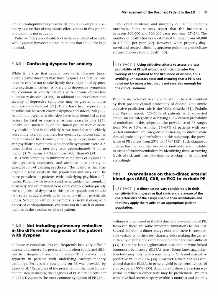
limited cardiopulmonary reserve. To rely soley on pulse oxi-
metry as a marker of respiratory effectiveness in this patient
population is not prudent.
Pulse oximetry is a valuable tool in the evaluation of patients
with dyspnea; however, it has limitations that should be kept
in mind.
Pitfall | Confusing dyspnea for anxiety
While it is true that several psychiatric illnesses (most
notably panic disorder) may have dyspnea as a feature, one
must be careful not to take lightly the complaint of dyspnea
in a psychiatric patient. Anxiety and depressive symptoms
are common in elderly patients with chronic obstructive
pulmonary disease (COPD). In addition the prevalence and
severity of depressive symptoms may be greater in those
who are most disabled [21]. There have been reports of a
possible link between chronic dyspnea and suicide risk [22].
In addition, psychiatric disorders have been identified as risk
factors for fatal or near-fatal asthma exacerbations [23].
Finally, in a Dutch study on the clinical presentation of acute
myocardial infarct in the elderly, it was found that the elderly
were more likely to manifest less-specific symptoms such as
breathlessness, heart failure, dizziness, syncope, neurologic,
and psychiatric symptoms. Non-specific symptoms were 2–5
times higher and mortality was approximately 4 times
higher (31% versus 7.7%) in those older than 65 [24].
It is very tempting to minimize complaints of dyspnea in
the psychiatric population and attribute it to anxiety or
exacerbation of existing psychoses. The reality is that true
organic disease exists in this population and may even be
more prevalent in patients with underlying psychiatric ill-
nesses. Patients with hypoxia and hypercarbia often complain
of anxiety and can manifest behavioral changes. Subsequently
the complaint of dyspnea in this patient population should
be treated as aggressively as in patients without psychiatric
illness. Screening with pulse oximetry is essential along with
a focused cardiopulmonary examination in search of abnor-
malities in the anxious or altered patient.
Pitfall | Not including pulmonary embolismin the differential diagnosis of the patientwith dyspnea
Pulmonary embolism (PE) can frequently be a very difficult
disease to diagnose. Its presentation is often subtle and diffi-
cult to distinguish from other diseases. This is even more
apparent in patients with underlying cardiopulmonary
pathology. Perhaps the best quote on PE was provided by
Laack et al. “Regardless of the presentation, the most funda-
mental step in making the diagnosis of PE is first to consider
it” [25]. Dyspnea is the most common symptom of PE [26].
The exact incidence and mortality due to PE remains
uncertain. Some sources report that the incidence is
between 200,000 and 300,000 cases per year [27–29]. The
number of deaths has been estimated to range from 50,000
to 100,000 per year [29]. However, when properly diag-
nosed and treated, clinically apparent pulmonary emboli are
an uncommon cause of death [30].
Patients suspected of having a PE should be risk stratified
by their pre-test clinical probability of disease. One simple
objective prediction rule is the Wells Criteria [31]. Fedullo
and Tapson report, “25–65% of patients with suspected
embolism are categorized as having a low clinical probability
of embolism; in this subgroup, the prevalence of PE ranges
from 5% to 10%. Another 25–65% of patients with sus-
pected embolism are categorized as having an intermediate
clinical probability of embolism; in this subgroup, the preva-
lence of PE ranges from 25% to 45%” [32]. Such diagnostic
criteria has the potential to reduce morbidity and mortality
in cases of thromboembolism by identifying those at varying
levels of risk and thus allowing the workup to be adjusted
accordingly.
Pitfall | Over-reliance on the D-dimer, arterialblood gas (ABG), CXR, or EKG to exclude PE
D-dimer is often used in the ED during the evaluation of PE.
However, there are some important limitations to this test.
Several different D-dimer assays exist and there is consider-
able variability in their test characteristics making the gener-
alizability of published estimates of D-dimer accuracy difficult
[33]. There are latex agglutination tests and enzyme-linked
immunosorbent assay (ELISA) tests. Some latex agglutina-
tion tests may only have a sensitivity of 65% and a negative
predictive value of 81% [34]. However, a meta-analysis con-
cluded that the ELISAs in general yield better sensitivities (of
approximately 95%) [35]. Additionally, there are certain sce-
narios in which D-dimer tests may be problematic. Patients
who have had recent surgery (within 3 months) and patients
Management of the Dyspneic Patient in the ED | 19
KEY FACT | Using objective criteria to assess pre-test
probability of PE will allow the clinician to cater the
workup of the patient to the likelihood of disease, thus
avoiding unnecessary tests and ensuring that a PE is not
ruled out by using a test that is not sensitive enough for
the clinical scenario.
KEY FACT | D-dimer assays vary considerably in their
sensitivity. It is imperative that clinicians are aware of the
characteristics of the assays used in their institutions and
that they apply the results on an appropriate patient
population.

with cancer may have elevated D-dimer levels making this
test non-specific in this patient population. Furthermore, in
these high-risk patients, because the pre-test probability is
high at baseline, the negative predictive value of the D-dimer
test is only 80% [36]. Finally, D-dimer assays may be affected
by clot burden and location of the emboli. There is greater
accuracy in excluding segmental or larger emboli (sensitivity
93%) than subsegmental emboli (sensitivity � 50%). D-dimer
concentration and the accuracy of D-dimer assays are clearly
dependent on embolus location and smaller, subsegmental
emboli may be missed when D-dimer assays are used as a sole
test to exclude PE [37]. Patients with a low pre-test probabil-
ity of PE are the only ones in whom a D-dimer has any utility
to exclude disease. Furthermore, only ultrasensitive D-dimers
should be used to exclude venothromboembolism in these
patients. Presently, the sensitivity of qualitative and latex
agglutination D-dimer assays is not sufficient to exclude dis-
ease in even these low-risk patients.
There have been multiple proposed diagnostic algorithims
which incorporate clinical assessment, imaging studies (lower
extremity ultrasound, V/Q scan, computed tomography (CT)
scan), and D-dimer tests. While some advocate the cessation
of a workup when there is a low pre-test clinical probability
and an ELISA D-dimer �500 ng/ml, none currently advocate
the use of a negative D-dimer with a high or intermediate
pre-test probability.
ABG data alone or in combination with other clinical data
is not useful in the assessment of suspected PE [38]. Patients
with acute PE are classically described as having hypoxemia,
hypocapnia, an elevated alveolar–arterial oxygen gradient
and a respiratory alkalosis. However, not all patients will
exhibit these findings. Some patients, particularly the young,
may not present with hypoxia or an elevated alveolar–arterial
oxygen gradient [39]. Up to 6% of patients with PE will have
a normal alveolar–arterial oxygen gradient. [26] In addition,
patients with a large PE may actually have hypercapnia and
respiratory acidosis secondary to circulatory and ventilatory
collapse. The ABG should not be not be used to exclude or
establish the diagnosis of PE.
The chest X-ray (CXR) cannot prove or exclude PE. A nor-
mal chest radiograph in the setting of severe dyspnea and
hypoxemia without other cardiopulmonary findings is
strongly suggestive of PE [40]. However, the majority of
patients with PE will have an abnormal chest radiograph.
Common findings include atelectasis, pulmonary infiltrates,
pleural effusion, and elevated hemidiaphragm. Classically
taught findings such as Hampton’s hump or decreased vas-
cularity (Westermark’s sign) are suggestive but uncommon
[26, 40, 41]. Findings of pulmonary infarction can some-
times be mistaken for infiltrates, thereby causing clinicians to
mistakenly diagnose patients with pneumonia. In addition,
the presence of a pleural effusion increases the likelihood
of PE in young patients who present with acute pleuritic
chest pain [42]. This again highlights the importance of con-
sidering the diagnosis of PE. If it is not in the differential
diagnosis, the possibility of PE will not be entertained.
The classic electrocardiographic findings with PE have
been described as the traditional S1Q3T3 pattern character-
ized by a large S-wave in lead I, and a Q-wave and T-wave
inversion in lead III. While often considered the hallmark of
PE, these findings are neither sensitive nor specific for PE.
Though sinus tachycardia is the most frequently encoun-
tered rhythm, normal EKGs are encountered in approxi-
mately 10–25% of patients with PE [43]. Patterns indicative
of right ventricular strain are also suggestive of PE [44].
Pitfall | Relying on a normal CXR to rule outcardiopulmonary disease
The seasoned clinician should immediately recognize that a
normal CXR does very little to help one exclude many car-
diopulmonary causes of dyspnea. With the exception of ten-
sion pneumothorax, most life-threatening causes of dyspnea
may demonstrate a normal CXR. Included in this partial list
are airway obstruction, PE, myocardial infarction, congestive
heart failure, cardiac tamponade, asthma, cor pulmonale,
pneumonia, pericarditis, COPD exacerbation, pulmonary
hypertension, myocarditis, carbon monoxide poisoning, and
methemoglobinemia. Small pneumothoracies may also be
missed in recumbent patients, especially when the pneu-
mothorax is located anteriorly. In addition, if the CXR is an
anteroposterior (AP) or portable view, the sensitivity is per-
haps even lower. The negative predictive value of a chest
radiograph is not sufficient to exclude cardiopulmonary dis-
ease. In a case where a CXR is read as normal in a hypoxic and
KEY FACT | In patients with unexplained dyspnea and
normal findings on routine studies, clinicians should have
a heightened suspicion for PE and myocardial or
pericardial disease.
KEY FACT | Radiographic findings with PE may mimic
those of pneumonia or pleural effusions.
20 | Chapter 2
Electrocardiographic findings of right ventricular strain
suggestive of PE [44].
• Incomplete or complete right bundle branch block
• Large S-waves in leads I and aVL
• Shift in transition zone in the precordial leads to V5
• Q-waves in leads III and aVF
• Right axis deviation
• Low-voltage QRS in limb leads
• T-wave inversion in inferior and anterior leads

dyspneic patient, the likelihood of cardiac and thromboem-
bolic pathology increases. This warrants the further investi-
gation of venothromboembolism and pericardial/ myocardial
disease states with the consideration of bedside echocardio-
graphy, EKG, cardiac enzymes, and possibly chest CT. In fact,
in one ED study, of 103 patients with unexplained dyspnea
who then underwent bedside echocardiography, 14 (13.6%)
had pericardial effusions [45]. Of these four were felt to be
large enough to explain the patients’ dyspnea.
Pitfall | Overly or underly aggressive oxygenadministration in patients with COPD
The traditional teaching has been that administration of
oxygen will cause the spontaneously breathing COPD patient
in respiratory distress to “stop breathing.” It was traditionally
taught that suppression of the hypoxic ventilatory drive would
lead to hypercapnia and subsequent hypercapnic respiratory
failure. However, it appears as though our traditional under-
standing of the pathophysiology was incorrect. Three mech-
anisms have been shown to worsen hypercapnia when
oxygen is administered in the COPD patient:
1. Worsened V/Q matching due to a decrease in hypoxic
pulmonary vasoconstriction (HPV);
2. Decreased binding affinity of hemoglobin for CO2;
3. Decreased minute ventilation [46, 47].
HPV is a physiologic response to alveolar hypoxia. Through
this mechanism blood is diverted away from poorly ventilated
lung units. When hypoxia affects the entire lung (typically
occurs at PaO2 levels �60 mmHg), HPV occurs throughout
the lung. This increases the pulmonary artery pressure and
subsequently recruits many previously unperfused pulmonary
capillaries, increasing the surface area available for gas diffu-
sion, and improving the matching of ventilation and perfusion.
If the hypoxia is resolved with supplemental oxygen, HPV
is blunted, thus worsening V/Q matching. This essentially
increases the physiologic deadspace and shunt and subse-
quently leads to elevations in PCO2 [46, 48].
The CO2 dissociation curve shifts to the right in the pres-
ence of increased oxygen saturation. This is known as the
Haldane effect. Subsequently, in the presence of increased
oxygen saturation from supplemental oxygen, oxyhemoglo-
bin binds CO2 less avidly than deoxyhemoglobin, thus
increasing the amount of CO2 dissolved in blood. This in
turn is reflected as an increased PaCO2 [49].
Administration of supplemental oxygen to a COPD
patient in respiratory distress has also been show to decrease
the patient’s minute ventilation. This is due to a small
decrease in respiratory rate that will also increase PCO2 [47].
As a general rule, prevention of tissue hypoxia is more
important than CO2 retention concerns. Many patients with
COPD have a chronic compensated respiratory acidosis.
While normal individuals may begin to experience depressed
level of consciousness at PaCO2 levels of 60–70 mmHg,
COPD patients may not exhibit these symptoms until their
PaCO2 reaches a level in excess of 90–100 mmHg. COPD
patients are at greater risk from hypoxia than from hyper-
capnia and oxygen should not be withheld from the hypoxic
COPD patient. Oxygen therapy should be emergently initiated
in all hypoxic COPD patients with an initial goal oxygen sat-
uration of 88–92% unless there is evidence of active cardiac
or cerebral ischemia.
The primary goal of oxygen therapy is to prevent tissue
hypoxia. The American Thoracic Society/European Respiratory
Society recommends a SaO2 greater than 90% or a PaO2 of
60–70 mmHg [50]. In addition, Kelly et al. reported that oxy-
gen saturation by pulse oximetry may be an effective screen-
ing test for systemic hypoxia, with the screening cutoff of
92% having sensitivity for the detection of systemic hypoxia
of 100% with specificity of 86% [51]. Further increases in
the FiO2 above the level needed to achieve these goals do not
add significantly to the oxygen content of blood, but do
increase the potential for more severe secondary hypercapnia
[50]. Venturi masks should be used when possible to permit
tight regulation of FiO2.
Finally, if CO2 retention occurs, monitor for acidemia. If
acidemia occurs, consider a trial of non-invasive or invasive
mechanical ventilation [50].
Pitfall | Not considering non-invasivepositive pressure ventilation; that is,CPAP/BiPAP as an alternative to intubationin selected patients
Non-invasive positive pressure ventilation (NPPV) has been
shown to reduce length of hospital stay, morbidity, mortal-
ity, and need for invasive mechanical ventilation. The data
supporting the use of NPPV is strongest for COPD exacerba-
tions; however, there is growing evidence to support its use
in cardiogenic pulmonary edema [52–55], hypoxic respira-
tory failure [56–60], pneumonia [56, 61], immunocompro-
mised states [62, 63], and asthma [64, 65].
There is a wealth of information showing the benefit of
NPPV in acute exacerbations of COPD. This data includes
randomized controlled trials, systemic reviews, and meta-
analysis [66–74]. NPPV has been shown to decrease intuba-
tion rates (RR 0.42, 95% CI 0.31–0.59), decrease mortality
(RR 0.41, 95% CI 0.26–0.64), decrease complications (RR
0.32, 95% CI 0.18–0.56), and decrease hospital length of
stay (weight mean difference �3.24 days, 95% CI �4.42
to �2.26) [68].
It is important to note that early institution of NPPV is
vital. Conti et al. showed that if one waits for failure of con-
ventional medical therapy, the benefits conferred by NPPV
(length of ICU stay, number of days on mechanical ventila-
tion, overall complications, ICU mortality, and hospital mor-
tality) are eliminated [75]. As such, patients in respiratory
Management of the Dyspneic Patient in the ED | 21

distress, not requiring emergent intubation and without
contraindications should be considered for NPPV. The
American Association for Respiratory Care recommends the
early use of NPPV when two or more of the following are
present:
1. Respiratory distress with moderate to severe dyspnea.
2. Arterial pH �7.35 with PaCO2 above 45 mmHg.
3. Respiratory rate of �25/min [76].
Improvement should be seen within 30–60 min, and if
improvement does not occur within 1–2 h, invasive
mechanical ventilation should be initiated. One should also
be aware that when using NPPV, frequent checks and
adjustments are required. Failure to do so may lead to the
patient’s condition worsening and a delay in the institution
of invasive mechanical ventilation.
Pearls for Improving Patient Outcomes
• Do not rely solely on a normal physical examination to exclude
disease.
• Patients with a normal pulse oximetry may still suffer from venti-
latory failure.
• Shortness of breath should be attributed to anxiety only when
all other causes have been definitively excluded.
• Risk stratification of patients with suspected venous throm-
boembolism utilizing clinical decision rules will aid in the diagno-
sis of PE.
• A normal CXR does very little to exclude cardiopulmonary
causes of dyspnea.
• The negative predictive value of a normal ABG or CXR does not
allow one to exclude PE.
• A D-dimer test should not be used to exclude PE in patients with
moderate or high clinic pre-test probability.
• Only high-sensitivity D-Dimer tests should be used to exclude PE
in patients but should only be used in patients with a low pre-
test probability.
• Do not withhold oxygen from a hypoxic COPD patient; however,
be cautious with its use and follow PCO2 levels.
• Consider the use of NPPV in selected patients with respiratory
distress if they do not require immediate emergent intubation.
This is especially true with patients in COPD exacerbation.
References
1. Dyspnea. Mechanisms, assessment, and management: a con-
sensus statement. American Thoracic Society. Am J Respir Crit
Care Med 1999; 159:321.
2. Banzett RB, Mulnier HE, Murphy K, Rosen SD, Wise RJ,
Adams L. Breathlessness in humans activates insular cortex.
NeuroReport 2000; 11(10):2117–20.
3. O’Connor GT, Anderson KM, Kannel WB, et al. Prevalence and
prognosis of dyspnea in the Farmingham Study. Chest 1987;
92:90S.
4. Hirshberg A, Thomson SR, Huizinga WK. Reliability of physical
examination in penetrating chest injuries. Injury 1988; 19(6):
407–9.
5. Chen SC, Chang KJ, Hsu CY. Accuracy of auscultation in the detec-
tion of haemopneumothorax. Eur J Surg 1998; 164(9):643–5.
6. Chen SC, Markmann JF, Kauder DR, Schwab CW.
Hemopneumothorax missed by auscultation in penetrating
chest injury. J Trauma 1997; 42(1):86–89.
7. Bokhari F, Brakenridge S, Nagy K, Roberts R, Smith R, Joseph
K, An G, Wiley D, Barrett J. Prospective evaluation of the sensi-
tivity of physical examination in chest trauma. J Trauma 2002;
53(6):1135–8.
8. Wipf JE, Lipsky BA, Hirschmann JV, Boyko EJ, Takasugi J,
Peugeot RL, Davis CL. Diagnosing pneumonia by physical
examination: relevant or relic? Arch Intern Med 1999; 159 (10):
1082–7.
9. Metlay JP, Kapoor WN, Fine MJ. Does this patient have
community-acquired pneumonia? Diagnosing pneumonia
by history and physical examination. J Am Med Assoc 1997;
278(17):1440–5.
10. Chakko S, Woska D, Martinez H, et al. Clinical, radiographic,
and hemodynamic correlations in chronic congestive heart fail-
ure: conflicting results may lead to inappropriate care. Am J Med
1991; 90:353–9.
11. Witting MD, Hsu S, Granja CA. The sensitivity of room-air
pulse oximetry in the detection of hypercapnia. Am J Emerg Med
2005; 23(4):497–500.
12. Witting MD, Lueck CH. The ability of pulse oximetry to screen
for hypoxemia and hypercapnia in patients breathing room air.
J Emerg Med 2001; 20(4):341–8.
13. Ayas N, Bergstrom LR, Schwab TR, Narr BJ. Unrecognized
severe postoperative hypercapnia: a case of apneic oxygena-
tion. Mayo Clin Proc 1998; 73(1):51–54.
14. Potkin RT, Swenson ER. Resuscitation from severe acute hyper-
capnia. Determinants of tolerance and survival. Chest 1992;
102(6):1742–5.
15. Stoneham MD. Uses and limitations of pulse oximetry. Br J
Hosp Med 1995; 54(1):35–41.
16. Hampson NB. Pulse oximetry in severe carbon monoxide poi-
soning. Chest 1998; 114(4):1036–41.
17. Miner JR, Heegaard W, Plummer D. End-tidal carbon dioxide
monitoring during procedural sedation. Acad Emerg Med 2002;
9(4):275–80.
18. Rohling R, Biro P. Clinical investigation of a new combined
pulse oximetry and carbon dioxide tension sensor in adult
anaesthesia. J Clin Monit Comput 1999; 15(1):23–27.
19. Senn O, Clarenbach CF, Kaplan V, Maggiorini M, Bloch KE.
Monitoring carbon dioxide tension and arterial oxygen satura-
tion by a single earlobe sensor in patients with critical illness or
sleep apnea. Chest 2005; 128(3):1291–6.
20. Chhajed PN, Kaegi B, Rajasekaran R, Tamm M. Detection of
hypoventilation during thoracoscopy: combined cutaneous car-
bon dioxide tension and oximetry monitoring with a new digi-
tal sensor. Chest 2005; 127(2):585–8.
21. Yohannes AM, Roomi J, Baldwin RC, Connolly MJ. Depression
in elderly outpatients with disabling chronic obstructive pul-
monary disease. Age Ageing 1998; 27(2):155–60.
22. Horton-Deutsch SL, Clark DC, Farran CJ. Chronic dyspnea and
suicide in elderly men. Hosp Commun Psychiatr 1992; 43(12):
1198–203.
22 | Chapter 2

23. Ben-Dov I. Fatal asthma: who, when, why and how to pre-
vent? Harefuah 2002; 141(6):524–6, 579, 578.
24. de Fockert JA. Clinical presentation of acute myocardial infarct
in the elderly. Tijdschr Gerontol Geriatr 1987; 18(6):305–8.
25. Laack TA, Goyal DG. Pulmonary embolism: an unsuspected
killer. Emerg Med Clin N Am 2004; 22(4):961–83.
26. Stein, PD, Terrin, ML, Hales, CA, et al. Clinical, laboratory,
roentgenographic, and electrocardiographic findings in patients
with acute pulmonary embolism and no pre-existing cardiac or
pulmonary disease. Chest 1991; 100:598.
27. Anderson Jr FA, Wheeler HB, Goldberg RJ, et al. A population-
based perspective of the hospital incidence and case-fatality
rates of deep vein thrombosis and pulmonary embolism: the
Worcester DVT Study. Arch Intern Med 1991; 151:933–8.
28. Silverstein MD, Heit JA, Mohr DN, Petterson TM, O’Fallon
WM, Melton III LJ. Trends in the incidence of deep vein throm-
bosis and pulmonary embolism: a 25-year population-based
study. Arch Intern Med 1998; 158:585–93.
29. Goldhaber SZ. Pulmonary embolism. New Engl J Med 1998;
339:93–104.
30. Carson JL, Kelley MA, Duff A, et al. The clinical course of pul-
monary embolism. New Engl J Med 1992; 326:1240–5.
31. Wolf SJ, McCubbin TR, Feldhaus KM, Faragher JP, Adcock DM.
Prospective validation of wells criteria in the evaluation of
patients with suspected pulmonary embolism. Ann Emerg Med
2004; 44(5):503–10.
32. Fedullo PF, Tapson VF. The evaluation of suspected pulmonary
embolism. New Engl J Med 2003; 349:1247–56.
33. Becker DM, Philbrick JT, Bachhuber TL, Humphries JE. D-dimer
testing and acute venous thromboembolism. A shortcut to
accurate diagnosis? Arch Intern Med 1996; 156(9):939–46.
34. Farrell S, Hayes T, Shaw M. A negative SimpliRED D-dimer
assay result does not exclude the diagnosis of deep vein throm-
bosis or pulmonary embolus in emergency department patients.
Ann Emerg Med 2000; 35(2):121–5.
35. Stein PD, Hull RD, Patel KC, Olson RE, Ghali WA, Brant R, Biel
RK, Bharadia V, Kalra NK. D-dimer for the exclusion of acute
venous thrombosis and pulmonary embolism: a systematic
review. Ann Intern Med 2004; 140(8):589–602.
36. Lee AY, Julian JA, Levine MN, Weitz JI, Kearon C, Wells PS,
Ginsberg JS. Clinical utility of a rapid whole-blood D-dimer
assay in patients with cancer who present with suspected acute
deep venous thrombosis. Ann Intern Med 1999; 131(6):417–23.
37. De Monye W, Sanson BJ, Mac Gillavry MR, Pattynama PM,
Buller HR, van den Berg-Huysmans AA, Huisman MV. Embolus
location affects the sensitivity of a rapid quantitative D-dimer
assay in the diagnosis of pulmonary embolism. Am J Respir Crit
Care Med 2002; 165(3):345–8.
38. Rodger MA, Carrier M, Jones GN, Rasuli P, Raymond F,
Djunaedi H, Wells PS. Diagnostic value of arterial blood gas
measurement in suspected pulmonary embolism. Am J Respir
Crit Care Med 2000; 162(6):2105–8.
39. Green RM, Meyer TJ, Dunn M, Glassroth J. Pulmonary
embolism in younger adults. Chest 1992; 101(6):1507–11.
40. ATS guidelines: the diagnostic approach to acute venous
thromboembolism. Am J Respir Crit Care Med 1999; 160:1043.
41. Worsley DF, Alavi A, Aronchick JM, Chen JT, Greenspan RH,
Ravin CE. Chest radiographic findings in patients with acute
pulmonary embolism: observations from the PIOPED study.
Radiology 1993; 189(1):133–6.
42. McNeill BJ, Hessel SJ, Branch WT, et al. Measures of clinical
efficacy: 3. The value of the lung scan in the evaluation of
young patients with pleuritic chest pain. J Nucl Med 1976;
17:163.
43. Ullman E, Brady WJ, Perron AD, Chan T, Mattu A. Electrocar-
diographic manifestations of pulmonary embolism. Am J Emerg
Med 2001; 19:514–9.
44. Sreeram N, Cheriex EC, Smeets JLRM, et al. Value of the 12-
lead electrocardiogram at hospital admission in the diagnosis of
pulmonary embolism. Am J Cardiol 1994; 73:298–303.
45. Blaivas M. Incidence of pericardial effusion in patients present-
ing to the emergency department with unexplained dyspnea.
Acad Emerg Med 2001; 8(12):1143–6.
46. Aubier M, Murciano D, Milic-Emili J, Touaty E, Daghfous J,
Pariente R, Derenne JP. Effects of the administration of O2 on
ventilation and blood gases in patients with chronic obstructive
pulmonary disease during acute respiratory failure. Am Rev
Respir Dis 1980; 122(5):747–54.
47. Aubier M, Murciano D, Fournier M, Milic-Emili J, Pariente R,
Derenne JP. Central respiratory drive in acute respiratory fail-
ure of patients with chronic obstructive pulmonary disease. Am
Rev Respir Dis 1980; 122(2):191–9.
48. Levitzky MG. Pulmonary Physiology, 6th edn. New York:
McGraw-Hill/Appleton & Lange, 2003, p. 107.
49. Levitzky MG. Pulmonary Physiology, 6th edn. New York:
McGraw-Hill/Appleton & Lange, 2003, pp. 158–9.
50. Standards for the diagnosis and care of patients with chronic
obstructive pulmonary disease. American Thoracic Society. Am
J Respir Crit Care Med 1995; 152:S77.
51. Kelly AM, McAlpine R, Kyle E. Respir Med 2001; 95(5):336–40.
52. Wigder HN, Hoffman P, Mazzolini D, et al. Pressure support
noninvasive positive pressure ventilation treatment of acute
cardiogenic pulmonary edema. Am J Emerg Med 2001; 19:
179–81.
53. Pang D, Keenan S, Cook D, et al. The effect of positive pressure
airway support on mortality and the need for intubation in car-
diogenic pulmonary edema. A systematic review. Chest 1998;
114(4):1185–92.
54. Bersten AD, Holt AW, Vedig AE, Skowronski GA, Baggoley CJ.
Treatment of severe cardiogenic pulmonary edema with con-
tinuous positive airway pressure delivered by face mask. New
Engl J Med 1991; 325(26):1825–30.
55. Nava S, Carbone G, DiBattista N, Bellone A, Baiardi P, Cosentini
R, Marenco M, Giostra F, Borasi G, Groff P. Noninvasive ventila-
tion in cardiogenic pulmonary edema: a multicenter randomized
trial. Am J Respir Crit Care Med 2003; 168(12):1432–7.
56. Ferrer M, Esquinas A, Leon M, Gonzalez G, Alarcon A, Torres
A. Noninvasive ventilation in severe hypoxemic respiratory
failure: a randomized clinical trial. Am J Respir Crit Care Med
2003; 168(12):1438–44.
57. Antonelli M, Conti G, Rocco M, Bufi M, De Blasi RA, Vivino G,
Gasparetto A, Meduri GU. A comparison of noninvasive posi-
tive-pressure ventilation and conventional mechanical ventila-
tion in patients with acute respiratory failure. New Engl J Med
1998; 339(7):429–35.
58. Martin TJ, Hovis JD, Costantino JP, Bierman MI, Donahoe MP,
Rogers RM, Kreit JW, Sciurba FC, Stiller RA, Sanders MH.
A randomized, prospective evaluation of noninvasive ventila-
tion for acute respiratory failure. Am J Respir Crit Care Med 2000;
161(3 Pt 1):807–13.
Management of the Dyspneic Patient in the ED | 23

59. Delclaux C, L’Her E, Alberti C, Mancebo J, Abroug F, Conti G,
Guerin C, Schortgen F, Lefort Y, Antonelli M, Lepage E,
Lemaire F, Brochard L. Treatment of acute hypoxemic nonhy-
percapnic respiratory insufficiency with continuous positive
airway pressure delivered by a face mask: a randomized con-
trolled trial. J Am Med Assoc 2000; 284(18):2352–60.
60. Keenan SP, Sinuff T, Cook DJ, Hill NS. Does noninvasive posi-
tive pressure ventilation improve outcome in acute hypoxemic
respiratory failure? A systematic review. Crit Care Med 2004;
32(12):2516–23.
61. Confalonieri M, Potena A, Carbone G, Porta RD, Tolley EA,
Umberto Meduri G. Acute respiratory failure in patients with
severe community-acquired pneumonia. A prospective ran-
domized evaluation of noninvasive ventilation. Am J Respir Crit
Care Med 1999; 160(5 Pt 1):1585–91.
62. Confalonieri M, Calderini E, Terraciano S, Chidini G, Celeste E,
Puccio G, Gregoretti C, Meduri GU. Noninvasive ventilation for
treating acute respiratory failure in AIDS patients with
Pneumocystis carinii pneumonia. Intens Care Med 2002;
28(9):1233–8.
63. Hilbert G, Gruson D, Vargas F, Valentino R, Gbikpi-Benissan G,
Dupon M, Reiffers J, Cardinaud JP. Noninvasive ventilation in
immunosuppressed patients with pulmonary infiltrates, fever,
and acute respiratory failure. New Engl J Med 2001;
344(7):481–7.
64. Soroksky A, Stav D, Shpirer I. A pilot prospective, randomized,
placebo-controlled trial of bilevel positive airway pressure in
acute asthmatic attack. Chest 2003; 123(4):1018–25.
65. Meduri GU, Cook TR, Turner RE, et al. Noninvasive positive
pressure ventilation in status asthmaticus. Chest 1996;
110(3):767–74.
66. Brochard L, Mancebo J, Wysocki M, Lofaso F, Conti G, Rauss A,
Simonneau G, Benito S, Gasparetto A, Lemaire F. Noninvasive
ventilation for acute exacerbations of chronic obstructive pul-
monary disease. New Engl J Med 1995; 333(13):817–22.
67. Wysocki M, Tric L, Wolff MA, Millet H, Herman B. Noninvasive
pressure support ventilation in patients with acute respiratory
failure. A randomized comparison with conventional therapy.
Chest 1995; 107(3):761–8.
68. Ram FS, Picot J, Lightowler J, Wedzicha JA. Non-invasive posi-
tive pressure ventilation for treatment of respiratory failure due
to exacerbations of chronic obstructive pulmonary disease.
Cochrane Database Syst Rev 2004; (3):CD004104.
69. Peter JV, Moran JL, Phillips-Hughes J, Warn D. Noninvasive
ventilation in acute respiratory failure—a meta-analysis
update. Crit Care Med 2002; 30(3):555–62.
70. Lightowler JV, Wedzicha JA, Elliott MW, Ram FS. Non-invasive
positive pressure ventilation to treat respiratory failure result-
ing from exacerbations of chronic obstructive pulmonary dis-
ease: cochrane systematic review and meta-analysis. Br Med J
2003; 326(7382):185.
71. Keenan SP, Sinuff T, Cook DJ, Hill NS. Which patients with
acute exacerbation of chronic obstructive pulmonary disease
benefit from noninvasive positive-pressure ventilation? A sys-
tematic review of the literature. Ann Intern Med 2003;
138(11):861–70.
72. Plant PK, Owen JL, Elliott MW. Early use of noninvasive venti-
lation for acute exacerbations of chronic obstructive pulmonary
disease on general respiratory wards: a multicentre randomised
controlled trial. Lancet 2000; 355:1931–5.
73. Keenan SP, Gregor J, Sibbald WJ, et al. Noninvasive positive
pressure ventilation in the setting of severe, acute exacerba-
tions of chronic obstructive pulmonary disease: more effective
and less expensive. Crit Care Med 2000; 28(6):2094–103.
74. Celikel T, Sungur M, Ceyhan B, et al. Comparisons of noninva-
sive positive pressure ventilation with standard medical therapy
in acute hypercapneic respiratory failure. Chest 1998;
114(6):1636–42.
75. Conti G, Antonelli M, Navalesi P, et al. Noninvasive vs. conven-
tional mechanical ventilation in patients with chronic obstruc-
tive pulmonary disease after failure of medical treatment in the
ward: a randomized trial. Intens Care Med 2002; 28(12):1701–7.
76. Bach, JR, Brougher, P, Hess, DR, et al. Consensus conference:
noninvasive positive pressure ventilation. Respir Care 1997;
42:364–9.
24 | Chapter 2

Introduction
Abdominal pain is the most commonly cited reason for a visit
to an emergency department (ED), accounting for nearly 7%
of all ED encounters [1]. A great majority of patients will be
discharged; many with the diagnosis of undifferentiated
abdominal pain (UDAP). Approximately 10% of these patients
will require urgent or emergent surgery [2]. The evaluation of
the patient with abdominal pain is fraught with hazards rang-
ing from the subjectivity of the pain itself, to the myriad of
diagnostic possibilities. Serious pathology can affect patients of
all ages, from the newborn with malrotation to the geriatric
patient with intestinal ischemia. The astute clinician must be
able to distinguish between those patients that can safely be
discharged home, those patients that need further evaluation
in the ED or inpatient setting, and those that require emergent
intervention to ward off impending mortality.
Pitfall | Misdiagnosis of cardiac ischemia asintra-abdominal pathology (and vice versa)
Pathology on one side of the diaphragm can cause symp-
toms on the opposite side. The most crucial example of this
is acute coronary syndrome (ACS) masquerading as an
intra-abdominal process. It has long been recognized that a
large percentage of patients with ACS will present without
chest pain. Atypical presentations are more common in
women, diabetics, and elderly patients [3, 4]. Canto et al.
examined elderly patients presenting with unstable angina
and found that 45% had no chest pain; 8% of patients pre-
sented with epigastric pain, 38% with nausea, and 11%
with vomiting [5]. A review of the data from the National
Registry of Myocardial Infarction-2 again showed that one-
third of patients had no chest pain. Upper abdominal pain
was often instead the presenting complaint. Patients pre-
senting without chest pain were on average 7 years older,
more likely to be women, and a significantly higher number
were diabetics. Patients that presented atypically experienced
longer delays and underwent reperfusion less often. They
were also less likely to receive medications such as aspirin,
heparin, or beta-blockers. Most importantly, they had a
mortality rate of 23% as opposed to 9% in the typical group
[3]. This data underscores the importance of considering
cardiac ischemia in patients presenting with gastrointestinal
complaints. In patients at risk for cardiac disease, an electro-
cardiogram should be obtained just as urgently as if the patient
were presenting with chest pain.
Conversely, it is well-reported in the literature that some
intra-abdominal conditions can mimic myocardial infarction,
most notably pancreatitis, but also acute cholecystitis (AC).
The literature is replete with cases of acute pancreatitis (AP)
in which an electrocardiogram shows changes of myocardial
ischemia, including ST-segment elevation [6–9]. In several
of these cases, the patients have been mistakenly adminis-
tered thrombolytics [10, 11]. While patients with ACS may
complain of abdominal pain, it is unusual for there to be
objective abdominal tenderness. If the diagnosis is unclear,
consideration should be made to further evaluate the patient
with serial electrocardiograms or emergent coronary angiog-
raphy. Thrombolytic therapy should be withheld if there is any
concern that the patient’s symptoms may actually represent
pancreatitis, peptic ulcer disease (PUD), or aortic dissection.
Pitfall | Over-reliance on laboratory valuesand ancillary testing in suspectedmesenteric ischemia
The incidence of mesenteric ischemia is low, approximately
1 in 1000 hospital admissions; however, it accounts for 1 out
of every 100 admissions for abdominal pain [12]. Clearly, most
emergency physicians (EPs) are seeing patients with mesen-
teric ischemia, whether or not they recognize it. The diagnosis
of mesenteric ischemia should be entertained in every patient
with a complaint of severe abdominal pain and a paucity of
physical findings (the classic “pain out of proportion to
exam”). Elucidation of risk factors can also suggest the diagno-
sis (see Table 3.1). Acute superior mesenteric artery (SMA)
embolus is more common in patients with underlying cardiac
disease. The most common cardiac conditions that predispose
KEY FACT | [Of] elderly patients presenting with
unstable angina … 45% had no chest pain. Eight
percent … presented with epigastric pain.
Chapter 3 | Evaluation and Management of the Patient withAbdominal Pain
Joseph P. Martinez
25

to SMA embolus are atrial fibrillation, other arrhythmias,
previous myocardial infarction with ventricular aneurysm
formation, valvular heart disease, or dilated cardiomyopathy
[13]. Physical findings of previous embolic disease such as
prior stroke or amputated toes may increase suspicion. SMA
thrombosis is seen in patients with classic cardiovascular risk
factors. These patients should be specifically interrogated for
symptoms of previous “intestinal ischemia” – abdominal
pain provoked by meals. They may also demonstrate signifi-
cant unintentional weight loss. One-half of patients with
mesenteric venous thrombosis will have a personal or family
history of deep venous thrombosis or pulmonary embolus
[14]. They should be interviewed as would any patient in
whom you suspect a thromboembolic process. Close atten-
tion should be paid to clues that suggest inherited throm-
bophilias – family history of thromboembolic events,
multiple first trimester pregnancy losses, or previous per-
sonal clot history. Acquired risks such as smoking or oral
contraceptive use should also be investigated. Non-occlusive
mesenteric ischemia should be near the top of the differen-
tial diagnosis list in any patient that exhibits a “low-flow
state” and presents with abdominal pain. This would include
patients that are septic, severely volume depleted, or with
end-stage congestive heart failure, and those that use
cocaine, digitalis, or are on hemodialysis [15–17].
Mortality from mesenteric ischemia is nearly 80%. This
mortality rate has remained remarkably constant through
the years. A landmark study by Boley et al. finally demon-
strated improvement in mortality only through the aggres-
sive use of early angiography. His group showed that with
immediate angiography, the mortality rate could be reduced
to 54% [18]. These results have since been duplicated in a
number of other studies [19, 20]. Unfortunately, many
physicians are unwilling to obtain angiography based solely
on the patient’s presentation and risk factors. They will wait
for collateral laboratory data, and obtain other, suboptimal
imaging. This is akin to delaying reperfusion in ACS until
the cardiac enzymes have peaked. While an elevated lactate
level, leukocytosis, metabolic acidosis, and hyperamylasemia
KEY FACT | Mortality from mesenteric ischemia is
nearly 80% … With immediate angiography, the mortality
rate could be reduced to 54%.
26 | Chapter 3
Table 3.1 Risk factors for mesenteric ischemia.
Type of mesenteric ischemia Risk factors Special notes
SMA embolus Cardiac disease One-third have a history of a previous embolic event
• Atrial fibrillation or other arrhythmia
• Valvular disease
• Ventricular aneurysm
• Cardiomyopathy
SMA thrombosis Vascular disease risks Acute event may be preceded by period of
• Hypertension “intestinal angina” and prolonged period of
• Hypercholesterolemia significant weight loss
• Diabetes mellitus
• Smoking
Mesenteric venous thrombosis Hypercoaguable state One-half have personal or family history of DVT/PE
• Inherited (factor V Leiden Subacute presentation
mutation, etc.) Women � men
• Acquired (malignancy, oral
contraceptives, etc.)
NOMI Low-flow states Often ICU patients
• Sepsis
• Heart failure
• Volume depletion
Hemodialysis
Drugs
• Digitalis
• Ergot derivatives
• Cocaine
• Norepinephrine
Post-surgery
DVT: deep venous thrombosis; PE: pulmonary embolus; NOMI: non-occlusive mesenteric ischemia; ICU: intensive care unit.

support the diagnosis, none of them are sensitive enough to
rule it out completely. In addition, obtaining other imaging
studies merely delays the time required to obtain a definitive
angiographic diagnosis and start treatment. If the patient has
risk factors for mesenteric ischemia, and the history and
physical examination suggest this diagnosis, arrangements
should be made for immediate angiography and surgical
consultation. The following caveats apply: (1) If mesenteric
venous thrombosis is suspected, computed tomography
(CT) is better than angiography [21], unless the angiogra-
pher is specifically instructed to perform venous phase
angiography. (2) If there will be a long delay in obtaining
angiography and the ED has access to a multi-detector row
CT scanner, a “mesenteric ischemia protocol,” and interpre-
tation of the images in a timely fashion, CT-angiography
shows a great deal of promise as a screening modality (see
Figure 3.1) [22].
Pitfall | Failure to consider heterotopicpregnancy in women receivingreproductive assistance
Women of child-bearing age are another high-risk demo-
graphic group when presenting with abdominal pain. Most
EPs are well versed in the need to evaluate these patients for
possible ectopic pregnancy should they present while early
in pregnancy. The standard practice for ruling out an ectopic
pregnancy is to obtain an ultrasound, transabdominal or
endovaginal depending on their gestational age at presenta-
tion and/or quantitative ß-HCG count. The diagnosis of
ectopic pregnancy is considered “ruled out” if an intrauter-
ine pregnancy (IUP) with fetal heart rate is visualized [23].
In general practice, this is a reasonable approach: hetero-
topic pregnancy (simultaneous occurrence of two or more
implantation sites, usually intrauterine and ectopic) is a
rare event, with most estimates in the range of 1:30,000
pregnancies [24].
With the advent of assisted reproductive technology (ART),
this ratio has changed. Women receiving ART have rates of
heterotopic pregnancy as high as 1:100 pregnancies [25]. This
appears to be independent of whether the patient received an
ovulation-inducing agent (e.g., clomiphene), an exogenous
gonadotropin, or in vitro fertilization with embryo transfer
[26–28]. Heterotopic pregnancy in natural conception cycles is
rare, though the incidence appears to be increasing [29].
Diagnosing a heterotopic pregnancy requires an extremely
high index of suspicion. Women have often already been
diagnosed with an IUP by ultrasound. The level of ß-HCG
may be increasing appropriately due to the presence of a
normal IUP. Adnexal masses and free fluid in the cul-de-sac
may be mistakenly attributed to ruptured corpus luteum
cysts, an admittedly more common diagnosis. In fact, only
10% of heterotopic gestations are diagnosed prior to operative
intervention [25].
A retrospective review of 66 cases of heterotopic preg-
nancy elucidated 4 common signs and symptoms to help
suggest the diagnosis: (1) abdominal pain, (2) adnexal mass,
(3) peritoneal irritation, and (4) enlarged uterus [24]. If the
diagnosis is made in the first trimester, an enlarged uterus
may be absent. More recently, it has been demonstrated that
KEY FACT | Women receiving ART have rates of
heterotopic pregnancy as high as 1:100 pregnancies.
Evaluation and Management of the Patient With Abdominal Pain | 27
A
B
Figure 3.1 Normal mesenteric CT angiogram.
Sagittal reformatting of multi-detector row CT scan (CT angiography)
demonstrating normal celiac axis (arrow A) and normal SMA (arrow B).

patients with an abnormal amount of free fluid in the cul-de-
sac on ultrasound were five times as likely to have an ectopic
pregnancy [30]. This finding should not be discounted in any
patient, even if a viable IUP is seen. Other findings that should
raise a red flag for clinicians include abnormalities seen in the
adnexa on ultrasound, especially an echogenic ring surround-
ing a cystic structure, and relative bradycardia in the face of
hemorrhagic shock [31, 32].
Pitfall | Over-reliance on “classic”presentations and laboratory results inpopulations at high risk for atypicalpresentations of appendicitis
Appendicitis is a common surgical emergency in the general
population with a lifetime risk of 7% [33]. Perforation rates
range from 17% to 20% [34]. Once thought to be a disease
of the young, it is now recognized that 10% of all cases of
appendicitis occur in patients over the age of 60 years. While
the overall mortality from appendicitis is less than 1%, in
the elderly population it ranges from 4% to 8%. This likely
reflects the fact that the incidence of perforation in elderly
patients is as high as 70% [35].
Dr. Alfredo Alvarado established a practical score known as
MANTRELS to assist with the early diagnosis of appendicitis
(see Table 3.2). The score is based on typical signs, symptoms,
and laboratory values often seen in acute appendicitis, namely:
Migration of pain, Anorexia (or ketonuria), Nausea and vomit-
ing, Tenderness in the right lower quadrant, Rebound tender-
ness, Elevated temperature (�100.4°F, �38°C), Leukocytosis
(white blood cell (WBC) count �10,400 cells/ml), and Shift to
the left (�75% neutrophils). A point is assigned to each, with
right lower quadrant tenderness and leukocytosis receiving 2
points, for a total of 10 possible points. A score of 5 or 6 indi-
cates possible appendicitis, 7 or 8 probable appendicitis, and 9
or 10 is very probable appendicitis [36]. This scoring system
has been found to be useful as a diagnostic tool in assisting cli-
nicians. Depending on the practice setting, especially when
abdominal CT is not readily available for diagnostic purposes,
the score can be used to risk stratify patients for discharge with
early follow-up, observation, or emergent consultation. If the
MANTRELS score is used, it should be weighed in conjunction
with physician judgement and should not be used as a means
to definitively rule out the possibility of acute appendicitis.
Whether or not physicians formally utilize the MANTRELS
score, the findings that encompass it are thought to be highly
representative of appendicitis.
Unfortunately, atypical presentations of appendicitis appear
to be the norm in elderly patients. Only 20% of elderly
patients will have the classic findings of fever, anorexia, right
lower quadrant pain, and leukocytosis [37]. Fever is seen in
less than one-third of elderly patients at the time of presenta-
tion, right lower quadrant tenderness may be absent in 30%,
and one-quarter will have a normal WBC count [38]. This
may account for the staggering statistic that nearly one-quarter
of elderly patients subsequently proven to have appendicitis
are discharged home at the first ED visit [39].
It is no surprise then that the MANTRELS score underper-
forms in elderly patients. A study in 2001 showed that in
143 cases of surgically proven appendicitis, 12 cases would
have been missed using the MANTRELS scoring system (i.e.,
MANTRELS score �5). Ten of the 12 cases were patients
between the age of 60 and 80 years [40].
A higher than normal index of suspicion is required to
accurately diagnose appendicitis in a timely fashion in elderly
KEY FACT | Only 20% of elderly patients will have the
classic findings of fever, anorexia, right lower quadrant
pain, and leukocytosis.
28 | Chapter 3
Findings suspicious for heterotopic pregnancy.
• Relative bradycardia in the face of hypotension
• Free fluid in cul-de-sac, despite normal IUP
• Adnexal abnormality, especially echogenic ring
surrounding cystic structure
• Abdominal pain or peritoneal irritation unexplained
by IUP
Points
Symptoms Migration 1
Anorexia 1
Nausea/vomiting 1
Signs Tenderness in right lower quadrant 2
Rebound 1
Elevated temperature (�100.4°F, �38.0°C) 1
Laboratory values Leukocytosis (WBC �10,400 cells/ml) 2
Shift to the left (�75% neutrophils on differential) 2
Total 10
Score: 5–6: possible appendicitis; 7–8: probable appendicitis; 9–10: very probable appendicitis.
Table 3.2 MANTRELS (Alvarado) Score for
suspected acute appendicitis.

patients. Low clinical suspicion or low MANTRELS score are
not sufficient to exclude the diagnosis in this population.
Liberal use of imaging with CT scan is encouraged early on
in the course of elderly patients with abdominal pain (see
Figure 3.2).
Conversely, another group of patients that the MANTRELS
scoring system is less suited for is women. In this case, a high
score has a lower positive predictive value than in children or
men. Several studies have shown that even with scores of 7 or
more, the percent of negative appendectomies is around one-
third [41, 42]. In women, especially of child-bearing age, a
high MANTRELS score may still require either confirmatory
testing (i.e., CT scan) or testing to exclude other pelvic pathol-
ogy (e.g., ultrasound or diagnostic laparoscopy) rather than
proceeding directly to surgery.
Pitfall | Failure to appreciate atypical signsand symptoms in the elderly
The patient that presents in extremis with a rigid board-
like abdomen presents little diagnostic dilemma for the EP.
Stabilization, a call to the covering surgeon, and rapid mobi-
lization of operative resources are quickly accomplished.
There exist many patients however, that similarly require
urgent or emergent surgical intervention, but present much
less dramatically. This proportion increases considerably with
advancing age. EPs may be falsely lulled into complacency
when evaluating an elderly person with abdominal pain, as
their presentation is often less dramatic. They often have
atypical symptoms of disease, delays in presenting for evalua-
tion, and a paucity of physical findings. Despite this, nearly
one-third will require urgent surgical intervention, and fully
one-half will require hospital admission [43]. Of those that
are discharged home, the recidivism rate is approximately
one-third, significantly higher than younger patients with the
same complaint. As noted above, appendicitis in the elderly is
challenging to diagnose. Other difficult disease entities include
PUD, biliary tract disease, and pancreatitis.
Peptic Ulcer Disease
PUD remains a highly prevalent disease throughout the world.
The mortality rate and number of hospitalizations is slowly
decreasing, except in the geriatric population. Painless ulcers
were found in 35% of patients older than 60 years with endo-
scopically proven PUD, compared with only 8% in those
below the age of 60 years [44]. One-half of all ulcers in the
geriatric population will have a complication as their pre-
senting symptom [45]. Complications include hemorrhage,
obstruction, or perforation/penetration.
Hemorrhagic complications of PUD are more common in
the elderly. Twenty percent of cases have no prior symptoms
of PUD. Elderly patients with hemorrhagic PUD are more
likely to require blood transfusions, require surgery to con-
trol hemorrhage, and to rebleed [46]. To further complicate
matters, the tachycardic response to hemorrhage may be
absent due to intrinsic conduction system defects or masked
by concomitant beta-blocker use.
Penetrations may be mistaken for pancreatitis or muscu-
loskeletal back pain. In elderly patients, even anterior perfora-
tions may be difficult to diagnose. Acute onset of pain is found
in only half the cases. Abdominal tenderness is often less
severe, with overt rigidity seen in only 20% [47]. The yield of
plain radiographs is much lower. Free intraperitoneal air is
seen on only 40% of radiographs [48] (see Figure 3.3). This
contributes to the increased mortality in elderly patients,
which approaches 30%, compared to 10% in the general
population [46]. Much of this mortality is directly attributable
to delayed diagnosis. If the diagnosis is delayed by 24 h, the
mortality rate increases 8-fold [48]. Some patients can be
managed non-operatively, but all patients will require surgical
consultation. A randomized, controlled trial of medical versus
surgical treatment of perforated peptic ulcers showed that the
group of patients older than 70 years was less likely to
respond to conservative treatment [49].
Biliary Tract Disease
Cholelithiasis is an extremely common disease, affecting up
to 10% of adults. Acute cholecystitis remains the most com-
mon reason for acute abdominal surgery in the elderly pop-
ulation [50]. The severity of biliary tract disease is much
higher in the elderly. Complications including ascending
KEY FACT | One-half of all ulcers in the geriatric
population will have a complication as their presenting
symptom.
Evaluation and Management of the Patient With Abdominal Pain | 29
Figure 3.2 Acute appendicitis.
Axiai view of CT scan demonstrating enlarged appendix that does not
fill with contrast material, consistent with acute appendicitis.
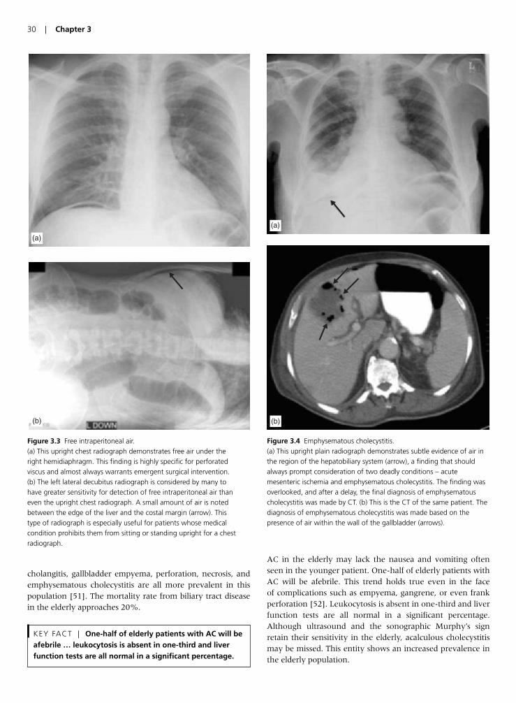
cholangitis, gallbladder empyema, perforation, necrosis, and
emphysematous cholecystitis are all more prevalent in this
population [51]. The mortality rate from biliary tract disease
in the elderly approaches 20%.
AC in the elderly may lack the nausea and vomiting often
seen in the younger patient. One-half of elderly patients with
AC will be afebrile. This trend holds true even in the face
of complications such as empyema, gangrene, or even frank
perforation [52]. Leukocytosis is absent in one-third and liver
function tests are all normal in a significant percentage.
Although ultrasound and the sonographic Murphy’s sign
retain their sensitivity in the elderly, acalculous cholecystitis
may be missed. This entity shows an increased prevalence in
the elderly population.
KEY FACT | One-half of elderly patients with AC will be
afebrile … leukocytosis is absent in one-third and liver
function tests are all normal in a significant percentage.
30 | Chapter 3
(a)
(b)
Figure 3.3 Free intraperitoneal air.
(a) This upright chest radiograph demonstrates free air under the
right hemidiaphragm. This finding is highly specific for perforated
viscus and almost always warrants emergent surgical intervention.
(b) The left lateral decubitus radiograph is considered by many to
have greater sensitivity for detection of free intraperitoneal air than
even the upright chest radiograph. A small amount of air is noted
between the edge of the liver and the costal margin (arrow). This
type of radiograph is especially useful for patients whose medical
condition prohibits them from sitting or standing upright for a chest
radiograph.
(a)
(b)
Figure 3.4 Emphysematous cholecystitis.
(a) This upright plain radiograph demonstrates subtle evidence of air in
the region of the hepatobiliary system (arrow), a finding that should
always prompt consideration of two deadly conditions – acute
mesenteric ischemia and emphysematous cholecystitis. The finding was
overlooked, and after a delay, the final diagnosis of emphysematous
cholecystitis was made by CT. (b) This is the CT of the same patient. The
diagnosis of emphysematous cholecystitis was made based on the
presence of air within the wall of the gallbladder (arrows).

Ascending cholangitis is seen more commonly in elderly
patients, presumably due to the increased incidence of
choledocholithiasis. Elderly patients often present in septic
shock, with no prodrome of biliary colic-type symptoms.
While conservative therapy with broad-spectrum antibiotics
induces a response in 70–80% of cases, severely ill patients
require emergent decompression. This may be open, percu-
taneous, or via endoscopic retrograde cholangiopancreato-
graphy (ERCP).
Gallbladder empyema is a complication of AC where the
gallbladder fills with pus due to complete obstruction. It also
is more common in the elderly, and carries a mortality rate
of 25% [53]. In addition to antibiotics, urgent surgical
decompression is required.
Emphysematous cholecystitis is an infection of the gall-
bladder wall by gas-forming organisms. The incidence rises
with age, and it is associated with diabetes and peripheral
vascular disease. It is the only biliary tract disease that is
more common in men. Patients are typically toxic appear-
ing. Diagnosis is made by visualizing air in the wall of the
gallbladder on plain radiographs or CT (see Figure 3.4), or
by the demonstration of “effervescent bile” on ultrasound.
Surgery is the mainstay of therapy.
Pancreatitis
AP is an inflammatory process where the pancreatic enzymes
autodigest the gland. It ranges from a disease of mild severity
to a life-threatening entity with hemorrhage or frank necrosis
of the organ. It remains the most common non-surgical
abdominal condition in the elderly population. The incidence
of AP increases 200-fold among those aged 65 years and
older [54]. One-third of all cases of pancreatitis occur in the
elderly, and they tend to have a more severe course with
mortality rates approaching 40% [55].
The clinical presentation in the elderly is quite varied. It
may demonstrate the classic “boring” epigastric pain radiating
to the back, or it may exhibit a hypermetabolic state resembling
systemic inflammatory response syndrome. Unfortunately, in
10% of case of AP in the elderly, it merely presents with
altered mental status and shock [45]. The risk of necrotizing
pancreatitis is higher, especially as the patient’s age approaches
80 years [56]. These patients are at high risk for rapid deterio-
ration. As such, a low threshold for CT scanning in the elderly
should be maintained, particularly if there are signs of impend-
ing sepsis. If CT scanning shows necrotizing pancreatitis and
bacterial infection is established (usually through percuta-
neous aspiration), surgical intervention has been shown to be
beneficial [57].
Pearls for Improving Patient Outcomes
• Always consider myocardial infarction as a cause of abdominal
pain, especially in patients with traditional cardiac risk factors.
• Maintain a high index of suspicion for mesenteric ischemia in
elderly patients and pursue early diagnostic imaging based on
clinical suspicion alone.
• Do not assume that visualization of an IUP on ultrasound
definitively rules out ectopic pregnancy in women undergoing
ART. These patients are at high risk for heterotopic pregnancy.
• The MANTRELS scoring system is useful in selected patient pop-
ulations, especially at both ends of the scoring spectrum. It
should be used with caution (if at all) in elderly patients and
women. Low MANTRELS scores in elderly patients should not
preclude imaging with CT scan. High MANTRELS scores in
women (especially of child-bearing age) should not reflexively
trigger surgery, as they may still have other pelvic pathology not
requiring operative treatment.
• Elderly patients often present with atypical signs and symptoms
of common disease entities. A broad diagnostic net should
be cast in these cases, and a period of observation with early
follow-up should be routine, even if the patient appears well.
References
1. McCaig LF, Burt CW. National Hospital Ambulatory Medical
Care Survey: 2003 emergency department summary. Advance
data from vital and health statistics; no 358. Hyattsville,
Maryland: National Center for Health Statistics. 2005.
2. Powers RD, Guertler AT. Abdominal pain in the ED: stability
and change over 20 years. Am J Emerg Med 1995; 13:301–3.
3. Canto JG, Shlipak MG, Rogers WJ, Malmgren J, Frederick P,
Lambrew CT, Ornato JP, Kiefe CI. Prevalence, clinical charac-
teristics and mortality among patients with myocardial infarc-
tion presenting without chest pain. J Am Med Assoc 2000;
283:3223–9.
4. Lusiani L, Perrone A, Pesavento R, Conte G. Prevalence, clinical
features and acute course of atypical myocardial infarction.
Angiology 1994; 45:49–55.
5. Canto JG, Fincher C, Kiefe CI, Allison JJ, Li Q, Funkhouser E,
et al. Atypical presentations among Medicare beneficiaries with
unstable angina pectoris. Am J Cardiol 2002; 90:248–53.
6. Yu AC, Riegert-Johnson DL. A case of acute pancreatitis pre-
senting with electrocardiographic signs of acute myocardial
infarction. Pancreatology 2003; 3(6):515–17.
7. Albrecht C, Laws FA. ST segment elevation pattern of acute
myocardial infarction induced by acute pancreatitis. Cardiol Rev
2003; 11(3):147–51.
8. Hung SC, Chiang CE, Chen JD, Ding PY. Images in cardiovascu-
lar medicine: pseudo-myocardial infarction. Circulation 2000;
101(25):2989–90.
9. Khairy P, Marsolais P. Pancreatitis with electrocardiographic
changes mimicking acute myocardial infarction. Can J Gas-
troenterol 2001; 15(8):522–6.
10. Cafri C, Basok A, Katz A, Abuful A, Gilutz H, Battler A.
Thrombolytic therapy in acute pancreatitis presenting as acute
myocardial infarction. Int J Cardiol 1995; 49:279–81.
KEY FACT | One-third of all cases of pancreatitis occur
in the elderly … with mortality rates approaching 40%.
Evaluation and Management of the Patient With Abdominal Pain | 31

11. Wagner AM, Santolo M. A correct decision? Lancet 2002;
359(9301):157.
12. Stoney RJ, Cunningham CG. Acute mesenteric ischemia.
Surgery 1993; 114:489–90.
13. Ruotolo RA, Evans SRT. Mesenteric ischemia in the elderly. Clin
Geriatr Med 1999; 15:527–57.
14. Rhee RY, Gloviczki P, Mendonca CT, Petterson TM, Serry RD,
Sarr MG, et al. Mesenteric venous thrombosis: still a lethal dis-
ease in the 1990s. J Vasc Surg 1994; 20:688–97.
15. Diamond S, Emmett M, Henrich WL. Bowel infarction as a cause
of death in dialysis patients. J Am Med Assoc 1986; 256:2545.
16. Newman TS, Magnuson TH, Ahrendt SA, Smith-Meek MA,
Bender JS. The changing face of mesenteric infarction. Am Surg
1998; 64:611.
17. Sai-Sudhakar CB, Al-Hakeem M, MacArthur JD, Sumpio BE.
Mesenteric ischemia secondary to cocaine abuse: case reports
and literature review. Am J Gastroenterol 1997; 92:1053.
18. Boley SJ, Sprayregen S, Siegelman SJ, Veith FJ. Initial results
from an aggressive roentgenologic and surgical approach to
acute mesenteric ischemia. Surgery 1977; 82:848.
19. Clark RA, Gallant TE. Acute mesenteric ischemia: angiographic
spectrum. Am J Roentgenol 1984; 142:555.
20. Lobo Martinez E, Carvajosa E, Sacco O, Martinez Molina E.
Embolectomy in mesenteric ischemia. Rev Esp Enferm Dig 1993;
83:351.
21. Bradbury MS, Kavanaugh PV, Chen MY, Weber TM, Bechtold RE.
Noninvasive assessment of portomesenteric venous thrombosis:
current concepts and imaging strategies. J Comput Assist Tomogr
2002; 26:392–404.
22. Hellinger JC. Evaluating mesenteric ischemia with multidetector-
row CT angiography. Tech Vasc Interv Radiol 2004; 7(3):160–6.
23. Simon BC, Snoey ER. Ultrasound in emergency and ambula-
tory medicine. St. Louis: Mosby; 1997.
24. Reece EA, Petrie RH, Sirmans MF, Finster M, Todd WD.
Combined intrauterine and extrauterine gestations: a review.
Am J Obstet Gynecol 1983; 146:323–30.
25. Habana A, Dokras A, Giraldo J, et al. Cornual heterotopic preg-
nancy: contemporary management options. Am J Obstet Gynecol
2000; 182:1264–70.
26. Sotrel G, Rao R, Scommegna A. Heterotopic pregnancy follow-
ing clomid treatment. J Reprod Med 1976; 16:78–80.
27. Botta G, Fortunato N, Merlino G. Heterotopic pregnancy follow-
ing administration of human menopausal gonadotropin in vitro
fertilization and embryo transfer: two case reports and review of
the literature. Eur J Obstet Gynecol Reprod Biol 1995; 59:2112–15.
28. Sucov A, Deveau L, Feola P, et al. Heterotopic pregnancy after
in vitro fertilization. Am J Emerg Med 1995; 13:641–3.
29. Jerrard D, Tso E, Salik R, Barish RA. Unsuspected heterotopic
pregnancy in a woman without risk factors. Am J Emerg Med
1992; 10:58–60.
30. Dart R, McLean S, Dart L. Isolated fluid in the cul-de-sac: how
well does it predict ectopic pregnancy? Am J Emerg Med 2002;
20:1–4.
31. Breyer MJ, Costantino TG. Heterotopic gestation: Another pos-
sibility for the emergency bedside ultrasonographer to consider.
J Emerg Med 2004; 26(1):81–4.
32. Somers MP, Spears M, Maynard AS, Syverud SA. Ruptured het-
erotopic pregnancy presenting with relative bradycardia in a
woman not receiving reproductive assistance. Ann Emerg Med
2004; 43:382–5.
33. Irvin TT. Abdominal pain: a surgical audit of 1190 emergency
admissions. Br J Surg 1989; 76:1121–5.
34. Schwartz SI. Appendix. In: Schwartz SI (ed.), Principles of sur-
gery, 6th edn. New York: McGraw Hill, 1994, pp. 1307–18.
35. Yamini D, Hernan V, Bongard F, et al. Perforated appendicitis: is
it truly a surgical emergency? Am Surg 1995; 221:279–82.
36. Alvarado A. A practical score for the early diagnosis of acute
appendicitis. Ann Emerg Med 1986; 15:557–64.
37. Horattas MC, Guyton DP, Wu D. A reappraisal of appendicitis in
the elderly. Am J Surg 1990; 160:291–3.
38. Storm-Dickerson TL, Horattas MC. What have we learned over
the past 20 years about appendicitis in the elderly? Am J Surg
2003; 185:198–201.
39. Rogers J. Abdominal Pain: Forsight. Dallas: American College
of Emergency Physicians; Issue 3, Dec 1986.
40. Gwynn LK. The diagnosis of acute appendicitis: Clinical assess-
ment versus computed tomography evaluation. J Emerg Med
2001; 21(2):119–23.
41. Malik AA, Wani NA. Continuing diagnostic challenge of acute
appendicitis: evaluation through modified Alvarado score. Aust
NZ J Surg 1998; 68:504–5.
42. Kalan M, Talbot D, Cunliffe WJ, Rich AJ. Evaluation of the
modified Alvarado score in the diagnosis of acute appen-
dicitis: a prospective study. Ann Roy Coll Surg Engl 1994;
76:418–419.
43. Brewer RJ, Golden GT, Hitsch DC, et al. Abdominal pain: an
analysis of 1,000 consecutive cases in a university hospital
emergency room. Am J Surg 1976; 131:219–24.
44. Leverat M. Peptic ulcer disease in patients over 60: experience
in 287 cases. Am J Dig Dis 1966; 11:279–85.
45. Caesar R. Dangerous complaints: The acute geriatric abdomen.
Emerg Med Report 1994; 15:191–202.
46. Borum ML. Peptic-ulcer disease in the elderly. Clin Geriatr Med
1999; 15(3):457–71.
47. Fenyo G. Acute abdominal disease in the elderly: experience
from two series in Stockholm. Am J Surg 1982; 143:751–4.
48. Wakayama T. Risk factors influencing the short-term results of
gastroduodenal perforation. Surg Today 1994; 24(8):681–7.
49. Crofts TJ, Park KG, Steele RJ. A randomized trial of nonopera-
tive treatment for perforated peptic ulcer. New Engl J Med 1989;
320(15):970–3.
50. Rosenthal RA, Anderson DK: Surgery in the elderly: observa-
tions on the pathophysiology and treatment of cholelithiasis.
Exp Gerontol 1993; 28:459–72.
51. Bedirli A. Factors effecting the complications in the natural his-
tory of acute cholecystitis. Hepatogastroenterol 2001; 48(41):
1275–8.
52. Morrow DJ, Thompson J, Wilson SE. Acute cholecystitis in the
elderly. Arch Surg 1978; 113:1149–52.
53. Sen M, Williamson RCN. Acute cholecystitis: surgical manage-
ment. Balliere’s Clin Gastroenterol 1991; 5:817–37.
54. Martin SP, Ulrich CD. Pancreatic disease in the elderly. Clin
Geriatr Med 1999; 15(3):579–605.
55. Hoffman E, Perez E. Acute pancreatitis in the upper age groups.
Gastroenterol 1959; 36:675–85.
56. Paajanen H. AP in patients over 80 years. Eur J Surg 1996;
162(6):471–5.
57. Bradley EL III, Allen K. A prospective longitudinal study of
observation versus surgical intervention in the management of
necrotizing pancreatitis. Am J Surg 1991; 161:19–24.
32 | Chapter 3

Introduction
Up to 90% of adults will have at least one episode of back
pain during their lifetime [1]. In the US alone, approxi-
mately 15 million patients present each year to a physician
with acute back pain [2]. Many of these patients arrive
at local emergency departments (ED) seeking evaluation
and symptom relief. In fact, back pain accounts for almost
2% of all ED visits per year [3]. Thankfully, 90–95% of
patients presenting with acute back pain have non-life-
threatening etiologies [4]. Although most patients cannot be
given an exact diagnosis, nearly all recover within 4–6
weeks [1, 5].
In the remaining 5–10% of patients, acute back pain is a
manifestation of serious pathology. Thoracic aortic dissec-
tion, abdominal aortic aneurysm, spinal epidural abscess,
spinal epidural metastasis, and spinal cord compressive syn-
dromes may all initially present with a complaint of acute
back pain. These “back pain emergencies” are missed in a
significant percentage of patients upon initial evaluation. As
a result of delays in diagnosis and treatment, patients with
these conditions continue to have unacceptably high mor-
bidity and mortality. Although challenging, it is crucial that
emergency physicians identify patients with acute back pain
with a potentially life-threatening disorder.
This chapter will focus on common pitfalls in the ED eval-
uation of patients with acute back pain. With this informa-
tion, the emergency physician can more effectively detect
and treat patients with potentially catastrophic causes of
back pain.
Pitfall | Over-reliance on the “Classic” ClinicalPresentations of Aortic Dissection, RupturingAbdominal Aortic Aneurysm, Spinal EpiduralAbscess, and Pulmonary Embolism
In emergency medicine, it is well recognized that the textbook
clinical presentations of disease are often the exception rather
than the rule. This holds true for the clinical presentations of
several dangerous back pain emergencies [6]. The emergency
physician must be able to diagnose these life-threatening
disorders even in the absence of “classic” presentations.
The classic clinical description of aortic dissection is the sud-
den onset of ripping chest pain that radiates to the inter-
scapular region. In reality, this textbook description is
present in less than one-third of patients with aortic dissec-
tion [6]. Although the abrupt onset of pain is reported in
85% of patients, many will not describe their pain as either
ripping or tearing [7, 8]. Patients with aortic dissection often
describe their pain as either sharp, pressure-like, pleuritic, or
even burning [7]. In addition to a variety of descriptions of
pain, patients with aortic dissection often report varying
locations of pain. Isolated back pain, especially thoracic, is
common in dissections of the descending aorta.
Textbook physical examination findings of aortic dissection
include hypertension, pulse deficits, blood pressure differen-
tials, and a new diastolic murmur. These classic abnormalities
are inconsistent and unreliable findings in patients with dis-
section. Up to one-third of patients with an acute aortic dis-
section are normotensive upon evaluation [9]. Although
considered specific for aortic dissection, a pulse deficit occurs
in just 25–38% of cases [6, 9] and its absence cannot be used
to exclude the diagnosis of aortic dissection. Bilateral blood
pressure measurements are frequently obtained in cases of
suspected aortic dissection. A difference of 20 mmHg or more
is considered significant. However, 20% of normal individuals
have blood pressure differentials greater than 20 mmHg,
thereby lowering the specificity of this finding [10–12]. The
sensitivity of a new diastolic murmur in the detection of aor-
tic dissection is reportedly just 28% [6]. Thus, it is easy to see
how the clinical examination is inadequate for diagnosing or
excluding an acute aortic dissection [9]. For the emergency
physician, suspicion for aortic dissection begins by identifying
risk factors for aortic pathology. A thorough risk factor analy-
sis for aortic disease must be performed in any patient
presenting with acute back pain. Risk factor assessment has
been shown to increase the rate of diagnosis of aortic catas-
trophes [10]. Thoracic aortic dissection must be considered
in any patient with risk factors for the same (discussed
below) who presents with the abrupt onset of severe thoracic
back pain.
KEY FACT | The “classic” description of aortic dissection
pain is absent in 2/3 of cases.
Chapter 4 | Management of Patients with Acute Back Pain inthe ED
Michael E. Winters
33

The textbook triad of rupturing abdominal aortic aneurysm
consists of abdominal pain, hypotension, and a pulsatile
abdominal mass. Unfortunately, less than 20% of patients
with a rupturing aneurysm present with this triad [13]. It is
important for the emergency physician to recognize that a
rupturing abdominal aortic aneurysm can present with left
lower quadrant pain, flank pain, or isolated back pain. As a
result, many patients with a ruptured aneurysm are misdiag-
nosed as having diverticulitis, nephrolithiasis, pyelonephritis,
or musculoskeletal strain [13]. Misdiagnosis leads to signifi-
cant morbidity and mortality. Mortality rates as high as 75%
have been reported when patients with a rupturing abdomi-
nal aortic aneurysm are initially misdiagnosed [10]. In addi-
tion to recognizing back or flank pain as the initial presenting
symptom, the emergency physician must also understand the
limitations of physical examination.
The sensitivity of abdominal palpation for an aneurysm
ranges from 45% to 97% [13]. Sensitivity increases with
increasing sizes of aneurysm, however, nearly 25% of abdom-
inal aneurysms greater than 5 cm cannot be palpated [13].
Similar to aortic dissection, suspicion for an abdominal aor-
tic aneurysm begins with a thorough risk factor assessment.
A rupturing aneurysm must be considered in any elderly
patient with acute back, or flank pain, who has risk factors
for an abdominal aortic aneurysm.
Spinal epidural abscess is classically associated with the triad
of fever, back pain, and neurologic deficits. Although these
symptoms are helpful if present, this triad occurs in as few as
13% of patients [14]. Up to 75% of patients with a spinal
epidural abscess will be afebrile and up to two-thirds of
patients will have a normal initial neurologic exam [14]. As a
result, patients with spinal epidural abscess incur significant
diagnostic delays. Often, patients with spinal epidural abscess
have multiple ED visits before the diagnosis is made. Since
rapid neurologic deterioration can occur, it is imperative that
the emergency physician identify patients with a spinal
epidural abscess. Rather than relying on the presence of
fever and back pain, the emergency physician should focus
on identifying risk factors for spinal epidural abscess in any
patient with acute back pain. It is reported that risk factor
assessment offers the highest sensitivity and negative predic-
tive value in identifying patients with spinal epidural abscess
before the onset of neurologic symptoms [14].
The clinical presentation of pulmonary embolism is classi-
cally described as the acute onset of pleuritic chest pain associ-
ated with dyspnea and hemoptysis. Atypical presentations of
pulmonary embolism, however, are common. Thoracic back
pain is a recognized presentation of pulmonary embolism [15].
In addition to aortic catastrophes and infectious etiologies, the
emergency physician should include a risk factor assessment
for venous thromboembolic disease when confronted with the
patient with acute thoracic back pain.
Pitfall | Failure to Perform a Risk FactorAssessment for Serious Etiologies ofBack Pain
Since the majority of patients with back pain have benign
etiologies, identification of the patient in whom to pursue
an extensive diagnostic workup is challenging. Suspicion for
serious pathology begins with an assessment of patient risk
factors for disease. Unfortunately, many physicians fail to
perform a risk factor profile in patients presenting with back
pain. In fact, failure to inquire about risk factors is one of
the most common reasons cited in cases of missed aortic
dissection [10]. Thus, it is imperative that the emergency
physician inquire about risk factors, perform a risk factor
assessment, and guide the workup accordingly. In many
cases, risk factors may be the only clue to serious underlying
pathology.
Risk factors for aortic catastrophes are well established and
include male gender, older age, hypertension, connective
tissue disorders, chromosomal syndromes, smoking, and
inflammatory conditions of the aorta [16]. Additional risk
factors for aortic dissection include a bicuspid aortic valve,
coarctation, aortic instrumentation, decelerating trauma,
and pregnancy [16]. In fact, over 50% of aortic dissections
in women less than 40 years of age occur during pregnancy,
most often during the third trimester and the initial stages of
labor [16]. Aortic dissection must be considered in the dif-
ferential diagnosis of any pregnant patient presenting during
the third trimester or labor with the abrupt onset of severe
back pain.
KEY FACT | Pregnancy is an often overlooked risk
factor for aortic dissection.
KEY FACT | Because the vast majority of patients with
spinal epidural abscesses present atypically, assessment of
patients for risk factors for this disease is essential for
identifying it prior to the onset of irreversible
neurological sequelae.
KEY FACT | Abdominal palpation is insensitive for
detecting abdominal aortic aneurysm.
KEY FACT | Rupturing abdominal aortic aneurysm often
presents with symptoms mimicking renal colic,
diverticulitis, and musculoskeletal pain.
34 | Chapter 4

Lastly, the emergency physician must always inquire about
the use of illicit substances in patients with back pain. Cocaine
use is an established risk factor for aortic dissection, particu-
larly of the descending aorta, in otherwise healthy individuals
[16]. Hypertensive patients presenting with the abrupt onset
of back pain in the setting of cocaine use should be imaged to
exclude acute aortic dissection.
Although traditionally taught as a disease of older individuals,
spinal epidural abscess can affect patients of any age. Risk fac-
tors for spinal epidural abscess include diabetes mellitus, alco-
holism, chronic renal failure, corticosteroid therapy, human
immunodeficiency virus/acquired immunodeficiency syn-
drome, and intravenous drug use [17]. Intravenous drug use is
considered one of the primary risk factors for spinal epidural
abscess, accounting for over 50% of cases in some populations
[18]. Any patient with back pain who uses intravenous drugs
must be considered to have a spinal epidural abscess until
proven otherwise. Regardless of the location of pain, these
patients require imaging to exclude an epidural abscess. It is
also important for the emergency physician to inquire about
recent invasive procedures. Back pain that develops in the
setting of a recent procedure, especially one known to have a
high incidence of bacteremia, is highly suspicious for either
vertebral osteomyelitis or spinal epidural abscess [1].
Epidural spinal cord compression is a true medical emergency
that cannot be missed. Over 90% of cases are due to spinal
epidural metastases. The primary risk factor for spinal epidural
metastases is a history of malignancy. Although prostate,
breast, and lung cancer most commonly cause bony metas-
tases, it is important to realize that any systemic malignancy
can metastasize to the spine. Lymphoma, renal cell cancer, gas-
trointestinal malignancies, and multiple myeloma are fre-
quently overlooked, yet account for a significant percentage of
cases. Spinal epidural metastases with resultant spinal cord
compression must be considered in any patient with back pain
and a history of malignancy. Additional etiologies for spinal
cord compression include massive midline disc herniation,
spinal hematoma, epidural abscess, traumatic compression,
and transverse myelitis [19].
Pitfall | Failure to Perform a CompleteNeurologic Exam
Arguably, the most important portion of the physical exam-
ination in patients with back pain is the neurologic exami-
nation. In many cases, subtle neurologic deficits are the only
indication of serious pathology, namely spinal cord com-
pression. Unfortunately, many physicians either fail to per-
form a complete neurologic exam or fail to appropriately
document their findings. In a review of missed cases of
spinal epidural abscess, Davis et al. report that, in each case,
minimal notation was made with regards to the neurologic
exam [14]. Exams were documented as either “nonfocal” or
“normal” without reference to the motor, sensory, deep ten-
don reflex, rectal, or cerebellar components.
The importance of performing a complete neurologic exam
cannot be overstated. Any patient with acute back pain and
neurologic deficits requires emergent diagnostic imaging. In
cases of spinal cord compression, motor deficits are the most
common neurologic finding and are present in up to 85%
of patients [20]. The emergency physician must pay close
attention to the motor examination of the lower extremi-
ties. Appropriate examination should include an assessment
of hip flexion and extension, leg flexion and extension, ankle
dorsiflexion and inversion, and great toe dorsiflexion. In
cases of thoracic spinal cord compression, the iliopsoas mus-
cles are preferentially affected, producing weakness of the
proximal lower extremities when testing hip flexion [20].
Sensory abnormalities occur slightly less often than motor
deficits, whereas bowel and/or bladder dysfunction is a late
finding in patients with epidural spinal cord compression
[20]. Any patient with a report of fecal retention or inconti-
nence and/or saddle anesthesia must have a rectal examina-
tion performed and documented. Additional indications for
rectal examination include severe pain and/or the presence
of any neurologic deficit. In addition to the rectal examina-
tion, patients with suspected spinal cord compression should
have a post-void residual measurement. A post-void residual
greater than 100–200 ml is indicative of acute urinary reten-
tion. Regardless of a history of prostatic abnormalities, any
patient with acute back pain and an abnormal post-void
residual should be suspected of having spinal cord compres-
sion until proven otherwise. Acute urinary retention is
reported to have 90% sensitivity and 95% specificity for
cauda equina syndrome [19].
KEY FACT | It is imperative to perform a complete
neurologic examination including, when indicated, a
rectal examination and post-void residual measurement.
KEY FACT | Spinal metastases must be considered in
patients with back pain and a history of malignancy,
regardless of the type.
KEY FACT | Patients with back pain using injection
drugs must have diagnostic imaging to definitively
exclude spinal epidural abscess.
KEY FACT | Aortic dissection must be excluded in
hypertensive patients with back pain and a history of
recent cocaine use.
Management of Patients with Acute Back Pain in the ED | 35

Pitfall | Failure to Recognize the Limitationsof Plain Radiography in Excluding a BackPain Emergency
Given their low cost and ready availability, plain radiographs
are frequently obtained in the evaluation of patients with
acute back pain. In the setting of trauma, plain films can be
useful to exclude fracture or dislocation. However, plain films
are often erroneously utilized to exclude other potential back
pain emergencies, such as spinal metastases or vertebral
osteomyelitis. For vertebral metastases, plain radiographs are
the least sensitive of all imaging modalities [4]. The pooled
sensitivity of plain radiographs for spinal metastases is just
60% [4]. Furthermore, up to 17% of patients with epidural
spinal cord compression due to metastatic disease have nor-
mal plain radiographs [20]. For the patient presenting with
acute back pain and neurologic deficits, plain films should not
be relied upon as the sole imaging modality to exclude spinal
epidural metastases or spinal cord compression.
Plain radiographs are equally insensitive in the diagnosis of
infectious etiologies of back pain, namely osteomyelitis and
spinal epidural abscess. Abnormalities that indicate a potential
infectious etiology include endplate erosion, loss of vertebral
disc height, and bony lysis, but these abnormalities usually
take several weeks to develop [4]. Thus, patients with infec-
tious etiologies of back pain may have entirely normal plain
films. For osteomyelitis, �33% of patients will have an
abnormality on plain film during the first 7–10 days [21]. The
overall sensitivity and specificity of plain radiographs for
osteomyelitis or epidural abscess is 82% and 57%, respec-
tively, and therefore cannot be used solely to rule out spinal
infection [4]. Given the potential for rapid neurologic deterio-
ration, once these disease entities are suspected, definitive
diagnostic imaging is indicated rather than false reassurance
with plain radiography.
Pitfall | Failure to Consider AorticCatastrophes in Patients with Acute BackPain and Neurologic Symptoms
The emergency physician must be knowledgeable regarding
atypical presentations of aortic dissection and abdominal
aortic aneurysm. It is imperative to recognize that aortic
catastrophes can present with back pain and focal neurologic
abnormalities. In fact, neurologic deficits are found in
18–36% of patients with aortic dissection [10]. Deficits seen
in patients with aortic dissection range from acute stroke to
unilateral lower extremity numbness. Unilateral lower
extremity deficits are caused by involvement of the great
radicular artery, a branch of the descending aorta. Thoracic
aortic dissection must be excluded in any patient with sud-
den thoracic back pain and focal neurologic deficits.
For rupturing abdominal aortic aneurysm, up to 5% of
patients will have associated neurologic deficits [10]. It is
believed that an expanding retroperitoneal hematoma
causes direct nerve compression. Since the femoral and
obturator nerves are the most susceptible to compression,
patients present with weakness of hip and knee flexion [10].
The emergency physician must consider aortic catastrophes
in the differential diagnosis of patients presenting with back
pain and neurologic deficits.
Pitfall | Failure to Recognize the Limitationsof Laboratory Studies in the Evaluation ofPatients with Suspected Spinal EpiduralAbscess or Vertebral Osteomyelitis
When considering an infectious etiology of back pain, physi-
cians frequently rely on the presence of a leukocytosis to guide
further evaluation. This finding is variable in infectious causes
of back pain. White blood cell counts above 12,000/mm3 are
seen in just 40% of patients with spinal infection [22]. In addi-
tion, the presence of a left shift has been shown to be unreli-
able in the detection of an infectious etiology [17]. Emergency
physicians should never exclude the possibility of a spinal
infection based upon a normal white blood cell count.
In contrast to the peripheral white blood cell count, inflam-
matory markers, such as the erythrocyte sedimentation rate
and C-reactive protein, have been shown to be useful in the
identification of patients with a spinal infection. These mark-
ers are elevated in approximately 90% of patients with an
infectious cause of back pain [21]. Up to 10% of patients can
have normal levels, therefore, these markers should not be
used as the sole criterion for excluding spinal infection.
Pitfall | Inadequate Radiographic Imaging inthe Diagnosis of Spinal Cord Compressionand Spinal Epidural Abscess
36 | Chapter 4
KEY FACT | There is virtually no indication for plain
radiography in the evaluation of nontraumatic back pain.
KEY FACT | Hematoma surrounding an abdominal aortic
aneurysm can cause weakness of hip and knee flexion.
KEY FACT | Lab studies cannot be used exclusively to
rule out an infectious cause of back pain.
KEY FACT | Magnetic resonance imaging is currently
the only imaging modality that can be used to exclude
spinal cord compression or abscess.

Emergency physicians recognize that spinal cord compres-
sion is a true emergency. Therefore, once the diagnosis is sus-
pected, emergent imaging must be obtained. As discussed,
plain films may be falsely negative in up to 17% of patients
with cord compression [20]. Bone scanning, computed
tomography, and positron-emission tomography have all
been used in the evaluation of patients with spinal cord com-
pression. These modalities, however, have not been shown
to be superior to magnetic resonance imaging. Magnetic res-
onance imaging is the imaging modality of choice when
diagnosing or excluding spinal cord compression. The over-
all diagnostic accuracy of magnetic resonance imaging for
spinal cord compression is 95% [20]. For patients suspected
of cord compression due to metastatic disease, magnetic res-
onance imaging of the entire spine is recommended, as com-
pression can occur at multiple levels [23].
Definitive diagnostic imaging must also be obtained when
considering the diagnosis of spinal epidural abscess. Magnetic
resonance imaging is the most sensitive modality for identifying
and characterizing spinal epidural abscess. Magnetic resonance
imaging accurately defines the extent of an abscess, highlights
any surrounding inflammation, evaluates the degree of thecal
sac compression, and aids in the planning of surgical drainage.
Sensitivity and specificity of magnetic resonance imaging for
spinal infection is 96% and 92%, respectively [4]. Computed
tomography myelography remains an option for patients
unable to undergo magnetic resonance imaging; however, this
procedure is invasive and can result in transmission of the
infection into the subarachnoid space. Currently, there is no
data on the accuracy of computed tomography without myel-
ography in the diagnosis of osteomyelitis or spinal epidural
abscess [4]. Similar to spinal epidural metastases, infectious eti-
ologies of back pain can occur at multiple spinal levels, espe-
cially in the patient abusing intravenous drugs. A magnetic
resonance scan that images only isolated sections of the spine
will undoubtedly miss lesions at other spinal levels. Therefore,
the entire spine should be imaged when evaluating patients for
spinal epidural abscess.
Pitfall | Inadequate Antibiotic Treatment in Patients with Infectious Etiologies ofBack Pain
Although optimal management of spinal epidural abscess
remains controversial, all patients with an infectious etiol-
ogy of acute back pain require prompt antibiotic administra-
tion. Ideally, antibiotics are given after tissue cultures are
obtained. Because patients can develop rapid, irreversible
neurologic deterioration, it is crucial for the emergency
physician to begin antibiotic therapy promptly.
Staphylococcal aureus is the most common isolate in cases
of osteomyelitis and epidural abscess, but up to one-third of
cases are caused by gram-negative organisms [24, 25].
Therefore, initial antibiotic coverage must be broad spectrum
and effective against both gram-positive and gram-negative
organisms. Acceptable regimens include a penicillinase-resist-
ant penicillin plus a third-generation cephalosporin, or in
cases of suspected methicillin-resistant Staphylococcal aureus,
vancomycin and a third-generation cephalosporin [21].
Pitfall | Failure to Initiate Prompt Treatmentin the Patient with Spinal Cord Compression
Definitive treatment of epidural spinal cord compression
requires a multi-disciplinary approach. Once the diagnosis is
suspected, appropriate consultation must be obtained. Often,
this involves consultation with neurosurgery, orthopedic
surgery, and radiation oncology. While awaiting definitive
treatment, the emergency physician must provide support-
ive care. Analgesics and corticosteroids are an important
component of that supportive care. Corticosteroids have
been shown to reduce edema, inhibit further inflammatory
damage, and delay the progression of neurologic deficits [20].
Recent randomized trials have demonstrated that, in the
case of cord compression due to metastatic disease, patients
who received corticosteroids were more likely to be ambula-
tory at long-term follow up [23, 26]. Dexamethasone is the
corticosteroid of choice given its low cost and relatively low
mineralocorticoid activity [23]. Currently, there is no con-
census on the optimal dose. Dosing regimens in the litera-
ture have ranged from 16 to 100 mg/day [20]. An acceptable
regimen consists of an initial dose of 10 mg of dexametha-
sone, followed by 6 mg every 4 h.
Pitfall | Failure to Promptly Refer Patientswith an Acute Motor Radiculopathy
Only 2–3% of patients presenting with acute low back pain
will have evidence of a lumbar radiculopathy [1]. Although
there are many potential etiologies, the most common cause
of a radiculopathy is an acute disc herniation. The size, level,
Management of Patients with Acute Back Pain in the ED | 37
KEY FACT | In suspected spinal infection, antibiotics such
as a third-generation cephalosporin plus a penicillinase-
resistant penicillin or vancomycin should be administered as
soon as possible after cultures are obtained.
KEY FACT | Parenteral pain medications and
dexamethasone (10 mg followed by 6 mg every 4 h)
should be administered to patients with suspected spinal
cord compression.
KEY FACT | Patients with radiculopathy and motor
deficits should have urgent neurosurgical referral.

and location of herniation determine a patient’s symptoms
and physical examination findings. In contrast to the patient
with low back pain and a normal physical exam, those with a
lumbar motor radiculopathy may require early surgical treat-
ment. The most important factor in determining whether an
early surgical procedure is needed is the presence of signifi-
cant weakness [27]. If the weakness is substantial, delaying
surgery could result in a permanent deficit. Therefore, it is
incumbent upon the emergency physician to ensure that the
patient with an acute lumbar motor radiculopathy is seen
within seven days of their ED visit. Urgent magnetic reso-
nance imaging, as well as neurosurgical referral, is needed to
ensure optimal outcomes.
Pearls for Improving Patient Outcomes
• “Classic” presentations are the exception rather than the rule in
back pain emergencies and they often present with symptoms
mimicking other disease.
• Risk factors assessment is crucial to help identify patients requir-
ing emergent imaging.
• Pregnancy is a risk factor for aortic dissection.
• A history of drug use should heighten suspicion for emergent
causes of back pain.
• Patients with back pain require a careful neurological examina-
tion to identify those requiring emergent treatment.
• Plain films of the back are almost never indicated for nontrau-
matic back pain.
• Magnetic resonance imaging is currently the only test that can
exclude spinal cord compression or abscess.
• For spinal infections, early administration of appropriate antibi-
otics is crucial.
• Steroids are indicated for patients with spinal cord compression.
• Patients with radicular symptoms and motor deficits should have
urgent neurosurgical referral.
References
1. Della-Giustina DA. Orthopedic emergencies: emergency
department evaluation and treatment of back pain. Emerg Med
Clin N Am 1999; 17(4):877–93.
2. Hart L, Deyo R, Churkin D. Physician office visits for low back
pain. Spine 1995; 20:11–19.
3. 1995 National Hospital Ambulatory Care Survey. National
Center for Health Statistics, Center for Disease Control and
Prevention, Hyattsville, Maryland.
4. Jarvik JG, Deyo RD. Diagnostic evaluation of low back pain
with emphasis on imaging. Ann Intern Med 2002; 137:586–97.
5. Bigos S, Bowyer O, Braen G, et al. Acute low back problems in
adults. In: Clinical Practice Guideline, Quick Reference Number
14, Rockville MD, U.S. Department of Health and Human
Services, Public Health Service, Agency for Health Care Policy
and Research. AHCPR Pub No. 95-0643. 1994.
6. Haro LH, Krajicek M, Lobl JK. Challenges, controversies, and
advances in aortic catastrophes. Emerg Med Clin N Am 2005;
23:1159–77.
7. Sullivan PR, Wolfson AB, Leckey RD, Burke JL. Diagnosis of
acute thoracic aortic dissection in the emergency department.
Am J Emerg Med 2000; 18:46–50.
8. Hagan PG, Nienaber CA, Isselbacher EM, et al. The International
Registry of Acute Aortic Dissection (IRAD): new insights into an
old disease. J Am Med Assoc 2000; 283;897–903.
9. Knaut AL, Cleveland JC. Aortic emergencies. Emerg Med Clin N
Am 2003; 21:817–45.
10. Rogers RL, McCormack R. Aortic disasters. Emerg Med Clin N Am
2004; 22:887–908.
11. Lane D, Beevers M, Barnes N, et al. Inter-arm differences in
blood pressure: when are they clinically relevant? J Hypertension
2002; 20:1089.
12. Pesola GR, Pesola HR, Lin M, et al. The normal difference in
bilateral blood indirect blood pressure recordings in hyperten-
sive individuals. Acad Emerg Med 2002; 9:342–5.
13. Carpenter CR. Abdominal palpation for the diagnosis of
abdominal aortic aneurysm. Ann Emerg Med 2005; 4:556–8.
14. Davis DP, Wold RM, Patel RJ, et al. The clinical presentation and
impact of diagnostic delays on emergency department patients
with spinal epidural abscess. J Emerg Med 2004; 26:285–91.
15. Laack TA, Goyal DG. Pulmonary embolism: an unsuspected
killer. Emerg Med Clin N Am 2004; 22:961–83.
16. Khan IA, Nair CK. Clinical, diagnostic, and management per-
spectives of aortic dissection. Chest 2002; 122:311–28.
17. Soehle M, Wallenfang T. Spinal epidural abscess: clinical mani-
festations, prognostic factors, and outcomes. Neurosurgery 2002;
51:79–87.
18. Calder KK, Severyn FA. Surgical emergencies in the intra-
venous drug user. Emerg Med Clin N Am 2003; 21:1089–116.
19. Small SA, Perron AD, Brady WJ. Orthopedic pitfalls: cauda
equina syndrome. Am J Emerg Med 2005; 23:159–63.
20. Prasad D, Schiff D. Malignant spinal-cord compression. Lancet
Oncol 2005; 6:15–24.
21. Perron AD, Brady WJ, Miller MD. Orthopedic pitfalls:
osteomyelitis. Am J Emerg Med 2003; 21:61–7.
22. Lurie JD, Gerber PD, Sox HC. A pain in the back. New Engl J
Med 2000; 343:723–6.
23. Schiff D. Spinal cord compression. Neurol Clin N Am 2003;
21:67–86.
24. Lu C, Chang W, Lui C, et al. Adult spinal epidural abscess: clin-
ical features and prognostic factors. Clin Neurol and Neurosurg
2002; 104:306–10.
25. Tunkel AR, Pradham SK. Central nervous system infections in
injection drug users. Infect Dis Clin N Am 2002; 16:589–605.
26. Sorensen PS, Helweg-Larsen S, Mouridsen H, et al. Effect of
high-dose dexamethasone in carcinomatous metastatic spinal
cord compression treated with radiotherapy: a randomized
trial. Eur J Cancer 1994; 30A:22–7.
27. Devereaux MW. Low back pain. Prim Care Clin Office Pract 2004;
31:33–51.
38 | Chapter 4

Introduction
In the course of a single year, 90% of people will experience
headache [1]. Headaches account for fully 1–2% of emer-
gency department (ED) visits, and the causes are myriad [2].
The International Headache Society classification breaks
headaches into 13 categories. Still of those patients with
headache that find their way to the ED, perhaps as few as 1%
will be found to have a serious underlying neurologic disorder
[3]. In Emergency medicine (EM), identifying a specific cause
of headache is less important than differentiating those
headaches with associated morbidity and treating them
appropriately. The potential for encountering pitfalls is large.
Headache is a common ED complaint and only a minority of
patients have serious underlying pathology. The pitfalls are
many. It is easy to err in diagnosis and miss the opportunity to
treat the one or two seriously ill patients from every hundred
who present with headache.
Pitfall | Failure to identify “red flag”features of headaches
Because headaches present frequently to the ED and the
majority are benign, the emergency physician (EP) can easily
be lulled into complacency. Determining which headaches
require neuroimaging can be a challenge. While not all seri-
ous headaches will present with “red flags,” failure to elicit
high-risk features will increase the likelihood of missing seri-
ous pathology.
The classic “do not miss” headaches are a simple, short
list: meningitis, brain tumor or other mass lesion, and
intracranial hemorrhage. The most effective and commonly
available initial diagnostic tests to evaluate for these four
items are computed tomography (CT) and spinal fluid analy-
sis after lumbar puncture (LP), but it is inefficient and
wasteful to simply perform a CT and LP on everyone who
presents with headache. Downsides include time, discomfort,
false positive results, radiation exposure, infection, and – in
the ultimate irony of LP for headache – post-dural puncture
headache. Determining who should receive diagnostic
imaging and/or LP is crucial to the management of this
complaint.
It is important to determine whether the presenting
headache represents a change from the typical headache
pattern experienced by the patient. Likewise, headaches in
patients with cancer, immunosuppression, the elderly, or
with associated neurologic findings should raise concern
and prompt further investigations.
It may be difficult to make the distinction between the
need for emergency scanning versus that which can wait a
few days or even weeks for follow-up. For many, practicing
in an environment where a head CT is readily available, and
where follow-up may be more difficult, the distinction
becomes less important. Though dependent on social con-
siderations (such as the likelihood of follow-up and resource
availability), headaches with associated “red flags” should
have as complete a workup in the ED as possible (Table 5.1).
Regarding meningitis, it’s worth pondering a 2004 Dutch
study by van de Beek and colleagues [4]. In reviewing
KEY FACT | The classic triad of fever, neck stiffness, and
altered mental status occurs in only 44% of cases of
meningitis.
KEY FACT | Headaches in the elderly, patients with a
history of malignancy or immunosuppression, those that
represent a change from a patient’s typical headache
pattern, or those that are associated with abnormal
neurologic findings should prompt consideration for
further diagnostic testing.
Chapter 5 | Headache Management
Stephen Schenkel
39
Table 5.1 “Red flag” indications for ED neuroimaging (CT or MRI)
[3, 11].
• First or worst headache of life, especially if rapid onset, such as a
“thunderclap” headache.
• Change in the nature of a headache, including increased frequency,
worsening severity, or new associated symptoms.
• A new headache in a patient with cancer or HIV.
• New-onset headache in a patient more than 50-year old.
• Headache associated with seizures.
• An abnormal neurologic examination including focal deficits or
altered mental status.

almost 700 cases of community-acquired bacterial meningi-
tis, they found that the classic triad of fever, neck stiffness,
and a change in mental status was relatively rare, occurring
in only 44% of cases. On the other hand, at least two of four
signs – fever, neck stiffness, change in mental status, and
headache – occurred in 95% of cases; 4% of cases had only
one of these symptoms, and 1% none.
Pitfall | Waiting for results before treatingsuspected bacterial meningitis
In medicine, order is greatly emphasized. The clinician gath-
ers information, develops a differential, conducts tests,
makes a diagnosis, then treats. In EM we frequently chal-
lenge this doctrine and when faced with central nervous
system (CNS) infections, it is imperative to do so. Two inde-
pendent, retrospective studies demonstrated that delays in
antibiotic therapy increased adverse outcomes and death in
the setting of bacterial meningitis [5, 6]. In the case of sus-
pected meningitis, this delay can be deadly, especially when
the time to antibiotics exceeds 6 h. The risk imparted by the
delay carries more risk than the potential adverse reaction
from a single dose of antibiotics. The algorithm, therefore, is
antibiotics, then CT and LP. This holds true even when hos-
pital transfer is required to obtain the CT.
The argument that antibiotics will render the cere-
brospinal fluid (CSF) non-diagnostic is also unfounded.
Initial antibiotic therapy does not change CSF cell counts or
protein or glucose. Gram stain and culture may also remain
positive despite antibiotic administration. Blood cultures,
drawn prior to antibiotic administration, may also reflect the
responsible bacterial agent [6, 7].
The 2002 study by de Gans and colleagues added another
wrinkle to this series of actions [8]. It demonstrated the
importance of not only timely antibiotic therapy, but also
timely and coordinated provision of corticosteroids in treat-
ing adult bacterial meningitis. In this study of 301 adult
patients with meningitis, dexamethasone 10 mg intravenously
15 min prior to antibiotic therapy substantially improved
response to therapy, reducing both unfavorable outcomes
and death. For patients with suspected meningitis who meet
the inclusion criteria of the study – suspicion of meningitis
but no cerebrospinal shunt, no pregnancy, no recent antibiotic
therapy, no active tuberculosis or fungal infection, and no
recent head trauma, neurosurgery, or peptic ulcer disease –
both dexamethasone and antibiotics should be given prior to
the diagnostic procedures. Because the theoretical mechanism
for the steroid’s efficacy is to prevent immunologic response
to lysed bacterial proteins, the dexamethasone should be
given before or, at the latest, with the antibiotics [9]. If the
patient proves not to have meningitis, the therapy can be
discontinued, otherwise, it should be continued for 4 days.
Pitfall | Failing to recognize the limitationsof a non-contrast head CT
By the time we’ve completed our discussion of pitfall num-
ber 1, and we’ve all looked for red flags in the diagnosis and
treatment of headache (Table 5.2), we are ordering non-
contrast CTs on a large percentage of people who enter our
departments with headaches. This is not surprising, as the
technology is available and common. But it can be decep-
tively comforting, too, as not all diagnoses will be made on
the non-contrast CT. It is for this reason that magnetic reso-
nance imaging (MRI) has become the most desirable test for
headache, though it is not as readily available in EM. Even
the standard argument that CT scanning is to look for bleed
and the MRI for everything else may be falling by the way-
side. MRI may be equally as accurate as CT in seeking out
acute intracerebral hemorrhage [10].
What might be missed on the non-contrast CT? The list is
long, but falls into a few specific categories: vascular disease,
neoplastic disease, posterior and cervicomedullary lesions,
and infections [11]. As we seek to uncover the odd causes of
concerning headaches, therefore, we try to bear in mind
that aneurysms themselves will not be visualized on CT, nor
will arterial dissections, venous thromboses, early infarc-
tions, or vasculitides. Neoplasms can hide on a non-contrast
scan, as can brain abscesses. Posterior lesions and infarctions
will hide amidst the scatter of the posterior skull. Pituitary
lesions may not be well visualized on the non-contrast CT.
Until we all have MRI available 24 hours daily, it will be
beneficial to recall when CT scanning with contrast might be
beneficial, improving on the sensitivity of the non-contrast
scan. There are, as always, downsides to this approach.
There are the risks of renal injury from contrast, along with
the risks of contrast reaction, and unintentional injection of
contrast into the subcutaneous tissue. The advantage of con-
trast lies in its ability to show where the blood–brain barrier
has broken down. The normal, healthy areas of the barrier
will not demonstrate uptake of contrast [12]. Therefore,
tumors and inflammatory processes enhance. For the patient
with HIV, or in whom metastatic disease is suspected, con-
trast may be particularly helpful.
In the patient with the worst headache ever, the “thun-
derclap” headache, or the headache with nuchal rigidity, we
often ponder whether to head all the way down the path of
KEY FACT | Corticosteroids administered prior to
antibiotics may improve outcomes in patients with
meningitis.
KEY FACT | In cases of suspected meningitis, antibiotics
should be administered as soon as possible and should not
be delayed pending results from diagnostic studies.
40 | Chapter 5

the CT and LP. A year 2000 review found three basic cate-
gories of reasons for failure to diagnose a subarachnoid
hemorrhage: failure to appreciate the spectrum of presenta-
tions, failure to understand the limitations of head CT, and
failure to perform and interpret a LP [2]. To make matters
more complicated, a 2003 review of 90 studies suggested
that the only 10–43% of patients with subarachnoid hemor-
rhage reported a prior “sentinel headache [13].” In other
words, the sudden, severe headache, on its own, is not a
sensitive indicator for subarachnoid hemorrhage.
The limited sensitivity of CT for subarachnoid hemorrhage
continues to be a matter of debate as our technology improves.
Equally important, though, is recognizing that the sensitivities
of the test varies with time. Within 12 h of a subarachnoid
hemorrhage, a third-generation CT may identify up to 100%
of bleeds. After 12 h however, the sensitivity may fall to 82%,
and then continue to fall with time [2, 14]. On the other hand,
CT scanners themselves improve with each generation of
machines. At least one study suggests that with the advent of
new generations of scanners, the LP will eventually become
unnecessary in the effort to rule out subarachnoid hemor-
rhage [15]. Until this is demonstrated consistently, however, a
negative CT should never be used in isolation to rule out sub-
arachnoid hemorrhage.
Pitfall | Trusting the laboratory evaluationof xanthochromia
Like the CT, CSF analysis is also time dependent.
Hemoglobin metabolizes to pink oxyhemoglobin and yellow
bilirubin, the colors that make CSF “xanthochromic.”
Development of the discoloration, though, can take up to
12 h. A traumatic LP may lead to a falsely positive finding of
xanthochromia, though more often, it will result in the
presence of erythrocytes in the CSF [16].
Ideally, the hospital laboratory will evaluate CSF with the
use of spectrophotometry. Few hospitals, though, actually
have appropriate equipment for this [17]. Visual evaluation is
much less reliable. This is a small point, but a vital one. If your
hospital is not using spectrophotometry, the laboratory techni-
cian’s estimation of “yellowing” of the spinal fluid is no differ-
ent from your own. Simply hold the test tube up to the light.
Unfortunately, visual inspection can miss xanthochromia up
to 50% of the time [2].
Pitfall | Neglecting those sources ofheadache that lie outside the skull
Not all serious headaches derive from intracranial pathol-
ogy. Unfortunately, “exogenous” causes of headache can be
difficult to recall and may present atypically. It is critical to
consider these to avoid significant morbidity.
Temporal Arteritis
Headache is the single most common symptom of temporal
arteritis. Failure to diagnose and treat may result in perma-
nent vision loss [18]. Almost all sufferers of temporal arteri-
tis are aged 50 or older. The majority are women and the
disease is more common among whites than African
Americans. While headache is the most common complaint,
it is not necessarily a temporal headache. The most helpful
corroborating features are jaw claudication and diplopia.
Temporal artery findings – including a prominent, tender, or
pulseless temporal artery – are the most helpful physical
examination findings. Any elderly headache sufferer should
be considered at risk for temporal arteritis and suspicion
should lead one to obtain an erythrocyte sedimentation rate
(ESR). There is no clear cut-off for what level of ESR neces-
sitates therapy, but a value less than 50 is reassuring and
makes the disease unlikely. A value greater than 50 should
prompt consideration of steroid treatment and urgent tempo-
ral artery biopsy.
Acute Angle-Closure Glaucoma
Typically, glaucoma occurs quietly and painlessly, but acute
angle-closure glaucoma represents 10% of cases and may
present with a chief complaint of headache [19]. It appears
more commonly in women, elderly patients, Asians, and
those with a family history. Typically patients will have
symptoms other than headache alone, potentially including
ocular pain and redness, blurred vision, halos around lights,
and vomiting. Physical examination may reveal a red eye
and a hazy cornea. Any suspicion for the disease should
prompt evaluation of intra-ocular pressure.
Carbon Monoxide Poisoning
Headache is the most common presenting symptom among
patients with carbon monoxide poisoning [20]. Unfortunately,
KEY FACT | Clusters of headaches in individuals with a
common exposure should prompt consideration of carbon
monoxide poisoning.
KEY FACT | Elderly patients with atypical headaches
should have an erythrocyte sedimentation rate checked
and steroids should strongly be considered for those with
values greater than 50.
KEY FACT | Visual inspection of CSF for xanthochromia
can miss up to 50% of cases.
KEY FACT | The sensitivity of CT to identify
subarachnoid hemorrhage decreases with time.
Headache Management | 41

other acute effects of carbon monoxide poisoning are diffuse
and non-specific. They include nausea, vomiting, weakness,
dizziness, blurred vision, and confusion. The nature of the
headache itself offers little suggestion of the diagnosis, as the
headache associated with carbon monoxide poisoning has few
consistent or specific characteristics [21]. With such vague
symptoms, it is the history that must drive suspicion. Presen-
tation in the early winter with newly introduced heating
systems, new appliances, or multiple family members with
similar presentations should prompt consideration of the diag-
nosis and direct one toward obtaining a carboxyhemoglobin
level and having carbon monoxide levels checked in the area
of potential exposure.
Pre-eclampsia
The pregnant patient presenting with a headache deserves spe-
cial consideration for the possibility of pre-eclampsia [22]. The
maternal complications of pre-eclampsia are extensive and
may include abruption placentae, renal failure, liver failure,
preterm delivery, and development of the HELLP syndrome or
eclampsia. The complaint of headache in a pregnant patient
over 20-week gestation warrants extra attention to blood pres-
sure measurement. If there is any question, patients should be
evaluated for proteinuria, elevated liver function tests, and
thrombolysis. While the physical symptoms of pre-eclampsia
can include blurred vision, epigastric pain, right upper quad-
rant pain, and mental status changes, headache may be the
earliest and most frequent presenting complaint [23].
Pitfall | Neglecting a few odd sources ofheadache that dwell inside the skull
A few unusual causes of headache remain among those
discussed so far.
Pseudotumor Cerebri
The classic patient with pseudotumor cerebri is an over-
weight female between 15 and 44 who presents most typi-
cally with a daily, retro-ocular headache that is made worse
with eye movement. Nausea, vomiting, photophobia, neck,
and back pain, diplopia, and visual loss may also be associ-
ated with the disorder. Physical examination findings may
include pipilledema and visual field constriction. The diag-
nosis, though, is confirmed via LP by the finding of an
intracranial pressure �250 mm of water. Treatment—with
volume CSF removal, carbonic anhydrase inhibitors, other
diuretics, and short-term corticosteroids – may prevent per-
manent visual loss [24].
Cerebral Venous Thrombosis
Cerebral venous thrombosis presents with severe headache in
more than 90% of patients [25]. Focal neurologic signs and
seizures appear in approximately half of patients. The presenta-
tion, however, is highly variable and – the challenge for us – the
CT scan can be entirely normal. CT venography, if available,
may be helpful. The patient at risk – and for whom the diag-
nosis should be considered – is young or middle aged with
unusual headache or stroke-like symptoms. Risk factors
include recent head injury, delivery, and oral contraceptive
use. Treatments for the condition, including anti-coagulation
and control of intracranial hypertension, remain unproven.
Viral Encephalitis
Typical features of a viral encephalitis include headache,
fever, seizures, and focal neurologic signs [26]. The respon-
sible viruses may include Herpes viruses, adenoviruses,
influenza viruses, and multiple others. In ED decision-
making, if headache and fever are severe enough to warrant
LP for bacterial meningitis, consideration should be given to
empiric coverage with acyclovir for the possibility of a her-
pes encephalitis, as untreated this disease carries a mortality
rate of over 70%.
Pitfall | Failure to pursue immediateneurosurgical intervention
It’s 2 a.m. An 18-year-old male appears on your doorstep. He
is unresponsive. His brother says, he sat down on the stoop,
yelled, “I have a headache,” and collapsed. His CT shows an
acute intra cerebral hemorrhage. Identifying the types of
hemorrhage that require neurosurgical intervention is crucial
to optimizing outcomes.
It’s not always clear what intracerebral hemorrhages ben-
efit from surgical therapy and which do not. Even in the
neurosurgical literature, there is considerable debate regard-
ing which cases would benefit from acute intervention.
Comparative trials date back to the 1960s [27].
For hemorrhage that is neither aneurysmal nor caused by
arteriovenous malformation, it is important to determine
the size and location of the bleed. In some settings, immedi-
ate and aggressive medical care may be the most beneficial
approach, especially for patients with only mild disability. For
patients with large hematomas, there may be some survival
42 | Chapter 5
Table 5.2 Headaches for the EP to find and treat.
Threatening headaches that respond to specific therapies
• Subarachnoid hemorrhage
• Meningitis
• Encephalitis
• Cervico–cranial–artery dissection
• Temporal arteritis
• Acute angle-closure glaucoma
• Hypertensive emergency
• Carbon monoxide poisoning
• Pseudotumor cerebri
• Cerebral venous and dural sinus thrombosis
• Acute stroke: either hemorrhagic or ischemic
• Mass lesions including tumor, abscess, and hematoma

benefit to hematoma evacuation. For the patient with a
mid-size bleed and mid-level functional disability, the deci-
sion to evacuate will depend on timing, location of bleed,
and neurosurgical opinion. Consider surgical evacuation in
young patients with moderate or large lobar hemorrhages
associated with clinical deterioration and in patients with
basal ganglionic hemorrhages with larger volume, expand-
ing volume, or progressive neurologic deterioration [28].
Infratentorial, or cerebellar bleeds, offer a much simpler per-
spective. Even small bleeds can progress rapidly, but the cere-
bellum can be approached with reduced risk of injury to
other cortical structure. Generally any cerebellar bleed with a
diameter �3 cm is a candidate for surgical evacuation [28].
The indications for surgery in the case of a ruptured
aneurysm are complicated and depend on neurological state
of the patient and age. The key in this population is to make
the diagnosis of aneurysmal bleeding, as the patient with
significant aneurysmal bleeding may benefit from rapid sur-
gical intervention where an equally devastated patient with
a non-aneurysmal intracerebral hemorrhage might not [29].
Pitfall | Not responding fully to the medicalmanagement of intracerebral hemorrhagewhile arranging for neurosurgicalevaluation
The effort to obtain neurosurgical consultation and determi-
nation of operability can divert one’s attention from the
intensive medical therapy that may, ultimately, be more
beneficial. The goal of medical therapy is to control intracra-
nial pressure and maintain hemodynamic stability while
preventing further bleeding from elevated blood pressure.
The most common means of controlling intracranial pres-
sure (ICP) is through control of its proxy, arterial blood pres-
sure. Unfortunately, with impaired autoregulation, arterial
blood pressure gives an inaccurate assessment of ICP [28].
There is no clear answer as to what might be the “right”
level of blood pressure control, though there is some sugges-
tion that a mean arterial pressure �130 mHg should be con-
trolled [30]. Labetalol and esmolol are appropriate agents
for this. Hypotension should also be avoided, with systolic
blood pressure kept above 90.
Intubation is warranted for patients who have lost their
airway reflexes. Avoid aggressive hyperventilation; the
pCO2 should stay above 28 mmHg to avoid excessive cere-
bral vasoconstriction. Mannitol is appropriate in the setting
of impending or completed herniation. In this setting, the
head of the bed should be elevated to 30 degrees. Anti-
convulsants reduce the risk of convulsive status epilepticus.
Seizing patients should receive benzodiazepines and subse-
quent phenytoin, valproic acid, or phenobarbital.
Corticosteroids have been used in both subarachnoid hem-
orrhage and intracerebral hemorrhage in order to reduce
swelling in the area of the bleed. There appears, however, to
be little evidence to either support or condemn this approach
[31]. There may be benefit to treatment with Recombinant
Activated Factor VIIA [32]. In any event, anti-coagulation
should be reversed in patients on warfarin.
Pitfall | Attributing headache to elevatedblood pressure
You need only read this particular pitfall if you are familiar
with this chief complaint from triage: “I always get a
headache when my blood pressure is up. I’d like you to check
my blood pressure.” Is there any validity to this complaint?
While a 2004 abstract suggests that there might be an asso-
ciation between blood pressure and daily headache in a large
screened population in Romania [33], this does not appear to
be the case in a large study in a US emergency department. A
Philadelphia study evaluating 551 patients found no rela-
tionship between blood pressure and symptoms, including
headache [34]. A Norwegian study found an inverse rela-
tionship between headache and hypertension [35].
A circular argument develops easily in these situations: is
the elevated blood pressure causing the headache, or is the
pain of the headache leading to an elevated blood pressure?
In either case, presuming that a patient’s headache arose
purely because of underlying hypertension – and is therefore
benign – is a dangerous approach.
Pitfall | Failure to consider a broadarmamentarium in the treatment ofheadache
Multiple therapies, specific and general, exist for the treat-
ment of headache pain. Many medications have demon-
strated efficacy, even though not specifically designed for
headache. Fortunately, EPs frequently seem to take advan-
tage of the many options [36]. A review of the 1998 US
based National Hospital Ambulatory Medical Care Survey
revealed over two-dozen oral and parenteral agents used to
treat both migraine and unspecified headache [37]. Common
oral medications included acetaminophen, NSAIDS (most
commonly ibuprofen and naproxen), and combination med-
ications including hydrocodone/acetaminophen and oxy-
codone/acetaminophen. Less common medications included
KEY FACT | Treatment of headache pain should be
individualized to the inciting cause.
KEY FACT | Patients with infratentorial or cerebellar
hemorrhages require emergent neurosurgical consultation
for hematoma evacuation.
Headache Management | 43
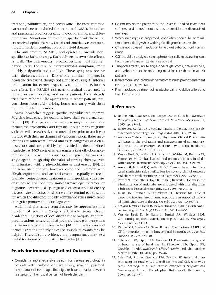
tramadol, zolmitriptan, and prednisone. The most common
parenteral agents included the parenteral NSAID ketorolac,
and parenteral prochlorperazine, metoclopramide, and chlor-
promazine. Almost one-third of non-specific headache suffer-
ers received opioid therapy. Use of anti-emetics was common,
though mostly in combination with opioid therapy.
The anti-emetics, NSAIDS, and opiates all provide non-
specific headache therapy. Each delivers its own side effects,
as well. The anti-emetics, prochlorperazine, and promet-
hazine, carry the risk of extrapyramidal symptoms, most
notably a dystonia and akathisia. These symptoms reverse
with diphenhydramine. Droperidol, another non-specific
headache treatment, though not alone in causing QT interval
prolongation, has earned a special warning in the US for this
side effect. The NSAIDS risk gastrointestinal upset and, in
long-term use, bleeding, and many patients have already
tried them at home. The opiates tend to sedate patients, pre-
vent them from safely driving home and carry with them
the potential for dependence.
Some headaches suggest specific, individualized therapy.
Migraine headaches, for example, have their own armamen-
tarium [38]. The specific pharmacologic migraine treatments
include the ergotamines and triptans, though many migraine
sufferers will have already tried one of these prior to coming to
the ED. With their mechanism of vasoconstriction, these med-
ications are somewhat limited and even dangerous as a diag-
nostic tool and are probably best avoided in the undefined
headache. A 2005 meta-analysis suggests that dihydroergota-
mine is less effective than sumatriptan or phenothiazines as a
single agent – suggesting the value of starting therapy, even
for migraines, with a phenothiazine or anti-emetic [39]. In
the same meta-analysis, however, combined treatment with
dihydroergotamine and an anti-emetic – typically metoclo-
pramide – outperformed treatment with meperidine, valproate,
or ketorolac. The long-term non-pharmacologic therapies for
migraine – exercise, sleep, regular diet, avoidance of dietary
triggers – are all tactics of which we may remind patients, but
for which the diligence of daily compliance relies much more
on regular primary and neurologic care.
Additional alternative remedies may be appropriate in a
number of settings. Oxygen effectively treats cluster
headaches. Injection of local anesthetic at occipital and tem-
poral locations where applied pressure increases symptoms
may relieve recalcitrant headaches [40]. If muscle strain and
torticollis are the underlying cause, muscle relaxants may be
helpful. There is some evidence that acupuncture may be a
useful treatment for idiopathic headache [41].
Pearls for Improving Patient Outcomes
• Consider a more extensive search for serious pathology in
patients with headache who are elderly, immunosuppressed,
have abnormal neurologic findinigs, or have a headache which
is atypical of their usual pattern of headache pain.
• Do not rely on the presence of the “classic” triad of fever, neck
stiffness, and altered mental status to consider the diagnosis of
meningitis.
• When meningitis is suspected, antibiotics should be adminis-
tered immediately while waiting for diagnostic test results.
• CT cannot be used in isolation to rule out subarachnoid hemor-
rhage.
• CSF should be analyzed spectophotometrically to assess for xan-
thochromia to maximize diagnostic yield.
• Temporal arteritis, acute angle-closure glaucoma, pre-eclampsia,
and carbon monoxide poisoning must be considered in at risk
patients.
• Infratentorial and cerebellar hematomas must prompt emergent
neurosurgical consultation.
• Pharmacologic treatment of headache pain should be tailored to
the likely etiology.
References
1. Raskin NH. Headache. In: Kasper DL, et al. (eds), Harrison’s
Principles of Internal Medicine, 16th ed. New York: McGraw-Hill,
2005, pp. 85–94.
2. Edlow JA, Caplan LR. Avoiding pitfalls in the diagnosis of sub-
arachnoid hemorrhage. New Engl J Med 2000; 342:29–36.
3. American College of Emergency Physicians. Clinical Policy: criti-
cal issues in the evaluation and management of patients pre-
senting to the emergency department with acute headache.
Ann Emerg Med 2002; 39:108–22.
4 Van de Beek D, de Gans J, Spanjaard L, Weisfelt M, Reitsma JB,
Vermeulen M. Clinical features and prognostic factors in adults
with bacterial meningitis. New Engl J Med 2004; 351:1849–59.
5. Aronin SI, Peduzzi P, Quagliarello VJ. Community-acquired bac-
terial meningitis: risk stratification for adverse clinical outcome
and effect of antibiotic timing. Ann Intern Med 1998; 129:862–9.
6. Proulx N, Frechette D, Toye B, Chan J, Kravcik S. Delays in the
administration of antibiotics are associated with mortality from
adult acute bacterial meningitis. QJM 2005; 98:291–8.
7. Talan DA, Hoffman JR, Yoshikawa TT, Overturf GD. Role of
empiric antibiotics prior to lumbar puncture in suspected bacter-
ial meningitis: state of the art. Rev Infect Dis 1988; 10:365–76.
8. deGans J, Van de Beek D. Dexamethasone in adults with bacte-
rial meningitis. New Engl J Med 2002; 347:1549–56.
9. Van de Beek D, de Gans J, Tunkel AR, Wijdicks EFM.
Community-acquired bacterial meningitis in adults. New Engl J
Med 2006; 354:44–53.
10. Kidwell CS, Chalela JA, Saver JL, et al. Comparison of MRI and
CT for detection of acute intracerebral hemorrhage. J Am Med
Assoc 2004; 292:1823–30.
11. Silberstein SD, Lipton RB, Goadsby PJ. Diagnostic testing and
ominous causes of headache. In: Silberstein SD, Lipton RB,
Goadsby PJ (eds), Headache in Clinical Practice, 2nd edn. London:
Martin Dunitz Ltd, 2002, pp. 35–46.
12. Sklar EM, Ruiz A, Quencer RM, Falcone SF Structural neu-
roimaging. In: Bradley WG, Daroff RB, Fenichel GM, Jankovic J
(eds), Neurology in Clinical Practice: Principles of Diagnosis and
Management, 4th ed. Philadelphia: Butterworth Heinemann,
2004, pp. 521–97.
44 | Chapter 5

13. Polmear A. Sentinel headaches in aneurismal subarachnoid
haemorrhage: what is the true incidence? A systematic review.
Cephalgia 2003; 23:935–41.
14. Sidman R, Connolly E, Lemke T. Subarachnoid hemorrhage
diagnosis: lumbar puncture is still needed when the computed
tomography scan is normal. Acad Emerg Med 1996; 3:827–31.
15. Boesiger BM, Shiber JR. Subarachnoid hemorrhage diagnosis
by computed tomography and lumbar puncture: are fifth gen-
eration CT scanners better at identifying subarachnoid hemor-
rhage? J Emerg Med 2005; 29(1):23–27.
16. Graves P, Sidman R. Xanthochromia is not pathognomonic for
subarachnoid hemorrhage. Acad Emerg Med 2004; 11(2):131–35.
17. Edlow JA, Bruner KS, Horowitz GL. Xanthochromia: a survey
of laboratory methodology and its clinical implications. Arch
Pathol Lab Med 2002; 126:413–15.
18. Smetana GW, Schmerling RH. Does this patient have temporal
arteritis? J Am Med Assoc 2002; 287:92–101.
19. Lee DA, Higginbotham EJ. Glaucoma and its treatment: a review.
Am J Health-Syst Pharm 2005; 62:691–99.
20. Kao LW, Nanagas HA. Carbon monoxide poisoning. Med Clin N
Am 2005; 89:1161–94.
21. Hampson NB, Hampson LA. Characteristics of headache associ-
ated with acute carbon monoxide poisoning. Headache 2002;
42:220–23.
22. Sibai B, Dekker G, Kupferminc M. Pre-eclampsia. Lancet 2005;
365:785–99.
23. Sibai BM. Diagnosis, prevention, and management of eclamp-
sia. Obstet & Gynecol 2005; 105:402–10.
24. Friedman DI. Pseudotumor cerebri. Neurol Clin N Am 2004;
22:99–131.
25. Stam J. Thrombosis of the cerebral veins and sinuses. New Engl
J Med 2005; 352(17):1791–98.
26. Kennedy PGE. Viral encephalitis. J Neurol 2005; 252:268–72.
27. Singh RVP, Prusmack CJ, Morcos JJ. Spontaneous intracerebral
hemorrhage: non-arteriovenous malformation, nonaneurysm.
In: Winn HR (ed). Youmans Neurological Surgery, 5th ed.
Philadelphia: Saunders, 2004, pp. 1733–68.
28. Qureshi AI, Tuhrim S, Broderick JP, Batjer HH, Hondo H,
Hanley DF. Spontaneous intracerebral hemorrhage. New Engl J
Med 2001; 344:1450–60.
29. Roux PDL, Winn HR. Surgical decision making for the treat-
ment of cerebral aneurysms. In: Winn HR (ed). Youmans
Neurological Surgery, 5th ed. Philadelphia: Saunders, 2004,
pp. 1793–1812.
30. Mayer SA, Rincon F. Treatment of intracerebral haemorrhage.
Lancet Neurol 2005; 4:662–72.
31. Feigin VL, Anderson N, Rikel GJE, Algra A, van Gijn J, Bennett
DA. Corticosteroids for aneurysmal subarachnoid haemorrhage
and primary intracerebral haemorrhage. The Cochrane Database
of Systematic Reviews 2005; 3: CD004583.
32. Juvela S, Kase C. Advances in intracerebral hemorrhage man-
agement. Stroke 2006; 37(2):301–04.
33. Gordon PE, Chiang WK, Hexdall WK, Kovacs M, Levy P,
Compton S, Arafat R. Headache and hypertension – is there an
association? [Abstract] Acad Em Med 2004; 11(5)521.
34. Karras DJ, Ufberg JW, Harrigan RA, Wald DA, Botros MS,
McNamara RM. Lack of relationship between hypertension-
associated symptoms and blood pressure in hypertensive ED
patients. Am J Emerg Med 2005; 23:106–10.
35. Hagen K, Stovner LJ, Vatten L, et al. Blood pressure and risk
of headache: a prospective study of 22,685 adults in Norway.
J Neurol Neurosurg Psychiatry 2002; 72:463–66.
36. Vinson DR, Hurtado TR, Vandenberg JT, Banwart L. Variations
among emergency departments in the treatment of benign
headache. Ann Emerg Med 2003; 41:90–97.
37. Vinson DR. Treatment patterns of isolated benign headache in
US emergency departments. Ann Emerg Med 2002; 39:215–22.
38 Goadsby PJ, Lipton RB, Ferrari MD. Migraine – current under-
standing and treatment. New Engl J Med 2002; 346:257–70.
39. Colman I, Brown MD, Innes GD, Grafstein E, Roberts TE, Rowe
BH. Parenteral dihydroergotamine for acute migraine headache:
a systematic review of the literature. Ann Emerg Med 2005; 45:
393–401.
40. Brofeldt BT, Panacek EA. Pericranial injection of local anesthet-
ics for the management of resistant headaches. Acad Emerg Med
1998; 5:1224–29.
41. Melchart D, Linde K, Berman B, White A, Vickers A, Allais G,
Brinkhaus B. Acupuncture for idiopathic headache. The Cochrane
Database of Systematic Reviews 2001; 1: CD001218.
Headache Management | 45

Introduction
Neck pain can be a daunting problem in the emergency depart-
ment, with etiologies ranging from benign to life threatening.
The neck contains vascular, nerve, airway, digestive, and
bony structures, any of which may be the source of pain. In
addition, referred pain from other regions of the body may
present with neck pain. Fascial planes connect compartments
of the neck with the mediastinum and prevertebral spaces,
posing a risk of spread of infection from the neck to these
regions. Pitfalls in the management of the patient with neck
pain include failure to consider the diverse differential diagno-
sis, diagnostic failures due to reliance on insensitive features
of the history and exam, misunderstanding of the limitations
of diagnostic laboratory and imaging tests, and failure to treat
with specific therapy in a timely fashion. In this chapter we’ll
consider some unusual but dangerous causes of neck pain
which are prone to pitfalls in diagnosis and treatment, includ-
ing infections, vascular disasters, and spinal cord compres-
sion syndromes. Pitfalls in the diagnosis and treatment of
cervical spine trauma are discussed separately in the chapter
pertaining to the trauma.
Pitfall | Failure to consider a vascular causeof neck pain
Cervical artery dissection (CAD) is a rare condition. Failure to
recognize it can lead to subsequent devastating neurologic
impairment from ischemic or thrombotic events. Unlike aor-
tic dissection, CAD is a disease predominantly of the young
with 70% occurring in people aged 20–40 years [1]. Internal
carotid artery dissection (ICAD) is the most common cause of
stroke in young adults. Headache and neck pain are promi-
nent features and up to 25% of people experience neck pain
alone [1, 2]. Isolated neck pain is more common in ICAD
than in vertebral artery dissection (VAD). The neck pain of
CAD is usually sudden, severe, and persistent pain.
CAD occurs after major blunt trauma, but also after trivial
trauma such as coughing, defecation, sexual activity, sporting
activities, and chiropractic manipulation. One study found
that 16% of CAD occurred after chiropractic treatments [3].
Apparently spontaneous dissections are also well documented
in the literature [3–10]. Other risk factors include history of
migraines, hypertension, tobacco use, connective tissue dis-
orders, and vasculitides.
Pitfall | Reliance on a normal physical examto rule out arterial dissection
The most common presentation of ICAD is headache and/or
neck pain with ischemic symptoms that may not present
until several hours or even days after the onset of neck pain.
36–58% of patients will have an incomplete Horner’s syn-
drome (ptosis and miosis without anhydrosis) and �15% will
have lower cranial nerve palsies [1]. In addition to headache
or neck pain, VAD presents with posterior circulation stroke
or cerebellar signs such as vertigo (57%), nausea/vomiting
(53%), unilateral facial numbness (46%), unsteadiness/gait
disturbance (42%), diplopia (23%), dysarthria (15%), dys-
phagia (11%), or extremity paresthesias or weakness (11%)
[2]. Symptoms can be transient and neurological deficits can
be subtle or absent at presentation.
When CAD is suspected based on history or physical exami-
nation, radiologic imaging should be obtained to rule in or rule
out the diagnosis. Helical computed tomographic (CT) cervi-
cal angiography is the diagnostic test of choice for CAD in
the emergency department due to its excellent sensitivity
and specificity (both approaching 100%), availability and
rapidity of testing, ability to evaluate several alternative
diagnoses, and the non-invasive nature of the test (see
Figure 6.1). Alternative diagnostic techniques include con-
ventional cervical angiography (78–84% sensitive), vascular
ultrasound, or MRI/MRA of the head and neck which
is �95% sensitive. As with any testing technique, false posi-
tive and false negative results may rarely occur with CT
angiography, sometimes for technical reasons such as metal-
lic streak artifacts in the image field or failure to include the
entire artery in question. If significant suspicion for this
diagnosis exists despite a normal diagnostic imaging exam,
an additional exam using a different test modality should be
considered [11].
KEY FACT | Internal carotid artery dissection is the most
common cause of stroke in young adults.
Chapter 6 | Evaluation and Management of the Patient withNeck Pain
Joshua Broder & Anita L’Italien
46

CAD should be considered in any adult patient complaining of
a sudden onset of significant neck pain with a history of
trauma, however minor, or recent chiropractic manipulation.
Ischemic symptoms, visual changes and symptoms of posterior
stroke should be sought in the history. A thorough neurologi-
cal examination, especially of the cranial nerves, is essential.
A normal neurological exam in a patient with a suspicious his-
tory does not rule out the diagnosis and should prompt radio-
logic testing.
Pitfall | Failure to consider a cardiac cause ofneck pain
Neck pain is a well-known manifestation of cardiac ischemia,
although usually as an additional feature of classical anginal
symptoms such as chest pain or dyspnea. Rare case reports
describe patients with cervical pain as the sole expression of
coronary artery disease, although often in retrospect more
typical features of cardiac chest pain are evident, though subtle
[12]. The presence of arm pain associated with neck pain may
be interpreted as radiculopathy when in fact both symptoms
are related to coronary ischemia. Unfortunately, symptoms
of either radiculopathy or angina pectoris can be exacer-
bated by ambulation, position, or arm motions, confounding
the diagnosis. Consideration of a cardiac cause of neck pain
should be given in patients with multiple cardiac risk factors,
no history of trauma, and particularly when diagnostic eval-
uations for cervical pathology have been negative. A careful
review of associated symptoms should be performed.
Women may be more likely than men to describe neck pain
as a symptom of cardiac ischemia [13].
Pitfall | Failure to recognize an infectiouscause of neck pain
Physicians may fail to recognize an infectious cause of neck
pain for a variety of reasons, including incomplete differential
diagnosis or over-reliance on specific history, exam, laboratory,
and imaging data. Treatment failures may also occur, including
failure to administer antibiotics or failure to involve a surgi-
cal consultant.
A variety of significant infections within the neck may present
with neck pain (see Table 6.1). These include peritonsillar,
parapharyngeal, and retropharyngeal abscesses, epiglottitis,
Ludwig’s angina, meningitis, osteomyelitis, diskitis, cervical
epidural abscesses (CEA), mastoiditis, otitis, and dental infec-
tions. Infections of congenital structures such as branchial cleft
cysts and thyroglossal duct cysts may occur. Some unusual
forms of pharyngitis may cause severe neck or throat pain,
including diphtheria, and esophagitis from herpes, CMV
(cytomegalovirus), or candida infection. Lemierre’s syndrome,
an infectious thrombophlebitis of the internal jugular venous
system, can be devastating and may present with neck pain.
Deep cervical plane infections may spread via fascial planes
to the mediastinum, resulting in mediastinitis with a mortal-
ity as high as 60% [14].
Epiglottitis has seen a decline in children since the introduc-
tion of the Hib vaccine in the early 1990s. However, the dis-
ease continues to be found in adults at rates similar to or
higher than in the pre-vaccine era [15, 16]. In fact, in the vac-
cine era, adult cases of acute epiglottitis (83%) outnumber
KEY FACT | In the vaccine era, adult cases of
acute epiglott i t is outnumber pediatr ic cases.
KEY FACT | Deep cervical infections may spread via fascial
planes to the mediastinum, resulting in mediastinitus
with a mortality as high as 60%.
KEY FACT | CAD should be considered in any adult
patient complaining of sudden onset of significant neck
pain with a history of trauma, however minor, or recent
chiropractic manipulation.
Evaluation and Management of the Patient with Neck Pain | 47
A
B
Figure 6.1 VAD on CT with IV contrast.
The patient’s right vertebral artery shows a stenotic region (arrow A),
with near complete loss of the lumen at the level of the C2 ring in the
extraforaminal portion of the vessel. The dark gray region (arrow B) is a
thrombosed region of dissection.

pediatric cases (17%) [17]. In addition, although rare,
Haemophilus influenza-related epiglottitis can be found in fully
vaccinated children. A 10-year retrospective study in New
England found 19 cases of childhood epiglottitis in a single
children’s hospital, 6 positive for H. influenzae, and 5 of those
6 in children with up-to-date immunizations [18].
Pitfall | Failure to elicit historical or examfindings suggesting infection
A history of injection drug use should prompt consideration of
venous thrombosis or deep neck abscesses, including epidural
spinal abscess. Retained foreign bodies such as needle frag-
ments may also provide a nidus for infection. In a retrospective
review, patients failed to disclose a history of cervical injection
in 50% of cases [19, 20]. Cervical osteomyelitis has been
reported as a complication of cervical injection [21, 22].
Swallowed foreign bodies such as bone fragments may also
cause deep space infection. A history of incomplete immuniza-
tion or immigrant status may place the patient at risk of diph-
theria infection. Immune compromise such as HIV/AIDS
should prompt consideration of infection with herpes, CMV,
or candida. Neurological complaints may suggest CEA. Recent
infections, including distant sources such as UTI or skin
abscesses, can lead to deep space infections, meningitis or CEA
by hematogenous inoculation.
Patients with a “locus minoris resistantiae” of the cervical spine
may be at increased risk for hematogenous seeding. Examples
include spondylosis, degenerative joint disease, previous
laminectomy or non-penetrating trauma from a fall or motor
vehicle collision. Some underlying diseases also predispose
patients to developing CEAs such as diabetes mellitus, alco-
holism, cancer, chronic renal failure and those with compro-
mised immune states. Any previous invasive spinal procedure,
even several months prior to presentation, should increase
the suspicion of CEA. Patients typically will not offer this infor-
mation unless asked specifically; 18% percent of patients will
have no identifiable predisposing factors for CEA [23, 24].
Physical exam should be thorough, but normal findings
should be incorporated into the overall picture, not used in
isolation to exclude a disease entity. Careful attention should
be paid to temperature, heart rate, blood pressure, and respira-
tory rate. The neck should be inspected for asymmetry or
swelling. Palpation for fluctuance or adenopathy should be
performed. The thyroid should be examined. Any stridor or
pooling of secretions should be noted. The oropharynx should
be inspected, looking for lesions or swelling. The dentition
and sublingual spaces should be examined. The uvula position
should be noted, especially any deviation. Tonsils should be
examined for swelling or exudates. Trismus and spitting of
secretions are also ominous signs. Brawny edema of the sub-
mandibular region and elevation of the tongue are signs of
Ludwig’s angina. A pseudomembrane should prompt con-
sideration of diphtheria. A careful neurological examination
of the cranial nerves and upper extremities should be per-
formed if suggested by history or symptoms.
Pitfall | Reliance on the absence of classicphysical exam findings to rule out infection
The classic physical exam findings of many dangerous neck
infections are not reliably present in patients with disease,
and their absence should not be used to rule out pathology.
Although neck stiffness is a common complaint in meningi-
tis (88%) [25], classic exam signs of meningitis should not be
relied upon due to poor sensitivity: a prospective study found
Kernig’s sign and Brudzinski’s signs to be only 5% sensitive
for meningitis, and nuchal rigidity to be only 30% sensitive
[26]. According to a retrospective review comparing physical
exam and symptoms with CT diagnosis of deep neck infection,
the physical exam for deep cervical infections is relatively
insensitive, underestimating the extent of infection in 70%
of patients. Classic signs and symptoms of infection were pres-
ent with the following frequency: fever (75.4%), pain (89.2%),
odynophagia (63.1%), dysphagia (47.7%), neck swelling
(84.6%), localized pain (76.9%), local erythema (66.7%),
and localized increase in temperature (55.4%) [27]. In
epiglottitis, drooling is a presenting symptom in only 15.2%,
and stridor or dyspnea in only 8.6%. Although “hot potato”
voice may suggest epiglottitis [28], muffled voice is found in
just over half of patients (52.1%) [29].
Pitfall | Over-reliance on fever as a sign ofinfection
KEY FACT | In patients with meningitis … absence of
a fever at presentation is associated with nearly 40-fold
increased mortality.
48 | Chapter 6
Table 6.1 Infectious causes of neck pain.
Peritonsillar abscess
Parapharyngeal abscess
Retropharyngeal abscess
Epiglottitis
Ludwig’s angina
Meningitis
Osteomyelitis
Diskitis
Cervical epidural abscess
Mastoiditis
Otitis
Dental infections
Infections of congenital structures (e.g. branchial cleft cysts,
thyroglossal duct cysts)
Pharyngitis
Esophagitis
Lemierre’s syndrome
Deep cervical plane infections

Evaluation and Management of the Patient with Neck Pain | 49
Fever is an unreliable finding. Reviews of presenting symp-
toms of adult patients with epiglottis found mean presenting
temperature to be 37.4ºC [28, 29]. In patients with meningi-
tis, fever is sometimes absent (7–33%), and absence of a
fever status at presentation is associated with nearly 40-fold
increased mortality (odds ratio 39.4), perhaps due to delay
in antibiotic administration [30, 31]. In patients with CEA,
fever was present in only 79% of patient in one review [23]
and was �38.4 in only 32% of patients in another large
review [32]. In yet another study, the mean presenting tem-
perature was only 38° C [33]. The classic triad of localized
spine tenderness, progressive neurological deficits, and fever
occurs in only 37% of patients with CEA [34].
Pitfall | Reliance on peripheral WBC count to exclude infection
Laboratory tests may assist in the diagnosis of infection, but
over-reliance on test results such as WBC count may result
in misdiagnosis. A review of epiglottis presentations found
mean WBC counts of 14,134.4 5556.1/mm3, meaning that
a significant percentage of patients presented with counts
within the normal range (5000–10,000/mm3) [29]. Cases
have been reported of epiglottitis with normal WBC counts
and no fever [35].
In children younger than 90 days, a low WBC count (less than
5000 cells/mm3) actually is associated with a seven times
higher risk of meningitis compared to the risk of bacteremia
[36]. In adults, serum leukocyte count has not been shown to
be a useful test to rule out meningitis. Published cohorts of
community-acquired meningitis include significant numbers
of patients with low, normal, and high peripheral WBC counts
[37]. The peripheral WBC count should not be used as a
decision node to discontinue a workup for meningitis.
In CEA, an elevated WBC count is an unreliable finding as it
is normal in up to 40% of patients [32]. 95% of patients will
have an elevated erythrocyte sedimentation rate (ESR) with
an average value of 51 mm/h but this is also a non-specific
finding [32, 38].
WBC counts may not be elevated in the presence of signifi-
cant infection or abscess, and low or normal WBC counts may
occur in acutely infected but immunocompromised hosts.
Pitfall | Misinterpretation of cerebrospinalfluid results
Cerebrospinal fluid (CSF) testing may be misinterpreted to
rule out meningitis inappropriately. Among patients with
relatively low CSF WBC counts, the presence of markedly
abnormal protein, neutrophil, or glucose values increases the
likelihood of bacterial meningitis. Intermediate values of these
CSF markers have little diagnostic value. Specifically, in chil-
dren with CSF WBC less than 30/mm3, high likelihood ratios
for bacterial meningitis are found for protein greater than
120 mg/dl (LR 22), neutrophil count greater than 75% (LR
57), glucose less than 20 mg/dl (LR 15), and glucose greater
than 120 mg/dl (LR 20) [39]. The same investigators have
reported a formula to discriminate between bacterial and viral
meningitis in children with CSF white cell counts greater than
7/mm3, based on CSF glucose and protein, CSF neutrophil
count, and patient age. They report a “high” sensitivity; how-
ever, the actual range of sensitivities reported varies between
92% and 98% [40], which may be unacceptable to an indi-
vidual practitioner. This rule has not been validated, and no
similar rule has been used in adults. Other decision rules for
discriminating bacterial from viral meningitis in children
have been derived and validated. The Bacterial Meningitis
Score has been described as a sensitive discriminator between
bacterial and viral meningitis. The score (1 point each for
CSF protein �80 mg/dl, peripheral absolute neutrophil
count �10,000 cells/mm3, seizure before or at presentation,
and CSF neutrophil count �1000 cells/mm3, and 2 points
for a CSF Gram stain showing bacteria) has a reported nega-
tive predictive value of 100% when the score is 0, but a sen-
sitivity of only 87% for bacterial meningitis when the score
is 2 or more [41].
An additional consideration is the difference between sen-
sitivity and negative predictive value. Sensitivity is the abil-
ity of a test to be positive in patients with disease, and it is
independent of the prevalence of a disease in a population.
In contrast, negative predictive value refers to the likelihood
of absence of disease when the test is negative. Negative pre-
dictive value is subject to change, depending on the preva-
lence of disease. In a pediatric study, a CSF WBC count less
than 30 cells/mm3 was associated with a negative predictive
value for bacterial meningitis of 99.3%, in a population of
1617 patients undergoing lumbar puncture. While this
value seems quite good, in fact only 44 patients (2.7%) had
confirmed meningitis, so almost any test, even a poor one,
could indicate the absence of disease 97.3% of the time. But
of the 44 cases of bacterial meningitis which were identified,
5 had 0–3 cells/mm3, and 6 had 4–30 cells/mm3, giving a
cutoff of 30 cells a sensitivity of only 75% [42]!
Correction of the CSF results for suspected traumatic lum-
bar puncture represents another opportunity for pitfall.
Evidence is scant that a corrected CSF value is a valid test for
meningitis, with a single published article suggesting that a
CSF WBC count greater than 10 times that attributable to
trauma is a sensitive cut-off for meningitis [43].
Pitfall | Reliance on plain film to excludedeep neck infection
Classic plain film findings of epiglottitis (see Figure 6.2; see
Table 6.2), while helpful if present, may be inadequate to
KEY FACT | In children younger than 90 days, a low
WBC count … is associated with a seven times higher risk
of meningitis compared to the risk of bacteremia.

50 | Chapter 6
rule out the diagnosis [44]. CT may be used to diagnose
equivocal cases and to delineate complications such as local
abscess formation – of course with careful attention to the
patient’s airway while at CT [45].
Lateral neck X-ray (see Figure 6.3a) for suspected retropha-
ryngeal abscess has a limited sensitivity, around 80%, while
CT (see Figure 6.3b) is extremely sensitive (100%) for soft tis-
sue abnormalities associated with retropharyngeal abscesses
and cellulitis [46, 47].
When evaluating for retained foreign body, reliance on plain
X-ray is a potential pitfall. Cadaver studies comparing CT and
plain film for a variety of fish bones found CT uniformly
superior, with a sensitivity around 90% compared with 39%
for X-ray [48, 49].
CT itself may have limitations that restrict its use in the
management decision for deep neck infections. In studies
comparing the ability of CT to discriminate between celluli-
tis and abscess, with surgical findings used as the gold stan-
dard, CT is between 68% and 73.5% sensitive and 56%
specific for abscess [50, 51].
Figure 6.2 Epiglottitis on plain film
This lateral soft tissue neck film demonstrates a classic “thumbprint
sign” (arrow) of an inflamed epiglottis. The hyoid bone is also seen
(arrowhead). Of note, this is an adult patient, as shown by the mature
cervical vertebrae.
Table 6.2 Sensitivities and specificities for plain radiograph findings in
epiglottitis
Plain radiograph findings
for epiglottitis Sensitivity (%) Specificity (%)
Epiglottis width to anterior- 96 100
posterior (AP) width of
C4 �0.33
Prevertebral soft tissue to C4 37 100
ratio �0.5
Width of hypopharyngeal airway 44 87
to width of C4 �1.5
Aryepiglottic fold enlargement 85 100
Arytenoid swelling 70 100
(a)
(b)
Figure 6.3. Retropharyngeal abscess in a 6-year-old male with sore
throat.
(a) Lateral view of the neck shows abnormal widening of the
prevertebral soft tissues (double arrow). Although a variety of criteria
have been described, soft tissues which exceed the width of the
adjacent vertebral bodies should raise suspicion of abscess. (b) CT with
IV contrast shows an ill-defined hypoattenuation and soft tissue
prominence in the right retropharyngeal space (arrowhead),
representing an abscess. The airway (arrow) is displaced anteriorly
by the abscess.

Pitfall | Delaying treatment of seriousinfections
Patients at risk for meningitis or diphtheria should immedi-
ately undergo respiratory isolation and begin antibiotic ther-
apy. Delay greater than 6 h in antibiotic therapy for meningitis
is associated with a significant increase in mortality, with esti-
mates from nearly 2-fold to perhaps 40-fold. Delay in diag-
nosis due to CT scan preceding lumbar puncture increases the
risk of delay in antibiotic administration by a factor of 5.6.
In the case of CEA, emergent neurosurgical consultation
should be obtained after the administration of intravenous
antibiotics. Neurological function continues to deteriorate in
a significant portion of patients after initiation of antibiotics
and many experts believe that surgery is necessary even if no
neurological deficits are present on diagnosis. About 50% of
patients treated medically and admitted without neurologic
deficits will have neurological deficits on discharge com-
pared to 20% of those treated surgically before the develop-
ment of deficits [52]. Additionally, total paralysis can occur
in a matter of a few hours. Once neurological symptoms
develop, simple removal of the mass may not result in com-
plete recovery because other processes likely occur, in addi-
tion to the mechanical compression, that result in cord
ischemia such as venous thrombosis or focal septic emboli.
Ultimate outcome is likely determined by a number of fac-
tors including the duration of symptoms, the only factor
where it is possible for emergency physicians to effect a
change (by prompt diagnosis, antibiotic therapy, and consul-
tation with a neurosurgeon). Once paralysis develops, it is
usually irreversible if surgery is not performed within 24 h
of onset [23]. In a review of 188 patients, treatment initiated
after 36 h of weakness or paralysis resulted in complete
recovery in only 39% of patients [53].
Pitfall | Failure to consider a neck mass
Neck pain may be due to the presence of a previously undiag-
nosed cervical mass. These include malignant and benign neo-
plasms and congenital structures such as branchial cleft cysts
or thyroglossal duct cysts, which may be super-infected. Asso-
ciated symptoms of weight loss, dysphagia or odynophagia,
hoarseness, or neurological symptoms such as ptosis may sug-
gest malignancy. Cervical neoplasms increase in incidence
with age, although some neoplasms, such as lymphomas,
may occur in younger patients. In patients above the age of
40 years with solitary, benign-appearing cervical cysts, metasta-
tic squamous cell carcinoma may be found in up to 23.5%
[54]. Cigarette smoking and alcohol use are risk factors [55].
Congenital abnormalities may present with neck pain.
These include thyroglossal duct cysts in the midline and
branchial cleft cysts in the lateral neck. Both thyroglossal
duct cysts and branchial cleft cysts may be occult until infec-
tion occurs, presenting then with pain and swelling.
Pitfall | Failure to evaluate the patient forevidence of malignancy
The physical exam should include inspection of the orophar-
ynx for lesions, as oral cancers may spread locally or metasta-
size within the neck. The neck should be inspected for
asymmetry, and palpation should be performed, including the
thyroid gland and anterior and posterior lymph nodes. The
voice quality should be noted, and the patient should be asked
about changes in voice character. Although hoarseness may
be associated with a benign diagnosis such as laryngitis,
hoarseness or change in voice could indicate local involve-
ment of laryngeal structures or involvement of the recurrent
laryngeal nerve. A Horner’s syndrome (ptosis, miosis, anhy-
drosis) can occur from compression of sympathetic nerves
innervating the face, so the patient’s eyes should be exam-
ined for eyelid position and pupillary size and reactivity.
Unfortunately, characteristics from exam and history, includ-
ing location, number of lesions, bilaterality, size, and dura-
tion, have poor positive and negative predictive values for
malignancy, in the range of 60–80% [55].
Pitfall | Reliance on plain film to exclude amass
Due to the insensitivity of history and exam for masses,
patients with unexplained neck pain may require imaging
for further diagnosis. Plain films of the neck are insensitive
and not generally useful. If a mass is considered, enhanced
CT (CT with IV contrast) is the preferred test, with excellent
sensitivity. MRI is helpful in patients with contraindications
to iodinated contrast, and ultrasound plays a role in differ-
entiating solid from cystic masses [56].
Pitfall | Failure to involve a consultant inpatients with mass lesions
Newly diagnosed cervical neoplasms require prompt follow-up,
though admission may not be required if the airway is not
threatened, the patient is tolerating oral intake, and pain is
adequately controlled. A firm outpatient follow-up plan should
be in place, with a clear understanding from the patient of the
significance of the emergency department findings and poten-
tial diagnosis. Otolaryngology referral is advisable for all cer-
vical masses, even those that appear to be benign or local
infections, as malignancy may co-exist. Importantly, incision
KEY FACT | In patients above the age of 40 years with
solitary, benign-appearing cervical cysts, metastatic
squamous cell carcinoma may be found in up to 23.5%.
Evaluation and Management of the Patient with Neck Pain | 51

and drainage of cervical masses should rarely be performed by
emergency physicians. Some cervical masses, including thy-
roglossal duct lesions, require complete excision for cure [57].
Emergency department incision and drainage may also be
contraindicated because in rare cases these lesions contain the
patient’s only functional thyroid tissue. Moreover, incision of
an unsuspected malignancy may promote spread and com-
promise chance for surgical cure, due to seeding of malignant
cells in needle tracks [58, 59]. Missed diagnoses of laryngeal
malignancy can have devastating medical consequences for
patients, with high rates of laryngectomy (63%) and mortal-
ity (35%). Patients with misdiagnosed laryngeal carcinoma
present at an early age (mean 47 years), compared with the
peak incidence of the disease (age 70–74 years). Malpractice
awards occur in over half of cases [60].
Pitfall | Failure to consider spinal epiduralhematoma in patients with neck pain andupper extremity paresthesias or weakness
Cervical epidural hematomas (CEH) are rare, but life threat-
ening. Mortality is 30% for CEH located above C5 [61]. CEH
are almost uniformly fatal secondary to respiratory failure if
not corrected [62]. Failure to recognize the diagnosis promptly
leads to a less favorable outcome and more severe permanent
neurological deficits. Because they are so rare, can occur spon-
taneously, and present with non-specific symptoms, they
present a difficult diagnostic dilemma.
CEHs are caused by trauma, underlying hematological dis-
orders or systemic disease, or as a complication of epidural
anesthesia [63]. However, there are many reports of sponta-
neous spinal epidural hematomas (SSEH) in the literature
which are defined as occurring in the absence of one of the
above causes [61, 62, 64–69]. They occur in as many as
40–50% of cases. SSEH has been reported to occur after
minor physical exertion, sneezing, coitus, straining, vomiting,
micturition, coughing and bending over. There are several case
reports of CEHs occurring after chiropractic manipulation
[70–73]. Other risk factors include anticoagulation, prior
thrombolytic therapy, pregnancy, alcoholism, liver disease,
and rheumatologic disorders.
Pitfall | Reliance on a normal neurologicexam to exclude spinal epidural hematoma
The usual presentation is that of an acute onset of severe pos-
terior neck pain that progresses to para- or tetraplegia in hours
or days. Cervical radicular pain is one of the earliest symptoms
and a hallmark of cervical epidural hematoma [74]. Although
most neck pain due to trauma that presents with radicular
symptoms will be due to an injured cervical disk or nerve
root, the emergency physician should consider the diagnosis
of SSEH and perform a thorough neurological examination.
The presence of other risk factors may necessitate imaging to
rule out a hematoma.
Pitfall | Failure to perform timely andappropriate imaging and consultationfor spinal epidural hematoma
Diagnosis requires MRI or CT myelography, and the diagnosis
may be missed on non-contrast CT or CT angiography per-
formed for suspected CAD. Treatment involves emergent
decompressive laminectomy with clot evacuation. It is also
imperative to reverse any existing coagulopathy.
Early neurosurgical involvement is essential. The duration
of symptoms affects the outcome as does the level and
extent of the lesion and the severity of the neurological
deficits. Patients obtain a useful recovery �50% of the time
when surgery is delayed for more than 36 h after symptom
onset [75]. In one review, only 15/27 recovered completely
when surgery was delayed for more than 36 h after symp-
toms onset [76].
Pearls for Improving Patient Outcomes
• Consider CAD in patients complaining of sudden onset of signif-
icant neck pain with history of trauma, however minor.
Neurological symptoms may be absent at presentation.
• Assess for a cardiac cause of neck pain, especially in women, and
evaluate for other symptoms or risk factors which may suggest
coronary artery disease.
• Suspect CEA in patients with significant neck pain and risk fac-
tors such as injection drug use, diabetes, immunocompromised,
chronic renal failure, cancer, alcoholism, recent distant or con-
tiguous infections, or recent invasive spinal procedure.
• Never rule out meningitis using Kernig’s and Brudzinski’s signs,
as they are poorly sensitive.
• Do not rely on the absence of fever or a normal WBC count alone
to rule out infection.
• Do not rule out meningitis using a corrected CSF value for trau-
matic lumbar punctures.
• Do not rely on plain films to exclude deep neck infections.
• Give immediate antibiotics for suspected meningitis, as delay in
antibiotic administration for CT or lumbar puncture significantly
increases mortality.
• Arrange specific follow-up for patients with undifferentiated
neck masses.
• Exercise caution in incision and drainage of cervical masses in
the emergency department due to the possibility of malignancy.
Consultation may be advisable.
• Suspect cervical epidural hematoma in a patient with neck pain
and radicular symptoms who has any of the following risk factors:
anticoagulation, pregnancy, liver disease, rheumatologic disor-
ders, preceding minor trauma or chiropractic manipulation.
52 | Chapter 6
Evaluation and Management of the Patient with Neck Pain | 53
References
1. Lee WW, Jensen ER. Bilateral internal carotid artery dissection
due to trivial trauma. J Emerg Med 2000; 19(1):35–41.
2. Saeed AB, Shuaib A, Al-Sulaiti G, Emery D. Vertebral artery
dissection: warning symptoms, clinical features and prognosis
in 26 patients. Can J Neurol Sci 2000; 27(4):292–6.
3. Dziewas R, Konrad C, Drager B, Evers S, Besselmann M,
Ludemann P, Kuhlenbaumer G, Stogbauer F, Ringelstein EB.
Cervical artery dissection – clinical features, risk factors, therapy
and outcome in 126 patients. J Neurol 2003; 250(10):1179–84.
4. Wessels T, Rottger C, Kaps M, Traupe H, Stolz E. Upper cranial
nerve palsy resulting from spontaneous carotid dissection. J
Neurol 2005; 252(4):453–6.
5. Chiche L, Praquin B, Koskas F, Kieffer E. Spontaneous dissection
of the extracranial vertebral artery: indications and long-term
outcome of surgical treatment. Ann Vasc Surg 2005; 19(1):5–10.
6. Wessels T, Sparing R, Neuschaefer-Rube C, Klotzsch C. Vocal
cord palsy resulting from spontaneous carotid dissection.
Laryngoscope 2003; 113(3):537–40.
7. Guidetti D, Pisanello A, Giovanardi F, Morandi C, Zuccoli G,
Troiso A. Spontaneous carotid dissection presenting lower cra-
nial nerve palsies. J Neurol Sci 2001; 184(2):203–7.
8. Rees JH, Valentine AR, Llewelyn JG. Spontaneous bilateral
carotid and vertebral artery dissection presenting as a Collet-
Sicard syndrome. Br J Radiol 1997; 70(836):856–8.
9. Silbert PL, Mokri B, Schievink WI. Headache and neck pain in
spontaneous internal carotid and vertebral artery dissections.
Neurology 1995; 45(8):1517–22.
10. Mokri B, Houser OW, Sandok BA, Piepgras DG. Spontaneous
dissections of the vertebral arteries. Neurology 1988; 38(6):
880–5.
11. Munera F, Cohn S, Rivas LA. Penetrating injuries of the neck:
use of helical computed tomographic angiography. J Trauma
2005; 58(2):413–18.
12. Lipetz JS, Ledon J, Silber J. Severe coronary artery disease pre-
senting with a chief complaint of cervical pain. Am J Phys Med
Rehabil 2003; 82(9):716–20.
13. Chen W, Woods SL, Puntillo KA. Gender differences in symp-
toms associated with acute myocardial infarction: a review of
the research. Heart Lung 2005; 34(4):240–7.
14. Cardenas-Malta KR, Cortes-Flores AO, Fuentes-Orozco C, Del
Carmen Martinez-Oropeza L, Lopez-Ramirez MK, Gonzalez-
Ojeda A. Necrotizing mediastinitis in deep neck infections. Cir
Cir 2005; 73(4):263–7. [Article in Spanish]
15. McVernon J, Trotter CL, Slack MP, Ramsay ME. Trends in
Haemophilus influenzae type b infections in adults in England and
Wales: surveillance study. Br Med J 2004; 329(7467):655–8.
16. Berger G, Landau T, Berger S, Finkelstein Y, Bernheim J, Ophir
D. The rising incidence of adult acute epiglottitis and epiglottic
abscess. Am J Otolaryngol 2003; 24(6):374–83.
17. Wood N, Menzies R, McIntyre P. Epiglottitis in Sydney before
and after the introduction of vaccination against Haemophilus
influenzae type b disease. Inter Med J 2005; 35(9):530–5.
18. Shah RK, Roberson DW, Jones DT. Epiglottitis in the
Hemophilus influenzae type B vaccine era: changing trends.
Laryngoscope 2004; 114(3):557–60.
19. Williams MF, Eisele DW, Wyatt SH. Neck needle foreign bodies
in intravenous drug abusers. Laryngoscope 1993; 103(1 Pt 1):
59–63.
20. Myers EM, Kirkland LS Jr, Mickey R. The head and neck seque-
lae of cervical intravenous drug abuse. Laryngoscope 1988;
98(2):213–18.
21. Smith MA, Trowers NR, Klein RS. Cervical osteomyelitis caused
by Pseudomonas cepacia in an intravenous-drug abuser. J Clin
Microbiol 1985; 21(3):445–6.
22. Sapico FL, Montgomerie JZ. Vertebral osteomyelitis in intra-
venous drug abusers: report of three cases and review of the lit-
erature. Rev Infect Dis 1980; 2(2):196–206.
23. Darouiche RO, Hamill RJ, Greenberg SB, Weathers SW,
Musher DM. Bacterial spinal epidural abscess. Review of 43 cases
and literature survey. Medicine (Baltimore) 1992; 71(6):369–85.
24. Ravicovitch MA, Spallone A. Spinal epidural abscesses: surgical
and parasurgical management. Eur Neurol 1982; 21:347–57.
25. Durand ML, Calderwood SB, Weber DJ, Miller SI, Southwick FS,
Caviness Jr. VS, Swartz MN. Acute bacterial meningitis in adults.
A review of 493 episodes.
26. Thomas KE, Hasbun R, Jekel J, Quagliarello VJ. The diagnostic
accuracy of Kernig’s sign, Brudzinski’s sign, and nuchal rigidity
in adults with suspected meningitis. Clin Infect Dis 2002;
35(1):46–52. Epub 2002 Jun 5.
27. Crespo AN, Chone CT, Fonseca AS, Montenegro MC, Pereira R,
Milani JA. Clinical versus computed tomography evaluation in
the diagnosis and management of deep neck infection. Sao
Paulo Med J 2004; 122(6):259–63. Epub 2005 Feb 2.
28. Shah RK, Roberson DW, Jones DT. Epiglottitis in the Hemophilus
influenzae type B vaccine era: changing trends. Laryngoscope
2004; 114(3):557–60.
29. Chang YL, Lo SH, Wang PC, Shu YH. Adult acute epiglottitis:
experiences in a Taiwanese setting. Otolaryngol Head Neck Surg
2005; 132(5):689–93.
30. Proulx N, Frechette D, Toye B, Chan J, Kravcik S. Delays in the
administration of antibiotics are associated with mortality from
adult acute bacterial meningitis. Q J Med 2005; 98(4):291–8.
Epub 2005 Mar 10.
31. van de Beek D, de Gans J, Spanjaard L, Weisfelt M, Reitsma JB,
Vermeulen M. Clinical features and prognostic factors in
adults with bacterial meningitis. N Engl J Med 2004; 351(18):
1849–59.
32. Rigamonti D, Liem L, Sampath P, Knoller N, Namaguchi Y,
Schreibman DL, Sloan MA, Wolf A, Zeidman S. Spinal epidural
abscess: contemporary trends in etiology, evaluation, and man-
agement. Surg Neurol 1999; 52(2):189–96.
33. Redekop GJ, Del Maestro RF. Diagnosis and management of
spinal epidural abscess. Can J Neurol Sci 1992; 19(2):180–7.
34. Ekbom DC, D-Elia J, Isaacson B, Lamarca F, Chepeha DB,
Bradford CR. Spinal epidural abscess after cervical pharyngoe-
sophageal dilation. Head Neck 2005; 27(6):543–8.
35. Jerrard DA, Olshaker J. Simultaneous uvulitis and epiglottitis
without fever or leukocytosis. Am J Emerg Med 1996; 14(6):551–2.
36. Bonsu BK, Harper MB. A low peripheral blood white blood cell
count in infants younger than 90 days increases the odds of
acute bacterial meningitis relative to bacteremia. Acad Emerg
Med 2004; 11(12):1297–301.
37. Aronin SI, Peduzzi P, Quagliarello VJ. Community-acquired
bacterial meningitis: risk stratification for adverse clinical out-
come and effect of antibiotic timing. Ann Intern Med 1998;
129(11):862–9.
38. Wong D, Raymond NJ. Spinal epidural abscess. NZ Med J 1998;
111:345–7.

54 | Chapter 6
39. Bonsu BK, Harper MB. Accuracy and test characteristics of
ancillary tests of cerebrospinal fluid for predicting acute bacter-
ial meningitis in children with low white blood cell counts in
cerebrospinal fluid. Acad Emerg Med 2005; 12(4):303–9.
40. Bonsu BK, Harper MB. Differentiating acute bacterial meningi-
tis from acute viral meningitis among children with cere-
brospinal fluid pleocytosis: a multivariable regression model.
Pediatr Infect Dis J 2004; 23(6):511–7.
41. Nigrovic LE, Kuppermann N, Malley R. Development and valida-
tion of a multivariable predictive model to distinguish bacterial
from aseptic meningitis in children in the post-Haemophilus
influenzae era. Pediatrics 2002; 110(4):712–19.
42. Freedman SB, Marrocco A, Pirie J, Dick PT. Predictors of bacte-
rial meningitis in the era after Haemophilus influenzae. Arch
Pediatr Adolesc Med 2001; 155(12):1301–6.
43. Mayefsky JH, Roghmann KJ. Determination of leukocytosis in
traumatic spinal tap specimens. Am J Med 1987; 82(6):1175–81.
44. Nemzek WR, Katzberg RW, Van Slyke MA, Bickley LS. A reap-
praisal of the radiologic findings of acute inflammation of the
epiglottis and supraglottic structures in adults. Am J Neuroradiol
1995; 16(3):495–502.
45. Smith MM, Mukherji SK, Thompson JE, Castillo M. CT in adult
supraglottitis. Am J Neuroradiol 1996; 17(7):1355–8.
46. Boucher C, Dorion D, Fisch C. Retropharyngeal abscesses: a clini-
cal and radiologic correlation. J Otolaryngol 1999; 28(3):134–7.
47. Nagy M, Backstrom J. Comparison of the sensitivity of lateral
neck radiographs and computed tomography scanning in pedi-
atric deep-neck infections. Laryngoscope 1999; 109(5):775–9.
48. Palme CE, Lowinger D, Petersen AJ. Fish bones at the cricopha-
ryngeus: a comparison of plain-film radiology and computed
tomography. Laryngoscope 1999; 109(12):1955–8.
49. Lue AJ, Fang WD, Manolidis S. Use of plain radiography and
computed tomography to identify fish bone foreign bodies.
Otolaryngol Head Neck Surg 2000; 123(4):435–8.
50. Vural C, Gungor A, Comerci S. Accuracy of computerized
tomography in deep neck infections in the pediatric population.
Am J Otolaryngol 2003; 24(3):143–8.
51. Stone ME, Walner DL, Koch BL, Egelhoff JC, Myer CM.
Correlation between computed tomography and surgical find-
ings in retropharyngeal inflammatory processes in children. Int
J Pediatr Otorhinolaryngol 1999; 49(2):121–5.
52. Curry WT, Hoh BL, Amin-Hanjani S, Eskandar EN. Spinal
epidural abscess: clinical presentation, management, and out-
come. Surg Neurol 2005; 63(4):364–71.
53. Williams JW, Powell T. Epidural abscess of the cervical spine:
case report and literature review. Br J Radiol 1990; 63(751):576–8.
54. Gourin CG, Johnson JT. Incidence of unsuspected metastases in
lateral cervical cysts. Laryngoscope 2000; 110(10 Pt 1):1637–41.
55. Bhattacharyya N. Predictive factors for neoplasia and malig-
nancy in a neck mass. Arch Otolaryngol Head Neck Surg 1999;
125(3):303–7.
56. Eskey CJ, Robson CD, Weber AL. Imaging of benign and malig-
nant soft tissue tumors of the neck. Radiol Clin N Am 2000;
38(5):1091–104.
57. Josephson GD, Spencer WR, Josephson JS. Thyroglossal duct
cyst: the New York Eye and Ear Infirmary experience and a litera-
ture review. Ear Nose Throat J 1998; 77(8):642–4, 646–7, 651.
58. Gritzmann N, Hollerweger A, Macheiner P, Rettenbacher T.
Sonography of soft tissue masses of the neck. J Clin Ultrasound
2001; 30(6):356–73.
59. Mighell AJ, High AS. Histological identification of carcinoma in
21 gauge needle tracks after fine needle aspiration biopsy of
head and neck carcinoma. J Clin Pathol 1998; 51(3):241–3.
60. Lydiatt DD. Medical malpractice and cancer of the larynx.
Laryngoscope 2002; 112(3):445–8.
61. Demierre B, Unger PF, Bongioanni F. Sudden cervical pain:
spontaneous cervical epidural hematoma. Am J Emerg Med
1991; 9(1):54–6.
62. Williams JM, Allegra JR. Spontaneous cervical epidural
hematoma. Ann Emerg Med 1994; 23(6):1368–70.
63. Stoll A, Sanchez M. Epidural hematoma after epidural block:
implications for its use in pain management. Surg Neurol 2002;
57(4):235–40.
64. Tender GC, Awasthi D. Spontaneous cervical spinal epidural
hematoma in a 12-year old girl: case report and review of the
literature. J La State Med Soc 2004; 156(4):196–8.
65. Steinmetz MP, Kalfas IH, Willis B, Chalavi A, Harlan RC.
Successful surgical management of a case of spontaneous
epidural hematoma of the spine during pregnancy. Spine J
2003; 3(6):539–42.
66. Ferry P, Reisner C, Ashpole R. A case of spontaneous cervical
extradural haematoma. Hosp Med 2001; 62(7):436–7.
67. Vaya A, Resureccion M, Ricart JM, Ortuno C, Ripoll F, Mira Y,
Aznar J. Clin Appl Thromb Hemost 2001; 7(2):166–8.
68. Lobitz B, Grate I. Acute epidural hematoma of the cervical spine:
an unusual cause of neck pain. South Med J 1995; 88(5):580–2.
69. Nagel MA, Taff IP, Cantos EL, Patel MP, Maytal J, Berman D.
Spontaneous spinal epidural hematoma in a 7-year-old girl.
Diagnostic value of magnetic resonance imaging. Clin Neurol
Neurosurg 1989; 91(2):157–60.
70. Lin IY. Diagnostic pitfall: nontraumatic spinal epidural hematoma
mimicking a brainstem stroke. Ann Emerg Med 2004; 44(2):183–4.
71. Saxler G, Barden B. Extensive spinal epidural hematoma—an
uncommon entity following cervical chiropractic manipulation.
Z Orthop Ihre Grenzgeb 2004; 142(1):79–82.
72. Segal DH, Lidov MW, Camins MB. Cervical epidural hematoma
after chiropractic manipulation in a healthy young woman:
case report. Neurosurgery 1996; 39(5):1043–5.
73. Zupruk GM, Mehta Z. Brown-sequard syndrome associated
with posttraumatic cervical epidural hematoma: case report
and review of the literature. Neurosurgery 1989; 25(2):278–80.
74. Sakamoto N, Yanaka K, Matsumaru Y, Nose T. Cervical epidural
hematoma causing hemiparesis. Arch Neurol 2003; 60(5):783.
75. McDonnell GV, Bell KE, Hawkins SA. A pain in the neck.
Postgrad Med J 2000; 76(891):229–30.
76. Patel H, Boaz JC, Phillips JP, Garg BP. Spontaneous spinal epidural
hematoma in children. Pediatr Neurol 1998; 19(4):302–7.
Introduction
Emergency medicine physicians (EPs) provide the initial
trauma care of most trauma patients. Although we often
work in concert with the trauma surgeons on the more seri-
ously injured patients, it behooves us to be on the cutting
edge of trauma care for all of our injured patients. Several
pitfalls that frequently surface as areas of concern for many
EPs are addressed here.
Pitfall | Failure to implement surrogates to crystalloid during resuscitation
Crystalloid fluid resuscitation is crucial to maintain perfusion
to vital organs. It is important, however, to frequently reassess
the patient between boluses to determine whether he/she is a
responder, partial responder, or non-responder to the fluids.
This allows one to determine the need for blood products,
surgical intervention, or other therapies. Therefore, infusing
predetermined boluses of fluid is essential to allowing one to
determine whether alternate therapies need be initiated.
Overzealous fluid administration can have several deleteri-
ous physiologic effects. Elevating the blood pressure above
the tamponade pressure of post-traumatic clot formation may
cause clot disruption and worsen bleeding and thus shock.
Also, hemodilution causing a progressive anemia decreases
the oxygen carrying capacity of the intravascular space and
decreases the viscosity of the blood allowing it to more easily
elute around an incomplete thrombus [1]. By judiciously uti-
lizing fluid resuscitation, selecting and implementing proper
blood products or surrogates when required (red blood cells
(RBC), plasma, platelets, clotting factors, rFVIIa), and better
monitoring the response to fluids, we should be able to
improve outcomes by limiting further hemorrhage and min-
imizing the complications of hemodilution.
As optimal perfusion pressure must be maintained, elevation
of the blood pressure above that of the tamponade pressure
of the clot may be required. In doing so, a clinical trade off is
made and must be recognized. This allows for more aggres-
sive management of the point of active bleeding, as well as
resuscitation with oxygen carrying products to replace the
lost hemoglobin. Administration of other perfusion adjuncts
can minimize the volume of crystalloid necessary to main-
tain perfusion. The rapidity of which definitive hemorrhage
control is established is paramount to the survival of the
patient. Likewise, identifying and controlling a bleeding source
is critical to minimize ongoing blood loss.
Pitfall | Over-reliance on focused abdominal sonography in trauma
Focused abdominal sonography in trauma (FAST) examina-
tions has rapidly replaced diagnostic peritoneal lavage (DPL)
in the evaluation of unstable or stable trauma patients as
part of the primary or secondary survey. Multiple studies
have reported the sensitivity of the FAST examination for
determining the presence of intra-abdominal fluid to be
90%, with a specificity of 99%, and an accuracy of 98%
[2–4]. As such, the FAST examination has its primary utility
in the unstable trauma patient who should not go to com-
puted tomography (CT) scan, but rather the operating room
due to increased suspicion for intra-abdominal injury as the
cause of instability. In patients who are hypotensive after
blunt abdominal trauma and not hemodynamically stable
enough to undergo diagnostic CT, negative US findings vir-
tually exclude intra-abdominal surgical injury as the cause
of hemodynamic instability, while positive US findings indi-
cate surgical injury in approximately 70% of cases [5]. The
FAST examination also may be helpful in a multi-patient
trauma to triage critically ill patients when not all can get a
CT scan in a timely manner.
In general, a positive FAST examination in the stable patient
does not mandate surgical intervention as non-operative
management for a solid organ injury may be indicated based
on patient stability, age, physiologic reserve, and severity of
injury based on CT grading. When indicated, CT imaging of
KEY FACT | In stable patients, a negative FAST
examination does not obviate the need for CT and a
positive one does not mandate operative intervention.KEY FACT | Patients must be reassessed regularly after
boluses of predefined quantities of crystalloid. Only by
doing so can the need for other adjuncts be identified.
Chapter 7 | Trauma Management in the ED
David E. Manthey & Bret A. Nicks
55
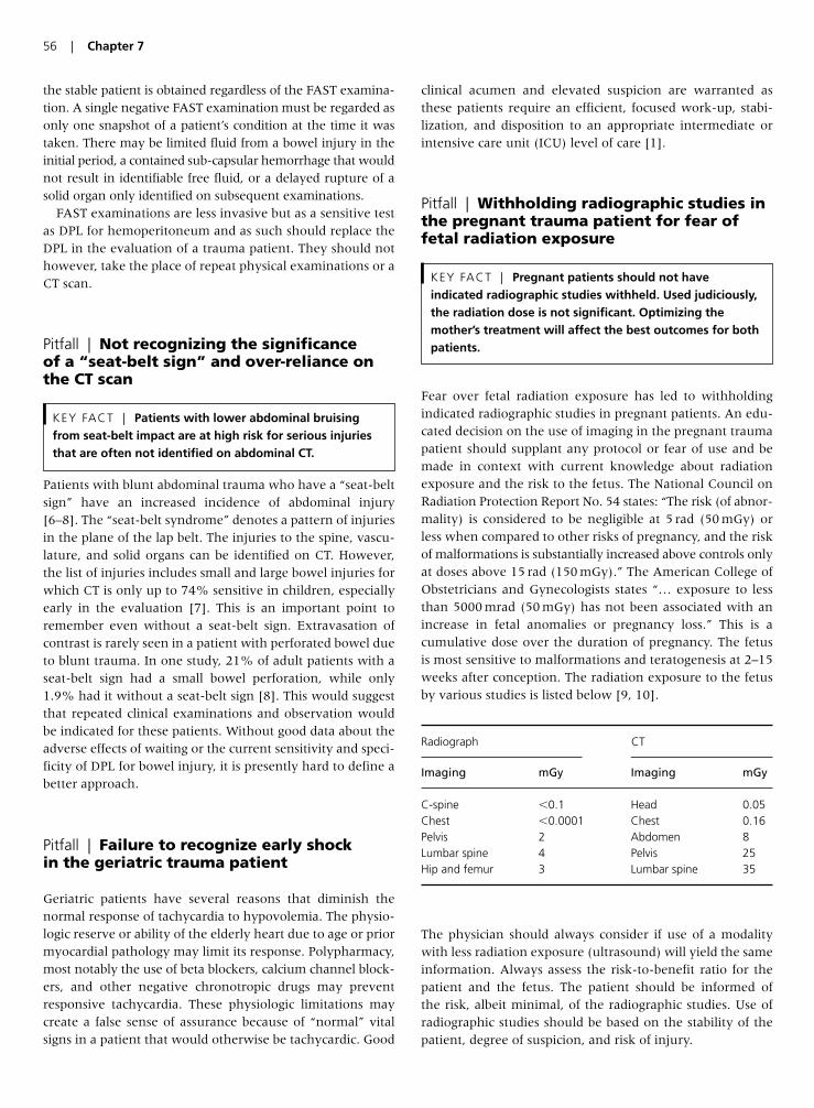
the stable patient is obtained regardless of the FAST examina-
tion. A single negative FAST examination must be regarded as
only one snapshot of a patient’s condition at the time it was
taken. There may be limited fluid from a bowel injury in the
initial period, a contained sub-capsular hemorrhage that would
not result in identifiable free fluid, or a delayed rupture of a
solid organ only identified on subsequent examinations.
FAST examinations are less invasive but as a sensitive test
as DPL for hemoperitoneum and as such should replace the
DPL in the evaluation of a trauma patient. They should not
however, take the place of repeat physical examinations or a
CT scan.
Pitfall | Not recognizing the significance of a “seat-belt sign” and over-reliance onthe CT scan
Patients with blunt abdominal trauma who have a “seat-belt
sign” have an increased incidence of abdominal injury
[6–8]. The “seat-belt syndrome” denotes a pattern of injuries
in the plane of the lap belt. The injuries to the spine, vascu-
lature, and solid organs can be identified on CT. However,
the list of injuries includes small and large bowel injuries for
which CT is only up to 74% sensitive in children, especially
early in the evaluation [7]. This is an important point to
remember even without a seat-belt sign. Extravasation of
contrast is rarely seen in a patient with perforated bowel due
to blunt trauma. In one study, 21% of adult patients with a
seat-belt sign had a small bowel perforation, while only
1.9% had it without a seat-belt sign [8]. This would suggest
that repeated clinical examinations and observation would
be indicated for these patients. Without good data about the
adverse effects of waiting or the current sensitivity and speci-
ficity of DPL for bowel injury, it is presently hard to define a
better approach.
Pitfall | Failure to recognize early shock in the geriatric trauma patient
Geriatric patients have several reasons that diminish the
normal response of tachycardia to hypovolemia. The physio-
logic reserve or ability of the elderly heart due to age or prior
myocardial pathology may limit its response. Polypharmacy,
most notably the use of beta blockers, calcium channel block-
ers, and other negative chronotropic drugs may prevent
responsive tachycardia. These physiologic limitations may
create a false sense of assurance because of “normal” vital
signs in a patient that would otherwise be tachycardic. Good
clinical acumen and elevated suspicion are warranted as
these patients require an efficient, focused work-up, stabi-
lization, and disposition to an appropriate intermediate or
intensive care unit (ICU) level of care [1].
Pitfall | Withholding radiographic studies inthe pregnant trauma patient for fear offetal radiation exposure
Fear over fetal radiation exposure has led to withholding
indicated radiographic studies in pregnant patients. An edu-
cated decision on the use of imaging in the pregnant trauma
patient should supplant any protocol or fear of use and be
made in context with current knowledge about radiation
exposure and the risk to the fetus. The National Council on
Radiation Protection Report No. 54 states: “The risk (of abnor-
mality) is considered to be negligible at 5 rad (50 mGy) or
less when compared to other risks of pregnancy, and the risk
of malformations is substantially increased above controls only
at doses above 15 rad (150 mGy).” The American College of
Obstetricians and Gynecologists states “… exposure to less
than 5000 mrad (50 mGy) has not been associated with an
increase in fetal anomalies or pregnancy loss.” This is a
cumulative dose over the duration of pregnancy. The fetus
is most sensitive to malformations and teratogenesis at 2–15
weeks after conception. The radiation exposure to the fetus
by various studies is listed below [9, 10].
The physician should always consider if use of a modality
with less radiation exposure (ultrasound) will yield the same
information. Always assess the risk-to-benefit ratio for the
patient and the fetus. The patient should be informed of
the risk, albeit minimal, of the radiographic studies. Use of
radiographic studies should be based on the stability of the
patient, degree of suspicion, and risk of injury.
KEY FACT | Pregnant patients should not have
indicated radiographic studies withheld. Used judiciously,
the radiation dose is not significant. Optimizing the
mother’s treatment will affect the best outcomes for both
patients.
KEY FACT | Patients with lower abdominal bruising
from seat-belt impact are at high risk for serious injuries
that are often not identified on abdominal CT.
56 | Chapter 7
Radiograph CT
Imaging mGy Imaging mGy
C-spine �0.1 Head 0.05
Chest �0.0001 Chest 0.16
Pelvis 2 Abdomen 8
Lumbar spine 4 Pelvis 25
Hip and femur 3 Lumbar spine 35

Pitfall | Inappropriate use of CTs and plainradiographs of the spine
Less than 50% of current academic emergency departments
continue to obtain additional lumbar and thoracic spine plain
films after obtaining a CT of the chest and abdomen. These
additional radiographs occur at additional expense and radi-
ation to the patient without additional diagnostic value. The
CT images can be reformatted to deliver images of the spinal
column with a sensitivity of 97% for thoracic spine fractures
and 95% for lumbar spine fractures. The CT may miss non-
operative compression fractures. The sensitivity of plain
radiographs is 62% for the thoracic spine and 86% for the
lumbar spine [11]. Two recent reviews of more than 3000
patients suggest that plain radiographs of the spine should
no longer be utilized in the trauma patient as CT has sup-
planted that radiographic modality [12]. The charges and
dose of radiation must be taken into consideration with
plain films charge about $145, and delivering 6.36 mSv of
radiation and CT charge about $880 and delivering
19.42 mSv CT for imaging of the lumbar spine [13]. Due to
the additional radiation exposure and cost for CT, perhaps
a more rational approach may be to reserve CT for those
in whom it is not possible to obtain adequate plain films
or those with a high suspicion of injury, however, further
research will be required to best identify a risk stratifying
approach.
Pitfall | Failure to appropriately apply C-spine clearance rules
Two C-spine clearance rules have come to the forefront in
recent years, the NEXUS rule and the Canadian C-spine
rule. Without getting into the discussion of which might be
better, the authors would like to emphasize the limitations
of such rules in general. We can all recall that NEXUS
requires a patient to have normal level of alertness, but how
many can define what that really means. The NEXUS study
defines it as an altered level of alertness which can include
any of the following:
1. Glasgow Coma Scale (GCS) score of 14 or less.
2. Disorientation to person, place, time, or events.
3. Inability to remember three objects at 5 min.
4. Delayed or inappropriate response to external stimuli.
This is not a downfall of the study, it is an issue with how we
often oversimplify a rule to make it easier to apply and
therefore utilize. But the NEXUS authors looked at their
criteria and state it best: “Because each of the five low-risk
criteria was the only marker of non-low-risk status in at least
a few patients with significant cervical spine injury, modifica-
tion of the overall NEXUS decision instrument by eliminating
any one of the criteria would markedly reduce sensitivity
and make the instrument unacceptable for clinical use [14].”
Neither rule unambiguously defines distracting injuries. A
recent study suggests that proximity of injury due to the gat-
ing theory that a noxious stimuli from the upper extremity
may provide enough counter-irritation through the spinal
pain pathways that other pain signals may go unnoticed
[15]. This may make upper extremity injuries distracting
even if they don’t fit the guidelines of either NEXUS or the
Canadian C-spine rules.
The Canadian C-spine rule removes anyone over the age
of 64. The NEXUS rule does not, and has looked at the geri-
atric population of the study citing a sensitivity of the NEXUS
criteria for all cervical spine injuries in the geriatric group of
98.5% (95% CI, 94.8–99.7%), and the sensitivity for clini-
cally significant injury was 100% (95% CI, 97.1–100%).
There are various case reports of elderly patients with injury,
but they often cite inappropriate evaluation of the level of
alertness. The NEXUS group urged caution in using the crite-
ria among children, especially those younger than 9 as there
was a limited number of patients and injuries in this group.
The Canadian C-spine rule was for patients 16 years and
older.
The Canadian C-spine rule [16] included the presence of a
concerning mechanism (fall from 1 m or greater, axial load
to head, motorized recreational vehicles, bicycle collision, or
motor vehicle collision (MVC) with high speed, rollover, or
ejection). The NEXUS study recognized that it was not utiliz-
ing a criterion for mechanism but all of these were certainly
included in the group that was studied.
The goal of both of these rules is to decrease the use of
unnecessary radiographs in the evaluation of the cervical
spine in blunt trauma. We doubt that either group wanted
their algorithms to supplant good clinical acumen and judg-
ment. Nevertheless, appropriate application of these rules
can identify patients at risk for cervical spine injury while
significantly reducing the number of cervical spine X-rays
necessary and should be encouraged. When doing so, how-
ever, it is imperative that the clinician know how to apply
the rules appropriately.
Pitfall | Failure to obtain CT imaging of thecervical spine in patients at high risk forinjury
KEY FACT | Because of its superior sensitivity, patients
in whom there is high suspicion for spinal injury should
have spinal CT.
KEY FACT | With newer CT scanners, spinal CTs can be
reformatted from abdominal CTs. Because of its superior
sensitivity, patients in whom there is high suspicion for
spinal injury should have spinal CT.
Trauma Management in the ED | 57
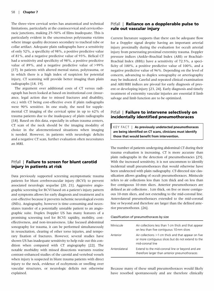
The three-view cervical series has anatomical and technical
limitations, particularly at the craniocervical and cervicotho-
racic junctions, making 25–50% of films inadequate. This is
particularly evident in the unconscious polytrauma victims
where image quality decreases secondary to tracheal tube or
collar artifact. Adequate plain radiographs have a sensitivity
of only 52%, a specificity of 98%, a positive predictive value
of 81%, and a negative predictive value of 93%. Helical CT
had a sensitivity and specificity of 98%, a positive predictive
value of 89%, and a negative predictive value of �99%
[17]. In patients with altered mental status, polytrauma, or
in which there is a high index of suspicion for potential
injury, CT scanning will provide better imaging than plain
radiographs [18, 19].
The argument over additional costs of CT versus radi-
ograph has been looked at based on institutional cost (insur-
ance, legal action due to missed fractures and disability,
etc.) with CT being cost-effective even if plain radiographs
were 90% sensitive. In one study, the need for supple-
mental CT imaging of the cervical spine in up to 62% of
trauma patients due to the inadequacy of plain radiographs
[18]. Based on this data, especially in urban trauma centers,
CT scan of the neck should be the imaging modality of
choice in the aforementioned situations when imaging
is needed. However, in patients with neurologic deficits
and a negative CT scan, further evaluation often necessitates
an MRI.
Pitfall | Failure to screen for blunt carotidinjury in patients at risk
Data previously supported screening asymptomatic trauma
patients for blunt cerebrovascular injury (BCVI) to prevent
associated neurologic sequelae [20, 21]. Aggressive angio-
graphic screening for BCVI based on a patient’s injury pattern
and symptoms allows for early diagnosis and treatment and is
cost-effective because it prevents ischemic neurological events
(INEs). Angiography, however is time-consuming and neces-
sitates transfer of a potentially unstable patient to an angio-
graphic suite. Duplex Doppler US has many features of a
promising screening tool for BCVI: rapidity, mobility, cost-
effectiveness, and non-invasiveness. Like focused abdominal
sonography for trauma, it can be performed simultaneously
in resuscitation, clearing of other torso injuries, and tempo-
rary fixation of fractures. However, several studies have
shown US has inadequate sensitivity to help rule out this con-
dition when compared with CT angiography [22]. The
notable morbidity with missed dissections warrants routine
contrast-enhanced studies of the carotid and vertebral vessels
when injury is suspected in blunt trauma patients with direct
injury to the neck, evidence of ecchymosis or swelling over
vascular structures, or neurologic deficits not otherwise
explained.
Pitfall | Reliance on a dopplerable pulse torule out vascular injury
Current literature supports that there can be adequate flow
for a Doppler signal despite having an important arterial
injury proximally during the evaluation for occult arterial
injury from penetrating proximal extremity trauma. Doppler
pressure indices (Ankle–Brachial Index (ABI) or Brachial–
Brachial Index (BBI)) have a sensitivity of 72.5%, a speci-
ficity of 100%, a positive predictive value of 100%, and a
negative predictive value of 96%. Depending on the level of
concern, advancing to duplex sonography or arteriography
may be indicated. Careful and repeated clinical examination
and ABI/BBI indices are pivotal for early diagnosis of pres-
ent or developing injury [23, 24]. Early diagnosis and timely
treatment of extremity vascular injuries are essential if limb
salvage and limb function are to be optimized.
Pitfall | Failure to intervene selectively onincidentally identified pneumothoraces
The number of patients undergoing abdominal CT during their
trauma evaluation is increasing. CT is more accurate than
plain radiographs in the detection of pneumothoracies [25].
With the increased sensitivity, it is not uncommon to identify
incidental small pneumothoraces that would otherwise have
been undetected with plain radiography. CT-directed size clas-
sification allows grading of occult pneumothoraces. Miniscule
refers to air collections less than 1cm thick and on less than
five contiguous 10-mm slices. Anterior pneumothoraces are
defined as air collections . 1cm thick, on five or more contigu-
ous 10-mm slices, and not extending to the mid-coronal line.
Anterolateral pneumothoraces extended to the mid-coronal
line or beyond and therefore are larger than the defined ante-
rior pneumothorax. [26].
Because many of these small pneumothoraces would likely
have resorbed spontaneously and are therefore clinically
KEY FACT | As previously undetected pneumothoraces
are being identified on CT scans, clinicians must identify
those that would benefit from intervention.
58 | Chapter 7
Classification of pneumothoraces by size
Miniscule Air collections less than 1 cm thick and that appear
on less than five contiguous 10 mm slices
Anterior Air collections �1 cm thick and that appear on five
or more contiguous slices but do not extend to the
mid-coronal line
Anterolateral Extend to the mid-coronal line or beyond and are
therefore larger than anterior pneumothoraces

insignificant, it is important to selectively intervene only on
those that are likely to benefit. The stability of the patient, the
degree of patient’s symptoms, the current size of the pneu-
mothorax, the change in size over time, the etiology of the
pneumothorax, and the degree of underlying lung disease
must all be considered when deciding whether to intervene.
Treatment with tube thoracostomy should be considered
in occult pneumothoraces found in trauma patients if they
are more than anterior in size, the patient is undergoing
positive pressure ventilation, those associated with underly-
ing lung disease, or if the patient is considered unstable. A
stable patient has been described as one with a respiratory
rate of �24 bpm, a heart rate between 60 and 120 bpm, a
normal blood pressure, room air saturations greater than
90%, and can speak in full sentences between breaths [27].
Pitfall | Avoiding succinylcholine in theacutely burned or crushed patient
The concern in a burn or crush victim is the pathologic
hyperkalemic response associated succinylcholine. However,
this response is caused by receptor up-regulation which
takes about 5 days post injury to occur but lasts an indefin-
able period of time. As well, there has been no correlation
between the percent of body surface area burned and the
likelihood of this hyperkalemic response, especially in acute
injuries. As most intubations in the ED occur well within the
5-day window, it is safe and more appropriate to utilize suc-
cinylcholine due to its rapidity of onset and short duration
of action unmatched by any non-depolarizing agent. If, for
some reason, the patient arrives needing to be intubated after
several days, a non-depolarizing agent such as rocuronium
or vecuronium would be advised. Recall that the up-regulated
immature nicotinic receptors are relatively refractory to non-
depolarizing agents and therefore may require larger doses
for appropriate rapid sequence intubation (RSI) response.
Pitfall | Failure to provide pelviccompression for open book pelvic fractures
Circumferential pelvic compression is a standard component
of the acute stabilization of patients with pelvic fractures.
This is initiated in the pre-hospital setting, as well as in the
ED until stabilization and definitive management can be
established. Recent compression devices have been intro-
duced that provide consistent compression force better pelvic
closure and stabilization than standard sheet technique and
at least one can be set to specific amount of torque. By decreas-
ing the intra-pelvic volume the potential blood loss is reduced,
minimizing subsequent hypovolemic shock [28]. Application
of these simple corset devices is easy and rapid, and does not
interfere with subsequent radiographic imaging. The device
will aid in controlling venous bleeding. However, if the patient
remains unstable, an arteriogram to evaluate for and treat
arterial bleeding may be indicated.
Pitfall | Discharging a mild head injurypatient with a normal head CT and GCSscore of 15 without close observation
The term “mild head injury” is used to denote amnesia or a
loss of consciousness from a closed head injury. Current man-
agement protocols for mild head injury include observation,
helical CT, or frequently a combination of both [29]. Several
studies addressing patients presenting to the emergency
department that have regained a normal level of conscious-
ness, have no abnormal neurologic findings, lack additional
body system injuries, and have a normal acute helical head
CT state that home management is appropriate [30, 31]. The
key to this approach is continued and appropriate observa-
tion of the patient. Variables not completely addressed in
these studies were CT misinterpretations, and social factors
limiting the level of outpatient home care. Further consider-
ation of available home monitoring, distance from the near-
est care facility, and those on concurrent anticoagulation is
crucial prior to discharge.
Important Issues That Lack Formal Data
Pitfall | Inappropriate choice of vascularaccess
Vascular access can be accomplished via two large bore
peripheral intravenous (i.v.) lines. The rate of flow through
a 14 g antecubital is twice that of a 16 g central venous catheter
due to pressure and the catheter length. In the absence of
peripheral lines, the placement of an 8.5 French sheath intro-
ducer centrally will allow for faster administration of resus-
citative fluids and blood products than a triple lumen. As for
the location of the line, there is significant debate. The femoral
access is logistically easiest because it is remote from any air-
way and thoracic evaluation and resuscitative efforts (intuba-
tion, chest tubes, etc.) as well as unhindered by the cervical
collar. However, it carries a significantly higher rate of venous
thrombosis and infection when compared to a subclavian
line. Subclavian line thrombosis rate are between 1.5% and
KEY FACT | Unstable patients with pelvic fractures
should have a pelvic compression device placed if their
bleeding source is not in their abdomen.
KEY FACT | Patients with significant burns are not at
risk for succinylcholine-induced hyperkalemia until 5 days
after the injury.
Trauma Management in the ED | 59

12.5% whereas femoral line thrombosis rates reach up to
25% [32]. Catheter-related clinical sepsis is only 1.5% in sub-
clavian catheters, while it approaches 4.4% in femoral
catheters [33]. In the clinical situation of hemodynamic insta-
bility with inability to obtain peripheral access, the femoral
line should be utilized, but promptly replaced when the clini-
cal situation improves.
Pitfall | Failure to remove patients frombackboards
Formal data gives us three significant points of information.
First is that the backboard does not adequately stabilize the
spine, nor maintain it in the appropriate position any better
than lying flat on the gurney. Log rolling minimizes exces-
sive movement of the patient’s spine. Finally, even in
healthy patients, increased duration of time on a backboard
leads to increased discomfort and poor tissue oxygenation
[34]. Backboards allow the convenient transport of a patient
from the emergency medical services (EMS) stretcher to the
gurney without detriment to the spine. Removal from the
backboard rapidly after transfer, CT, or radiographic imaging
improves patient comfort without detriment to spine care,
and possibly decreases skin breakdown [35, 36].
Pitfall | Errors in transporting patients totertiary care facilities
The answer to this depends on the degree of certainty that
an injury has occurred, and the transferring facilities ability
to both diagnose and initiate treatment of the injury. Trans-
porting all trauma patients to regional trauma centers is
inefficient; however, the bypass of nearer, non-designated
hospitals in deference to regional trauma centers decreases
mortality in the severely injured [37–39]. In general, after
the initial assessment, the transferring facility should initiate
the transfer as soon as possible. Appropriate stabilization of the
ABC’s of a trauma patient should always be undertaken and
therefore at least a CXR should be done to evaluate for a
pneumothorax (PTX). Appropriate resuscitation should con-
tinue before, during, and after the transfer. Blood products
can be sent with the patient if they are available.
Radiographic studies are often repeated at the accepting
institution even if they can accompany the patient on CD or
via the Internet. Postponing a transfer in order to perform
these studies delays the evaluation at the center that can
address any life-threatening issues. Continuing the work-up
while awaiting the transfer team and transferring all data
and images with them can quicken the involvement of the
appropriate subspecialty team. Sending the patients without
a full radiographic evaluation will increase the number who
arrive at tertiary care centers but do not require specialty
intervention, but that is the necessary and acceptable cost of
an aggressive stance in the treatment of trauma.
Pitfall | Over-Reliance on laboratory data intrauma
In most cases of trauma, laboratory data has minimal utility.
A complete blood count can be misleading, as it only tells
you the state of the intravascular concentration of RBC at
the time of the draw. Therefore it tells you the state of ane-
mia, but not whether or not the patient bled a significant
amount, or whether or not there is ongoing hemorrhage.
A subsequent CBC to identify any change from the initial
CBC is essential. An electrolyte panel is most commonly
normal or not relevant to the care of the patient unless a
concurrent co-morbid condition caused the traumatic event.
Amylase is neither sensitive nor specific in the evaluation of
a pancreatic or viscous injury. A type and screen are important
if the use of blood products is being considered. However,
point-of-care laboratory assessment, and more importantly
the trend in the patient’s laboratories associated with their
clinical course remain most valuable. In the hemodynami-
cally unstable patient, the emphasis should be on resuscita-
tion and blood draws for laboratories should not be allowed
to interfere with this.
Pitfall | Intubation of trauma patients withan endotracheal tube (ETT) that is too small
When intubating a trauma patient, keep in mind that the
patient may need to undergo instrumentation of the endo-
tracheal tube for cultures or bronchoscopy. Smaller tubes
limit the ability of these evaluations, and are more prone to
mucopurulent plugging from airway secretions as early as
24 hours post-intubation [40]. Placement of the largest tube
possible is beneficial to both future treatment and ventila-
tion weaning of trauma or ICU patients.
Pitfall | Failure to provide adequateanalgesia to trauma patients
Under-treatment of pain in the emergency department has
received notable attention over the past years. Studies have
documented an increase in the use of analgesics, but little
evidence supports whether the treatment provides adequate
analgesia. The i.v. morphine is considered the criterion stan-
dard for the treatment of severe pain. Although morphine
given in a single dose of 0.1 mg/kg is the appropriate weight-
based dose, fear and overestimation of potential respiratory
KEY FACT | Patients should be removed from
backboards as soon as possible.
60 | Chapter 7

depression or hypotension lead to suboptimal doses amongst
clinicians. The efficacy of 0.1 mg/kg of i.v. morphine to
decrease the pain in half approaches only 33% – without any
significant adverse effects [41]. Appropriate treatment, not
inadequate analgesia, should remain the goal and may require
re-education of physicians and nurses.
Pearls for Improving Patient Outcomes
• Patients should be reassessed between crystalloid boluses to
determine the best resuscitation strategy.
• FAST examination do not replace CT in stable patients.
• Up to 21% of patients with a seat-belt sign may have hollow vis-
cous injuries, for which CT is only 76% sensitive.
• Elderly patients may not show tachycardia in the face of shock
due to medications and underlying physiology.
• Indicated radiographic studies should be obtained on pregnant
patients.
• C-spine clearance rules should be used appropriately and in the
proper population to obtain the same negative predictive value.
• Doppler pulse alone in a higher-risk patient does not rule out
vascular injury in the trauma patient.
• Occult pneumothoraces found on CT may not need intervention.
• Succinylcholine is still paralytic of choice in acutely burned
patients.
• Open book pelvic fractures should be treated with a compres-
sion device.
• Despite a normal GCS and head CT, mild head injury patients
deserve continued monitoring, even in home settings.
• Most trauma patients can be resuscitated with two large bore
peripheral i.v. access lines.
• Trauma patients should be removed from backboards as soon as
possible for comfort and to prevent skin breakdown.
• Critically ill trauma patients should be transferred to trauma cen-
ters after limited life-sustaining interventions without delay for
further evaluation.
• Most laboratory data has limited utility in a trauma patient.
• Utilization of a larger size ETT tube facilitates future airway man-
agement and evaluation.
• Pain should be treated with adequate analgesia with relevant
regard to hemodynamic stability.
References
1. Scalea TM, Simon HM, Duncan AO, et al. Geriatric blunt multi-
ple trauma: improved survival with early invasive monitoring.
J Trauma 1990; 30(2):129–34; discussion 134–6.
2. Rozycki GS, Ochsner MG, Schmidt JA, et al. A prospective
study of surgeon-performed ultrasound as the primary adjunct
modality for injured patient assessment. J Trauma 1995;
39:492–500.
3. Ma OJ, Mateer JR, Ogata M, et al. Prospective analysis of a
rapid trauma ultrasound examination performed by emergency
physicians. J Trauma 1995; 38:879–85.
4. Hoffman R, Nerlich M, Muggia-Sullam M, et al. Blunt abdomi-
nal trauma in cases of multiple trauma evaluated by ultra-
sonography: a prospective analysis of 291 patients. J Trauma
1992; 32:452–8.
5. Farahmand N, Sirlin CB, Brown MA, et al. Hypotensive
patients with blunt abdominal trauma: performance of screen-
ing US. Radiology 235(2):436–43.
6. Sokolove PE, Kuppermann N, Holmes JF. Association between
the “seat belt sign” and intra-abdominal injury in children with
blunt torso trauma. Acad Emerg Med 2005; 12:808–13.
7. Albanese CT, Meza MP, Gardner MJ, et al. Is computed
tomography a useful adjunct to the clinical examination for the
diagnosis of pediatric gastrointestinal perforation from blunt
abdominal trauma in children? J Trauma 1996; 40(3):417–21.
8. Wotherspoon S, Chu K, Brown AF. Abdominal injury and the
seat-belt sign. Emerg Med 2001; 13(1):61–65.
9. Toppenberg KS, Hill DA, Miller DP. Safety of radiographic imaging
during pregnancy. Am Fam Physician 1999; 59(7):1813–8, 1820.
10. Parry RA, Glaze SA, Archer BR. The AAPM/RSNA physics tuto-
rial for residents. Typical patient radiation doses in diagnostic
radiology. Radiographics 1999; 19(5):1289–302.
11. Sheridan R, Peralta R, Rhea J, et al. Reformatted visceral protocol
helical computed tomographic scanning allows conventional
radiographs of the thoracic and lumbar spine to be eliminated in
the evaluation of blunt trauma patients. J Trauma 2003;
55(4):665–9.
12. Hauser CJ, Visvikis G, Hinrichs C, et al. Prospective validation
of computed tomographic screening of the thoracolumbar spine
in trauma. J Trauma 2003; 55(2):228–34.
13. Wintermark M, Mouhsine E, Theumann N, et al. Thoracolumbar
spine fractures in patients who have sustained severe trauma:
depiction with multi-detector row CT. Radiology 2003;
227(3):681–9.
14. Panacek EA, Mower WR, Holmes JF, et al. NEXUS Group. Test
performance of the individual NEXUS low-risk clinical screen-
ing criteria for cervical spine injury. Ann Emerg Med 2001;
38(1):22–25.
15. Heffernan DS, Schermer CR, Lu SW. What defines a distracting
injury in cervical spine assessment? J Trauma 2005; 59(6):
1396–9.
16. Bandiera G, Stiell IG, Wells GA, et al. Canadian C-Spine and CT
Head Study Group. The Canadian C-spine rule performs better
than unstructured physician judgment. Ann Emerg Med 2003;
42(3):395–402.
17. McCulloch PT, France J, Jones DL, et al. Helical computed
tomography alone compared with plain radiographs with
adjunct computed tomography to evaluate the cervical spine
after high-energy trauma. J Bone Joint Surg Am 2005; 87(11):
2388–94.
18. Gale SC, Gracias PH, Reilly PM, et al. The inefficiency of plain
radiography to evaluate the cervical spine after blunt trauma.
J Trauma 2005; 59(5):1121–5.
19. Holmes JF, Akkinepalli R. Computed tomography versus plain
radiography to screen for cervical spine injury: a meta-analysis.
J Trauma 2005; 58(5):902–5.
20. Fabian TC, Patton JH Jr, Croce MA, et al. Blunt carotid injury.
Importance of early diagnosis and anticoagulant therapy. Ann
Surg 1996; 223:513–25.
21. Miller PR, Fabian TC, Bee TK, et al. Blunt cerebrovascular
injuries: diagnosis and treatment. J Trauma 2001; 51:279–86.
Trauma Management in the ED | 61

22. Nederkoorn PJ, van der Graaf Y, HuninkMG. Duplex ultra-
sound and magnetic resonance angiography compared with
digital subtraction angiography in carotid artery stenosis: a sys-
tematic review. Stroke 2003; 34:1324–32.
23. Modrall JG, Weaver FA, Yellin AE. Diagnosis and management
of penetrating vascular trauma and the injured extremity.
Emerg Med Clin N Am 1998; 16(1):129–44.
24. Nassoura ZE, Ivatury RR, Simon RJ, et al. A reassessment of
Doppler pressure indices in the detection of arterial lesions in
proximity penetrating injuries of extremities: a prospective
study. Am J Emerg Med 1996; 14(2):151–6.
25. Bungay HK, Berger J, Traill ZC, et al. Pneumothorax post
CT-guided lung biopsy: a comparison between detection on
chest radiographs and CT. Br J Radiol 1999; 72(864):1160–3.
26. Wolfman NT, Myers WS, Glauser SJ, Meredith JW, Chen MY.
Validity of CT classification on management of occult
Pneumothorax: a prospective study. Am J Roentgenol 1998; 171:
1317–20.
27. Baumann MH, Strange C. The clinician’s perspective on pneu-
mothorax management. Chest 1997; 112(3):822–8.
28. Bottlang M, Simpson T, Sigg J, et al. Non-invasive reduction of
open book pelvic fractures by circumferential compression.
J Ortho Trauma 2002; 16(6):367–73.
29. Nagy KK, Joseph KT, Krosner SM, et al. The utility of head
computed tomography after minimal head injury. J Trauma
1999; 46(2):268–70.
30. Livingston DH, Lavery RF, Passannante MR, et al. Emergency
department discharge of patients with a negative cranial com-
puted tomography scan after minimal head injury. Ann Surg
2000; 232(1):126–32.
31. Geijerstam JL, Britton M. Mild head injury: reliability of early
computed tomographic findings in triage for admission. Emerg
Med J 2005; 22:103–7.
32. Timsit JF, Farkas JC, Boyer JM, et al. Central vein catheter-
related thrombosis in intensive care patients: incidence, risks
factors, and relationship with catheter-related sepsis. Chest
1998; 114:207–13.
33. Merrer J, DeJonghe B, Golliot F, et al. Complications of femoral
and subclavian venous catheterization in critically ill patients:
a randomized controlled trial. J Am Med Assoc 2001;
286(6):700–7.
34. Hauswald M, Hsu M, Stockoff C: Maximising comfort and min-
imizing ischemia: a comparison of four methods of spinal
immobilization. Prehosp Emerg Care 2000; 4(3):250–52.
35. Kwan I, Bunn F. Effects of prehospital spinal immobilization: a
systematic review of randomized trials on healthy subjects.
Prehosp Disast Med 2005; 20(1):47–53.
36. Black CA, Buderer NMF, Blaylock B, et al. Comparative study of
risk factors for skin breakdown with cervical orthotic devices:
Philadelphia and Aspen. J Trauma Nurs 1998; 5(3):62–66.
37. Kupas DF, Dula DJ, Pino BJ. Patient outcome using medical
protocol to limit “lights and siren” transport. Prehosp Disaster
Med 1994; 9(4):226–9.
38. Biggers WA, Zachariah BS, Pepe PE. Emergency medical vehicle
collisions in an urban system. Pre-hospital Disaster Med 1996;
11(3):195–201.
39. Nathens AB, Maier RV, Brundage SI, et al. The effect of interfacil-
ity transfer on outcome in an urban trauma system. J Trauma
2003; 55(3):444–9.
40. Boque MC, Gualis B, Sandiumenge A, et al. Endotracheal tube
intraluminal diameter narrowing after mechanical ventilation:
use of acoustic reflectometry. Intens Care Med 2004;
30(12):2204–9.
41. Bijur PE, Kenny MK, Gallagher EJ. Intravenous morphine at
0.1 mg/kg is not effective for controlling severe acute pain in the
majority of patients. Ann Emerg Med 2005; 46:362–67.
62 | Chapter 7

Introduction
The most common cause of death worldwide is infectious dis-
ease. Infections kill millions every year around the world,
especially in third-world countries. But even in the most
developed countries, infections are still a leading killer.
Researchers’ efforts to develop new ways to prevent infections,
such as vaccination, and to treat infections, such as new antimi-
crobials, are constantly met with new obstacles. Increasing
antibiotic resistance leading to new strains of common organ-
isms, as well as the development of completely new types of
infections. Methicillin-resistant Staphylococcus aureus (MRSA)
and sepsis have recently gained significant attention in aca-
demic circles as well as the popular press; meanwhile, “old”
infections such as pneumonia and meningitis continue to be
leading causes of mortality in many populations. Emergency
physicians must remain well informed about the trends and
changing nature of infectious disease, while at the same time
not ignoring the older diseases, in order to avoid the many pit-
falls that are encountered in this field.
Pitfall | Failure to consider acute bacterialendocarditis in febrile patients
Infective endocarditis is relatively uncommon, but given the
morbidity and mortality associated with a missed diagnosis it
is important that emergency physicians consider the diagnosis
to avoid the inappropriate discharge of patients with life-
threatening infections. Furthermore, while infective endo-
carditis generally cannot be diagnosed in the emergency
department (ED), the emergency physician often plays a critical
role in obtaining initial blood cultures that are of vital impor-
tance in determining management. Failure to obtain necessary
cultures may result long-term therapy that is unnecessarily
protracted or even inappropriate.
The most common signs and symptoms of bacterial endo-
carditis are fever, malaise, night sweats, and a heart murmur,
typically in a patient with underlying valvular heart disease
[1]. The disease should be considered in any patient with an
unexplained fever and a murmur. If evaluation for endocardi-
tis is initiated in the ED or if hospitalization is warranted, three
sets of blood cultures should be obtained from different sites
over 1–2 h before initiating antimicrobial therapy. Careful skin
prepping should be performed before drawing cultures to min-
imize the chances of contamination. Appropriately acquired
cultures obtained in the ED may be the only opportunity for
the pathogen and optimal therapy to be determined.
Pitfall | Over-reliance on exam findings inacute bacterial endocarditis
Fever is the most common manifestation of infective endo-
carditis, and is noted 90% of cases [2]. However, fever is a
ubiquitous complaint among ED patients, and therefore is
an exceedingly non-specific finding. Conversely, fever may
be absent in the elderly, uremic, or debilitated patient with
endocarditis, and should not be considered an essential
component to the diagnosis [1].
Cardiac murmurs are often the physical finding that prompts
the emergency physician to consider infectious endocarditis.
Although the vast majority of patients with infectious endo-
carditis will develop a new murmur or change in character of
a previously recognized murmur at some point in their illness,
the finding will not necessarily be evident early in the course
of the illness. In one series a murmur on initial presentation
was only 35% sensitive for acute bacterial endocarditis [3].
Some specific populations are even less likely to have mur-
murs, including patients with right-sided valvular lesions
and the elderly [4]. Absence of a cardiac murmur, therefore,
should not cause the emergency physician to exclude the
diagnosis of acute bacterial endocarditis if other features sug-
gest the disease.
Other physical findings are associated with endocarditis.
Roth spots are pale, ovoid retinal hemorrhages visible on
fundoscopic exam. These are also seen in patients with anemia
and collagen vascular disease, and are present in only about
5% patients with endocarditis [5]. Osler nodes are tender,
indurated nodules on the palms of the hands and soles of
KEY FACT | A murmur on initial presentation was only
35% sensitive for acute bacterial endocarditis … absence
of a cardiac murmur, therefore, should not cause the
emergency physician to exclude the diagnosis.
Chapter 8 | Management of Infectious Diseases
David J. Karras, Wayne A. Satz & Jeffrey Barrett
63

the feet, and Janeway lesions are non-tender macules in the
same areas. These lesions are more specific to infective
endocarditis and should strongly suggest the diagnosis, but
are insensitive and noted in �20% of cases [4]. Splinter
hemorrhages of the nailbeds and mucosal petechaie are sim-
ilarly insensitive and also non-specific, being found in
leukemia, lymphoma, and uremia [2]. These findings can be
very helpful when noted, but their absence should not dis-
suade the emergency physician from considering infective
endocarditis as a diagnostic possibility.
Pitfall | Failure to consider endocarditis inintravenous drug users with neurologicaldeficits
Neurologic complications occur in approximately one-third
of cases of infective endocarditis and typically present as a
stroke syndrome. Valvular vegetations may embolize to the
brain and causes cerebral infarction, usually in a branch of
the middle cerebral artery, causing contralateral motor or
sensory symptoms. However, infarction can occur anywhere
in the cerebral circulation, producing a myriad of possible
neurologic symptoms [2, 5].
Embolized infectious vegetations can impact at branch
points in the cerebral vascular tree, causing the formation of
mycotic aneurysms. These are noted in up to 2% of patients
with infective endocarditis, causing symptoms due to mass
effect, vascular leakage, or sudden rupture [5]. This can lead
to a broad spectrum of clinical presentations ranging from
subarachnoid hemorrhage to meningitis to coma. Infective
endocarditis is therefore an important consideration when a
young person who has no risk factors for premature cere-
brovascular disease presents with stroke symptoms, particu-
larly when the patient has risk factors for the development
of infective endocarditis, such as intravenous drug use or
valvular heart disease, or presents with a fever. Appropriate
antimicrobial therapy, when promptly initiated, can reduce
the rate of subsequent embolic events 10-fold [3, 5].
Pitfall | Misapplication of the pneumoniaseverity index
Community-acquired pneumonia (CAP) is very common,
with over 5 million annual cases in the US. About 80% of
cases can be appropriately managed as outpatients, and emer-
gency physicians play a pivotal role in the management of
this disease. The majority of patients with CAP remain on
the antibiotic regimen started in the ED [6]. Furthermore,
the emergency physician usually determines the treatment
setting – and will bear responsibility if a poor outcome ensues
in an individual with CAP who is discharged to home.
In 1997, Fine and colleagues published a rule to predict
the mortality associated with CAP in patients with specific
co-morbidities, vital signs, and laboratory results [7]. Alter-
nately known as the Pneumonia Severity Index (PSI), Patient
Outcomes Research Trial (PORT) score, and the Fine rule,
the algorithm is sometimes utilized to determine whether
outpatient or inpatient therapy is appropriate for specific
patients with CAP. Information immediately available upon
ED presentation is used to determine whether patients with
CAP meet Class I criteria (see Table 8.1): 50 years of age or
younger, no history of congestive heart failure or neoplastic,
cerebrovascular, renal, or liver disease, normal mental status,
pulse �125/min, respiratory rate �30/min, systolic blood
pressure �90 mmHg, and temperature between 35°C and
40°C. When all these criteria were met, CAP-related mortality
was predicted to be 0.1% and outpatient therapy was con-
sidered appropriate.
Calculating CAP-related mortality in patients not meeting
Class I criteria requires a complex point system based on
20 demographic, vital sign, and laboratory characteristics.
Outpatient therapy was presumed to be suitable for patients
assigned to Class II, who have a predicted mortality of 0.6%.
A short-stay setting or home intravenous therapy might be
appropriate for patients in Class III, with a predicted mortal-
ity of 0.9%. Patients in higher risk classes have predicted
mortality of at least 9% and were obvious candidates for
hospitalization.
Initial enthusiasm regarding use of the PSI to determine a
patient’s disposition, however, has been tempered by some
basic but important limitations in its design – the rule does not
account for several factors that may have a significant impact
on outcome. The index does not include immunocompro-
mised individuals, and thus cannot be used in patients with
HIV/AIDS, those undergoing treatment for malignancies, or
others who are immunosuppressed. The study also excluded
KEY FACT | Initial enthusiasm regarding use of the PSI
to determine a patient’s disposition has been tempered by
some basic but important flaws in its design.
KEY FACT | Neurologic complications occur in
approximately one-third of cases of infective endocarditis.
64 | Chapter 8
Table 8.1 Low-risk (“Class I”) criteria for patients with CAP.
Age 50 years
No prior history of congestive heart failure, neoplasm, cerebrovascular
disease, renal disease, or liver disease
Normal mental status
Pulse �125/min
Respiratory rate �30/min
Systolic blood pressure �90 mmHg
Temperature between 35°C and 40°C

children and patients who had recently been hospitalized, and
the rule cannot be applied to either of these groups.
The PSI algorithm also does not incorporate oxygen satu-
ration. The PaO2 enters into the equation only when its
value is �60 mmHg. Most emergency physicians do not rou-
tinely obtain an arterial blood gas in patients with CAP
unless the pulse oximetry value is low. It would be wise,
therefore, to consider obtaining an arterial blood gas on any
patient with CAP who is on the borderline between mortal-
ity risk classes, and certainly in any patient with a low pulse
oximetry value. The result may make an important differ-
ence in disposition decision-making.
Another fundamental limitation of the PSI is its failure
to incorporate social circumstances in the risk assessment.
Homelessness and other unacceptable living arrangements,
inability to obtain medication, absence of follow-up care, and
inadequate social support may detrimentally affect the likeli-
hood of successful outpatient therapy, but are not contained
in the PSI algorithm. It is incumbent upon emergency physi-
cian to assess the intended outpatient treatment setting and,
should it appear unsuitable for successful therapy, attempt to
remedy deficiencies, involve social service workers, or simply
override the PSI and admit the patient to the hospital.
In its revised treatment guidelines, the Infectious Disease
Society of America (IDSA) urges physicians to use clinical
judgment and consider the patient’s overall health and social
environment when making disposition decisions, rather than
relying on the PSI alone [8]. This is important acknowledg-
ment of the pitfalls inherent in blind application of decision
rules. When the emergency physician rejects the PSI algo-
rithm, medical decision-making should be documented to
facilitate quality assurance reviews, reimbursement, and
social service consultation.
Pitfall | Use of antibiotics for uncomplicatedbronchitis
“Acute bronchitis” refers to a respiratory tract infection
of �2–3 weeks’ duration in which cough is a predominant
feature and pneumonia has been excluded. Acute bronchitis
in patients without underlying lung disease, immuno-
compromise, or co-morbidity is termed “acute uncomplicated
bronchitis” and in 90% of cases is viral in etiology [9].
Placebo-controlled trials have failed to demonstrate a benefit
to antibiotic therapy in the treatment of acute uncomplicated
bronchitis, and meta-analyses demonstrate no salutary effect
of antibiotics on duration of illness or lost work days. There
is no evidence that the color, volume, or consistency of sputum
predicts the presence of bacteria or need for antibiotics in
acute uncomplicated bronchitis. Furthermore, smokers with-
out obstructive lung disease do not appear to benefit from
routine administration of antibiotics for acute bronchitis.
Antimicrobials may be justified in patients with acute exac-
erbations of chronic bronchitis (AECB), which is usually
considered to be an increase in sputum purulence, increase in
sputum volume, or worsening dyspnea in a patient with
chronic bronchitis. In contrast to acute uncomplicated bron-
chitis, AECB is associated with a bacterial respiratory infection
in two-thirds of cases [10]. The presence of green sputum
alone is a sensitive but fairly non-specific predictor of the
presence of a bacterial pathogen [11]. A meta-analysis of nine
comparable trials of antimicrobials in AECB favored antibiotic
therapy by a small but statistically significant margin [12].
Most authorities agree that antibiotic therapy is appropriate
for patients with moderate-to-severe cases of AECB and those
with severe underlying lung disease [13]. There are multiple
appropriate antibiotic options if therapy is warranted.
Pitfall | Inappropriate antibioticsprescription for sinusitis
Sinusitis is another heterogeneous disease with multiple eti-
ologies and diagnostic uncertainties. In its broadest sense,
sinusitis is an inflammation of the paranasal sinuses.
Manifestations include nasal congestion, periorbital pain,
purulent nasal discharge, maxillary toothache, fever, and
percussion tenderness of the infected sinus. Acute sinusitis
is a frequent complication of the common cold. These infec-
tions, representing the vast majority of acute sinusitis cases,
are usually viral. Symptoms generally resolve within 10 days
of onset, and antimicrobial therapy has no benefit.
Bacterial sinusitis usually results from superinfection of a
pre-existing viral sinusitis. Unfortunately, no signs or symp-
toms of acute sinusitis – either individually or in combination –
can reliably distinguish bacterial from viral infections. The
“classic” findings of purulent nasal discharge, facial pain on
bending forward, sinus tenderness, and maxillary toothache
each identify only about half of patients with acute bacterial
sinusitis, and are almost as frequently seen in patients with
viral infections [14]. Fever is noted in only about a quarter of
patients with bacterial sinusitis, but is far more suggestive of
bacterial infection than the other signs. Plain sinus films have
very little diagnostic utility: there are no findings on com-
puted tomography (CT) that distinguish viral from bacterial
infection, and sinus CT reveals a very high rate of abnormal-
ities even in asymptomatic patients [15].
KEY FACT | … classic signs of acute (bacterial) sinusitis:
fever, marked pain, tenderness, or swelling in the sinus
region, and molar pain without evidence of dental
etiology.
KEY FACT | There is no evidence that the color, volume,
or consistency of sputum predicts the presence of bacteria
or need for antibiotics in acute uncomplicated bronchitis.
Management of Infectious Diseases | 65
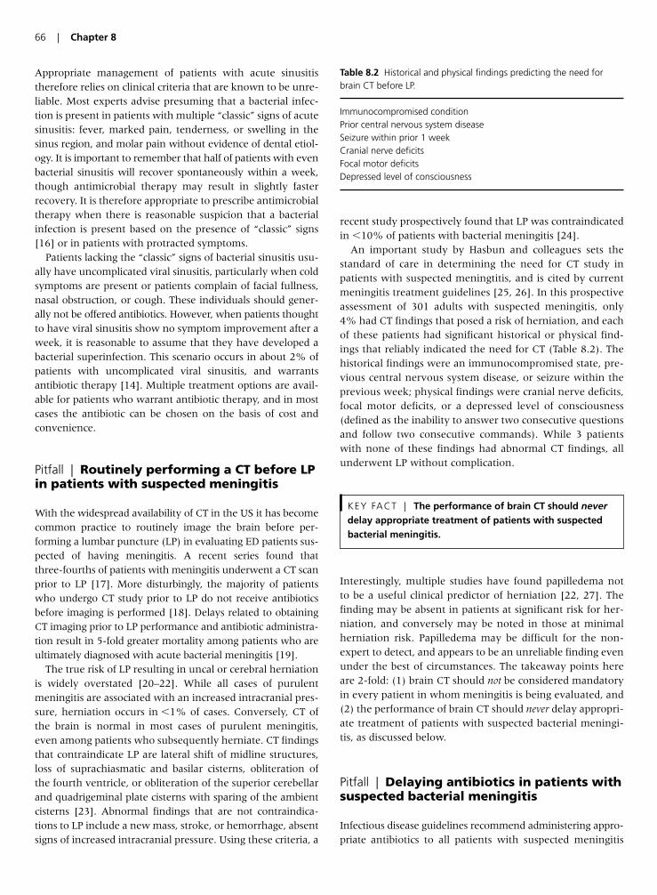
Appropriate management of patients with acute sinusitis
therefore relies on clinical criteria that are known to be unre-
liable. Most experts advise presuming that a bacterial infec-
tion is present in patients with multiple “classic” signs of acute
sinusitis: fever, marked pain, tenderness, or swelling in the
sinus region, and molar pain without evidence of dental etiol-
ogy. It is important to remember that half of patients with even
bacterial sinusitis will recover spontaneously within a week,
though antimicrobial therapy may result in slightly faster
recovery. It is therefore appropriate to prescribe antimicrobial
therapy when there is reasonable suspicion that a bacterial
infection is present based on the presence of “classic” signs
[16] or in patients with protracted symptoms.
Patients lacking the “classic” signs of bacterial sinusitis usu-
ally have uncomplicated viral sinusitis, particularly when cold
symptoms are present or patients complain of facial fullness,
nasal obstruction, or cough. These individuals should gener-
ally not be offered antibiotics. However, when patients thought
to have viral sinusitis show no symptom improvement after a
week, it is reasonable to assume that they have developed a
bacterial superinfection. This scenario occurs in about 2% of
patients with uncomplicated viral sinusitis, and warrants
antibiotic therapy [14]. Multiple treatment options are avail-
able for patients who warrant antibiotic therapy, and in most
cases the antibiotic can be chosen on the basis of cost and
convenience.
Pitfall | Routinely performing a CT before LPin patients with suspected meningitis
With the widespread availability of CT in the US it has become
common practice to routinely image the brain before per-
forming a lumbar puncture (LP) in evaluating ED patients sus-
pected of having meningitis. A recent series found that
three-fourths of patients with meningitis underwent a CT scan
prior to LP [17]. More disturbingly, the majority of patients
who undergo CT study prior to LP do not receive antibiotics
before imaging is performed [18]. Delays related to obtaining
CT imaging prior to LP performance and antibiotic administra-
tion result in 5-fold greater mortality among patients who are
ultimately diagnosed with acute bacterial meningitis [19].
The true risk of LP resulting in uncal or cerebral herniation
is widely overstated [20–22]. While all cases of purulent
meningitis are associated with an increased intracranial pres-
sure, herniation occurs in �1% of cases. Conversely, CT of
the brain is normal in most cases of purulent meningitis,
even among patients who subsequently herniate. CT findings
that contraindicate LP are lateral shift of midline structures,
loss of suprachiasmatic and basilar cisterns, obliteration of
the fourth ventricle, or obliteration of the superior cerebellar
and quadrigeminal plate cisterns with sparing of the ambient
cisterns [23]. Abnormal findings that are not contraindica-
tions to LP include a new mass, stroke, or hemorrhage, absent
signs of increased intracranial pressure. Using these criteria, a
recent study prospectively found that LP was contraindicated
in �10% of patients with bacterial meningitis [24].
An important study by Hasbun and colleagues sets the
standard of care in determining the need for CT study in
patients with suspected meningtitis, and is cited by current
meningitis treatment guidelines [25, 26]. In this prospective
assessment of 301 adults with suspected meningitis, only
4% had CT findings that posed a risk of herniation, and each
of these patients had significant historical or physical find-
ings that reliably indicated the need for CT (Table 8.2). The
historical findings were an immunocompromised state, pre-
vious central nervous system disease, or seizure within the
previous week; physical findings were cranial nerve deficits,
focal motor deficits, or a depressed level of consciousness
(defined as the inability to answer two consecutive questions
and follow two consecutive commands). While 3 patients
with none of these findings had abnormal CT findings, all
underwent LP without complication.
Interestingly, multiple studies have found papilledema not
to be a useful clinical predictor of herniation [22, 27]. The
finding may be absent in patients at significant risk for her-
niation, and conversely may be noted in those at minimal
herniation risk. Papilledema may be difficult for the non-
expert to detect, and appears to be an unreliable finding even
under the best of circumstances. The takeaway points here
are 2-fold: (1) brain CT should not be considered mandatory
in every patient in whom meningitis is being evaluated, and
(2) the performance of brain CT should never delay appropri-
ate treatment of patients with suspected bacterial meningi-
tis, as discussed below.
Pitfall | Delaying antibiotics in patients withsuspected bacterial meningitis
Infectious disease guidelines recommend administering appro-
priate antibiotics to all patients with suspected meningitis
KEY FACT | The performance of brain CT should never
delay appropriate treatment of patients with suspected
bacterial meningitis.
66 | Chapter 8
Table 8.2 Historical and physical findings predicting the need for
brain CT before LP.
Immunocompromised condition
Prior central nervous system disease
Seizure within prior 1 week
Cranial nerve deficits
Focal motor deficits
Depressed level of consciousness

within 30 min of presentation [25]. Despite these universal
recommendations, recent studies continue to report significant
delays in the administration of antibiotics [18, 25].
Administration of antimicrobials in an inpatient unit, rather
than in the ED, is associated with a 5-h delay in medication
delivery and a 3-fold increase in mortality [28]. A recent
study reported that a delay �6 h between ED presentation
and administration of antibiotics independently increased
mortality in patients with bacterial meningitis by more than
8-fold [19]. Furthermore, timing of antibiotic administration
was incrementally and closely related to mortality. It is impor-
tant to note that many studies relating antibiotic administra-
tion to outcomes are confounded by factors that may have
made early diagnosis of meningitis difficult.
A common argument against immediate administration of
antibiotics is that the practice may impair the diagnosis of
bacterial meningitis on subsequent CSF testing. Antibiotic
administration does not appear to cause changes in CSF
white blood cell, protein, or glucose levels that would impede
their diagnostic utility, and does not change CSF Gram stain
findings [29, 30]. Almost all patients with acute bacterial
meningitis still have diagnostic CSF abnormalities even if LP
is delayed by 3 days. CSF cultures may be positive even after
one dose of antibiotic administration, although the yield
decreases with more fastiditious organisms such as meningic-
occus [31]. Importantly, other modalities can reliably identify
the causative organism. Blood cultures obtained prior to anti-
biotic administration reveal the causative organism in 86% of
patients with bacterial meningitis. Given the enormous mor-
tality benefit associated with early administration of antibi-
otics, it is imperative to administer appropriate antimicrobial
therapy as soon as bacterial meningitis is suspected.
Pitfall | Failure to appreciate the range ofCSF findings associated with bacterialmeningitis
The CSF white blood cell (WBC) count, protein, and glucose
values are often unreliable tools for differentiating between
viral and bacterial meningitis. Commonly accepted indicators
of bacterial meningitis are total WBC count �5 cells/mm3,
polymorphonuclear WBC �75% of total WBC, CSF to serum
glucose ratio �50%, and CSF protein �45 mg/dl [32]. WBC
differentials may be misleading early in the course of menin-
gitis, as more than 10% of patients with bacterial infection
will have an initial lymphocytic predominance, and viral
meningitis can be initially be dominated by neutrophils
[30, 33, 34]. While each of these parameters is relatively
insensitive, most authorities conclude that if any of these
indices are abnormal, the incidence of bacterial meningitis
is high enough to warrant presumptive diagnosis and aggres-
sive treatment until culture results are available [18, 25,
30, 31, 35].
Pitfall | Over-reliance on physical findings indiagnosing bacterial meningitis
Failure to diagnose bacterial meningitis is in the top five
malpractice claims in emergency medicine [36]. Virtually all
patients with bacterial meningitis will have a fever, neck
stiffness, headache, or altered mental status, and therefore
the absence of all these findings can essentially exclude the
diagnosis provided that the patient is capable of providing a
relevant history [37]. Physical exam findings, however, are
far less reliable. The largest prospective study to date noted
the classic findings of fever, nuchal rigidity, and altered
mental status in less than half of patients with bacterial
meningitis, and a recent smaller study noted these classic find-
ings in only 20% of cases [18].
Negative tests of nuchal rigidity, including the Kernig
and Brudzinski signs, are commonly documented to indicate
absence of meningitis. The Brudzinski sign refers to the
patient’s spontaneous flexion of the hips during attempts to
passively flex the neck. The Kernig sign refers to the patient’s
reluctance to allow full extension of the knee when the hip is
flexed 90°. Nuchal rigidity is more loosely defined, but is gen-
erally described as inability or discomfort during neck flexion.
Each of these signs were developed and tested in patients with
severe, late stage meningitis. While Kernig and Brudzinski
signs are each about 95% specific for meningitis, their sensitiv-
ity is �10% [38]. Nuchal rigidity is slightly more sensitive, but
less specific. Documenting the absence of meningeal signs,
therefore, is not sufficient to exclude meningitis. Particularly in
immunocompromised hosts, the presentation of meningitis is
likely to be subacute, and negative exam findings are even
more likely.
Jolt accentuation of headache may be a more sensitive
maneuver for the diagnosis of meningitis. This test is per-
formed by horizontally rotating the head two to three times
per second; increased headache severity is considered a pos-
itive test. In a single small study, the maneuver displayed
97% sensitivity but only 60% specificity for the presence of
meningitis [39].
KEY FACT | The classic findings of fever, nuchal rigidity,
and altered mental status [were found] in less than half of
patients with bacterial meningitis.KEY FACT | Antibiotic administration does not appear
to cause changes in cerebrospinal fluid (CSF) white blood
cell, protein, or glucose levels … and does not change CSF
Gram stain findings.
Management of Infectious Diseases | 67

Pitfall | Failing to recognize methicillin-resistant Staphylococcus aureus as acommon pathogen
While MRSA emerged in the 1960s, until recently it has
been associated only with patients exposed to antibiotics for
extended periods of time or quartered in places harboring
resistant pathogens. Traditional risk factors for MRSA
included recent surgery or hospitalization, residence in a
long-term care facility, prolonged antibiotic administration,
hemodialysis, presence of indwelling devices or catheters,
some chronic illnesses, and intravenous drug use. In 1998, a
pediatric inpatient study made that made it clear that MRSA
was relevant in patients with community-acquired infec-
tions [40]. While 87% of children hospitalized with MRSA
infections between 1988 and 1990 had traditional risk factors
for resistant infections, only 29% of patients with MRSA
infections between 1993 and 1995 had such risk factors. The
prevalence of MRSA infections in hospitalized children rose
25-fold in the 5 years between the study periods.
Subsequent reports have confirmed the importance of
MRSA as a significant pathogen in a number of settings,
including daycare, prisons, and among athletes [41–43].
A recent study of 1600 individuals with community-acquired
MRSA infections found that 87% had skin or wound infec-
tions, but a small number had infections atypical for MRSA,
including urinary tract infections, meningitis, osteomyelitis,
sinusitis, and bacteremia [44]. Case reports have implicated
MRSA in fatal cases of pneumonia in immunocompetent
adults and children [45, 46] and in necrotizing fasciitis, a dis-
ease rarely associated with staphylococcal infections [47].
Community-acquired MRSA is now a common and serious
problem among ED patients. In a recent multicenter study
of 422 ED patients with skin infections, 59% were found to
have MRSA [48]. All isolates were sensitive to trimethoprim-
sulfamethoxizole, but only 90–95% were sensitive to
clindamycin or tetracycline. A larger study of inpatients and
outpatients found that infections were sensitive to van-
comycin and 97% to trimethoprim-sulfamethoxizole, but only
about 85% were sensitive to either clindamycin or tetracy-
cline [44].
Despite the prevalence of MRSA skin lesions, antimicrobial
therapy is not necessary for all ED patients with abscesses.
Uncomplicated abscesses generally only require incision and
drainage; antibiotics have not been shown to improve out-
comes in these patients. Antibiotics are indicated for patients
with abscesses complicated by cellulitis or fever, however,
and the selected agent should be active against MRSA.
Trimethoprim-sulfamethoxizole is an effective and inexpen-
sive option. Patients allergic to sulfa may be offered clin-
damycin or tetracycline, but it should be recognized that a
significant number of organisms have developed resistance
to these antibiotics. Linezolid is another option, but a course
of therapy costs over $1000. Some authorities advise adding
rifampin to a course of trimethoprim-sulfamethoxizole to
improve efficacy [49]. Furthermore, it has been noted that
unlike abscesses, cellulitis is more commonly due to Strepto-
coccus pyogenes than to Staphylococcus aureus, and trimethoprim-
sulfamethoxizole may be less effective for these infections.
Some experts therefore advise that patients who have
cellulitis without abscess be treated with a combination
of a first-generation cephalosporin and trimethoprim-
sulfamethoxizole, or with clindamycin alone [49].
Pitfall | Failure to aggressively treat sepsis
Emergency physicians frequently encounter sepsis, in
widely varying degrees of severity. Sepsis is broadly defined
as the presence of at least two components of the systemic
inflammatory response syndrome (SIRS) and a suspected
source of infection (see Table 8.3). When associated with
organ dysfunction it is termed “severe sepsis,” while “septic
shock” is associated with hypotension unresponsive to intra-
venous fluid resuscitation without another cause for
hypotension [50]. A recent analysis determined that there
are 751,000 cases of severe sepsis annually in the US, carry-
ing an estimated mortality rate of 27% – killing as many
people annually as acute myocardial infarction [51]. Although
septic shock is not usually a diagnostic dilemma, subtle
manifestations of organ dysfunction or tissue hypoxia can be
difficult to appreciate without frank hypotension.
In a 2001 study, Rivers et al. showed that early, goal-
directed therapy (EGDT) of patients with severe sepsis and
septic shock significantly decreased in-hospital mortality
from 47% to 31% [52]. This approach to resuscitation tar-
gets the normalization of central venous oxygen saturation
instead of the traditional target of blood pressure normaliza-
tion. The goals of EGDT during the initial resuscitation
period (first 6 h) include maintenance of the central venous
KEY FACT | Early, goal-directed therapy of patients …
significantly decreased in-hospital mortality from 47%
to 31%.
KEY FACT | Severe sepsis (carries) an estimated
mortality rate of 27% – killing as many people annually as
acute myocardial infarction.
KEY FACT | Community-acquired MRSA is now a
common and serious problem among ED patients … in a
recent study of ED patients with skin infections, 59%
were found to have MRSA.
68 | Chapter 8

pressure between 8 and 12 mmHg, mean arterial pressure
�65 mmHg, central venous (superior vena cava) or mixed
venous oxygen saturation �70%, and urine output
�0.5 ml/kg/h (see Table 8.4). If fluid resuscitation is accom-
plished with a resulting central venous pressure of
8–12 mmHg, but the central venous oxygen saturation or
mixed venous oxygen saturation remains �70%, then a
dobutamine infusion should be administered to accomplish
the goal. If the patient’s hematocrit is �30%, packed red blood
cells should be transfused to a goal of �30% prior to or during
the dobutamine infusion.
A post hoc analysis of this data showed that EGDT made an
even more dramatic difference in normotensive patients with
elevated serum lactate levels (�4 mmol/l), dropping mortality
at 60 days from 70% to 24% [53]. Nguyen et al. also showed
that failure to improve an elevated lactate level during the ini-
tial resuscitation of patients with severe sepsis more than dou-
bled mortality [54]. These data suggest that microcirculatory
defects causing global tissue hypoxia may exist without frank
hypotension, and that correction of this dysfunction may
reverse the process before organ failure and death ensues.
Pitfall | Failure to ensure adequateantimicrobial treatment in severe sepsisand septic shock
It seems intuitive that adequately treating patients with
severe sepsis or septic shock entails using antibiotics that are
effective against the infecting microorganism and initiating
therapy as quickly as possible. In fact, recent guidelines for
the management of severe sepsis and septic shock call for the
administration of intravenous antibiotics within 1 h of recog-
nizing the syndrome [55]. What may not be readily apparent
is the impact on outcome that inappropriate antibiotic ther-
apy may have, or how frequently an error in coverage is
made. A recent study found that 15% of patients with sepsis
received antibiotics ineffective against the infecting microor-
ganism. While the effect on mortality was most pronounced
in those with septic shock, it was significant in all patients. In
patients with septic shock, inappropriate antibiotic treatment
doubled 14-day mortality from 40 to 80% [56]. The practic-
ing emergency physician should be aware of the importance
of antimicrobial efficacy when treating sepsis, and cover
broadly when the source is unclear.
Pearls for Improving Patient Outcomes
• Do not discount the diagnosis of bacterial endocarditis based
simply on the absence of a murmur.
• The disposition of patients with pneumonia should be based
primarily on the physician’s clinical judgement and not purely on
the PSI or other such decision instruments.
• Avoid prescribing antibiotics in all cases of bronchitis and sinusi-
tis. Consider the patient co-morbidities and disease severity
before dispensing antibiotics.
• Use of brain CT before all LPs is unnecessary and often results in
delays in care of the patient. Never delay antibiotics while await-
ing the CT in patients with suspected bacterial meningitis.
• Do not withhold antibiotics in patients with suspected bacterial
meningitis out of fear of impairing the CSF analysis. CSF results
remain diagnostic even after antibiotic administration.
• Antibiotic regimens for community-acquired cellulitis should
include coverage for MRSA in many ED populations now.
• Severe sepsis and septic shock must be treated aggressively.
EGDT and broad-spectrum antibiotics can significantly decrease
mortality in these patients.
References
1. Mylonakis E, Calderwood SB. Infective endocarditis in adults.
New Eng J Med 2001; 345(16):1318–30.
2. Crawford MH, Durack DT. Clinical presentation of infective
endocarditis. Cardiol Clin 2003; 21:159–66.
3. Saccent M, Cobbs CG. Clinical approach to infective endocardi-
tis. Cardiol Clin 1996; 14(3):352–62.
4. Cunha BA, Gill MV, Lazar JM. Acute infective endocarditis:
diagnostic and therapeutic approach. Inf Dis Clin N Am 1996;
10:811–31.
5. Kaye AR. Neurologic complications of infective endocarditis.
Neurol Clin 1993; 11:419–37.
6. Marrie TJ. Community-acquired pneumonia: epidemiology,
etiology, treatment. Infect Dis Clin N Am 1998; 12:723–40.
7. Fine MJ, Auble TE, Yealy DM, et al. A prediction rule to identify
low-risk patients with community-acquired pneumonia. New
Engl J Med 1997; 336:243–50.
Management of Infectious Diseases | 69
Table 8.3 Definitions of sepsis states [50].
Components of SIRS. SIRS is present when at least two criteria
are present
Temperature �36°C or �38°C
Pulse �90 beats/min
Respiratory rate �20 breaths/min or PaCO2 �32 mmHg
WBC �4 k or �12 k
Sepsis syndromes
Sepsis SIRS associated with an infection, proven
or suspected
Severe sepsis Sepsis associated with organ dysfunction
Septic shock Sepsis associated with hypotension
despite adequate volume resuscitation
Table 8.4 Initial resuscitation goals in patients with sepsis.
Central venous pressure 8–12 mmHg
Mean arterial pressure �65 mmHg
Central venous (superior vena cava) or mixed venous oxygen
saturation �70%
Urine output �0.5 ml/kg/h

8. Mandell LA, Bartlett JG, Dowell SF, et al. Update of practice
guidelines or the management of community-acquired pneu-
monia in immunocompetent adults. Clin Infect Dis 2003; 37:
1405–33.
9. Gonzales R, Bartlett JG, Besser RE, et al. Principles of appropri-
ate antibiotic use for treatment of uncomplicated acute bron-
chitis: background. Ann Emerg Med 2001; 37:720–7.
10. Sethi S. Infectious exacerbations of chronic bronchitis: diagnosis
and management. J Antimicr Chemother 1999; 43:A97–105.
11. Stockley RA, O’Brien C, Pye A, Hill SL. Relationship of sputum
color to nature and outpatient management of COPD. Chest
2000; 117:1638–45.
12. Saint S. Antibiotics in chronic obstructive pulmonary disease:
a meta-analysis. J Am Med Assoc 1995; 273:957–60.
13. Grossman RF. Guidelines for the treatment of acute exacerba-
tions of chronic bronchitis. Chest 1997; 112:110–15.
14. Picirillo JF. Acute bacterial sinusitis. New Engl J Med 2004;
351:902–10.
15. Gwaltney JM, Phillips CD, Miller RD, Riker DK. Computed
tomographic study of the common cold. New Engl J Med 2004:
330:25–30.
16. Snow V, Mottur-Pilson C, Hickner JM. Principles of appropriate
antibiotic use for acute sinusitis in adults. Ann Intern Med 2001;
134:495–7.
17. Durand ML, Calderwood SB, Weber DJ, et al. Acute bacterial
meningitis: a review of 493 episodes. New Engl J Med 1993;
328:21–8.
18. van de Beek D, de Gans J, Spanjaard L, et al. Clinical features
and prognostic factors in adults with bacterial meningitis. New
Engl J Med 2004; 351:1849.
19. Proulx N, Fréchette D. Delays in the administration of antibi-
otics are associated with mortality from adult acute bacterial
meningitis. Q J Med 2005; 98(4):291–8.
20. Haslam RH. Role of computerized tomography in the early man-
agement of bacterial meningitis. J Pediatr 1991; 119:157–9.
21. Richards PG, Towu-Aghanste E. Dangers of lumbar puncture.
Br Med J 1986; 292:605–6.
22. Duffy GP. Lumbar puncture in the presence of raised intracra-
nial pressure. Br Med J 1969; 1:407–9.
23. Gower DJ, Baker AL, Bell WO, Ball MR. Contraindications to
lumbar puncture as defined by computed cranial tomography.
J Neurol Neurosurg Psychiatry 1987; 50:1071–4.
24. Gopal AK, Whitehouse JD, Simel DL, Corey GR. Cranial com-
puted tomography before lumbar puncture: a prospective clini-
cal evaluation. Arch Intern Med 1999; 159:2681–5.
25. Hasbun R, Abrahams J, Jekel J, Quagliarello VJ. Computed
tomography of the head before lumbar puncture in adults with
suspected meningitis. New Engl J Med 2001; 345:1727–33.
26. Tunkel AR, Hartman BJ, Kaplan SL, et al. Practice guidelines for
the management of bacterial meningitis. Clin Infect Dis 2004;
39:1267.
27. Sharp CG, Steinhart CM. Lumbar puncture in the presence of
increased intracranial pressure: the real danger. Pediatr Emerg
Care 1987; 3:39–43.
28. Miner JR, Heegaard W, Mapes A, Biros M. Presentation, time to
antibiotics, and mortality of patients with bacterial meningitis at
an urban county medical center. J Emerg Med 2001; 21:387–92.
29. Blazer S, Berant M, Alon U. Bacterial meningitis. Effect of
antibiotic treatment on cerebrospinal fluid. Am J Clin Pathol
1983; 80:386–7.
30. Talan DA, Zibulewsky. Relationship of clinical presentation to
time to antibiotics for the emergency department management
of suspected bacterial meningitis. Ann Emerg Med 1993;
22:1733–8.
31. Coant PN, Kornberg AE, Duffy LC, Dryja DM, Hassan SM. Blood
culture results as determinants in the organism identification of
bacterial meningitis. Pediatr Emerg Care 1992; 8:200–5.
32. Seehusen DA, Reeves MM. Cerebrospinal fluid analysis. Am
Fam Pract 2003; 68:6.
33. Spach DH, Jackson LA. Central nervous system infections.
Neurol Clin 1999; 17:711–35.
34. Bonsu BK, Harper MB. Accuracy and test characteristics of
cerebral spinal fluid for predicting meningitis in children with
low white blood cell counts in cerebral spinal fluid. Acad Emerg
Med 2005; 12(4):303–9
35. McMillan DA, Lin CY, Aronin SI, Quagliarello VJ. Community-
acquired bacterial meningitis in adults: categorization of causes
and timing of death. Clin Infect Dis 2001; 33:969–75.
36. Selbst SM, Friedman MJ. Epidemiology and etiology of malprac-
tice lawsuits involving children in US emergency departments
and urgent care centers. Pediatr Emerg Care 2005; 21:165–9.
37. Attia J, Hatala R, Cook DJ, Wong JG. The rational clinical
examination: does this adult patient have acute meningitis?
J Am Med Assoc 1999; 282:175.
38. Thomas KE, Hasbun R, Jekel J, Quagliarello VJ. The diagnostic
accuracy of Kernig’s sign, Brudzinski’s sign, and nuchal rigidity
in adults with suspected meningitis. Clin Infect Dis 2002; 35:46.
39. Uchihara T, Tsukagoshi H. Jolt accentuation of headache: the
most sensitive sign of CSF pleocytosis. Headache 1991; 31:167.
40. Herold BC, Immergluck LC, Maranan MC, et al. Community-
acquired methicillin-resistant Staphylococcus aureus in children
with no identified predisposing risk. J Am Med Assoc 1998;
279:593–8.
41. CDC. Methicillin-resistant Staphylococcus aureus infections in cor-
rectional facilities – Georgia, California, and Texas, 2001–2003.
MMWR 2003; 52:992–5.
42. CDC. Methicillin-resistant Staphylococcus aureus infections
among competitive sports participants, 2000–2003. MMWR
2003; 52:992–6.
43. Kazakova SV, Hageman JC, Matava M, et al. A clone of
methicillin-resistant Staphylococcus aureus among professional
football players. New Engl J Med 2005; 352:468–75.
44. Fridkin SK, Hageman JC, Morrison M, et al. Methicillin-
resistant Staphylococcus aureus disease in three communities.
New Engl J Med 2005; 352:1436–44.
45. Gillet Y, Issartel B, Vanhems P, et al. Association between
Staphylococcus aureus strains carrying gene for Panton-Valentine
leukocidin and highly lethal necrotising pneumonia in young
immunocompetent patients. Lancet 2002; 359:753–9.
46. Frazee BW, Salz TO, Lambert L, Perdreau-Remington F. Fatal
community-associated methicillin-resistant Staphylococcus aureus
pneumonia in an immunocompetent young adult. Ann Emerg
Med 2005; 46:401–4.
47. Miller LG, Perdreau-Remington F, Rieg G, et al. Necrotizing
fascitis caused by community-associated methicillin-resistant
Staphylococcus aureus in Los Angeles. New Engl J Med 2005;
352:1445–53.
48. Moran GJ, Krishnadasan A, Gorwitz RJ, et al. Methicillin-
resistant S. aureus infections among patients in emergency
department. New England Journal of Medicine 2006; 355:666–74.
70 | Chapter 8

49. Moran GJ, Talan DA. Community-associated methicillin-
resistant Staphylococcus aureus: is it in your community and should
it change practice? Ann Emerg Med 2005; 45:321–2.
50. Levy MM, Fink MP, Marschall JC, et al. 2001 SCCM/ ESICM/
ACCP/ATS/SIS International sepsis definitions conference. Crit
Care Med 2003; 31:1250–6.
51. Angus DC, Linde-Zwirble WT, Lidicker J, et al. Epidemiology of
severe sepsis in the United States: analysis of incidence, out-
come, and associated costs of care. Crit Care Med 2001;
29:1303–10.
52. Rivers E, Nguyen B, Havstad S, et al. Early goal-directed ther-
apy in the treatment of severe sepsis and septic shock. New Engl
J Med 2001; 345:1368–77.
53. Donnino MW, Nguyen B, Jacobsen G, et al. Cryptic septic
shock: a sub-analysis of early, goal-directed therapy. Chest 2003;
124(4):90S.
54. Nguyen HB, Rivers EP, Knoblich BP, et al. Early lactate clear-
ance is associated with improved outcome in severe sepsis and
septic shock. Crit Care Med 2004; 32:1637–42.
55. Dellinger RP, Carlet JM, Masur H, et al. Surviving sepsis cam-
paign guidelines for the management of severe sepsis and septic
shock. Crit Care Med 2004; 32:858–73.
56. Valles J, Rello J, Ochangavia A, et al. Community-acquired
bloodstream infection in critically Ill adult patients – impact of
shock and inappropriate antibiotic activity on survival. Chest
2003; 123:1615–24.
Management of Infectious Diseases | 71

Introduction
Emergency physicians are the primary providers of post-
traumatic wound management. Over 11 million traumatic
wounds are managed in emergency departments (ED) in the
US annually [1]. Of these, over 50% occur as a result of blunt
trauma, while the remainder involve the penetration of sharp
objects, such as metal, glass, or wood. The majority of these
wounds occur on patients’ heads and upper extremities, both
highly visible areas [2]. As a result, the cosmetic appearance of
healed wounds is a priority for both patients and emergency
physicians. According to a survey of ED patients, the top four
concerns of patients with a laceration include the cosmetic
appearance, preservation of function, decreasing the pain of
the repair, and avoiding a wound infection [3]. As these key
outcomes are closely related to the specific wound care tech-
niques used during the initial ED encounter, it is of utmost
importance that emergency physicians stay abreast of the
biggest pitfalls in wound care in order to avoid negative out-
comes. A national survey of wound care techniques revealed
that many practitioners manage wounds in ways that are
contrary to the recommendations of published literature [4].
Furthermore, between 5% and 20% of all emergency physi-
cian malpractice claims involve issues of wound care, with up
to 11% of all malpractice dollars being awarded for these cases
[2]. These data further emphasize the importance of recogniz-
ing and avoiding many of the common pitfalls of wound care
reviewed in the subsequent sections.
Pitfall | Failure to properly prepare a woundprior to closure
Beyond anesthesia, inspection, and evaluation for foreign
body, proper preparation of a wound improves its outcome.
Despite available evidence-based recommendations, many
practitioners continue to employ techniques and utilize prod-
ucts that are unnecessary, and in some cases detrimental. In a
survey distributed to predominantly board certified emergency
physicians, investigators found that despite the existence of
wound management guidelines, 90% of physicians treated
wounds based on personal preference. Thirty-eight percent
soaked wounds, 67% scrubbed wounds, 27% irrigated
wounds with inadequate technique, and 21% used concen-
trated povidone–iodine solution or hydrogen peroxide [4].
Wound irrigation with high pressure and adequate volume has
been shown to have a dramatic impact on wound infection
rates. Simply soaking a wound provides no advantage and
may be detrimental to its ultimate outcome [2]. The necessary
pressure for wound irrigation can be accomplished by pushing
fluid in a 30–60 ml syringe through a 19-gauge catheter. This
is a highly effective means of reducing potentially infective
material from a wound [5, 6]. The pressure generated through
this technique cannot be reproduced by puncturing containers
of irrigation fluid and squeezing with maximal hand pressure
[1]. The data regarding the volume of irrigation fluid necessary
is limited. Most authorities recommend 50–100 ml of irrigation
fluid per cm of laceration. However, these sources agree
that the volume of irrigation should be adjusted to the wound
characteristics and degree of contamination [2, 7]. For instance,
a contaminated scalp wound may require less irrigation solu-
tion than an extremity wound.
Irrigation of highly vascularized structures such as the
face and scalp may not be as critical. Hollander et al. demon-
strated that not irrigating clean-appearing, simple facial and
scalp lacerations did not alter the rates of infection [8]. In
addition, at very high irrigation pressures, research suggests
that infection rates may actually increase due to tissue dam-
age. Therefore, care must be taken not to use high-pressure
irrigation in vascular wounds with loose alveolar tissue [7].
The choice of irrigation solution can have significant effects
on wound healing. Antiseptic solutions such as chlorhexi-
dine, povidone–iodine 10% solution, hydrogen peroxide,
and detergents are toxic to tissues and impede wound heal-
ing [9, 10]. Studies comparing diluted 1% povidone–iodine
to normal saline have not shown any difference in infection
rates [11]. Furthermore, given the prevalence of iodine sen-
sitivity, the use of diluted iodine irrigation is discouraged.
More recently, published studies have concluded that there is
no clinically significant difference in infection rates between
simple wounds irrigated with potable tap water and sterile
normal saline [12, 13].
Another misguided practice which has been shown to
increase infection rates is shaving the area surrounding a
KEY FACT | The necessary pressure for wound
irrigation can be accomplished by pushing fluid in a
30–60 ml syringe through a 19-gauge catheter.
Chapter 9 | Wound Care in Emergency Medicine
Siamak Moayedi & Mercedes Torres
72

wound prior to repair [14, 15]. If hair removal is required
for adequate access to or visualization of a wound, it is rec-
ommended that the hair is clipped, rather than shaved, at
the skin level [16]. One effective alternative is to use petro-
leum products to comb hair away from the wound. Care
must be taken to remove small pieces of hair from the
wound prior to closure.
Finally, many practitioners routinely use sterile gloves for
the repair of simple lacerations. Studies supporting this prac-
tice are lacking. In a prospective study of 816 patients with
simple wounds randomized to wound repaired using sterile
versus clean gloves, there was no difference in subsequent
infection rates [17].
Pitfall | Failure to detect the presence of aforeign body in a wound
When unrecognized, retained foreign bodies can cause sig-
nificant morbidity, including local inflammation, infection,
and compression of vital structures [18]. Emergency physi-
cians have a variety of tools at their disposal to assist with
the detection of foreign bodies. The key to addressing a foreign
body is finding it.
Detection of foreign bodies by clinical examination alone
has proven unreliable. If the bottom of a wound is visible and
no foreign body is seen, 7% of subsequent X-rays have been
shown to be positive for the presence of a foreign body (see
Figure 9.1). Furthermore, when the bottom of a wound is not
visualized, clinically undetected foreign bodies have been
found on X-rays in 21% of cases [1]. There are clinical clues
that can raise suspicion for the presence of a foreign body:
These include persistent pain, drainage, a palpable mass, sur-
face discoloration, or a blood-stained tract. Obtaining a his-
tory of glass breaking while in contact with the skin, blunt
trauma with tooth fragments, clenched-fist injuries, puncture
wounds, motor vehicle accidents, and foreign body sensation
should raise suspicion for retained foreign bodies in a wound
[18]. According to a study of wounds caused by glass, 15%
had retained glass within the wound. Patients reporting for-
eign body sensation had a positive predictive value of 31%
and a negative predictive value of 89%. However, using patient
perception alone to determine X-ray eligibility would have
missed 57% of wounds with retained glass [19].
One of the primary diagnostic modalities used for detec-
tion of foreign bodies in wounds is X-ray. Legal precedents
have shown that practitioners who failed to X-ray wounds
caused by glass were unsuccessful in defending themselves
in 60% of cases [1]. Radio opaque foreign bodies include
glass, metal, bone, teeth, pencil graphite, certain plastics,
gravel, some aluminum, and some sand [2, 18]. X-rays can
detect greater than 50% of glass with a single dimension
between 0.5 and 2.0 mm, and virtually all with a dimension
greater than 2.0 mm [1, 7, 18]. Underpenetrated X-rays,
highlighting the soft tissues are more useful for detecting
retained foreign bodies not visualized with standard X-rays
[18, 20]. In addition, it is possible to visualize a filling defect
on a soft tissue X-ray created by a radiolucent foreign body
[20]. One problem with X-rays is their two-dimensional
quality. If detected, it is often difficult to determine the exact
location of the foreign body and its spatial relation to other
vital structures. Obtaining multiple views and using markers
such as paper clips or needles can assist with localization of
the foreign body using X-ray alone [18].
When suspicion is high for a radiolucent foreign body, or a
foreign body in close proximity to vital structures, a computed
tomography (CT) scan is often the study of choice. CTs are
useful for detecting radiolucent objects such as wood, certain
plastics, thorns, and spines. In addition, they can detect the
presence of vegetative material, which has a high rate of
KEY FACT | Missed retained foreign bodies are the fifth
leading cause of malpractice claims against emergency
physicians in the US [1].
Wound Care in Emergency Medicine | 73
Persistent pain
Drainage from wound
Palpable mass at wound site
Surface discoloration
Blood-stained wound tract
History of contact with broken glass
Clenched-fist injury
Puncture wounds
Motor vehicle accident
Foreign body sensation by patient
Figure 9.1 Plain radiograph demonstrating metallic foreign body in the
foot.

infection and inflammatory complication. This benefit is
optimized within the first 48 hours because vegetative for-
eign bodies absorb water within tissues, changing their den-
sity to appear very similar to soft tissue, and therefore more
difficult to differentiate [1]. Regardless the age of a wound,
CTs are still 100 times more sensitive for detecting various
densities of objects as compared with X-ray. In addition,
they provide a visualization of the location of foreign bodies
in relation to surrounding structures [18]. The ability to
localize a foreign body assists the practitioner in deciding
whether it should be removed and if removal should be sur-
gical. Magnetic resonance imaging (MRI) has also been
shown to be useful in identifying retained vegetative matter,
but cannot be used if there is any suspicion for metal or
gravel foreign bodies. The cost and “inconvenience” of MRI
has excluded this as a primary imaging modality for the
detection of retained wound foreign bodies [18].
Another diagnostic modality that has been shown to be
efficient and less costly is bedside ultrasound. The value of
ultrasound in detecting and localizing wound foreign bodies
is dependent upon the provider’s skill and the size of the
object. A 7.5 MHz linear probe is optimal for wound exami-
nation, allowing approximately 3 cm of tissue penetration.
The technique involves rotating the probe in a perpendicu-
lar position to the skin in order to attempt to localize the for-
eign body in cross section [21]. Increasing the frequency of
the probe improves its ability to detect smaller foreign bod-
ies [22]. One recent study examining the utility of bedside
ultrasound in the detection of wood foreign bodies demon-
strated a sensitivity of 95% and specificity of 89% [23]. Others
have cited ranges of sensitivity from 50% to 90%, and speci-
ficity from 70% to 97%. Certain types of foreign bodies have
characteristic appearances on ultrasound. Metal or glass is
described as having a comet-tail appearance, while wood,
plastic, sand, and pebbles often demonstrate acoustic shad-
owing similar to that seen with gallstones [21]. Unlike CT
scans, ultrasound may be more helpful when patients pres-
ent greater than 48 hours after suffering their wound, as the
inflammation around a foreign body makes it easier to iden-
tify its location [23]. However, the use of bedside ultrasound
is complicated by false positives. These include echo artifacts
that can be made by air pockets, calcifications, old scar tissue,
blood, sutures, sesamoid bones, or purulence [18]. In addi-
tion, the presence of multiple tissue planes can complicate
ultrasound guided localization [24]. Once a foreign body is
located, ultrasound can provide real-time visualization of its
removal as well as identifying other smaller surrounding
foreign bodies [25].
Pitfall | Failure to recognize the morbidityassociated with plantar puncture wounds
Plantar puncture wounds represent a unique challenge for
physicians. The soles of the feet are the most common site of
puncture wounds in the lower extremities. The force of a
patient’s weight onto a foreign body and the relative small dis-
tance from the skin to highly susceptible structures such as
bones and joints make this injury especially prone to infection
and significant morbidity. Care of the patient presenting with
an acute plantar puncture remains controversial. The two
most controversial areas involve the extent of wound explo-
ration/debridement and the question of antibiotic prophylaxis.
Many authors have asserted that plantar punctures have a
high predilection for retained foreign bodies. Schwab and
Powers reported a 3% rate of retained foreign body after
initial surface cleansing without wound exploration [26]. A
larger study of 887 patients also reported a 3% incidence of
foreign body retention. Half of these were pieces of foot
wear, while the remainder included rust and dirt [27].
However, because many patients with this injury do not
present to the ED unless they experience continued pain or
infection, the true incidence of retained foreign body may be
lower [28].
There are no prospective, randomized trials that demon-
strate the utility of prophylactic antibiotics in these injuries.
One of the most quoted studies recommending antibiotic
prophylaxis was an uncontrolled, observational study, with
optional use of antibiotics [29]. Most authorities defer to the
individual clinician regarding the use of antibiotics with the
caveat that antibiotics do not compensate for inadequate
wound care [18].
Uncomplicated puncture wounds without concern for
retained foreign bodies may not require any wound explo-
ration, but close follow up is advised [26]. Many aggressive
and time-consuming procedures, such as coring and wound
extension, have been described for the evaluation of these
wounds. Coring refers to excision of a block of tissue down
to the subcutaneous layer to allow visualization and access
to foreign bodies and contaminated tissue. There is no defin-
itive study showing that routinely coring puncture wounds
reduces the incidence of infection. Furthermore, extensive
debridement and coring may actually delay wound healing
and cause undue pain [18]. Authors have argued that high-
risk wounds which may require aggressive debridement
include injuries through rubber-soled shoes, injuries over
metatarsal phalangeal joints, and injuries in patients with
high-risk comorbidities, such as diabetes and neuropathy [2].
KEY FACT | The rate of osteomyelitis caused by plantar
puncture wounds is between 0.04% and 2%.
KEY FACT | High-risk wound which may require
aggressive debridement include injuries through rubber-
soled shoes, injuries over metatarsal phalangeal joints,
and injuries in patients with high-risk comorbidities.
74 | Chapter 9

A concerning clinical scenario involves a previous puncture
wound presenting days later with evidence of infection.
Delayed presentation is a significant marker for deep-seated
infection [30]. These cases should prompt a thorough inves-
tigation for a retained foreign body [18]. Referral to special-
ists for removal of all but superficial foreign bodies and
aggressive debridement is recommended. A high level of
concern for osteomyelitis is raised when a wound relapses
or fails to respond to the initial therapy. Overall, the rate of
osteomyelitis caused by plantar puncture wounds is
between 0.04% and 2% [18]. Advanced imaging techniques
such as MRI may be required for diagnosis.
Although gram-positive organisms remain the most com-
mon cause of infection, several studies have demonstrated
the role of Pseudomonas aeruginosa when the puncture occurs
through rubber-soled foot wear [7]. It is presumed that the
moist inner sole of the shoe provides a suitable environment
for this bacterium [31]. When considering antibiotic pro-
phylaxis, ciprofloxacin is still the antibiotic of choice. The
newer quinolones, despite demonstrating better efficacy
against gram-positive organisms, are less effective against
Pseudomonas.
Pitfall | Indiscriminate use of prophylacticantibiotics for bite wounds
On a yearly basis, several million people are bitten by mam-
mals in the USA. Almost one half of all children in the USA
will be bitten by a dog at some point during their childhood
[32]. A small proportion of adults and children with bite
wounds, somewhere between 1 and 2 million, seek treat-
ment. Animal bites comprise 1% of all ED visits annually,
with dog bites accounting for 80% of those visits [2, 33, 34].
Although bite wounds are a commonly encountered com-
plaint among emergency physicians, their management
remains controversial. They are thought to be at increased
risk for infections and wound complications, leading to the
recommended use of prophylactic antibiotics. However, stud-
ies of infection rates after mammalian bites have failed to
demonstrate any significant difference between those who
received prophylactic antibiotics and those who received
placebo except in cases of high-risk wounds [7, 32, 35, 36].
One of the primary concerns with any bite wound is possi-
ble inoculation of the soft tissue with oral flora. The specific
organisms of concern vary based on the type of mammal
which has bitten the patient. While over 80% of animal bites
reported are from dogs, only 4–25% of these become infected.
Cat bites, although less frequent, are more commonly compli-
cated by infection rates that range between 30% and 80%,
depending on the study referenced [34, 37]. Human bites
are considered to have the highest risk of infection when
compared with dogs and cats. Although most bite wound
infections contain mixed flora, pasturella is the primary cul-
prit in both dog and cat bite wounds. Pasturella causes a rapid
onset of signs and symptoms of infection, typically within 24
hours of the initial injury [37]. Streptococci, Staphylococci,
Moraxella, Corynebacterium, and Neisseria are the next most
common pathogens cultured from animal bite wounds.
Infections attributed to Staphylococci and Streptococci typi-
cally manifest as nonpurulent wounds with lymphangitis. In
addition to the aforementioned flora, over 50% of bite
wounds contain anaerobic organisms [37, 38]. Therefore,
optimal prophylactic agents would include beta lactams with
a beta lactamase inhibitor, second-generation cephalosporins
with anaerobic activity, or a combination of clindamycin and
a fluoroquinolone. Traditional choices such as first-generation
cephalosporins, ampicillin, or penicillin alone, have proven
ineffective and are not recommended [37].
Although there is evidence that bite wounds in general
have higher infection rates as compared with other types of
wounds, the utility of prophylactic antibiotics for all bite
wounds has been questioned. According to a 2005 Cochrane
Database review of eight randomized controlled trials, pro-
phylactic antibiotics were associated with a statistically signif-
icant reduction in the rate of infections after human bites, but
not after cat or dog bites [32]. However, other studies suggest
that even human bite wounds that involve minor injuries
have not been shown to benefit from prophylactic antibiotics
[36]. Thus, physicians must consider more than just the
species of the biting mammal when evaluating the benefit of
prophylactic antibiotics. It is safer and more cost-effective to
reserve prophylactic antibiotics for high-risk wounds and
high-risk patients (Table 9.1).
Focusing on the characteristics of the wound, deep punc-
ture wounds, wounds with extensive crush injury or devital-
ized tissue, and wounds involving underlying muscle or
tendons are at higher risk of infection. In addition, any wound
involving an underlying bone fracture, retained foreign body,
or joint should be considered an increased infection risk [2].
KEY FACT | Studies of infection rates after mammalian
bites have failed to demonstrate any significant
difference between those who received prophylactic
antibiotics and those who received placebo except in
cases of high-risk wounds.
Wound Care in Emergency Medicine | 75
Table 9.1 Wound and patient characteristics that increase the risk of
wound infection.
Wound characteristics
Deep punctures
Wounds with extensive crush injury or devitalized tissue
Underlying muscle or tendon involvement
Patient characteristics
Diabetes
Immunocompromised state
History of endocarditis
Presence of orthopedic prostheses

Bites on the hand, including fight bites, maintain a particu-
larly high risk of infection and current evidence supports the
use of prophylactic antibiotics in these cases [32].
Patient-specific factors which would support the use of pro-
phylactic antibiotics include a history of diabetes mellitus,
immunocompromise, endocarditis, and orthopedic prosthe-
ses. The time between the bite and presentation to the ED is
another important factor in determining the value of antibi-
otic prophylaxis. Studies demonstrate that pasturella infec-
tions typically manifest clinical signs and symptoms within
24 hours. Infections caused by other organisms typically
develop within 72 hours [7, 38]. Therefore, patients who
present greater than 72 hours from the initial bite with no
signs of infection do not require antibiotic prophylaxis.
In summary, recent literature supports the use of prophy-
lactic antibiotics in nontrivial human bites, mammalian bite
wounds with high-risk characteristics, and/or mammalian
bite wounds suffered by high-risk patients. There is no sub-
stitute for the value of adequate wound irrigation, cleansing,
and debridement in the prevention of bite wound infections
for all bites. If a bite wound meets the aforementioned
criteria of high infection risk, it is important to consider the
time since injury and the offending mammal in order to
select the most appropriate antibiotic. These practices will
help minimize mammalian bite wound infection rates, while
preventing unnecessary side effects and risks of antibiotic
prophylaxis.
Pitfall | Failure to provide appropriatewound after care instructions
There are several factors that affect the outcome of a
wound. These include the choice of dressing placed on
the wound and patient education regarding proper care of
the wound. Experience demonstrates that there can be a
wide variation in outcome despite proper wound prepara-
tion and suturing [39]. In order to optimize the cosmetic
outcome of a wound, the physician must be mindful of the
effects of UV light exposure and the choice of dressing
applied to the wound.
The data to support avoidance of sun light exposure to a
wound comes from plastic surgery and dermatology literature.
In a retrospective study of patients undergoing dermabrasion,
the authors observed that wounds exposed to sunlight within
6 months from the time of the injury can develop permanent
hyperpigmentation [40]. Subsequent to this study, multiple
animal experiments have demonstrated that UV light expo-
sure on laser- and knife-induced wounds significantly alters
normal skin structures and induces hyperpigmentation [41,
42]. Although these findings have not been investigated in
humans, the recommendation is to use sunscreen products on
a new wound for 6 months after epithelialization, which is
typically complete within 48 hours. An optimal opportunity to
discuss this with a patient is during a wound check visit. The
patient should also be cautioned about the risk of hypersensiti-
zation to sunscreen products. It may be advantageous to rec-
ommend hypoallergenic products which are readily available
over the counter.
The choice of dressing applied to a wound also impacts
its cosmetic outcome. Dressings that provide moist, warm
environments improve the rate of epithelialization [43].
Furthermore, they prevent contamination, sunlight expo-
sure, and repeated trauma. Petroleum ointment and topical
antibiotics such as bacitracin or triple antibiotic preparations
provide the requisite moist environment for epithelializa-
tion. The value of antibiotic over petroleum ointment is con-
troversial. In one prospective, randomized study topical
antibiotics yielded lower infection rates [44]. However, a
similar study published the following year found essentially
no difference between antibiotic and petroleum ointments
[45]. Some authors have suggested the use of antibiotic
ointments, since there is no disadvantage linked to their use
[1]. More recently, academic dermatology literature has
warned against the routine use of bacitracin, citing that it
is the seventh most common cause of contact dermatitis in
North America. In addition to the adverse effects of contact
dermatitis on cosmetic outcome, they cite more than 26
cases of anaphylaxis related to bacitracin use [46]. Of note,
both topical antibiotics and petroleum products cause dis-
solving of cyanoacrylates and are not to be used on wounds
closed with skin glue preparations.
Pearls for Improving Patient Outcomes
• When cleansing a wound, avoid antiseptic solutions or prepara-
tions, as they are tissue toxic. High-pressure irrigation with potable
tap water is a safe and effective means of cleaning a wound.
• Clip or comb hair away from a wound rather than shaving at
the skin.
• Wounds with high suspicion for retained foreign bodies should
be further investigated with radiographs. If concern for radiolu-
cent foreign bodies exists, CT or ultrasound is recommended.
• Bedside ultrasound is an efficient and cost-effective tool for
localizing and assisting with the removal of foreign bodies.
• Maintain a high clinical suspicion for the presence of retained
foreign bodies in plantar puncture wounds; especially if there is
a delayed presentation.
KEY FACT | Wounds exposed to sunlight within
6 months from the time of the injury can develop
permanent hyperpigmentation.
KEY FACT | Patients who present greater than 72 hours
after the initial bite with no signs of infection do not
require antibiotic prophylaxis.
76 | Chapter 9

Wound Care in Emergency Medicine | 77
• Universal prescription of prophylactic antibiotics for all bite
wounds is not indicated. Clenched-fist human bite wounds are
at highest risk for infection and mandate prophylactic antibiotic
therapy.
• Optimal wound aftercare involves maintenance of a moist,
warm environment until epithelialization occurs, followed by
avoidance of UV light exposure for up to 6 months.
References
1. Wedmore IS. Wound care: modern evidence in the treatment of
man’s age-old injuries. Emerg Med Pract 2005; 7(3):1–24.
2. Pfaff JA, Moore GP. ED wound management: identifying and
reducing risk. ED Legal Lett 2005; 16(9):97–108.
3. Singer AJ, Mach C, Thode HC, et al. Patient priorities with trau-
matic lacerations. Am J Emerg Med 2000; 18(6):683–86.
4. Howell JM, Chisholm CD. Outpatient wound preparation and
care: a national survey. Ann Emerg Med 1992; 21(8):976–81.
5. Stevenson TR, Thacker JG, Rodeheaver GT, et al. Cleansing the
traumatic wound by high pressure syringe irrigation. JACEP
1976; 5(1):17–21.
6. Longmire AW, Broom LA, Burch J. Wound infection following
high-pressure syringe and needle irrigation. Am J Emerg Med
1987; 5(2):179–81.
7. Capellan O, Hollander JE. Management of lacerations in the
emergency department. Emerg Med Clin N Am 2003; 21:205–31.
8. Hollander JE, Richman PB, Werblud M, et al. Irrigation in facial
and scalp lacerations: does it alter outcome? Ann Emerg Med
1998; 31(1):73–77.
9. Oberg MS. Povidone–iodine solutions in traumatic wound
preparations. Am J Emerg Med 1987; 5(6):553–55.
10. Oberg MS, Lindsey D. Do not put hydrogen peroxide or povi-
done–iodine into wounds. Am J Dis Child 1987; 141(1):27–28.
11. Dire DJ, Welsh AP. A comparison of wound irrigation solutions
used in the emergency department. Ann Emerg Med 1990;
19(6):704–8.
12. Bansal BC, Wiebe RA, Perkins SD, et al. Tap water for irrigation
of lacerations. Am J Emerg Med 2002; 20:469–72.
13. Valente JH, Forti RJ, Zandieh SO, et al. Wound irrigation in
children: saline solution or tap water? Ann Emerg Med 2003;
41(5):609–16.
14. Tang K, Yeh JS, Sgouros S. The influence of hair shave on the
infection rate in neurosurgery. Ped Neurosurg 2001; 35(1):13–7.
15. Horgan MA, Piatt JH. Shaving of the scalp may increase the rate
of infection in CSF shunt surgery. Ped Neurosurg 1997;
26(4):180–4.
16. Seropian R, Reynolds BM. Wound infections after preoperative
depilatory versus razor preparation. Am J Surg 1971;
121(3):251–4.
17. Perelman VS, Francis GJ, Rutledge T, et al. Sterile versus non-
sterile gloves for repair of uncomplicated lacerations in the
emergency department: a randomized controlled trial. Ann
Emerg Med 2004; 43(3):362–70.
18. Singer AJ, Hollander JE (eds). Lacerations and acute wounds.
Philadelphia: F.A. Davis Company, 2003.
19. Steele MT, Tran LV, Watson WA, et al. Retained glass foreign
bodies in wounds: predictive value of wound characteristics,
patient perception, and wound exploration. Am J Emerg Med
1998; 16(7):627–30.
20. Lammers RL, Magill T. Detection and management of foreign
bodies in soft tissue. Emerg Med Clin N Am 1992; 10(4):767–81.
21. Schlager D. The use of ultrasound in the emergency depart-
ment. Emerg Med Clin N Am 1997; 15(4):896–913.
22. Hung YT, Hung LK, Griffith JF, et al. Ultrasound for the detec-
tion of vegetative foreign body in hand – a case report. Hand
Surg 2004; 9(1):83–87.
23. Graham DD. Ultrasound in the emergency department: detec-
tion of wooden foreign bodies in the soft tissues. J Emerg Med
2002; 22(1):75–79.
24. Dean AJ, Gronczewski CA, Constantino TG. Technique for
emergency medicine bedside ultrasound identification of a
radiolucent foreign body. J Emerg Med 2003; 24(3):303–08.
25. Dumarey A, De Maeseneer M, Ernst C. Large wooden foreign
body in the hand: recognition of occult fragments with ultra-
sound. Emerg Radiol 2004; 10:337–39.
26. Schwab RA, Powers RD. Conservative therapy of plantar punc-
ture wounds. J Emerg Med 1995; 13(3):291–5.
27. Fitzgerald RH, Cowan JDE. Puncture wounds of the foot.
Orthop Clin N Am 1975; 6(4):965–72.
28. Weber EJ. Plantar puncture wounds: A survey to determine the
incidence of infection. J Accid & Emerg Med 1996; 13(4):274–7.
29. Pennycook A, Makower R, O’Donnell AM. Puncture wounds of
the foot: can infective complications be avoided? J Roy Soc Med
1994; 87(10):581–3.
30. Eidelman M, Bialik V, Miller Y, et al. Plantar puncture wounds
in children: analysis of 80 hospitalized patients and late seque-
lae. Isr Med Assoc J 2003; 5(4):268–71.
31. Fisher MC, Goldsmith JF, Gilligan PH. Sneakers as a source of
Pseudomonas aeruginosa in children with osteomyelitis follow-
ing puncture wounds. J Ped 1985; 106(4):607–9.
32. Medeiros I, Saconato H. Antibiotic prophylaxis for mammalian
bites. Cochrane Database of Systematic Reviews 2005; 4.
33. Dire DJ. Emergency management of dog and cat bite wounds.
Emerg Med Clin N Am 1992; 10(4):719–36.
34. Taplitz RA. Managing bite wounds. Postgrad Med 2004; 116(2).
35. Smith MR, Walker A, Brenchley J. Barking up the wrong tree?
A survey of dog bite wound management. Emerg Med J 2003;
20:253–55.
36. Broder J, Jerrard D, Olshaker J, et al. Low risk of infection in
selected human bites treated without antibiotics. Am J Emerg
Med 2004; 22(1):10–13.
37. Talan DA, Citron DM, Abrahamian FM, et al. Bacteriologic
analysis of infected dog and cat bites. New Engl J Med 1999;
340(2):85–92.
38. Brook I. Management of human and animal bite wounds: an
overview. Adv Skin & Wound Care 2005; 18(4):197–203.
39. Hollander JE, Blasko B, Singer AJ, et al. Poor correlation of
short- and long-term appearance of repaired lacerations. Acad
Emerg Med 1995; 2(11):983–7.
40. Ship AG, Weiss PR. Pigmentation after dermabrasion: an avoid-
able complication. Plast Reconst Surg 1985; 75(4):528–32.
41. Haedersal M, Bech-Thomsen N, Poulsen T, et al. Ultraviolet
exposure influences laser-induced wounds, scars and hyperpig-
mentation: a murine study. J Am Soc Plast Surg 1998;
101(5):1315–22.
42. Davidson SF, Brantley SK, Das SK. The effects of ultraviolet radi-
ation on wound healing. Br J Plast Surg 1991; 44(3):210–4.

78 | Chapter 9
43. Pollack SV. Wound healing: environmental factors affecting
wound healing. J Derm Surg Oncol 1979; 5(6):477–81.
44. Dire DJ, Coppola M, Dwyer DA, et al. Prospective evaluation of
topical antibiotics for preventing infections in uncomplicated
soft-tissue wounds repaired in the ED. Acad Emerg Med 1995;
2(1):4–10.
45. Smack DP, Harrington AC, Dunn C, et al. Infection and allergy
incidence in ambulatory surgery patients using white petrola-
tum vs. bacitracin ointment. A randomized controlled trial.
J Am Med Assoc 1996; 276(12):972–7.
46. Jacob SE, James WD. Bacitracin after clean surgical procedures
may be risky. J Am Acad Derm 2004; 51(6):1036.

Introduction
Caring for a pregnant patient can evoke anxiety in even the
most seasoned emergency physicians (EP). The responsibility of
ensuring the health of both the mother and fetus depends on
accurate diagnosis and treatment of the mother without doing
harm to the developing child. To make matters worse, preg-
nancy produces physiologic changes that lead to atypical pre-
sentations of illness and misinterpretation of diagnostic studies.
This section includes some of the most common and most
deadly pitfalls encountered in the care of a pregnant patient.
Pitfall | Failure to suspect pregnancy
While it seems obvious, the pearl here is to trust no one. All
women of childbearing age should be presumed to be preg-
nant until proven otherwise. Every emergency department
(ED) physician has a story of a patient who has a positive
pregnancy test after being assured that there was “no chance”
that she could be pregnant. In fact, one study found that 10%
of women with abdominal pain or vaginal bleeding and a
positive pregnancy test had denied the possibility of pregnancy
[1]. Unless there is an operative report of a complete hys-
terectomy and bilateral oophrectomy, obtain a pregnancy
test regardless of the sexual and menstrual history.
Pitfall | Failure to detect ectopic due toheterotopic pregnancy
Heterotopic pregnancies are rare in the general population;
however with the increasing incidence of pelvic inflamma-
tory disease and in vitro fertilization (IVF), these cases are
becoming more frequent with some estimates as high as
1:100 after assisted reproductive technology procedures [2].
Traditionally, ectopic pregnancy was ruled out once an
intrauterine pregnancy (IUP) was identified. However, the
increasing rate of IVF and resultant increase in heterotopic
pregnancy necessitates a more careful interpretation of the
ultrasound examination. The presence of a viable IUP does
not rule out a heterotopic pregnancy unless the remainder
of the exam is completely normal. Findings such as free fluid
in the abdomen or abnormal appearing adnexae should be
considered heterotopic pregnancy until proven otherwise,
especially in patients who have conceived using assisted
reproductive technology. Identification of an IUP in patients
who have conceived using IVF who have signs and symp-
toms consistent with ectopic pregnancy should not be used
to definitively rule out ectopic pregnancy. If unstable, these
patients should be treated as if they have a rupturing ectopic
pregnancy. If stable and close follow-up is available, they
can be dismissed after consultation with their obstetrician.
Pitfall | Failure to correctly interpret vitalsigns and lab values in the setting ofpregnancy
The physiologic changes of pregnancy affect nearly all organ
systems leading to changes in what are considered “normal”
values for vital signs and many diagnostic studies. Misinter-
pretation of these indices can lead to missed diagnoses. Recall
that values considered normal in the majority of the population
can be a sign of significant pathology in the pregnant patient. A
list of clinically significant changes is provided in Table 10.1.
For a complete review, please refer to the maternal physiology
section in the 22nd edition of Williams Obstetrics, 2005 [3].
Pitfall | Failure to document Rh status andoffer alloimmunization prophylaxis topatients at risk for maternal–fetalhemorrhage
Screening for Rh status and offering anti-D immune globu-
lin to pregnant patients who are at risk for maternal–fetal
hemorrhage is critical in the ED. In order to be most effective,
KEY FACT | Pregnancy physiology must be considered
when interpreting vital signs and labs.
KEY FACT | Visualization of an intrauterine pregnancy
in patients with risk factors for heterotopic pregnancy
does not exclude ectopic pregnancy.
KEY FACT | All women of childbearing age should be
assumed to be pregnant until proven otherwise, regardless
of sexual and menstrual history.
Chapter 10 | Management of the Pregnant Patientin the ED
Kristine Thompson
79

prophylaxis must be given within 72 h of exposure. Although
patients at risk are frequently seen in the ED, surveys have
demonstrated that physicians fail to perform an Rh type and
appropriately treat those who could benefit from immune
globulin in approximately 85% of cases [4, 5]. The reasons
that this intervention is so frequently missed are not clear but
may include deferring the decision to the follow-up physician
or lack of familiarity with the current guidelines. Failure to
provide prophylaxis to an Rh negative mother who is exposed
to Rh positive blood from her fetus will result in alloimmu-
nization approximately 16% of the time [6]. The danger is the
resulting insult to fetal red cells in subsequent pregnancies
leading to varying degrees of fetal injury ranging from mild
hemolytic anemia to fetal demise.
Controversy exists as to which patients have a high enough
risk of exposure to warrant prophylaxis. The most current rec-
ommendations from the American College of Obstetricians
and Gynecologists are summarized below [7].
The main area of debate is whether to offer immune globu-
lin to patients with threatened abortions under 12 weeks ges-
tation. To date, therehavebeennodefinitivestudiesexamining
the incidence of fetomaternal transfusion in this group. The
Royal College of Obstetricians and Gynecologists do not
recommend anti-D immune globulin in women with a viable
fetus before 12 weeks gestation who experience a brief episode
of spotting [8], whereas the US guidelines recommend “strong
consideration” for providing immune globulin to this group.
Rh isoimmunization is a potentially devastating event that
can be prevented with appropriate screening and therapy.
Treating physicians must carefully consider the possibility
of maternal-fetal hemorrhage in every pregnant patient.
Although controversy still exists regarding what constitutes a
significant exposure, the treatment carries little risk of harm
and withholding it could have devastating consequences. All
Rh negative women who have the potential to be exposed to
an Rh positive fetus should be offered anti-D immune globulin
in the ED.
Pitfall | Failure to perform a timelyperimortem cesarean section
Perimortem cesarean section has the potential to be life saving
for both mother and child. It is also one of the most stressful
situations an EP will ever face. It is one of those procedures
that is practiced, simulated, and tested, but rarely performed.
The procedure itself is dramatic and intimidating, and the win-
dow of opportunity to maximize survival is brief. The decision
to proceed must be made quickly, but physicians often hesi-
tate hoping someone with more experience will arrive.
Unfortunately, that hesitation can be costly as the best out-
comes are seen if the child is delivered within 5 min of the
loss of spontaneous maternal circulation [9].
KEY FACT | When the uterine height is 24 cm or greater,
the fetus should be assumed viable and, when indicated,
a perimortem cesarean section should be performed.
80 | Chapter 10
Table 10.1 Normal physiological changes during pregnancy.
Blood pressure Decreases 2nd trimester, then approaches baseline at term. Pathologic if �30 mmHg systolic or 15 mmHg diastolic over
baseline, sustained �6 h
Heart rate Increases 10–15 beats/min at rest
Respiratory rate Unchanged, but hyperpnea is common, increased tidal volume, lower PCO2, mild respiratory alkalosis is normal –
hypoxemia occurs much more rapidly with hypoventilation or apnea
Hemoglobin Lower – anemic if �11 g/dl in 1st and 3rd trimester, �10.5 in 2nd (CDC criteria for anemia in children and in
childbearing-aged women. MMWR 1989; 38(22):400)
Plasma volume Increases 50% (or 1500 ml) – significant blood loss may occur before clinical signs appear
Leukocyte count Can be slightly higher, but depressed PMN function leading to “immunosuppressed state” (Krause P. Host defenses
during pregnancy: Neutrophil chemotaxis and adherence. Am J Obstet Gynecol 1987; 157:274)
PT, PTT Shortened secondary to increased levels of coagulation factors, plasma fibrinogen increases 50% – hypercoaguable state
Sedimentation rate Increased
Serum Bicarbonate Decreased (19–20 meq/l) – compensation for respiratory alkalosis
BUN, Creatinine, Increased GFR, creatinine decreased (upper limit
GFR of normal is 0.8 mg/dl)
Urinalysis Small amount of glycosuria is normal, large may indicate gestational diabetes, proteinuria and hematuria are pathologic
Indications for Rh screening and prophylaxis in the ED (7):
• Level A recommendation
– After a first trimester pregnancy loss
• Level C recommendations
– Threatened abortion
– Second or third trimester antenatal bleeding
(placenta previa, abruption)
– Abdominal trauma

The decision to proceed with perimortem cesarean section
must take into account both the benefits to the mother and to
the fetus. In cases where the mother has sustained a lethal
injury, the procedure is done to save the life of the child.
However, maternal hemodynamic status is often improved
when the fetus is extracted [10]. Indications for the procedure
include loss of spontaneous maternal circulation and evidence
of a viable fetus. As a history is rarely available, the assessment
of fetal viability must rely solely on physical examination. At
20 weeks gestation, the fundus should be at the level of the
umbilicus. After 20 weeks, the gestational age roughly corre-
lates with the distance in centimeters from the pubic symph-
ysis to the uterine fundus. If the fundal height is at least
24 cm at the time of maternal cardiopulmonary arrest, peri-
mortem cesarean delivery is indicated.
During the procedure itself, CPR should continue to maximize
placental blood flow. Massive fluid resuscitation is continued
in lieu of pressors as placental vasoconstriction may compro-
mise fetal outcome. Do not stop to prepare the abdomen as
this only delays delivery. Begin the procedure as soon as possi-
ble, but no later than 4 min after loss of maternal pulse
to ensure delivery by 5 min. A pre-assembled perimortem
cesarean delivery kit can save time and frustration, however
if there is no time for preparation, a large scalpel is all that is
required. Perform a vertical midline incision from the epigas-
trium to the symphysis pubis following the linea nigra if pres-
ent. Rapidly cut through all layers of the abdomen until the
uterus is exposed. Next, open the uterus through a midline
incision, thereby avoiding the uterine vessels which attach lat-
erally. The safest practice is to start the incision at the fundus
with a scalpel, then extend it with something more blunt such
as large scissors. Once the first incision is made into the uterus,
place your hand inside between the fetus and the uterine wall
to prevent injury to the child as you finish the incision. Deliver
the child head first if possible. Hold the child in a position
lower than the mother, suction the mouth, and then clamp
and cut the umbilical cord.
EPs must be prepared to act quickly and efficiently in order
to offer both patients the best chance of survival. Because this
skill is used infrequently and under very stressful circum-
stances, it is critical that EPs are very familiar with both the
indications and the technique. Practicing the scenario under
simulated conditions provides an excellent opportunity to
practice both the cognitive decision to initiate the procedure,
as well as the psychomotor skills for its performance. The
recent advances in simulator technology provide a great
opportunity for physicians to enhance their skills in the man-
agement of this high risk, low incidence procedure.
Pitfall | Avoiding necessary radiologicimaging for fear of harm to the fetus
A common myth circulating in both the medical and lay com-
munities is that routine X-rays are harmful and will lead to
fetal anomalies if performed on a pregnant patient. In fact, the
risk to the fetus for most imaging studies is minimal, while
the risk of a missed diagnosis to both the mother and the fetus
may be substantial. There is no increased risk to the fetus of
anomalies, growth restriction, or abortion with radiation
exposure of less than 5000 mrad [11]. There may be a slightly
higher incidence of childhood leukemia, but only if the fetus is
exposed to more than 1000–2000 mrad in the first trimester
[12]. As demonstrated in Table 10.2, most emergency evalua-
tions fall far short of the amount of radiation associated with
harm to the fetus.
To date, there have been no adverse fetal effects reported
with ultrasonography or MRI thus these are the diagnostic
modalities of choice in pregnant patients. Non-ionic oral and
intravenous contrast agents are also felt to be safe. However,
iodinated agents have been associated with neonatal hypothy-
roidism and should be avoided [12].
The decision to use radiologic imaging in a pregnant patient
must take into account the risk to the fetus, the risk of harm to
the mother, and the risk to the mother and fetus of missing a
diagnosis because of a reluctance to use available imaging.
Although the health of the fetus must be considered, appro-
priate diagnostic imaging should not be withheld from preg-
nant patients.
Pitfall | Attributing dyspnea in pregnancy toa gravid uterus
Dyspnea is a very common complaint during the third
trimester of pregnancy. It is estimated that 75% of women
in their third trimester complain of shortness of breath. The
gravid uterus causes mass effect on the diaphragm limiting
excursion, progesterone induces a mild respiratory alkalosis,
and oxygen consumption is increased all leading to the sensa-
tion of dyspnea. However, the dyspnea of pregnancy tends to
KEY FACT | When performing a perimortem cesarian
section, CPR should be continued, vasopressors should be
avoided, and massive fluid resuscitation should be
continued.
Management of the Pregnant Patient in the ED | 81
Table 10.2 Radiation exposure with common ED diagnostic
procedures (values from multiple sources).
Study Fetal Radiation Exposure (mrad)
Chest X-ray (shielding abdomen) 0.02–0.07
Extremity 1–2
Abdomen (2 view) 250
Pelvis 150–200
VQ scan 100–500
CT pulmonary angiogram 100–900
CT abdomen and pelvis 3000–5000

worsen gradually as the pregnancy progresses. A sudden onset
or worsening of dyspnea should not be attributed to the gravid
uterus alone. Potentially life-threatening causes of dyspnea
in pregnancy include severe mitral valve stenosis (MS), peri-
partum cardiomyopathy (PPCM), and pulmonary embolus.
Cardiogenic dyspnea is an uncommon, but dangerous cause
of shortness of breath in pregnancy. MS is the most common
valvular lesion newly diagnosed in pregnancy [13]. The
physiologic rise in heart rate and stroke volume may greatly
increase the pressure gradient across the narrowed mitral
valve leading to increased left atrial pressure resulting in
congestive heart failure and atrial arrhythmias.
PPCM is defined as a dilated cardiomyopathy distinct from
a pre-existing cardiomyopathy worsened by the stressors of
pregnancy. The cause is unknown, but thought to be most
likely immunologic, drug induced, nutritional, familial, or
infectious [14]. The challenge in recognizing PPCM is that it is
rare and the symptoms mimic those of normal third trimester
pregnancies. Complaints such as pedal edema, dizziness, dys-
pnea, and fatigue are common in both normal late pregnancy
and early congestive heart failure. There are no specific crite-
ria to help identify these patients. Instead, the diagnosis relies
on clinical signs of heart failure, the timing of symptoms occur-
ring in the last month of pregnancy or the first 3 months post-
partum, and echocardiographic evidence of left ventricular
systolic dysfunction. Because PPCM carries a high mortality,
patients with signs of heart failure or rapidly worsening dys-
pnea should be referred for echocardiography.
Pregnancy is an established risk factor for the development
of venous thromboembolism. It is associated with marked ele-
vation of several coagulation factors and increased produc-
tion of fibrin leading to a hypercoaguable state. In addition,
the gravid uterus compresses the lower venous system mak-
ing the legs vulnerable to stasis and the formation of clot.
The risk of venous thromboembolism is five times higher in
pregnant women than in non-pregnant women of the same
age [15]. Although the incidence of thrombotic events in
pregnancy is relatively low, it is the leading cause of mater-
nal death in developed countries [16].
Clinicians should not hesitate to order the appropriate studies
to make an accurate diagnosis for fear of harm to the fetus.
The health of the fetus is dependant on the health of the
mother and a missed pulmonary embolism (PE) could be life
threatening for both patients. The diagnostic algorithm mir-
rors that of someone who is not pregnant. The use of D-dimer
to risk-stratify patients with pregnancy cannot be recom-
mended. The pre-test probability for PE in the parturient is,
by definition moderate, owing to the hypercoaguable state of
pregnancy. This negates the value of the test because it is ide-
ally suited for patients at a low baseline pretest probability
for disease. Ultrasound of the lower extremities is helpful if
positive, but again, an isolated negative exam in a moderate
probability patient is not sufficient to rule out the diagnosis.
If DVT without PE is suspected, serial non-invasive extremity
exams are recommended [17]. Ventilation–perfusion scans
had been the screening test of choice for PE in pregnancy.
However, increasing evidence is supporting the use of helical
chest computed tomography (CT). Although this practice
has historically been discouraged, recent studies have demon-
strated a lower risk of radiation to the fetus than with VQ
scanning [18] and a higher sensitivity to detect disease.
Additionally, CT provides information about structures in
the chest in addition to the pulmonary vasculature.
Once the diagnosis of PE is made, the mother will likely
require anticoagulation with heparin for the remainder of
pregnancy as warfarin is contraindicated. She will also need to
consider prophylaxis in subsequent pregnancies. Throm-
bolytics have been used in pregnancy, but the risk of placen-
tal abruption and fetal death due to these medications is
unknown thus their use should be limited to life-threatening
situations [19].
Pitfall | Over reliance on physical examand laboratory data to diagnoseappendicitis
The diagnosis of appendicitis in pregnancy can be very chal-
lenging. The typical symptoms including nausea, vomiting,
and anorexia are present in normal pregnancies. Leukocytosis
and elevated CRP are normal in pregnancy and thus are not
helpful in ruling in or excluding the diagnosis. There also limi-
tations in the physical exam. The expected findings of rigid-
ity, guarding, and rebound tenderness signaling peritoneal
irritation are frequently absent as gradual stretching of the
abdomen leads to peritoneal desensitization [20]. Interestingly,
the most commonly discussed change in the physical exam of
pregnant patients with appendicitis may be a myth. It is widely
believed and taught that the appendix migrates laterally and
into the right upper quadrant with progression of pregnancy.
This is based on a study using barium radiographs done in
1932 [21], however more recent clinical studies demonstrate
that appendiceal pain, even in pregnancy, is most commonly
located in the right lower quadrant [22]. Mourad et al. found
that more than 80% of gravid patients with appendicitis
reported pain in the typical location of the right lower quad-
rant. A subsequent study also did not support the displace-
ment of the appendix by the gravid uterus [23]. The migration
theory is likely a myth that continues to be perpetuated
because it intuitively makes sense.
KEY FACT | Pregnant patients with suspected
appendicitis should have a confirmatory ultrasound
performed prior to surgery.
KEY FACT | In pregnant patients with suspected PE, CT
angiography is the test of choice.
82 | Chapter 10

A diagnosis of appendicitis in pregnancy should not be based
on exam and laboratory findings alone. Appendectomy during
pregnancy is associated with a fetal loss/miscarriage rate of
5.6%. This climbs to 10% if the appendix is perforated [24].
Because an unnecessary appendectomy could have devastating
consequences for the pregnancy, radiologic imaging should be
utilized to support the diagnosis prior to operative interven-
tion. The diagnostic modality of choice is ultrasound which has
been shown to be around 84% sensitive. If it is a technically
difficult study or the results are inconclusive, helical CT scan-
ning is recommended. CT in non-pregnant patients has been
demonstrated to have a sensitivity of 98%, and a small case
series in pregnant patients found CT to be 100% sensitive [25].
Although the dose of radiation to the fetus is substantial at
approximately 3000 mrad, it does not reach the level of radia-
tion believed to be harmful to the fetus (5000 mrad). As MRI
becomes more widely available, it may replace ultrasound as
diagnostic modality of choice as it combines little risk of
harm to the fetus with excellent anatomic visualization [26].
The diagnosis of appendicitis is often made by clinical exam
alone. This practice is not recommended in gravid patients as
appendectomy in pregnancy carries a significant risk of mis-
carriage and fetal loss. That being said, delay in diagnosis has
been associated with higher rates of perforation and fetal com-
promise. A rapid and accurate diagnosis will ensure the best
outcome for the mother and child. Although current surgical
practice still includes diagnostic laparotomy based on high
clinical suspicion, every effort should be made to avoid unnec-
essary appendectomy during pregnancy.
Pitfall | Failure to monitor patients whohave sustained minor blunt trauma in thethird trimester
Trauma in pregnancy is not uncommon complicating at least
1 in 12 pregnancies [27]. Blunt trauma is more common than
penetrating, with the most common mechanisms including
motor vehicle accidents, assaults, and falls. Of these, car acci-
dents occur most frequently accounting for nearly 60% of
blunt trauma in pregnancy [28]. Many of these accidents are
minor and significant injury is not suspected. Unfortunately,
what may seem like minimal injury to the mother can cause
life-threatening consequences in the child. Without appro-
priate monitoring, placental abruption and uterine rupture
may be missed.
Placental abruption is the most common cause of fetal demise
outside of maternal death in blunt trauma. The shearing forces
experienced in blunt trauma can lead to separation of the
rigid placenta from the elastic uterine wall. Approximately
2–4% of pregnant women experiencing minor traumatic
insults will suffer a placental abruption [29]. Clinical signs
and symptoms such as vaginal bleeding, uterine or abdomi-
nal tenderness, and abdominal pain are helpful, but are not
always present and cannot be relied upon. Ultrasound has
been proposed as an initial screening exam, however it has a
sensitivity of less than 50% for placental abruption [30]. In
contrast, uterine irritability has been strongly associated with
abruption [31]. Cardiotocographic monitoring (CTM) has
emerged as the standard of care for all patients in the third
trimester who sustain blunt trauma regardless of the sever-
ity of the mechanism of injury.
Ideally, an obstetrician or obstetrical nurse would be present to
review CTM data. However, EPs should be able to recognize
signs of fetal distress and uterine irritability. Fetal distress is
manifested by fetal tachycardia, bradycardia, loss of beat to
beat variability, or recurrent fetal heart rate decelerations. The
definition of uterine irritability after trauma varies in the lit-
erature, but is most commonly referred to as more than one
contraction every 10–15 min. CMT monitoring should be
initiated as soon as possible in the ED and continued for a min-
imum of 4 h. Those with uterine irritability require admis-
sion for prolonged CMT monitoring. Any sign of fetal distress
after 24 weeks gestation is an indication for emergent cesarean
section. In cases of minor trauma, patients without evidence
of fetal distress or uterine irritability during 4 h of monitor-
ing may be safely discharged home.
Pearls for Improving Patient Outcomes
• Assume all women of childbearing age are pregnant until proven
otherwise.
• Suspect heterotopic pregnancy in pregnant patients with abdomi-
nal pain and a viable IUP in the setting of fertility-promoting tech-
niques, or with free fluid or abnormal adnexae on ultrasound.
• Carefully interpret diagnostic studies and vital signs in pregnant
patients.
• Consider the potential for maternal-fetal hemorrhage in all
pregnant patients presenting to the ED. Provide anti-D immune
globulin to all women at risk for isoimmunization.
• Do not withhold appropriate diagnostic imaging from pregnant
patients at any period of gestation.
• Recognize the indications for and be prepared to perform an
emergent cesarian section on the hemodynamically unstable preg-
nant patient with potentially viable fetus.
• Patients with acute or significant progressive dyspnea in pregnancy
should be investigated for causes other than the gravid uterus.
KEY FACT | All patients in the third trimester of
pregnancy with any degree of blunt abdominal trauma
require CTM for a minimum of 4 hours.
KEY FACT | Abdominal pain, tenderness, and vaginal
bleeding may be absent with placental abruption.
Management of the Pregnant Patient in the ED | 83

• Utilize all available data, including radiologic studies, to make an
accurate diagnosis of acute appendicitis and avoid unnecessary
appendectomy.
• Patients with viable gestations require at least 4 h of CTM moni-
toring after even minor trauma.
References
1. Ramoska E. Reliability of patient history in determining the
possibility of pregnancy. Ann Emerg Med 1989; 18:48–50.
2. Habana A. Cornual heterotopic pregnancy: contemporary man-
agement options. Am J Obstet Gynecol 2000; 182:1264–70.
3. Cunninham et al. Maternal Physiology. Williams Obstetrics,
22nd edition, New York: McGraw-Hill, 2005; pp 121–50.
4. Grant J. Underutilization of Rh prophylaxis in the emergency
department: a retrospective survey. Ann Emerg Med 1991;
21:181–3.
5. Weinberg L. Use of anti-D immunoglobulin in the treatment of
threatened miscarriage in the accident and emergency depart-
ment. Emerg Med J 2001; 18:444–7.
6. Bowman J. Controversies in Rh prophylaxis. Am J obstet Gynecol
1985; 151:289–94.
7. ACOG practice bulletin. Prevention of Rh D alloimmunization.
Number 4, May 1999. Clinical management guidelines for
obstetrician–gynecologists. American College of Obstetrics and
Gynecology. Int J Gynecol Obstetr 1999; 66(1):63–70.
8. Royal College of Obstetricians and Gynaecologist. Green top
Guidelines. Anti-D immunoglobulin for Rh prophylaxis. London:
RCOG, 2002.
9. Katz VL. Perimortem cesarean delivery. Obstet gynecol 1986;
68:571–6.
10. Katz VL. Perimortem cesarean delivery: Were our assumptions
correct? Am J Obstet Gynecol 2005; 192:1916–21.
11. Brent R. The effect of embryonic and fetal exposure to X-ray,
microwaves, and ultrasound: counseling the pregnant and non-
pregnant patient about these risks. Semin Oncol 1989; 16:347–68.
12. Guidelines for diagnostic imaging during pregnancy. ACOG
Committee Opinion No. 299, American College of Obstetricians
and Gynecologists. Obstet Gynecol 2004; 104:647–51.
13. Elkayam et al. Valvular heart disease in pregnancy part I: native
valves. J Am Coll Cardiol 2005; 46(2):223–30.
14. Ro, A. Peripartum cardiomyopathy. Cardiology in Review 2006;
14(1):35–42.
15. National Institutes of Health Consensus Development Confer-
ence. Prevention of venous thrombosis and pulmonary
embolism. J Am Med Assoc 1986; 256: 744–9.
16. Berg C. et al. Pregnancy-related mortality in the United States,
1991–1997. Obstet Gynecol 2003; 101(2):289–96.
17. Toglia M. Venous thromboembolism during pregnancy. New
Engl J Med 1996; 335(2):108–14.
18. Winer-Muram H. Pulmonary embolism in pregnant patients:
fetal radiation dose with helical CT. Radiology 2002; 224:487–92.
19. Patel RK. Thrombolysis in pregnancy. Thromb Haemost 2003;
90:1216–17.
20. Martin C. Physiologic changes in pregnancy: surgical implica-
tions. Clin Obstet Gynecol 1994; 37:241–55.
21. Baer J. Appendicitis in pregnancy with changes in the position
and axis of the normal appendix in pregnancy. J Am Med Assoc
1932; 98:1359–64.
22. Mourad J. Appendicitis in pregnancy: New information that
contradicts long-held clinical beliefs. Am J Obstet Gynecol 2000;
182:1027–9.
23. Hodjati H. Location of the appendix in the gravid patient:
a re-evaluation of the established concept. Int J Gynaecol Obstetr
2003; 81(3):245–7.
24. Cohen-Kerem R. Pregnancy outcome following non-obstetric
surgical intervention. Am J Surg 2005; 190(3):467–3.
25. Castro M. The use of helical computed tomography in preg-
nancy for the diagnosis of acute appendicitis. Am J Obstet Gynecol
2001; 184:954–7.
26. Birchard K. MRI of acute abdominal and pelvic pain in preg-
nant patients.. Am J Roentgenology 2005; 184(2):452–8.
27. Rosenfeld J. Abdominal trauma in pregnancy: When is fetal
monitory necessary? Postgrad Med 1990; 88:89–91.
28. Shah A. Trauma in pregnancy. Emerg med Clin N Am 2003;
21:615–29.
29. Rothenberger D. Blunt maternal trauma: a review of 103 cases.
J Trauma 1978; 18(3):173–9.
30. Dahmus M. Blunt abdominal trauma: are there any predictive
factors for abruption placentae or maternal-fetal distress. Am J
Obstet Gynecol 1993; 169(4):1054–9.
31. Pearlman M. A prospective controlled study of outcome after
trauma during pregnancy. Am J Obstet Gynecol 1990;
162(6):1502–10.
84 | Chapter 10

Introduction
The care of children in the emergency department (ED) can
be quite challenging as the patient may be non-verbal,
extremely anxious or present with a vague history. Several
high-risk areas have been identified in pediatric emergency
medicine. A recent retrospective study by Steve Selbst et al.
discovered that the most common diagnoses involved in pedi-
atric lawsuits in the US were meningitis, appendicitis, arm
fracture, and testicular torsion [1]. Other areas of potential
liability include medication errors, delayed diagnosis of intus-
susception and inappropriate evaluation of fever in children.
This chapter will discuss some of the more common pitfalls in
the acute care of children.
Pitfall | Overdiagnosis of otitis media
The American Academy of Pediatrics and the American
Academy of Family Physicians formed a subcommittee on
management of acute otitis media (OM). Their published
practice guideline was designed to give recommendations for
management of acute OM in children 2 months to 12 years
of age (Table 11.1) [2]. In order to diagnose acute OM the
following must be present.
In addition, the subcommittee recommended an option to
withhold antibiotics for 48 h in children over 6 months of age
who do not have severe infections or temperatures greater
than 39°C. Children who have severe disease or infants
�6 months of age with confirmed OM using insufflation,
should be treated. High-dose amoxicillin (80–90 mg/kg) is still
the first line drug of choice.
Pitfall | Failure to differentiate a simplefebrile seizure from other causes ofseizures
It is imperative that emergency providers know the definition
and age range for simple and complex febrile seizures. These
characteristics are listed in Table 11.2. As an example of a
potentially devastating error that can occur by misclassifying a
seizure as being a simple febrile one, a 12-year-old girl pre-
sented to an ED with the report of a fever and a generalized
seizure that lasted 10 min. The Emergency Physician (EP)
noted that the patient was back to her baseline mental status
and no further workup was initiated. A diagnosis of a simple
febrile seizure was made and the patient was discharged
home. She returned later that day with status epilepticus. She
is now neurologically impaired as a result of encephalitis.
Therefore, a complex febrile seizure occurs when there is a
focal seizure, more than one seizure in a 24-h period, or
duration of seizure activity exceeding 15 min. The workup
of a simple febrile seizure vastly differs from that which
KEY FACT | Though patients with simple febrile
seizures require only an age-appropriate fever workup,
patients with worrisome features should undergo lumbar
puncture.
KEY FACT | Antibiotics can be safely withheld for
48 hours in children older than 6 months of age without
severe disease.
Chapter 11 | Pediatric Care in the ED
Ghazala Sharieff
85
Table 11.1 Diagnostic Criteria for OM [2].
In order to diagnose acute OM the following must be present:
Acute onset of symptoms
Signs of middle ear infection
(a) Bulging TM
(b) Limited or absent mobility of the TM – middle ear effusion is best
confirmed by pneumatic otoscopy! (Pooled sensitivity of 94%
and specificity of 80%.)
(c) Otorrhea
(d) Air-fluid level behind the TM
Signs of middle-ear inflammation
(a) Distinct erythema of the TM or
(b) Distinct otalgia that interferes with normal activity or sleep
Table 11.2 Characteristics of a simple febrile seizure.
To qualify as a simple febrile seizure, it must:
Be a generalized seizure
Last �15 min
Not recur within a 24-h period
Occur in a patient aged 6 months to 5 years
Occur in a patient with no evidence of meningitis on examination

should be performed in patients with complex febrile
seizures. Trainer et al. [3] noted that patients with simple
febrile seizures had no higher incidence of serious bacterial
illness than patients with febrile illnesses alone. Therefore,
the authors concluded that patients with simple febrile
seizures need only undergo an age-appropriate fever evalu-
ation. Routine electrolyte testing, CT scans and EEG’s are
not necessary. Warden et al. [4] in a review article concluded
that routine lumbar punctures are not necessary in children
�12 months of age with simple febrile seizures. However, a
fever evaluation should be performed that is appropriate for
the patient’s age. A lumbar puncture should be strongly con-
sidered in patients �18 month of age who have: (1) a history
of irritability, decreased feeding, or lethargy; (2) a prolonged
postical period; (3) physical examination findings of
meningitis such as a bulging fontanelle, photophobia, severe
headache, Kernig or Brudzinski signs; (4) an abnormal
appearance or mental status findings on initial observation
following the postictal period; (5) any complex febrile seizure
features; or (6) pretreatment with antibiotics. If these factors
are not present and the child is well appearing a lumbar
puncture can be deferred. In children over 18 months of age
who have no signs of CNS infections, lumbar puncture can
also be safely deferred.
Pitfall | Over-reliance on the peripheralwhite blood cell (WBC) count to determinewhether a child needs to have a lumbarpuncture
For years the peripheral blood count has been used as a
marker for identifying patients with serious bacterial illness.
According to published fever policies, patients who have a
peripheral WBC count between 5 and 15,000 cells/mm3
without a significant bandemia have a low risk of serious
bacterial illness [5, 6]. However, this theory has been
recently challenged. In a study by Bonsu et al. [7], the
peripheral WBC count was found to be an unreliable marker
in patients with subsequent meningitis. The authors exam-
ined the presenting WBC count in 22 patients who were
subsequently found to have bacterial meningitis based on
cerebrospinal fluid (CSF) analysis. The median value of the
peripheral WBC count for patients with bacterial meningitis
was 10,200 cells/mm3 versus a median peripheral WBC
count of 11,200 cells/mm3 for infants without bacterial
meningitis. Alarmingly, if a WBC count of 15 k was used as
the sole criteria to identify patients requiring evaluation for
meningitis, 73% of patients would have erroneously been
missed. If the cutoff was raised to 20 k, 96% of patients with
meningitis would not have been identified. Using peripheral
WBC counts of 5–15 k as low-risk criteria would have failed
to identify 41% of cases of bacterial meningitis. The same
authors evaluated the utility of the WBC count to identify
children at risk for bacteremia and again found this test to be
an inaccurate screening tool [8].
A lumbar puncture should be performed if the patient has
clinical findings that are consistent with meningitis such as
extreme irritability or lethargy, petechiae or purpura, bulging
fontanelle, prolonged seizure activity, or any other abnormal
physical examination findings. The reliance on the peripheral
WBC count to guide the decision on whether or not to per-
form a lumbar puncture or to obtain blood cultures will only
be misleading and grant a false sense of security in the
patient’s differential diagnosis.
Pitfall | Misdosing medications for children
Furthermore, medication dosing errors are also frequent. In
a retrospective chart review of 1532 children treated at a
pediatric tertiary care hospital, prescribing errors were
found in 10.1% (154/1532) of the charts [9]. The following
variables accounting for an increased incidence of errors:
1. Patients seen between 4 a.m. and 8 a.m.
2. Patients with severe disease.
3. Medication ordered by a trainee – higher incidence at the
beginning of the academic year.
4. Patients seen on weekends.
The most common drugs involved in errors were: acetamin-
ophen, antibiotics, asthma medications and antihistamines.
There were two severe errors (drug error that could cause
death or that was ordered was not administered) including
one case in which a child with meningitis did not receive
antibiotic. The take-home message is that when dosing
medications for children, 47.5% of errors were ranked as
significant – a drug error that could cause a non-life-threat-
ening consequence or less effective treatment (e.g. 10-fold
lower dose of amoxicillin for OM).
A common cause of medication errors is the lack of stan-
dardization of the units in which children are weighed.
Medication dosing is typically determined by the weight of
the individual in kilograms. If a child’s weight is mistakenly
reported in pounds and dosing is based on this number, seri-
ous medication errors can occur. Alternatively, even if the
weight is recognized as being in pounds, errors can arise by
miscalculating when converting to kilograms. As an exam-
ple, a 10-day-old male presented to the ED with complaints
KEY FACT | The weight of children should only be
recorded in kilograms and dosage calculations and
concentrations must be checked prior to administration.
KEY FACT | The peripheral WBC count should not be
used to identify patients requiring a workup for
meningitis or serious bacterial illness.
86 | Chapter 11

of poor feeding and vomiting. His weight was recorded as
6 pounds. The attending physician calculated the newborn
as weighing 13 kg (instead of 2.7 kg!) and ordered a 280 ml
bolus of normal saline rather than the recommended bolus
of 54 ml. The patient began to develop respiratory distress
and seemed to have a delayed capillary refill after the bolus
was administered. The baby was given a repeat bolus of
280 ml of normal saline. The patient subsequently required
intubation and transfer to a pediatric facility due to fluid
overload. It is imperative that children be weighed in kilo-
grams and this should start from the very first measurement.
Requiring an additional step to convert from pounds to kilo-
grams is inherently fraught with error. The take-home mes-
sage is that when dosing medications for children, clinicians
must check and recheck the dosing to not only ensure that
their calculations are correct but that the dose administered
is the dose ordered. This is particularly true in the setting of
the high-risk variables listed above.
Pitfall | Diagnosing colic in an infantwithout ruling out other causes of crying
While colic is a common cause of persistent crying in infancy,
other serious causes must be considered and excluded. The
classic definition of colic follow a “rule of 3’s” first described
by Wessel [10]. A child with colic typically cries for 3 h/day,
3 days/week for at least 3 weeks and this excessive crying
typically resolves by age 3 months. The physical examination
will help to determine the cause of crying in approximately
41% of patients [11]. A systematic approach in imperative
and the infant should be fully undressed and examined. Hair
tourniquet syndrome can easily be missed and there have
been many case reports of infants with hair tourniquets not
only around the digits but also around the penis and clitoris.
Corneal abrasions are also in the differential diagnosis and
are readily diagnosed by flourescein examination. Intussus-
ception also presents with crying and colicky abdominal pain
and therefore a high index of suspicion should be maintained
in patients who are described by the parents as “drawing up
their legs” and then returning to normal baseline in between
episodes. “IT CRIES” is a useful mnemonic that has been
described to highlight common causes of unexplained crying
in infants (Table 11.3) [12]. It is important that life-threatening
and readily treatable causes of unexplained crying are identi-
fied and managed appropriately.
Pitfall | Relying on the physical appearanceof a newborn to determine the need for afull sepsis evaluation
Neonates are at high risk for serious infection and are typically
infected with virulent bacteria (such as group B streptococci,
E. coli, and Listeria monocytogenes). Group B streptococci, is
associated with high rates of meningitis (39%), non-
meningeal foci of infection (10%), and sepsis (7%) [13].
Several criteria have been tested in order to determine
which infants are at risk for serious bacterial illness. Baskin
et al. described the “Boston criteria” for febrile children
�38.0°C between 1 and 3 months of age presenting to the
ED [14]. Infants were discharged after an injection of intra-
muscular ceftriaxone (50 mg/kg) if the child was well
appearing and had no ear, soft tissue, joint or bone infec-
tions on physical examination. These patients must have
had CSF with �10 WBC/high-power field (hpf), microscopic
urinalysis with �10 WBC/hpf or urine dipstick negative for
leukocyte esterase, peripheral WBC count �20,000/mm3,
and a normal chest X-ray in patients in whom a chest X-ray
was obtained.
Baker et al. developed the Philadelphia criteria to identify
neonates who were at low risk for serious bacterial illness
[15]. Patients who looked well (as defined by an Infant
Observation Score of 10 or less) and had a peripheral WBC
count �15,000/mm3, a band-to-neutrophil ratio of �0.2,
uric acid with fewer than 10 WBC/hpf and few or no bacte-
rial on a spun urine specimen, CSF with fewer than
8 WBC/mm3 and a negative Gram stain, negative chest
X-ray (obtained on all patients), and stool negative for blood
and few or no WBC on microscopy (sent on those patients
with watery diarrhea) were considered to have a negative
screen and were not treated with antibiotics. The neonates
that were placed into the high-risk category had a higher
incidence of bacterial disease (18.6%) but 4.6% of neonates
classified as low risk had a serious bacterial infection. Kadish
et al. found a similar rate of SBI in neonates that they cate-
gorized as low risk when they retrospectively applied both
the Philadelphia criteria and the Boston criteria [16].
Another screening tool known as the “Rochester criteria”
has been applied to infants 60-day old into high- and low-
risk groups [17]. The children who met these criteria looked
well, had been previously healthy, and had no evidence of
skin, soft tissue, bone, joint, or ear infection. Additionally,
they had normal peripheral WBC counts (5–15,000/mm3),
normal absolute band counts (1500/mm3), 10 WBC/hpf
of spun urine sediment, and for those patients with diarrhea,
5 WBC/hpf on stool smear. The low-risk group identified
children who were unlikely to have serious bacterial infec-
tion, with a negative predictive value of 98.9%.
Pediatric Care in the ED | 87
Table 11.3 Causes of crying in infants.
I–Infections: otitis media, urinary tract infection, meningitis
T–Trauma: consider head CT, skeletal survey, focused radiographs
C–Cardiac: Congestive heart failure, supraventricular tachycardia
R–Rectal fissure, reaction to medications (immunizations), reflux
I–Intussusception
E–Eye: corneal abrasion or ocular foreign body
S–Surgical disease such as inguinal hernia, testicular torsion
S–Strangulation: hair/fiber tourniquet syndrome

88 | Chapter 11
Temperature � 38.0° and age 0–3 months
Age <28 days or toxicappearance
1. Blood culture2. Rapid urine testing2. Urine culture4. Cerebrospinal fluid Gram stain and culture5. Chest X-ray and stool studies if indicated6. Peripheral WBC with differential7. Consider HSV studies8. IV antibiotics9. Hospital admission
Baselinehigh risk
1. Peripheral WBC with differential
2. Blood culture
3. Rapid urine testing
4. Urine culture
6. Chest X-ray and stool studies if indicated
1. Peripheral WBC with differential2. Blood culture3. Rapid urine testing4. Urine culture5. Cerebrospinal fluid Gram stain and culture6. Chest X-ray and stool studies if indicated
Abnormallabs or X-ray
1. Cerebrospinal fluid Gram stain and culture if not initially performed
2. Ceftriaxone 50 mg/kg IV or IM
3. Hospital admission
1. Follow-up assured in 24 h.
2. Adequate social situation (e.g., access to telephone and transportation)
3. Parents and primary care physician agreeable to outpatient approach
4. Consider ceftriaxone 50 mg/kg IV/IM but only if lumbar puncture is performed
Discharge
Age 29 days–2 months
A
B
C
D
Yes
No
Age 2–3 months
Option 1 Option 2
E
Yes
No
No
No
Yes
Yes
Figure 11.1 Guidelines for fever management in infants �3 months of age. Courtesy of Paul Ishimine. Children’s Hospital and Health
Center/University of California, San Diego. Fever without apparent source in children 0–3 months of age (adapted from [5, 6]).

Despite the number of different criteria available, none
have been shown to be as accurate in the evaluation of the
febrile neonate. Using the Rochester criteria, Jaskiewicz
found that 2 of 227 children �30-day old who met low-risk
criteria had serious bacterial illness (SBI) [18]. However,
Ferrera et al. found a 6% incidence of serious bacterial illness
in neonates that were retrospectively classified as low risk
by the Rochester criteria [19]).
Another recent study compared the Philadelphia criteria
to the Rochester criteria [20]. Infants �56 days of age who
presented with fever were enrolled and all patients under-
went a full sepsis evaluation (CBC, blood culture, urinalysis,
urine culture and lumbar puncture). An investigator blinded
to the final diagnosis assigned a risk category (low, interme-
diate or high risk of serious bacterial illness) based on the
initial screening results. Ultimately the Rochester criteria
would have failed to identify 2/65 infants with serious bac-
terial illness and the Philadelphia criteria would have missed
one of these patients as well.
Because of inability to accurately predict serious infec-
tions in the �2-month-old age group, recommendations for
these patients include obtaining blood cultures, urine for
rapid urine testing, urine cultures, and lumbar puncture.
Figures 11.1 and 11.2 show the current guidelines for fever
evaluation in infants �3 months of age and infants and chil-
dren 3–36 months of age [21].
Pitfall | Misdiagnosing children withvomiting with “gastroenteritis”
Vomiting is a non-specific symptom associated with many
different disease processes. While rotavirus and other causes
of gastroenteritis can present with vomiting as the initial
complaint, in the absence of diarrhea, the clinician must
maintain a high index of suspicion for serious pathology.
Though one should always use extreme caution when mak-
ing the diagnosis of “gastroenteritis” even greater caution
should be exercised in children, especially those without
diarrhea. It is also important to determine whether or not
the emesis is bilious in nature. Bilious emesis is typically
seen in patients with a bowel obstruction below the ampulla
of Vater. Table 11.4 lists some causes of vomiting and the fol-
lowing case examples serve as a reminder to check for the
underlying cause of vomiting in a child.
An 8-month-old female presents with vomiting and low-
grade fever of 100.4°F. She looks dehydrated on examina-
tion but is not toxic appearing. Vitals signs show a heart rate
of 160 beats/min, O2 saturation of 100% and a respiratory
rate of 20. The ED physician gives a bolus of IV fluids and
since the patient is now tolerating fluids, she is discharged
home. No laboratory data was obtained. The patient returned
later that day with a heart rate of 180 beats/min, a tempera-
ture of 104°F, a systolic blood pressure of 60 mmHg and was
minimally responsive on examination. She was found to have
urosepsis. A common cause of vomiting is urinary tract infec-
tion and this is easy to check for. Bag urinalysis is not accept-
able and has a high rate of contamination. Al-Orifi discovered
that the contamination rate for bag urines was 68% versus
9% for catheterized specimens. A culture should also be sent,
particularly in children �2 years of age as there is an approx-
imate 10% incidence of bacteria without pyuria [22].
Another example of vomiting as a non-specific sign is that
of a 1 year-old male who presented with non-bilious emesis
and lethargy. Vital signs were within normal limits except for
a heart rate of 170 beats/min. The patient seemed to have
some mild abdominal discomfort but no focal tenderness. He
did not have any diarrhea and was not febrile. The patient
KEY FACT | Vomiting is a non-specific symptom and
should not automatically presumed to be caused by a
self-limited infection. Serious infections, abdominal
catastrophes, and inborn errors of metabolisms should be
considered in all children presenting with this symptom.
Pediatric Care in the ED | 89
Figure 11.1 (Continued)
A. Urine testing can be accomplished by either microscopy, Gram stain or urine dipstick. Chest X-rays are indicated in patients with hypoxia,
tachypnea, abnormal lung sounds, or respiratory distress. Stool studies are indicated in patients with diarrhea. Herpes simplex virus testing should
be considered in the presence of risk factors (see text for details). HSV testing is best accomplished by polymerase chain reaction or viral culture.
Neonates should receive both ampicillin (50 mg/kg IV). Older children should receive ceftriaxone (50 mg/kg IV). A WBC count with differential may
be sent but the results should not dissuade the clinician from pursuing a full evaluation and treatment with antibiotics.
B. Young patients who have increased underlying risk include children who were premature, had prolonged hospital stays after birth, those with
underlying medical conditions, patients with indwelling medical devices, fever �5 days, or patients already on antibiotics.
C. Urine testing can be accomplished by either microscopy, Gram stain or urine dipstick. Chest X-rays are indicated in patients with hypoxia,
tachypnea, abnormal lung sounds, or respiratory distress. Stool studies are indicated in patients with diarrhea.
D. Abnormal labs: Peripheral WBC count: �5000/mm3 or band-to-enutrophil ration: �0.2; Urine testing: 5 WBC/hpf, bacteria on Gram stain, or
positive leukocyte esterase or nitrite; Cerebrospinal fluid: 8 WBC/mm3 or bacteria on Gram stain; Stool Specimen: 5 WBC/hpf; Chest X-ray: inflitrate
on chest X-ray.
E. Administering ceftriaxone (50 mg/kg IV or IM) is optional, but should only be considered in patients who have undergone lumbar puncture.
Patients who have not undergone lumbar puncture should not get ceftriaxone.

90 | Chapter 11
Age 3–36 months Healthy without underlying medical conditionsNontoxic appearance and no obvious source
1. No diagnostic testing required2. Discharge home with follow-up in 48 hours if fever persists
3. Reevaluation if condition worsens
Evaluate for occult infection
Occult pneumonia
1. Obtain CXR if patient has hypoxia, tachypnea, respiratory distress, abnormal breath sounds
2. Consider CXR if no other source identified and WBC
�20,000/mm (if obtained)
Occult UTI
1. Obtain rapid urine testing and culture in girls �24 months if 1 risk factor present:
Fever � 2 daysAge �12 monthsWhite raceNo alternative sourceof fever
2. Strongly consider rapid urine testing and culture for uncircumcised boys �12 m
Rapid urine test positive?
1. Outpatient oral antibiotics (e.g., cefixime, cephalexin)
2. Consider giving first dose of parenteral antibiotics (e.g., ceftriaxone) in ED or office
Findings suggestive of pneumonia?
1. Assess clinical stability for discharge
2 Ensure ability to obtain follow-up care
3. Follow-up in 24–48 hours for persistent symptoms
4. Immediate follow-up for worsening condition
5. Immediate follow-up for positive blood culture
6. Discharge home
1. Outpatient oral antibiotics (e.g., amoxicillin, azithromycin)
2. Consider giving first dose of parenteral antibiotics (e.g., ceftriaxone) in ED or office
Occult bacteremia
1. Increased risk with
Temperature � 40ºC
WBC � 15,000/mm3
or ANC �10,000/mm3
Age 6–24 months
Contacts withmeningococcal disease
Petechiae
Prolongedgastroenteritis
Temperature � 39°C
Yes No
Yes No
No
Yes
Option 2
Blood culture
Option 3For patients with � 3 PCV7doses:
No blood
Option 1
CBC with diffIf WBC >15k,blood culture and
Figure 11.2 Guidelines for fever management in infants 3–36 months of age. Courtesy of Paul Ishimine, Children’s Hospital and Health
Center/University of California, San Diego (adapted from [5, 6] ).
was given IV fluids and discharged home. He returned the
next day with bilious emesis and frankly bloody stools. He
underwent an unsuccessful barium enema and was taken to
the operating room for a difficult reduction of an intussuscep-
tion. The emergency care provider should have a high index
of suspicion for this disease process in a child who present
with vomiting only and either no fever or a low-grade fever.
A rectal examination should be performed with the realization
that rectal bleeding is found in �40% of patients with proven
intussusception. Furthermore, the classic triad of vomiting,
paroxysmal pain and bloody stools is present in less than one
third of patients [23]. However, the absence of bloody or
heme-positive stools does not reliably rule out the presence of
intussuception. If the history is suspicious, a kidneys, ureter,
bladder (KUB) or preferably, a screening ultrasound can be per-
formed. It is important to note that a normal plain radiograph

should not preclude further investigation. At our facility, we
have had 2 patients within the last year who presented with
classic histories for intussuscption, vomiting and colicky
abdominal pain, who had negative rectal examinations. The
ED physican was astute enough to order enema studies in
both of these children who did indeed have intussusception.
A final case for consideration is that of a 9-month-old
female who presented to the ED with lethargy and vomiting.
She did not have a fever. Her vital signs show a heart rate of
160 BPM, RR of 30, oxygen saturation of 99% and blood
pressure of 90/50 mmHg. She appears to be encephalopathic
on examination. CBC, CT scan and lumbar puncture are nor-
mal. Electrolytes are normal except for a glucose of 48 and a
bicarbonate of 12 meq. An ammonia level was obtained at this
point and was found to be markedly elevated. This child was
ultimately diagnosed with an inborn error of metabolism.
Key points to address in these patients are: (1) hydration sta-
tus – most of these patients are dehydrated and many have
been vomiting; (2) glucose replacement – these children are
typically hypoglycemic and may require a central line to
administer high dextrose infusions; and (3) these patients are
at risk for infections as they may have low absolute neu-
trophil counts. In patients with organic acidemias, bicarbon-
ate therapy (initial bolus of 1 meq/kg) may be life saving.
Pearls for Improving Patient Outcomes
• Utilize the published criteria to diagnose otitis media. In equivo-
cal cases, withholding antibiotics for 48 h is safe to allow this
entity to declare itself.
• Specific criteria should be used to differentiate simple from com-
plex febrile seizures.
• Weigh children in kilograms not pounds.
• Do not rely on the peripheral WBC count to determine whether
or not to perform a lumbar puncture on an infant.
• Do not diagnose colic without first searching for other causes of
crying.
• Fully undress each child that you examine.
• Do not diagnose gastroenteritis in children who present with
vomiting without diarrhea.
Acknowledgement
Special thanks to Paul Ishimine, MD for his tremendous
help in preparing the fever section.
References
1. Selbst SM, Friedman MJ, Singh SB. Epidemiology and etiology
of malpractice lawsuits involving children in US emergency
departments and urgent care centers. Pediatr Emerg Care 2005;
21:165–9.
2. American Academy of Pediatrics and American Academy of
Family Physicians. Subcommittee on management of acute oti-
tis media. Diagnosis and Management of acute otitis media.
Pediatrics 2004; 113:1451–65.
3. Trainor JL, Hampers LC, Krug SE, Listernick R. Children with
first-time simple febrile seizures are at low risk of serious bacte-
rial illness. Acad Emerg Med 2001; 8:781–7.
Pediatric Care in the ED | 91
Gastrointestinal/surgical
Appendicitis
Cholecystitis
Cyclic vomiting
Food poisoning
Gastroesophageal reflux
Gastroenteritis
Hepatitis
Hirschsprung disease
Incarcerated hernia
Inflammatory bowel disease
Intussusception
Malrotation with midgut volvulus
Necrotizing enterocolitis
Pancreatitis
Peptic ulcer disease
Pyloric stenosis
Small bowel obstruction
Testicular torsion
Infections
Otitis media
Meningitis
Pelvic inflammatory disease
Respiratory disease→ post-tussive emesis
(RSV, pertussis)
Streptococcal pharyngitis
Urinary tract infection/pyelonephritis
Other Causes
Pregnancy
Drug ingestions
Henoch Schonlein Purpura
Nephrolithiasis
Uremia
Metabolic
Adrenal insufficiency
Diabetic ketoacidosis
Inborn errors of metabolism
Neurologic
Head trauma
Intracranial lesions/hemorrhage
Migraine headache
Seizures
Vertigo
Table 11.4 Causes of vomiting in children.

4. Warden CR, Zibulewsky J, Mace S, Gold C, Gausche-Hill M.
Evaluation and management of febrile seizures in the out-of-
hospital and emergency department settings. Ann Emerg Med
2003; 41:215–22.
5. American College of Emergency Physicians Clinical Policies
Committee; American College of Emergency Physicians Clinical
Policies Subcommittee on Pediatric Fever. Clinical policy for chil-
dren younger than three years presenting to the emergency
department with fever. Ann Emerg Med 2003; 42:530–45.
6. Baraff LJ. Management of fever without source in infants and
children. Ann Emerg Med 2000 Dec; 36(6):602–14.
7. Bonsu B, Harper M. Utility of the peripheral blood white blood
cell count for identifying sick young infants who need lumbar
puncture. Ann Emerg Med 2003; 41:206–14.
8. Bonsu B, Harper M. Identifying febrile young infants with bac-
teremia: Is the peripheral white blood cell count an accurate
screen? Ann Emerg Med 2003; 42(2):216–25.
9. Kizer E, Scolnik D, Macpherson A, et al. Variables associated
with medication errors in pediatric emergency medicine.
Pediatrics 2002; 110;737–42.
10. Wessel MA, Cobb JC, Jackson EB, et al. Paroxysmal fussing in
infancy sometimes called colic. Pediatrics 1954; 14:421–34.
11. Poole SR. The infant with acute, unexplained, excessive crying.
Pediatrics 1991; 88:450–5.
12. Herman M, Nelson R. Critical Decision in emergency medicine
2006; 20:2–10.
13. Pena BM, Harper MB, Fleisher GR. Occult bacteremia with
group B streptococci in an outpatient setting. Pediatrics 1998;
102:67–72.
14. Baskin MN, O’Rourke EJ, Fleisher GR. Outpatient treatment of
febrile infants 28 to 89 days of age with intramuscular adminis-
tration of ceftriaxone. J Pediatr 1992; 120(1):22–7.
15. Baker MD, Bell LM, Avner JR. Outpatient management with-
out antibiotics of fever in selected infants. New Engl J Med 1993;
329(20):1437–41.
16. Kadish HA, Loveridge B, Tobey J, Bolte RG, Corneli HM.
Applying outpatient protocols in febrile infants 1–28 days of
age: can the threshold be lowered? Clin Pediatr 2000; 39:81–8.
17. Dagan R, Sofer S, Phillip M, Shachak E. Ambulatory care of
febrile infants younger than 2 months of age classified as being
at low risk for having serious bacterial infections. J Pediatr 1988;
112(3):355–60.
18. Jaskiewicz JA, McCarthy CA, Richardson AC, et al. Febrile
infants at low risk for serious bacterial infection – an appraisal
of the Rochester criteria and implications for management.
Febrile Infant Collaborative Study Group. Pediatrics 1994;
94(3):390–6.
19. Ferrera PC, Bartfield JM, Snyder HS. Neonatal fever: utility of
the Rochester criteria in determining low risk for serious bacte-
rial infections. Am J Emerg Med 1997; 15(3):299–302.
20. Garra G, Cunningham SJ, Crain EF. Reappraisal of criteria used
to predict serious bacterial illness in febrile illness in febrile
infants less than 8 weeks of age. Acad Emerg Med 2005;
12:921–5.
21. Ishimine P. Fever without source in children 0–36 months of
age. Pediatr Clin N Am 2006; 53:167–94.
22. Al-Orifi F, McGillivray D, Tange S, et al. Urine culture from bag
specimens in young children: are the risks too high?
J Pediatr 2000; 137(2):221–6.
23. Yamamoto LG, Morita SY, Boychuk RB, et al. Stool appearance
in intussusception: assessing the value of the term “currant
jelly”. Am J Emerg Med 1997; 15:293.
92 | Chapter 11

Introduction
The number of patients living with some form of hemato-
logic or solid organ malignancy is rising. As treatment effi-
cacy continues to improve, the number of patients with
malignancies is increasing. Emergency physicians (EP) can
expect to see increasing numbers of patients with these dis-
eases presenting to emergency departments (EDs). Cancer is
currently the second leading cause of death in the US, and it
is estimated that one death in four is due to cancer [1].
Most of the oncologic urgencies and emergencies that pres-
ent to EDs are actually, to some degree, amenable to therapy or
intervention. Patients with these malignancies may present
with a condition that, once treated, will allow them to lead rel-
atively normal lives. Since hematologic and oncologic malig-
nancies are so common, EPs must be experts in the recognition
and initial management of cancer-related emergencies.
From a practical standpoint, hematologic and oncologic
emergencies can be divided into three main categories: infec-
tious, mechanical, and metabolic. The most important infec-
tious complication is fever in the setting of neutropenia.
Mechanical emergencies include airway obstruction, superior
vena cava (SVC) syndrome, cardiac tamponade, and spinal
cord compression. Metabolic emergencies make up the third
group of emergencies and include entities such as the hyper-
viscosity syndrome, hypercalcemia, syndrome of inappropriate
antidiuretic hormone secretion (SIADH), and acute tumor
lysis syndrome. Because of some of the unique characteristics
of cancer patients, it is essential that EPs seek out and treat
these emergencies to optimize the outcomes for their patients.
The purpose of the following review is to emphasize the
subtleties and atypical nature of some of the more common
hematologic and oncologic urgencies and emergencies, and
to highlight important diagnostic and treatment errors that
occur in daily clinical practice. By having an appreciation of
non-classic disease presentations and the clinical errors that
occur in these patients, the EP can truly have an impact on
morbidity and mortality in the cancer patient.
Pitfall | Failure to recognize and rapidlyinitiate appropriate antibiotic therapywhen treating neutropenic fever
Of all the emergencies in hematology and oncology, fever
in the setting of neutropenia is one for which expeditious
recognition and management may significantly lessen mor-
bidity and mortality. Rapid treatment of this condition is
only possible if the treating physician recognizes that a pre-
existing hematologic or oncologic diagnosis exists or that
chemotherapy has been recently instituted. Absent of a
hematologic malignancy treated by chemotherapy, other
conditions that lead to neutropenia, such as agranulocytosis
are quite rare.
Neutropenia is said to exist when the total white blood cell
(WBC) count is �500 cells/�l or a count of �1000 cells/�l
with an anticipated decrease to �500 cells/�l. Most patients
will have their nadir between 2 and 4 weeks after
chemotherapy is initiated. Thus, patients currently receiving
chemotherapy with a total WBC count �1000/�l may be on
their way to approaching 500 cells/�l, depending on the
time frame. In equivocal cases, the patient’s oncologist
should be contacted to collaboratively develop a treatment
plan with the EP. Rates of serious infection climb signifi-
cantly when total WBC counts fall below 500 cells/�l [2].
Fever in the neutropenic patient is present if the temperature
exceeds �38.3° (101°F) or if the temperature � 38.0°C
(100.4°F) for �1 . Special care must be taken with patients
who complain of fever but who arrive afebrile. Patients who
report a fever meeting the above criteria prior to arrival in
the ED should be treated as if they have a fever.
Patients with neutropenia may have few of the classic
signs and symptoms of bacterial infections because of an
inadequate number of circulating WBCs. Specific examples
of altered presentations include the following:
1. A patient with a significant pneumonia may have no
cough and no infiltrates on plain film.
2. A patient with a urinary tract infection may present in a sub-
tle fashion because WBCs do not deposit in the urinary tract
and produce the typical symptoms of dysuria and frequency.
3. Meningitis may present without clinical evidence of
meningismus or cerebral spinal fluid pleocytosis.
Therefore, febrile patients with confirmed or suspected neu-
tropenia should have cultures of blood and urine obtained
Chapter 12 | Management of Hematology/Oncology Patients in the ED
Robert L. Rogers
93
KEY FACT | Afebrile patients who report a history of
fever should be treated as if they are currently febrile.

and a chest X-ray performed. It is important to initiate
empiric antibiotic therapy early and to provide broad cover-
age since the source of infection is often unknown. There is
some data that a chest computed tomography (CT) may be
the best initial choice to evaluate for pulmonary infection.
Neutropenic patients with significant pulmonary infections,
such as aspergillosis, may have a normal chest X-ray despite
a remarkably abnormal chest CT [2]. If the patient has an
indwelling central line in place, at least one set of blood cul-
tures should be obtained through the line. Fever in the set-
ting of neutropenia can rapidly deteriorate despite initially
being stable. A thorough physical examination should
always be performed to search for an underlying infection.
Progression of infection in neutropenic patients may be rapid,
so empiric therapy should be started promptly at onset of
fever. Initial antibiotic therapy should be tailored to the sus-
pected offending pathogens. In the not too distant past, neu-
tropenic patients were empirically treated with two
antibiotics, typically a third-generation cephalosporin in com-
bination with an aminoglycoside. Gram-negative bacilli,
including Pseudomonas, Escherichia coli, and Klebsiella, have
been the most common offending pathogens. Recently, how-
ever, there has been an increase in the rate of infections
caused by gram-positive bacteria, many of them being methi-
cillin-resistant organisms [2]. Staphylococcus aureus and coagu-
lase-negative staphylococci are the most common causes,
particularly if the patient has an indwelling catheter for
chemotherapy or transfusion. There is now evidence to sup-
port single agent antimicrobial therapy. A third- or fourth-
generation cephalosporin (ceftazidime or cefipime) or a
carbapenem (imipenem–cilastatin or meropenem) can be used
as monotherapy [2]. Including vancomycin in the treatment
regimen would be indicated if a catheter-related infection is
suspected, if the patient has known colonization with methi-
cillin-resistant Staphylococcus aureus (MRSA), positive blood
culture results with gram-positive bacteria before an identifica-
tion is available, or if hypotension or sepsis is present [2].
Pitfall | Failure to identify malignant spinalcord compressive lesions
Back pain is a common and generally benign complaint
encountered in the ED. As such, it is easy to minimize the
etiology of patients with this complaint. In patients with a
history of malignancy, spinal metastases must be considered.
What can EPs do to lessen their risk of missing this potentially
catastrophic entity? And what therapies can be rapidly insti-
tuted to lessen morbidity?
As many as 5% of all cancer patients will develop metas-
tases to the spine and spinal cord at some point in the course
of their disease [3]. Epidural spinal cord compression is a
devastating complication of many malignancies, including
breast, lung, and prostate cancer. Epidural spinal cord com-
pression may be the first clinical manifestation of malig-
nancy. Patient outcomes have been shown to be related to
early diagnosis and rapid institution of therapy [4]. This may
offer patients with advanced cancer the opportunity to max-
imize their quality of life during their final days.
Often, patients with a known diagnosis of cancer (breast,
lung, prostate, renal) present for evaluation of back pain or
new onset radiculopathy. EPs should maintain a high index
of suspicion for potentially dangerous back pain entities, such
as cancer, cauda equina syndrome, and abdominal aortic
aneurysm, Any patient with a known history of cancer, no
matter how remote, should be worked-up for this entity if the
back pain is unexplained. Likewise, in patients with recent
weight loss or other signs or symptoms of malignancy such as
unexplained fever, an undiagnosed malignancy should be
considered. There have been some case reports of recurrence
of a previously diagnosed and treated cancer presenting as an
epidural metastasis with or without cord compression [5].
Patients with early spinal cord compression may present with
a variety of clinical findings. Common complaints include
numbness, tingling, sensory loss, bowel or bladder inconti-
nence, or abnormalities of proprioception, such as loss of
vibratory sense. A detailed motor, sensory, and deep tendon
reflex examination should be performed in all patients with
suspected spinal cord compression, as well as observation of
gait and evaluation of sphincter tone. Back pain worsened by
particular maneuvers, such as coughing or lying in a supine
position, should be considered suspect, as should pain that is
worse at night. Most mechanical back pain entities do not
cause worsening of pain with these maneuvers. New-onset
radiculopathy from compression of a spinal root may also be
the first manifestation of tumor. Many cases of spinal cord
metastasis have been missed because of failure to consider
metastasis to the epidural space and spine [5].
KEY FACT | Back pain that with neurological findings,
atypical features, or in patients with symptoms
suggestive of undiagnosed cancer should have urgent
followup arranged.
94 | Chapter 12
KEY FACT | Appropriate initial empiric antibiotic
choices include a third or fourth-generation cephalosporin
a carbapenum. Vancomycin should be added for patients
at risk for MRSA.
KEY FACT | Patients with suspected spinal malignancies
require magnetic resonance imaging (MRI) thought the
urgency of the scan depends on the patients symptoms
and findings. Patients with identified compressive lesions
should receive corticosteroids and neurosurgical
consultation.

For patients with suspected spinal lesions, MRI has become
the modality of choice in most centers and should be
obtained in all cases of suspected spinal cord compression. The
timing of the study depends on the patients symptoms and
findings. Patients with new motor deficits or symptoms consis-
tent with cauda equina syndrome should have definitive
imaging while in the ED. Those without findings should have
urgent follow-up arranged so the study can be performed and
followed-up rapidly. Patients with cord compression require
neurosurgical consultation and high-dose corticosteroids.
Pitfall | Failure to consider the diagnosis ofSVC syndrome
Generally, SVC syndrome is an urgency rather than an
emergency. Two situations in which this entity becomes an
emergency are cases in which cerebral edema or in which
laryngeal edema have developed. This can lead to life-
threatening cerebral herniation and airway compromise,
respectively. In the past, the most common causes of SVC
syndrome were tuberculosis, aortic aneurysm, and fibrosing
mediastinitis. Today, lung carcinoma and other malignancies
such as lymphoma are the most common causes. One par-
ticular cause with an increasing in prevalence is catheter-
related SVC thrombosis [6].
Clinical manifestations of SVC syndrome are protean and
depend on the degree of vena cava obstruction. Patients
may be relatively asymptomatic and have only mild facial
fullness when bending forward or may have florid symp-
toms of facial swelling, headache, and airway compromise.
The key to understanding the variability of the clinical pres-
entation is the anatomy of the azygous vein. The azygous
vein is a large vessel that enters the proximal SCV and
drains blood from the thorax. Obstruction above the level of
the azygous entry point may lead to relatively few symp-
toms due to the ability of the azygous to decompress the
upper extremities, head, and neck. If a compressive lesion or
thrombus obstructs the SVC below the entry point there is
no mechanism for upper torso, head, and neck decompres-
sion, and patients may present with marked venous collat-
eral formation on the chest wall, neck, and shoulders and
facial swelling on examination [7].
A clue to the diagnosis is the presence of asymmetric neck,
chest, or upper arm venous distension. It is often helpful to
compare the patient’s appearance to that of a picture of the
patient. If the patient does not have any photographs, a driv-
ers license or other identification card picture can often be
helpful to identify changes the patient’s appearance.
Traditional teaching is that most patients will have a sen-
sation of facial fullness, facial swelling, cough, variable arm
swelling, and dilated neck and upper chest wall veins. SCV
syndrome would not be a difficult diagnosis to make in this
scenario. However, SNV syndrome may present with few if
any clinical findings. The one symptom reported by many
patients is a sensation of fullness when bending forward, or a
vague, chronic cough. However, if venous decompression is
able to occur, there will be no prominent veins or facial
swelling. Thus, the diagnosis may not even be considered.
Treatment is aimed at relieving congestive symptoms. In
cases where suspected or confirmed lung cancer or lym-
phoma is the cause, institution of radiation therapy may
help lessen venous congestion and reduce upper torso,
head, and neck venous pressure. Though SVC syndrome
was previously considered an indication for emergent radia-
tion therapy, this treatment should be reserved for patients
with life-threatening laryngeal or cerebral edema. Patients
with a confirmed or suspected diagnosis of SVC syndrome
based on clinical grounds should undergo CT scanning to
define the degree of SVC obstruction and to evaluate for the
possibility of thrombotic SVC occlusion [8]. If there are no
life-threatening features, this can be done as an outpatient
if urgent follow-up can be arranged. Adjunctive treatments
such as steroids and diuretic therapy may be used, particu-
larly if head and neck edema is a prominent feature.
Pitfall | Failure to identify malignantpericardial tamponade as a cause ofdyspnea
Pericardial effusions and tamponade are known complications
of many malignancies including lung and breast cancer, lym-
phoma, and leukemia. Patients with malignancy may present
for evaluation with a stable pericardial effusion, or may pres-
ent in extremis or in cardiac arrest secondary to hemodynami-
cally significant cardiac tamponade. Because malignancies are
often identified late in their course, cardiac tamponade may be
the initial manifestation of malignancy. In a small study of 23
cases of neoplastic cardiac tamponade, 8 cases were the initial
presentation of the underlying malignancy [9].
It is critical to remember that patients with tamponade
physiology may be relatively hemodynamically stable [10].
Guberman et al. studied 56 cases of tamponade on a medical
service and noted that most of the patients did not fit the
classic description. Importantly, most of the patients had a
normal blood pressure and no jugular venous distension on
initial presentation [11]. Other studies have evaluated the
classic physical examination findings and have found that as
many as 30% of patients with tamponade lack classic find-
ings [12]. More importantly, it is evident from the literature
that patients with significant tamponade on the verge of
decompensation and cardiac arrest may exhibit very few
clinical indicators suggesting the severity of the tamponade
[11]. Many cases of neoplastic cardiac tamponade present
Management of Hematology/Oncology Patients in the ED | 95
KEY FACT | Patients with tamponade physiology are
often hemodynamically stable on initial presentation.

with dull or vague chest tightness, shortness of breath, and
fatigue. Other clues to the diagnosis include low blood pres-
sure, a narrowed pulse pressure, jugular venous distension,
low voltage ECG, or cardiomegaly without evidence of heart
failure on chest radiography, however their absence should
not be used in isolation to exclude this process.
Treatment of neoplastic cardiac tamponade depends upon
the hemodynamic stability of the patient. Unstable patients
or patients who present in cardiac arrest (pulseless electrical
activity) should undergo needle pericardiocentesis. In a car-
diac arrest situation this is commonly performed blindly. For
patients who are hemodyamically stable pericardiocentesis
can also be performed by ECG guidance or under fluo-
roscopy. With the increased use of bedside ultrasonography
in EDs, pericardial effusions can be more effectively local-
ized and safely performed [13]. Consultation with a cardiol-
ogist and a cardiothoracic surgeon should be undertaken
because in some cases a definitive procedure such as a peri-
cardial drain or window will need to be performed.
Pitfall | Failure to identify hyperviscositysyndrome as a cause for dyspnea or alteredmental status
Hyperviscosity syndrome and leukostasis syndrome may be
one of the most difficult hematologic emergencies to detect
in the ED. Hyperviscosity syndrome is caused by increased
blood viscosity while leukostasis syndrome is caused by
marked leukocytosis.
Signs and symptoms of hyperviscosity typically manifest
when serum viscosity reaches five times the viscosity of water.
Normal human serum is 1.4 to 1.8 times as viscous as water.
Clinical manifestations are directly related to high viscosity
and can be caused by multiple hematologic disorders, such
as Waldenstrom’s macroglobulinemia, multiple myeloma,
acute leukemia with blast crisis, and polycythemia vera
[14]. Patients with a known diagnosis of any one of these
disorders who present with mental status changes, unex-
plained dyspnea, or headache should be suspected of having
a hyperviscosity syndrome and should have appropriate
therapy instituted. Clinical presentations will be dictated by
flow characteristics of different blood vessels and are directly
related to microcirculatory occlusion [15]. This may manifest
as derangements in central nervous system (CNS) function
or hypoxemia and respiratory failure.
The important dilemma for EPs is in detecting this disorder
de novo in a patient without a known hematologic diagnosis.
One clinical pearl is to consider a hyperviscous state in
patients when the laboratory has trouble running their blood
through the laboratory equipment. Serum viscosity is
reported in Centipoises. A normal serum viscosity is less than
1.8, and clinical symptoms are rare unless serum viscosity is
greater than 4.0 Centipoises. In some hospitals the test can be
performed in an hour, whereas in others it is a send out labo-
ratory test with a longer turnaround time. The EP should also
strongly consider this diagnosis in cases of unexplained coma
or unexplained shortness of breath [14]. Given that there is
no known gold standard for diagnosis, EPs should have a high
index of suspicion for this disorder in patients with clinical
deterioration with an underlying hematologic disease.
Treatment in the ED should be aimed at lowering serum
viscosity. If a patient already has a diagnosis, this will almost
always involve consultation with the patient’s hematologist.
Therapy is instituted to correct the causative factor of the
hyperviscosity syndrome. Management of this condition
should involve lowering of the cellular or protein components
of blood [15]. In cases of multiple myeloma, this might
involve plasmapheresis to lower serum protein levels. The
primary role of the EP is to assess the patient’s airway, breath-
ing, and circulation, initiate resuscitative efforts, and to begin
aggressive intravenous hydration. Phlebotomy of 2–3 units of
blood is also an effective temporizing treatment modality.
Leukostasis, or hyperleukocytosis, is a condition defined by tis-
sue hypoperfusion secondary to elevated WBCs. This is seen in
some cases of acute leukemia. This particular syndrome is typ-
ically seen when total WBC counts approach 100,000/�l but
can be seen in certain types of monoblastic leukemias when
the total WBC count is only 25–50,000/�l [16].
As with hyperviscosity syndrome, the central nervous and
pulmonary systems are the most commonly involved in
leukostasis syndrome. Patients may present with new onset
or unexplained shortness or breath, respiratory failure, and
depressed mental status. Infiltration of the gastrointestinal
(GI) tract or liver may lead to GI bleeding or liver failure
[16]. Diagnosis is typically made in the setting of organ dys-
function and a markedly elevated peripheral WBC count.
96 | Chapter 12
KEY FACT | Treatment of malignant pericardial effusion
involves drainage of the pericardial fluid. The technique
and timing must be determined by the hemodynamic
stability of the patient.
KEY FACT | Hyperviscosity syndrome must be
considered in patients with mental status changes or
dyspnea and a history of hematologic malignancy.
KEY FACT | Leukostasis syndrome is most commonly
symptomatic when leukocyte counts approach 100,000/�l
but can be seen with counts of 25000–50,000/�l
KEY FACT | Patients with leukostasis syndrome should
receive aggressive hydration and undergo whole blood
phlebotomomy in the ED.

Treatment requires a multifaceted approach. Aggressive intra-
venous hydration should be rapidly instituted in the ED. A
hematologist should be contacted in order to plan definitive
chemotherapy. In the ED, the key principles of treatment
include measures to rapidly lower peripheral WBC counts
and to prepare for the inevitable development of the tumor
lysis syndrome. This syndrome is composed of hyperkalemia,
hyperuricemia, hyperhosphatemia, and renal failure and
results when leukemic blasts or lymphoma cells undergo cell
death with release of intracellular contents [17, 18].
Treatments include oral administration of phosphate binders
and allopurinol along with aggressive intravenous hydration
[19]. An initial dose of 600 mg of oral allopurinol can be given
in the ED. Alternatively, intravenous Rasbiricase, a recombi-
nant urate oxidase, can be given in an initial dose of
0.05–0.2 mg/kg intravenously. This drug has been shown to
be extremely effective at reducing serum urate levels and can
prevent the development of acute renal failure [19]. The most
commonly used phosphate binder is aluminum hydroxide
and is generally given in a dose of 50–150 mg/kg orally daily.
Leukapheresis or whole blood phlebotomy can be used to
lower the white count until chemotherapy has begun. This
can be performed in the ED as a temporizing measure until
definitive care is undertaken. Whole blood phlebotomy can
be performed by withdrawing 1–2 units of whole blood in an
effort to temporarily lower serum viscosity. In addition,
hydroxyurea in a dosage of 50–100 mg/kg daily can be given
while awaiting chemotherapy.
Another important pitfall is transfusing red blood cells to
these patients. The combination of WBCs, red blood cells,
platelets, and other serum constituents make up an individ-
ual patient’s cytocrit. The cytocrit is related to the serum vis-
cosity. Higher the cytocrit, higher is the viscosity. Since
many, if not most, patients with leukostasis have leukemia
as the underlying diagnosis, almost all will be severely ane-
mic secondary to bone marrow suppression. The temptation
is to transfuse blood products. This should be performed
with extreme caution since even small elevations of the
patient’s cytocrit may cause worsening small vessel ischemia
and result in worsening hypoxia and CNS hemorrhage.
Pitfall | Not Considering the Diagnosis ofthe SIADH and Failure to AppropriatelyTreat
One of the more common oncologic entities seen in EDs is
the SIADH. This syndrome is caused by release of antidiuretic
hormone from ectopic sites such as a lung cancer or CNS
infection and leads to hyponatremia. When severe, hypona-
tremia constitutes a true oncologic emergency [20].
Hyponatremia is the most frequent electrolyte disorder in
clinical medicine. This frequently chronic and often stable
condition may become life threatening when serum sodium
concentration continues to fall and patients develop CNS
manifestations such as coma or seizures. Symptoms may
also include anorexia, weakness, confusion, and changes in
mental status.
It is imperative that EPs know how fast to treat hypona-
tremia. The most feared complication of excessively rapid cor-
rection of hyponatremia is the osmotic demyelination
syndrome (ODS), formerly known as central pontine myeli-
nolysis (CPM), a devastating condition in which there is
destruction of neurons and edema of the pons and midbrain
[21]. It is generally agreed that acute, symptomatic hypona-
tremia (seizures or coma) should be treated aggressively to
raise the serum sodium rapidly to a level above a theoretical
seizure threshold. The literature most often quotes a level of
somewhere between 120 and 130 meq/dl [21]. A good rule of
thumb for dealing with hyponatremia in the ED is to correct
chronic hyponatremia slowly and acute hyponatremia quickly.
The problem with this traditional teaching is that most of the
time EPs are presented with an ill-appearing hyponatremic
patient with an unknown history and unknown duration of
hyponatremia. Frequently the decision to institute therapy
must be done with little if any past medical history.
What is evident from the literature is that acute, sympto-
matic hyponatremia (seizures and/or coma) should be
treated rapidly with hypertonic (3%) saline [21]. Hypertonic
saline can be administered at a rate of approximately
1–2 cc/kg/h in order to raise the serum sodium by 2.5 meq/h.
In the ED, this can be safely accomplished by administering
100–200 cc of 3% saline over a few hours. Thus, if a patient
with a sodium of 105 is actively seizing, hypertonic saline is
given to correct the serum level to the seizure threshold,
somewhere around 115–120 meq/l. The whole point of this
treatment is to raise the serum sodium by approximately
10 meq/l, which in many cases will stop seizure activity. The
hypertonic saline should then be stopped. Frequent labora-
tory assessments must be performed to assure that the
desired rate and level correction is not exceeded. It can also
be determined precisely how much hypertonic saline to
administer and how fast according to the patient’s sodium
level on presentation and the goal sodium level. Since this
can be tedious and time consuming for the practicing EP, it is
Management of Hematology/Oncology Patients in the ED | 97
KEY FACT | Blood transfusion should be avoided in
patients with leukostasis syndrome as it can worsen small
vessel ischemia.
KEY FACT | Rapidity of correction of hyponatremia is
determined by the patient’s symptoms. Hypertonic saline
should be reserved for those actively seizing. Normal
saline or fluid restriction should be administered to those
with milder disease.

not discussed here. As a rule of thumb, hyponatremia that is
chronic in nature should be corrected slowly at a rate of no
more than 0.5 meq/h or 12 meq over a 24-h period.
Treatment of mild hyponatremia secondary to SIADH
includes free water restriction (500 cc – 1 l/day) with close
follow-up by the patient’s oncologist or administration of
demeclocycline, a tetracycline derivative, which produces
nephrogenic diabetes insipidus. It is usually given in a dose
of 250 mg four times a day and is usually started after dis-
cussion with the patient’s oncologist.
Pearls for Improving Patient Outcomes
• All febrile patients with a history of cancer undergoing treat-
ment with chemotherapy should be considered to be neu-
tropenic until proven otherwise. They should receive broad
spectrum antibiotics early.
• Spinal metastasis and cord compression may be the first symp-
tom of a malignancy.
• Neoplastic cardiac tamponade may be the first clinical manifesta-
tion of cancer. Cancer patients who present with unexplained
cardiopulmonary symptoms should have this diagnosis ruled out.
• The diagnosis of hyperviscosity syndrome should be considered
in all patients with hematologic malignancies who present with
a constellation of unexplained signs and symptoms. Prompt
institution of measures to decrease serum viscosity should be
initiated in the ED.
• Blood transfusion is to be avoided unless absolutely necessary in
patients with leukostasis syndrome.
• Hypertonic saline should be reserved for hyponatremic patients
who are actively seizing or have altered mental status.
References
1. Current Clinical Issues-Cancer chemotherapy: Teaching old
drugs new tricks. Annals of Int Med 2001; 135:1107–10.
2. Hughes WT, Armstrong D, Bodey GP, et al. Guidelines for the
use of antimicrobial agents in neutropenic patients with cancer-
IDSA guidelines. Clin Infect Dis 2002; 34:730–51.
3. Posner JB. Back pain and epidural spinal cord compression.
Med Clin N Am 1987; 71:185–204.
4. Kim RY, Spencer SA, Meridith RF, et al. Extradural spinal cord
compression: analysis of factors determining functional
progress, prospective study. Radiology 1990; 176:279–82.
5. Helweg-Larson S, Sorensen PS. Symptoms and signs in metasta-
tic spinal cord compression: a study of progression from first
symptoms until diagnosis in 153 patients. Eur J Cancer 1994;
3A:396–99.
6. Nieto AF, Doty DB. Superior vena cava obstruction: clinical
syndrome, etiology, and treatment. Curr Probl Cancer 1986;
10:441–84.
7. Maguire WM. Mechanical complications of cancer. Emerg Med
Clin N Am 1993; 11:421–30.
8. Schwartz EE, Goodman LR, Haskin ME. Role of CT scanning in
the superior vena cava syndrome. Am J Clin Oncol 1986;
9:71–78.
9. Haskel RJ, French WJ. Cardiac tamponade as the initial presen-
tation of malignancy. Chest 1985; 88(1):70–73.
10. Kralstein J, Fishman WH. Malignant pericardial disease: diag-
nosis and treatment. Cardiol Clin 1987; 5:583–89.
11. Guberman BA, Fowler NO, Gueron M, et al. Cardiac tampon-
ade in medical patients. Circulation 1981; 64(3):633–40.
12. Levine MJ, Lorell BH, Diver DJ, et al. Implications of
echocardiographically-assisted diagnosis of pericardial tampon-
ade in contemporary medical patients: detection before
hemodynamic embarrassment. J Am Coll Cardiol 1991; 17(1):
59–65.
13. Blavais M. Incidence of pericardial effusion in patients present-
ing to the emergency department with unexplained dyspnea.
Acad Emerg Med 2001; 8(12):1143–6.
14. Kwaan HC, Bongu A. The hyperviscosity syndromes. Semin
Thromb Hemost 1999; 25(2):199–208.
15. Coppell J. Consider ‘hyperviscosity syndrome’ in unexplained
breathlessness. Acta Haematol 2000; 104(1):52–53.
16. Markman M. Common complications and emergencies associ-
ated with cancer and its therapy. Cleve Clin J Med 1994;
12:105–14.
17. Arrambide K, Toto RD. Tumor lysis syndrome. Semin Nephrol
1993; 13:273–80.
18. Alkhuja S, Ulrich H. Acute renal failure from spontaneous
acute tumor lysis syndrome: a case report and review of the lit-
erature. Ren Fail 2002; 24(2):227–32.
19. Davidson MB, Thakkar S, Hix JK, et al. Pathophysiology, clini-
cal consequences, and treatment of tumor lysis syndrome. Am J
Med 2004; 116:546–54.
20. Lin M, Liu SJ, Lim IT. Disorders of water imbalance. Emerg Med
Clin N Am 2005; 23(3):749–70.
21. Adrogue HJ, Madias NE. Primary Care: Hyponatremia. New Eng
J Med 2000; 342:1581–89.
98 | Chapter 12

Introduction
Patients who are intoxicated, violent, or both often receive
superficial or delayed medical evaluations in the emergency
department (ED). A number of reasons for this exist, as these
patients may be unpleasant, disrespectful to staff, and often
have poor hygiene. Some staff may prefer that these patients
be “quickly dispositioned,” as they may not be comfortable
caring for them, may not feel appreciated by them, or may
feel their safety is threatened. Furthermore, these patients can
be disruptive to the ED and other patients. Often, intoxicated
or violent patients are given premature diagnoses based on
stereotypes or presumptions, especially if they have had prior
ED visits. They may be kept in the waiting room or relegated
to corners of the department so that priority can be given to
other patients.
Unfortunately, the management of intoxicated and vio-
lent patients involves several high-risk medical and legal
issues. Both groups may have serious underlying illnesses
that must be rapidly identified and addressed. At the same
time, emergency physicians (EPs) need to institute appropri-
ate measures to protect themselves, other staff, the patient,
and innocent third parties. Finally, EPs must be aware of and
comply with local, state, and federal regulations with respect
to intoxicated and/or violent patients.
Pitfall | Failure to perform a comprehensivemedical evaluation
Intoxicated patients should be evaluated with the assump-
tion that an underlying medical or traumatic condition exists
until proven otherwise. Some patients may appear intoxi-
cated despite an alternative diagnosis causing their symptoms.
Behaviors associated with intoxication, such as slurred
speech, ataxia, sleepiness, or confusion may also be the result
of metabolic disorders (hypoglycemia, hypoxia, uremia,
Wernicke’s encephalopathy, diabetic ketoacidosis, or elec-
trolyte imbalances), neurological illnesses (trauma, ischemia,
neoplasms, dementia, or postictal states), infections, or
endocrinopathies. One reason clinicians may be misled is
their over-reliance on a patient’s breath odor. Ethanol has
little or no odor. The odor that physicians misidentify comes
from aromatic substances in an alcoholic drink, which per-
sist even after the ethanol has been metabolized. Correlation
does not exist between the strength of odor on a patient’s
breath and his or her level of intoxication.
Even if a patient is intoxicated, it remains imperative to
consider and rule out co-existing illnesses and trauma. Some
patients become intoxicated to mask the pain of underlying
life-threatening conditions. Doing so alters the presentations
typical of these conditions, leading to delayed or missed
diagnoses. Patients who drink alcohol chronically are espe-
cially at increased risk for serious medical problems because
of underlying liver disease, cardiomyopathies, and nutri-
tional deficiencies. Furthermore, intoxicated patients are
more likely to fall, experience closed head injuries, and have
sequelae related to bleeding diatheses.
Violent patients also run the risk of having underlying
medical illnesses missed, as their behavior is often attributed
to underlying psychiatric diagnoses. One study reported that
EPs documented “medically clear” in 80% of patients later
identified to have medical disease [1]. Another study
demonstrated that 46% of consecutive newly hospitalized
psychiatric patients had an unrecognized medical illness that
either caused or exacerbated their psychiatric illness [2].
While several psychiatric illnesses may cause violent behav-
ior, organic (medical) illnesses are more commonly respon-
sible. One frequently cited study of 100 consecutive alert
adult ED patients with new psychiatric symptoms reported
that 63 had an organic etiology for their symptoms [3].
Alcohol or drug intoxication and withdrawal are among
the most common causes for violent behavior. Medication
overdoses and drug–drug interactions are also commonly
responsible. Elderly patients and patients taking psychiatric
medications are at particular risk for drug toxicity due to
polypharmacy, complicated dosing regimens, and narrow
therapeutic windows. Metabolic, neurologic, infectious, and
endocrine disorders must also be considered as underlying
causes of violent behavior.
KEY FACT | Psychiatric conditions rarely present
suddenly or with visual, tactile, or olfactory hallucinations.
KEY FACT | Ethanol has little or no odor. Correlation
does not exist between the strength of odor on a
patient’s breath and his or her level of intoxication.
Chapter 13 | Management of Intoxicated/Violent Patients
Yesha Patel & Gus M. Garmel
99

Missing an organic condition increases the morbidity of a
patient who has been declared “medically clear” and admitted
to a psychiatric ward, as further medical evaluation may be
limited or not occur. Given the prevalence of missed medical
diagnoses, it is wise to assume that patients exhibiting violent
behavior have an associated organic illness until proven oth-
erwise. Several screening criteria exist which may rapidly dif-
ferentiate between medical and psychiatric etiologies (see
Table 13.1). Disorientation, fluctuating levels of conscious-
ness, abnormal vital signs, and age greater than 40 without
previous psychiatric history strongly suggest organic illness
[4]. In addition, psychiatric conditions rarely present sud-
denly or with visual, tactile, or olfactory hallucinations [5].
Given that a history is often unobtainable in an intoxicated
or violent patient, a thorough physical examination is impor-
tant. One study showed that failure to perform an appropriate
examination contributed to 43% of missed medical diagnoses
in patients admitted from the ED to a psychiatric unit [6]. EPs
should take precautions to ensure that this evaluation is done
completely and safely. The patient’s cervical spine should be
immobilized if the possibility of trauma exists. A complete set
of vital signs should be measured and reviewed on every
patient, including the temperature. Fever in the presence of
new psychiatric symptoms suggests an infectious etiology.
Patients who abuse alcohol or certain stimulants are at partic-
ular risk for hypothermia and hyperthermia, respectively.
A bedside glucose measurement should be considered the
sixth “vital sign.” Hypoglycemia is a readily correctable life-
threatening cause of altered mental status in an apparently
intoxicated or violent patient. Many chronic alcoholics have
malnutrition and depleted glycogen stores, placing them at
increased risk of hypoglycemia.
It is important that patients are disrobed prior to the phys-
ical examination. Although this is often met with protest by
the patient and staff, EPs must aggressively search for clues
to medical illnesses or occult trauma. Disrobing patients
allows for weapon searches, which minimizes the possibility
of self-inflicted or staff injuries. Keys and wallets left in back
pockets may cause skin lacerations and neurapraxias; these
items should be removed. Without their clothing, keys, wal-
lets, or shoes, patients are less likely to leave the ED before
their evaluation is completed.
The neurologic examination is particularly useful in cor-
rectly identifying medical conditions in both intoxicated and
violent patients. Unfortunately, it is frequently neglected,
incompletely or incorrectly performed, or not documented
[1, 5]. In a random sample of 120 EPs from 1983, Zun found
that few EPs performed an entire mental status examination
(MSE); 72% took �5 min to perform one, using selected,
unvalidated components [7]. In another study, failure to
perform an adequate MSE was the single most important
error contributing to the misdiagnosis of patients inappro-
priately admitted to psychiatric units from EDs. In this study,
none of the 64 misdiagnosed cases had an adequate MSE
documented [6] (See appendices on page 106).
Serial reevaluations (including repeated vital signs) are
prudent for every ED patient, especially those who are
intoxicated. Intoxicated patients are at risk for delayed pre-
sentations of bleeding or aspiration. Those involved in
motor vehicle collisions are five times more likely to have
traumatic injuries missed during their ED evaluation [8].
Patients with chronic alcohol use can develop hypoglycemia
and/or withdrawal. Frequent reevaluation helps ensure that
illnesses with latent phases, such as toxic alcohol poisoning,
are not missed. Although there is a great deal of variability,
the mental status of most uncomplicated intoxicated patients
normalizes 3–5 hr from ED admission [9]. Further work-up
is warranted if the mental status deteriorates or fails to
improve, or if new signs or symptoms develop, such as pain,
focal neurologic or abnormal vital signs, or abdominal guard-
ing. A low threshold should exist for laboratory and radio-
logic testing of patients with new psychiatric symptoms,
medical comorbidities, and advanced age [7, 10].
Pitfall | Inappropriate use of blood alcohollevels during management and disposition
Ordering a serum blood alcohol level (BAL) on every sus-
pected intoxicated patient is unnecessary. Such an approach
is time–consuming, costly, may be invasive, and rarely
changes patient management. Being selective about when
and how to measure a BAL is recommended to prevent
errors and delays in patient care, as well as to reduce costs.
One circumstance in which the BAL is important is when
differentiating altered mental status secondary to alcohol
from alternative or additional causes, such as head trauma,
metabolic disturbances, seizures, psychosis, or multiple drug
overdose [11]. A BAL may also be useful when the diagno-
sis is uncertain or an intoxicated patient’s mental status does
not clear over time. One study of 918 consecutive trauma
patients concluded that a patient’s GCS is not statistically
affected by alcohol until the BAL is 200 mg/dl or higher [12].
KEY FACT | Intoxicated patients with BALs
�200–240 mg/dl and a Glasgow Coma Scale (GCS) of 13 or
less should be evaluated for additional causes of altered
mental status.
100 | Chapter 13
Table 13.1 Factors favoring organic illness over psychiatric illness.*
Age �40 or �12, without previous history of psychiatric illness
Abnormal vital signs
Abrupt onset of symptoms
Absent family history of psychiatric disease/diagnosis
Disorientation or fluctuating levels of consciousness
Presence of visual, tactile, or olfactory hallucinations
*Overlap is possible; exceptions to these exist.

Another study evaluating GCS scores in intoxicated assault
patients found that in most patients, a BAL �240 mg/dl
did not reduce one’s GCS by more than 2–3 points [13].
These studies suggest that intoxicated patients with BALs
�200–240 mg/dl and a GCS of 13 or less should be evaluated
for additional causes of altered mental status. For example, a
patient with a GCS of 12 and a BAL of 80 mg/dl should be
further evaluated for an alternative diagnosis or diagnoses.
On the other hand, if a patient’s BAL is consistent with the
degree of altered mental status, then supportive care with
frequent reevaluations is appropriate.
The results of a traditional serum BAL may take 30–120 min,
which, in some circumstances, may cause dangerous delays in
patient care. Breath and saliva analyses allow correlation of a
patient’s alcohol level with their clinical condition within 5 min.
This rapid turnaround allows EPs to be more efficient if changes
to patient management are necessary. These alternative meth-
ods are less expensive, require minimal training, and are as
accurate as serum analysis if properly performed [14–17].
Once the patient’s alcohol level is known, repeat levels are
rarely needed. Because alcohol is rapidly absorbed in the
stomach, the level should be near its maximum when a
patient arrives at the ED. After this, the rate of elimination of
alcohol follows zero-order kinetics: about 12 mg/dl/hr in non-
drinkers, 15 mg/dl/hr in social drinkers, and 30 mg/dl/hr in
chronic drinkers [11]. Furthermore, relying on a repeat alco-
hol level to determine whether a patient is ready for discharge
is inappropriate. More important than an actual level is the
patient’s decisional capacity and ability for airway control.
Chronic users can function with minimal mental or motor
dysfunction at levels as high as 400 mg/dl. In fact, waiting for
the BAL to decrease in these patients not only may take many
hours, but can also result in symptoms of withdrawal.
Conversely, a patient with a BAL legally deemed safe to drive
may still have motor or decisional deficits due to a lack of tol-
erance or an occult medical condition. Finally, using BALs for
legal purposes is state-dependent. Several states have manda-
tory reporting laws or require EPs to disclose a BAL at police
request. However, other states may require police to obtain a
court order before EPs can share patient information, includ-
ing BAL results, without consent.
Pitfall | Failure to protect the patient, staff,and third parties from harm
The uncontrolled behavior of some patients can hinder their
evaluation and subsequent diagnosis of a serious medical
condition. Such behavior can also cause self-inflicted injuries,
as well as injuries to ED staff or innocent third parties. The
challenge for EPs is to ensure such patients receive a compre-
hensive evaluation while protecting themselves and others.
Numerous studies show that ED personnel are at high risk
for assault by agitated patients. In a survey of 127 EDs at US
teaching hospitals, 43% reported physical attacks against staff
at least once a month. In the preceding 5 years, 80% had at
least one staff member injured by a violent patient, 57%
reported at least one threat of violence with a weapon,
including two hostage incidents, and 7% reported fatal violent
acts. Eighteen percent of these EDs surveyed reported weapon
threats at least once a month [18].
Recognizing historical and physical predictors of impending
violence may prevent such attacks (see Tables 13.2 and 13.3).
The best predictor of a violent episode is a previous history of
violence [19]. Patients brought in under arrest also represent
a high-risk group. Those individuals restrained by police prior
to ED arrival will likely need restraints in the ED [20].
Another risk factor for impending violence is a history of psy-
chiatric illness. Organic disorders such as dementia, delirium,
chemical intoxication with alcohol or stimulant drugs, and
alcohol or chemical withdrawal also possess high potential for
violent behavior. Certain physical clues can help identify
potentially violent patients as well. Patients who are easily
startled, use pressured speech, pace, clench their fists, invade
other people’s personal space, or make abrupt body motions
are more likely to behave violently [21, 22]. Another
extremely important predictor of impending violence is a
patient who provokes anxiety or fear in the ED staff [19].
These patients should always be presumed dangerous.
Once a potentially violent patient has been identified,
most laws dictate that the least restrictive intervention is
KEY FACT | The rate of elimination of alcohol follows
zero-order kinetics: about 12 mg/dl/hr in non-drinkers,
15 mg/dl/hr in social drinkers, and 30 mg/dl/hr in chronic
drinkers.
Management of Intoxicated/Violent Patients | 101
Table 13.2 Historical clues that predict violent behavior.
Prior history of violence
Patient arrival to ED in police custody or in restraints
Known history of psychiatric illness
History of substance abuse, including intoxication or withdrawal
Table 13.3 Clues that predict violent behavior.
Acute intoxication with (or withdrawal from) alcohol or other drugs,
especially stimulants
Startles easily
Pressured speech
Frequent pacing in room
Repeated clenching of fists
Invasion of other people’s personal space
Abrupt body motions
Physically imposing individual
Clinical signs of prior altercations, such as scars, fight injuries, or
bruises

employed to control behavior. Thus, de-escalation strategies
should be attempted first. Minimizing the time a patient is
kept waiting decreases the potential for violence. He or she
should be checked for weapons and placed in a quiet, pri-
vate environment with minimal external stimuli. The room
should be free of dangerous objects (needles, equipment
cords, laceration trays, pill bottles, etc.). Staff should be
aware that many objects have been used to cause injury,
such as IV poles, bedpans, or water basins. It is also impor-
tant to separate patients from potentially violent family
members and friends. Two recent studies established that
between 11% and 89% of assaults against ED staff came
from the family or friends of an agitated patient [23, 24].
Items to increase patient comfort and gain trust should be
provided, such as blankets and/or food. The proper position-
ing of security may serve to de-escalate potentially violent
situations as well as provide protection. Although it may
compromise confidentiality, EPs should leave a potentially
violent patient’s door open during the evaluation, and
remain between the patient and the door at all times [25].
It is important for EPs to appear calm, convey a sense of
control, and express appropriate concern for the patient.
Empathic verbal interventions and collaborative approaches
are recommended by emergency psychiatrists and highly
desired by agitated patients, but are not frequently instituted
by EPs [19, 22, 26, 27]. These include repeating phrases
back to the patient to demonstrate understanding (without
necessarily agreeing), sharing concern, and involving the
patient in treatment decisions when possible. The patient
must not feel rushed. Another staff member should sit and
calmly talk with the patient if the EP is too busy [22].
Providers should avoid prolonged eye contact with the
patient, crossing their arms, or keeping their arms behind
their backs, as these actions may threaten the patient and
worsen agitation [19]. Other dangerous actions include
ignoring a violent patient, arguing with him, ordering him
to calm down, threatening to call security, or underestimat-
ing the inherent risk of the situation [28].
If less restrictive methods fail to de-escalate the situation,
the next step is a “show of force” (also called a “show of
numbers”). Patients who are not psychotic will usually coop-
erate on seeing several security personnel. If a patient con-
tinues to be a danger to himself or others, and/or a
life-threatening injury cannot be ruled out, the patient may
need to be restrained. Restraint options include chemical,
physical, or both. If the situation warrants, and it becomes
necessary to temporarily seclude a patient, extreme caution
should be exercised. In 1982, the US Supreme Court made
an historical decision that has served as legal precedent for
seclusion and restraint use by EPs. In Youngberg v. Romeo, it
was held that reasons definitely exist to restrain patients “to
protect them as well as others from violence.” Furthermore,
“courts must show deference to the judgment exercised by a
qualified professional” [29]. Since then, courts have upheld
cases requiring restraints and procedures, without patient
consent, to diagnosis and treat mentally incapacitated
patients in emergency situations [30, 31]. Strict adherence to
national, state, and hospital guidelines must be maintained
to prevent or minimize untoward outcomes. Using seclusion
or restraints to punish patients or for staff convenience is
prohibited by the Joint Commission on Accreditation of
Healthcare Organizations (JCAHO), Medicare regulations,
and most state laws.
Pitfall | Failure to consider the indicationsfor and adverse effects of chemical restraint
Many chemical restraints have similar efficacy and speed of
onset for reducing acute agitation, but differ greatly in terms
of specific indications, available formulations, adverse
effects, cost, and patient preference. Medical outcomes and
patient satisfaction can improve if these factors are consid-
ered prior to using chemical restraint.
Agitation from alcohol and benzodiazepine withdrawal,
as well as cocaine and amphetamine hyperactivity, should
be treated with benzodiazepines. Lorazepam (Ativan) is the
only benzodiazepine absorbed well orally (PO), sublingually
(SL), intramuscularly (IM), and intravenously (IV), and has
no active metabolites. The typical dose for each route is 0.5–
2 mg. Midazolam (Versed) may have a more rapid onset of
action and a shorter duration of action than lorazepam. An
effective starting dose of midazolam appears to be 5 mg IM
[10]. Regardless of the benzodiazepine chosen, lower doses
or alternative agents should be used in patients with alcohol
intoxication, underlying chronic obstructive pulmonary dis-
ease (COPD), or the elderly to avoid excessive sedation, res-
piratory depression, and hypotension [32].
Agitation from alcohol intoxication or known psychiatric ill-
ness can be effectively treated with haloperidol (Haldol), a
high-potency butyrophenone or typical antipsychotic. The
standard dose for younger patients is 5–10 mg PO/IM/IV.
This dose is best reduced to 2 mg in the elderly or those
with comorbidities. There is no risk of respiratory depression
or hypotension, but distressing extrapyramidal symptoms
can occur. These can be avoided or treated with doses of
2 mg benztropine (Cogentin), 25–50 mg diphenhydramine
(Benadryl), or 2 mg lorazepam PO/IM/IV. Akathisia (the sen-
sation of uneasiness and motor restlessness) should not be
confused with worsening agitation, and should not be treated
with repeat doses of antipsychotics. Another potential compli-
cation of most typical antipsychotics is QT-interval prolonga-
tion, which predisposes to ventricular dysrhythmias. Patients
at greatest risk appear to have cardiac problems, electrolyte
KEY FACT | A potential complication of most typical
antipsychotics is QT-interval prolongation, which
predisposes to ventricular dysrhythmias.
102 | Chapter 13

disturbances, or take medications with quinidine-like car-
diac effects. The high potency butyrophenone droperidol
(Inapsine), a favorite of many EPs for acute agitation, was
recently taken off the market in Europe and given a Black
Box warning in the US due to concerns about QT-interval
prolongation leading to dysrhythmias. Many authors have
disputed these findings, and, despite these concerns, prefer
droperidol 5 mg IM/IV for more rapid sedation compared to
haloperidol [10, 26, 32].
In 2006, the American College of Emergency Physicians
(ACEP) issued a clinical policy statement giving a level B
recommendation for the use of benzodiazepines (lorazepam
or midazolam) or typical antipsychotics (haloperidol or
droperidol) as monotherapy for undifferentiated causes of
acute agitation [10]. The combination of a benzodiazepine
and an antipsychotic is felt to cause fewer extrapyramidal
symptoms, and allows less of each drug to be used [26, 32].
One popular regimen for emergency control of acute agita-
tion is 5 mg haloperidol and 2 mg lorazepam IM.
The use of atypical antipsychotics has recently been
replacing the use of typical antipsychotics in the outpatient
setting, including psychiatric EDs. The atypical antipsy-
chotics appear to be as effective as the typical antipsychotics,
with less risk of extrapyramidal side effects. There also
appears to be an overall decreased incidence of QT prolonga-
tion. In its clinical policy, ACEP gives a level B recommenda-
tion for the use of these agents as monotherapy for acutely
agitated patients with known psychiatric illness [10].
Risperidone (Risperdal) is available as a liquid as well as an
oral disintegrating tablet (ODT), which can be useful for
cooperative patients who are moderately agitated. The typi-
cal dose is 2 mg PO. However, risperidone’s utility is limited
in severely combative and uncooperative patients because
there currently is no rapidly-acting IM preparation.
Olanzapine (Zyprexa) can be used as an ODT for moderately
agitated patients or IM for severely agitated patients. A dose
of 10 mg IM appears to be safe and effective for most
patients, with half that dose for the elderly or debilitated.
Because of possible risks of bradycardia and hypotension,
olanzapine should not be given within 1 hr of a benzodi-
azepine. Therefore, it should be used with extreme caution
in patients with alcohol or sedative intoxication. Ziprasidone
(Geodon) is a new agent for severely agitated or uncoopera-
tive patients. The most effective dose appears to be 20 mg
IM. There is concern over measurable prolongation of the
QT interval, although these findings have been refuted by
some studies [26, 32, 33]. Research is ongoing to identify
the safest and most effective pharmacologic agent or combi-
nation of agents for controlling all causes of acute agitation.
Consumer groups and consensus guidelines from emer-
gency psychiatrists recommend that patients be asked to
willingly take medications whenever possible prior to admin-
istering them by force, as this increases patient satisfaction
and improves therapeutic alliance. It is also recommended to
ask patients about a history of response to or adverse effects
from chemical restraints prior to selecting a particular medica-
tion. Most patients prefer benzodiazepines to decrease acute
agitation, followed by atypical antipsychotics. Patients least
prefer conventional antipsychotics. Not surprisingly, patients
overwhelmingly prefer the oral administration of any drug to
involuntary IM administration [26, 27].
Regardless of the medication(s) chosen, it is important to
be aware that the half-life of some of these agents is several
hours. For patients who are being released (ideally to the
care of responsible adults) or transferred to psychiatric facil-
ities, potential side effects of these medications must be
anticipated and communicated.
Pitfall | Incomplete or dangerous physicalrestraint application
Physical restraints are often necessary in addition to chemi-
cal restraints. On rare occasions, physical restraints may be
used instead of chemical restraints. However, their use can
be dangerous not only from a medical perspective, but also
from a legal one. Because of their inherent risks, any use of
physical restraints requires proper technique, frequent
reevaluation, and meticulous documentation.
Many healthcare workers have been injured during
attempts to restrain combative patients. To reduce the likeli-
hood of injury, no one should restrain a patient alone. Ideally,
a six-person team should do this – one for each extremity, one
to control the head, and one leader to apply the restraints.
Every team member should have pre-assigned roles, having
received education in and previously rehearsed restraint appli-
cation. Team members should remove objects that could be
used as weapons against them, such as stethoscopes, eye-
glasses, neckties or scarves, pens, scissors, or lapel pins. Similar
to a well-run cardiac resuscitation, only the team leader should
give orders. The team should advance to the patient from all
directions on the leader’s command. Prior to their application,
the patient should have the reason for restraints explained.
There should be no negotiation once the decision to restrain
has been made.
Patient-related complications from physical restraints are
probably under-reported in the literature. Lavoie’s survey
showed that in the preceding 5 years, 13% of EDs had
caused significant patient injuries from restraints; 16% were
sued over restraining a patient, failing to restrain a patient,
or injuries sustained by a patient during restraint application
or while restrained [18]. Restraints must be secure enough
to prevent injury or escape, but loose enough to prevent
neurovascular complications. Leather restraints applied to
both ankles and wrists are the safest and most secure [21,
22, 26]. One common error made by ED staff is incom-
pletely restraining a patient, which allows the patient to
harm himself, others, or release his or her restraints and
elope. Patients can remove restraints applied only to the legs
or only to one arm. Cloth or soft restraints should not be
Management of Intoxicated/Violent Patients | 103

used because they are less secure than leather restraints.
Gauze bandages are discouraged because they risk neurovas-
cular injury to the extremities. Other protective measures
include soft cervical collars to prevent head banging. A non-
rebreather oxygen mask connected to high flow oxygen can
protect staff from spitting, as can placing a clear shield over
the patient’s face. Once restraints are applied, they should
remain in place only as long as the reason for their use
remains.
It is equally important that restraints are applied properly to
prevent iatrogenic injuries. Skin tears, fractures, dislocations,
and head injuries have been reported, as has dehydration. In
the past 10 years, over 130 deaths have been reported from
restraints in all settings [34]. The number one cause of these
deaths is inadequate staff orientation to and training in the
use of restraints [35]. One ED active in research in this area
reviewed its own restraint-related complications. It identified
documented complications only 7% of the time during the
year analyzed. Severe injuries and deaths related to restraints
were not reported [36]. A 2004 survey found that a large
percentage of EM residencies and pediatric EM fellowships
do not teach the proper application of physical restraints or
the appropriate situations in which restraints should be used
[37]. These findings suggest that hospitals and training pro-
grams should increase formal education on, including simu-
lated exposure to, restraining patients.
The most common cause of restraint-related death is
asphyxiation [38]. Patients restrained in the prone position,
including those brought to the ED by police in hobble
restraints (“hog-tied”) may be at greatest risk. The prone
position can cause airway obstruction and restriction of the
diaphragm and intercostal muscles. When prone-restrained
agitated patients struggle with police or ED staff, cate-
cholamine surges can quickly result in unmet oxygen
demands. Hypoxia, cardiac dysrhythmias, and sudden death
may ensue [38, 39]. Other actions which cause asphyxia
include putting a sheet over the patient’s head, “burying” the
patient’s head into the gurney, and inadvertently obstruct-
ing the patient’s airway when pulling his arms across his
neck [40].
Deaths while restrained have also been reported by stran-
gulation. Patients with waist or vest restraints have strangled
themselves when trying to get out of confinement. Patients
who are altered and/or combative are also at high risk for
entrapment of the head, neck, or thorax in the spaces created
by side rails. To prevent this, patients should be securely
restrained to gurney rails; gaps no greater than 5 in. should
be present between the mattress and the side rails.
In response to restraint-related complications in various
settings, the Centers for Medicare and Medicaid Services
(CMS) and JCAHO implemented regulations concerning the
proper use of restraints. Only physicians or other licensed
independent practitioners (LIPs) can order restraints, which
must be done in the least restrictive manner possible for the
least specified period of time. If an adult patient is placed in
restraints by other staff members, a physician or LIP must
evaluate the patient within 1 hr [42, 43]. Once restrained,
patients must be continually monitored. This is important
because the second most common cause of restraint-related
deaths is inadequate patient assessment [35]. Besides assess-
ing for complications, patients must be evaluated for under-
lying illnesses. One ED study reported that nearly 50% of
patients restrained for behavioral reasons ultimately required
medical or surgical admissions [44].
Finally, documentation should be complete when using
chemical or physical restraints, or both. Meticulous docu-
mentation is the best defense against potential future inves-
tigations of battery and false imprisonment lawsuits. The
medical record should document the following:
1. The emergent indication for restraint (i.e., the patient
was a danger to himself or others, and a comprehensive
evaluation was impossible without restraints).
2. The need for restraints and treatment was explained to
the patient. If the patient lacked the mental capacity to
understand the nature of his condition and the risks/
benefits of treatment versus no treatment, this must be
documented.
3. Less restrictive options had been tried and failed.
4. The type of restraints used, and that no injuries were sus-
tained during their application (or any injuries that
occurred).
5. Orders for and the results of frequent reassessments,
position changes, and vital signs [21].
Pitfall | Failure to assess for and recognizesuicide and/or homicide risk
Many EPs ignore the suicidal statements of intoxicated
patients, assuming they are “just drunk” or “don’t really mean
it.” These are dangerous assumptions given that 40–60% of
people who commit suicide are intoxicated with alcohol at
the time of their death [45]. Alcohol intoxication increases
suicide risk in both chronic and occasional drinkers [46]. It
impairs judgment, increases aggressiveness and impulsive-
ness, and may potentiate or obscure the presence of danger-
ous co-ingestants. Patients who are alcohol-dependent also
often have additional independent risk factors for suicide,
such as psychiatric illness or social and financial problems [45].
KEY FACT | Alcohol intoxication increases suicide risk.
40–60% of people who commit suicide are intoxicated
with alcohol at the time of their death.
KEY FACT | Restraints must be applied in the least
restrictive manner possible for the shortest period of time.
104 | Chapter 13
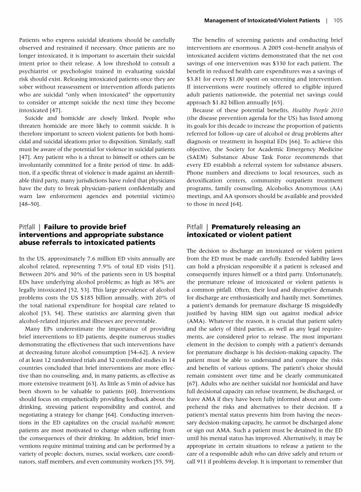
Patients who express suicidal ideations should be carefully
observed and restrained if necessary. Once patients are no
longer intoxicated, it is important to ascertain their suicidal
intent prior to their release. A low threshold to consult a
psychiatrist or psychologist trained in evaluating suicidal
risk should exist. Releasing intoxicated patients once they are
sober without reassessment or intervention affords patients
who are suicidal “only when intoxicated” the opportunity
to consider or attempt suicide the next time they become
intoxicated [47].
Suicide and homicide are closely linked. People who
threaten homicide are more likely to commit suicide. It is
therefore important to screen violent patients for both homi-
cidal and suicidal ideations prior to disposition. Similarly, staff
must be aware of the potential for violence in suicidal patients
[47]. Any patient who is a threat to himself or others can be
involuntarily committed for a finite period of time. In addi-
tion, if a specific threat of violence is made against an identifi-
able third party, many jurisdictions have ruled that physicians
have the duty to break physician–patient confidentially and
warn law enforcement agencies and potential victim(s)
[48–50].
Pitfall | Failure to provide briefinterventions and appropriate substanceabuse referrals to intoxicated patients
In the US, approximately 7.6 million ED visits annually are
alcohol related, representing 7.9% of total ED visits [51].
Between 20% and 30% of the patients seen in US hospital
EDs have underlying alcohol problems; as high as 38% are
legally intoxicated [52, 53]. This large prevalence of alcohol
problems costs the US $185 billion annually, with 20% of
the total national expenditure for hospital care related to
alcohol [53, 54]. These statistics are alarming given that
alcohol-related injuries and illnesses are preventable.
Many EPs underestimate the importance of providing
brief interventions to ED patients, despite numerous studies
demonstrating the effectiveness that such interventions have
at decreasing future alcohol consumption [54–62]. A review
of at least 12 randomized trials and 32 controlled studies in 14
countries concluded that brief interventions are more effec-
tive than no counseling, and, in many patients, as effective as
more extensive treatment [63]. As little as 5 min of advice has
been shown to be valuable to patients [60]. Interventions
should focus on empathetically providing feedback about the
drinking, stressing patient responsibility and control, and
negotiating a strategy for change [64]. Conducting interven-
tions in the ED capitalizes on the crucial teachable moment;
patients are most motivated to change when suffering from
the consequences of their drinking. In addition, brief inter-
ventions require minimal training and can be performed by a
variety of people: doctors, nurses, social workers, care coordi-
nators, staff members, and even community workers [55, 59].
The benefits of screening patients and conducting brief
interventions are enormous. A 2005 cost–benefit analysis of
intoxicated accident victims demonstrated that the net cost
savings of one intervention was $330 for each patient. The
benefit in reduced health care expenditures was a savings of
$3.81 for every $1.00 spent on screening and intervention.
If interventions were routinely offered to eligible injured
adult patients nationwide, the potential net savings could
approach $1.82 billion annually [65].
Because of these potential benefits, Healthy People 2010
(the disease prevention agenda for the US) has listed among
its goals for this decade to increase the proportion of patients
referred for follow-up care of alcohol or drug problems after
diagnosis or treatment in hospital EDs [66]. To achieve this
objective, the Society for Academic Emergency Medicine
(SAEM) Substance Abuse Task Force recommends that
every ED establish a referral system for substance abusers.
Phone numbers and directions to local resources, such as
detoxification centers, community outpatient treatment
programs, family counseling, Alcoholics Anonymous (AA)
meetings, and AA sponsors should be available and provided
to those in need [64].
Pitfall | Prematurely releasing anintoxicated or violent patient
The decision to discharge an intoxicated or violent patient
from the ED must be made carefully. Extended liability laws
can hold a physician responsible if a patient is released and
consequently injures himself or a third party. Unfortunately,
the premature release of intoxicated or violent patients is
a common pitfall. Often, their loud and disruptive demands
for discharge are enthusiastically and hastily met. Sometimes,
a patient’s demands for premature discharge IS misguidedly
justified by having HIM sign out against medical advice
(AMA). Whatever the reason, it is crucial that patient safety
and the safety of third parties, as well as any legal require-
ments, are considered prior to release. The most important
element in the decision to comply with a patient’s demands
for premature discharge is his decision-making capacity. The
patient must be able to understand and compare the risks
and benefits of various options. The patient’s choice should
remain consistent over time and be clearly communicated
[67]. Adults who are neither suicidal nor homicidal and have
full decisional capacity can refuse treatment, be discharged, or
leave AMA if they have been fully informed about and com-
prehend the risks and alternatives to their decision. If a
patient’s mental status prevents him from having the neces-
sary decision-making capacity, he cannot be discharged alone
or sign out AMA. Such a patient must be detained in the ED
until his mental status has improved. Alternatively, it may be
appropriate in certain situations to release a patient to the
care of a responsible adult who can drive safely and return or
call 911 if problems develop. It is important to remember that
Management of Intoxicated/Violent Patients | 105

alcohol consumption or an elevated BAL does not necessarily
mean that a patient lacks decisional capacity.
It is not permissible to use a psychiatric hold in order to
detain an intoxicated or aggressive patient unless very clear
conditions are met (danger to self, danger to others, or
gravely disabled). However, many EDs have what is com-
monly referred to as an “ED Hold,” in which ED policy at
that institution allows security to prevent a patient from
leaving. Another alternative is contacting the police, who
can encourage patients to listen to instructions or behave in
a more appropriate manner. It is advisable to have clear
guidelines and be familiar with institutional policies, as
these situations occur commonly.
Appropriate documentation is crucial. Documentation of a
final reassessment, including vital signs, and a lack of suicidal
or homicidal ideations should occur, with the date and time.
Any patient who is released AMA should have documentation
of full decisional capacity, and that he has been fully informed
about the risks and benefits of all options. For an intoxicated
patient, outpatient referrals should be provided, and the
patient should be instructed not to drink and drive. These
instructions should be documented as well. If possible, docu-
ment that the patient was discharged to the care of a responsi-
ble adult driver to whom the discharge instructions were
provided, explained, and understood. For a patient who elopes
from the ED, efforts used to prevent elopement, attempts to
find or contact the patient, and notification of the appropriate
authorities and third parties should be documented.
Pearls for Improving Patient Outcomes
• A comprehensive medical evaluation is essential to rule out
alternative or co-existing medical conditions resulting in or from
an intoxicated or violent state.
• There are a few specific circumstances when a BAL is necessary;
consider using rapid saliva or breath tests if these are available.
• Predictors of violent behavior should be recognized. Necessary
measures to protect the patient, staff, and third parties, such as
de-escalation techniques, seclusion, and restraints should be used.
• Chemical restraints should be administered based on suspected
diagnosis and known adverse effects. Whenever possible,
involve the patient in the selection of the medication and ask
him/her to take it willingly.
• Physical restraint application should be rehearsed. Restraints
should be applied to each extremity completely, safely, and with
frequent reevaluation.
• The potential for suicide and homicide in intoxicated or violent
patients must not be overlooked or minimized.
• The ED is a cost-effective and ideal place for providing brief
interventions and social services to individuals who abuse alco-
hol or other substances.
• Do not allow patients who lack decision-making capacity to
elope or be discharged AMA.
• Appropriate documentation is essential. Mental status examina-
tions, frequent reevaluations, reasons for restraints, lack of suici-
dality or homicidality, notification of appropriate authorities and
potential victims, and social service referrals should be clearly
documented.
Appendix A
Mini-Mental State (MMS) examination, from Folstein MF, Folstein
SE, McHugh PR. “ Mini-Mental State:” A Practical Method for
Grading the Cognitive State of Patients for the Clinician.
J. Psych. Res. 1975; 12(3)189–198.
Appendix B
Confusion Assessment Method, from Inouye SK, van Dyck CH,
Alessi CA, et al. Clarifying Confusion. The Confusion Assess-
ment Method. Ann Intern Med 1990; 113:941–948.
References
1. Tintinalli JE, Peacock FW, Wright MA. Emergency medical
evaluation of psychiatric patients. Ann Emerg Med 1994; 23(4):
859–62.
2. Hall R, Gardner E, Stickney S, et al. Physical illness manifesting
as psychiatric disease. II. Analysis of a state hospital inpatient
population. Arch Gen Psych 1980; 37(9):989–995.
3. Henneman PL, Mendoza R, Lewis RS. Prospective evaluation
of emergency department medical clearance. Ann Emerg Med
1994; 24(4):672–7.
4. Dubin WR, Weiss KJ, Zeccardi JA. Organic brain syndrome. The
psychiatric imposter. J Am Med Assoc 1983; 249(1):60–2.
5. Lagomasino I, Daly R, Stoudemire A. Medical assessment of
patients presenting with psychiatric symptoms in the emer-
gency setting. Psych Clin N Am 1999; 22(4):819–50.
6. Reeves RR, Pendarvis EJ, Kimble R. Unrecognized medical
emergencies admitted to psychiatric units. Am J Emerg Med
2000; 18(4):390–3.
7. Zun LS. Evidence-based evaluation of psychiatric patients.
J Emerg Med 2005; 28(1):35–9.
8. Fabrri A, Marchesini G, Morselli-Labate A, et al. Blood alcohol
concentration and management of road trauma patients in the
emergency department. J Trauma 2001; 50(3):521–8.
9. Todd K, Berk WA, Welch RD, et al. Prospective analysis of men-
tal status progression in ethanol-intoxicated patients. Am J
Emerg Med 1992; 10(4):271–3.
10. Lukens TW, Wolf SJ, Edlow JA, et al. American College of
Emergency Physicians Clinical Policies Subcommittee (Writing
Committee) on critical issues in the diagnosis and management
KEY FACT | Any patient who is released AMA should
have documentation of full decisional capacity, and that
he has been fully informed about the risks and benefits of
all options.
106 | Chapter 13

of the adult psychiatric patient in the emergency department.
Ann Emerg Med 2006; 47(1):79–99.
11. Gibb K. Serum alcohol levels, toxicology screens, and use of the
breath alcohol analyzer. Ann Emerg Med 1986; 15(3):349–53.
12. Galbraith S, Murray W, Patel A, et al. The relationship between
alcohol and head injury and its effect on the conscious level.
Br J Surg 1976; 63(2):128–30.
13. Brickley M, Shepherd J. The relationship between alcohol intox-
ication, injury severity and Glasgow Coma Score in assault
patients. Injury 1995; 26(5):311–14.
14. Keim ME, Bartfield JM, Raccio-Robak N. Blood ethanol estima-
tion: a comparison of three methods. Acad Emerg Med 1996;
3(1):85–7.
15. Smolle K, Hofmann G, Kaufmann P, et al. QED alcohol test:
a simple and quick method to detect ethanol in saliva of
patients in emergency departments. Comparison with the con-
ventional determination in blood. Intens Care Med 1999; 25(5):
492–5.
16. Degutis L, Rabinovici R, Sabbaj A, et al. The saliva strip test is
an accurate method to determine blood alcohol concentration
in trauma patients. Acad Emerg Med 2004; 11(8):885–7.
17. Gibb K, Oak R, Yee A, et al. Accuracy and usefulness of a breath
alcohol analyzer. Ann Emerg Med 1984; 13(7):516–20.
18. Lavoie FW, Carter G, Danzi D, et al. Emergency department
violence in United States teaching hospitals. Ann Emerg Med
1998; 17(11):1227–33.
19. Hill S, Petit J. The violent patient. Emerg Med Clin N Am 2000;
18(2):301–15.
20. Blomhoff S, Seim S, Friis S. Can prediction of violence among
psychiatric inpatients be improved? Hosp Comm Psychiatr 1990;
41(7):771–5.
21. Salkin M. Use of restraints and seclusion in the ED. ED Legal
Letter 1998; 9(6):57–68.
22. Dubin W. Violent patients. In: Bosker, G. (ed.), The Emergency
Medicine Reports Textbook of Adult and Pediatric Emergency Medicine.
2nd edition. Atlanta, Georgia: American Health Consultants
Incorporated, 2002, pp. 2069–76.
23. Kowalenko T, Walters BL, Khare RK, et al. Workplace violence:
a survey of emergency physicians in the state of Michigan. Ann
Emerg Med 2005; 46(2):142–7.
24. Ayranci U. Violence toward health care workers in emer-
gency departments in west Turkey. J Emerg Med 2005; 28(3):
361–5.
25. Meyers T, Garmel GM. Abnormal behavior. In: Mahadevan SV,
Garmel GM (eds), An Introduction to Clinical Emergency Medicine:
Guide for Practitioners in the Emergency Department. Cambridge, UK:
Cambridge University Press, 2005, pp. 161–70.
26. Allen MH, Currier GW, Hughes DH, et al. Treatment of behav-
ioral emergencies. Post Grad Med May 2001: 1–88.
27. Allen M, Carpenter D, Sheets J, et al. What do consumers say
they want and need during a psychiatric emergency? J Psych
Practice 2003; 9(1):39–58.
28. Rice M, Moore G. Management of the violent patient.
Therapeutic and legal considerations. Emerg Med Clinics of N Am
1991; 9(1):13–30.
29. Youngberg v. Romeo, 457 US 307 (1982).
30. Miller v. Rhode Island Hospital, 625 A.2d 778, 785 (R.I. 1993).
31. Blackman v. Rifkin, 759 P.2d 54, 58 (Colo Ct. App 1988).
32. Battaglia J. Pharmacological management of acute agitation.
Drugs 2005; 65(9):1207–22.
33. Zimbroff DL, Allen MH, Battaglia J. Best clinical practice with
ziprasidone IM: update after 2 years of experience. CNS Spectrums
2005; 10(9):1–15.
34. Joint Commission for Accreditation of Healthcare Organizations.
Sentinel Event Statistics. September 30, 2005, www.jcaho.org
35. Joint Commission for Accreditation of Healthcare Organizations.
Root Causes of Restraint Deaths (1995–2004). May 13, 2002,
www.jcaho.org
36. Zun LS. A prospective study of the complication rate of use of
patient restraint in the emergency department. J Emerg Med
2003; 24(2):119–24.
37. Dorfman DH, Kastner B. The use of restraint for pediatric psy-
chiatric patients in emergency departments. Pediatr Emerg Care
2004; 20(3):151–6.
38. Mohr WK, Petti TA, Mohr BD. Adverse effects associated with
physical restraint. Can J Psych 2003; 48(5):330–7.
39. Abdon-Beckman D. An awkward position: restraints and sud-
den death. J Emerg Med Ser 1997; 22(3):88–94.
40. Joint Commission for Accreditation of Healthcare Organizations.
Sentinel Event Alert: Preventing Restraint Deaths. Issue 8, November
18, 1998. www.jcaho.org
41. FDA Safety Alert: Entrapment Hazards with Hospital Bed Side
Rails, 1995.
42. Joint Commission for Accreditation of Healthcare Organiza-
tions. PC.12.130 and PC.12.140. Revised April 1, 2005. www.
jcaho.org
43. Centers for Medicare & Medicaid Services. Conditions of
Participation for Hospitals. 482.13. Revised October 1, 1999.
www.cms.hhs.gov
44. Lavoie FW. Consent, involuntary treatment, and the use of force
in an urban emergency department. Ann Emerg Med 1992;
21(1):25–32.
45. Mental health and suicide facts. Department of Health and
Human Services. National Strategy for Suicide Prevention; 2004.
http://www.mentalhealth.org/suicideprevention/suicidefacts.asp
46. Hufford MR. Alcohol and suicide behavior. Clin Psych Rev 2001;
21(5):797–811.
47. Dwyer B. The suicidal patient. In: Bosker, G. (ed.), The Emergency
Medicine Reports Textbook of Adult and Pediatric Emergency Medicine.
2nd edition. Atlanta, Georgia: American Health Consultants
Incorporated, 2002, pp. 2053–60.
48. Tarasoff v. Regents of the University of California, 17 Cal. 3d 425,
131 Cal. Rptr. 14, 551 P.2d 334 (1976).
49. Thompson v. County of Alameda, 27 Cal. 3d 741, 167 Cal. Rptr. 70,
614 P.2d 728 (1980).
50. Brady v. Hopper, 570 F. Supp. 1333, 1338 D. Colo. 1983.
51. McDonald A, Wang N, Camargo C. US emergency department
visits for alcohol-related diseases and injuries between 1992
and 2000. Arch Intern Med 2004; 164(5):531–7.
52. Centers for Disease Control and Prevention. Injury Fact Book.
2002. www.cdc.gov/ncipc/fact_book/09_Alcohol_%20Injuries_
%20ED.htm
53. D’Onofrio G, Bernstein E, Bernstein J, et al. Patients with alco-
hol problems in the emergency department, Part 1: Improving
detection. SAEM Substance Abuse Task Force. Acad Emerg Med
1998; 5(12):1200–9.
54. D’Onofrio G, Degutis LC. Preventive care in the emergency
department: screening and brief intervention for alcohol prob-
lems in the emergency department: a systematic review. Acad
Emerg Med 2002; 9(6):627–38.
Management of Intoxicated/Violent Patients | 107

55. Bernstein E, Bernstein J, Levenson S. Project ASSERT: an
ED-based intervention to increase access to primary care, pre-
ventive services, and the substance abuse treatment system.
Ann Emerg Med 1997; 30(2):181–9.
56. Wright LM, Moran L, Meyrick M, et al. Intervention by an alco-
hol health worker in an accident and emergency department.
Alcohol 1998; 33:651–6.
57. Crawford M, Patton R, Touquet R, et al. Screening and referral
for brief intervention of alcohol-misusing patients in an emer-
gency department: a pragmatic randomized controlled trial.
Lancet 2004; 364(9442):1334–9.
58. Green M, Setchell J, Hames P, et al. Management of alcohol
abusing patients in accident and emergency departments. J R
Soc Med 1993; 86(7):393–5.
59. Gentilello LM, Rivara FP, Donovan DM, et al. Alcohol interven-
tions in a trauma center as a means of reducing the risk of
injury recurrence. Ann Surg 1999; 230(4):473–80.
60. Babor TF, Acuda W, Campillo C, et al. World Health
Organization Brief Intervention Study Group. A cross-national
trial of brief intervention with heavy drinkers. Am J Public
Health 1996; 86:948–55.
61. Wilk A, Jensen N, Havighurst T. Meta-analysis of randomized
control trials addressing brief interventions in heavy alcohol
drinkers. J Gen Intern Med 1997; 12(5):274–83.
62. Monti PM, Colby SM, Barnett NP, et al. Brief intervention for
harm reduction with alcohol-positive older adolescents in a
hospital emergency department. J Consult Clin Psych 1999;
67(6):989–94.
63. Bien TH, Miller WR, Tonigan JS. Brief interventions for alcohol
problems: a review. Addiction 1993; 88(3):315–35.
64. D’Onofrio G, Bernstein E, Bernstein J, et al. Patients with alcohol
problems in the emergency department, Part 2: Intervention and
referral. Acad Emerg Med 1998; 5(12):1210–17.
65. Gentilello LM, Ebel B, Wickizer T, et al. Alcohol interventions for
trauma patients treated in emergency departments and hospitals:
a cost benefit analysis. Ann Surg 2005; 241(4):541–50.
66. US Department of Health and Human Services. Healthy People
2010: Understanding and Improving Health. Washington, DC: US
Government Printing Office, 2000.
67. Marco C, Derse A. Leaving against medical advice: should you
take no for an answer? ED Legal Letter 2004; 15(11):121–32.
108 | Chapter 13

Abdominal pain, evaluation and
management
appendicitis presentation, 28–29
heterotopic pregnancy, 27–28
intra-abdominal pathology
cardiac ischemia, 25
mesenteric ischemia, 25–27
signs and symptoms, in elderly, 29–31
Acute angle-closure glaucoma, 41–42
Acute back pain management, 33–38
acute motor radiculopathy, 37–38
antibiotic treatment, spinal infection, 37
aortic catastrophes, 33, 34, 36
aortic dissection, 33, 34, 35, 36
corticosteroids, 37
laboratory studies, spinal infection, 36
MRI for spinal cord compression or
abscess, 36–37
neurologic exam, 35
plain radiography, 36
pulmonary embolism, 34
risk factor assessment, 34–35
rupturing abdominal aortic aneurysm,
34, 36
spinal epidural abscess, 34
Acute bacterial endocarditis
exam findings, 63–64
in febrile patients, 63
Acute bronchitis, 65
Acute cholecystitis (AC), 25, 29, 30–31
Acute coronary syndrome (ACS), 1, 25
Acute exacerbations of chronic bronchitis
(AECB), 65
Acute myocardial infarction, 1–2, 6, 11
anginal equivalents, 1
risk factors, 1
Acute stroke, 8
Alcohol, 99, 100–101, 104
Amylase, 60
Analgesia, 60–61
Antibiotic therapy, 37, 40, 51, 65, 66,
69, 94
Anticeptic solutions, 72
Aortic dissection (AD), 7–8, 33, 34, 35, 36
cocaine, 35
risk factor, 34
Appendicitis, 28–29, 82–83
Arterial blood gas (ABG), 18, 19–20, 65
Ascending cholangitis, 31
Assisted reproductive technology (ART), 27
Ativan see lorazepam
Auscultation, 10, 17
Bacitracin, 76
Backboards, 60
Bacterial meningitis, 40, 42, 49, 66–67, 86
CSF range, 67
physical findings, 67
Bacterial Meningitis Score, 49
Benzodiazepines see individual entries
Biliary tract disease, 29–31
Bite wounds, 75–77
Blunt cerebrovascular injury (BCVI), 58
Boerhaave’s syndrome, 13
Boston criteria, 87
Brudzinski sign, 67
Calcium, 8
Canadian C-spine rule, 57
Carbon monoxide poisoning, 42
Carboxyhemoglobin, 18, 42
Cardiac markers see individual entries
Cardiac murmur, 63
Cardiac troponins, 6
Cardiotocographic monitoring (CTM), 83
Centers for Medicare and Medicaid Services
(CMS), 104
Cephalosporin, 37, 68, 75, 94
Cerebral venous thrombosis, 42
Cerebrospinal fluid (CSF), 49, 67
Cervical artery dissection (CAD), 46, 47
Cervical epidural abcesses (CEA), 48, 49, 51
Cervical epidural hematomas (CEH), 52
Chemoreceptor, 17
Chest syndromes, evaluation and
management, 1
acute MI diagnosis, 1–2
AD diagnosis, 7–8
Boerhaave’s syndrome, 13
cardiac ischemia , 2, 5–6
chest wall tenderness, 2
chest X-ray, 8, 12–13
ECG findings, 9–10
GI cocktail, 5
herpes zoster, 13–14
LBBB, 2–3
negative cardiac enzymes, 6–7
PE evaluation
pleuritic chest pain and dyspnea
presence, 9
pericarditis differentiation, 10–12
pneumothorax, 12–13
VPR, 3–5
Chest wall tenderness
musculoskeletal pain, 2
Chest X-ray, 8–9, 12–13, 19–20, 94
Cholelithiasis, 29
Chronic obstructive pulmonary disease
(COPD), 11, 19, 21, 102
CK-MB, 6–7
Colic, 87
Community-acquired pneumonia (CAP), 64
Computed tomography (CT), 27, 42, 51,
74, 95
Corticosteroids, 37, 40, 43, 95
Crystalloid fluid resuscitation, 55
C-spine clearance rules, 57
CT angiography, 27, 46, 52, 58, 82
Cytocrit, 97
D-dimer, 19–20, 82
Doppler signal, 58
Dyspenic patient management
ABG, 20
cardiopulmonary disease, rule out of,
20–21
CXR, 20
D-dimer, 19–20
dyspnea, definition, 17
dyspneic for anxiety, confusion, 19
EKG, 20
NPPV use, 21–22
oxygen administration in COPD
patients, 21
PE, in differential diagnosis, 19
physical examination, 17–18
pulse oximetry measurement, 18–19
Early, goal-directed therapy (EGDT), 68
ECG see electrocardiogram
Echocardiography, 10, 19–20, 21, 82
EKG see electrocardiogram
Electrocardiogram, 2, 3, 5–6, 9–10, 11
Emergency physicians (EP), 2, 25, 33–37,
63, 65, 72, 73, 85
Emphysematous cholecystitis, 30, 31
Endotracheal tube (ETT), 60
End-tidal CO2 detectors, 18
Enzyme-linked immunosorbent assay
(ELISA) test, 19, 20
Index
109

Epiglottitis, 47–48, 49–50
in plain film, 50
sensitivities and specificities, 50
Fetal distress, 83
Fever management in infants, 88, 89, 90
Focused abdominal sonography in trauma
(FAST), 55–56
Gallbladder empyema, 31
GI cocktail, 5
Haemophilus influenza, 48
Haldane effect, 21
Headache management
bacterial meningitis, 40
broad armamentarium, 43–44
elevated blood pressure, 43
skull
inside pain, 42
outside pain, 41–42
intracerebral hemorrhage, 43
limitations of CT, 40–41
neurosurgical intervention, 42–43
red flag features, 39–40
xanthochromia, 41
Hematology/oncology patients management
hyperviscosity and leukostasis
syndromes, 96–97
neutropenic fever treatment, 93–94
pericardial effusions and cardiac
tamponade, 95–96
SIADH diagnosis, 97–98
spinal cord compression, 94–95
SVC syndrome diagnosis, 95
hemoglobin, 18, 41, 80
Herpes zoster, 13–14
Heterotopic pregnancy, 27, 28, 79
Horner’s syndrome, 8, 46, 51
Hypercapnia, 17, 18, 20, 21
Hyperviscosity syndrome, 96–97
Hyponatremia, 97–98
Hypoxia, 17, 18, 19, 20, 21, 69
Hypoxic pulmonary vasoconstriction
(HPV), 21
In vitro fertilization (IVF), 27, 79
Infectious Disease Society of America
(IDSA), 65
Infectious diseases management
acute bacterial endocarditis, 63–64
bacterial meningitis, 66–67
intravenous drug users, with neurological
deficits, 64
meningitis, CT before LP, 66–67
MRSA, 68
PSI, 64–65
sepsis, treatment, 68–69
septic shock, antimicrobial treatment, 69
sinusitis, antibiotics prescription,
65–66
uncomplicated bronchitis, antibiotics
use, 65
Infective endocarditis, 63
Internal carotid artery dissection (ICAD), 46
Intestinal ischemia, 27
Intoxicated/violent patients management,
99–106
blood alcohol levels, during management
and disposition, 100
chemical restraint, indication and adverse
effects, 102–103
comprehensive medical evaluation,
99, 106
intoxicated patients
interventions and appropriate
substance abuse referrals, 105
patient, staff, and third parties protection,
101–102
physical restraint application, 103–104
prematurely releasing, 105
recognize suicide and/or homicide risk,
104–105
Intra-abdominal pathology, 25
Intrauterine pregnancy (IUP), 27, 79
IT CRIESS, 87
JCAHO, 102, 104
Kernig’s sign, 67
latex agglutination tests, 19, 20
Left bundle branch block (LBBB), 2–3
Lemierre’s syndrome, 47
Leukostasis syndrome, 96–97
Lorazepam, 102, 103
Lumbar puncture (LP), 39, 49, 51, 66,
86, 89
Magnetic resonance imaging (MRI), 37, 38,
40, 41, 42, 52, 74, 75, 84, 96
MANTRELS, 28–29
Maternal–fetal hemorrhage, 79, 80, 83
Mechanoreceptor, 17
Meningitis, 39–40, 40, 48, 49, 51, 86
CT before LP, 66
see also bacterial meningitis
Mental status examination (MSE), 100
Mesenteric ischemia, 25–27
risk factors, 26
Methemoglobinemia, 18, 20
Methicillin-resistant Staphylococcus aureus
(MRSA), 68, 94
Midazolam, 102, 103
Migraine headaches, 44
Mild head injury, 59
Misdosing medications, 86–87
Myoglobin, 6, 7
Neck pain, evaluation and management
arterial dissection, rule out of, 46–47
cardiac ischemia, 47
classic physical exam, neck infection, 48
CSF test, 49
elicit history/exam findings, infection
suggestion, 48
fever, infection sign, 48–49
infectious cause recognition, 47–48
malignancy evidence, 51
mass lesion patients, 51–52
neck mass consideration, 51
peripheral WBC count, 49
plain film findings of epiglottitis, 49–50
serious infection, delaying treatments, 51
spinal epidural hematoma, 52
image appropriation and consultation,
52
normal neurologic exam, 52
vascular cause consideration, 46
Negative cardiac enzymes, 6–7
Neurological deficits, 64
Neutropenia, 93–94
NEXUS study, 57
Non-invasive positive pressure ventilation
(NPPV), 21–22
Nuchal rigidity, 67
Olanzapine, 103
Osteomyelitis, 37, 49, 75
Otitis media (OM), 85
Oxygen therapy, 21
Pancreatitis, 31
Pediatric care, in ED
colic, 87
misdosing medications, 86–87
OM, 85
peripheral WBC count, 86
sepsis evaluation, 87–89
simple febrile seizure, 85–86
vomiting with gastroenteritis, 89–91
Peptic ulcer disease (PUD), 29, 40
Pericardial effusions and cardiac tamponade
neoplastic cardiac tamponade, 95–96
Pericarditis, 10–12
Peripartum cardiomyopathy (PPCM), 82
Peripheral WBC count, 36, 49, 86, 87, 96
Phildadelphia criteria, 87, 89
Placental abruption, 83
Plain film, 36, 37, 49–50, 51, 57
Plain radiography, 36, 50, 57, 58
Pleuritic chest pain, 9
Pneumonia severity index (PSI), 64–65
Pneumothorax, 12–13, 17, 19, 20, 58–59
Pre-eclampsia, 42
Pregnant patient management
appendicitis, diagnosis of, 82–83, 84
dyspnea in pregnancy, 81–82, 83
heterotopic pregnancy, 79, 83
maternal–fetal hemorrhage, 79–80
minor blunt trauma, in third
trimester , 83
perimortem cesarean section, 80–81
pregnancy suspection, 79
radiologic imaging, risk for fetus, 81, 83
vital signs and lab values interpretation, 79
Pseudomonas aeruginosa, 75
Pseudotumor cerebri, 42
Pulmonary embolism (PE), 19, 20, 33,
34, 82
pleuritic chest pain and dyspnea
presence, 9
Pulmonary infarction, 9
Pulse oximetry, 18–19, 21, 65
Radio opaque foreign bodies, 73
Red flag features, of headaches, 39
Retropharyngeal abscess, 50
110 | Index

Risperidal see risperidone
Risperidone, 103
Rochester criteria, 87, 89
Rupturing abdominal aortic aneurysm,
34, 36
Seat-belt syndrome, 56
Sepsis, 68–69, 87
antimicrobial treatment, 69
definitions of, 69
resuscitation goals, 69
evaluation, of newborn, 87–89
Boston criteria, 87
Phildadelphia criteria, 87, 89
Rochester criteria, 87, 89
Sgarbossa’s criteria, 10
SIADH diagnosis, 97–98
Simple febrile seizure, 85–86
Sinusitis, 65–66
Society for Academic Emergency Medicine
(SAEM), 105
Spectrophotometry, 41
Spinal cord compression, 35, 94–95
Spinal epidural abscess, 34, 35, 36, 37
risk factor, 35
Spinal epidural hematoma, 52
Spontaneous spinal epidural hematomas
(SSEH), 52
ST-segment elevation MI (STEMI), 2, 3
Sgarbossa’s criteria, 3
Staphylococcal aureus, 37
Subarachnoid hemorrhage, 41, 43
Succinylcholine, 59
Superior mesenteric artery (SMA)
embolus, 25–26
thrombosis, 26
Superior vena cava (SVC) syndrome
diagnosis, 95
Supplemental oxygen, 18, 21
Temporal arteritis, 41
Thromboembolism, 19, 82
Thrombolytic therapy, 25, 52
Toxidromes see individual entries
Trauma management, in ED
acutely burned or crushed patient,
succinylcholine
avoidance in, 59
analgesia, 60–61
backboards, 60
blunt carotid injury, 58
cervical spine, CT imaging, 57–58
crystalloid fluid resuscitation, 55
C-spine clearance rules, 57
Doppler signal, 58
ETT, 60
FAST examination, 55–56
geriatric patient, 56
laboratory data, 60
mild head injury, 59
pelvic compression, for pelvic fractures,
59
pneumothoraces, 58–59
pregnant patient, fetal radiation exposure
fear, 56
seat-belt syndrome, 56
spine, CTs and plain radiographs of, 57
tertiary care facilities, transporting
errors, 60
vascular access, inappropriate choice,
59–60
Trimethoprim-sulfamethoxizole, 68
Troponin, 6, 10
Ultrasound, 27, 28, 30, 51, 74, 75, 79,
82, 83
Uncomplicated bronchitis, 65
Vascular access, 59–60
Venous thromboembolism, 22, 82
Ventricular paced rhythm (VPR), 3–5
Vertebral artery dissection (VAD), 46, 47
Viral encephalitis, 42
Vomiting, causes of, 91
Wound care
after care instructions, 76
foreign body detection, 73–74
plantar puncture wounds, 74–75
proper preparation, to closure, 72–73
prophylactic antibiotics, for bite wounds,
75–76
Wound infection, characteristic, 75
Wound irrigation, 72
Xanthochromia, 41
Ziprasidone, 103
Zyprexa see olanzapine
Index | 111


![[Diagnostic pitfalls with Cushing's syndrome]](https://img.pdfslide.net/doc/110x75/634c738d7f5a138881007d0f/diagnostic-pitfalls-with-cushings-syndrome.jpg)

![Coda-avoiding: Some Evidence from Portuguese [1]](https://img.pdfslide.net/doc/110x75/631297b6aca2b42b580ce326/coda-avoiding-some-evidence-from-portuguese-1.jpg)


