Embed Size (px)
Citation preview
epptwtctasrasptfltpitospattt
RA
The Journal of Emergency Medicine, Vol. 40, No. 1, pp. 103–112, 2011Copyright © 2011 Elsevier Inc.
Printed in the USA. All rights reserved0736-4679/$–see front matter
doi:10.1016/j.jemermed.2009.10.019
ClinicalReviews
ENCOUNTERS WITH VENOMOUS SEA-LIFE
Isaac Fernandez, MS,* Genaro Valladolid, MS,† Joseph Varon, MD, FACP, FCCP, FCCM,‡§�and George Sternbach, MD¶
*Universidad de Monterrey, Monterrey, Nuevo León, México, †Universidad Autónoma de Baja California, Tijuana, Baja California,México, ‡University of Texas Health Science Center of Houston, Houston, Texas, §The University of Texas Medical Branch at
Galveston, Galveston, Texas, �St. Luke’s Episcopal Hospital, Houston, Texas, and ¶Department of Emergency Medicine, StanfordMedical Center, Stanford, California
Reprint Address: Joseph Varon, MD, FACP, FCCP, FCCM, Dorrington Medical Associates P.A., 2219 Dorrington St., Houston,
TX 77030-3209ec
SdffnElovmeaal
C
Te
Abstract—Background: Sea-life with envenomation ca-abilities are quite abundant and diverse worldwide, beingredominantly found in tropical waters. Most envenoma-ions occur not as an attack, but as a result of self defensehen the animal perceives danger; and often when locals or
ourists are engaged in recreational activities. Most of theseases have only minor injuries, and few are fatal. Objec-ives: To describe the impact, clinical features, and man-gement of life-threatening marine envenomations. Discus-ion: Recognition of the injury and identification of theesponsible animal is crucial for quick and successful man-gement. Medical professionals should be cognizant of pre-enting symptoms such as respiratory distress, musclearalysis, or cardiovascular decompensation. For these pa-ients, antivenom should be given immediately if available,ollowed by pharmacological and physical therapy to re-ieve symptoms and pain. If any foreign bodies are left athe site of the injury, they must be removed. Tetanus pro-hylaxis should also be considered in case of puncture, and
f signs of early infection are present, broad-spectrum an-ibiotics should be administered. Conclusion: Managementf envenomations from marine animals should be empha-ized not only to health centers, but also to the generalopulation, so that initial treatment can be started as soons possible. Educational programs regarding risks and ini-ial management for these incidents are also recommendedo reduce the incidence and associated morbidity and mor-ality of the encounters. © 2011 Elsevier Inc.
ECEIVED: 11 April 2009; FINAL SUBMISSION RECEIVED: 10
CCEPTED: 27 October 2009103
Keywords—marine envenomations; jellyfish; stonefish;one snail; blue ringed octopus; antivenom; stingray
INTRODUCTION
ea-life with envenomation capabilities are quite abun-ant and diverse worldwide; they are predominantlyound in tropical waters. Each year, thousands of injuriesrom stingrays and jellyfish take place (1–3). Fortu-ately, few of these reported incidents are fatal (4–7).nvenomations from the scorpion fish family or mol-
usks are not as common as envenomations from jellyfishr stingrays. Venomous lesions from aquatic creaturesary greatly in presentation; they range from simple,ild, localized pain with erythema and papulovesicular
ruption, to severe shock and death (Table 1). Thisrticle will review animals with envenomations that aressociated with a high rate of morbidity and with serious,ife-threatening events.
DISCUSSION
oelenterates
here are approximately 10,000 species of marine co-lenterates, of which more than 100 are considered dan-
er 2009;
Octob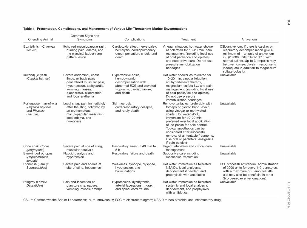
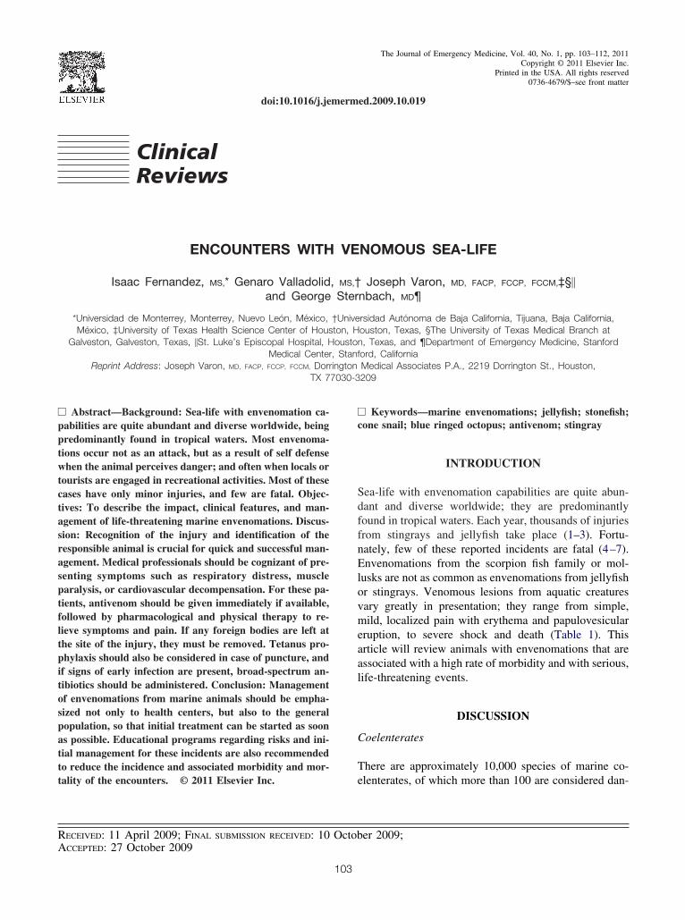
Table 1. Presentation, Complications, and Management of Various Life-Threatening Marine Envenomations
Offending AnimalCommon Signs and
Symptoms Complications Treatment Antivenom
Box jellyfish (Chironexfleckeri)
Itchy red maculopapular rash,burning pain, edema, andthe classical ladder-rungpattern lesion
Cardiotoxic effect, nerve palsy,hemolysis, cardiopulmonarydecompensation, shock, anddeath
Vinegar irrigation, hot water showeras tolerated for 10–20 min, painmanagement (including local useof cold packs/ice and opiates),and supportive care. Do not usepressure immobilizationbandages
CSL-antivenom. If there is cardiac orrespiratory decompensation give aminimum of 1 ampule of antivenomi.v. (20,000 units diluted 1:10 withnormal saline). Up to 3 ampules maybe given consecutively if response isinadequate in addition to magnesiumsulfate bolus i.v.
Irukandji jellyfish(Carukia barnesi)
Severe abdominal, chest,limbs, or back pain;generalized muscular pain,hypertension, tachycardia,vomiting, nausea,diaphoresis, piloerection,and local erythema
Hypertensive crisis,hemodynamicdecompensation withabnormal ECG and elevatedtroponins, cardiac failure,and death
Hot water shower as tolerated for10–20 min, vinegar irrigation,antihypertensive therapy,magnesium sulfate i.v., and painmanagement (including local useof cold packs/ice and opiates).Do not use pressureimmobilization bandages
Unavailable
Portuguese man-of-war(Physalia physalisand Physaliautriculus)
Local sharp pain immediatelyafter the sting, followed byan erythematousmaculopapular linear rash,local edema, andnumbness
Skin necrosis,cardiorespiratory collapse,and rarely death
Remove tentacles, preferably withforceps or gloved hand. Avoidusing vinegar or methylatedspirits. Hot water (45°C)immersion for 10–20 minpreferred over local applicationof ice-packs for pain control.Topical anesthetics can beconsidered after successfulremoval of all tentacle fragments.Use oral or parenteral analgesicsif pain persists
Unavailable
Cone snail (Conusgeographus)
Severe pain at site of sting,muscular paralysis
Respiratory arrest in 40 min to5 h
Urgent intubation and critical caremanagement
Unavailable
Blue-ringed octopus(Hapalochlaenalunulata)
Flaccid paralysis andhypotension
Respiratory failure and death Supportive care includingmechanical ventilation
Unavailable
Stonefish (Family:Scorpaenidae)
Severe pain and edema atsite of sting, headaches
Weakness, syncope, dyspnea,hypotension, andhallucinations
Hot water immersion as tolerated,NSAIDs, local analgesia,debridement if needed, andprophylaxis with antibiotics
CSL stonefish antivenom. Administrationof 2000 units for every 1–2 punctures,with a maximum of 3 ampules. (Itsuse may also be beneficial in otherScorpaenidae envenomations)
Stingray (Family:Dasyatidae)
Pain and laceration atpuncture site, nausea,vomiting, muscle cramps
Hypotension, dysrhythmia,arterial lacerations, thorax,and spinal cord trauma
Hot water immersion as tolerated,systemic and local analgesia,debridement, and prophylaxiswith antibiotics
Unavailable
CSL � Commonwealth Serum Laboratories; i.v. � intravenous; ECG � electrocardiogram; NSAID � non-steroidal anti-inflammatory drug.
104I.Fernandez
etal.
gcAspflWafit
CkttpT6
rocmasjav
f91(3rhmoiporssdTjln
fiac
1iimdsTarsoc
CmcI1iofTm
fcac
Itppte
Ftp
Sea-life Envenomations 105
erous. Creatures of the phylum Cnidaria are carnivoresontaining venom-charged nematocysts. Phospholipase2 (PLA2), an enzyme found in the venom of many
nakes, has also been identified in all classes of thehylum cnidaria, including the box jellyfish (Chironexeckeri) and the Irukandji (Carukia barnesi) (8,9).hereas many of the creatures in the phylum Cnidaria
re known to be venomous, the Irukandji and box jelly-sh have a high incidence of mortality associated with
heir stings.
hironex fleckeri (box jellyfish). The box jellyfish, alsonown as the sea wasp and marine stinger, is native tohe Western Australian coast, across the Northern Terri-ory coast and down the east coast of Queensland, and isredominantly seen during the summer months (10).his feared and deadly animal has accounted for at least7 deaths in Australia (11,12).
Large doses of box jellyfish venom can produce aapid cardiotoxic effect and can present with an absencef electrocardiographic abnormalities (13,14). In severeases, the patient decompensates and collapses withininutes, and often by the time medical attention is avail-
ble, the patient is beyond recovery. Delayed hypersen-itivity reactions from jellyfish stings, including boxellyfish, present with an itchy red maculopapular rashfter several days or weeks in more than half of theictims (15,16).
A recent prospective study of 225 confirmed stingsrom the box jellyfish by Currie and Jacups revealed that2% of the stings took place between October 1 and June
(i.e., stinger season), 83% were in shallow water�1 m), and were significantly more common between:00 p.m. and 6:00 p.m. (p � 0.001) (4). In almost alleports, the onset of pain was immediate. Approximatelyalf of the study population reported pain intensity asoderate, and one-fourth stated it to be severe. One-third
f the patients required parenteral narcotics despite thenduction of vinegar and cold ice-packs, and 8% of stungatients required hospitalization. There have been reportsf �65 deaths that occurred over this last century as aesult of jellyfish stings (17,18). In the aforementionedtudy, a 3-year-old child died as a result of a box jellyfishting. In addition, the authors related that two additionaleaths outside the scope of the study were also reported.he authors went on to state that the last 10 deaths from
ellyfish envenomations were all children. This is mostikely related to the smaller body mass contacted by theematocysts, producing a more virulent effect (19).
It is important to differentiate the box jellyfish stingrom others in its species, as response to therapeuticnterventions may vary (20). It may be helpful to perform
rapid inspection of the affected area, to look for the
haracteristic cross-hatched ladder pattern lesion (Figure f) (21). Microscopic identification of the jellyfish speciess also helpful in deciding the best approach to treatment,ncluding possible need for antivenom (15,22). Twoethods to identify the box jellyfish sting have been
escribed; the skin-scraping method with a blade and theticky tape method used for nematocyst identification.he latter method is considered faster, easier to perform,nd more accurate, with one study showing positiveesults in 85% of patients studied (n � 20) (22). Anothertudy demonstrated positive identification results in 74%f patients (n � 39), but identification can still be diffi-ult if the skin lesion is small (15,22).
arukia barnesi (Irukandji). This small bullet-shapededusa containing four contractile tentacles was first
aptured by Barnes in 1961 (23). It is responsible for therukandji syndrome described previously by Flecker in952, and it is named after an aboriginal tribe thatnhabited the coastal waters around Cairns, where mostf the cases had been reported (24). The Irukandji isound in Australia along the northern and western coasts.he size of the bell of this animal ranges from 3 to 19m (25).The stingers (nematocysts) from this creature are
ound not only on the tentacles but also on the bell of thisarybdeid (23). Although nematocysts found on the bellre morphologically different from those on the tenta-les, they can also cause envenomation (26).
The most common initial signs and symptoms afterrukandji sting include raising a wheal, local erythema athe sting site, severe abdominal, chest, limb, or backain, cough, nausea, vomiting, and generalized muscularain; followed by symptoms of sympathetic hyperac-ivity (e.g., sweating, hypertension, tachycardia, pilo-rection, and mild pyrexia), which can lead to cardiac
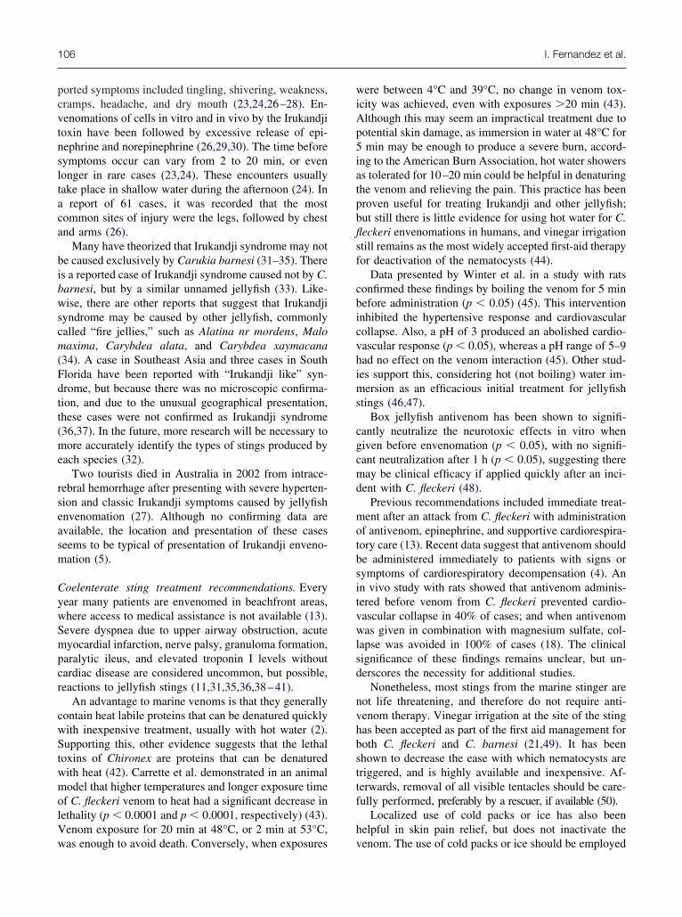
igure 1. Box jellyfish (Chironex fleckeri). The animal’s ten-acles with the characteristic ladder-like pattern. Figurerinted with permission of Dr. Zoltan Takacs.
ailure and pulmonary edema. Other infrequently re-
pcvtnsltaca
bibwscm(Fdtt(me
rseasm
CywSmpcr
cwStwmolVw
wiAp5iatpbflsf
cbicvhims
cgcmd
motbsitvwlsd
nvhbsttf
h
106 I. Fernandez et al.
orted symptoms included tingling, shivering, weakness,ramps, headache, and dry mouth (23,24,26–28). En-enomations of cells in vitro and in vivo by the Irukandjioxin have been followed by excessive release of epi-ephrine and norepinephrine (26,29,30). The time beforeymptoms occur can vary from 2 to 20 min, or evenonger in rare cases (23,24). These encounters usuallyake place in shallow water during the afternoon (24). In
report of 61 cases, it was recorded that the mostommon sites of injury were the legs, followed by chestnd arms (26).
Many have theorized that Irukandji syndrome may note caused exclusively by Carukia barnesi (31–35). Theres a reported case of Irukandji syndrome caused not by C.arnesi, but by a similar unnamed jellyfish (33). Like-ise, there are other reports that suggest that Irukandji
yndrome may be caused by other jellyfish, commonlyalled “fire jellies,” such as Alatina nr mordens, Maloaxima, Carybdea alata, and Carybdea xaymacana
34). A case in Southeast Asia and three cases in Southlorida have been reported with “Irukandji like” syn-rome, but because there was no microscopic confirma-ion, and due to the unusual geographical presentation,hese cases were not confirmed as Irukandji syndrome36,37). In the future, more research will be necessary toore accurately identify the types of stings produced by
ach species (32).Two tourists died in Australia in 2002 from intrace-
ebral hemorrhage after presenting with severe hyperten-ion and classic Irukandji symptoms caused by jellyfishnvenomation (27). Although no confirming data arevailable, the location and presentation of these caseseems to be typical of presentation of Irukandji enveno-ation (5).
oelenterate sting treatment recommendations. Everyear many patients are envenomed in beachfront areas,here access to medical assistance is not available (13).evere dyspnea due to upper airway obstruction, acuteyocardial infarction, nerve palsy, granuloma formation,
aralytic ileus, and elevated troponin I levels withoutardiac disease are considered uncommon, but possible,eactions to jellyfish stings (11,31,35,36,38–41).
An advantage to marine venoms is that they generallyontain heat labile proteins that can be denatured quicklyith inexpensive treatment, usually with hot water (2).upporting this, other evidence suggests that the lethal
oxins of Chironex are proteins that can be denaturedith heat (42). Carrette et al. demonstrated in an animalodel that higher temperatures and longer exposure time
f C. fleckeri venom to heat had a significant decrease inethality (p � 0.0001 and p � 0.0001, respectively) (43).enom exposure for 20 min at 48°C, or 2 min at 53°C,
as enough to avoid death. Conversely, when exposures vere between 4°C and 39°C, no change in venom tox-city was achieved, even with exposures �20 min (43).lthough this may seem an impractical treatment due tootential skin damage, as immersion in water at 48°C formin may be enough to produce a severe burn, accord-
ng to the American Burn Association, hot water showerss tolerated for 10–20 min could be helpful in denaturinghe venom and relieving the pain. This practice has beenroven useful for treating Irukandji and other jellyfish;ut still there is little evidence for using hot water for C.eckeri envenomations in humans, and vinegar irrigationtill remains as the most widely accepted first-aid therapyor deactivation of the nematocysts (44).
Data presented by Winter et al. in a study with ratsonfirmed these findings by boiling the venom for 5 minefore administration (p � 0.05) (45). This interventionnhibited the hypertensive response and cardiovascularollapse. Also, a pH of 3 produced an abolished cardio-ascular response (p � 0.05), whereas a pH range of 5–9ad no effect on the venom interaction (45). Other stud-es support this, considering hot (not boiling) water im-ersion as an efficacious initial treatment for jellyfish
tings (46,47).Box jellyfish antivenom has been shown to signifi-
antly neutralize the neurotoxic effects in vitro wheniven before envenomation (p � 0.05), with no signifi-ant neutralization after 1 h (p � 0.05), suggesting thereay be clinical efficacy if applied quickly after an inci-
ent with C. fleckeri (48).Previous recommendations included immediate treat-
ent after an attack from C. fleckeri with administrationf antivenom, epinephrine, and supportive cardiorespira-ory care (13). Recent data suggest that antivenom shoulde administered immediately to patients with signs orymptoms of cardiorespiratory decompensation (4). Ann vivo study with rats showed that antivenom adminis-ered before venom from C. fleckeri prevented cardio-ascular collapse in 40% of cases; and when antivenomas given in combination with magnesium sulfate, col-
apse was avoided in 100% of cases (18). The clinicalignificance of these findings remains unclear, but un-erscores the necessity for additional studies.
Nonetheless, most stings from the marine stinger areot life threatening, and therefore do not require anti-enom therapy. Vinegar irrigation at the site of the stingas been accepted as part of the first aid management foroth C. fleckeri and C. barnesi (21,49). It has beenhown to decrease the ease with which nematocysts areriggered, and is highly available and inexpensive. Af-erwards, removal of all visible tentacles should be care-ully performed, preferably by a rescuer, if available (50).
Localized use of cold packs or ice has also beenelpful in skin pain relief, but does not inactivate the
enom. The use of cold packs or ice should be employed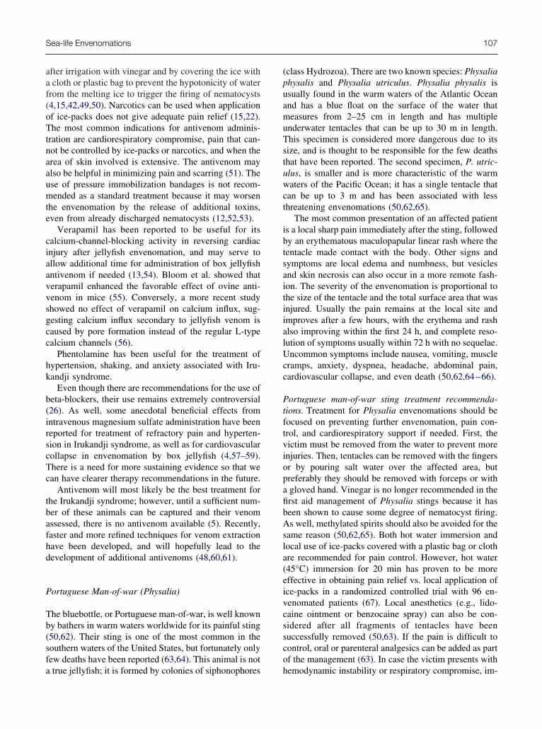
aaf(oTtnaaumte
ciaavvsgcc
hk
b(irscTc
tbafhd
P
Tb(sfa
(puamuTstuwct
ibtsaitiialUcc
PtftviopafibAsla(eivcssco
Sea-life Envenomations 107
fter irrigation with vinegar and by covering the ice withcloth or plastic bag to prevent the hypotonicity of water
rom the melting ice to trigger the firing of nematocysts4,15,42,49,50). Narcotics can be used when applicationf ice-packs does not give adequate pain relief (15,22).he most common indications for antivenom adminis-
ration are cardiorespiratory compromise, pain that can-ot be controlled by ice-packs or narcotics, and when therea of skin involved is extensive. The antivenom maylso be helpful in minimizing pain and scarring (51). These of pressure immobilization bandages is not recom-ended as a standard treatment because it may worsen
he envenomation by the release of additional toxins,ven from already discharged nematocysts (12,52,53).
Verapamil has been reported to be useful for itsalcium-channel-blocking activity in reversing cardiacnjury after jellyfish envenomation, and may serve tollow additional time for administration of box jellyfishntivenom if needed (13,54). Bloom et al. showed thaterapamil enhanced the favorable effect of ovine anti-enom in mice (55). Conversely, a more recent studyhowed no effect of verapamil on calcium influx, sug-esting calcium influx secondary to jellyfish venom isaused by pore formation instead of the regular L-typealcium channels (56).
Phentolamine has been useful for the treatment ofypertension, shaking, and anxiety associated with Iru-andji syndrome.
Even though there are recommendations for the use ofeta-blockers, their use remains extremely controversial26). As well, some anecdotal beneficial effects fromntravenous magnesium sulfate administration have beeneported for treatment of refractory pain and hyperten-ion in Irukandji syndrome, as well as for cardiovascularollapse in envenomation by box jellyfish (4,57–59).here is a need for more sustaining evidence so that wean have clearer therapy recommendations in the future.
Antivenom will most likely be the best treatment forhe Irukandji syndrome; however, until a sufficient num-er of these animals can be captured and their venomssessed, there is no antivenom available (5). Recently,aster and more refined techniques for venom extractionave been developed, and will hopefully lead to theevelopment of additional antivenoms (48,60,61).
ortuguese Man-of-war (Physalia)
he bluebottle, or Portuguese man-of-war, is well knowny bathers in warm waters worldwide for its painful sting50,62). Their sting is one of the most common in theouthern waters of the United States, but fortunately onlyew deaths have been reported (63,64). This animal is not
true jellyfish; it is formed by colonies of siphonophores hclass Hydrozoa). There are two known species: Physaliahysalis and Physalia utriculus. Physalia physalis issually found in the warm waters of the Atlantic Oceannd has a blue float on the surface of the water thateasures from 2–25 cm in length and has multiple
nderwater tentacles that can be up to 30 m in length.his specimen is considered more dangerous due to itsize, and is thought to be responsible for the few deathshat have been reported. The second specimen, P. utric-lus, is smaller and is more characteristic of the warmaters of the Pacific Ocean; it has a single tentacle that
an be up to 3 m and has been associated with lesshreatening envenomations (50,62,65).
The most common presentation of an affected patients a local sharp pain immediately after the sting, followedy an erythematous maculopapular linear rash where theentacle made contact with the body. Other signs andymptoms are local edema and numbness, but vesiclesnd skin necrosis can also occur in a more remote fash-on. The severity of the envenomation is proportional tohe size of the tentacle and the total surface area that wasnjured. Usually the pain remains at the local site andmproves after a few hours, with the erythema and rashlso improving within the first 24 h, and complete reso-ution of symptoms usually within 72 h with no sequelae.ncommon symptoms include nausea, vomiting, muscle
ramps, anxiety, dyspnea, headache, abdominal pain,ardiovascular collapse, and even death (50,62,64–66).
ortuguese man-of-war sting treatment recommenda-ions. Treatment for Physalia envenomations should beocused on preventing further envenomation, pain con-rol, and cardiorespiratory support if needed. First, theictim must be removed from the water to prevent morenjuries. Then, tentacles can be removed with the fingersr by pouring salt water over the affected area, butreferably they should be removed with forceps or withgloved hand. Vinegar is no longer recommended in therst aid management of Physalia stings because it haseen shown to cause some degree of nematocyst firing.s well, methylated spirits should also be avoided for the
ame reason (50,62,65). Both hot water immersion andocal use of ice-packs covered with a plastic bag or clothre recommended for pain control. However, hot water45°C) immersion for 20 min has proven to be moreffective in obtaining pain relief vs. local application ofce-packs in a randomized controlled trial with 96 en-enomated patients (67). Local anesthetics (e.g., lido-aine ointment or benzocaine spray) can also be con-idered after all fragments of tentacles have beenuccessfully removed (50,63). If the pain is difficult toontrol, oral or parenteral analgesics can be added as partf the management (63). In case the victim presents with
emodynamic instability or respiratory compromise, im-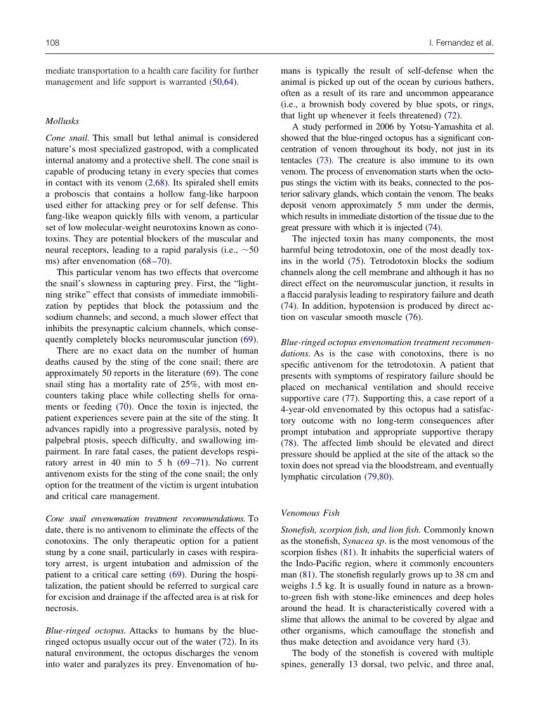
mm
M
Cniciaufstnm
tnzsiq
dascmpappraoa
Cdcstptfn
Brni
mao(t
sctvptdwg
hicda(t
Bdspps4tp(ptl
V
Sastmwtasot
108 I. Fernandez et al.
ediate transportation to a health care facility for furtheranagement and life support is warranted (50,64).
ollusks
one snail. This small but lethal animal is consideredature’s most specialized gastropod, with a complicatednternal anatomy and a protective shell. The cone snail isapable of producing tetany in every species that comesn contact with its venom (2,68). Its spiraled shell emits
proboscis that contains a hollow fang-like harpoonsed either for attacking prey or for self defense. Thisang-like weapon quickly fills with venom, a particularet of low molecular-weight neurotoxins known as cono-oxins. They are potential blockers of the muscular andeural receptors, leading to a rapid paralysis (i.e., �50s) after envenomation (68–70).This particular venom has two effects that overcome
he snail’s slowness in capturing prey. First, the “light-ing strike” effect that consists of immediate immobili-ation by peptides that block the potassium and theodium channels; and second, a much slower effect thatnhibits the presynaptic calcium channels, which conse-uently completely blocks neuromuscular junction (69).
There are no exact data on the number of humaneaths caused by the sting of the cone snail; there arepproximately 50 reports in the literature (69). The conenail sting has a mortality rate of 25%, with most en-ounters taking place while collecting shells for orna-ents or feeding (70). Once the toxin is injected, the
atient experiences severe pain at the site of the sting. Itdvances rapidly into a progressive paralysis, noted byalpebral ptosis, speech difficulty, and swallowing im-airment. In rare fatal cases, the patient develops respi-atory arrest in 40 min to 5 h (69–71). No currentntivenom exists for the sting of the cone snail; the onlyption for the treatment of the victim is urgent intubationnd critical care management.
one snail envenomation treatment recommendations. Toate, there is no antivenom to eliminate the effects of theonotoxins. The only therapeutic option for a patienttung by a cone snail, particularly in cases with respira-ory arrest, is urgent intubation and admission of theatient to a critical care setting (69). During the hospi-alization, the patient should be referred to surgical careor excision and drainage if the affected area is at risk forecrosis.
lue-ringed octopus. Attacks to humans by the blue-inged octopus usually occur out of the water (72). In itsatural environment, the octopus discharges the venom
nto water and paralyzes its prey. Envenomation of hu- sans is typically the result of self-defense when thenimal is picked up out of the ocean by curious bathers,ften as a result of its rare and uncommon appearancei.e., a brownish body covered by blue spots, or rings,hat light up whenever it feels threatened) (72).
A study performed in 2006 by Yotsu-Yamashita et al.howed that the blue-ringed octopus has a significant con-entration of venom throughout its body, not just in itsentacles (73). The creature is also immune to its ownenom. The process of envenomation starts when the octo-us stings the victim with its beaks, connected to the pos-erior salivary glands, which contain the venom. The beakseposit venom approximately 5 mm under the dermis,hich results in immediate distortion of the tissue due to thereat pressure with which it is injected (74).
The injected toxin has many components, the mostarmful being tetrodotoxin, one of the most deadly tox-ns in the world (75). Tetrodotoxin blocks the sodiumhannels along the cell membrane and although it has noirect effect on the neuromuscular junction, it results inflaccid paralysis leading to respiratory failure and death
74). In addition, hypotension is produced by direct ac-ion on vascular smooth muscle (76).
lue-ringed octopus envenomation treatment recommen-ations. As is the case with conotoxins, there is nopecific antivenom for the tetrodotoxin. A patient thatresents with symptoms of respiratory failure should belaced on mechanical ventilation and should receiveupportive care (77). Supporting this, a case report of a-year-old envenomated by this octopus had a satisfac-ory outcome with no long-term consequences afterrompt intubation and appropriate supportive therapy78). The affected limb should be elevated and directressure should be applied at the site of the attack so theoxin does not spread via the bloodstream, and eventuallyymphatic circulation (79,80).
enomous Fish
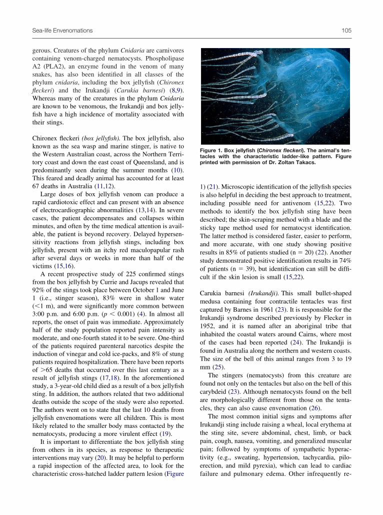
tonefish, scorpion fish, and lion fish. Commonly knowns the stonefish, Synacea sp. is the most venomous of thecorpion fishes (81). It inhabits the superficial waters ofhe Indo-Pacific region, where it commonly encountersan (81). The stonefish regularly grows up to 38 cm andeighs 1.5 kg. It is usually found in nature as a brown-
o-green fish with stone-like eminences and deep holesround the head. It is characteristically covered with alime that allows the animal to be covered by algae andther organisms, which camouflage the stonefish andhus make detection and avoidance very hard (3).
The body of the stonefish is covered with multiple
pines, generally 13 dorsal, two pelvic, and three anal,
asaafir
titevnpaadsfibtts
ow3ttiybit
eltc(eot
Oslpgste
StarTwp
mAem4aaiPs
FcTc
Sea-life Envenomations 109
nd venom is released from two lateral glands in everypine base upon mechanical pressure. This is a defensend self-preservation mechanism that is not used forttacking prey (Figure 2) (82). The venom of the stone-sh is one of the most powerful known to man, compa-able in potency to that of the cobra (83).
The stonefish inflicts its defensive damage firsthrough a wound produced by the spine of the fish, whichntroduces the venom into the tissue. The injected venomhen increases capillary permeability, inducing severedema of the affected limb. The lethal portion of theenom is a highly hypotensive agent with myotoxic andeurotoxic components (84). Systemic effects mayresent as muscle weakness, syncope, dyspnea, head-ches, and hallucinations (81). Fortunately, there is anntivenom available; however, the diagnosis must beone by clinical correlation, and tetanus prophylaxishould be started (85). Other members of the scorpionsh family are also venomous, the most well knowneing the lion fish. As with many other species in nature,he scorpion fishes have an attack-defense mechanismhat includes the secretion of venoms and other toxicubstances with biological actions (86).
The lion fish, a well-known exotic fish, is also capablef causing envenomation injuries when contact is madeith its venomous dorsal, anal, and pelvic spines (Figure). These wounds often occur to the hands of those tryingo manipulate the fish. Lion fish venom is the weakest ofhe scorpion fish family, yet it still creates a sharp,ntense, throbbing pain that radiates to other areas be-ond the puncture site (83). Along with the other mem-ers of its family, wounds from this fish can be dividednto three grades, depending on the severity: 1) ery-
igure 2. Stonefish. This camouflaged animal can easily beonfused with a rock or coral from the bottom of the sea.he dorsal spines shown contain dangerous venom thatould threaten a person’s life.
hema, pallor, ecchymosis or even cyanosis are the firstFs
vents that present, and result from the increased capil-ary permeability; 2) vesicle formation, as an effect of theoxins; 3) local necrosis observed within days, which isonsidered a grave complication and requires debriding87). If left untreated, the pain may persist for days orven weeks. Systemic effects can be very similar to thosef the stonefish, and are always relative to the concen-ration of venom injected into the victim (87,88).
Mostly found in the coastal waters of the Atlanticcean (usually Brazil, Uruguay, and Argentina), the
corpion fish remains an understudied fish due to itsimited global distribution (89). The spines of the scor-ion fish are strong, short, and have more developedlands than the lion fish (90). Like its “cousin” thecorpion fish, the lion fish is able to generate a pro-einaceous venom, with cardiotoxic and neurotoxicffects (91).
tonefish, scorpion fish, and lion fish envenomationreatment recommendations. Specific antivenom is avail-ble for the different species of scorpion fish and helps inelieving the pain and systemic effects of envenomation.he dose is 2000 units for every one to two punctures,ith a maximum of three ampules for more than fourunctures (85).
All the spines must be removed, and the affected limbust be elevated and cleaned with clear running water.fterwards, direct pressure should be applied to prevent
xcessive bleeding. The affected limb should be im-ersed in water heated to an approximate temperature of
5°C (never boiling water) to relieve acute pain. Inddition, supplemental local or oral analgesia may bedministered. Tetanus prophylaxis should also be admin-stered and the patient should be observed for 6–12 h.lain radiographs to ensure the removal of all spineshould also be performed. There is no need for antibiotic
igure 3. Lion fish. This fish has many long dorsal and finpines, all containing venom.
pin
SatttsStetSwo
ifistttmdnd
ancter
codtiTl
Scptaptsomafiota
T
Amm1mhav
MaRissvmmtipwF
s
110 I. Fernandez et al.
rophylaxis, even though antibiotics are required if theres any sign of infection or if Gram-positive bacteria areoted (81,83,87).
tingrays. Stingrays belong to the chondrichthyes familylong with sharks and skates. They are broad, flat crea-ures with a whip-like tail. They are inhabitants of theropical warm waters and are found practically all overhe world (2,92). There are approximately 11 differentpecies of stingrays in the coastal waters of the Unitedtates, and about 150 species worldwide (92). Encoun-
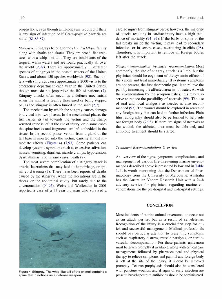
ers with stingrays cause approximately 2000 visits to themergency department each year in the United States,hough most do not jeopardize the life of patients (7).tingray attacks often occur as a defense mechanismhen the animal is feeling threatened or being steppedn, as the stingray is often buried in the sand (2,7).
The mechanism by which the stingray causes damages divided into two phases. In the mechanical phase, thesh lashes its tail towards the victim and the sharp,errated spine is left at the site of injury, or in some caseshe spine breaks and fragments are left embedded in theissue. In the second phase, venom from a gland at theail base is injected into the victim, causing almost im-ediate effects (Figure 4) (7,93). Some patients can
evelop systemic symptoms such as excessive salivation,ausea, vomiting, diarrhea, muscle cramps, hypotension,ysrhythmias, and in rare cases, death (7).
The most severe complication of a stingray attack isrterial lacerations that may lead to hemorrhage, or spi-al cord trauma (7). There have been reports of deathsaused by the stingrays, when the lacerations are in thehorax or the abdominal cavity, but rarely due to thenvenomation (94,95). Weiss and Wolfenden in 2001eported a case of a 33-year-old man who survived a
pigure 4. Stingray. The whip-like tail of the animal contains apine that functions as a defense weapon.
ardiac injury from stingray barbs; however, the majorityf attacks resulting in cardiac injury have a high inci-ence of mortality (94–97). If the barbs or spine of theail breaks inside the victim, it may lead to secondarynfection, or in severe cases, necrotizing fasciitis (98).herefore, it is important to remove all foreign bodies
eft after the attack.
tingray envenomation treatment recommendations. Mostommonly, the site of stingray attack is a limb, but thehysician should be cognizant of the systemic effects ofhe venom and treat immediately. If systemic symptomsre not present, the first therapeutic goal is to relieve theain by immersing the affected area in hot water. As withhe envenomation by the scorpion fishes, this may alsoerve to reduce the possibility of necrosis (99). The usef oral and local analgesia as needed is also recom-ended (93). The wound should be explored in search of
ny foreign body that can lead to further infection. Plainlm radiography should also be performed to help ruleut foreign body (7,93). If there are signs of necrosis athe wound, the affected area must be debrided, andntibiotic treatment should be started.
reatment Recommendations Overview
n overview of the signs, symptoms, complications, andanagement of various life-threatening marine enveno-ations described above is presented below and in Table
. It is worth mentioning that the Department of Phar-acology from the University of Melbourne, Australia
as the Australian Venom Research Unit with a 24-hdvisory service for physicians regarding marine en-enomations for the pre-hospital and in-hospital settings.
CONCLUSION
ost incidents of marine animal envenomation occur nots an attack per se, but as a result of self-defense.ecognition of the injury is a crucial first step for qu-
ck and successful management. Medical professionalshould pay particular attention to presenting symptomsuch as respiratory distress, muscle paralysis, or cardio-ascular decompensation. For these patients, antivenomust be given promptly if available, along with critical careanagement, followed by pharmaceutical and physical
herapy to relieve symptoms and pain. If any foreign bodys left at the site of the injury, it should be removedromptly. Tetanus prophylaxis should also be consideredith puncture wounds, and if signs of early infection are
resent, broad-spectrum antibiotics should be administered.
tawtmap
1
1
1
1
1
1
1
1
1
1
2
2
2
2
2
2
2
2
2
2
3
3
3
3
3
3
3
3
3
3
4
4
4
4
4
4
4
Sea-life Envenomations 111
Antivenoms against the box jellyfish and stonefishoxins have been developed and it is hoped that theirvailability will continue to spread. Scientists continue toork to develop antivenoms for conotoxin and tetrodo-
oxin. In the absence of antivenom, therapeutic optionsay include use of hot water, analgesia with non-steroidal
nti-inflammatory drugs or narcotics, and critical sup-ortive care.
REFERENCES
1. White J. Envenoming and antivenom use in Australia. Toxicon1998;36:1483–92.
2. Auerbach PS. Envenomation by aquatic invertebrates. In: Auer-bach PS, ed. Wilderness medicine, 4th edn. St. Louis, MO: Mosby;2001:1450–85.
3. Auerbach PS. Envenomation by aquatic vertebrates. In: AuerbachPS, ed. Wilderness medicine, 4th edn. St. Louis, MO: Mosby;2001:1488–505.
4. Currie BJ, Jacups SP. Prospective study of Chironex fleckeri andother box jellyfish stings in the “Top End” of Australia’s NorthernTerritory. Med J Aust 2005;183:631–6.
5. Bailey PM. Fatal envenomation by jellyfish causing Irukandjisyndrome. Med J Aust 2003;178:139; author reply 139–40.
6. Russell FE, Panos TC, Kang LW, Warner AM, Colket TC 3rd.Studies on the mechanism of death from stingray venom; a reportof two fatal cases. Am J Med Sci 1958;235:566–84 passim.
7. Clark RF, Girard RH, Rao D, Ly BT, Davis DP. Stingray enveno-mation: a retrospective review of clinical presentation and treat-ment in 119 cases. J Emerg Med 2007;33:33–7.
8. Balsinde J, Winstead MV, Dennis EA. Phospholipase A(2) regu-lation of arachidonic acid mobilization. FEBS Lett 2002;531:2–6.
9. Nevalainen TJ, Peuravuori HJ, Quinn RJ, et al. Phospholipase A2in cnidaria. Comp Biochem Physiol B Biochem Mol Biol 2004;139:731–5.
0. Currie B, McKinnon M, Whelan B, Alderslade P. The Govechirodropid: a box jellyfish appearing in the “safe season”. Med JAust 2002;177:649.
1. Ulrich H, Landthaler M, Vogt T. Granulomatous jellyfish derma-titis [German]. J Dtsch Dermatol Ges 2007;5:493–5.
2. Little M. Is there a role for the use of pressure immobilizationbandages in the treatment of jellyfish envenomation in Australia?Emerg Med (Fremantle) 2002;14:171–4.
3. Burnett JW. The use of verapamil to treat box-jellyfish stings. MedJ Aust 1990;153:363.
4. Ramasamy S, Isbister GK, Seymour JE, Hodgson WC. Pharma-cologically distinct cardiovascular effects of box jellyfish (Chi-ronex fleckeri) venom and a tentacle-only extract in rats. ToxicolLett 2005;155:219–26.
5. O’Reilly GM, Isbister GK, Lawrie PM, Treston GT, Currie BJ.Prospective study of jellyfish stings from tropical Australia, in-cluding the major box jellyfish Chironex fleckeri. Med J Aust2001;175:652–5.
6. Uri S, Marina G, Liubov G. Severe delayed cutaneous reaction dueto Mediterranean jellyfish (Rhopilema nomadica) envenomation.Contact Dermatitis 2005;52:282–3.
7. Currie BJ. Clinical toxicology: a tropical Australian perspective.Ther Drug Monit 2000;22:73–8.
8. Ramasamy S, Isbister GK, Seymour JE, Hodgson WC. The in vivocardiovascular effects of box jellyfish Chironex fleckeri venom inrats: efficacy of pre-treatment with antivenom, verapamil andmagnesium sulphate. Toxicon 2004;43:685–90.
9. Suntrarachun S, Roselieb M, Wilde H, Sitprija V. A fatal jellyfishencounter in the Gulf of Siam. J Travel Med 2001;8:150–1.
0. Ramasamy S, Isbister GK, Seymour JE, Hodgson WC. The in vivo
cardiovascular effects of an Australasian box jellyfish (Chiropsal-mus sp.) venom in rats. Toxicon 2005;45:321–7.1. Beadnell CE, Rider TA, Williamson JA, Fenner PJ. Managementof a major box jellyfish (Chironex fleckeri) sting. Lessons from thefirst minutes and hours. Med J Aust 1992;156:655–8.
2. Currie BJ, Wood YK. Identification of Chironex fleckeri enveno-mation by nematocyst recovery from skin. Med J Aust 1995;162:478–80.
3. Barnes JH. Cause and effect in Irukandji stingings. Med J Aust1964;14:897–904.
4. Flecker H. Irukandji sting to North Queensland bathers withoutproduction of weals but with severe general symptoms. Med J Aust1952;2:89–91.
5. Underwood AH, Seymour JE. Venom ontogeny, diet and morphol-ogy in Carukia barnesi, a species of Australian box jellyfish thatcauses Irukandji syndrome. Toxicon 2007;49:1073–82.
6. Fenner PJ, Williamson J, Callanan VI, Audley I. Further under-standing of, and a new treatment for, “Irukandji” (Carukia barnesi)stings. Med J Aust 1986;145:569, 572–4.
7. Fenner PJ, Hadok JC. Fatal envenomation by jellyfish causingIrukandji syndrome. Med J Aust 2002;177:362–3.
8. Baguley IJ. The excitatory:inhibitory ratio model (EIR model): anintegrative explanation of acute autonomic overactivity syn-dromes. Med Hypotheses 2007;70:26–35.
9. Winkel KD, Tibballs J, Molenaar P, et al. Cardiovascular actionsof the venom from the Irukandji (Carukia barnesi) jellyfish: effectsin human, rat and guinea-pig tissues in vitro and in pigs in vitro.Clin Exp Pharmacol Physiol 2005;32:777–88.
0. Ramasamy S, Isbister GK, Seymour JE, Hodgson WC. The in vivocardiovascular effects of the Irukandji jellyfish (Carukia barnesi)nematocyst venom and a tentacle extract in rats. Toxicol Lett2005;155:135–41.
1. Huynh TT, Seymour J, Pereira P, et al. Severity of Irukandjisyndrome and nematocyst identification from skin scrapings. MedJ Aust 2003;178:38–41.
2. Gershwin L. Jellyfish responsible for Irukandji syndrome. QJM2006;99:801–2; author reply 3–4.
3. Little M, Seymour J. Another cause of “Irukandji stingings”. MedJ Aust 2003;179:654.
4. Little M, Pereira P, Carrette T, Seymour J. Jellyfish responsible forIrukandji syndrome. QJM 2006;99:425–7.
5. McD Taylor D, Pereira P, Seymour J, Winkel KD. A sting from anunknown jellyfish species associated with persistent symptoms andraised troponin I levels. Emerg Med (Fremantle) 2002;14:175–80.
6. de Pender AM, Winkel KD, Ligthelm RJ. A probable case ofIrukandji syndrome in Thailand. J Travel Med 2006;13:240–3.
7. Grady JD, Burnett JW. Irukandji-like syndrome in South Floridadivers. Ann Emerg Med 2003;42:763–6.
8. Armoni M, Ohali M, Hay E. Severe dyspnea due to jellyfishenvenomation. Pediatr Emerg Care 2003;19:84–6.
9. Salam AM, Albinali HA, Gehani AA, Al Suwaidi J. Acute myo-cardial infarction in a professional diver after jellyfish sting. MayoClin Proc 2003;78:1557–60.
0. Ponampalam R. An unusual case of paralytic ileus after jellyfishenvenomation. Emerg Med J 2002;19:357–8.
1. Laing JH, Harrison DH. Envenomation by the box-jellyfish—anunusual cause of ulnar nerve palsy. J R Soc Med 1991;84:115–6.
2. Winter KL, Isbister GK, Seymour JE, Hodgson WC. An in vivoexamination of the stability of venom from the Australian boxjellyfish Chironex fleckeri. Toxicon 2007;49:804–9.
3. Carrette TJ, Cullen P, Little M, Peiera PL, Seymour JE. Temper-ature effects on box jellyfish venom: a possible treatment forenvenomed patients? Med J Aust 2002;177:654–5.
4. Yoshimoto CM, Yanagihara AA. Cnidarian (coelenterate) enveno-mations in Hawai’i improve following heat application. Trans RSoc Trop Med Hyg 2002;96:300–3.
5. Winter KL, Fernando R, Ramasamy S, Seymour JE, Isbister GK,Hodgson WC. The in vitro vascular effects of two chirodropid(Chironex fleckeri and Chiropsella bronzie) venoms. Toxicol Lett2007;168:13–20.
6. Nomura JT, Sato RL, Ahern RM, Snow JL, Kuwaye TT,
Yamamoto LG. A randomized paired comparison trial of cutane-
44
4
5
5
5
5
5
5
5
5
5
5
6
6
6
6
6
6
6
6
6
6
7
7
7
7
7
7
7
7
7
7
8
8
8
8
8
8
8
8
8
8
9
9
9
9
9
9
9
9
9
9
112 I. Fernandez et al.
ous treatments for acute jellyfish (Carybdea alata) stings. Am JEmerg Med 2002;20:624–6.
7. Taylor JG. Treatment of jellyfish stings. Med J Aust 2007;186:43.8. Ramasamy S, Isbister GK, Seymour JE, Hodgson WC. The in vitro
effects of two chirodropid (Chironex fleckeri and Chiropsalmus sp.)venoms: efficacy of box jellyfish antivenom. Toxicon 2003;41:703–11.
9. Fenner PJ, Williamson JA, Burnett JW, Rifkin J. First aid treat-ment of jellyfish stings in Australia. Response to a newly differ-entiated species. Med J Aust 1993;158:498–501.
0. Tibballs J. Australian venomous jellyfish, envenomation syn-dromes, toxins and therapy. Toxicon 2006;48:830–59.
1. King GK. Acute analgesia and cosmetic benefits of box-jellyfishantivenom. Med J Aust 1991;154:365–6.
2. Carrette T, Alderslade P, Seymour J. Nematocyst ratio and prey intwo Australian cubomedusans, Chironex fleckeri and Chiropsal-mus sp. Toxicon 2002;40:1547–51.
3. Pereira PL, Carrette T, Cullen P, Mulcahy RF, Little M, SeymourJ. Pressure immobilisation bandages in first-aid treatment of jelly-fish envenomation: current recommendations reconsidered. Med JAust 2000;173:650–2.
4. Burnett JW, Calton GJ. Response of the box-jellyfish (Chironexfleckeri) cardiotoxin to intravenous administration of verapamil.Med J Aust 1983;2:192–4.
5. Bloom DA, Burnett JW, Hebel JR, Alderslade P. Effects of vera-pamil and CSL antivenom on Chironex fleckeri (box-jellyfish)induced mortality. Toxicon 1999;37:1621–6.
6. Bailey PM, Bakker AJ, Seymour JE, Wilce JA. A functional com-parison of the venom of three Australian jellyfish—Chironex fleckeri,Chiropsalmus sp., and Carybdea xaymacana—on cytosolic Ca2�,haemolysis and Artemia sp. lethality. Toxicon 2005;45:233–42.
7. Barnett FI, Durrheim DN, Speare R, Muller R. Management ofIrukandji syndrome in northern Australia. Rural Remote Health2005;5:369.
8. Corkeron M, Pereira P, Makrocanis C. Early experience withmagnesium administration in Irukandji syndrome. Anaesth Inten-sive Care 2004;32:666–9.
9. Corkeron MA. Magnesium infusion to treat Irukandji syndrome.Med J Aust 2003;178:411.
0. Bloom DA, Burnett JW, Alderslade P. Partial purification of boxjellyfish (Chironex fleckeri) nematocyst venom isolated at thebeachside. Toxicon 1998;36:1075–85.
1. Carrette T, Seymour J. A rapid and repeatable method for venomextraction from cubozoan nematocysts. Toxicon 2004;44:135–9.
2. Nimorakiotakis B, Winkel KD. Marine envenomations. Part1—Jellyfish. Aust Fam Physician 2003;32:969–74.
3. Kaufman MB. Portuguese man-of-war envenomation. PediatrEmerg Care 1992;8:27–8.
4. Stein MR, Marraccini JV, Rothschild NE, Burnett JW. Fatal Por-tuguese man-o’-war (Physalia physalis) envenomation. AnnEmerg Med 1989;18:312–5.
5. Fenner PJ. Dangers in the ocean: the traveler and marine enveno-mation. I. jellyfish. J Travel Med 1998;5:135–41.
6. Giordano AR, Vito L, Sardella PJ. Complication of a Portugueseman-of-war envenomation to the foot: a case report. J Foot AnkleSurg 2005;44:297–300.
7. Loten C, Stokes B, Worsley D, Seymour JE, Jiang S, Isbister GK.A randomised controlled trial of hot water (45 degrees C) immer-sion versus ice packs for pain relief in bluebottle stings. Med JAust 2006;184:329–33.
8. Schulz JR, Norton AG, Gilly WF. The projectile tooth of a fish-hunting cone snail: Conus catus injects venom into fish prey usinga high-speed ballistic mechanism. Biol Bull 2004;207:77–9.
9. Haddad V 2nd, de Paula Neto JB, Cobo VJ. Venomous mollusks:the risks of human accidents by conus snails (gastropoda: conidae)in Brazil. Rev Soc Bras Med Trop 2006;39:498–500.
0. White L, White J. Clinical toxicology of conus snail stings. In:Meier J, White J, eds. Handbook of clinical toxicology of animalvenoms and poisons. Boca Raton, FL: CRC Press; 1995:117–28.
1. Rice RD, Halstead BW. Report of fatal cone shell sting by Conus
geographus Linnaeus. Toxicon 1968;5:223–4.2. Walker DG. Survival after severe envenomation by the blue-ringedoctopus (Hapalochlaena maculosa). Med J Aust 1983;2:663–5.
3. Yotsu-Yamashita M, Mebs D, Flachsenberger W. Distribution oftetrodotoxin in the body of the blue-ringed octopus (Hapaloch-laena maculosa). Toxicon 2007;49:410–2.
4. Flecker H, Cotton BC. Fatal bite from octopus. Med J Aust1955;42:329–31.
5. Sheumack DD, Howden ME, Spence I, Quinn RJ. Maculotoxin: aneurotoxin from the venom glands of the octopus Hapalochlaenamaculosa identified as tetrodotoxin. Science 1978;199:188–9.
6. Kao CY. Tetrodotoxin, saxitoxin and their significance in the studyof excitation phenomena. Pharmacol Rev 1966;18:997–1049.
7. Nimorakiotakis B, Winkel KD. Marine envenomations. Part 2—Othermarine envenomations. Aust Fam Physician 2003;32:975–9.
8. Cavazzoni E, Lister B, Sargent P, Schibler A. Blue-ringed octopus(Hapalochlaena sp.) envenomation of a 4-year-old boy: a casereport. Clin Toxicol (Phila) 2008;46:760–1.
9. Sutherland SK, Duncan AW. New first-aid measures for enveno-mation: with special reference to bites by the Sydney funnel-webspider (Atrax robustus). Med J Aust 1980;1:378–9.
0. Edmonds C. A non-fatal case of blue-ringed octopus bite. Med JAust 1969;2:601.
1. Lee JY, Teoh LC, Leo SP. Stonefish envenomations of thehand—a local marine hazard: a series of 8 cases and review of theliterature. Ann Acad Med Singapore 2004;33:515–20.
2. Gopalakrishnakone P, Gwee MC. The structure of the venom glandof stonefish Synanceja horrida. Toxicon 1993;31:979–88.
3. Vetrano SJ, Lebowitz JB, Marcus S. Lionfish envenomation.J Emerg Med 2002;23:379–82.
4. Austin L, Gillis RG, Youatt G. Stonefish venom: some biochemical andchemical observations. Aust J Exp Biol Med Sci 1965;43:79–90.
5. Limited AC. Stonefish antivenom sheet [updated 2007]. Availableat: http://www.csl.com.au. Accessed September 9, 2009.
6. Junqueira ME, Grund LZ, Orii NM, et al. Analysis of the inflam-matory reaction induced by the catfish (Cathorops spixii) venoms.Toxicon 2007;49:909–19.
7. Kizer KW, McKinney HE, Auerbach PS. Scorpaenidae envenomation. Afive-year poison center experience. JAMA 1985;253:807–10.
8. Trestrail JH 3rd, al-Mahasneh QM. Lionfish string experiences ofan inland poison center: a retrospective study of 23 cases. Vet HumToxicol 1989;31:173–5.
9. Haddad V Jr, Martins IA, Makyama HM. Injuries caused byscorpionfishes (Scorpaena plumieri Bloch, 1789 and Scorpaenabrasiliensis Cuvier, 1829) in the Southwestern Atlantic Ocean(Brazilian coast): epidemiologic, clinic and therapeutic aspects of23 stings in humans. Toxicon 2003;42:79–83.
0. Halstead BW. Marine animals that sting. In: Halstead BW, ed.Dangerous marine animals. Cambridge, MD: Cornell MaritimePress; 1959:73–81.
1. Schaeffer RC Jr, Carlson RW, Russell FE. Some chemical prop-erties of the venom of the scorpionfish Scorpaena guttata. Toxicon1971;9:69–78.
2. Forrester MB. Pattern of statin exposures reported to Texas poisoncenters, 1998–2004. J Toxicol Environ Health A 2007;70:1424–32.
3. Cook MD, Matteucci MJ, Lall R, Ly BT. Stingray envenomation.J Emerg Med 2006;30:345–7.
4. Fenner PJ, Williamson JA, Skinner RA. Fatal and non-fatal stin-gray envenomation. Med J Aust 1989;151:621–5.
5. Cross TB. An unusual stingray injury—the skindiver at risk. MedJ Aust 1976;2:947–8.
6. Weiss BF, Wolfenden HD. Survivor of a stingray injury to theheart. Med J Aust 2001;175:33–4.
7. Cadzow WH. Puncture wound of the liver by stingray spines. MedJ Aust 1960;47:936–7.
8. Barber GR, Swygert JS. Necrotizing fasciitis due to Photobacte-rium damsela in a man lashed by a stingray. N Engl J Med2000;342:824.
9. Campbell J, Grenon M, You CK. Psuedoaneurysm of the superfi-cial femoral artery resulting from stingray envenomation. Ann
Vasc Surg 2003;17:217–20.