Embed Size (px)
Citation preview
412 VOLUME 20 NUMBER 4 APRIL 2013 nature structural & molecular biology
r e V i e W
Determining the fate of collisions between the machineries of DNA replication and transcription has been a challenge for many years. Initial attempts to examine the outcome of the interaction of rep-lication forks with stationary and/or transcribing RNA-polymerase molecules were made in 1983 by Bruce Alberts1 and colleagues. Since then, many additional examples of transcription-replication colli-sions in bacteria, yeast and mammalian cells have been described. In prokaryotes, both codirectional and head-on encounters appear to be unavoidable, as replication and transcription proceed simultane-ously on the same template DNA, but replisomes move approximately 12-fold faster (~600–730 nucleotides (nt) s−1)2,3 than do RNA polymer-ases (~50 nt s−1)4,5. The first evidence in vivo for the occurrence of transcription-replication collisions came from EM visualization of ColE1-induced replication forks and transcribing Escherichia coli RNA polymerase6. Since then, many studies in bacteria have described tran-scription complexes as natural impediments to replication (reviewed in ref. 7). Paused or stalled RNA polymerases form stable barriers to replisomes and lead to DNA damage. At collision sites with back-tracked RNA polymerases, DNA double-strand breaks (DSBs) were shown to depend on RNA hybridization with the homologous DNA strand, to form the so-called R loop8.
Whether replication and transcription interfere in eukaryotes has long been debated. In eukaryotes, genome duplication is limited to S phase, which leaves other cell-cycle phases as a temporal win-dow in which transcription can occur in the absence of replication. Although transcription can also occur during S phase9–11, it is gener-ally restricted to domains that are spatially separated from the repli-cation territories12,13. Moreover, in contrast to those of prokaryotes,
eukaryotic replication and transcription machineries progress with comparable speeds of 17–33 nt s−1 (ref. 14) and 17–72 nt s−1, respec-tively15–17. Therefore, the occurrence of collisions seemed unlikely in eukaryotes.
However, recent studies show that eukaryotic transcription and replication machineries do indeed meet under specific circumstances. In Saccharomyces cerevisiae, a genome-wide study identified 96 sites in which DNA-polymerase binding was particularly enriched. These sites are located within the open reading frames (ORFs) of highly transcribed genes, for which elevated transcription rates require mul-tiple RNA polymerase II (Pol II) complexes per gene18,19. In mam-malian cells, transcription and replication complexes also meet at specific genomic positions. In human cells depleted of topoisomerase I (Top1), replication pausing or stalling and the resulting DNA breaks preferentially occur at transcribed loci, such as the highly expressed SRSF3 or the S phase–transcribed histone genes19. Also, the longest human genes, which need more time than one complete cell cycle to be fully transcribed, contain regions of encounters between replica-tion forks and elongation complexes20.
Here we will review mechanisms that are known to coordinate replication and transcription spatially and temporally to minimize collisions. The influence of directionality and the consequences of transcription-replication collisions, which result in replication fork stalling, DNA damage and recombination, will be discussed. To ensure genomic stability, multiple transcription- and/or replication-coupled processes have evolved, and their misbalances may lead to DNA damage. As a direct cause of collision-related DNA fragility, two seemingly independent mechanisms have been identified, which are based on either R-loop formation or topological tension generation. In addition, we propose a hypothetical model that could explain how transcribing RNA polymerase on very long genes, once removed from the DNA template by the passage of the replication fork, may continue and finish started transcripts.
Transcriptionandreplication:whichinfluenceswhich?A number of cellular mechanisms are implicated in avoiding and controlling transcription-replication encounters. Because codirec-tionality preserves genome integrity21, bacterial genomes seem to
1Institut de Génétique et de Biologie Moléculaire et Cellulaire, Centre National de la Recherche Scientifique UMR 7104, Institut National de la Santé et de la Recherche Médicale U 964, Université de Strasbourg, Illkirch, France. 2Department of Biochemistry, New York University School of Medicine, New York, New York, USA. 3Present addresses: Institute of Cell Biology, University of Bern, Bern, Switzerland (A.H.) and Department of Biology and Biotechnology Charles Darwin, Sapienza University of Rome, Rome, Italy (M.B.). 4These authors contributed equally to this work. Correspondence should be addressed to L.T. ([email protected]).
Received 9 October 2012; accepted 7 February 2013; published online 3 April 2013; doi:10.1038/nsmb.2543
Transcription-replication encounters, consequences and genomic instabilityAnne Helmrich1,3,4, Monica Ballarino1,3,4, Evgeny Nudler2 & Laszlo Tora1
Toensureaccurateduplicationofgeneticmaterial,thereplicationforkmustovercomenumerousnaturalobstaclesonitsway,includingtranscriptioncomplexesengagedalongthesametemplate.HerewereviewthevariouslevelsofinterdependencebetweentranscriptionandreplicationprocessesandhowdifferenttypesofencountersbetweenRNA-andDNA-polymerasecomplexesmayresultinclashesofthosemachineriesontheDNAtemplateandthusincreasegenomicinstability.Inaddition,wesummarizestrategiesevolvedinbacteriaandeukaryotestominimizetheconsequencesofcollisions,includingR-loopformationandtopologicalstresses.
npg
© 2
013
Nat
ure
Am
eric
a, In
c. A
ll rig
hts
rese
rved
.
nature structural & molecular biology VOLUME 20 NUMBER 4 APRIL 2013 413
r e V i e W
have evolved to minimize the frequency of head-on collisions. The complete sequence of the Bacillus subtilis genome reveals a strong transcription orientation bias with respect to replication fork progres-sion, as 75% of the predicted genes are transcribed in the direction of replication22.
Whereas coordination between prokaryotic transcription and rep-lication is limited to controlling the orientation of routinely occurring encounters23, eukaryotes have developed diverse regulatory mecha-nisms to separate both the location and the timing of transcription and replication. The first evidence of such mechanisms protecting replication machineries from clashes with transcription complexes came from the discovery of replication-fork barriers downstream of S. cerevisiae 35S rRNA23 and tRNA transcription units24 that separate highly expressed genes from replication forks. Transcription-dependent replication pause sites were also mapped in plants25, Xenopus laevis26, Tetrahymena thermophila27 and mammals28,29. In addition to these spatial control mechanisms, replication and transcription timing is also coordinated. Changes in gene expression, as for example during differentiation, can coincide with changes in replication timing at newly transcribed loci30. Moreover, genome-wide mapping of rep-lication timing revealed that, in general, the transcribed regions of metazoan genomes are replicated early in S phase, although later-replicating active genes have also been described31–33.
S phase–transcribed genes that are involved in nucleosome assem-bly, DNA repair or cell proliferation34 have evolved specific ways to avoid cotemporal transcription and replication. The multicopy organization of the replication-dependent histone H4 genes, each with distinct regulatory sequences35, suggests that transcription and replication can be temporally separated at individual loci. The single-copy gene encoding cyclin B1 is transcriptionally active in late S phase but is replicated in early S phase20. Replication of human rDNA oper-ons is initiated from two different sites: silent rDNA repeats replicate in mid and late S phase, initiating from the nontranscribed intergenic spacers, and active rDNA transcription units replicate early, initiating from sites throughout the rDNA units, preferentially from promoter-proximal sequences. During ongoing replication, these active DNA units remain silent, and transcription is activated once replication is complete36,37.
Because replication origins are usually enriched within promoter regions, CpG sequences and DNase-hypersensitivity sites38–40, the open chromatin structure at such sites may favor the binding of a replication preinitiation complex. In agreement with this pos-sibility, origin activity can be stimulated by transcription factors or chromatin-remodeling complexes41,42. Moreover, the timing of replication can be influenced by chromatin modifications that also affect gene expression, such as histone acetylation and DNA meth-ylation at replication origins30,43. For example, the histone deacety-lase complex Sin3–Rpd3, which is a gene-specific transcriptional repressor, delays the firing of origins that are adjacent to Sin3–Rpd3- binding promoters43. In human cells, replication initiation occurs more frequently at methylated CpG sites. Replication initiation sites are also linked to transcriptionally active sites but are depleted from highly transcribed regions40. Active transcription was shown to suppress replication initiation in early Xenopus embryos at rDNA loci44, at the Chinese hamster dihydrofolate reductase (Dhfr) gene locus45, at the Msh4 gene in yeast46 and at the long human genes FHIT and WWOX47. Together, these studies indicate that the open chromatin structure associated with active transcription can facilitate replication-fork assembly as long as the transcription- initiation-factor recruitment does not inhibit the binding of replication initiation components.
However, it has also been suggested that replication can inhibit transcription initiation. At the highly expressed gene encoding β-globin in human and mouse erythroid cells, Pol II was shown to be dislodged from the gene locus during replication48. Moreover, on extremely long genes (>800 kilobases) that require more than one cell cycle to be transcribed, Pol II restarts a new round of transcription in G2, only after replication is completed20.
In summary, the above examples demonstrate the diversity of regulatory mechanisms that evolved to coordinate transcription and replication and to minimize clashes.
Replication-transcriptioncollisionscausegenomicinstabilityThe consequences of collisions between DNA-dependent DNA polymerases and DNA-dependent RNA polymerases present a high risk for cells, as they may give rise to chromosomal recombination events, tumor formation20, DNA damage or cell death (reviewed in refs. 7,49,50). EM, two-dimensional gel electrophoresis, DNA comb-ing and DNA-polymerase chromatin immunoprecipitation6,18,19,51–54 have demonstrated that, in both prokaryotic and eukaryotic cells, transcription-replication encounters cause replication-fork stalling that is often accompanied by DNA damage or recombination6,18,19,51–54. As perturbed replication results in DNA lesions, the activities of the replicating polymerases as well as the function of replicative helicases and topoisomerases, such as Rrm3, Top1 and Top2, protect the genome from deleterious replication-transcription encounters20,51,55–57. Thus, mutations in these factors or perturbation of their activities will lead to genomic instability. However, DNA damage can also be increased at collision sites when one of the various transcription-related phases, including cotranscriptional transcript processing, release of stalled or backtracked RNA polymerase, RNA degradation or nuclear export, is deficient8,19,58–63.
Oncogenes that provide aberrant growth signals to cells can also cause DNA damage at replication forks. Overexpression of the cyclin E onco-protein results in replication slowing accompanied by an increase in replication-transcription interference64. Thus, replication stress in cyclin E–overexpressing cells seems to result from increased replication ini-tiation events coupled to replication and transcription conflicts, which in turn lead to DNA damage and genomic instability through a com-bination of incomplete DNA replication, formation of DSBs at stalled replication forks and transcription-associated recombination64.
When replication-fork progression is slowed in human cells, the frequency of cotemporal replication and transcription increases on very long genes, which results in common fragile site (CFS) break formation20. CFSs are specific genomic loci with elevated sensitivity to replication stress, and their instability, especially in precancerous cells, is thought to have a role in oncogenic transformation65. The contribution of active transcription in replication stress–related CFS instability was originally suggested in 2006, when a number of CFSs were mapped to uncommonly long ORFs spanning >700 kilobases of genomic DNA66. Subsequently, the comparison of the relative timing of transcription and replication within these long CFS-containing genes revealed the simultaneous presence of both transcription complexes and replication forks within the long coding regions20. Moreover, their fragility, which arises from the abnormally high frequency of fork-stalling events during DNA replication, depends on the RNA synthesis along the CFS region.
In contrast to the deleterious effects described above, DNA rear-rangements induced by replication-transcription encounters can be functionally very important under certain physiological conditions. In vertebrate B cells, immunoglobulin class switch recombination is a site-directed gene rearrangement allowing immunoglobulin
npg
© 2
013
Nat
ure
Am
eric
a, In
c. A
ll rig
hts
rese
rved
.
414 VOLUME 20 NUMBER 4 APRIL 2013 nature structural & molecular biology
r e V i e W
isotype switching. Class switching depends both on transcription and on replication co-orientation, which suggests that collisions provide the underlying mechanism in the recombination process67.
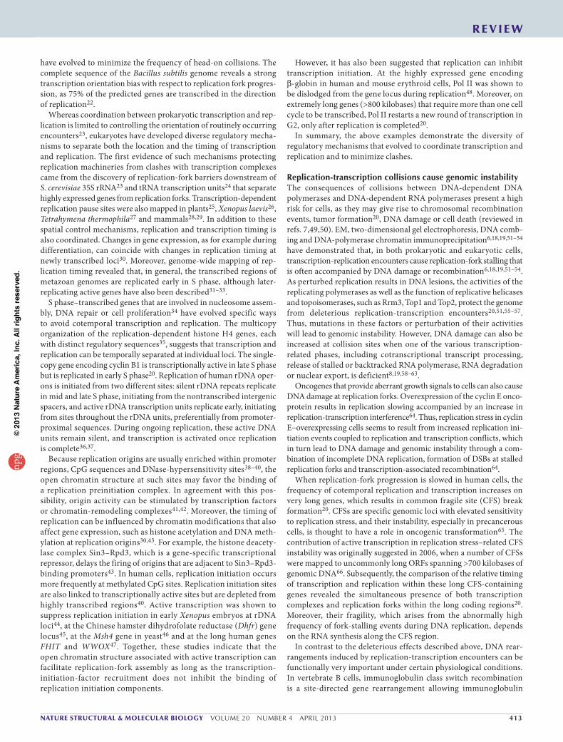
WhendirectionalitymakesthedifferenceDepending on the orientation of a particular gene, the replication fork can face RNA polymerases in either a head-on or a codirectional manner. In the head-on conflicts, a given gene encoded on the lag-ging strand is transcribed in the opposite direction from leading- strand replication. In contrast, conflicts are codirectional when genes are coded on the leading strand and transcription occurs in the same direction as leading-strand replication. Consequently, the mode of transcription-replication encounters depends on the direction of transcription.
Although it was initially widely believed that head-on collisions between transcription and replication machineries were more detri-mental than codirectional ones (described above), a number of recent studies suggest that the reality is more complicated. DNA damage can be caused by head-on or codirectional encounters or even upon collisions in both directions. The following sections (and Fig. 1) sum-marize the studies supporting each of these different scenarios.
Head-on collisions. In bacteria, the majority of genes are encoded on the leading strand in the direction of DNA replication, which argues for a selection toward co-orientation of replication and transcription. When chromosomal fragments are inverted to a head-on orientation, replication rates are reduced, levels of the recombination protein RecA are increased, and the SOS DNA-damage response is activated21,68. Replication is particularly impaired at the highly transcribed rRNA operons but only when the head-on orientation is induced6. Notably, under these conditions, the RecBC recombination protein is required for cell viability69. In yeast, Pol III–transcribed tRNA genes only form replication fork blocks when the orientation of replication is opposite to that of transcription24. Moreover, head-on but not codirectional transcription causes replication-fork pausing and increases recombi-nation of plasmid-based recombination constructs with strong induc-ible promoters51,53. As the speeds of progressing transcription and replication complexes in eukaryotes seem to be similar, it is reasonable
to hypothesize that the two machineries progressing in the same direction will follow each other rather than collide.
Co-oriented collisions. Codirectional collisions are detrimental when transcription elongation is compromised by extensive backtracking, which is the reversible sliding of the RNA-polymerase elongation complex backward along DNA and RNA70. Bacteria have several mechanisms to suppress backtracking or to eliminate backtracked complexes, including the presence of active ribosomes that move just behind RNA polymerase5, the induction of transcript cleavage by GreA and GreB factors71,72 or the induction of termination by Rho and Mfd73. In E. coli, all these mechanisms contribute to suppression of DSBs associated with the codirectional collisions between repli-somes and backtracked RNA polymerase8. Also, at immunoglobulin class switch loci in mammalian B cells, nondeleterious replication-fork impediments and DNA rearrangements depend on the co-orientation of replication and transcription67,74 (also described above).
Directionality does not matter. In yeast, the relative directions of replication and transcription do not matter on a genome-wide scale, as the naturally occurring sites of replication-fork pausing at tran-scribed regions appear to be independent of polarity18. Moreover, in yeast strains with RNA-processing defects, sites of DNA damage locate to ORFs regardless of their orientation with respect to the closest replication origin75. Therefore, replication forks may meet a transcription-elongation complex from either direction, which suggests that both codirectional and head-on collisions have the potential to result in DNA breakage, at least when transcription is hampered (Fig. 1).
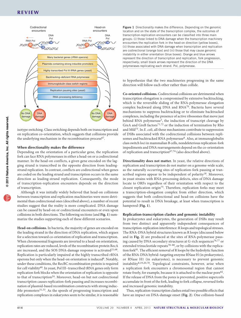
Replication-transcriptionclashesandgenomicinstabilityIn prokaryotes and eukaryotes, the generation of DSBs may result from two distinct and apparently independent consequences of transcription-replication interference: R loops and topological stresses. The RNA-DNA hybrid structures known as R loops (discussed below and in Fig. 2) are produced at the sites of RNA-polymerase paus-ing caused by DNA secondary structures at G-rich sequences76,77 or extended trinucleotide repeats78–80, or by collisions with the replica-tion fork81. The efficient removal of R loops by the hydrolytic function of the RNA-DNA hybrid–targeting enzyme RNase H (in prokaryotes), or RNase H1 (in eukaryotes), is necessary to prevent genomic instability8,19,20,78. Topological constraints, however, arise when a replication fork encounters a chromosomal region that cannot rotate freely, for example, because it is attached to the nuclear pore82. If the release of DNA from the pores is prevented, positive supercoils accumulate in front of the fork, leading to fork collapse, reversed forks and increased genomic instability.
Thus, replication-transcription clashes entail two possible effects that have an impact on DNA-damage onset (Fig. 2). One collision-based
Head-onencounters
Many bacterial genes (rRNA operons)
Highly transcribed Pol III tRNA genes (yeast)
Immunoglobulin class switch regions
Backtracking–deficient RNA polymerase
Replication pausing sites (yeast)
RNA–processing deficiency
Plasmids containing strong inducible promoters
Codirectional encounters
DNAPol
RNA Pol
DNAdamage
No DNA damage
DNAdamage
No DNA damage
DNAPol
RNA Pol
Figure 1 Directionality makes the difference. Depending on the genomic location and on the state of the transcription complex, the outcomes of transcription-replication encounters can be classified into three main groups: (i) those linked to DNA damage when the transcription machinery encounters the replication fork in the head-on direction (yellow boxes), (ii) those associated with DNA damage when transcription and replication are codirectional (orange box) and (iii) those that may cause genomic instability in either orientation (blue boxes). Orange and blue arrows represent the direction of transcription and replication, fork progression, respectively; small black arrows represent the direction of the DNA polymerases replicating each strand. Pol, polymerase.
npg
© 2
013
Nat
ure
Am
eric
a, In
c. A
ll rig
hts
rese
rved
.
nature structural & molecular biology VOLUME 20 NUMBER 4 APRIL 2013 415
r e V i e W
problem is the creation of R loops. Several observations argue for R-loop formation and topological tensions being independent from each other. First, R loops and their impact on DNA damage were detected in bacteria8, where transcribed loci are not bound to fixed membrane structures. Second, in E. coli plasmids that experience a deletion upon induced transcription-replication collisions, the formation of tran-scription-induced supercoiling domains does not induce deletions53. Moreover, topological tension generated between replicating and tran-scribing sites, when unresolved, leads to reversed replication forks, fork breakage and recombination. Because RNase H treatment does not suppress cell death upon fork reversal, collisions with topological constraints are unlikely to be linked with R loops69,82. Still, it is not clear whether R loops and topological tensions are mutually exclusive or whether they can occur together at the same collision site.
R loops. According to the ‘thread-back’ model83, the newly synthesized RNA transcript can invade the DNA duplex to form a three-stranded nucleic acid molecule containing an RNA-DNA duplex and a single-stranded DNA84: the R loop (Fig. 2). R loops are natural intermedi-ates of several transcription- or replication-linked processes. Under normal conditions, the formation of short, ~8-base-pair-long R loops within the transcription bubble helps the transcription complexes to move forward70 and influences the rate of elongation85. Another physiological function of R loops, which was initially defined in bac-teriophage T4 (reviewed in ref. 86) and later extended to other organ-isms, occurs during replication initiation. Although R loops occur at low frequency in normal cells20,60, the number and/or the length of the R loops may increase when transcription or RNA processing is disturbed8,19,60,63,74,75,87. However, perturbing replication, either by hindering DNA-polymerase progression or by depleting replicative topoisomerases, also favors the creation or stabilization of R loops, which leads to increased DNA damage19,20,56. To avoid the deleterious outcome of long and stable R loops, the activity of RNase H1 needs
to be tightly controlled by regulating either its cellular concentra-tion and/or its target accessibility. The low number of chromosomal lesions remaining upon RNase H1 overexpression even in nonper-turbed cells20,60 may suggest the existence of an R loop–independent DNA-damage process.
Topological constraints. Because, in eukaryotes, mRNA export occurs cotranscriptionally, DNA instability can arise from topological tension that occurs when transcription and replication machineries meet at loci that are bound to the nuclear periphery88. When yeast cells defective in the Mec1 (ATR) replication-damage checkpoint were used to study replication-fork instability, replication stress induced by low doses of hydroxyurea caused accumulation of reversed forks and hemireplicated intermediates82 (Fig. 2). Because overexpression or ablation of endogenous RNase H1 did not influence hydroxyurea sensitivity, the authors concluded that the mechanism leading to replication-fork stalling was independent of R loops. Instead, under these conditions fork reversal appears to require both transcription and the localization of the region to the nuclear periphery, as ablation of nuclear pore proteins or the introduction of DNA double-strand breaks between the transcribed unit and the replication fork suppress hydroxyurea sensitivity. Thus, when DNA is attached to the nuclear pore and is unable to freely rotate, the topological tension generated in front of the replisome may lead to replication-fork stalling. According to the topological model, the nuclear pore complex (NPC)-bound DNA region will be removed from the NPC by the activities of the Mec1 (ATR) replication-damage checkpoint proteins to permit pro-gression of the replication fork82. This model is also supported by data showing the participation of the Rrm3 helicase in facilitating replication-fork movement through transcribed loci or sites occupied by DNA-binding proteins51,57.
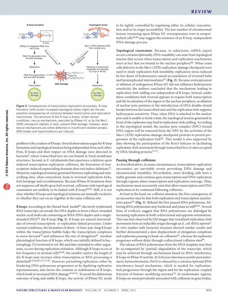
PassingthroughcollisionsAs described above, in many circumstances, transcription-replication encounters are inevitable events provoking DNA damage and chromosomal instability. Nevertheless, most dividing cells have a stable genome and continue gene transcription and DNA replication through regions where transcription and replication converge. Thus, mechanisms must necessarily exist that allow transcription and DNA replication to be continued following collisions.
At least in the head-on-collision situation, the first consequence of an encounter may be that both replication and transcription machin-eries pause89 (Fig. 3). Behind the first paused RNA polymerase, fol-lowing RNA polymerases may backtrack and pause as well90,91. Several lines of evidence suggest that RNA polymerases are dislodged by incoming replication in both codirectional and opposite orientations. This was first observed by EM images that visualized replication-fork movement from an inducible origin through an E. coli rRNA operon6. In vitro studies with bacterial enzymes showed similar results and further demonstrated a slow displacement of elongation complexes and replisome pausing in head-on collisions92, whereas the replisome progresses without delay through codirectional collision sites89.
The release of RNA polymerases from the DNA template may then be accompanied by (partial) degradation of the transcript, which may be achieved through mechanisms based on RNA interference, R loops or RNase H activity. At Schizosaccharomyces pombe pericentro-meric heterochromatin, Pol II is released by a cotranscriptional RNA interference–based mechanism, which is needed for replication-fork progression through the region and for the replication-coupled function of histone-modifying enzymes54. In euchromatic regions, R loops are most prevalently associated with collisions in gene bodies.
R-loop formation Topological stress
RNAse H1 degrades RNA fromthe R loop
Mec1 (ATR) detaches DNAfrom nuclear pore
DNA damage
RNA Pol dislocation fromthe DNA template
Relaxing of supercoiledfragments (for example
by Top2 or Rrm3)
Replication
Replication
RNA Pol reassociationwith the DNA template
Transcription
R-loop accumulation
Mec1 (
ATR) che
ckpo
int
defic
iency
R loop
No DNA damage
No DNA damage
RNAPol DNA Pol
Figure 2 Consequences of transcription-replication encounters. R-loop formation (left) and/or increased topological stress (right) are the two possible consequences of collisions between transcription and replication machineries. The structure of the R loop is boxed. Under normal conditions, rescue mechanisms, executed by RNase H1 or by the Mec1 (ATR) checkpoint (details in text), prevent DNA damage. However, when rescue mechanisms are either defective or insufficient (dashed arrows), DNA breaks and hypermutations are induced.
npg
© 2
013
Nat
ure
Am
eric
a, In
c. A
ll rig
hts
rese
rved
.
416 VOLUME 20 NUMBER 4 APRIL 2013 nature structural & molecular biology
r e V i e W
Although R loops at collision sites are primarily studied in eukaryo-tes19,60,81, their implication in the generation of DNA damage has also been reported in E. coli8.
Paused RNA polymerases are effectively displaced from the DNA template by the progressing replication machinery itself, as shown in vitro89. In E. coli, replicative helicases Rep, DinG and UvrD help to resolve potentially deleterious encounters due to head-on collisions93. Also, loading of the DnaB, and DnaD proteins and the PriA rep-lication-restart protein (in B. subtilis), along with antibacktracking transcription factors GreA, GreB, DksA and active ribosomes, pre-vents DNA damage associated with codirectional collisions8,58,94. In mammals, unfettered DNA-polymerase progression also prevents DNA instability at regions of transcription-replication interference20. RNA transcripts may persist at the DNA-duplication site, as shown by single-cell real-time RNA transcription visualization of the human cyclin D1 gene95. DNA polymerases were suggested to use this RNA as a primer for replication89.
Once forks have passed, mRNAs can either be recovered by new transcript initiation, which is probably the case at short prokaryotic genes, or by recruitment of RNA-elongation complexes to the template- associated 5′ portion of the transcript (Fig. 3). On very long genes, where transcription of a full-length transcript takes more than one cell cycle and where, thus, obligatory collisions occur within each round of transcription20, RNA elongation must continue after the collisions to allow generation of full-length transcripts. Figure 3 depicts a hypothetical model in which RNA polymer-ases may continue the elongation of the started transcripts after the collision. In line with such a model, elongation-competent forms of RNA polymerase are able to bind RNA-DNA bubbles that contain a free (single-stranded) 5′-terminal RNA tail and to carry on RNA synthesis with good efficiency96, at least in vitro. RNA-DNA
hybrids located within intronic sequences would then allow the transcript to be correctly spliced once transcription elongation at the 3′ intronic fragment reaches the next branch site, as may be the case within long genes20. Thus, a sequential progression through a collision site (depicted in the hypothetical scenario in Fig. 3) may allow the timely completion of replication and the generation of complete mRNAs.
ConclusionsFor a cell, it is crucial to ensure proper replication and gene transcrip-tion. As transcription-replication interference is deleterious at most genomic loci, multiple regulatory mechanisms have evolved to control and minimize such encounters. However, at many loci in bacteria and also within distinct regions of eukaryotic DNA, transcription-replication interference is unavoidable and may sometimes even be beneficial for the cell. Researchers are still just beginning to under-stand the complex mechanisms operating at the sites of collisions, and we therefore anticipate the elucidation of many more details of these mechanisms in the future. A better comprehension of these molecular events will clarify the view of how genomic stability is maintained. At present, very little is known about how epigenetic or chromatin-associated changes influence transcription-replication interference globally or at specific genomic loci. In addition, the way in which defined chromatin structures with specific epigenetic marks influence genomic stability awaits further investigation. Thus, we are expecting the discovery of an expanding array of functional links between epige-netics, chromatin architecture, transcriptional regulation, replication initiation and DNA repair.
AckNowLEdgMENTsWe apologize to colleagues whose work could only be cited indirectly, owing to space limitations. We thank S. Bour for help with preparing the figures. A.H. and M.B. were supported by fellowships from Fondation pour la Recherche Médicale. E.N. was supported by grants from the US National Institutes of Health (R01 GM058750) and the Biogerontology Research Foundation, UK. This work was supported by funds from Centre Nationale de la Recherche Scientifique (LEA-SkinChroma), Agence Nationale de la Recherche (ANR-09-BLAN-0266 and ANR-09-BLAN-056), Institut National du Cancer (2008-Ubican) and European Community (EPIDICAN) grants to L.T.
coMPETINg FINANcIAL INTEREsTsThe authors declare no competing financial interests.
Reprints and permissions information is available online at http://www.nature.com/reprints/index.html.
Replicationmachinery
RNA Pol
Fork progression causesthe release of RNA Pol; RNA stays associatedwith the template
Recruitment of RNA Poland transcription elongation
DNA polymerases mayuse the transcript asprimer for replication
Replication restartproteins
Figure 3 Stepwise passage through a collision course. Certain steps of the presented scenario are based on in vitro and in vivo studies of collisions occurring on very long genes (described in text; other steps have not been described biochemically). Our hypothetical model explains the potential sequences of events upon transcription-replication collisions, leading to unperturbed continuation of both replication and transcription processes. Encounters cause a transient pause of both transcription (orange) and replication (blue) machineries and result in the invasion of the transcript (represented by an orange line) into DNA. The recruitment of replication-restart proteins stimulates the movement of the replication fork, resulting in displacement of the RNA polymerase (RNA Pol) transcription complex. DNA polymerases may use the transcript to prime replication. Following passage of the replication fork, the RNA polymerase complex may reassociate with the RNA annealed to one of the daughter strands and thus resume elongation of previously started transcripts. Orange and blue arrows represent the direction of transcription and replication, respectively. Small red arrows indicate the release and the hypothesized reassociation of the transcription complex on long genes, following collisions.
npg
© 2
013
Nat
ure
Am
eric
a, In
c. A
ll rig
hts
rese
rved
.
nature structural & molecular biology VOLUME 20 NUMBER 4 APRIL 2013 417
r e V i e W
1. Bedinger, P., Hochstrasser, M., Jongeneel, C.V. & Alberts, B.M. Properties of the T4 bacteriophage DNA replication apparatus: the T4 dda DNA helicase is required to pass a bound RNA polymerase molecule. Cell 34, 115–123 (1983).
2. Breier, A.M., Weier, H.U. & Cozzarelli, N.R. Independence of replisomes in Escherichia coli chromosomal replication. Proc. Natl. Acad. Sci. USA 102, 3942–3947 (2005).
3. Mok, M. & Marians, K.J. The Escherichia coli preprimosome and DNA B helicase can form replication forks that move at the same rate. J. Biol. Chem. 262, 16644–16654 (1987).
4. Epshtein, V., Toulme, F., Rahmouni, A.R., Borukhov, S. & Nudler, E. Transcription through the roadblocks: the role of RNA polymerase cooperation. EMBO J. 22, 4719–4727 (2003).
5. Proshkin, S., Rahmouni, A.R., Mironov, A. & Nudler, E. Cooperation between translating ribosomes and RNA polymerase in transcription elongation. Science 328, 504–508 (2010).
6. French, S. Consequences of replication fork movement through transcription units in vivo. Science 258, 1362–1365 (1992).
This is the first in vivo study of transcription-replication encounters, showing that RNA polymerases are dislodged from a bacterial rRNA operon when a replication fork passes from either the same or the opposite direction.
7. McGlynn, P., Savery, N.J. & Dillingham, M.S. The conflict between DNA replication and transcription. Mol. Microbiol. 85, 12–20 (2012).
8. Dutta, D., Shatalin, K., Epshtein, V., Gottesman, M.E. & Nudler, E. Linking RNA polymerase backtracking to genome instability in E. coli. Cell 146, 533–543 (2011).
9. Klevecz, R.R., Bolen, J., Forrest, G. & Murray, D.B. A genomewide oscillation in transcription gates DNA replication and cell cycle. Proc. Natl. Acad. Sci. USA 101, 1200–1205 (2004).
10. Reinke, H. & Gatfield, D. Genome-wide oscillation of transcription in yeast. Trends Biochem. Sci. 31, 189–191 (2006).
11. Cho, R.J. et al. A genome-wide transcriptional analysis of the mitotic cell cycle. Mol. Cell 2, 65–73 (1998).
12. Wansink, D.G. et al. RNA polymerase II transcription is concentrated outside replication domains throughout S-phase. J. Cell Sci. 107, 1449–1456 (1994).
13. Wei, X. et al. Segregation of transcription and replication sites into higher order domains. Science 281, 1502–1506 (1998).
14. Hiratani, I. et al. Global reorganization of replication domains during embryonic stem cell differentiation. PLoS Biol. 6, e245 (2008).
15. Darzacq, X. et al. In vivo dynamics of RNA polymerase II transcription. Nat. Struct. Mol. Biol. 14, 796–806 (2007).
16. Pérez -Ortin, J.E., Alepuz, P.M. & Moreno, J. Genomics and gene transcription kinetics in yeast. Trends Genet. 23, 250–257 (2007).
17. Singh, J. & Padgett, R.A. Rates of in situ transcription and splicing in large human genes. Nat. Struct. Mol. Biol. 16, 1128–1133 (2009).
18. Azvolinsky, A., Giresi, P.G., Lieb, J.D. & Zakian, V.A. Highly transcribed RNA polymerase II genes are impediments to replication fork progression in Saccharomyces cerevisiae. Mol. Cell 34, 722–734 (2009).
19. Tuduri, S. et al. Topoisomerase I suppresses genomic instability by preventing interference between replication and transcription. Nat. Cell Biol. 11, 1315–1324 (2009).
20. Helmrich, A., Ballarino, M. & Tora, L. Collisions between replication and transcription complexes cause common fragile site instability at the longest human genes. Mol. Cell 44, 966–977 (2011).
This is the first report of naturally occurring collisions in human cells; the authors show that transcription of very long genes takes more than one cell cycle to be completed, and the instability of common fragile sites within those genes is linked to a spatial and temporal overlap between transcription and replication.
21. Srivatsan, A., Tehranchi, A., MacAlpine, D.M. & Wang, J.D. Co-orientation of replication and transcription preserves genome integrity. PLoS Genet. 6, e1000810 (2010).
22. Kunst, F. et al. The complete genome sequence of the gram-positive bacterium Bacillus subtilis. Nature 390, 249–256 (1997).
23. Brewer, B.J. When polymerases collide: replication and the transcriptional organization of the E. coli chromosome. Cell 53, 679–686 (1988).
24. Deshpande, A.M. & Newlon, C.S. DNA replication fork pause sites dependent on transcription. Science 272, 1030–1033 (1996).
25. López-Estraño, C., Schvartzman, J.B., Krimer, D.B. & Hernandez, P. Characterization of the pea rDNA replication fork barrier: putative cis-acting and trans-acting factors. Plant Mol. Biol. 40, 99–110 (1999).
26. Maric, C., Levacher, B. & Hyrien, O. Developmental regulation of replication fork pausing in Xenopus laevis ribosomal RNA genes. J. Mol. Biol. 291, 775–788 (1999).
27. Zhang, Z., Macalpine, D.M. & Kapler, G.M. Developmental regulation of DNA replication: replication fork barriers and programmed gene amplification in Tetrahymena thermophila. Mol. Cell Biol. 17, 6147–6156 (1997).
28. López-Estraño, C., Schvartzman, J.B., Krimer, D.B. & Hernandez, P. Co-localization of polar replication fork barriers and rRNA transcription terminators in mouse rDNA. J. Mol. Biol. 277, 249–256 (1998).
29. Putter, V. & Grummt, F. Transcription termination factor TTF-I exhibits contrahelicase activity during DNA replication. EMBO Rep. 3, 147–152 (2002).
30. Hiratani, I., Takebayashi, S., Lu, J. & Gilbert, D.M. Replication timing and transcriptional control: beyond cause and effect–part II. Curr. Opin. Genet. Dev. 19, 142–149 (2009).
31. Schübeler, D. et al. Genome-wide DNA replication profile for Drosophila melanogaster: a link between transcription and replication timing. Nat. Genet. 32, 438–442 (2002).
32. White, E.J. et al. DNA replication-timing analysis of human chromosome 22 at high resolution and different developmental states. Proc. Natl. Acad. Sci. USA 101, 17771–17776 (2004).
33. Woodfine, K. et al. Replication timing of the human genome. Hum. Mol. Genet. 13, 191–202 (2004).
34. van der Meijden, C.M. et al. Gene profiling of cell cycle progression through S-phase reveals sequential expression of genes required for DNA replication and nucleosome assembly. Cancer Res. 62, 3233–3243 (2002).
35. Holmes, W.F. et al. Coordinate control and selective expression of the full complement of replication-dependent histone H4 genes in normal and cancer cells. J. Biol. Chem. 280, 37400–37407 (2005).
36. Coffman, F.D., He, M., Diaz, M.L. & Cohen, S. DNA replication initiates at different sites in early and late S phase within human ribosomal RNA genes. Cell Cycle 4, 1223–1226 (2005).
37. Dimitrova, D.S. DNA replication initiation patterns and spatial dynamics of the human ribosomal RNA gene loci. J. Cell Sci. 124, 2743–2752 (2011).
This study shows that on active rDNA loci, replication initiates randomly throughout the early-replicating rDNA, whereas silent rDNA replicates inside the nucleoli during mid and late S phase, which ensures efficient replication and reduces the risk of chromosome breaks and rDNA hyper-recombination.
38. Cadoret, J.C. et al. Genome-wide studies highlight indirect links between human replication origins and gene regulation. Proc. Natl. Acad. Sci. USA 105, 15837–15842 (2008).
39. Cayrou, C. et al. Genome-scale analysis of metazoan replication origins reveals their organization in specific but flexible sites defined by conserved features. Genome Res. 21, 1438–1449 (2011).
By performing a genome-scale purification of DNA replication origins, the authors show that replication-initiation events are most frequent at CpG island–containing promoters in mice and at CpG island–like regions in Drosophila.
40. Martin, M.M. et al. Genome-wide depletion of replication initiation events in highly transcribed regions. Genome Res. 21, 1822–1832 (2011).
41. Chang, V.K., Donato, J.J., Chan, C.S. & Tye, B.K. Mcm1 promotes replication initiation by binding specific elements at replication origins. Mol. Cell Biol. 24, 6514–6524 (2004).
42. Danis, E. et al. Specification of a DNA replication origin by a transcription complex. Nat. Cell Biol. 6, 721–730 (2004).
43. Knott, S.R., Viggiani, C.J., Tavare, S. & Aparicio, O.M. Genome-wide replication profiles indicate an expansive role for Rpd3L in regulating replication initiation timing or efficiency, and reveal genomic loci of Rpd3 function in Saccharomyces cerevisiae. Genes Dev. 23, 1077–1090 (2009).
44. Hyrien, O., Maric, C. & Mechali, M. Transition in specification of embryonic metazoan DNA replication origins. Science 270, 994–997 (1995).
45. Dimitrova, D.S. Nuclear transcription is essential for specification of mammalian replication origins. Genes Cells 11, 829–844 (2006).
46. Mori, S. & Shirahige, K. Perturbation of the activity of replication origin by meiosis-specific transcription. J. Biol. Chem. 282, 4447–4452 (2007).
47. Letessier, A. et al. Cell-type-specific replication initiation programs set fragility of the FRA3B fragile site. Nature 470, 120–123 (2011).
The authors show that common fragile sites are epigenetically defined loci corresponding to the latest initiation-poor regions to complete replication in a given cell type.
48. Vieira, K.F. et al. Recruitment of transcription complexes to the β-globin gene locus in vivo and in vitro. J. Biol. Chem. 279, 50350–50357 (2004).
49. Kim, N. & Jinks-Robertson, S. Transcription as a source of genome instability. Nat. Rev. Genet. 13, 204–214 (2012).
50. Merrikh, H., Zhang, Y., Grossman, A.D. & Wang, J.D. Replication-transcription conflicts in bacteria. Nat. Rev. Microbiol. 10, 449–458 (2012).
51. Prado, F. & Aguilera, A. Impairment of replication fork progression mediates RNA polII transcription-associated recombination. EMBO J. 24, 1267–1276 (2005).
52. Takeuchi, Y., Horiuchi, T. & Kobayashi, T. Transcription-dependent recombination and the role of fork collision in yeast rDNA. Genes Dev. 17, 1497–1506 (2003).
53. Vilette, D., Ehrlich, S.D. & Michel, B. Transcription-induced deletions in Escherichia coli plasmids. Mol. Microbiol. 17, 493–504 (1995).
54. Zaratiegui, M. et al. RNAi promotes heterochromatic silencing through replication-coupled release of RNA Pol II. Nature 479, 135–138 (2011).
55. Bermejo, R. et al. Genome-organizing factors Top2 and Hmo1 prevent chromosome fragility at sites of S phase transcription. Cell 138, 870–884 (2009).
56. El Hage, A., French, S.L., Beyer, A.L. & Tollervey, D. Loss of Topoisomerase I leads to R-loop–mediated transcriptional blocks during ribosomal RNA synthesis. Genes Dev. 24, 1546–1558 (2010).
57. Ivessa, A.S. et al. The Saccharomyces cerevisiae helicase Rrm3p facilitates replication past nonhistone protein-DNA complexes. Mol. Cell 12, 1525–1536 (2003).
58. Tehranchi, A.K. et al. The transcription factor DksA prevents conflicts between DNA replication and transcription machinery. Cell 141, 595–605 (2010).
59. Trautinger, B.W., Jaktaji, R.P., Rusakova, E. & Lloyd, R.G. RNA polymerase modulators and DNA repair activities resolve conflicts between DNA replication and transcription. Mol. Cell 19, 247–258 (2005).
60. Wahba, L., Amon, J.D., Koshland, D. & Vuica-Ross, M. RNase H and multiple RNA biogenesis factors cooperate to prevent RNA:DNA hybrids from generating genome instability. Mol. Cell 44, 978–988 (2011)
npg
© 2
013
Nat
ure
Am
eric
a, In
c. A
ll rig
hts
rese
rved
.
418 VOLUME 20 NUMBER 4 APRIL 2013 nature structural & molecular biology
This study demonstrates that RNA-DNA hybrids naturally occur in wild-type yeast cells, probably owing to transcriptional errors, and are removed by evolutionarily conserved RNase H enzymes.
61. Washburn, R.S. & Gottesman, M.E. Transcription termination maintains chromosome integrity. Proc. Natl. Acad. Sci. USA 108, 792–797 (2011).
62. Wellinger, R.E., Prado, F. & Aguilera, A. Replication fork progression is impaired by transcription in hyperrecombinant yeast cells lacking a functional THO complex. Mol. Cell Biol. 26, 3327–3334 (2006).
63. Li, X. & Manley, J.L. Inactivation of the SR protein splicing factor ASF/SF2 results in genomic instability. Cell 122, 365–378 (2005).
64. Jones, R.M. et al. Increased replication initiation and conflicts with transcription underlie Cyclin E-induced replication stress. Oncogene advance online publication, doi:10.1038/onc.2012.387 (3 September 2012).
65. Ozeri-Galai, E., Bester, A.C. & Kerem, B. The complex basis underlying common fragile site instability in cancer. Trends Genet. 28, 295–302 (2012).
66. Helmrich, A., Stout-Weider, K., Hermann, K., Schrock, E. & Heiden, T. Common fragile sites are conserved features of human and mouse chromosomes and relate to large active genes. Genome Res. 16, 1222–1230 (2006).
67. Yu, K., Chedin, F., Hsieh, C.L., Wilson, T.E. & Lieber, M.R. R-loops at immunoglobulin class switch regions in the chromosomes of stimulated B cells. Nat. Immunol. 4, 442–451 (2003).
68. Wang, J.D., Berkmen, M.B. & Grossman, A.D. Genome-wide coorientation of replication and transcription reduces adverse effects on replication in Bacillus subtilis. Proc. Natl. Acad. Sci. USA 104, 5608–5613 (2007).
69. De Septenville, A.L., Duigou, S., Boubakri, H. & Michel, B. Replication fork reversal after replication-transcription collision. PLoS Genet. 8, e1002622 (2012).
70. Nudler, E. RNA polymerase backtracking in gene regulation and genome instability. Cell 149, 1438–1445 (2012).
71. Borukhov, S., Sagitov, V. & Goldfarb, A. Transcript cleavage factors from E. coli. Cell 72, 459–466 (1993).
72. Toulmé, F. et al. GreA and GreB proteins revive backtracked RNA polymerase in vivo by promoting transcript trimming. EMBO J. 19, 6853–6859 (2000).
73. Park, J.S. & Roberts, J.W. Role of DNA bubble rewinding in enzymatic transcription termination. Proc. Natl. Acad. Sci. USA 103, 4870–4875 (2006).
74. Gan, W. et al. R-loop–mediated genomic instability is caused by impairment of replication fork progression. Genes Dev. 25, 2041–2056 (2011).
75. Stirling, P.C. et al. R-loop–mediated genome instability in mRNA cleavage and polyadenylation mutants. Genes Dev. 26, 163–175 (2012).
76. Skourti-Stathaki, K., Proudfoot, N.J. & Gromak, N. Human senataxin resolves RNA/DNA hybrids formed at transcriptional pause sites to promote Xrn2-dependent termination. Mol. Cell 42, 794–805 (2011).
R loops can be key determinants in transcription termination by recruiting human senataxin and Xrn2 at 3′ cleavage poly(A) sites.
77. Mischo, H.E. et al. Yeast Sen1 helicase protects the genome from transcription-associated instability. Mol. Cell 41, 21–32 (2011).
78. Lin, Y., Dent, S.Y., Wilson, J.H., Wells, R.D. & Napierala, M. R loops stimulate genetic instability of CTG.CAG repeats. Proc. Natl. Acad. Sci. USA 107, 692–697 (2010).
79. Reddy, K. et al. Determinants of R-loop formation at convergent bidirectionally transcribed trinucleotide repeats. Nucleic Acids Res. 39, 1749–1762 (2011).
80. Grabczyk, E., Mancuso, M. & Sammarco, M.C. A persistent RNA.DNA hybrid formed by transcription of the Friedreich ataxia triplet repeat in live bacteria, and by T7 RNAP in vitro. Nucleic Acids Res. 35, 5351–5359 (2007).
81. Huertas, P. & Aguilera, A. Cotranscriptionally formed DNA:RNA hybrids mediate transcription elongation impairment and transcription-associated recombination. Mol. Cell 12, 711–721 (2003).
82. Bermejo, R. et al. The replication checkpoint protects fork stability by releasing transcribed genes from nuclear pores. Cell 146, 233–246 (2011).
83. Westover, K.D., Bushnell, D.A. & Kornberg, R.D. Structural basis of transcription: separation of RNA from DNA by RNA polymerase II. Science 303, 1014–1016 (2004).
84. Reaban, M.E., Lebowitz, J. & Griffin, J.A. Transcription induces the formation of a stable RNA.DNA hybrid in the immunoglobulin α switch region. J. Biol. Chem. 269, 21850–21857 (1994).
85. Bochkareva, A., Yuzenkova, Y., Tadigotla, V.R. & Zenkin, N. Factor-independent transcription pausing caused by recognition of the RNA-DNA hybrid sequence. EMBO J. 31, 630–639 (2012).
The authors show that the recognition of RNA-DNA hybrid sequence by multisubunit RNA polymerases is involved in transcription regulation and may determine the overall rate of transcription elongation.
86. Kreuzer, K.N. & Brister, J.R. Initiation of bacteriophage T4 DNA replication and replication fork dynamics: a review in the Virology Journal series on bacteriophage T4 and its relatives. Virol. J. 7, 358 (2010).
87. Gómez-González, B., Felipe-Abrio, I. & Aguilera, A. The S-phase checkpoint is required to respond to R-loops accumulated in THO mutants. Mol. Cell Biol. 29, 5203–5213 (2009).
88. Bermejo, R., Lai, M.S. & Foiani, M. Preventing replication stress to maintain genome stability: resolving conflicts between replication and transcription. Mol. Cell 45, 710–718 (2012).
The authors show that mRNA-export complex (TREX-2 or THO)-dependent coupling of transcription, gene gating and mRNA biogenesis causes aberrant transitions at stalled forks in replication checkpoint–defective cells.
89. Pomerantz, R.T. & O’Donnell, M. The replisome uses mRNA as a primer after colliding with RNA polymerase. Nature 456, 762–766 (2008).
The authors investigate the stability of the E. coli replisome after encounters with a head-on RNA polymerase and discover a new role for the transcription-coupled repair pathway in facilitating replication through arrested transcription complexes.
90. Saeki, H. & Svejstrup, J.Q. Stability, flexibility, and dynamic interactions of colliding RNA polymerase II elongation complexes. Mol. Cell 35, 191–205 (2009).
91. Hobson, D.J., Wei, W., Steinmetz, L.M. & Svejstrup, J.Q. RNA polymerase II collision interrupts convergent transcription. Mol. Cell 48, 365–374 (2012).
92. Pomerantz, R.T. & O’Donnell, M. Direct restart of a replication fork stalled by a head-on RNA polymerase. Science 327, 590–592 (2010).
93. Boubakri, H., de Septenville, A.L., Viguera, E. & Michel, B. The helicases DinG, Rep and UvrD cooperate to promote replication across transcription units in vivo. EMBO J. 29, 145–157 (2010).
94. Merrikh, H., Machon, C., Grainger, W.H., Grossman, A.D. & Soultanas, P. Co-directional replication–transcription conflicts lead to replication restart. Nature 470, 554–557 (2011).
This study on highly transcribed rRNA genes was the first direct demonstration that in vivo the restart machinery is involved in resolving potentially deleterious encounters due to head-on and codirectional conflict.
95. Yunger, S., Rosenfeld, L., Garini, Y. & Shav-Tal, Y. Single-allele analysis of transcription kinetics in living mammalian cells. Nat. Methods 7, 631–633 (2010).
96. Daube, S.S. & von Hippel, P.H. Functional transcription elongation complexes from synthetic RNA-DNA bubble duplexes. Science 258, 1320–1324 (1992).
r e V i e Wnp
g©
201
3 N
atur
e A
mer
ica,
Inc.
All
right
s re
serv
ed.