Embed Size (px)
Citation preview

Endothelin Induces Rapid, Dynamin-mediated Budding ofEndothelial Caveolae Rich in ET-B*□S
Received for publication, January 3, 2012, and in revised form, March 27, 2012 Published, JBC Papers in Press, March 28, 2012, DOI 10.1074/jbc.M111.338897
Phil Oh, Thierry Horner, Halina Witkiewicz, and Jan E. Schnitzer1
From the Proteogenomics Research Institute for Systems Medicine, San Diego, California 92121
Background:Whether physiological ligands can regulate caveolae trafficking in endothelial cells is unknown.Results: ET-B is concentrated in endothelial caveolae. Endothelins are very potent stimulators of rapid dynamin-mediatedinternalization of caveolae and their cargo in endothelial cells in vitro and under native conditions in tissue.Conclusion: Caveolae can mediate rapid endocytosis when induced by select GPCR ligands.Significance:We reveal a new function for endothelin in regulated caveolae trafficking in endothelial cells.
Clathrin-independent trafficking pathways for internalizingG protein-coupled receptors (GPCRs) remain undefined. Clath-rin-mediated endocytosis of receptors including ligand-en-gaged GPCRs can be very rapid and comprehensive (<10 min).Caveolae-mediated endocytosis of ligands and antibodies hasbeen reported to be much slower in cell culture (>>10 min).Little is known about the role of physiological ligands and spe-cificGPCRs in regulating caveolae trafficking.Here,we find thatone receptor for endothelin, ET-B but not ET-A, resides onendothelial cell surfaces in both tissue and cell culture primarilyconcentrated within caveolae. Reconstituted cell-free buddingassays show that endothelins (ETs) induce the fissionof caveolaefrom endothelial plasma membranes purified from rat lungs.Electronmicrocopyof lung tissue sections and tissue subcellularfractionation both show that endothelin administered intravas-cularly in rats also induces a significant loss of caveolae at theluminal surface of lung vascular endothelium. Endothelial cellsin culture show that ET stimulates very rapid internalization ofcaveolae and cargo including caveolin, caveolae-targeting anti-body, and itself. The ET-B inhibitor BQ788, but not the ET-Ainhibitor BQ123, blocks the ET-induced budding of caveolae.Both the pharmacological inhibitor Dynasore and the geneticdominant negative K44A mutant of dynamin prevent thisinduced budding and internalization of caveolae. Also shRNAlentivirus knockdown of caveolin-1 expression prevents rapidinternalization of ET and ET-B. It appears that endothelin canengage ET-B already highly concentrated in caveolae of endo-thelial cells to induce very rapid caveolae fission and endocyto-sis. This transport requires active dynamin function. Caveolaetraffickingmay occurmore rapidly than previously documentedwhen it is stimulated by a specific ligand to signaling receptorsalready located in caveolae before ligand engagement.
Caveolae are flask-shaped plasmalemmal invaginations inmany cell types that endocytose select toxins (1–3), viruses (4),
and conformationally modified proteins (5). In vascular endo-thelial cells lining the blood vessels of specific organs wherethey are especially abundant, caveolae also transport selectblood-borne macromolecules and even specific targeting anti-bodies across this formidable cell barrier to reach cells insidethe tissue (5–7). Caveolar budding or fission from the plasmamembrane to form free vesicles requires dynamin-mediatedGTP hydrolysis at the caveolar neck (3, 8, 9).The dynamics of caveolar internalization are currently
unclear. Internalization of SV40 virus and sequestered cell sur-face proteins (i.e. glycosylphosphatidylinositol-anchored pro-teins) can be mediated by caveolae via a relatively slow processtaking 1–2 h (4, 10). It has even been reported that caveolae arestatic (11) and do not engage in constitutive vesicular traffick-ing (12). However, at least in endothelial cells in vitro, caveolaecan endocytose select molecules within 20–30min (5, 7, 9, 13).Select proteins and antibodies, usually linked to gold nanopar-ticles, can be transcytosed within 15min (6). Until recently thistrafficking pathway generally appeared to traffic more slowlythan clathrin-dependent endocytic pathways. Dynamic imag-ing using intravital fluorescence video microscopy demon-strated in vivo that caveolae transcytosis can be very rapid (14).After intravenous injection, only antibodies targeting proteinsconstitutively residing within caveolae are transported acrosslung vascular endothelium and into the lung tissue within sec-onds of binding. Within minutes of intravenous injection, thewhole lung tissue is flooded with antibody percolating throughthe lung interstitium. This very rapid transvascular transportoccurs against a substantial concentration gradient and there-fore is by definition active transport or pumping (14). Physio-logical ligands that bind their receptors already concentratedwithin caveolae for rapid trafficking into and/or across theendothelium are unknown.Clathrin-coated vesicles can mediate the internalization of
many GPCRs,2 a process that is important for receptor desen-sitization and possible signaling within the cell (15, 16).
* This work was supported, in whole or in part, by National Institutes of HealthGrants HL52766, HL58216, and HL67386.
□S This article contains supplemental Figs. S1 and S2.1 To whom correspondence should be addressed: Proteogenomics Research
Institute for Systems Medicine, 11107 Roselle St., San Diego, CA. Tel.: 858-450-9999; Fax: 858-450-9888; E-mail: [email protected].
2 The abbreviations used are: GPCR, G protein-coupled receptor; ET, endothe-lin; ACE, angiotensin-converting enzyme; P, luminal endothelial cellplasma membranes; V, caveolae of P; RLMVEC, rat lung microvascularendothelial cell(s); BAEC, bovine aortic endothelial cell(s); eGFP, enhancedGFP; shCav1, shRNA lentiviral transduction particle to caveolin-1; cav1,caveolin-1; 5�NT, 5�-nucleotidase.
THE JOURNAL OF BIOLOGICAL CHEMISTRY VOL. 287, NO. 21, pp. 17353–17362, May 18, 2012© 2012 by The American Society for Biochemistry and Molecular Biology, Inc. Published in the U.S.A.
MAY 18, 2012 • VOLUME 287 • NUMBER 21 JOURNAL OF BIOLOGICAL CHEMISTRY 17353

Although it appears quite clear that clathrin-independent path-ways for GPCR internalization exist (16, 17), alternative path-ways have yet to be well defined. Caveolin-GPCR interactionshave been reported along with caveolin-dependent GPCRinternalization; however, the caveolin binding motif has beenmapped to the extracellular portion of the GPCR not likely tofacilitate interactionwith caveolin (18–21), which extends into,but not across, the lipid membrane. Although select GPCR andkey signaling molecules may exist in caveolae and/or lipid rafts(22), their internalization by caveolae as well as their role inregulating caveolae budding and endocytosis remain substan-tially undefined. GPCR may sequester in caveolae and/or lipidrafts after ligand engagement (23–26). It is also unclearwhetherligand-induced budding can occur without sequestration viareceptors already localized a priori in caveolae. PhysiologicalGPCR ligands that induce rapid endocytosis comparable withthe clathrin pathway have yet to be identified for caveolae.Endothelins (ETs) are endogenous ligands that play a key role
in vascular homeostasis. They are among themost potent vaso-constrictors known and have been implicated in vascular dis-eases of several organ systems, including hypertension (27).Two endothelin receptor subtypes exist, endothelin receptortype A (ET-A) and type B (ET-B). In the vasculature, ET-A andET-B are expressed in vascular smooth muscle cells to mediatevasoconstriction (28, 29). ET-B is also expressed in endothelialcells, where it functions to remove ET from the circulation (30–32). Upon stimulation, both receptor types undergo internal-ization for signal termination or possibly, signal persistence (15,33, 34). Both ET-A and ET-B can be endocytosed throughclathrin-coated pits (33, 34), but ET-A has also been found incaveolin-rich fractions from smooth muscle cells (35). ET-B isinternalized and sorted into the late endosomal/lysosomalpathway, unlike ET-A, which is recycled (33, 34). The effects ofET on endothelial cells and how it is processed by endothelialcells remain unclear.Here, to begin to assess the role of caveolae in internalization
of ligand-engaged receptors, we used our in vitro, reconsti-tuted, cell-free budding assay for caveolae to screen for ligandsthat can directly induce the fission of caveolae from endothelialcell plasma membranes. We identify ETs as very potent stimu-lators of caveolae budding via ET-B, which are already concen-trated in caveolae before ligand engagement. We then confirmthat this novel function for ET occurs in intact cells by showingthat ET induces very rapid internalization of caveolae and itscontents in endothelial cells in cell culture, as well as undernative conditions in tissue.
EXPERIMENTAL PROCEDURES
Materials—The following materials were used: endothelin-1(ET-1), -2, and -3 (Sigma or Peninsula Laboratories, SanCarlos,CA); BQ123 and BQ788 (EMD Biosciences, Philadelphia, PA);ATP, GTP, Dynasore, and shRNA lentiviral transduction par-ticle to caveolin-1 (cav1) (clone ID TRCN0000008002) andcontrol shRNA (Sigma-Aldrich); antibodies to caveolin-1and -2 and transferrin receptor (rabbit polyclonal and mousemonoclonal #Z034; BD Biosciences, San Diego, CA and rabbitpolyclonal; Santa Cruz Biotechnology, Santa Cruz, CA); anti-bodies to angiotensin-converting enzyme (ACE) (Santa Cruz
Biotechnology); tetramethylrhodamine-conjugated ET-1 andTfn (iron-saturated) and biotin-conjugated ET-1 (PhoenixPharmaceuticals, Burlingame, CA); antibodies to clathrin (BDBiosciences); antibodies to �-actin and albumin (Sigma); TexasRed anti-rabbit IgG, BODIPY anti-mouse IgG, Alexa 568 anti-rabbit IgG, and Alexa 488 anti-rabbit IgG (Invitrogen); HRP-conjugated anti-mouse or anti-rabbit IgG secondary antibodiesand streptavidin (GE Healthcare); antibodies to ET-B (Dr.Miyoung Chun, Boston University School of Medicine); anti-bodies to 5�-nucleotidase (5�NT) (Dr. Paul Luzio, University ofCambridge); monoclonal antibodies mAPP2 (aminopeptidaseP2, a mAb to a caveolar protein) and TX3.406 (generated in-house); rat lung microvascular endothelial cells (RLMVEC; Dr.Karen Guice University, North Carolina, Chapel Hill); bovineaortic endothelial cells (BAEC; Ken Baker, Sandoz, Boston,MA); caveolin-1-eGFP expression construct (Dr. Giusy Fiucci,ORT, Paris, France); wild type dynamin2- andK44Adynamin2-eGFP expression constructs (Dr.MarkMcNiven,Mayo Clinic);Eps15-eGFP (mutant Eps15 delta 95–295) (Dr. Frances Brod-sky, UCSF); and tetramethylbenzidine liquid substrate systemfor ELISA (Sigma-Aldrich; St. Louis, MO).Isolation of Luminal Endothelial Cell Plasma Membranes
and Caveolae—The luminal endothelial cell plasma mem-branes (P) and their caveolae (V) were isolated directly from ratlung tissue using an in situ silica coating procedure as described(9, 13, 22, 36–40).Western Analysis—Proteins from the tissue subfractions
were solubilized with cell lysis buffer (2 M urea, 0.5 M Tris, pH6.8, 3 mM EDTA, 3% SDS), separated by SDS-PAGE, and trans-ferred to nitrocellulose filters for immunoblotting with theappropriate antibodies as described (14, 36, 38, 41).ET-1 Treatment of Rat Lungs in Situ—The lungs of male
Sprague-Dawley rats were perfused at 37 °C via the pulmonaryartery as described (36). Briefly, the lungs were flushed withmammalian Ringer’s solution at constant pressure (8 mm Hg)prior to perfusionwith either Ringer’s solution or ET-1 in Ring-er’s for 2min and then allowed to stand for 3min (for total ET-1incubation time of 5 min). The lungs were then flushed withMES buffered saline, pH 6, for 1.5 min at 4 °C followed by silicaperfusion and processing to isolate silica-coated luminal endo-thelial cell plasma membranes as described (9, 13, 22, 36–40).In Vitro Caveolar Fission Assay—As described in our past
work (3), a cell-free assay for caveolar fission was reconstitutedwith the silica-coated luminal endothelial cell plasma mem-branes (P) purified from rat lung. Briefly, P (20 �g) were incu-bated for 30min at 37 °Cwith calf brain cytosol (1mg/ml) 2mM
ATP, 30 �M GTP (3) plus ETs (as indicated). In some cases, Pwere preincubated at 37 °C with cytosol from HeLa cellsexpressing either wild type or K44A mutant dynamin for 60min (replacing brain cytosol in assay). Sucrose was added to afinal concentration of 40% in 20 mM KCl, layered with 0–35%continuous sucrose gradient, and then subjected to centrifuga-tion for 4 h at 50,000 rpm (TLA50 rotor in a BeckmanUltramaxcentrifuge) at 4 °C (3). The membrane pellet (P-Vb) was pro-cessed forWestern analysis as described (9). The extent of bud-ding was quantified followed by densitometry to measure lossof caveolin signal from themembrane pellet. In some cases, thefloating, budded caveolae (Vb) were collected and tested for
Endothelin-induced Budding of Caveolae
17354 JOURNAL OF BIOLOGICAL CHEMISTRY VOLUME 287 • NUMBER 21 • MAY 18, 2012

specific proteins of interest byWestern analysis. For the collec-tion of Vb, 100 �g of P was used.Immunofluorescence Microscopy—RLMVEC, human umbil-
ical vein endothelial cells, and BAEC were grown on cover-slips for dual immunofluorescence confocal microscopy asdescribed in our past work (9). The cells were incubated withTX3.406 (1 �g/ml) and/or antibodies to 5�NT (1 �g/ml) at 4 °Cfor 30 min to label cell surface caveolae or lipid rafts, respec-tively. The cells were fixed and permeabilized with methanol at�20 °C, blocked with 2% goat serum, and then stained withcaveolin-1 (1:1000) or ET-B (1:100) antibodies. For ET-B andclathrin, the cells were fixed prior to labeling with antibodies toET-B (1:100), clathrin (1:500), and/or caveolin-1 (1:500). Thebound primary antibody was detected with a reporter IgG con-jugated to Texas Red (anti-rabbit IgG) or BODIPY (anti-mouseIgG). The immunofluorescence signal was visualized and pho-tographed using a confocal fluorescence microscope (PerkinElmer Wallac, Gaithersburg, MD).ET-1-induced Internalization of Caveolae—BAEC or
RLMVEC were transiently transfected to express caveolin-1fused to eGFP, Eps15mutant fused to eGFP, wild type, or K44ADynamin2 fused to eGFP using Lipofectamine 2000 followingthe manufacturer’s protocol. Alternatively, BAEC wereinfected for 24 h with shRNA lentiviral transduction particle tocaveolin-1 (shCav1) following the manufacturer’s protocol.Fluorescenated probes, ET-1 or Tfn, were bound to the cell
surface at 4 °C for 1 h. The cells were then warmed to 37 °C for5 min followed by fixation with 2% paraformadehyde, permea-bilization with 1% Triton X-100, and incubation with primaryantibody followed by appropriate fluorophore-conjugatedreporter antibody. Alternatively, TX406 or 5�NT antibody wasbound at 4 °C prior to ET-1 addition and incubation at 37 °C,fixation with 2% paraformadehyde, permeabilization with 1%Triton X-100, and labeling with a fluorophore-conjugatedreporter. In some experiments, the cellswere treatedwith ET-1,fixed, and permeabilized as above before labeling with the pri-mary antibody(s) to ET-B and/or cav1 followed by fluorophore-conjugated reporter as described above for dual immunofluo-rescence microscopy. The cells were visualized using confocalfluorescence microscopy.Enzyme-linked Avidin-based Detection Assay for Cell Surface
ET-1—BAEC were plated onto a 96-well plate and allowed togrow until confluent. In some cases, cells were infected for 24 hwith either caveolin-1 (shCav1) or a control shRNA lentiviraltransduction particle following the manufacturer’s protocol.Biotinylated ET-1was bound to intact, nonfixed cells at 4 °C for1 h. Some cells were immediately fixed, but not permeabilized,with 2% paraformadehyde followed by labeling with HRP-con-jugated streptavidin and signal development using tetrameth-ylbenzidine substrate followingmanufacturer’s protocol.Othercells were warmed to 37 °C for 5 min before processing asabove.Electron Microscopy—The lungs of male Sprague-Dawley
rats were perfused with ET-1 or vehicle control and allowed toincubate for 5 min. The lungs were then flushed with MESbuffered saline, pH 6, for 1.5min at 4 °C, fixed by perfusionwith30 ml of 4% paraformaldehyde, 0.1% glutaraldehyde in 0.1 M
sodium cacodylate, pH 7.4, and excised. The tissue was cut into
�1-mm3 pieces, incubated for 2 h at 4 °C in the same fixative,washed with 0.1 M sodium cacodylate-HCl buffer, pH 7.4 (threetimes for 15 min each time), and post-fixed for 60 min on ice in1% OsO4 in 0.1 M sodium cacodylate buffer, pH 7.0. The fixedspecimens were then processed through dehydration andembedding in Epon. The embedded blocks were cut into sec-tions, stained with lead and uranyl acetate by standard proce-dures as in our past work (9, 36), and analyzed using a FEIMorgani 268D transmission electron microscope with a CCDcamera system for EM digital imaging. Caveolar density at thecell surface was quantified by morphometric analysis of ran-domly selected fields (range of sample size was 15–76 fields) asdescribed in our past work (9, 36).
RESULTS
Ligand Stimulation of Caveolar Budding—We tested thedirect and immediate effects of various compounds on the bud-ding of caveolae using a reconstituted cell-free assay thatfocuses only on the last steps of the budding process, namelyfission, of the caveolae as described previously (3, 9). Briefly, weincubated silica-coated P isolated from rat lungs (36) withreconstituted cytosol supplemented with ET-1, insulin, masto-paran analog (MAS7), VEGF, or albumin, before separating anybudded caveolae from the plasma membranes by centrifuga-tion (3). The loss of cav1 signal from the membrane pellet wasmeasured byWestern analysis to quantify caveolar budding (3).Only the heterotrimeric G protein activators, ET-1 andMAS7,induced caveolar fission (data not shown). Looking for directeffectors of caveolae fission from plasmamembranes, we foundthat a well known physiological ligand ET-1 can stimulateendothelial caveolar budding.To characterize ET-1-induced caveolar fission in more
detail, we varied the ET-1 concentration and the incubationtime in our in vitro assay. At all time points tested, ET-1 with 30�MGTP in the cytosol stimulated caveolar budding (Fig. 1A) asindicated by the reduced signal for cav1. The level of ET-in-duced budding appeared comparable with the maximalresponse detected in this assay for 1 mM GTP alone (Fig. 1B) asdescribed previously (3). This caveolar fission was maximallyinduced at ET-1 doses of �30 nM. ET-B was also released fromthe endothelial cell plasma membranes in a dose-dependentmanner and to a similar extent as cav1. Conversely, cell surfacemarker proteins not concentrated in caveolae, such as ACE,remained constant (also true for the cytoskeletal marker, �-ac-tin, as well as lipid raftmarker, 5�NT (data not shown)).We alsoobserved similar induction with ET-2 and ET-3 (Fig. 1C anddata not shown). Thus, endothelins stimulated the fission ofcaveolae from the plasma membrane in a time- and dose-de-pendent manner. The concentration profile of ET-1 inducingcaveolar fission is consistentwith past experiments studying ETreceptor interactions and signaling in cultured cells (17, 33,42–44). Base on these data, we used ET1 at 30–100 nM for thestudies described below.Although it is well known that endothelial cells primarily
express ET-B and not ET-A and although we could not detectET-A byWestern analysis in the isolated endothelial cell mem-branes (data not shown), we still decided to assess the specificfunction of ET-B in this process by using pharmacological
Endothelin-induced Budding of Caveolae
MAY 18, 2012 • VOLUME 287 • NUMBER 21 JOURNAL OF BIOLOGICAL CHEMISTRY 17355

antagonist specific to either ET-A (BQ123) or ET-B (BQ788).Using our in vitro caveolae fission assay, BQ788 but not BQ123inhibited the ability of ET to induce caveolae fission (Fig. 1D).These data are consistentwith the involvement of ET-B and notET-A in ET-induced caveolae budding.ET-1 Stimulates Caveolae Budding in Endothelial Cells in
Tissue—To determine whether ET-1 stimulates the budding ofcaveolae natively in vascular endothelium in tissue, we per-fused the rat lung microvasculature with ET-1 or controlvehicle. After 5 min of exposure, we flushed the lungs andisolated their P for comparative Western analysis (Fig. 1E).ET-1 caused a �70% decrease (when compared with vehiclecontrol) in cav1 and APP2 (a lung endothelial caveolaemarker protein (14)), whereas the ACE and 5�NT levelsremained unchanged.We also performed electronmicroscopy withmorphometric
analysis on tissue sections from the ET-1-perfused rat lungs.ET-1 caused a significant decrease (p � 0.001) in caveolar den-sity in pulmonary microvessels (1.00 � 0.31 caveola/�m linearluminal endothelial cell surface membrane in ET-1-stimulatedlungs versus 1.84 � 0.52 caveolae/�m membrane in unstimu-lated rats). We similarly detected ET-1-induced budding ofcaveolae in endothelial cells of the descending aorta (data notshown). Thus, ET-1 also stimulated caveolar budding in intactendothelial cells growing under physiologically and native con-ditions in tissue.
ET1-induced Caveolin Internalization in Cultured Endothe-lial Cells—We used fluorescence imaging to assess ET-1-in-duced internalization of caveolae by transfecting BAEC toexpress caveolin-EGFP fusion protein (cav1-eGFP) tomark thecaveolae. These cells were incubated or not with ET-1 and thenimaged using fluorescence confocal microscopy. Within min-utes of exposure to ET-1, therewas a clear and significant redis-tribution of the cav1-eGFP from the cell surface into the cell. By10 min, large intracellular punctae were readily apparent invirtually all of the cells (Fig. 1F). This extensive and rapid inter-nalization of caveolae and redistribution of caveolin was notdetectedwithout ET-1 stimulation (Fig. 1G). Again BQ788 (Fig.1H) but not BQ123 (Fig. 1I) prevented this induction of cave-olae budding, resulting here in no redistribution of caveolin.Our data so far are consistent with ET-1 inducing caveolar bud-ding from the plasma membrane very rapidly via a mechanismrequiring specific engagement and activation of ET-B.ET-B Localizes to Dynamic Caveolae—Various GPCR, such
as BkR, appear to require ligand engagement to sequesterapparently in caveolae and/or lipid rafts (23–26). To determinewhether the receptor for endothelin in endothelial cells, ET-B,resides on its own already concentrated in caveolae, we per-formed Western analysis on subcellular fractions of rat lungtissue. cav1 and ET-B were highly enriched in P versus the totaltissue homogenate and even more enriched in the isolated Vover P (Fig. 2A). V was also enriched in other EM-validated
FIGURE 1. Endothelins induce endothelial caveolae budding in vitro, in cell culture, and in situ. A–D, in vitro reconstituted cell-free budding assay (see“Experimental Procedures”). Western blot analysis of repelleted silica-coated endothelial plasma membranes showing the signal for the indicated proteinseither after 100 nM ET-1 stimulation for the indicated times (A) or stimulation with indicated concentration (nM) of ET-1 for 30 min (B). The graphs represent thequantified signal from the Western blot analysis as a percentage of control signal (either ET-1 at time 0 or no ET-1, respectively) for the indicated protein andthen plotted as a function of incubation time (A) or ET-1 concentration (B). C, histogram showing relative cav1 signal remaining on repelleted silica-coatedendothelial plasma membranes after 30 min of treatment with of ET-1, ET-2, and ET-3. D, Western blot analysis of the repelleted silica-coated endothelialplasma membrane proteins showing the signal for the indicated primary antibodies after 30 min of pretreatment with either 50 nM BQ123 (ET-A antagonist) or5 nM BQ788 (ET-B antagonist) followed by 30 min of stimulation with ET-1. E, Western blot analysis of silica-coated endothelial plasma membranes isolated fromrat lungs showing the signal for the indicated primary antibodies after 5 min of stimulation with ET-1. F–I, confocal fluorescence microscopic images ofcav1-eGFP expressing BAEC showing caveolin-1 localization either untreated (F) or after 5 min of stimulation with ET-1 (G). Confocal fluorescence microscopicimages of BAEC showing localization of caveolin-1 after pretreated for 30 min with either 5 nM BQ788 (H) or 50 nM BQ123 (I) followed by 5 min of stimulationwith ET-1. The arrowheads indicate cav1 perinuclear intracellular localization. H, total lung homogenate; eNOS, endothelial nitric-oxide synthase. Cont, control.All of the graphs are the percentages of the control cav1 signal detected without ET1 exposure. n � 3 experiments. The bar represents 100 �m.
Endothelin-induced Budding of Caveolae
17356 JOURNAL OF BIOLOGICAL CHEMISTRY VOLUME 287 • NUMBER 21 • MAY 18, 2012
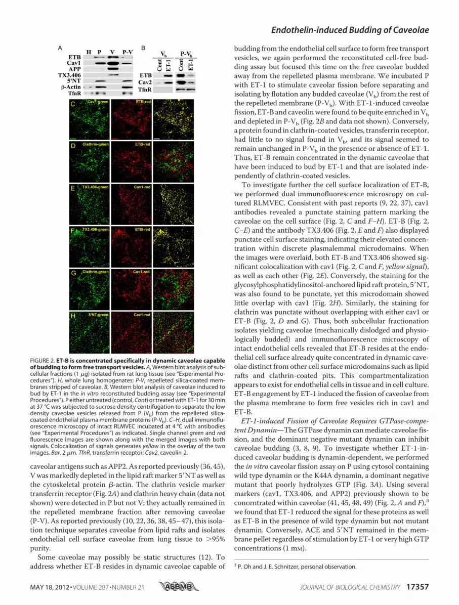
caveolar antigens such asAPP2.As reported previously (36, 45),Vwasmarkedly depleted in the lipid raftmarker 5�NTaswell asthe cytoskeletal protein �-actin. The clathrin vesicle markertransferrin receptor (Fig. 2A) and clathrin heavy chain (data notshown) were detected in P but not V; they actually remained inthe repelleted membrane fraction after removing caveolae(P-V). As reported previously (10, 22, 36, 38, 45–47), this isola-tion technique separates caveolae from lipid rafts and isolatesendothelial cell surface caveolae from lung tissue to �95%purity.Some caveolae may possibly be static structures (12). To
address whether ET-B resides in dynamic caveolae capable of
budding from the endothelial cell surface to form free transportvesicles, we again performed the reconstituted cell-free bud-ding assay but focused this time on the free caveolae buddedaway from the repelleted plasma membrane. We incubated Pwith ET-1 to stimulate caveolar fission before separating andisolating by flotation any budded caveolae (Vb) from the rest ofthe repelleted membrane (P-Vb). With ET-1-induced caveolaefission, ET-B and caveolinwere found to be quite enriched inVband depleted in P-Vb (Fig. 2B and data not shown). Conversely,a protein found in clathrin-coated vesicles, transferrin receptor,had little to no signal found in Vb, and its signal seemed toremain unchanged in P-Vb in the presence or absence of ET-1.Thus, ET-B remain concentrated in the dynamic caveolae thathave been induced to bud by ET-1 and that are isolated inde-pendently of clathrin-coated vesicles.To investigate further the cell surface localization of ET-B,
we performed dual immunofluorescence microscopy on cul-tured RLMVEC. Consistent with past reports (9, 22, 37), cav1antibodies revealed a punctate staining pattern marking thecaveolae on the cell surface (Fig. 2, C and F–H). ET-B (Fig. 2,C–E) and the antibody TX3.406 (Fig. 2, E and F) also displayedpunctate cell surface staining, indicating their elevated concen-tration within discrete plasmalemmal microdomains. Whenthe images were overlaid, both ET-B and TX3.406 showed sig-nificant colocalization with cav1 (Fig. 2, C and F, yellow signal),as well as each other (Fig. 2E). Conversely, the staining for theglycosylphosphatidylinositol-anchored lipid raft protein, 5�NT,was also found to be punctate, yet this microdomain showedlittle overlap with cav1 (Fig. 2H). Similarly, the staining forclathrin was punctate without overlapping with either cav1 orET-B (Fig. 2, D and G). Thus, both subcellular fractionationisolates yielding caveolae (mechanically dislodged and physio-logically budded) and immunofluorescence microscopy ofintact endothelial cells revealed that ET-B resides at the endo-thelial cell surface already quite concentrated in dynamic cave-olae distinct from other cell surfacemicrodomains such as lipidrafts and clathrin-coated pits. This compartmentalizationappears to exist for endothelial cells in tissue and in cell culture.ET-B engagement by ET-1 induced the fission of caveolae fromthe plasma membrane to form free vesicles rich in cav1 andET-B.ET-1-induced Fission of Caveolae Requires GTPase-compe-
tentDynamin—TheGTPase dynamin canmediate caveolae fis-sion, and the dominant negative mutant dynamin can inhibitcaveolae budding (3, 8, 9). To investigate whether ET-1-in-duced caveolar budding is dynamin-dependent, we performedthe in vitro caveolar fission assay on P using cytosol containingwild type dynamin or the K44A dynamin, a dominant negativemutant that poorly hydrolyzes GTP (Fig. 3A). Using severalmarkers (cav1, TX3.406, and APP2) previously shown to beconcentrated within caveolae (41, 45, 48, 49) (Fig. 2, A and F),3we found that ET-1 reduced the signal for these proteins as wellas ET-B in the presence of wild type dynamin but not mutantdynamin. Conversely, ACE and 5�NT remained in the mem-brane pellet regardless of stimulation by ET-1 or very highGTPconcentrations (1 mM).
3 P. Oh and J. E. Schnitzer, personal observation.
FIGURE 2. ET-B is concentrated specifically in dynamic caveolae capableof budding to form free transport vesicles. A, Western blot analysis of sub-cellular fractions (1 �g) isolated from rat lung tissue (see “Experimental Pro-cedures”). H, whole lung homogenates; P-V, repelleted silica-coated mem-branes stripped of caveolae. B, Western blot analysis of caveolae induced tobud by ET-1 in the in vitro reconstituted budding assay (see “ExperimentalProcedures”). P either untreated (control, Cont) or treated with ET-1 for 30 minat 37 °C was subjected to sucrose density centrifugation to separate the lowdensity caveolae vesicles released from P (Vb) from the repelleted silica-coated endothelial plasma membrane proteins (P-Vb). C–H, dual immunoflu-orescence microscopy of intact RLMVEC incubated at 4 °C with antibodies(see “Experimental Procedures”) as indicated. Single channel green and redfluorescence images are shown along with the merged images with bothsignals. Colocalization of signals generates yellow in the overlay of the twoimages. Bar, 2 �m. TfnR, transferrin receptor; Cav2, caveolin-2.
Endothelin-induced Budding of Caveolae
MAY 18, 2012 • VOLUME 287 • NUMBER 21 JOURNAL OF BIOLOGICAL CHEMISTRY 17357

To assess further the role of dynamin in ET-1-induced caveo-lar fission, we used a pharmacological inhibitor of dynamin,Dynasore (50, 51). To confirm for the first time that Dynasorecould inhibit caveolar fission and thus was functionally quitesimilar to the dominant negative mutant K44A dynamin, weperformed our in vitro reconstituted cell-free budding assay.Dynasore was preincubated in cytosol containing normaldynamin prior to incubation with P and ET-1 stimulation asdescribed above. Again there was little to no observable ET-1induced caveolar budding when dynamin was inhibited (Fig.3B). Thus, both pharmacological and genetic means of inhibi-tion showed the critical role of dynamin in endothelin-inducedcaveolae fission.We extended these in vitro studies to evaluating dynamin’s
function in intact endothelial cells in lung tissue by simplyincluding Dynasore with ET administered intravascularly inrats. Western analysis again showed that ET-1 within minuteswas able to induce substantial budding of caveolae rich in ET-Bas measured by loss of caveolin and ET-B signal in P isolatedfrom the lung tissue (Fig. 3C). Dynasore completely inhibitedthis effect of endothelin. Thus, ET specifically induced caveolarfission in reconstituted cell-free assay in vitro and in intactendothelial cells in lung tissue via a dynamin-dependentmechanism.ET-1 Internalization in Cultured Endothelial Cells—We
used ET-1 conjugated to tetramethylrhodamine to follow thecell surface dynamics and internalization of ET-1 and cav1simultaneously in intact cells. At 4 °C, ET-1 and cav1 colocalizesignificantly on the cell surface. Some cells showed �70% colo-calization (Fig. 4A), whereas others exhibited 30% colocaliza-tion. ET-1 was rapidly internalized within minutes after warmup to 37 °C (Fig. 4B). Most of the ET-1 at the cell surfaceappeared to move inside the cell within 10 min to producestrong intracellular punctate staining. Dominant negativemutant dynamin prevented this endocytosis when we per-formed the same experimental protocol this time using BAECtransiently transfected with K44A dynamin2 fused to eGFP.
The cells expressing K44A dynamin2-eGFP lacked any intra-cellular accumulation of ET-1 (Fig. 5, A and B). However, theneighboring cells not expressing K44A dynamin2-eGFP stillshowed ample intracellular perinuclear punctate staining of theET-1 probe (Fig. 5, A and B, outlined cell). Similarly inducedexpression of another dominant negative mutant protein,Eps15 mutant, an inhibitor of clathrin-mediated endocytosis(52), did not prevent rapid ET-1 (Fig. 5, C and D, arrowheads),yet expression of this Eps15 mutant did inhibit transferrininternalization (Fig. 5, E and F, arrowhead), and thus themutant form was active. In other experiments, we followedET-B internalization after ET-1 stimulation of the cells. Again,K44A dynamin (supplemental Fig. S1A) but not Eps15 mutant(supplemental Fig. S1B) prevented internalization in this caseof the ET-1 receptor ETB. Thus, the internalization of ET-1 andits receptor ET-B via caveolae in cultured endothelial cells isdependent on active dynamin.In these experiments, we did have some concern with the
ET-1-tetramethylrhodamine probe in part because of ourinability to competemuch of its cell surface binding with nakedET-1 not conjugated to a fluorophore. The hydrophobicity ofthe fluorophore attached to such a small molecule may causedirect lipid membrane interaction. Regardless, our data hereare still consistentwith rapid dynamin-mediated endocytosis ofET-1 by caveolae and not by clathrin-coated vesicles. However,better fluorescent ETprobes are neededbecause this probemaylack the desired specificity for ET-B and thus caveolae. Hence,we did not pursue further studies with this probe. It appearsthatwith ET-B already concentrated in caveolae, ET can rapidlystimulate dynamin-mediated fission of caveolae that drivesinternalization of caveolae and its cargo into the cell.ET-1 and ET-B Internalization Requires Caveolin-1—To
assess the role for caveolin in ET endocytosis andwhether cave-olae are indeed required for this internalization, we inhibitedcav1 expression in BAEC using shRNA lentiviral transductionparticle specific for cav1. Routinely we were able to reducecaveolin-1 expression by �75–80% (supplemental Fig. S2).Upon ET-1 treatment of control cells expressing caveolin-1, weobserved rapid ETB endocytosis as well as ET-B and caveolin-1
FIGURE 3. Role of dynamin in caveolae fission induced by ET-1. A, Westernblot analysis of repelleted endothelial cell plasma membranes after perform-ing the in vitro reconstituted budding assay in the presence of ET-1 for 30 min,GTP, and either wild type (wt) or K44A mutant dynamin containing cytosol asindicated (see “Experimental Procedures”). B, same as A but used standardcytosol and as indicated with 50 �M Dynasore (30 min of pretreatment beforeET-1 and also included with ET-1). C, Western analysis of silica-coated endo-thelial plasma membranes isolated from rat lungs that had been treated insitu (see “Experimental Procedures”) with ET-1 and/or 50 �M Dynasore. Dyna-sore was administered for 10 min and then also combined with ET-1 asindicated.
FIGURE 4. ET-1 is rapidly internalized by caveolae in cultured endothelialcells. Images from confocal fluorescence microscopy of tetramethylrhod-amine-ET1 localization (red) on intact BAEC immediately fixed after 4 °C bind-ing (A) or after the cells were warmed to 37 °C for 5 min (B) prior to fixationwith paraformadehyde. The cells were then permeabilized before incubationwith cav1 antibodies and fluorophore-conjugated reporter antibodies (cav1,green). The bar represents 50 �m.
Endothelin-induced Budding of Caveolae
17358 JOURNAL OF BIOLOGICAL CHEMISTRY VOLUME 287 • NUMBER 21 • MAY 18, 2012

intracellular perinuclear colocalization (Fig. 5G), but in cellsshowing minimal to no detectable caveolin-1 expression, weobserved little to no intracellular perinuclear signal and onlycell surface signal for ET-B (Fig. 5H). We also performed awarm-up internalization experiment using biotinylated-ET-1and an enzyme-linked, avidin-based detection assay to confirmthat ET-1 remained at the cell surface after cav1 knockdown(Fig. 5I). With 5 min of warming of the cells at 37 °C, the ET-1surface signal decreased by �80% both in the uninfected cells,as well as cells infected with a control shRNA lentivirus trans-duction particle (shCont). ET remained completely detectableat the cell surface when the cells were kept at 4 °C as well aswhen the cells were infected with shCav1. Thus, cav1 and cave-olae are necessary for rapid internalization of ET-1 and ET-B.ET-1 Induces the Internalization of TX3.406, a Caveolar Tar-
geting Antibody—To determine further whether the ET-in-duced, dynamin-dependent endocytosis was restricted to cave-olae and to avoid possible off target interactions of theET-fluorophore, we also evaluated the effect of ET-1 on endo-cytosis in intact, untransfected cultured cells by using two anti-body probes: 5�NT, which targets lipid rafts, and TX3.406,which targets caveolae from outside the cell. As discussed ear-lier, Fig. 2 (E and F) shows excellent colocalization with caveo-lin-1 and ET-B at the cell surface when TX3.406 was incubatedwith endothelial cells at 4 °C. In these new experiments,TX3.406 and 5�NT antibodies were allowed to bind the surfaceof RLMVEC at 4 °C prior towarming to 37 °C in the presence orabsence of ET-1. Within 5 min of warming, only the ET-1-exposed cells showed extensive internalization of TX3.406,which rapidly concentrated in discrete vesicular structures seenas large punctate signals inside the cells (Fig. 6, A–D). Theselarge intracellular vesicles still contained cav1 (Fig. 6D). Bycomparison, the antibody to 5�NT remained at the cell surfacein the absence or presence of ET-1 (Fig. 6, E and F). Consistent
with these results, TX3.406 but not 5�NT antibodies becamequite resistant to trypsin digestion within 5 min (data notshown). This induction of rapid internalization of the caveolaeprobe parallels our findings in BAEC using cav1-eGFP as amarker of caveolae (Fig. 1, F and G).We also performed the same experimental protocol using
RLMVEC transiently transfected with wild type normaldynamin2-eGFP and mutant K44Adynamin2-eGFP (Fig. 7).Cells expressing wild type dynamin2-eGFP, as well as nearbycells not effectively transfected, showed intracellular perinu-clear TX3.406 accumulation (indicated by arrowheads) (Fig.7B). The cells expressing K44A dynamin2-eGFP did not,despite neighboring cells (indicated by arrowheads) not
FIGURE 5. Role of dynamin and caveolin-1 but not Eps15 in rapid internalization of ET-1 and ET-B in cultured endothelial cells. A–F, images fromconfocal fluorescence microscopy of intact BAEC expressing either K44A Dynamin2-eGFP (green) (A and B) or Eps15-eGFP mutant (green) (C–F), and localizationof either tetramethylrhodamine-ET1 (red) (A–D) or tetramethylrhodamine-Tfn (red) (E and F) after binding at 4 °C followed by warming to 37 °C for 5 min priorto fixation with paraformaldehyde (see “Experimental Procedures”). The dashed lines in A and B outline the positions of untransfected cell. The arrowheads inD and F denote Eps15 mutant transfected cells. G and H, localization of cav1 and ETB after 5 min of stimulation with ET1 of BAEC that were uninfected (G) oralready infected for 24 h with shCav1 (H). I, histogram showing relative biotinylated ET-1 cell surface signal detected using an enzyme-linked, avidin-baseddetection assay (see “Experimental Procedures”). Intact, nonfixed BAEC were incubated with biotinylated ET-1 at 4 °C. Some cells (labeled 4 °C) were immedi-ately fixed and processed for biotin detection. The other cells (labeled 37 °C) were first warmed to 37 °C for 5 min before processing. The cells infected for 24 hwith shRNA lentivirus transduction particle are indicated as either �shCav1 for caveolin-1 shRNA or with control shRNA lentivirus transduction particle(�shCont) for control shRNA. *, p � 0.05 (analysis of variance/Tukey’s range test). n � 3 experiments of triplicate wells for each experiment. The bar represents50 �m.
FIGURE 6. ET-1-induced internalization of TX3.406, an antibody targetingcaveolae, in cultured cells. Images from confocal fluorescence microscopyof intact RLMVEC incubated at 4 °C with either TX3.406 (A–D) or 5�NT anti-body (E and F) to label caveolae and lipid rafts, respectively. The cells werethen incubated for 5 min at 37 °C in the presence (B–D and F) or absence(A and E) of ET-1 and then fixed and processed with fluorescent reporterantibody (green). C, magnified image of cell indicated by arrow and yellow boxin B. D, the RLMVEC stimulated with ET-1 were also permeabilized and incu-bated with polyclonal antibody to caveolin-1 followed by fluorescentreporter antibody to rabbit IgG (red). The arrowheads indicate internalizedsignal. The bar represents 50 �m (A, B, and D–F) or 10 �m (C).
Endothelin-induced Budding of Caveolae
MAY 18, 2012 • VOLUME 287 • NUMBER 21 JOURNAL OF BIOLOGICAL CHEMISTRY 17359

expressing K44A dynamin2-eGFP still showing caveolae inter-nalization of the probe as indicated by the intracellular perinu-clear punctate staining (Fig. 7,A,C, andD). Thus, ET-1 inducesthe rapid dynamin-mediated endocytosis of caveolae but notlipid rafts to form large cav1-containing intracellular vesicles.
DISCUSSION
Receptor-mediated endocytosis plays an important role inmany physiological processes including GPCR signaling andsequestration (53). Caveolae and clathrin-coated pits are plas-malemmal vesicles that pinch off from the cell surface in adynamin-dependent manner to internalize their cargo forintracellular sorting and trafficking (54). Although the role ofclathrin-coated pits in GPCR internalization has been wellcharacterized, it is becoming apparent that upon ligand engage-ment, many GPCR are rapidly internalized by other endocyticpathways not yet fully defined (17, 34, 35). Claing et al. (17)detected three distinct pathways for GPCR internalization.Pathway I (�1 adrenergic receptor, �2 adrenergic receptor,�-opioid receptor, adenosine 2B receptor, and M1 muscarinicreceptor) is inhibited by overexpressed mutant �-arrestin,K44Adynamin, andwild type smallGprotein-coupled receptorkinase-interacting protein, GIT1. Pathway II internalizes ET-Band vasoactive intestinal peptide 1 receptor and is inhibited byK44A dynamin but not by GIT1 or mutant �-arrestin. PathwayIII (angiotensin 1A receptor andM2muscarinic receptor) is notaffected by overexpression of these three proteins. Clathrin ves-icles clearly mediate Pathway I, but the endocytic mechanismfor the others are unknown, although it has been speculatedthat caveolae mediate pathway II (17).In this report, we use three different systems (an in vitro
caveolae fission assay, intact cultured endothelial cells, andendothelial cells existing natively in tissue) to showconclusively
that endothelins stimulates the dynamic internalization ofcaveolae rich in ET-B through a fission process requiringdynamin. Both Dynasore and the dominant negative mutant ofdynamin, K44A, prevent caveolar budding induced by ET.Although ET-B internalization can be mediated in other celltypes by clathrin-coated pits (34), we find, in endothelial cells,no evidence of clathrin colocalization with ET-B nor clathrin-mediated endocytosis of ET-1 or ET-B (no inhibition withdominant negative EPS-15). Endothelin induces the internal-ization of caveolae and its cargo in endothelial cells in cultureand in tissue.Unlike ET-B, which we show in endothelial cells resides
already concentrated in caveolae that can respond directly andthus very quickly to ligand engagement, various GPCR such asbradykinin receptors appear to first require ligand engagementfor sequestration into caveolae and/or lipid rafts, possibly fordelayed, slow internalization (19, 23–26, 55). These studies aremostly based on overexpressing transfected cells and/or bio-chemical subfractionation techniques that are known to coiso-late noncaveolar microdomains from the plasma membrane(i.e. lipid rafts) and even from intracellular compartments (i.e.Golgi and nuclei) (36, 45). Here, we show using lung tissue toavoid possible cell culture artifacts and using subcellular frac-tionation techniques that isolate caveolae separately fromotherdetergent-resistant microdomains (i.e. lipid rafts) (3, 9, 36, 38,45), that ET-B resides constitutively in caveolae rather thansequestering there only after ligand engagement. This finding isconfirmed in intact cultured endothelial cells by confocal fluo-rescencemicroscopy showing ET-B signal as punctae that colo-calize extensively withmarkers for caveolae but notmarkers forlipid rafts or clathrin-coated vesicles.It is now apparent that caveolae canmediate very rapid inter-
nalization of select ligands. ET-B is localized and concentratedin caveolae of microvascular endothelial cells in rat lung as wellas bovine aortic endothelial cells and rat lung microvascularendothelial cells. ET-1 can induce fission of caveolae in a recon-stituted cell-free budding assay from endothelial cell plasmamembranes purified from lungs and can stimulate the buddingand internalization of caveolae in cultured endothelial cells.The budding and internalization of caveolae with ET-1 engage-ment of its receptor is very rapid. Caveolae are indeed dynamicstructures capable of stimulated membrane trafficking. Herewe discover that the natural physiological ligand, ET, cangreatly speed up caveolae budding and internalization. Thus,caveolae may not be the slow endocytic pathway as envisionedpreviously (4–7, 9–13), but in fact their internalization may betightly regulated and quite rapid after ligand engagement ofspecific receptors. Prepackaging the receptors in caveolaebefore ligand engagement likely eliminates time lost to seques-tering receptors and probably is key to inducing very rapidendocytosis. This rapid internalization of ligand and receptor islikely important in the regulation of signal transduction eitherthrough homologous desensitization and/or delivery to specificinternal compartments yet to be determined.The rapid response in budding is also probably enabled by
having dynamin as well as the rest of the ET-B signalingmachinery (heterotrimeric G proteins and nonreceptor tyro-sine kinases (22, 37)) already concentrated in caveolae before
FIGURE 7. Role of dynamin in ET-1 induced internalization of TX3.406 incultured endothelial cells. Images from confocal fluorescence microscopyof RLMVEC already transfected to express either K44A dynamin2-eGFP (green)(A, C, and D) or wild type (wt) dynamin2-eGFP (green) (B). The cells were incu-bated with TX3.406 for 1 h at 4 °C and washed before 5 min of stimulation withET-1 at 37 °C and then fixation, permeabilization, and incubation with sec-ondary fluorescent reporter antibody to mouse IgG (red). The arrowheadsindicated intracellular perinuclear staining. Bar, 10 �m.
Endothelin-induced Budding of Caveolae
17360 JOURNAL OF BIOLOGICAL CHEMISTRY VOLUME 287 • NUMBER 21 • MAY 18, 2012

ligand engagement. Dynamin is the key mechanical effectormediating end stage budding and is already polymerized like aspring around the neck of caveolae ready for immediate fission(9, 22, 37, 40). It is clear from the in vitro reconstituted cell-freeassay, which focuses only on the last step of the caveolar bud-ding process, namely fission, and not the invagination processor formation of new caveolae, that endothelin can directly stim-ulate caveolar fission. The polymeric ring of dynamin can gen-erate themechanical force and extension necessary to pinch offthe mature caveolae from the plasma membrane to form freetransport vesicles (3, 9, 56). It seems that caveolae can bud byfirst invaginating to form their mature, stable, omega-shapedstructure and, at least in this case, by then waiting, poised andready, for specific ligands to trigger their rapid, fission from theplasma membrane. This mechanism implies a role for caveolaebeyond simple static signaling platforms toward acutely inte-grating or coupling receptor-mediated signaling to the inter-nalization and trafficking of cell surface molecules and ligands.Thus, key signaling and trafficking molecules may indeed beconcentrated a priori in caveolae to mediate very rapidresponses to ligand engagement, resulting in rapid budding ofcaveolae. Although the regulated caveolae trafficking revealedin this study is likely fundamental to the biology of the cell, theexact signaling mechanisms and patho-physiological ramifica-tions remain to be determined for this newly discovered biolog-ical process where a specific ligand can induce very rapid cave-olae budding and internalization in endothelial cells.
Acknowledgments—We thankDrs.MiyoungChun andPaul Luzio forthe kind gifts of antibodies to ET-B and 5�NT; Drs. Mark McNiven,Frances Brodsky, and Giussy Fiucci for the kind gifts of constructs todyn2GFP (wild type and K44A), Eps15-eGFP mutant, and cav1-eGFP; and Drs. Karen Guice and Ken Baker for the kind gifts of theendothelial cells RLMVEC and BAEC, respectively.
REFERENCES1. Montesano, R., Roth, J., Robert, A., and Orci, L. (1982) Non-coated mem-
brane invaginations are involved in binding and internalization of choleraand tetanus toxins. Nature 296, 651–653
2. Parton, R. G., Joggerst, B., and Simons, K. (1994) Regulated internalizationof caveolae. J. Cell Biol. 127, 1199–1215
3. Schnitzer, J. E., Oh, P., andMcIntosh, D. P. (1996) Role of GTP hydrolysisin fission of caveolae directly from plasma membranes. Science 274,239–242
4. Pelkmans, L., Kartenbeck, J., andHelenius, A. (2001) Caveolar endocytosisof simian virus 40 reveals a new two-step vesicular-transport pathway tothe ER. Nat. Cell Biol. 3, 473–483
5. Schnitzer, J. E., Oh, P., Pinney, E., and Allard, J. (1994) Filipin-sensitivecaveolae-mediated transport in endothelium: reduced transcytosis, scav-enger endocytosis, and capillary permeability of select macromolecules.J. Cell Biol. 127, 1217–1232
6. McIntosh, D. P., Tan, X. Y., Oh, P., and Schnitzer, J. E. (2002) Targetingendothelium and its dynamic caveolae for tissue-specific transcytosis invivo. A pathway to overcome cell barriers to drug and gene delivery. Proc.Natl. Acad. Sci. U.S.A. 99, 1996–2001
7. Milici, A. J., Watrous, N. E., Stukenbrok, H., and Palade, G. E. (1987)Transcytosis of albumin in capillary endothelium. J. Cell Biol. 105,2603–2612
8. Henley, J. R., Krueger, E. W., Oswald, B. J., and McNiven, M. A. (1998)Dynamin-mediated internalization of caveolae. J. Cell Biol. 141, 85–99
9. Oh, P., McIntosh, D. P., and Schnitzer, J. E. (1998) Dynamin at the neck of
caveolaemediates their budding to form transport vesicles byGTP-drivenfission from the plasma membrane of endothelium. J. Cell Biol. 141,101–114
10. Mayor, S., Rothberg, K. G., and Maxfield, F. R. (1994) Sequestration ofGPI-anchored proteins in caveolae triggered by cross-linking. Science264,1948–1951
11. Bundgaard, M. (1983) Vesicular transport in capillary endothelium. Doesit occur? Fed. Proc. 42, 2425–2430
12. Thomsen, P., Roepstorff, K., Stahlhut, M., and van Deurs, B. (2002) Cave-olae are highly immobile plasmamembranemicrodomains, which are notinvolved in constitutive endocytic trafficking.Mol. Biol. Cell 13, 238–250
13. McIntosh, D. P., and Schnitzer, J. E. (1999) Caveolae require intact VAMPfor targeted transport in vascular endothelium. Am. J. Physiol. 277,H2222–H2232
14. Oh, P., Borgström, P., Witkiewicz, H., Li, Y., Borgström, B. J., Chrastina,A., Iwata, K., Zinn, K. R., Baldwin, R., Testa, J. E., and Schnitzer, J. E. (2007)Live dynamic imaging of caveolae pumping targeted antibody rapidly andspecifically across endothelium in the lung. Nat. Biotechnol. 25, 327–337
15. Chun, M., Lin, H. Y., Henis, Y. I., and Lodish, H. F. (1995) Endothelin-induced endocytosis of cell surface ETA receptors. Endothelin remainsintact and bound to the ETA receptor. J. Biol. Chem. 270, 10855–10860
16. Ferguson, S. S. (2001) Evolving concepts in G protein-coupled receptorendocytosis. The role in receptor desensitization and signaling. Pharma-col. Rev. 53, 1–24
17. Claing, A., Perry, S. J., Achiriloaie,M.,Walker, J. K., Albanesi, J. P., Lefkow-itz, R. J., and Premont, R. T. (2000) Multiple endocytic pathways of Gprotein-coupled receptors delineated by GIT1 sensitivity. Proc. Natl.Acad. Sci. U.S.A. 97, 1119–1124
18. Ginés, S., Ciruela, F., Burgueño, J., Casadó, V., Canela, E. I.,Mallol, J., Lluís,C., and Franco, R. (2001) Involvement of caveolin in ligand-induced re-cruitment and internalization of A(1) adenosine receptor and adenosinedeaminase in an epithelial cell line.Mol. Pharmacol. 59, 1314–1323
19. Igarashi, J., and Michel, T. (2000) Agonist-modulated targeting of theEDG-1 receptor to plasmalemmal caveolae. eNOS activation by sphingo-sine 1-phosphate and the role of caveolin-1 in sphingolipid signal trans-duction. J. Biol. Chem. 275, 32363–32370
20. Schwencke, C., Okumura, S., Yamamoto, M., Geng, Y. J., and Ishikawa, Y.(1999) Colocalization of �-adrenergic receptors and caveolin within theplasma membrane. J. Cell. Biochem. 75, 64–72
21. Yamaguchi, T., Murata, Y., Fujiyoshi, Y., and Doi, T. (2003) Regulatedinteraction of endothelin B receptor with caveolin-1. Eur. J. Biochem. 270,1816–1827
22. Oh, P., and Schnitzer, J. E. (2001) Segregation of heterotrimeric G proteinsin cell surface microdomains. Gq binds caveolin to concentrate in cave-olae, whereas Gi and Gs target lipid rafts by default. Mol. Biol. Cell 12,685–698
23. deWeerd, W. F., and Leeb-Lundberg, L. M. (1997) Bradykinin sequestersB2 bradykinin receptors and the receptor-coupled Galpha subunits G�q
and G�i in caveolae in DDT1 MF-2 smooth muscle cells. J. Biol. Chem.272, 17858–17866
24. Haasemann, M., Cartaud, J., Muller-Esterl, W., and Dunia, I. (1998) Ago-nist-induced redistribution of bradykinin B2 receptor in caveolae. J. CellSci. 111, 917–928
25. Feron,O.,Han,X., andKelly, R. A. (1999)Muscarinic cholinergic signalingin cardiacmyocytes. Dynamic targeting ofM2AChR to sarcolemmal cave-olae and eNOS activation. Life Sci. 64, 471–477
26. Ishizaka, N., Griendling, K. K., Lassègue, B., and Alexander, R. W. (1998)Angiotensin II type 1 receptor. Relationship with caveolae and caveolinafter initial agonist stimulation. Hypertension 32, 459–466
27. Agapitov, A. V., and Haynes, W. G. (2002) Role of endothelin in cardio-vascular disease. J. Renin Angiotensin Aldosterone Syst. 3, 1–15
28. Seo, B., Oemar, B. S., Siebenmann, R., von Segesser, L., and Lüscher, T. F.(1994) Both ETA and ETB receptors mediate contraction to endothelin-1in human blood vessels. Circulation 89, 1203–1208
29. Batra, V. K., McNeill, J. R., Xu, Y., Wilson, T. W., and Gopalakrishnan, V.(1993) ETB receptors on aortic smooth muscle cells of spontaneouslyhypertensive rats. Am. J. Physiol. 264, C479–C484
30. Fukuroda, T., Fujikawa, T., Ozaki, S., Ishikawa, K., Yano, M., and Ni-
Endothelin-induced Budding of Caveolae
MAY 18, 2012 • VOLUME 287 • NUMBER 21 JOURNAL OF BIOLOGICAL CHEMISTRY 17361

shikibe,M. (1994) Clearance of circulating endothelin-1 by ETB receptorsin rats. Biochem. Biophys. Res. Commun. 199, 1461–1465
31. de Nucci, G., Thomas, R., D’Orleans-Juste, P., Antunes, E., Walder, C.,Warner, T. D., and Vane, J. R. (1988) Pressor effects of circulating endo-thelin are limited by its removal in the pulmonary circulation and by therelease of prostacyclin and endothelium-derived relaxing factor. Proc.Natl. Acad. Sci. U.S.A. 85, 9797–9800
32. Liu, S., Premont, R. T., Kontos, C. D., Huang, J., and Rockey, D. C. (2003)Endothelin-1 activates endothelial cell nitric-oxide synthase via heterotri-meric G-protein �� subunit signaling to protein jinase B/Akt. J. Biol.Chem. 278, 49929–49935
33. Oksche, A., Boese, G., Horstmeyer, A., Furkert, J., Beyermann,M., Bienert,M., and Rosenthal, W. (2000) Late endosomal/lysosomal targeting andlack of recycling of the ligand-occupied endothelin B receptor.Mol. Phar-macol. 57, 1104–1113
34. Bremnes, T., Paasche, J. D., Mehlum, A., Sandberg, C., Bremnes, B., andAttramadal, H. (2000) Regulation and intracellular trafficking pathways ofthe endothelin receptors. J. Biol. Chem. 275, 17596–17604
35. Chun, M., Liyanage, U. K., Lisanti, M. P., and Lodish, H. F. (1994) Signaltransduction of a G protein-coupled receptor in caveolae. Colocalizationof endothelin and its receptor with caveolin. Proc. Natl. Acad. Sci. U.S.A.91, 11728–11732
36. Schnitzer, J. E., McIntosh, D. P., Dvorak, A. M., Liu, J., and Oh, P. (1995)Separation of caveolae from associated microdomains of GPI-anchoredproteins. Science 269, 1435–1439
37. Liu, J., Oh, P., Horner, T., Rogers, R. A., and Schnitzer, J. (1997) Organizedendothelial cell surface signal transduction in caveolae distinct from gly-cosylphosphatidylinositol-anchored proteinmicrodomains. J. Biol. Chem.272, 7211–7222
38. Oh, P., and Schnitzer, J. E. (1998) Isolation and subfractionation of plasmamembranes to purify caveolae separately from glycosyl-phospatidylinosi-tol-anchored protein microdomains. In Cell Biology: A Laboratory Hand-book (Celis, J., ed) Academic Press, Orlando, FL
39. Rizzo, V., McIntosh, D. P., Oh, P., and Schnitzer, J. E. (1998) In situ flowactivates endothelial nitric oxide synthase in luminal caveolae of endothe-lium with rapid caveolin dissociation and calmodulin association. J. Biol.Chem. 273, 34724–34729
40. Schnitzer, J. E., Liu, J., and Oh, P. (1995) Endothelial caveolae have themolecular transport machinery for vesicle budding, docking, and fusionincluding VAMP, NSF, SNAP, annexins, and GTPases. J. Biol. Chem. 270,14399–14404
41. Oh, P., Li, Y., Yu, J., Durr, E., Krasinska, K.M., Carver, L. A., Testa, J. E., andSchnitzer, J. E. (2004) Subtractive proteomic mapping of the endothelialsurface in lung and solid tumours for tissue-specific therapy.Nature 429,629–635
42. Okamoto, Y., Ninomiya, H., Miwa, S., and Masaki, T. (2000) Cholesteroloxidation switches the internalization pathway of endothelin receptor
type A from caveolae to clathrin-coated pits in Chinese hamster ovarycells. J. Biol. Chem. 275, 6439–6446
43. Zhang, J., Barak, L. S., Anborgh, P. H., Laporte, S. A., Caron, M. G., andFerguson, S. S. (1999) Cellular trafficking of G protein-coupled receptor/�-arrestin endocytic complexes. J. Biol. Chem. 274, 10999–11006
44. Chen, X., Berrou, J., Nguyen, G., Sraer, J. D., and Rondeau, E. (1995) En-dothelin-1 induces rapid and long lasting internalization of the thrombinreceptor in human glomerular epithelial cells. Biochem. Biophys. Res.Commun. 217, 445–451
45. Oh, P., and Schnitzer, J. E. (1999) Immunoisolation of caveolae with highaffinity antibody binding to the oligomeric caveolin cage. Toward under-standing the basis of purification. J. Biol. Chem. 274, 23144–23154
46. Hooper, N.M. (1999) Detergent-insoluble glycosphingolipid/cholesterol-rich membrane domains, lipid rafts and caveolae (review). Mol. MembrBiol. 16, 145–156
47. Zheng, Y. Z., and Foster, L. J. (2009) Biochemical and proteomic ap-proaches for the study of membrane microdomains. J. Proteomics 72,12–22
48. García-Cardeña, G., Oh, P., Liu, J., Schnitzer, J. E., and Sessa,W. C. (1996)Targeting of nitric oxide synthase to endothelial cell caveolae via palmi-toylation. Implications for nitric oxide signaling. Proc. Natl. Acad. Sci.U.S.A. 93, 6448–6453
49. Shaul, P. W., Smart, E. J., Robinson, L. J., German, Z., Yuhanna, I. S., Ying,Y., Anderson, R. G., and Michel, T. (1996) Acylation targets emdothelialnitric-oxide synthase to plasmalemmal caveolae. J. Biol. Chem. 271,6518–6522
50. Macia, E., Ehrlich, M., Massol, R., Boucrot, E., Brunner, C., and Kirch-hausen, T. (2006) Dynasore, a cell-permeable inhibitor of dynamin. Dev.Cell 10, 839–850
51. Newton, A. J., Kirchhausen, T., and Murthy, V. N. (2006) Inhibition ofdynamin completely blocks compensatory synaptic vesicle endocytosis.Proc. Natl. Acad. Sci. U.S.A. 103, 17955–17960
52. Pula, G., Mundell, S. J., Roberts, P. J., and Kelly, E. (2004) Agonist-inde-pendent internalization ofmetabotropic glutamate receptor 1a is arrestin-and clathrin-dependent and is suppressed by receptor inverse agonists.J. Neurochem. 89, 1009–1020
53. Mukherjee, S., Ghosh, R. N., and Maxfield, F. R. (1997) Endocytosis.Physiol. Rev. 77, 759–803
54. Schnitzer, J. E. (2001) Caveolae. From basic trafficking mechanisms totargeting transcytosis for tissue-specific drug and gene delivery in vivo.Adv. Drug Deliv Rev. 49, 265–280
55. Dessy, C., Kelly, R. A., Balligand, J. L., and Feron, O. (2000) Dynaminmediates caveolar sequestration of muscarinic cholinergic receptors andalteration in NO signaling. EMBO J. 19, 4272–4280
56. Stowell, M. H., Marks, B., Wigge, P., and McMahon, H. T. (1999) Nucle-otide-dependent conformational changes in dynamin. Evidence for amechanochemical molecular spring. Nat. Cell Biol. 1, 27–32
Endothelin-induced Budding of Caveolae
17362 JOURNAL OF BIOLOGICAL CHEMISTRY VOLUME 287 • NUMBER 21 • MAY 18, 2012


