Embed Size (px)
Citation preview
lable at ScienceDirect
Biomaterials 32 (2011) 3413e3422
Contents lists avai
Biomaterials
journal homepage: www.elsevier .com/locate/biomater ia ls
Engineering spatial control of multiple differentiation fates within a stem cellpopulation
Elmer D.F. Ker a,d, Bur Chu b,d, Julie A. Phillippi e, Burhan Gharaibeh f, Johnny Huard f, Lee E. Weiss c,d,Phil G. Campbell a,b,d,*aDepartment of Biological Sciences, Carnegie Mellon University, 4400 Fifth Avenue, Pittsburgh, PA 15213, USAbDepartment of Biomedical Engineering, Carnegie Mellon University, 5000 Forbes Avenue, Pittsburgh, PA 15213, USAcRobotics Institute, Carnegie Mellon University, 5000 Forbes Avenue, Pittsburgh, PA 15213, USAd Institute for Complex Engineered Systems, Carnegie Mellon University, 1213 Hamburg Hall, 5000 Forbes Avenue, Pittsburgh, PA 15213, USAeUniversity of Pittsburgh, Thomas E. Starzl Biomedical Science Tower, Department of Surgery, PA 15260, USAf Stem Cell Research Centre of Children’s Hospital, UPMC, Bridgeside Point II Bldg, Suite 206 450 Technology Drive, Pittsburgh, PA 15219, USA
a r t i c l e i n f o
Article history:Received 31 December 2010Accepted 13 January 2011Available online 12 February 2011
Keywords:BoneBMP (bone morphogenetic protein)ECM (extracellular matrix)Fibroblast growth factorMuscleTendon
* Corresponding author. Institute for Complex EnMellon University, 1213 Hamburg Hall, 5000 Forbes AUSA. Tel.: þ1 412 268 4126; fax: þ1 412 268 5229.
E-mail address: [email protected] (P.G. Camp
0142-9612/$ e see front matter � 2011 Elsevier Ltd.doi:10.1016/j.biomaterials.2011.01.036
a b s t r a c t
The capability to engineer microenvironmental cues to direct a stem cell population toward multiplefates, simultaneously, in spatially defined regions is important for understanding the maintenance andrepair of multi-tissue units. We have previously developed an inkjet-based bioprinter to create patternsof solid-phase growth factors (GFs) immobilized to an extracellular matrix (ECM) substrate, and appliedthis approach to drive muscle-derived stem cells toward osteoblasts ‘on-pattern’ and myocytes ‘off-pattern’ simultaneously. Here this technology is extended to spatially control osteoblast, tenocyte andmyocyte differentiation simultaneously. Utilizing immunofluorescence staining to identify tendon-promoting GFs, fibroblast growth factor-2 (FGF-2) was shown to upregulate the tendon marker Scleraxis(Scx) in C3H10T1/2 mesenchymal fibroblasts, C2C12 myoblasts and primary muscle-derived stem cells,while downregulating the myofibroblast marker a-smooth muscle actin (a-SMA). Quantitative PCRstudies indicated that FGF-2 may direct stem cells toward a tendon fate via the Ets family members oftranscription factors such as pea3 and erm. Neighboring patterns of FGF-2 and bone morphogeneticprotein-2 (BMP-2) printed onto a single fibrin-coated coverslip upregulated Scx and the osteoblastmarker ALP, respectively, while non-printed regions showed spontaneous myotube differentiation. Thiswork illustrates spatial control of multi-phenotype differentiation and may have potential in theregeneration of multi-tissue units.
� 2011 Elsevier Ltd. All rights reserved.
1. Introduction
The musculoskeletal system comprises multiple tissue typesincluding bone, muscle, tendon, ligament and cartilage as well astheir respective tissue interfaces such as bone-to-tendon enthesesand muscle-to-tendon junctions. The maintenance and repair ofthese multi-tissue structures involves the spatial control of stemcell differentiation toward tissue-specific cells, such as osteoblasts,tenocytes and myocytes [1]. This process is regulated by physicaland biochemical microenvironmental cues imparted by the inter-actions of cells with their extracellular matrix (ECM), neighboring
gineered Systems, Carnegievenue, Pittsburgh, PA 15213,
bell).
All rights reserved.
cells, and secreted local and systemic signaling molecules,including growth factors (GFs) [1,2]. Signaling molecules, regulatethe pericellular environment where they reside in both the ‘liquid-phase’ (freely diffusing) form and the ‘solid-phase’ (immobilized)form that exists in an equilibrium state between desorption fromand adsorption to the ECM and cell surfaces [3]. The uniquearchitecture and biochemical composition of the ECM allows it tosequester (immobilize) and release GFs at picogram to nanogramlevels [3e7], and can negatively or positively regulate GF bioactivityand bioavailability [3]. As such, GF sequestration by the ECMimmobilizes GFs to specific locations, which in turn imparts thetemporal and spatial cues required for directing cell behaviors suchas cell adhesion, migration, proliferation, differentiation andapoptosis, which are vital for orchestrating complex processes suchas development, maintenance and wound healing [3,7e19].Therefore, developing toolsets that can be used to selectivelycontrol the physical placement and dosage of multiple exogenous
E.D.F. Ker et al. / Biomaterials 32 (2011) 3413e34223414
GFs in a physiologically-relevant manner in order to spatially directa stem cell population toward multiple cell fates simultaneously isa logical consideration for studying stem cell behaviors and mayalso have direct applications in regenerative medicine.
Prior work reported by our group and by others has shown thatECMs patterned with solid-phase GFs can be engineered to controlvarious aspects of stem cell behavior, including proliferation,migration and differentiation in vitro [8,13,14,16,20e23] as well asdifferentiation in vivo [10]. We previously demonstrated that a GF-patterned fibrin ECM created using an inkjet-based bioprintingtechnology can drive a single stem cell population toward osteo-blast and myocyte fates simultaneously, in registration to printedpatterns in vitro [16]. In the work presented here, we report on theextension of this approach to spatially drive stem cell differentia-tion toward a tendon fate simultaneously with osteoblast andmyocyte differentiation.
Using solid-phase GFs to direct stem cells to tenocytes in vitrohasnot been previously reported in literature. Therefore, prior tostudyingmulti-lineage patterning, candidate tendon-promotingGFshad to be identified and validated. Candidate GFs were screenedagainst mouse C3H10T1/2 mesenchymal fibroblasts, C2C12 myo-blasts and primary muscle-derived stem cells (MDSCs) using bothliquid- and solid-phase immunofluorescence staining for the tendonmarker Scleraxis (Scx) [24,25]. Quantitative PCR studies weresubsequently performed to elucidate themechanism bywhich stemcells differentiated toward a tendon lineage. Following this, solid-phase presentation of FGF-2 and/or BMP-2 on fibrin-coated glasscoverslips using either coarse hand-printing or high resolution, low-dose inkjet bioprinting was used to demonstrate spatial control ofstem cell differentiation toward multiple cell fates simultaneously.
2. Materials and methods
2.1. Cell culture
Multipotent mouse C3H10T1/2 cells (ATTC, Manassas, VA) were grown in Dul-becco’s Modified Eagle’s Media (DMEM; Invitrogen, Carlsbad, CA), 10% fetal bovineserum (Invitrogen, Carlsbad, CA) and 1% penicillin-streptomycin (PS; Invitrogen,Carlsbad, CA). Mouse C2C12 cells (ATTC, Manassas, VA) were grown in DMEM, 10%bovine serum (Invitrogen, Carlsbad, CA) and 1% PS. Multipotent MDSCs were iso-lated from primary mouse gastrocnemius muscle biopsies following a modifiedpreplate technique [26] and were grown in DMEM (high glucose), 10% horse serum(HS; Invitrogen, Carlsbad, CA), 10% FBS, 0.5% Chick Embryo Extract (AccurateChemical Co, Westbury, NY) as previously described [26,27]. For myogenic differ-entiation, cells were grown in low serum containing myogenic differentiation(DMEM, 2% HS, 1% PS) media for 3e5 days. This media is subsequently referred to asmyogenic media or myogenic conditions for the remainder of the text. All cells werekept at 37 �C, 5% CO2 in a humidified incubator.
2.2. Growth factor preparation and use
Recombinant human BMP-2 (Genetics Institute Inc, Cambridge, MA), FGF-2(Peprotech, Rockyhill, NJ), FGF-4 (Peprotech, Rockyhill, NJ) and GDF-7 (Prospc Bio,Rehovot, Israel) were reconstituted according to manufacturer’s instructions to1e2 mg/mL, aliquoted and stored at �80 �C. Prior to use, GFs were freshly diluted tothe desired concentration in 10 mM sodium phosphate, pH 7.4. For liquid-phase GFexperiments, cells were seeded at 2.6e3.1�104 cells/cm2 in the presence or absenceof GF (1e500 ng/mL) under proliferation (High serum) and myogenic (Low serum)media for 3e4 days. For solid-phase GF experiments, cells were seeded at3.1e3.6� 104 cells/cm2 over printed fibrin-coated coverslips under proliferation andmyogenic media for 3e4 days.
2.3. Preparation of fibrin coated coverslips
Homogenous fibrin films were prepared essentially as described by Campbellet al., 2005 [8]. Briefly, 18 � 18 mm epoxy-silanized glass coverslips (Thermo FisherScientific, Waltham, MA) were coated with 0.1 mg/mL fibrinogen (Aventis Behring,King of Prussia, PA or American Diagnostica Inc., Stanford, CT)and converted intofibrin by incubating coverslips in 4 U/mL thrombin (Enzyme Research Laboratories,South Bend, IN). Coverslips were then washed with phosphate buffered saline (PBS)and sterile deionized water before air-drying in a laminar flow hood. The thicknessof the fibrin films was previously estimated to be approximately 20 nm [8].
2.4. Growth factor printing
Prior to printing, GFs were freshly diluted to the desired concentration in 10 mM
sodium phosphate, pH 7.4. Prior to filling the inkjet with the GF, the printhead wassterilized by rinsing with 70% ethanol followed by sterile deionized water. The bio-ink, consisting of 100e200 mg/ml GFwas loaded into the printhead, and printed ontofibrin-coated glass coverslips as previously described [8,14]. The concentration ofinkjetted GFs can be modulated by overprinting, which is achieved by varying thenumber of times a GF is deposited in the same (x,y) location. In the case of hand-printed GF patterns, 1e2 mL of a 100 mg/mL GF solution was pipetted onto a fibrin-coated glass coverslip instead and a diamond scribe pen was used to mark thedroplet perimeter after it had been allowed to air-dry for 1 h at 37 �C. After printing,fibrin-coated coverslips were incubated in PBS for 5 min followed by serum-freeDMEM with 1% PS overnight at 37 �C, 5% CO2 to wash off unbound GF prior to cellseeding. The surface concentration of GF present on fibrin-coated coverslips duringcell seeding was estimated based on desorption measurements in previous studies[8,13,14,28].
2.5. Quantitative PCR
For the experiments investigating Scx expression during muscle differentiation,C2C12 cells were grown at 1.55 � 102 cells/cm2 under proliferation conditions andat 2.5 � 103 cells/cm2 under myogenic conditions for 4 days to ensure similarnumber of cells in both conditions prior to RNA extraction. C3H10T1/2 cells weregrown in proliferation medium at 1.5e2.0 � 104 cells/cm2 in the presence orabsence of FGF-2 (50 mg/mL) for 36 h and 72 h prior to extraction of total RNA(RNeasy Mini Kit; Qiagen, Valencia, CA). Quantitative polymerase chain reactionanalysis for pea3, erm and Scx were performed as previously described [29,30].Target gene expression was normalized to 18S internal control. Gene expression isdisplayed as the mean of five independent experiments and represented asmean � Standard Error Mean (SEM). Statistical analysis was performed asdescribed below.
2.6. Immunofluorescence staining
Cells were washed in PBS, fixed in methanol for 5 min, air-dried and blockedwith 10% donkey serum (Jackson Immunoresearch,West Gove, PA) for 20min at RT.For mouse-on-mouse staining, an additional blocking step was performed byincubating cells with 100 mg/mL donkey anti-mouse FAB (Jackson Immunor-esearch,West Gove, PA) for 1 h at RT. Cells were then rinsed with wash buffer (PBS,0.1%BSA) and incubated with primary antibodies: rabbit anti-Scx (10 mg/mL;Abcam, Cambridge, MA), mouse anti-myosin MF20 (1 mg/mL; DSHB, Iowa City,Iowa), mouse anti-a-smooth muscle actin (a-SMA; 1 mg/mL; Abcam, Cambridge,MA) or goat anti-myogenin (2 mg/mL; Santa Cruz Biotechnology Inc, Santa Cruz, CA)overnight at 4 �C. Cells were then rinsed three times with wash buffer and incu-bated with secondary antibodies for 1 h at RT e donkey anti-goat FITC (4 mg/mL;Santa Cruz Biotechnology Inc, Santa Cruz, CA), donkey anti-mouse Dylight 488 nmor donkey anti-rabbit Dylight 549 nm (15 mg/mL each; Jackson Immunoresearch,West Gove, PA). Lastly, cells were rinsed five times with wash buffer and imagedusing a Zeiss Axiovert 200M microscope (Carl Zeiss Microimaging, Thornwood,NY) equipped with a Colibri LED light source. Quantification of immunofluores-cence staining was performed using Adobe Photoshop 7.0 (Adobe Systems, SanJose, CA). Briefly, the rectangular marquee tool was used to draw a bounding box(Approximately 700 pixels by 700 pixels representing an area 0.9mmby 0.9mm insize) and the image histogram tool was used to measure average pixel intensity.Statistical analysis was performed as described below.
2.7. ALP stain
Cells were seeded onto GF patterns for 72 h, washed in PBS and fixed for 2min in3.7% formaldehyde. Alkaline phosphatase activity (ALP; SIGMAFAST) was detectedaccording to the manufacturer’s instructions (SigmaeAldrich, St. Louis, MO). Whererequired, alkaline phosphatase-stained images were converted to CMYK formatsince this color format is representative of reflected light colors as opposed toemitted light colors (RGB). Since cyan and magenta form the color blue, thesechannels were added together and inverted. The average pixel intensity wasdetermined using the image histogram tool in Adobe Photoshop 7.0 (Adobe Systems,San Jose, CA).
2.8. Statistical analysis
For both quantitative PCR and immunofluorescence analysis, one-way analysisof variance followed by Fisher’s least significant difference post hoc test usingSYSTAT 9 software (Systat Software Inc., Richmond, CA) was performed to determinesignificance among treatment groups. A p value �0.05 was considered statisticallysignificant.
E.D.F. Ker et al. / Biomaterials 32 (2011) 3413e3422 3415
3. Results
3.1. Effect of liquid-phase FGF-2 and FGF-4 on Scx expression inC3H10T1/2 cells
Liquid-phase experiments incorporating a combination of 200 ng/mL BMP-2, 500 ng/mL BMP-12/GDF-7 and 50 ng/mL FGF-2 indicatedthat C3H10T1/2 cells upregulated the tendon marker Scx in the pres-ence of FGF-2 alone but not with GDF-7 or BMP-2 (SupplementaryFig. 1). In addition, FGF-2-treated cells adopted an elongated spindle-like morphology reminiscent of tenocytes (Supplementary Fig. 1).When treatedwith50ng/mLFGF-4, C3H10T1/2 cells showedpunctatenuclear staining of Scx transcription factor (Supplementary Fig. 2).
3.2. Effect of liquid-phase FGF-2 on Scx expression in C2C12 cells
Scx expression in C2C12 cells was examined under proliferation(high serum) as well as myogenic (low serum) conditions to deter-mine if such cells could undergo tendon specification when treatedwith FGF-2. Under proliferation conditions, increasing amounts ofFGF-2 resulted in upregulation of the tendon marker Scx in a dose-dependentmannerwith punctate nuclear stainingof Scxobserved incells treated at 25 ng/mL FGF-2 and 50 ng/mL FGF-2 (SupplementaryFig. 3). Undermyogenic conditions, Scxexpressionwasunexpectedlyupregulated in nascent myotubes in the absence of FGF-2(Supplementary Fig. 4). Confocal sectioning studies determined thatthe thickness ofmyotubes, which is observed as a bright halo aroundcells inphase-contrast images (Supplementary Fig. 4), contributed inpart to the high levels of Scx (data not shown). Furthermore, quan-titative PCR analysis indicated that there was no fold change in Scxexpression between proliferating C2C12 cells (1.003 � 0.075 foldchange) anddifferentiatingmyotubes (1.022� 0.209 fold change). Inthe presence of FGF-2, myotube formation was inhibited and cellsshowed increased Scx expressionwhen compared to non-myocytesin untreated control (Supplementary Fig. 4).
3.3. Effect of liquid-phase FGF-2 on Scx expression in MDSCs
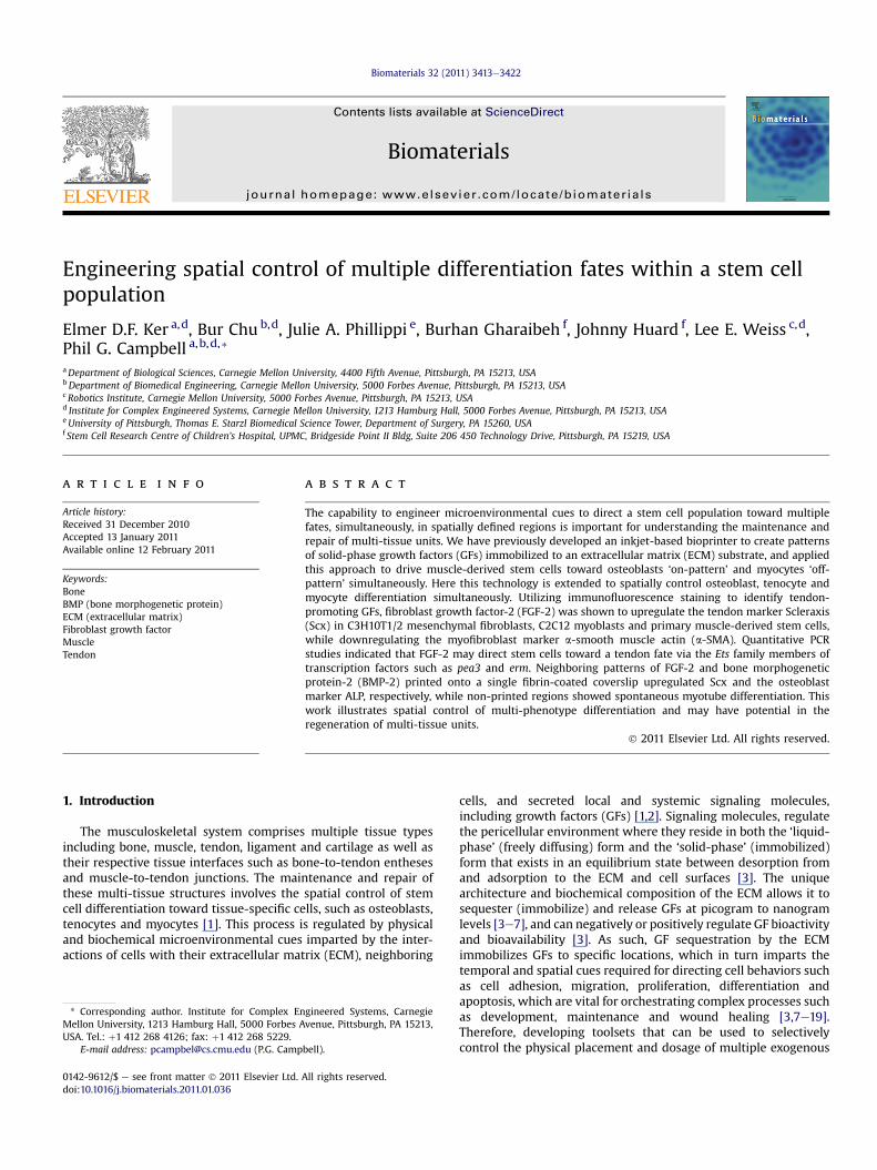
Tendon specification inMDSCswas examined in cells treatedwithFGF-2 under proliferation and myogenic conditions. Under prolifera-tion conditions, FGF-2 dose-dependently increased expression of thetendonmarker Scx with punctate nuclear staining of Scx occasionallyobserved at 50 ng/mL FGF-2 (Fig. 1). Similar to C2C12 cells, Scxexpression was upregulated in nascent myotubes in the absence ofFGF-2. In the presence of FGF-2, myotube formation was inhibitedwithMDSCsexhibiting lower levelsof themyogenicmarkermyogenin(Supplementary Fig. 5). In addition, Scx expression was upregulatedwhen compared to non-myocytes in untreated control and FGF-2-treated cells did not show increased expression for the myofibroblastmarker a-smooth muscle actin (a-SMA; Supplementary Fig. 6).
3.4. Regulation of Scx in C3H10T1/2 cells
As previous studies indicated that members of the Ets family oftranscription factors such as pea3 and erm were involved in regu-lation of Scx during tendon development in the chick, quantitativePCR analysis of these genes were performed to determine ifa similar mechanism was operating in these stem cell populations[31]. 50 ng/mL FGF-2 upregulated pea3 (43.1 � 27.5 fold change,p ¼ 0.005) and erm (16.5 � 6.5 fold change, p ¼ 0.178) at 36 hwhereas Scx levels remained constant. At 72 h, all three genes wereupregulated: pea3 (169.6 � 45.8 fold change, p ¼ 0.008), erm(72.5�16.2 fold change, p¼ 0.033) and Scx (22.8� 5.4 fold change,p ¼ 0.006). As such, the prior induction of pea3 and erm suggestthat these two genes lie upstream of Scx (Fig. 2).
3.5. Effect of solid-phase FGF-2 on Scx expression in C3H10T1/2cells
Having demonstrated that Scx expression was upregulated byliquid-phase FGFs, square patterns of FGF-2 (each measuring 1 by1mm)were inkjet printed ontofibrin-coated glass coverslipswith2,6 and 12 overprints to determine if solid-phase GF patterns canspatially direct tendon specification in a dose-dependent manner.Our previous studies have shown that the surface concentration ofGF that is deposited can bemodulated byoverprinting and that suchGF patterns can persist for up to 144 h under standard cell cultureconditions [10,13,14,16]. As shown in Fig. 3A, the amount of FGF-2deposited in 2, 6 and 12 overprints after washing and prior to cellseeding was estimated to be 40.8 pg/mm2, 122.4 pg/mm2 and244.8 pg/mm2 FGF-2 based on previous studies [8,13,14,28]. Underproliferation conditions (High serum), C3H10T1/2 cells showedupregulationof Scx in response to solid-phase patterningof FGF-2 ina dose-dependent manner (Fig. 3B). Although the lowest dose ofsolid-phase FGF-2 (40.8 pg/mm2) was not sufficient to induce anincrease in Scx expression relative to negative control/non-printedregions (p¼ 0.872), higher doses of solid-phase FGF-2 resulted in anincrease in Scx expression relative to negative control/non-printedregions (p ¼ 0.009 for 122.4 pg/mm2 FGF-2 and p ¼ 0.001 for244.8 pg/mm2 FGF-2; Fig. 3) in C3H10T1/2 cells. Thus, solid-phasepatterning of FGF-2 can spatially control tendon cell fate (Fig. 3).
3.6. Effect of solid-phase FGF-2 on MF20 and Scx expression inC2C12 cells
Similarly, square patterns of FGF-2 (each measuring 1 by 1 mm)were inkjet printed onto fibrin-coated glass coverslips with 5, 10and 30 overprints (corresponding to an estimated amount of102 pg/mm2, 203 pg/mm2 and 612 pg/mm2 FGF-2) to determine ifmultiple stem cell fates could be spatially controlled in a dose-dependent manner within the same construct. Under both prolif-eration and myogenic conditions, inkjet printed patterns of FGF-2resulted in a dose-dependent increase in Scx expression (Fig. 4B, C).
Under proliferation conditions, although the lowest dose of solid-phase FGF-2 (102 pg/mm2 FGF-2) was not sufficient to induce anincrease in Scx expression relative to negative control/non-printedregions (p¼ 0.099), higher doses of solid-phase FGF-2 resulted in anincrease in Scx expression relative to negative control/non-printedregions (p ¼ 0.01 for 203 pg/mm2 FGF-2 and p ¼ 0.000 for 612 pg/mm2 FGF-2) in C2C12 cells (Fig. 4C). Under myogenic conditions,although lower doses of solid-phase FGF-2 were not sufficient toinducean increase inScxexpressionrelative tonegative control/non-printed regions (p ¼ 0.139 for 102 pg/mm2 FGF-2 and p ¼ 0.053 for203 pg/mm2 FGF-2), the highest dose of solid-phase FGF-2 resultedin an increase in Scx expression relative to negative control/non-printed regions (p ¼ 0.022 for 612 pg/mm2 FGF-2) in C2C12 cells(Fig. 4C). Within and outside the printed regions, cells fused to formmultinucleated myotubes as a result of high cell density leading tospontaneous cell fusion under proliferation conditions or directmyogenic induction, as evidenced by the presence of musclemyosinor MF20 (Fig. 4B). No difference in MF20 staining was observedbetween printed and non-printed regions (p > 0.05 for all cases).Taken together, these results demonstrate the simultaneous speci-fication of myocyte and tenocyte fates within the same construct ina spatially defined manner in a dose-dependent fashion.
3.7. Effect of solid-phase BMP-2 and FGF-2 on ALP, MF20 and Scxexpression in C2C12 cells
Having demonstrated that multiple stem cell fates could bespatially controlled within the same construct, inkjet bioprinting
Fig. 1. Dose-dependent effect of FGF-2 on expression of tendon marker Scx in mouse MDSCs after 72 h in proliferation media. Increasing amounts of FGF-2 resulted in upregulationof tendon marker Scx. White box indicates magnified region (right). Note the punctate nuclear staining of Scx transcription factor in 50 ng/ml FGF-2. Scale bar 50 mm.
E.D.F. Ker et al. / Biomaterials 32 (2011) 3413e34223416
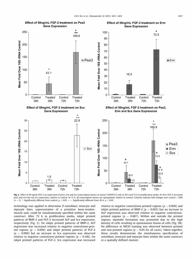
Fig. 2. Effect of 50 ng/mL FGF-2 on expression of pea3, erm and Scx transcription factors in mouse C3H10T1/2 cells at 36 h and 72 h in proliferation media. At 36 h, FGF-2 increasedpea3 and erm but not Scx expression, relative to control. At 72 h, all transcription factors are upregulated, relative to control. Columns indicate fold changes over control � SEM(n ¼ 5). *, Significantly different from control; p � 0.05. þ þ, Significantly different from 36 h; p � 0.05.
E.D.F. Ker et al. / Biomaterials 32 (2011) 3413e3422 3417
technology was applied to determine if osteoblast, tenocyte andmyocyte fates, representative of a primitive bone-tendon-muscle unit, could be simultaneously specified within the sameconstruct. After 72 h in proliferation media, inkjet printedpatterns of BMP-2 and FGF-2 increased ALP and Scx expression,respectively (Fig. 5). On inkjet printed patterns of BMP-2, ALPexpression was increased relative to negative control/non-prin-ted regions (p ¼ 0.000) and inkjet printed patterns of FGF-2(p ¼ 0.000) but no increase in Scx expression was observedrelative to negative control/non-printed regions (p ¼ 0.146). Oninkjet printed patterns of FGF-2, Scx expression was increased
relative to negative control/non-printed regions (p ¼ 0.000) andinkjet printed patterns of BMP-2 (p ¼ 0.003) but no increase inALP expression was observed relative to negative control/non-printed regions (p ¼ 0.887). Within and outside the printedregions, myotube formation was promoted due to the highdensity of cells resulting in spontaneous fusion of cells (Fig. 5B).No difference in MF20 staining was observed between printedand non-printed regions (p > 0.05 for all cases). Taken together,these results demonstrate the simultaneous specification ofosteoblast, tenocyte and myocyte fates within the same constructin a spatially defined manner.
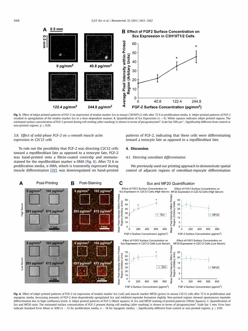
Fig. 3. Effect of inkjet printed patterns of FGF-2 on expression of tendon marker Scx in mouse C3H10T1/2 cells after 72 h in proliferation media. A. Inkjet printed patterns of FGF-2resulted in upregulation of the tendon marker Scx in a dose-dependent manner. B. Quantification of Scx Expression (n ¼ 6). White squares indicates inkjet printed regions. Theestimated surface concentration of FGF-2 present during cell seeding (after washing) is shown in terms of picograms/mm2. Scale bar 500 mm *, Significantly different from control ornon-printed regions; p � 0.05.
E.D.F. Ker et al. / Biomaterials 32 (2011) 3413e34223418
3.8. Effect of solid-phase FGF-2 on a-smooth muscle actinexpression in C2C12 cells
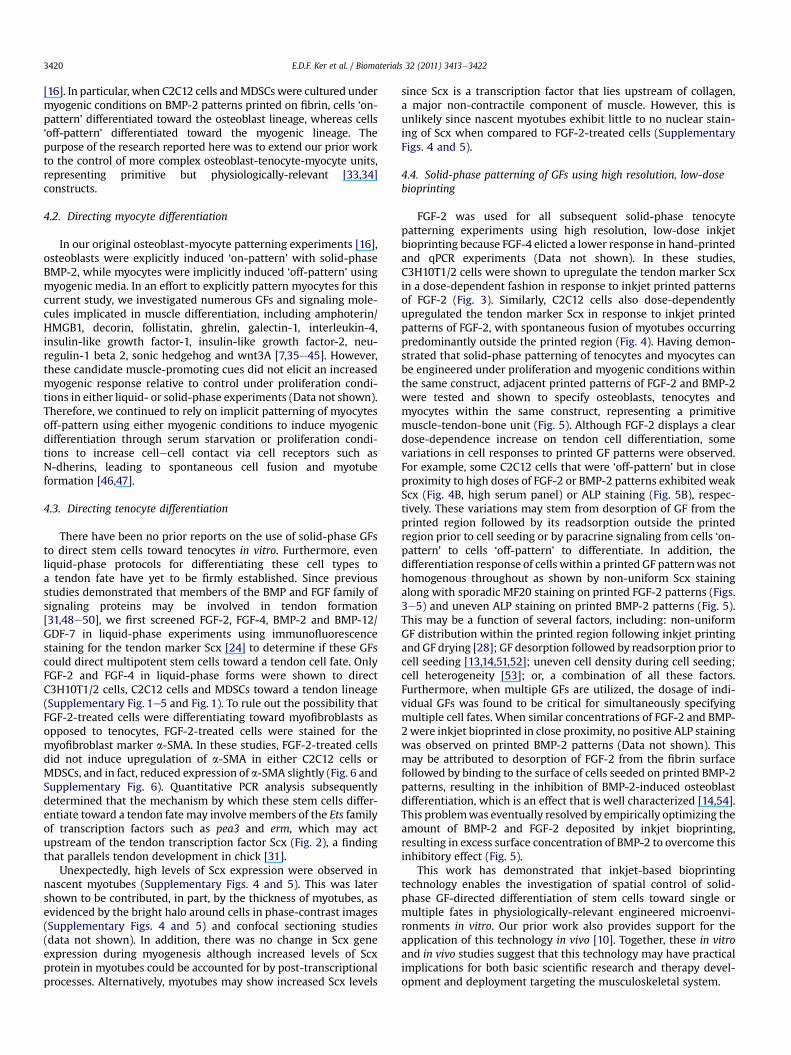
To rule out the possibility that FGF-2 was directing C2C12 cellstoward a myofibroblast fate as opposed to a tenocyte fate, FGF-2was hand-printed onto a fibrin-coated coverslip and immuno-stained for the myofibroblast marker a-SMA (Fig. 6). After 72 h inproliferation media, a-SMA, which is transiently expressed duringmuscle differentiation [32], was downregulated on hand-printed
Fig. 4. Effect of inkjet printed patterns of FGF-2 on expression of tendon marker Scx (red)myogenic media. Increasing amounts of FGF-2 dose-dependently upregulated Scx and inhidifferentiation due to high confluency levels. A. Inkjet printed patterns of FGF-2 (Black squaScx and MF20 stain. The estimated surface concentration of FGF-2 present during cell seedindicate Standard Error Mean or SEM (n ¼ 12 for proliferation media, n ¼ 16 for myogenic
patterns of FGF-2, indicating that these cells were differentiatingtoward a tenocyte fate as opposed to a myofibroblast fate.
4. Discussion
4.1. Directing osteoblast differentiation
We previously used our printing approach to demonstrate spatialcontrol of adjacent regions of osteoblast-myocyte differentiation
and muscle marker MF20 (green) in mouse C2C12 cells after 72 h in proliferation andbited myotube formation slightly. Non-printed regions showed spontaneous myotuberes). B. Scx and MF20 staining of printed patterns (White Squares). C. Quantification ofing (after washing) is shown in terms of picograms/mm2. Scale bar 1 mm. Error barsmedia). *, Significantly different from control or non-printed regions; p � 0.05.
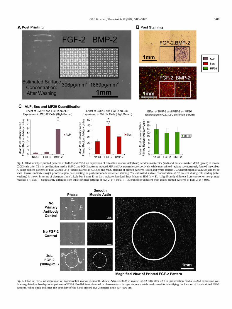
Fig. 5. Effect of inkjet printed patterns of BMP-2 and FGF-2 on expression of osteoblast marker ALP (blue), tendon marker Scx (red) and muscle marker MF20 (green) in mouseC2C12 cells after 72 h in proliferation media. BMP-2 and FGF-2 patterns induced ALP and Scx expression, respectively, while non-printed regions spontaneously formed myotubes.A. Inkjet printed patterns of BMP-2 and FGF-2 (Black squares). B. ALP, Scx and MF20 staining of printed patterns (Black and white squares). C. Quantification of ALP, Scx and MF20stain. Squares indicates inkjet printed region post-printing or post-immunofluorescence staining. The estimated surface concentration of GF present during cell seeding (afterwashing) is shown in terms of picograms/mm2. Scale bar 1 mm. Error bars indicate Standard Error Mean or SEM (n ¼ 8). *, Significantly different from control or non-printedregions; p � 0.05. þ, Significantly different from inkjet printed patterns of FGF-2; p � 0.05. þ þ, Significantly different from inkjet printed patterns of BMP-2; p � 0.05.
Fig. 6. Effect of FGF-2 on expression of myofibroblast marker a-Smooth Muscle Actin (a-SMA) in mouse C2C12 cells after 72 h in proliferation media. a-SMA expression wasdownregulated on hand-printed patterns of FGF-2. Parallel lines observed in phase-contrast images denote scratch marks used for identifying the location of hand-printed FGF-2patterns. White circle indicates the boundary of the hand-printed FGF-2 pattern. Scale bar 1000 mm.
E.D.F. Ker et al. / Biomaterials 32 (2011) 3413e3422 3419
E.D.F. Ker et al. / Biomaterials 32 (2011) 3413e34223420
[16]. In particular, when C2C12 cells andMDSCswere cultured undermyogenic conditions on BMP-2 patterns printed on fibrin, cells ‘on-pattern’ differentiated toward the osteoblast lineage, whereas cells‘off-pattern’ differentiated toward the myogenic lineage. Thepurpose of the research reported here was to extend our prior workto the control of more complex osteoblast-tenocyte-myocyte units,representing primitive but physiologically-relevant [33,34]constructs.
4.2. Directing myocyte differentiation
In our original osteoblast-myocyte patterning experiments [16],osteoblasts were explicitly induced ‘on-pattern’ with solid-phaseBMP-2, while myocytes were implicitly induced ‘off-pattern’ usingmyogenic media. In an effort to explicitly pattern myocytes for thiscurrent study, we investigated numerous GFs and signaling mole-cules implicated in muscle differentiation, including amphoterin/HMGB1, decorin, follistatin, ghrelin, galectin-1, interleukin-4,insulin-like growth factor-1, insulin-like growth factor-2, neu-regulin-1 beta 2, sonic hedgehog and wnt3A [7,35e45]. However,these candidate muscle-promoting cues did not elicit an increasedmyogenic response relative to control under proliferation condi-tions in either liquid- or solid-phase experiments (Data not shown).Therefore, we continued to rely on implicit patterning of myocytesoff-pattern using either myogenic conditions to induce myogenicdifferentiation through serum starvation or proliferation condi-tions to increase cellecell contact via cell receptors such asN-dherins, leading to spontaneous cell fusion and myotubeformation [46,47].
4.3. Directing tenocyte differentiation
There have been no prior reports on the use of solid-phase GFsto direct stem cells toward tenocytes in vitro. Furthermore, evenliquid-phase protocols for differentiating these cell types toa tendon fate have yet to be firmly established. Since previousstudies demonstrated that members of the BMP and FGF family ofsignaling proteins may be involved in tendon formation[31,48e50], we first screened FGF-2, FGF-4, BMP-2 and BMP-12/GDF-7 in liquid-phase experiments using immunofluorescencestaining for the tendon marker Scx [24] to determine if these GFscould direct multipotent stem cells toward a tendon cell fate. OnlyFGF-2 and FGF-4 in liquid-phase forms were shown to directC3H10T1/2 cells, C2C12 cells and MDSCs toward a tendon lineage(Supplementary Fig. 1e5 and Fig. 1). To rule out the possibility thatFGF-2-treated cells were differentiating toward myofibroblasts asopposed to tenocytes, FGF-2-treated cells were stained for themyofibroblast marker a-SMA. In these studies, FGF-2-treated cellsdid not induce upregulation of a-SMA in either C2C12 cells orMDSCs, and in fact, reduced expression of a-SMA slightly (Fig. 6 andSupplementary Fig. 6). Quantitative PCR analysis subsequentlydetermined that the mechanism by which these stem cells differ-entiate toward a tendon fate may involvemembers of the Ets familyof transcription factors such as pea3 and erm, which may actupstream of the tendon transcription factor Scx (Fig. 2), a findingthat parallels tendon development in chick [31].
Unexpectedly, high levels of Scx expression were observed innascent myotubes (Supplementary Figs. 4 and 5). This was latershown to be contributed, in part, by the thickness of myotubes, asevidenced by the bright halo around cells in phase-contrast images(Supplementary Figs. 4 and 5) and confocal sectioning studies(data not shown). In addition, there was no change in Scx geneexpression during myogenesis although increased levels of Scxprotein in myotubes could be accounted for by post-transcriptionalprocesses. Alternatively, myotubes may show increased Scx levels
since Scx is a transcription factor that lies upstream of collagen,a major non-contractile component of muscle. However, this isunlikely since nascent myotubes exhibit little to no nuclear stain-ing of Scx when compared to FGF-2-treated cells (SupplementaryFigs. 4 and 5).
4.4. Solid-phase patterning of GFs using high resolution, low-dosebioprinting
FGF-2 was used for all subsequent solid-phase tenocytepatterning experiments using high resolution, low-dose inkjetbioprinting because FGF-4 elicted a lower response in hand-printedand qPCR experiments (Data not shown). In these studies,C3H10T1/2 cells were shown to upregulate the tendon marker Scxin a dose-dependent fashion in response to inkjet printed patternsof FGF-2 (Fig. 3). Similarly, C2C12 cells also dose-dependentlyupregulated the tendon marker Scx in response to inkjet printedpatterns of FGF-2, with spontaneous fusion of myotubes occurringpredominantly outside the printed region (Fig. 4). Having demon-strated that solid-phase patterning of tenocytes and myocytes canbe engineered under proliferation and myogenic conditions withinthe same construct, adjacent printed patterns of FGF-2 and BMP-2were tested and shown to specify osteoblasts, tenocytes andmyocytes within the same construct, representing a primitivemuscle-tendon-bone unit (Fig. 5). Although FGF-2 displays a cleardose-dependence increase on tendon cell differentiation, somevariations in cell responses to printed GF patterns were observed.For example, some C2C12 cells that were ‘off-pattern’ but in closeproximity to high doses of FGF-2 or BMP-2 patterns exhibited weakScx (Fig. 4B, high serum panel) or ALP staining (Fig. 5B), respec-tively. These variations may stem from desorption of GF from theprinted region followed by its readsorption outside the printedregion prior to cell seeding or by paracrine signaling from cells ‘on-pattern’ to cells ‘off-pattern’ to differentiate. In addition, thedifferentiation response of cells within a printed GF patternwas nothomogenous throughout as shown by non-uniform Scx stainingalong with sporadic MF20 staining on printed FGF-2 patterns (Figs.3e5) and uneven ALP staining on printed BMP-2 patterns (Fig. 5).This may be a function of several factors, including: non-uniformGF distribution within the printed region following inkjet printingand GF drying [28]; GF desorption followed by readsorption prior tocell seeding [13,14,51,52]; uneven cell density during cell seeding;cell heterogeneity [53]; or, a combination of all these factors.Furthermore, when multiple GFs are utilized, the dosage of indi-vidual GFs was found to be critical for simultaneously specifyingmultiple cell fates. When similar concentrations of FGF-2 and BMP-2were inkjet bioprinted in close proximity, no positive ALP stainingwas observed on printed BMP-2 patterns (Data not shown). Thismay be attributed to desorption of FGF-2 from the fibrin surfacefollowed by binding to the surface of cells seeded on printed BMP-2patterns, resulting in the inhibition of BMP-2-induced osteoblastdifferentiation, which is an effect that is well characterized [14,54].This problemwas eventually resolved by empirically optimizing theamount of BMP-2 and FGF-2 deposited by inkjet bioprinting,resulting in excess surface concentration of BMP-2 to overcome thisinhibitory effect (Fig. 5).
This work has demonstrated that inkjet-based bioprintingtechnology enables the investigation of spatial control of solid-phase GF-directed differentiation of stem cells toward single ormultiple fates in physiologically-relevant engineered microenvi-ronments in vitro. Our prior work also provides support for theapplication of this technology in vivo [10]. Together, these in vitroand in vivo studies suggest that this technology may have practicalimplications for both basic scientific research and therapy devel-opment and deployment targeting the musculoskeletal system.
E.D.F. Ker et al. / Biomaterials 32 (2011) 3413e3422 3421
5. Conclusions
This report identified both liquid- and solid-phase FGF-2 asbeing capable of upregulating the tendon marker Scx in C3H10T1/2cells, C2C12 cells and MDSCs. Quantitative PCR analysis suggeststhat members of the Ets family of transcription factors such as pea3and erm may lie upstream of Scx. This report also demonstrateshow inkjet bioprinting technology can create persistent GF patternsthat direct a single stem cell population toward multiple fates,including tenocytes, myocytes or osteoblasts, within the sameconstruct in a spatially defined manner. This capability not onlyoffers an approach to study multi-lineage differentiation in vitro,but may also be translatable to new therapies to treat disease andtrauma of the musculoskeletal system.
Acknowledgments
We would like to thank James Fitzpatrick for assistance withfluorescence microscopy and Larry Schultz for assistance with GFprinting. This work was supported by NIH grants RO1EB004343and RO1EB007369 as well as funding from the PennsylvaniaInfrastructure Technology Alliance (PITA).
Appendix
Figures with essential color discrimination. Figs. 4 and 5 in thisarticle are difficult to interpret in black and white. The full colorimages can be found in the on-line version, at doi:10.1016/j.biomaterials.2011.01.036.
Appendix A. Supplementary material
Supplementary data associated with this article can be found, inthe online version, at doi:10.1016/j.biomaterials.2011.01.036.
References
[1] Scadden DT. The stem-cell niche as an entity of action. Nature 2006;441(7097):1075e9.
[2] Nelson CM, Bissell MJ. Of extracellular matrix, scaffolds, and signaling: tissuearchitecture regulates development, homeostasis, and cancer. Annu Rev CellDev Biol 2006;22:287e309.
[3] Taipale J, Keski-Oja J. Growth factors in the extracellular matrix. Faseb J1997;11(1):51e9.
[4] Canalis E. Growth factor control of bone mass. J Cell Biochem 2009;108(4):769e77.
[5] Chen D, Zhao M, Mundy GR. Bone morphogenetic proteins. Growth Factor2004;22(4):233e41.
[6] Choi YJ, Lee JY, Park JH, Park JB, Suh JS, Choi YS, et al. The identification ofa heparin binding domain peptide from bone morphogenetic protein-4 and itsrole on osteogenesis. Biomaterials 2010;31(28):7226e38.
[7] Unsicker K, Krieglstein K. Cell signaling and growth factors in development.Germany: WILEY-VCH; 2006.
[8] Campbell PG, Miller ED, Fisher GW, Walker LM, Weiss LE. Engineered spatialpatterns of FGF-2 immobilized on fibrin direct cell organization. Biomaterials2005;26(33):6762e70.
[9] Carinci P, Becchetti E, Baroni T, Carinci F, Pezzetti F, Stabellini G, et al.Extracellular matrix and growth factors in the pathogenesis of some cranio-facial malformations. Eur J Histochem 2007;51(Suppl. 1):105e15.
[10] Cooper GM, Miller ED, Decesare GE, Usas A, Lensie EL, Bykowski MR, et al.Inkjet-based biopatterning of bone morphogenetic protein-2 to spatiallycontrol calvarial bone formation. Tissue Eng Part A 2010;16(5):1749e59.
[11] Datta N, Holtorf HL, Sikavitsas VI, Jansen JA, Mikos AG. Effect of bone extra-cellular matrix synthesized in vitro on the osteoblastic differentiation ofmarrow stromal cells. Biomaterials 2005;26(9):971e7.
[12] DeCarlo AA, Whitelock JM. The role of heparan sulfate and perlecan in bone-regenerative procedures. J Dent Res 2006;85(2):122e32.
[13] Miller ED, Fisher GW,Weiss LE, Walker LM, Campbell PG. Dose-dependent cellgrowth in response to concentration modulated patterns of FGF-2 printed onfibrin. Biomaterials 2006;27(10):2213e21.
[14] Miller ED, Phillippi JA, Fisher GW, Campbell PG, Walker LM, Weiss LE. Inkjetprinting of growth factor concentration gradients and combinatorial arrays
immobilized on biologically-relevant substrates. Comb Chem HighThroughput Screen 2009;12(6):604e18.
[15] Nelson CM, Tien J. Microstructured extracellular matrices in tissue engi-neering and development. Curr Opin Biotechnol 2006;17(5):518e23.
[16] Phillippi JA, Miller E, Weiss L, Huard J, Waggoner A, Campbell P. Microenvi-ronments engineered by inkjet bioprinting spatially direct adult stem cellstoward muscle- and bone-like subpopulations. Stem Cells 2008;26(1):127e34.
[17] Ruhrberg C, Gerhardt H, Golding M, Watson R, Ioannidou S, Fujisawa H, et al.Spatially restrictedpatterning cues provided byheparin-bindingVEGF-A controlblood vessel branching morphogenesis. Genes Dev 2002;16(20):2684e98.
[18] Schultz GS, Wysocki A. Interactions between extracellular matrix and growthfactors in wound healing. Wound Repair Regen 2009;17(2):153e62.
[19] Wiradjaja F, DiTommaso T, Smyth I. Basement membranes in developmentand disease. Birth Defects Res C Embryo Today 2010;90(1):8e31.
[20] de Juan-Pardo EM, Hoang MB, Conboy IM. Geometric control of myogenic cellfate. Int J Nanomedicine 2006;1(2):203e12.
[21] Flaim CJ, Teng D, Chien S, Bhatia SN. Combinatorial signaling microenviron-ments for studying stem cell fate. Stem Cells Dev 2008;17(1):29e39.
[22] Ilkhanizadeh S, Teixeira AI, Hermanson O. Inkjet printing of macromoleculeson hydrogels to steer neural stem cell differentiation. Biomaterials 2007;28(27):3936e43.
[23] Lee YB, Polio S, Lee W, Dai G, Menon L, Carroll RS, et al. Bio-printing ofcollagen and VEGF-releasing fibrin gel scaffolds for neural stem cell culture.Exp Neurol 2010;223(2):645e52.
[24] Cserjesi P, Brown D, Ligon KL, Lyons GE, Copeland NG, Gilbert DJ, et al.Scleraxis: a basic helix-loop-helix protein that prefigures skeletal formationduring mouse embryogenesis. Development 1995;121(4):1099e110.
[25] Scott A, Sampaio A, Abraham T, Duronio C, Underhill TM. Scleraxis expressionis coordinately regulated in a murine model of patellar tendon injury. J OrthopRes 2011;29(2):289e96.
[26] Gharaibeh B, Lu A, Tebbets J, Zheng B, Feduska J, Crisan M, et al. Isolation ofa slowly adhering cell fraction containing stem cells from murine skeletalmuscle by the preplate technique. Nat Protoc 2008;3(9):1501e9.
[27] Qu-Petersen Z, Deasy B, Jankowski R, Ikezawa M, Cummins J, Pruchnic R, et al.Identification of a novel population of muscle stem cells in mice: potential formuscle regeneration. J Cell Biol 2002;157(5):851e64.
[28] Miller E. Inkjet printing of solid-phase growth factor patterns to direct cellfate [Doctor of Philosophy]. Pittsburgh: Carnegie Mellon University; 2007.
[29] Jadlowiec J, Dongell D, Smith J, Conover C, Campbell P. Pregnancy-associatedplasma protein-a is involved in matrix mineralization of human adultmesenchymal stem cells and angiogenesis in the chick chorioallontoicmembrane. Endocrinology 2005;146(9):3765e72.
[30] Jadlowiec J, Koch H, Zhang X, Campbell PG, Seyedain M, Sfeir C. Phospho-phoryn regulates the gene expression and differentiation of NIH3T3, MC3T3-E1, and human mesenchymal stem cells via the integrin/MAPK signalingpathway. J Biol Chem 2004;279(51):53323e30.
[31] Brent AE, Tabin CJ. FGF acts directly on the somitic tendon progenitorsthrough the Ets transcription factors Pea3 and Erm to regulate scleraxisexpression. Development 2004;131(16):3885e96.
[32] Springer ML, Ozawa CR, Blau HM. Transient production of alpha-smoothmuscle actin by skeletal myoblasts during differentiation in culture andfollowing intramuscular implantation. Cell Motil Cytoskeleton 2002;51(4):177e86.
[33] Clayton RA, Court-Brown CM. The epidemiology of musculoskeletal tendinousand ligamentous injuries. Injury 2008;39(12):1338e44.
[34] Yang PJ, Temenoff JS. Engineering orthopedic tissue interfaces. Tissue Eng PartB Rev 2009;15(2):127e41.
[35] Chan J, O’Donoghue K, Gavina M, Torrente Y, Kennea N, Mehmet H, et al.Galectin-1 induces skeletal muscle differentiation in human fetal mesen-chymal stem cells and increases muscle regeneration. Stem Cells 2006;24(8):1879e91.
[36] Elia D, Madhala D, Ardon E, Reshef R, Halevy O. Sonic hedgehog promotesproliferation and differentiation of adult muscle cells: involvement of MAPK/ERK and PI3K/Akt pathways. Biochim Biophys Acta 2007;1773(9):1438e46.
[37] Filigheddu N, Gnocchi VF, Coscia M, Cappelli M, Porporato PE, Taulli R, et al.Ghrelin and des-acyl ghrelin promote differentiation and fusion of C2C12skeletal muscle cells. Mol Biol Cell 2007;18(3):986e94.
[38] Georgiadis V, Stewart HJ, Pollard HJ, Tavsanoglu Y, Prasad R, Horwood J, et al.Lack of galectin-1 results in defects in myoblast fusion and muscle regener-ation. Dev Dyn 2007;236(4):1014e24.
[39] Gros J, Serralbo O, Marcelle C. WNT11 acts as a directional cue to organize theelongation of early muscle fibres. Nature 2009;457(7229):589e93.
[40] Horsley V, Jansen KM, Mills ST, Pavlath GK. IL-4 acts as a myoblast recruitmentfactor during mammalian muscle growth. Cell 2003;113(4):483e94.
[41] Kim D, Chi S, Lee KH, Rhee S, Kwon YK, Chung CH, et al. Neuregulin stimulatesmyogenic differentiation in an autocrine manner. J Biol Chem 1999;274(22):15395e400.
[42] Kishioka Y, Thomas M, Wakamatsu J, Hattori A, Sharma M, Kambadur R, et al.Decorin enhances the proliferation and differentiation of myogenic cellsthrough suppressing myostatin activity. J Cell Physiol 2008;215(3):856e67.
[43] Kocamis H, Gulmez N, Aslan S, Nazli M. Follistatin alters myostatin geneexpression in C2C12 muscle cells. Acta Vet Hung 2004;52(2):135e41.
[44] Sorci G, Riuzzi F, Arcuri C, Giambanco I, Donato R. Amphoterin stimulatesmyogenesis and counteracts the antimyogenic factors basic fibroblast growthfactor and S100B via RAGE binding. Mol Cell Biol 2004;24(11):4880e94.

E.D.F. Ker et al. / Biomaterials 32 (2011) 3413e34223422
[45] Straface G, Aprahamian T, Flex A, Gaetani E, Biscetti F, Smith RC, et al. Sonichedgehog regulates angiogenesis and myogenesis during post-natal skeletalmuscle regeneration. J Cell Mol Med 2009;13(8B):2424e35.
[46] Blau HM, Pavlath GK, Hardeman EC, Chiu CP, Silberstein L, Webster SG,et al. Plasticity of the differentiated state. Science 1985;230(4727):758e66.
[47] Goichberg P, Geiger B. Direct involvement of N-cadherin-mediated signalingin muscle differentiation. Mol Biol Cell 1998;9(11):3119e31.
[48] Edom-Vovard F, Schuler B, Bonnin MA, Teillet MA, Duprez D. Fgf4 positivelyregulates scleraxis and tenascin expression in chick limb tendons. Dev Biol2002;247(2):351e66.
[49] Hoffmann A, Pelled G, Turgeman G, Eberle P, Zilberman Y, Shinar H, et al.Neotendon formation induced by manipulation of the Smad8 signallingpathway in mesenchymal stem cells. J Clin Invest 2006;116(4):940e52.
[50] Wolfman NM, Hattersley G, Cox K, Celeste AJ, Nelson R, Yamaji N, et al. Ectopicinduction of tendon and ligament in rats by growth and differentiation factors 5,6, and7,membersof theTGF-betagenefamily. J Clin Invest1997;100(2):321e30.
[51] Morin R, Kaplan D, Perez-Ramirez B. Bone morphogenetic protein-2 binds asmultilayers to a collagen delivery matrix: an equilibrium thermodynamicanalysis. Biomacromolecules 2006;7(1):131e8.
[52] Sahni A, Odrljin T, Francis CW. Binding of basic fibroblast growth factor tofibrinogen and fibrin. J Biol Chem 1998;273(13):7554e9.
[53] Collins CA, Olsen I, Zammit PS, Heslop L, Petrie A, Partridge TA, et al. Stem cellfunction, self-renewal, and behavioral heterogeneity of cells from the adultmuscle satellite cell niche. Cell 2005;122(2):289e301.
[54] Quarto N, Wan DC, Longaker MT. Molecular mechanisms of FGF-2 inhibitoryactivity in the osteogenic context of mouse adipose-derived stem cells(mASCs). Bone 2008;42(6):1040e52.


