Embed Size (px)
Citation preview

RESEARCH ARTICLE Open Access
Enhanced efficacy of photodynamictherapy by inhibiting ABCG2 in coloncancersJu Hee Kim1, Jae Myung Park1,3*, Yoon Jin Roh1, In-Wook Kim1, Tayyaba Hasan2 and Myung-Gyu Choi1
Abstract
Background: Photodynamic therapy (PDT) contains a photosensitizing process, which includes cellular uptakeof photosensitizer and delivery of light to the target. ATP-binding cassette subfamily G2 (ABCG2) regulatesendogenous protoporphyrin levels. In human colon cancers, it is not fully examined the role of ABCG2 inporphyrin-based photodynamic therapy.
Methods: SW480 and HT29 cells were selected because they showed low and high ABCG2 expression levels,respectively. Pyropheophorbid-a (PPa) was used as a photosensitizer. Cells were exposed to a 670 nm diod laser.Cell viability and necrosi apoptosis was examined. Production level of singlet oxygen was detected with thephotomultiplier-tube s/ -based singlet oxygen detection system.
Results: SW480 cells, which expressed lower level of ABCG2, showed the higher uptake of PPa than HT-29 cells. Theuptake level of PPa was significantly correlated with the decreased cell viability after PDT. Pretreatment with a ABCG2inhibitor, Ko-143, significantly enhanced the PDT efficacy in HT29 cells compared to vehicle-pretreated cells. To confirmthe ABCG2 effect on PDT, we established ABCG2 over-expressing stable cells in SW480 cells (SW480/ABCG2).Furthermore, SW480/ABCG2 cells showed significantly decreased PDT effect compared to the control cells. The increasedor decreased cell survival was significantly correlated with the production level of singlet oxygen after PDT.
Conclusion: ABCG2 plays an important role in determining the PDT efficacy by controlling the photosensitizer effluxrate. This implies the control of ABCG2 expression may be a potential solution to enhance photosensitivity.
Keywords: Photodynamic therapy, Colon cancer, Protoporphyrin, ATP-binding cassette transporters, Photosensitizingagents
BackgroundPhotodynamic therapy (PDT) is a modality that uses aphotosensitizer that is selective to target cells, followedby exposure to a given specific light source [1, 2]. Photo-sensitizer is excited by irradiation, producing reactiveoxygen species such as singlet oxygen, other free radicalsand cause necrosis or apoptosis of the target cells [3].PDT is indicated in the treatment of various cancers and
is known to be particularly effective in basal cancer cellssuch as skin cancer [4].Pheophorbide a (PPa) is a chlorophyll-based photosen-
sitizer which is localized to the mitochondria [5]. Manystudies reported that PPa showed an anti-tumor effect invivo on human lung, liver, and skin cancer [6–8]. Irradi-ated PPa produced singlet oxygen which rapidly induceddepolarization of mitochondrial membrane potential andtumor cell death. PDT using PPa has been attempted onhepatocarcinoma, Jurkat leukemia cell, pigmented mel-anoma, and colon cancer cells [9–11]. PPa had structuralsimilarities with porphyrin which was the most com-monly used photosensitizer [12].The main problem in the chemotherapy of cancer is
that drug resistance may occur and such resistance is
* Correspondence: [email protected] Research Institute of Medical Science, The Catholic University ofKorea, Seoul, South Korea3Division of Gastroenterology, Department of Internal Medicine, Seoul St.Mary’s Hospital, The Catholic University of Korea, 222 Banpo-daero,Seocho-gu, Seoul 137-071, South KoreaFull list of author information is available at the end of the article
© 2015 Kim et al. This is an Open Access article distributed under the terms of the Creative Commons Attribution License(http://creativecommons.org/licenses/by/4.0), which permits unrestricted use, distribution, and reproduction in any medium,provided the original work is properly credited. The Creative Commons Public Domain Dedication waiver (http://creativecommons.org/publicdomain/zero/1.0/) applies to the data made available in this article, unless otherwise stated.
Kim et al. BMC Cancer (2015) 15:504 DOI 10.1186/s12885-015-1514-4

the main reason that makes cancer treatment difficult. Oneof the drug resistance mechanisms derived from the drugefflux system of cells. ATP-binding cassette subfamily G2(ABCG2) is a family of multi-drug resistance proteins,which protect cell from exogenous and endogenous toxinthrough the efflux system [13]. ABCG2 interacts with hemestructure and porphyrins and block their accumulation incells and tissues [14, 15]. Intracellular uptake of photosensi-tizer is accordingly regulated by ABC transporter superfam-ily member ABCG2. ABCG2 was regarded as the maincause of resistance to PDT. Studies with ABCG2 knock-outmice had shown the accumulation of porphyrin photosenti-zer and increase in photosensitivity compared to wild typemice, which means that the ABCG2 protein plays a majorrole in PDT efficacy [16]. Also, the overexpression ofABCG2 induced resistance to PDT by blocking the accu-mulation of photosensitizer in cell [13]. In colon cancers, astudy reported that the expression of ABCG2 correlatedwith the shortened patient survival [17].In this study, we hypothesized that ABCG2 is a target
protein in enhancing colon cancer PDT efficacy. To con-firm our hypothesis, we tested PDT effect depending onABCG2 expression level. We also checked the resultsafter the inhibition of ABCG2 with Ko-143. In order toshow that ABCG2 is directly related with PDT efficacy,we established ABCG2 overexpressing stable cells inSW480 and tested PDT efficacy in vitro. Furthermore,we checked the anti-tumor effect on xenograft modelusing cells with either low or high expression of ABCG2cell in vivo. The results from this study show a possiblecause of reduced sensitivity to photosensitizer-PDT incolon cancer cells. Inhibition of ABCG2 expression maybe a potential solution to enhance photosensitivity.
MethodsMaterialsPheophorbide a (PPa) was obtained from Frontier ScientificInc. (USA). Ko-143 and thiazolyl blue tetrazoliumbromide (MTT) were from Sigma Aldrich. Mousemonoclonal antibodies to ABCG2 and β-actin werefrom Santa Cruz (USA). Polyclonal rabbit antibody tocaspase (cleaved) was purchased from abcam (UK).Polyclonal rabbit antibody to LC3 was purchased fromCell Signaling Technology (USA).
Cell culture and in vitro photodynamic treatmentSW480, SW480/ABCG2, HT29, HCT116, LoVo, andDLD1 cells were maintained under an atmosphere of5 % CO2 in RPMI medium (Genedepot, USA) supple-mented with 10 % fetal bovine serum and 1 % penicillin-streptomicin (Genedepot, USA). Cells were incubatedwith or without PPa (0–400 nM) for 16 h at 37 °C inRPMI medium supplemented with 5 % fetal bovineserum and 1 % penicillin-streptomicin. Ko-143 (0-1uM)
were pretreated or treated for 1 h at 37 °C. The cellswere then photoirradiated using a diod laser emittingred light at 670 nm wavelength (equipment by Kuk-Je A& SL Co., Seoul, Korea). The power density at the illu-mination area was 800 mW/cm2 and total light dose was4 J/cm2. Thereafter, cells were harvested at 4 h, 8 h, and24 h, respectively.
Cell viability assayCells were cultured in 96-well culture plates (1 × 10⁴cells/well) overnight. Cells applied to PDT were incubated for4 h with 0.5 mg/ml thiazolyl blue tetrazolium bromide.Converted MTT formazan crystals were solubilizedDMSO. The absorbance at 540 nm was measured usingmicroplate reader (Biotek ELX-800 Absorbance Reader,USA).
Measurement of Singlet oxygen productionCells were incubated with or without PPa (0–400 nM)for 16 h at 37 °C, washed twice in PBS, and lysed inmethanol. Lysed cells were applied to PMT-based singletoxygen monitoring system (Physical Sciences Inc., USA).
Quantitative Real-time PCRTotal RNA was isolated with Trizol Reagent (Invitrogen,USA) and reverse-transcribed with a reverse transcrip-tion (RT)-PCR kit (Takara) according to manufacturer’sinstruction. The level of ABCG2 was measured using aMX-3000P (Stratagene, USA). SYBR green master mix(Takara, Japan) was used to quantify the mRNA expressionof ABCG2. The primers used for quantitative RT-PCR wereas follows: ABCG2 sense, 5’-TGGCTTAGACTCAAGCACAGC; ABCG2 antisense, 5’-TCGTCCCTGCTTAGACATCC-3; GAPDH sense, 5’-AGCCACATCGCTCAGACAC-3’; GAPDH antisense, 5’-GCCCAATACGACCAAATCC-3’.
Immunocytochemical assayCells were cultured on glass coverslip. Glass coverslipsof confluent cells were washed with PBS two times. Cellswere fixed for 20 min at room temperature in 4 % paraf-ormalaldehyde/PBS. After rinsing with PBS, cells wereincubated with 5%FBS/PBS for 30 min to block non-specific staining. After washing with PBS, cells were in-cubated with ABCG2 antibody in 5%FBS/PBS overnight(1:200). After washing with PBS, cells were incubatedwith secondary Alexa-488 antibody in 5%FBS/PBS for anhour. Cells were monitored by fluorescence microscopy(Axiovert 200 MAT, Zeiss, Germany).
Flow cytometryCells were applied to PDT and then incubated withAnnexin V-FITC or PI for 15 min at room temperature.Measurement of apoptosis or necrosis was done usingFACS Calibur flow cytometer (BD Bioscience, USA).
Kim et al. BMC Cancer (2015) 15:504 Page 2 of 9
Immunoblot analysisCells lysed in buffer containing 20 mM HEPES (pH 7.0),1 % Triton X-100, 150 mM NaCl, 10 % glycerol, 2 mMEGTA, 1 mM EDTA, 1 mM Glycerol 2-phosphate, 1 μg/ml leupeptin, 1 μg/μl aprotinin, 1 mM AEBSF, 50 mMNaF, and 1 mM NA3VO4. The proteins separated bySDS-PAGE were transferred to a nitrocellulose mem-brane using electrophoresis tank. After the membranewas incubated with specific antibodies, the signal wasenhanced with chemiluminesence reagents (Genedepot)and then measured by LAS-3000 Image Analysis System(Fujifilm, Japan).
Stable cell establishment in SW480 cells using retrovirusFor preparation of SW480/ABCG2, retrovirus encodingABCG2 genes were produced by using BglII/XhoI sites.HEK293T cells were transfected with pMSCV-ABCG2,pgag-pol, and pVSV-G, using Lipofectamine2000, andthen 48 h later, media including Prx III retroviruses werecollected and filtered to remove cell debris. SW480 cellswere incubated with ABCG2 retrovirus, and the cellswere inoculated with ABCG2 retrovirus. The cells ex-pressing ABCG2 were selected with puromycin.
Xenograft model and Photodynamic therapyFour-week-old male BALB/c nude mice were used forin vivo study. The animal experiments were conductedin accordance with the institutional guidelines of theCatholic University of Korea, College of medicine,Seoul, Korea. SW480 and SW480/ABCG2 cells (0.5 × 10⁶)were inoculated subcutaneously in 100 μl of phosphate-
buffered saline (PBS). The mice were then dividedinto a treatment and a control group. The treatmentgroup consisted of two subgroups: the control groupand the PDT applied group (670 nm). Animals wereinjected i.v. with Ko-143(10 mg/kg) followed by i.v.PPa after 1 h.
Statistical analysisData were expressed as means ± standard error of mean(SEM) using Student t test.
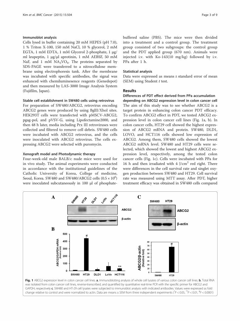
ResultsDifferences of PDT effect derived from PPa accumulationdepending on ABCG2 expression level in colon cancer cellThe aim of this study was to see whether ABCG2 is atarget protein in enhancing colon cancer PDT efficacy.To confirm ABCG2 effect in PDT, we tested ABCG2 ex-pression level in colon cancer cell lines (Fig. 1a, b). Incolon cancer cells, HT29 cell showed the highest expres-sion of ABCG2 mRNA and protein. SW480, DLD1,LOVO, and HCT116 cells showed low expression ofABCG2. Among them, SW480 cells showed the lowestABCG2 mRNA level. SW480 and HT29 cells were se-lected, which showed the lowest and highest ABCG2 ex-pression level, respectively, among the tested coloncancer cells (Fig. 1c). Cells were incubated with PPa for16 h and then irradiated with 4 J/cm2 red right. Therewere differences in the cell survival rate and singlet oxy-gen production between SW480 and HT29. Cell survivalrate was measured using MTT assay. After PDT, highertreatment efficacy was obtained in SW480 cells compared
Fig. 1 ABCG2 expression level in colon cancer cell lines. a, Immunoblotting analysis of whole cell lysates of various colon cancer cell lines. b, Total RNAwas isolated from colon cancer cell lines, reverse-transcribed, and quantified by quantitative real-time PCR with the specific primer for ABCG2 andGAPDH, respectively c, SW480 and HT-29 cell lysates were subjected to immunoblot analysis with indicated antibodies. Values were expressed as foldchange relative to control and were normalized to actin. Data are means ± SEM from three independent experiments (*P < 0.05, **P < 0.01, #P < 0.0001)
Kim et al. BMC Cancer (2015) 15:504 Page 3 of 9

to HT29 (Fig. 2a). After treating with 100 nM PPa, therewas differences of three times in phototoxicity betweenSW480 and HT29. Singlet oxygen played a main role inkilling cancer cells in PDT. We checked the singlet oxygenproduction using PMT-based singlet oxygen monitoringsystem. SW480 cell showed lower production rate of sing-let oxygen than HT29 (Fig. 2b). These results indicate thathigh expression of ABCG2 induced PPa release out of thecell by the efflux function. To further explain the effect ofABCG2 in PDT, we verified localization of ABCG2 andaccumulation of PPa using light fluorescence microscopy(Fig. 2c). SW480 cells showed no fluorescence of ABCG2,but strong red fluorescence of PPa. In contrast, HT29 cellsshowed ABCG2 fluorescence without accumulation ofPPa. These results indicate that ABCG2 is related with theresistance to PDT derived from the efflux of photosensi-tizer in colon cancer.
Enhanced efficacy of PDT by ABCG2 inhibitionThe above findings proved that ABCG2 plays a majorrole in the resistance of PDT, which could be preventedby using Ko-143, an inhibitor of ABCG2 transporter
[18]. To confirm whether the blockage of ABCG2 couldincrease the effect of PDT in colon cancer, we tested thecell survival rate and singlet oxygen production. Cellswere pretreated with 1 μM of Ko-143 for 1 h and thenincubated with PPa. There was no change in the SW480cell survival rate after the inhibition of ABCG2 (Fig. 3a).Contrastingly, HT29 cells showed decreased cell survivalrate derived from ABCG2 protection (Fig. 3a). Com-bined treatment of PPa with Ko-143 enhanced the sensi-tivity of HT29 cell to PDT. To further investigate theABCG2 inhibition effect, we measured singlet oxygenproduction of ABCG2 treated cells and non-treated cells.Singlet oxygen production rate was increased after-ABCG2 inhibition in both SW480 and HT29. SW480cells showed a little increase in the singlet oxygen, withno effect on cell survival rate. On the other hand, HT29cells treated with Ko-143 showed more singlet oxygenproduction compared to other cells (Fig. 3b). To clarifythat ABCG2 was inversely related with PPa accumula-tion, we measured fluorescence of PPa in Ko-143treatedcells using fluorescence microscope. The level of PPafluorescence in HT29 cells was remarkably increased by
Fig. 2 Differences of PDT effect between SW480 and HT29 cells depending on ABCG2 expression level. a, SW480 and HT29 cells were irradiatedwith a PDT laser (4 J/cm2) after a 16 h pretreatment of PPa at indicated concentrations. The MTT assay was performed in triplicate 24 h followingthe irradiation. b, Cells were incubated with 100 nM PPa for 16 h and then lysed with methanol, followed by application of PMT-based singletoxygen monitoring system. c, Cells were incubated with 200 nM PPa for 16 h and then subjected to immunocytochemical staining for anti-abcg2(Green), PPa (Red), and DAPI (blue). Data are means ± SEM from three independent experiments (*P < 0.05, **P < 0.01, #P < 0.0001)
Kim et al. BMC Cancer (2015) 15:504 Page 4 of 9

Ko-143 (Fig. 3c). It seems that ABCG2 inhibition in-creased the sensitivity to PDT by blocking PPa effluxand thereby inducing high level of singlet oxygen.
Effect of ABCG2 overexpressionAs mentioned above, ABCG2 plays a major role in indu-cing drug resistance and determining the efficacy ofPDT. In order to show that ABCG2 is directly relatedwith PDT efficacy, we established SW480 that stablyoverexpress ABCG2 using retrovirus. The expressionlevel of ABCG2 in SW480 and SW480/ABCG2 was con-firmed by western blot analysis and immunocytochem-istry with anti-ABCG2 antibody (Fig. 4a). SW/ABCG2cells with overexpression of ABCG2showed higher levelof survival rate compared to SW480 and HT29, whichindicates that overexpression of ABCG2 is directly re-lated with the efficacy of PDT and drug resistance(Fig. 4b). SW480/ABCG2 cells showed low fluorescenceof PPa, whereas SW480 cell showed strong signal of PPa(Fig. 4c). Also, singlet oxygen production rate was de-creased in SW480/ABCG2 cells (Fig. 5a). To investigatethe effect of PDT on cell viability, we checked for apop-tosis or necrosis using flow cytometry analysis withAnnexin V and PI. After irradiation, apoptosis and ne-crosis developed considerably in SW480. SW480 cellswith overexpression of ABCG2 showed little necrosis
while HT29 cells showed some areas of apoptosis andnecrosis (Fig. 5b). In order to verify the effect of ABCG2on PDT efficacy, we checked for the cleaved caspase-3and LC3II level. Cleaved caspase-3 is known as a markerof apoptosis and LC3II suggests autophagic cell death[19, 20]. Cleaved caspase-3 level in SW480 was furtherincreased at 24 h after PDT than SW480/ABCG2(Fig. 5c) and SW480/ABCG2 cells showed low level ofLC3II than SW480 at post-24 h PDT (Fig. 3c). Theabove data indicate that ABCG2 expression level incolon cancer cells is inversely related with PDT efficacy,and that ABCG2 inhibition could therefore increase can-cer cell death.
In vivo antitumor effects of PDTTo test for the effects of ABCG2 expression in vivo, PDTwas applied to xenograft tumor models established by sub-cutaneous injection of colon cancer cells (SW480, SW480/ABCG2). When tumor size reached150-200 mm3, PPawas administered to the tumors at the dose of1.25 mg/kg followed by irradiation with a 670 nmdiode light (150 J/cm2) 6 h later. There was nochange in size of the tumor between SW480 andSW480/ABCG2 injected mice immediately after PDT.However, at 7 days after the injection, the tumor volumeincreased by a greater extent in mice injected with
Fig. 3 Effect of Ko-143 on PPa treated colon cancer cell in PDT. a, Cells were irradiated by 670 nm light (4 J/cm2) after 16 h incubation with indicatedconcentration of PPa in the presence or absence of 1 μM Ko-143. At 24 h after PDT, the MTT assay was performed. b, Cells were incubated with 100 nMPPa for 16 h in the presence or absence of 1uM Ko-143 and then lysed with methanol, followed by application of PMT-based singlet oxygen monitoringsystem. c, Cells were incubated with 200 nM PPa for 16 h in the presence or absence of 1uM Ko-143 and then subjected to immunocytochemical stainingfor anti-abcg2 (Green), PPa (Red), and DAPI (blue). Data are means ± SEM from three independent experiments (*P < 0.05, **P < 0.01, #P < 0.0001)
Kim et al. BMC Cancer (2015) 15:504 Page 5 of 9

SW480/ABCG2 cells compared to mice injected withSW480 cells (Fig. 6a). To confirm this result, HT-29 cellsxenografted in nude mouse were treated with PBS alone,PPa + irradiation with or without Ko-143 pretreatment(Fig. 6b). As shown in cell experiments, combined treat-ment of PPa with Ko-143 enhanced the sensitivity ofHT29 cell to PDT. These results show that ABCG2 ex-pression influences the outcome of PDT in vivo.
DiscussionIn this study, we demonstrated that ABCG2 plays a mainrole in drug resistance related to PDT in colon cancercell. Our data showed that ABCG2 overexpressionstrongly induced photosensitizer efflux which means thatABCG2 inhibition may be the solution of drug resistancein colon cancer. Studies indicated that ABCG2 contrib-utes to protection of cellular accumulation of porphyrinand toxic heme components [14]. ABCG2 was first dis-covered in a breast cancer cell line, which is also calledbreast cancer resistance protein (BCRP/ABCG2) [21]. Ithas been reported that ABCG2 overexpression usingtemporarily ABCG2-transfected HEK293 cells showed
decreased cellular accumulation of PPIX, whereas otherABC-transporter overexpression (ABCB1 and ABCC1)did not effect on PPIX accumulation, which suggestedthat ABCG2 could be the main target of drug resistance[12]. In human colon cancer cell, ABCG2 is known asmain cause of resistance to drug such as SN-38 which isused for chemotherapy in colon cancer [22]. ABCG2 isimportant in developing drug resistance in PDT. How-ever, the association ABCG2 and PDT efficacy in coloncancer cell remain unclear. Our studies demonstrate thatPDT efficacy is regulated by ABCG2 expression level incolon cancer. First of all, we checked the ABCG2 ex-pression level in mRNA and protein. We found that highexpression level of ABCG2 cells showed low efficacy ofPDT which was resulted from low accumulation PPadue to ABCG2 induced efflux. Fluorescence microscopyexperiments also showed low level of PPa in ABCG2overexpressed cell. Also, ABCG2 inhibitor, Ko-143,treated cell showed increased PPa accumulation, whichinduced high PDT efficacy. Consistently, ABCG2 knock-down in HT-29 using siRNA showed increased PPa (datanot shown). PDT efficacy was correlated with ABCG2
Fig. 4 Effect of ABCG2 on the uptake of photosensitizer in SW480 and SW480/ABCG2 cells. a, Overexpression of ABCG2 in SW480 was confirmedby immunoblotting and immunocytochemical assay using ABCG2 antibody. b, MTT assay was performed in SW480 and SW480/ABCG2 cells. c, SW480and SW480/ABCG2 cells were incubated with 200 nM PPa for 16 h and then immunocytochemical assay for anti-abcg2 (Green), PPa (Red), and DAPI(blue) was applied. Data are means ± SEM from three independent experiments (*P < 0.05, **P < 0.01, #P < 0.0001)
Kim et al. BMC Cancer (2015) 15:504 Page 6 of 9

Fig. 5 Differences of singlet oxygen production rate & cell death between SW480 and SW480/ABCG2 cells. a, SW480, SW480/ABCG2, and HT29cells were incubated with 100 nM PPa for 16 h and then lysed with methanol, followed by application of PMT-based singlet oxygen monitoringsystem. b, Cells were harvested 4 h after photosensitization and stained with Annexin V/PI for FACS analysis. c, Cells were incubated with 100 nM PPafor 16 h followed by irradiation. Cell lysates were subjected to immunoblot with indicated antibodies. Data are means ± SEM from three independentexperiments (*P < 0.05, **P < 0.01, #P < 0.0001)
Fig. 6 Antitumor effects of PDT in in vivo experiments. a, SW480 and SW480/ABCG2 cells were collected and subcutaneously injected into BALB/cnude mice. The sizes of tumors were measured using a caliper, tumor volume was calculated using the formula: 0.523 × length × width2 (mm3).b, HT-29 cells xenografted in nude mouse were treated with PBS alone, PPa + irradiation with or without Ko-143 pretreatment. Combined treatment ofPPa with Ko-143 enhanced the sensitivity of HT29 cell to PDT. Data are means ± SEM from n = 4 (*P < 0.05, **P < 0.01, #P < 0.0001)
Kim et al. BMC Cancer (2015) 15:504 Page 7 of 9
expression level. SW480/ABCG2 expressing high level ofABCG2 showed no apoptosis or necrosis after PDT,whereas, SW480 cells were prone to apoptosis or necro-sis. These phenomena were also confirmed by higherlevel of cleaved caspase-3 and LC3II in SW480 cells.SW480 treated with PDT showed increased cleavedcaspase-3 and LC3II compared to SW480/ABCG2,which means that apoptotic and autophagic cell deathwas induced in SW480 cells. Tumor cell death in PDTwas induced by the release of singlet oxygen producedby photo-excitation of photosensitizer by visible light[23]. We found that singlet oxygen production was corre-lated with PPa accumulation in cells. These data suggestedthat PDT efficacy was dependent on PPa accumulationand PPa induced singlet oxygen production. According torecent report, ABCG2, drug transporter, regulates the out-come of hypericin-mediated photodynamic therapy inHT-29 [24].ABCG2 has been already found to have the main role
in porphyrin accumulation in mice. To show ABCG2directly influenced PDT efficacy, we established SW480stably overexpressing ABCG2 and also tested PDT effi-cacy using in vitro system, and then we checked theanti-tumor effect on xenograft model using cells differ-ently expressing ABCG2 in vivo studies. The tumor sizeof SW480 cells was significantly smaller in the grouptreated with PPa-PDT compared to the SW480/ABCG2injected group. These results indicate that antitumor ef-fects are dependent on ABCG2 expression level.Many researchers reported that ABCG2 inhibition had
decreased PDT efficacy through other protein regulation[25–27]. However, we directly showed ABCG2 effect incolon cancer cell using ABCG2 overexpressed cell lineboth in vitro and in vivo, providing direct evidences ofenhanced PDT effect when inhibiting the ABCG2. Inthis aspect, our research suggests a very important roleof ABCG2 in drug resistance and PDT efficacy in coloncancer cell, and provides reasonable and sufficient evi-dence to develop ABCG2 inhibitor for protecting drugresistance.
ConclusionIn summary, this study shows a possible cause of re-duced sensitivity to photosensitizer-PDT in colon cancercells. Our study provides a potential solution to enhancephotosensitivity by inhibition of ABCG2 expression.
Competing interestsThe authors declare that they have no competing interests.
Authors’ contributionsJHK carried out the all experiments, YJR carried out the immunoassays. IWKparticipated in the design of the study and performed the statistical analysis.JHK and JMP conceived of the study, and participated in its design andcoordination, and MGC and TH helped to draft the manuscript. All authorsread and approved the final manuscript.
AcknowledgmentsThis study was supported by the Global Research and Development Center.The funding of this foundation comes from the National ResearchFoundation of Korea through the Ministry of Science, ICT and FuturePlanning (NRF-2011-0031644).
FundingThis research was supported by program of Global Research and DevelopmentCenter through the National Research Foundation of Korea (NRF) funded by theMinistry of Science, ICT and Future Planning (NRF-2011-0031644).
Author details1Catholic Research Institute of Medical Science, The Catholic University ofKorea, Seoul, South Korea. 2Wellman Center for Photomedicine, Departmentof Dermatology, Massachusetts General Hospital, Harvard Medical School,Boston, MA, USA. 3Division of Gastroenterology, Department of InternalMedicine, Seoul St. Mary’s Hospital, The Catholic University of Korea, 222Banpo-daero, Seocho-gu, Seoul 137-071, South Korea.
Received: 27 December 2014 Accepted: 25 June 2015
References1. Dolmans DE, Fukumura D, Jain RK. Photodynamic therapy for cancer. Nat
Rev Cancer. 2003;3(5):380–7.2. Castano AP, Mroz P, Hamblin MR. Photodynamic therapy and anti-tumour
immunity. Nat Rev Cancer. 2006;6(7):535–45.3. Oleinick NL, Morris RL, Belichenko I. The role of apoptosis in response to
photodynamic therapy: what, where, why, and how. Photochem PhotobiolSci. 2002;1(1):1–21.
4. Choudhary S, Nouri K, Elsaie ML. Photodynamic therapy in dermatology: areview. Lasers Med Sci. 2009;24(6):971–80.
5. Tang PM, Liu XZ, Zhang DM, Fong WP, Fung KP. Pheophorbide a basedphotodynamic therapy induces apoptosis via mitochondrial-mediated pathwayin human uterine carcinosarcoma. Cancer Biol Ther. 2009;8(6):533–9.
6. Chan JY, Tang PM, Hon PM, Au SW, Tsui SK, Waye MM, et al. Pheophorbidea, a major antitumor component purified from Scutellaria barbata, inducesapoptosis in human hepatocellular carcinoma cells. Planta Med.2006;72(1):28–33.
7. Yin X, Zhou J, Jie C, Xing D, Zhang Y. Anticancer activity and mechanism ofScutellaria barbata extract on human lung cancer cell line A549. Life Sci.2004;75(18):2233–44.
8. Nakamura Y, Murakami A, Koshimizu K, Ohigashi H. Inhibitory effect ofpheophorbide a, a chlorophyll-related compound, on skin tumor promotionin ICR mouse. Cancer Lett. 1996;108(2):247–55.
9. Lee WY, Lim DS, Ko SH, Park YJ, Ryu KS, Ahn MY, et al. Photoactivation ofpheophorbide a induces a mitochondrial-mediated apoptosis in Jurkatleukaemia cells. J Photochem Photobiol B. 2004;75(3):119–26.
10. Hajri A, Wack S, Meyer C, Smith MK, Leberquier C, Kedinger M, et al. In vitroand in vivo efficacy of photofrin and pheophorbide a, a bacteriochlorin, inphotodynamic therapy of colonic cancer cells. Photochem Photobiol.2002;75(2):140–8.
11. Jin ZH, Miyoshi N, Ishiguro K, Umemura S, Kawabata K, Yumita N, et al.Combination effect of photodynamic and sonodynamic therapy onexperimental skin squamous cell carcinoma in C3H/HeN mice. J Dermatol.2000;27(5):294–306.
12. Robey RW, Steadman K, Polgar O, Bates SE. ABCG2-mediated transport ofphotosensitizers: potential impact on photodynamic therapy. Cancer BiolTher. 2005;4(2):187–94.
13. Ishikawa T, Nakagawa H, Hagiya Y, Nonoguchi N, Miyatake S, Kuroiwa T. KeyRole of Human ABC Transporter ABCG2 in Photodynamic Therapy andPhotodynamic Diagnosis. Adv pharmacol Sci. 2010;2010:587306.
14. Krishnamurthy P, Xie T, Schuetz JD. The role of transporters in cellular hemeand porphyrin homeostasis. Pharmacol Ther. 2007;114(3):345–58.
15. Wakabayashi K, Tamura A, Saito H, Onishi Y, Ishikawa T. Human ABCtransporter ABCG2 in xenobiotic protection and redox biology. Drug MetabRev. 2006;38(3):371–91.
16. Jonker JW, Buitelaar M, Wagenaar E, Van Der Valk MA, Scheffer GL, ScheperRJ, et al. The breast cancer resistance protein protects against a majorchlorophyll-derived dietary phototoxin and protoporphyria. Proc Natl AcadSci U S A. 2002;99(24):15649–54.
Kim et al. BMC Cancer (2015) 15:504 Page 8 of 9

17. Wang X, Xia B, Liang Y, Peng L, Wang Z, Zhuo J, et al. Membranous ABCG2expression in colorectal cancer independently correlates with shortenedpatient survival. Cancer Biomark. 2013;13(2):81–8.
18. Allen JD, van Loevezijn A, Lakhai JM, van der Valk M, van Tellingen O, ReidG, et al. Potent and specific inhibition of the breast cancer resistanceprotein multidrug transporter in vitro and in mouse intestine by a novelanalogue of fumitremorgin C. Mol Cancer Ther. 2002;1(6):417–25.
19. Nicholson DW, Ali A, Thornberry NA, Vaillancourt JP, Ding CK, Gallant M,et al. Identification and inhibition of the ICE/CED-3 protease necessary formammalian apoptosis. Nature. 1995;376(6535):37–43.
20. Codogno P, Meijer AJ. Autophagy and signaling: their role in cell survivaland cell death. Cell Death Differ. 2005;12 Suppl 2:1509–18.
21. Gottesman MM, Fojo T, Bates SE. Multidrug resistance in cancer: role ofATP-dependent transporters. Nat Rev Cancer. 2002;2(1):48–58.
22. Yamazaki R, Nishiyama Y, Furuta T, Hatano H, Igarashi Y, Asakawa N, et al.Novel acrylonitrile derivatives, YHO-13177 and YHO-13351, reverse BCRP/ABCG2-mediated drug resistance in vitro and in vivo. Mol Cancer Ther.2011;10(7):1252–63.
23. Mroz P, Yaroslavsky A, Kharkwal GB, Hamblin MR. Cell death pathways inphotodynamic therapy of cancer. Cancers. 2011;3(2):2516–39.
24. Sackova V, Kulikova L, Mikes J, Kleban J, Fedorocko P. Hypericin-mediatedphotocytotoxic effect on HT-29 adenocarcinoma cells is reduced by lightfractionation with longer dark pause between two unequal light doses.Photochem Photobiol. 2005;81(6):1411–6.
25. Choi BH, Ryoo IG, Kang HC, Kwak MK. The sensitivity of cancer cells topheophorbide a-based photodynamic therapy is enhanced by Nrf2 silencing.PLoS One. 2014;9(9), e107158.
26. Jendzelovsky R, Mikes J, Koval J, Soucek K, Prochazkova J, Kello M, et al.Drug efflux transporters, MRP1 and BCRP, affect the outcome of hypericin-mediated photodynamic therapy in HT-29 adenocarcinoma cells. PhotochemPhotobiol Sci. 2009;8(12):1716–23.
27. Jung KA, Choi BH, Kwak MK. The c-MET/PI3K signaling is associated withcancer resistance to doxorubicin and photodynamic therapy by elevatingBCRP/ABCG2 expression. Mol Pharmacol. 2015;87(3):465–76.
Submit your next manuscript to BioMed Centraland take full advantage of:
• Convenient online submission
• Thorough peer review
• No space constraints or color figure charges
• Immediate publication on acceptance
• Inclusion in PubMed, CAS, Scopus and Google Scholar
• Research which is freely available for redistribution
Submit your manuscript at www.biomedcentral.com/submit
Kim et al. BMC Cancer (2015) 15:504 Page 9 of 9


