Embed Size (px)
Citation preview

Enhanced Susceptibility of Nasal Polyp Tissues to Avianand Human Influenza VirusesOrnpreya Suptawiwat1, Pongsakorn Tantilipikorn2, Chompunuch Boonarkart1, Jate Lumyongsatien2,
Mongkol Uiprasertkul3, Pilaipan Puthavathana1, Prasert Auewarakul1*
1 Department of Microbiology, Faculty of Medicine Siriraj Hospital, Mahidol University, Bangkok, Thailand, 2 Department of Oto-Rhino-Laryngology, Faculty of Medicine
Siriraj Hospital, Mahidol University, Bangkok, Thailand, 3 Department of Pathology, Faculty of Medicine Siriraj Hospital, Mahidol University, Bangkok, Thailand
Abstract
Background: Influenza viruses bind and infect respiratory epithelial cells through sialic acid on cell surface. Differentialpreference to sialic acid types contributes to host- and tissue-tropism of avian and seasonal influenza viruses. Although thehighly pathogenic avian influenza virus H5N1 can infect and cause severe diseases in humans, it is not efficient in infectinghuman upper respiratory tract. This is because of the scarcity of its receptor, a2,3-linked sialic acid, in human upper airway.Expression of sialic acid can be influenced by various factors including inflammatory process. Allergic rhinitis and nasalpolyp are common inflammatory conditions of nasal mucosa and may affect expression of the sialic acid and susceptibilityto influenza infection.
Methodology/Principal Finding: To test this hypothesis, we detected a2,3- and a2,6-linked sialic acid in human nasal polypand normal nasal mucosal tissues by lectin staining and infected explants of those tissues with avian influenza viruses H5N1and seasonal influenza viruses. We show here that mucosal surface of nasal polyp expressed higher level of a2,3- and a2,6-linked sialic acid than normal nasal mucosa. Accordingly, both H5N1 avian influenza viruses and seasonal influenza virusesreplicated more efficiently in nasal polyp tissues explants.
Conclusions/Significance: Our data suggest a role of nasal inflammatory conditions in susceptibility to influenza infection,especially by avian influenza viruses, which is generally inefficient in infecting human upper airway. The increased receptorexpression may contribute to increased susceptibility in some individuals. This may contribute to the gradual adaptation ofthe virus to human population.
Citation: Suptawiwat O, Tantilipikorn P, Boonarkart C, Lumyongsatien J, Uiprasertkul M, et al. (2010) Enhanced Susceptibility of Nasal Polyp Tissues to Avian andHuman Influenza Viruses. PLoS ONE 5(9): e12973. doi:10.1371/journal.pone.0012973
Editor: Justin Brown, University of Georgia, United States of America
Received April 5, 2010; Accepted August 16, 2010; Published September 24, 2010
Copyright: � 2010 Suptawiwat et al. This is an open-access article distributed under the terms of the Creative Commons Attribution License, which permitsunrestricted use, distribution, and reproduction in any medium, provided the original author and source are credited.
Funding: This study was supported by Thailand Research Fund. O.S. and C.B. were supported by post-doctoral and research assistant grants of Faculty ofMedicine Siriraj Hospital. The sponsors of the study had no role in study design, collection, analysis, or interpretation of data, or in the writing of the report.
Competing Interests: The authors have declared that no competing interests exist.
* E-mail: [email protected]
Introduction
The viral surface protein, hemagglutinin, of influenza viruses
can bind to various types of sialic acid molecules on cell surface
glycans [1]. The sialic acid serves as main receptor for influenza
virus binding and entry into target cells. Preference to the two
major receptor types, a2,3- and a2,6-linked sialic acid, is a pivotal
difference between avian and human influenza viruses [2]. The
main target cell of H5N1 avian influenza viruses (AIVs) in humans
is type II alveolar epithelial cells [3], which express the a2,3-linked
sialic acid abundantly [4]. In contrast to alveoli, epithelia of
human upper airway express mainly a2,6-linked sialic acid and
lack a2,3-linked sialic acid [4]. Using a2,3-linked sialic acid, AIVs
including the highly pathogenic H5N1 viruses, do not infect
human upper airway efficiently. However, in vitro explants of
tissues from human nasopharynx and tonsil have been shown to be
susceptible to infection by H5N1 AIVs [5]. It is not known how
H5N1 AIV establishes infection in humans. Possibilities are direct
invasion of lung via aerosols and minimal infection in upper
airway with spreading into lung via minor aspiration. The
presence of virus in nasopharyngeal aspirates and throat swabs
suggest that infection of upper airway exists and may precede lung
infection.
Expression of sialic acid on cell surface can be affected by
multiple factors, including cellular differentiation, oncogenesis,
and inflammation [6,7]. Cell surface sialic acid was shown to affect
histamine release in allergic conditions [8]. Sialic acid content in
mucin produced by nasal mucosa can be altered by allergic
reaction [9]. Nasal polyp is a common condition caused by
chronic allergic or inflammatory process. We asked whether
mucosal surface of nasal polyps contained an altered level of sialic
acid. Because availability of suitable receptor can determine
efficiency of infection, altered levels of cell surface sialic acid may
affect the susceptibility to influenza viruses, especially for a2,3-
linked sialic acid, which is scarce in upper airway and if
upregulated may enhance susceptibility to H5N1 AIV.
Materials and Methods
Nasal polyp and mucosal tissuesSix nasal polyposis patients schedule for turbinate reduction
procedures (turbinoplasties) were recruited for the study. All
PLoS ONE | www.plosone.org 1 September 2010 | Volume 5 | Issue 9 | e12973

polyposis patients had the skin prick test positive for the common
allergens in Thailand. For the comparison group, four patients
with the diagnosis of inferior turbinate hypertrophy and scheduled
for partial inferior turbinectomies were recruited. Informed
consents were signed by the patients and the study was approved
by the Institutional Review Board of Faculty of Medicine Siriraj
Hospital. During the surgeries (polypectomies or turbinoplasties),
small pieces of tissues (polyps vs. mucosa) were cut and
immediately sent to the laboratory.
Tissue culture and viral infectionThe tissues were extensively washed and immediately placed
into culture medium (F-12K nutrient mixture with L-glutamine,
and antibiotics) (Gibco BRL, USA) in 24-well tissue culture plates.
The tissues were infected with 16106 tissue culture infectious doses
50% (TCID50) of influenza A viruses of subtypes H5N1 [A/
Thailand/3 (SP-83)/04] or H1N1 [A/Thailand/Siriraj-3/06
(H1N1)] within three hours after collection. After two hours
infection the unattached virus was removed by washing twice with
PBS. The tissues were incubated at 37uC in 5% CO2 incubator for
0, 20, 24 and 48 hours before the supernatants were collected for
virus titrating by plaque assay. The tissues were then fixed in 10%
neutral buffered formalin and processed for histological sections.
Lectin stainingTissue section were deparaffinized with xylene for 5 minutes
then sequentially hydrated with 100, 95 and 80% alcohol for 5
minutes at each step. The tissues were then blocked for non-
specific binding with 3% bovine serum albumin (Sigma, USA) in
phosphate buffer saline (PBS) for 1 hour. After discarding blocking
solution, tissues were incubated with 1 mg of FITC-conjugated
Maackia amurensis I lectin (MAA I) or Sambucus nigra lectin
SNA (Vector Laboratories, USA) in blocking solution for 1 hour at
room temperature, then washed twice with PBS and finally
counterstained with Evan’s blue for 10 minutes. The slides were
mounted and visualized under fluorescence microscopy. In some
experiments, fresh tissue were pre-digested with neuraminidase
from Clostridium perfringens (Sigma, USA) at a concentration of 1U/
ml in PBS for 1 hour at 37uC before performing paraffin section
and the lectin staining in order to confirm the specificity. Six
lectin-stained sections of nasal polyps and nasal turbinates from six
patients were counted for epithelial cells with positive and negative
staining in three fields of the slide to calculate average percentages
of sialic acid-positive cells. About 200 cells were counted in each
slide.
RNA extraction & Real time RT-PCRFresh tissues were cut into small pieces with a sterile surgical
blade. Total RNA was isolated from small quantities of tissue (1 to
10 mg) using 800 ml TRIZOL reagent (Invitrogen, USA) and then
purified using Qiagen RNAeasy kit according to the manufactur-
er’s instructions. One microgram of total RNA was reverse
transcribed with avian myeloblastosis virus reverse transcriptase
(Promega, USA) and a random hexamer. The amplification was
then performed in a Sybr green dye detection format (LightCycler;
Roche, USA). The amplification reactions contained 16Light-
Cycler Fast Start DNA Master Sybr Green dye I (LightCycler;
Roche, USA) and 0.4 mM of each forward and reverse primer.
Melting-curve analyses were performed from 65uC to 95uC. The
following primers were used to detect the expression of specific
genes: ST3GAL1 forward and reverse [10]; ST3GAL4 forward
and reverse [11]; E14134 and E14135 for glyceraldehyde-3-
phosphate dehydrogenase (GAPDH) mRNA [12]. Reactions were
performed in triplicates. All quantitations (threshold cycle [CT]
values) were normalized to that of GAPDH to generate DCT, and
the difference between the DCT value of the nasal polyp tissue and
that of the reference (nasal mucosa tissue) was calculated as DDCT.
The relative level of gene expression was expressed as 22DDCT.
Detection of viral infection in vitroParaffin-embedded, infected and non-infected control tissue
sections were deparaffinized and rehydrated. Endogenous perox-
idase activity was blocked by incubating the slides in 3% hydrogen
peroxide for 15 minutes at room temperature. Tissue sections were
treated with pre-warmed (37uC) 200 mg/mL proteinase K in TE
buffer (50 mM Tris-HCl, 10 mM EDTA, pH 8.0) for 15 minutes
at 37uC. A biotinylated anti-sense probe for in situ hybridization
was prepared by in vitro transcription from a full-length viral
nucleoprotein clone. Tissue sections and probes were heated in
hybridization buffer (50% formamide, 3X SSC, 1X Denhardt’s
solution, 200 mg/mL Yeast tRNA, 50 mM sodium phosphate
pH 7.4, and 1 mg/mL of dextran sulfate in diethyl pyrocarbo-
nate-treated water) for 10 minutes at 90uC, placed on ice for 10
minutes, then mixed and left to hybridize overnight at 37uC in
humidified chamber. The hybridization signal was developed with
conjugated Streptavidin-Horseradish Peroxidase (PIERCE, USA)
and diaminobenzidine. The sections were then counterstained
with hematoxylin.
Figure 1. Representative micrographs of nasal polyp and nasalturbinate mucosa showing distribution of a2,3- and a2,6-sialicacid. Tissue sections were stained with FITC conjugated lectin MAA I(left panel) or SNA (right panel), specific toward a2,3- or a2,6-linkedsialic acid, respectively (a). To confirm the specificity and the presenceof a2,3-linked sialic acid on nasal polyp, tissues were digested with 1U/ml of sialidase before MAA I staining. The sialidase-treated tissue (right)lost the staining signal on the apical surface, while non-treated sectionwas positive (left) (b).doi:10.1371/journal.pone.0012973.g001
Susceptibility to Influenza
PLoS ONE | www.plosone.org 2 September 2010 | Volume 5 | Issue 9 | e12973
Statistical analysisCorrelations between percentages of lectin-stained cells and
viral titers produced the same tissues were determined using
Pearson correlation analysis and linear regression analysis. All
statistical computations were performed using SPSS software
(version 16.0, SPSS Inc., Chicago, IL).
Results
Expression of sialic acid on nasal polypsInitially four nasal polyp and four nasal turbinate mucosa
samples were examined and tissue sections were stained with the
lectins. Histological examination showed normal intact nasal
mucosa in the turbinate specimens and submucosal infiltration of
eosinophils, lymphocytes, and plasma cells in the polyp specimens
typical of allergic reaction. The SNA and MAA I staining in polyp
tissues were more intense and covered more cells than that
observed in normal mucosal tissues. Both SNA and MAA I lectins
stained mucosal surface of polyps, whereas normal nasal mucosa
showed positive staining with SNA on the mucosal surface and
positive staining with MAA I in submucosal glands (Figure 1a). In
order to provide a quantitative measurement, we counted cells
with positive staining on their apical surface. While
79.93610.06% and 71.63622.21% of epithelial cells on nasal
polyps expressed a2,3- and a2,6-linked sialic acid, respectively,
only 17.94616.83% and 35.64612.51% of epithelial cells on
normal nasal mucosa expressed the receptors. These differences
are statistically significant at p,0.01 for a2,3-linked sialic acid and
p,0.05 for a2,6-linked sialic acid, by t-test. This indicates that the
a2,3-linked sialic acid, which is the receptor for AIV, is present on
mucosal surface of nasal polyps but not on normal nasal mucosa.
The lectin staining pattern of normal nasal mucosa is in agreement
with what has been previously reported [4]. In order to confirm
the expression of a2,3-linked sialic acid on the nasal polyps, we
digested fresh tissue of nasal polyp by 1U/ml sialidase before
performing the lectin staining. The sialidase digestion eliminated
the MAA I staining signal from the apical surface of the epithelial
cells confirming the specificity. Staining in basal cells was not
eliminated probably because they were not accessible to the
Figure 3. Correlation between the percentages of lectin-positive cells and the viral titers. Dot plots of percentages oflectin-positive cells versus maximum viral titers produced from thesame tissue samples show linear correlation with Pearson correlationcoefficient of 0.889 for SNA (p = 0.003) and 0.859 for MAA I (p = 0.006).The data were derived from the same experiments shown in Figure 1, 2and 4.doi:10.1371/journal.pone.0012973.g003
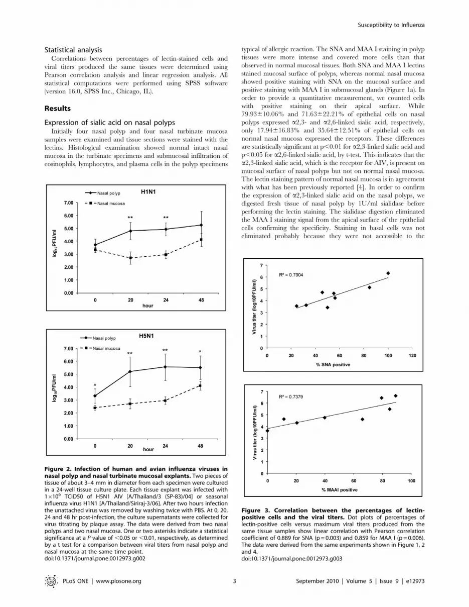
Figure 2. Infection of human and avian influenza viruses innasal polyp and nasal turbinate mucosal explants. Two pieces oftissue of about 3–4 mm in diameter from each specimen were culturedin a 24-well tissue culture plate. Each tissue explant was infected with16106 TCID50 of H5N1 AIV [A/Thailand/3 (SP-83)/04] or seasonalinfluenza virus H1N1 [A/Thailand/Siriraj-3/06]. After two hours infectionthe unattached virus was removed by washing twice with PBS. At 0, 20,24 and 48 hr post-infection, the culture supernatants were collected forvirus titrating by plaque assay. The data were derived from two nasalpolyps and two nasal mucosa. One or two asterisks indicate a statisticalsignificance at a P value of ,0.05 or ,0.01, respectively, as determinedby a t test for a comparison between viral titers from nasal polyp andnasal mucosa at the same time point.doi:10.1371/journal.pone.0012973.g002
Susceptibility to Influenza
PLoS ONE | www.plosone.org 3 September 2010 | Volume 5 | Issue 9 | e12973

enzyme as the digestion was done on an intact piece of tissue
(Figure 1b).
Infection of tissue explants by influenza virusesTo test whether the increased sialic acid expression on nasal
polyps would result in enhanced susceptibility to influenza
infection, tissue explants were infected with influenza viruses.
Tissue explants were derived from 2 nasal polyps and 2 normal
nasal mucosal tissue samples. Both the seasonal influenza virus [A/
Thailand/Siriraj-3/06 (H1N1)] and the highly pathogenic AIV
[A/Thailand/3 (SP-83)/04 (H5N1)] could infect normal nasal
mucosa explants as indicated by the increase of viral titer in the
culture supernatant. The titers of H5N1 AIV were comparable to
or even somewhat higher than those of seasonal H1N1. This may
reflect the higher replication efficiency of H5N1 as seen in various
cell lines (data not shown). Interestingly, both the seasonal
influenza and the H5N1 AIV replicated to higher titers in nasal
polyp explants (Figure 2). The maximum viral titers showed
Figure 4. Distribution of receptor and infection of nasal polyps and adjacent normal mucosa. Distribution of sialic acid receptors (a) andoutputs of viral infection (b) of tissue samples from nasal polyps and adjacent normal mucosa from the same patients. The data were derived fromexperiments using nasal polyps and adjacent normal nasal mucosal tissue samples from two patients.doi:10.1371/journal.pone.0012973.g004
Susceptibility to Influenza
PLoS ONE | www.plosone.org 4 September 2010 | Volume 5 | Issue 9 | e12973

correlation with the percentages of sialic acid-positive cells on the
same tissue samples (Figure 3).
To ensure that the difference between nasal polyps and normal
nasal mucosa was not because of normal variation among
individuals, we repeated the experiments using tissue samples
from nasal polyps and adjacent normal mucosa from the same
patients, and similar results were observed (Figure 4). This
indicated that nasal polyps were indeed more efficiently infected
by seasonal and avian influenza viruses.
We performed in situ hybridization on sections of infected and
non-infected polyp tissues. Positive hybridization signal was
observed on the mucosal surface of infected polyp, while the
non-infected polyp was negative (Figure 5). This indicates that the
infection occurred in the epithelial cells, where the viral receptor
was present, and further supports the association between the
presence of viral receptor and the infection.
Expression of ST3GAL1 and ST3GAL4 mRNAIn order to explore the mechanism of sialic acid upregulation,
we measured mRNA levels of ST3GAL1 and ST3GAL4, which
are the major enzymes responsible for adding sialic acid to
galactose through the a2,3-linkage [13]. Both of the enzymes
showed higher levels of mRNA in nasal polyp (Figure 6). This
suggests that up-regulation of the enzymes is responsible for the
observed sialic acid up-regulation.
Discussion
Previous reports showed that nasal polyp tissue explants could
be infected by influenza viruses [14,15]. The authors suggested
that nasal polyp tissue can be used as an in vitro model for
influenza infection. These reports are in agreement with our data
showing more efficient infection in nasal polyp explants as
compared to normal nasal mucosa. Furthermore, our data show
a correlation between cell surface sialic acid availability and the
efficiency of infection, suggesting that receptor availability may be
a major determinant for efficient infection in respiratory
epithelium.
In addition to sialic acid upregulation, allergic and inflamma-
tory conditions can cause a number of changes including
upregulation of various cytokines, such as IL-1, IL-4, IL-5, IL-8,
and TNF-a [16]. Whether these changes contribute to the sialic
acid upregulation and the increased efficiency of influenza
infection is not clear. NF-kB is required for influenza virus
replication [17], and it has been shown to be upregulated in nasal
polyp [18]. It is possible that upregulation of NF-kB may also
contribute to the enhanced influenza infection in the nasal polyp
tissue.
H5N1 AIV transmission in humans is inefficient [19]. Only a
small fraction of exposed individuals became infected [20].
Clusters of infected individuals are often within blood-related
individuals [21], suggesting host factors contributing to the
susceptibility to the infection. Variability of sialic acid expression
on mucosa of upper respiratory tract is likely to be one of the
factors. Here, we show that sialic acid on nasal mucosa and the
efficiency of H5N1 AIV infection can be upregulated by allergic
and inflammatory conditions and that the upregulated a2,3-linked
sialic acid can support H5N1 AIV infection [22,23,24]. This
suggests that conditions capable of upregulating a2,3-linked sialic
acid in human upper airway may contribute to susceptibility to
H5N1 AIV infection in exposed individuals. This variation in sialic
acid expression may provide an opportunity for AIVs to infect
certain individuals as entry points into human population, which
Figure 5. In situ hybridization of H5N1 AIV-infected (upper)and non-infected (lower) polyp tissues. The hybridization signal isshown in red-brown color in the epithelial cells on the mucosal surfaceof infected tissue.doi:10.1371/journal.pone.0012973.g005
Figure 6. Expression of ST3GAL1 and ST3GAL4 mRNA mea-sured by quantitative real time RT-PCR. The amount of total RNAwas normalized using GAPDH mRNA. The data are shown as the mean6 SD from an experiment done in triplicate. The data were derivedfrom two pieces of tissues from the same patient. The differencebetween mRNA expression in nasal polyp and nasal mucosa, marked byasterisks, is statistically significant (t test, P,0.01).doi:10.1371/journal.pone.0012973.g006
Susceptibility to Influenza
PLoS ONE | www.plosone.org 5 September 2010 | Volume 5 | Issue 9 | e12973

provide a chance for further adaptation, which will enable the
virus to gain full access into human population. Understanding
this may be crucial for preventing emergence of a pandemic virus.
Field evidences linking nasal polyp and increased influenza
susceptibility are lacking. However, allergic conditions such as
asthma, which is associated with allergic nasal conditions, have
been shown to be associated with upper respiratory tract infection
[22,23,24]. Most investigators consider this relationship as
triggering of asthmatic attack by viral infection. Nevertheless, the
association exists and whether allergic persons are more
susceptible to viral infection or influenza has not been fully
explored. As for nasal allergy, the signs and symptoms of the
allergy can be confused with those of viral infection, which further
complicates the observation of association between the two
conditions. Carefully designed clinical studies are needed to
explore this possible relationship.
Although our data are derived from in vitro experiments, our
experimental system involves only minimal in vitro handling of the
tissue and is likely to resemble what could happen in vivo.
Nevertheless, whether incidence of seasonal influenza and the risk
of H5N1 AIV infection in exposed individuals can be indeed
influenced by allergic conditions and levels of cell surface sialic
acid requires further exploration and clinical investigations.
Author Contributions
Conceived and designed the experiments: OS PT PA. Performed the
experiments: OS PT CB JL MU. Analyzed the data: OS PT CB JL MU PP
PA. Contributed reagents/materials/analysis tools: PP. Wrote the paper:
PA.
References
1. Suzuki Y (2005) Sialobiology of influenza: molecular mechanism of host range
variation of influenza viruses. Biol Pharm Bull 28: 399–408.2. Rogers GN, Paulson JC (1983) Receptor determinants of human and animal
influenza virus isolates: differences in receptor specificity of the H3 hemagglu-
tinin based on species of origin. Virology 127: 361–373.3. Uiprasertkul M, Puthavathana P, Sangsiriwut K, Pooruk P, Srisook K, et al.
(2005) Influenza A H5N1 replication sites in humans. Emerg Infect Dis 11:1036–1041.
4. Shinya K, Ebina M, Yamada S, Ono M, Kasai N, et al. (2006) Avian flu:
influenza virus receptors in the human airway. Nature 440: 435–436.5. Nicholls JM, Chan MC, Chan WY, Wong HK, Cheung CY, et al. (2007)
Tropism of avian influenza A (H5N1) in the upper and lower respiratory tract.Nat Med 13: 147–149.
6. Dall’Olio F, Chiricolo M (2001) Sialyltransferases in cancer. Glycoconj J 18:
841–850.7. Schauer R (2009) Sialic acids as regulators of molecular and cellular interactions.
Curr Opin Struct Biol 19: 507–514.8. Jensen C, Norn S, Skov PS, Dahl BT, Thastrup O, et al. (1987) Membrane sialic
acid influences basophil histamine release by interfering with calciumdependence. Agents Actions 20: 161–164.
9. Shimizu T, Hirano H, Shimizu S, Kishioka C, Sakakura Y, et al. (2001)
Differential properties of mucous glycoproteins in rat nasal epithelium. Acomparison between allergic inflammation and lipopolysaccharide stimulation.
Am J Respir Crit Care Med 164: 1077–1082.10. Petretti T, Kemmner W, Schulze B, Schlag PM (2000) Altered mRNA
expression of glycosyltransferases in human colorectal carcinomas and liver
metastases. Gut 46: 359–366.11. Wang PH, Lee WL, Juang CM, Yang YH, Lo WH, et al. (2005) Altered mRNA
expressions of sialyltransferases in ovarian cancers. Gynecol Oncol 99: 631–639.12. Nishimori H, Shiratsuchi T, Urano T, Kimura Y, Kiyono K, et al. (1997) A
novel brain-specific p53-target gene, BAI1, containing thrombospondin type 1repeats inhibits experimental angiogenesis. Oncogene 15: 2145–2150.
13. Kitagawa H, Paulson JC (1994) Differential expression of five sialyltransferase
genes in human tissues. J Biol Chem 269: 17872–17878.
14. Ginzburg VP, Rosina EE, Sharova OK, Ghendon YZ (1982) The replication of
influenza A viruses in organ cultures of human nasal polyps. Arch Virol 74:
293–298.
15. Ginzburg VP, Rozina EE, Sharova OK, Ghendon Yu Z (1985) Reproduction of
human and animal influenza viruses in human nasal polyp organ cultures. Acta
Virol 29: 424–427.
16. Fireman P (1996) Cytokines and allergic rhinitis. Allergy Asthma Proc 17:
175–178.
17. Kumar N, Xin ZT, Liang Y, Ly H, Liang Y (2008) NF-kappaB signaling
differentially regulates influenza virus RNA synthesis. J Virol 82: 9880–9889.
18. Valera FC, Queiroz R, Scrideli C, Tone LG, Anselmo-Lima WT (2008)
Expression of transcription factors NF-kappaB and AP-1 in nasal polyposis. Clin
Exp Allergy 38: 579–585.
19. Ungchusak K, Auewarakul P, Dowell SF, Kitphati R, Auwanit W, et al. (2005)
Probable person-to-person transmission of avian influenza A (H5N1).
N Engl J Med 352: 333–340.
20. Ortiz JR, Katz MA, Mahmoud MN, Ahmed S, Bawa SI, et al. (2007) Lack of
evidence of avian-to-human transmission of avian influenza A (H5N1) virus
among poultry workers, Kano, Nigeria, 2006. J Infect Dis 196: 1685–1691.
21. Olsen SJ, Ungchusak K, Sovann L, Uyeki TM, Dowell SF, et al. (2005) Family
clustering of avian influenza A (H5N1). Emerg Infect Dis 11: 1799–1801.
22. Lopez Perez G, Morfin Maciel BM, Navarrete N, Aguirre A (2009)
Identification of influenza, parainfluenza, adenovirus and respiratory syncytial
virus during rhinopharyngitis in a group of Mexican children with asthma and
wheezing. Rev Alerg Mex 56: 86–91.
23. Miller EK, Griffin MR, Edwards KM, Weinberg GA, Szilagyi PG, et al. (2008)
Influenza burden for children with asthma. Pediatrics 121: 1–8.
24. Fasano MB Combined airways: impact of upper airway on lower airway. Curr
Opin Otolaryngol Head Neck Surg 18: 15–20.
Susceptibility to Influenza
PLoS ONE | www.plosone.org 6 September 2010 | Volume 5 | Issue 9 | e12973


