Embed Size (px)
Citation preview

Enhancement of Radiation-Induced RegrowthDelay by Gemcitabine in a Human Tumor
Xenograft ModelMarion A. Joschko, Ph.D.,1 Lorraine K. Webster, Ph.D.,1*
Janice Groves, DIP. APPL. SCI.,1 Kally Yuen, M.Sc.,4Manuela Palatsides, B.Sc.,1 David L. Ball, M.B.B.S., F.R.A.C.R.,3 and
Michael J. Millward, M.B.B.S., F.R.A.C.P.21Experimental Chemotherapy and Pharmacology Unit, Trescowthick Research Laboratories,
Peter MacCallum Cancer Institute, Melbourne, Victoria, Australia2Division of Haemotology and Medical Oncology, Peter MacCallum Cancer Institute,
Melbourne, Victoria, Australia3Division of Radiation Oncology, Peter MacCallum Cancer Institute, Melbourne,
Victoria, Australia4Statistical Centre, Peter MacCallum Cancer Institute, Melbourne, Victoria, Australia
SUMMARY Gemcitabine, a cytidine nucleoside analogue, has schedule-dependent an-titumor activity in vitro and in vivo. Gemcitabine also has dose- and time-dependentradiosensitization properties in vitro. Thus it may have therapeutic application in combi-nation with radiation. The aims of this study were to investigate whether gemcitabinecould enhance radiation-induced tumor regrowth delay in a human squamous carcinoma(FaDu) xenograft in nude mice and to examine the effect of gemcitabine on radiation-induced apoptosis in in vivo tumors. Radiation was given locally to the tumors twice dailyin 2 Gy fractions over 2 weeks for 5 days/week. Significant regrowth delay enhancementwas observed which was dependent on gemcitabine schedule. Effective schedules usingmaximum tolerated gemcitabine doses were twice weekly and once weekly, but not daily.Significant toxicity occurred with radiation plus twice weekly gemcitabine, but enhance-ment was seen using gemcitabine doses well below the maximum tolerated dose. Bothgemcitabine and radiation led to apoptotic cell death, but this was not increased when bothtreatments were combined. These results indicate that gemcitabine may be of therapeuticvalue as a radiation enhancer in the treatment of human cancers. Preliminary studiessuggest that increased apoptotic cell death is not a mechanism leading to this enhance-ment. Radiat. Oncol. Invest. 5:62–71, 1997. © 1997 Wiley-Liss, Inc.
Key words: gemcitabine; radiosensitizer; xenograft; squamous carcinoma
INTRODUCTIONThe new pyrimidine antimetabolite gemcitabine(28,28-difluorodeoxycytidine, dFdC, Gemzart, EliLilly Australia Pty. Ltd., Sydney, Australia) hasshown antitumor activity in vitro and in humantumor xenografts that is superior to cytosine arabi-
noside (ara-C), a nucleoside analogue with a simi-lar structure [1]. Both gemcitabine and ara-C in-hibit cellular proliferation in the S-phase of the cellcycle and result in accumulation at G1S [1]. Likeara-C, gemcitabine requires activation by intracel-lular phosphorylation to the 58 triphosphate deriva-
Contract grant sponsor: National Health and Medical Research Council of Australia; Contract grant sponsor: Anti-CancerCouncil of Victoria.
*Correspondence to: Lorraine K. Webster, Ph.D., Trescowthick Research Laboratories, Peter MacCallum Cancer Insti-tute, Locked Bag No. 1, A’Beckett Street, Melbourne, Victoria 3000, Australia. E-mail: [email protected]
Received 4 December 1996; Revised 17 April 1997; Accepted 17 April 1997
© 1997 Wiley-Liss, Inc.
Radiation Oncology Investigations 5:62–71 (1997)

tive (dFdCTP), which can be incorporated into bothDNA and RNA, resulting in inhibition of DNAsynthesis [2]. Compared to the triphosphate ofara-C (ara-CTP), dFdCTP has higher intracellularconcentration and prolonged intracellular retentionin solid tumor cell lines [2–4].
Gemcitabine has broad-spectrum antitumoractivity against murine solid tumors and humantumor xenografts including lung, breast, colon,and oropharyngeal carcinomas [1,5]. In xeno-grafts derived from carcinomas of the head andneck, equitoxic doses of gemcitabine were moreactive than cisplatin, methotrexate, 5-fluoroura-cil, and cyclophosphamide [5]. The antitumoractivity of gemcitabine was schedule-dependentin in vivo models, with a 3-day interval betweeninjections being superior to daily or weekly injec-tions [6].
Early clinical trials of gemcitabine evaluateddifferent schedules and demonstrated schedule-dependent toxicity. The maximum tolerated dose(MTD) was substantially higher when gemcitabinewas given weekly or every 2 weeks, compared totwice weekly or daily administration [7]. Phase IItrials have principally used a weekly schedule, andantitumor activity has been demonstrated against anumber of tumors including non-small cell lungcancer, breast cancer, pancreatic cancer, and squa-mous cell cancer of the head and neck [7–9]. Theuse of a twice weekly schedule did not increase theantitumor activity against non-small cell lung can-cer [10].
Other antimetabolites, such as 5-fluorouracil,have been shown to act as radiation sensitizers inboth preclinical models and in clinical trials [11]. Invitro studies have examined the potential for gem-citabine to enhance the effect of radiation. Admin-istration of gemcitabine prior to and concurrentlywith radiation resulted in effective killing of EMT6mouse mammary tumor cells in vitro, but whetheror not the combination was synergistic could not bedetermined due to additional delayed toxicity asgemcitabine appeared to be released from dyingcells during clonogenic survival assays [12]. UsingHT29 human colon carcinoma cells in vitro, radia-tion sensitization by non-cytotoxic doses of gem-citabine was observed which was dose- and time-dependent, with maximal effect when cells wereirradiated immediately following gemcitabine ex-posure instead of prior to, or in middle of, drugtreatment [13]. Depletion of cellular dATP was acritical event in producing radiation sensitizationby gemcitabine [13].
Apoptosis is a distinctive mode of cell deathwith specific morphological features that distin-
guish it from necrosis. Apoptotic cell death can beinduced by cytotoxic drugs including cisplatin andpaclitaxel [14,15], and in murine tumors usinglarge single doses of radiation [16]. In vitro, gem-citabine and other nucleoside analogues includingara-C can trigger apoptotic cell death in humanleukemia cells [17,18]. Paclitaxel, when given priorto a single dose of radiation, can potentiate radia-tion-induced apoptotic cell death in murine tumorsin vivo, with the effect being dependent on theinterval between paclitaxel and radiation [19]. Incontrast, although the administration of cisplatinprior to radiation resulted in sensitization of a mu-rine mammary carcinoma in vivo, no potentiationof apoptosis by cisplatin was observed [20]. Singlefractions of radiation are rarely used for the treat-ment of human cancers, however, and whether in-creased apoptosis is a true consequence of a drug-radiation interaction needs to be examined usinglower radiation doses.
While studies have examined the combinationof gemcitabine and radiation in vitro [12,13], thishas not been investigated in human tumors in vivo.Drug-radiation studies using multiple radiationfractions reflect clinical protocols better than ex-periments using single large fractions, and there-fore these studies are helpful in the design of clini-cal trials combining cytotoxics and radiation, and todirect studies to appropriate mechanistic issues. Wehave therefore investigated the potential of gem-citabine in clinically relevant schedules to enhancethe radioresponse of a human squamous carcinomaxenograft model. In addition, apoptosis was exam-ined in tumors removed at selected times aftertreatment with gemcitabine, radiation, or both treat-ments.
MATERIALS AND METHODS
Animals
All animal procedures were approved by the insti-tutional Animal Experimentation Ethics Commit-tee. Female Balb C (nu/nu) athymic nude mice 5–6weeks old were housed in isolated, aseptic condi-tions and given free access to sterile food and wa-ter. During the 2-week irradiation period, the micewere nutritionally supplemented with Ensure (Ab-bott Laboratories, Abbott Park, IL), liquefied insterile water. Immunosuppression was ensured forthe duration of the experiment by exposing themice to 5 Gy whole body irradiation using a labo-ratory 137Cs irradiator 24 hr prior to tumor inocu-lation.
Joschko et al.: Gemcitabine Radiation Enhancement in Mice 63

Tumor Source and Inoculation
The FaDu cell line derived from the hypopharynx(American Type Culture Collection, Rockville,MD), propagated in vitro, was used as the tumorsource. Tumors removed from the mice were con-firmed to be of human origin by DNA extraction,and were characterized histologically as a poor tomoderately differentiated squamous carcinoma.Cells were thawed and maintained until confluentin RPMI with 10% fetal calf serum and 0.05%gentamicin. After disaggregation with pronase, 4 ×10⁶ cells in 50ml phosphate buffered saline (PBS)were injected subcutaneously into the right lateralthigh. Ten days later, mice with tumor volumes inthe range 250–400 mm3 were randomized for ex-periments. Each experiment consisted of groups of5–12 mice that were treated with either 1) vehiclecontrol, 2) gemcitabine alone at specified schedulesand doses, 3) radiation alone, or 4) combination ofgemcitabine and radiation.
Irradiation
A 250 kV orthovoltage X-ray unit [half-valuethickness (HVT) 0.5 mm Cu, dose rate 1 Gy/min]was used to irradiate the mice. Unanesthetized micewere restrained in a specially designed apparatusthat exposed only the tumor-bearing limb to theX-ray beam. The tumors were exposed to 40 Gy in20 fractions of 2 Gy twice daily, on days 1–5 and8–12. In the groups receiving drug plus radiation,the first daily irradiation occurred within 30 minafter gemcitabine injection, and was followed 6–7hr later by the second radiation exposure. Pilotstudies in our laboratory confirmed that the micewould tolerate this fractionated radiation schedule
with no local toxicity. No tumors were cured byradiation alone.
Drug Doses and Schedules
Gemcitabine hydrochloride (Eli Lilly Australia Pty.Ltd.) was dissolved in sterile saline and stored at4°C until use. Mice were briefly anesthetized, andgemcitabine diluted to the appropriate concentra-tion was injected intravenously at 10 ml/kg throughthe retro-orbital sinus, within 30 min prior to irra-diation. The MTD, defined as the highest dose re-sulting in less than a 10% reduction in body weight,was determined for each gemcitabine schedule innon-tumor-bearing Balb C nude mice. An addi-tional weight loss of approximately 5% has beenobserved in previous experiments in mice exposedto radiation together with cytotoxic agents [21].Equitoxic (MTD) doses were used in scheduleswith known antitumor activity in previous studieswith xenografts [5]. The schedules and doses ofgemcitabine are listed in Table 1. The experimentswere performed on two separate occasions, with thedaily, twice weekly at 160 mg/kg, and once weeklydone together, and the remaining two twice weeklydoses studied at another time with all the appropri-ate control groups. The two lower doses of 100 and50 mg/kg in the twice weekly schedule were inves-tigated after this schedule was found to be unac-ceptably toxic at the MTD when combined withradiation.
Determination of Tumor Size
Tumor size was measured twice weekly with digi-tal calipers (Maxcal, Extech Pty. Ltd., Melbourne,Australia). The tumor volumes were computed us-ing the formula V4 0.5 (L × W2), where V, L, and
Table 1. Schedules and Doses of Gemcitabine Given With and Without Concurrent TwiceDaily Radiation
Schedule Injection daysGemcitabine dose(mg/kg/injection)
Total gemcitabinedose (mg/kg)
Meanweight
loss (%)Toxicdeaths
Vehicle control — — — — —
Radiation alonea — — — 2 0/10
Daily 1–5, 8–12 2.3 (MTD) 23 2 0/10Daily + radiation 1–5, 8–12 2.3 23 10 1/10Once weekly 1, 8 430 (MTD) 860 2 1/10Once weekly + radiation 1, 8 430 860 11 0/10Twice weekly 1, 4, 8, 11 160 (MTD) 640 2 0/10Twice weekly + radiation 1, 4, 8, 11 160 640 23 6/10Twice weekly 1, 4, 8, 11 100 400 2 0/9Twice weekly + radiation 1, 4, 8, 11 100 400 19 4/12Twice weekly 1, 4, 8, 11 50 200 3 0/5Twice weekly + radiation 1, 4, 8, 11 50 200 22 2/9
aData from first set of experiments.
64 Joschko et al.: Gemcitabine Radiation Enhancement in Mice

W were the volume, length, and width of the twoperpendicular diameters, respectively. The volumeswere then expressed as a percentage of the pretreat-ment volume on day 1, which was designated as100%.
ApoptosisApoptosis was assessed by microscopic examina-tion [16,20] of tumors at two time points on the firstday in the once weekly schedule. The mice re-ceived either 430 mg/kg gemcitabine at time zero,or 2 Gy irradiation at 0.5 and 7.5 hr, or the com-bination of gemcitabine and radiation. At 8 and 24hr, 2 or 3 mice per group were sacrificed with flu-othane, and the tumors were immediately excisedand placed in PBS. The tumors were cut into#1 mm cubed pieces and fixed in 2% glutaralde-hyde 0.08 M Sorensen’s phosphate buffer, pH 7.4.The tissue was subsequently postfixed in 2% OsO4
0.1 M Sorensen’s phosphate buffer, pH 7.4. Thetissue was dehydrated and embedded in Spurr’sresin. Semithin sections (0.70mm) were cut on anUltracut E and stained with toluidine blue andazure II. Apoptosis was scored blind for each slideby microscopic examination at ×400 magnification.Between 5 and 10 non-necrotic fields were ran-domly selected for each treatment and the numbersof apoptotic nuclei per cell remnants were based onscoring approximately 200 cells per field. Thenumber of aberrant nuclei within each field wascounted and expressed as a percentage of totalnumber of cells. The percentage of aberrant nucleiwas analyzed for significant differences betweeneach treatment group.
Statistical MethodsFor each mouse, the day at which the tumorreached 200% of its initial size was determined bylinear interpolation of observed points on itsgrowth curve. Because of the presence of censoreddata, due to the death of some mice before thetumor reached 200% of initial size, the Kaplan-Meier product limit method was used to estimatethe median time to reach 200% of the initial tumorsize for each group. Treatment groups were com-pared using the log-rank test.
The absolute tumor growth delay (ATGD)was calculated as the difference between the esti-mated median times required for the vehicle controlgroup and each treatment group to reach 200% oftheir pretreatment volumes [20–22]. The expectedtumor growth delay (ETGD) under an additivemodel for each gemcitabine plus radiation groupwas determined by adding the ATGD in the gem-citabine alone and radiation alone groups [23]. TheATGD and the ETGD were compared using normal
approximations. Normalized tumor growth delay(NTGD) was defined as the ATGD in the gem-citabine plus radiation group minus the ATGD inthe gemcitabine alone group [20,22], thus provid-ing an estimate of the tumor growth delay (TGD)induced by the combined treatment without thecontribution of the drug. The regrowth delay en-hancement was defined as the ratio of NTGD forcombined drug and radiation over ATGD for radia-tion alone [20].
Comparisons of treatment groups at eachtime point in the apoptosis study were carriedout using the Kruskal-Wallis one-way analysisof variance test, adjusting for multiple compari-sons. The percentage of apoptotic cells in eachtreatment group was estimated by averaging thepercentages of apoptotic cells over all the non-necrotic fields.
BMDP statistical software was used [24]. Allstatistical tests performed were two-sided and re-sults withP < 0.05 were considered significant.
RESULTS
Growth Delay Studies
Figure 1 shows mean tumor volumes, indicatinggroup trends for each treatment, while Table 2gives the estimated median times to reach 200% ofinitial volumes for the calculation of growth delayand regrowth delay enhancement.
Vehicle Controls and RadiationAlone Groups
The estimated median time to reach 200% of initialtumor volume in the vehicle control groups rangedfrom 4 to 7 days. There was no difference in thetime to reach 200% of the initial tumor volume inthe four vehicle control groups studied (P 4 0.23).Localized radiation alone, in the first set of experi-ments, resulted in a decline of the mean tumor vol-ume to approximately 65% of the pretreatment vol-ume by day 18 (Fig. 1). The median time [± stan-dard error (S.E.)] to regrowth to 200% of the initialvolume was 43 ± 2 days (Table 2). In the second setof experiments, evaluating the two lower gemcitab-ine doses in the twice weekly schedule, the mediantime for regrowth to 200% for radiation alone was33 ± 3 days (P 4 0.13, comparison of the tworadiation alone groups). The radiation dose andschedule used in these studies have not led to FaDutumor cures in this or any previous studies per-formed in our laboratory.
Joschko et al.: Gemcitabine Radiation Enhancement in Mice 65

Daily Schedule
The median times to grow to 200% of initial vol-ume in the daily gemcitabine alone and vehiclecontrol groups were 5 ± 2 and 6 ±1days, respec-tively (P 4 0.42), showing that this schedule ofgemcitabine was not cytotoxic to the FaDu tumor.In the daily gemcitabine plus radiation group, ini-tial growth was similar to the control, with a rapidincrease in the mean tumor volume to approxi-mately 190% by day 2. However, the mean tumorvolume decreased to 35% of initial volume by day25, prior to regrowth to 200% (Fig. 1A). There wasno evidence of an enhanced effect on tumor re-growth delay by combining gemcitabine in thisschedule with radiation, as the ATGD after com-bined treatment (58 ± 20 days) was not signifi-cantly different from the ETGD (37 ± 3 days)(P 4 0.28). The regrowth delay enhancement was1.6 ± 0.5.
Once Weekly Schedule
The median times to grow to 200% of initial vol-ume in the weekly gemcitabine alone and the ve-hicle control groups were 13 ± 1 and 7 ± 2days,respectively (P < 0.0001). This result suggests thatgemcitabine given in this schedule at the MTD pro-duced significant antitumor activity. When com-bined with radiation, this schedule of gemcitabinecaused tumor regression to a mean volume of ap-proximately 2% by day 25 before regrowth to200% (Fig. 1B). Gemcitabine in this schedule didenhance the effect of radiation, as the ATGD aftercombined treatment (100 ± 25 days) was signifi-cantly greater than the ETGD (42 ± 4 days)(P 4 0.022). The regrowth delay enhancement was2.6 ± 0.7.
Twice Weekly Schedule
The median times to grow to 200% of initial vol-ume for twice weekly gemcitabine alone at theMTD and the vehicle control groups were 16 ± 2and 7 ± 1days, respectively (P 4 0.0015; Table 2),suggesting that gemcitabine alone in this schedulehas some antitumor activity. There was an en-hanced effect by combining gemcitabine with ra-diation, as the ATGD after combined treatment(130 ± 17 days) was significantly greater than theETGD (46 ± 3 days) (P < 0.0001). The regrowthdelay enhancement was 3.3 ± 0.5. No unexpectedtoxicity was observed when gemcitabine alone wasgiven twice weekly for 2 weeks at the MTD of 160mg/kg. However, when this dose and schedule of
Fig. 1. Effect of gemcitabine combined with acceleratedfractionated irradiation on the growth of FaDu tumors. Micewith tumor volumes of 250–400 mm3 (100% on day 1) weregiven vehicle only (h), or local irradiation to the tumor-bearing thigh (m), or gemcitabine alone (d), or irradiationplus gemcitabine (j). Gemcitabine doses were(A) daily at2.3 mg/kg/injection;(B) once weekly at 430 mg/kg/injection; or(C) twice weekly at 50 mg/kg/injection. Pointsare mean − S.E.M. for 5–10 mice.
66 Joschko et al.: Gemcitabine Radiation Enhancement in Mice
gemcitabine was combined with radiation, therewere 6/10 (60%) toxic deaths within the first 3weeks.
Two lower doses of 100 and 50 mg/kg (Fig.1C) were also investigated in this twice weeklyschedule. As with the MTD, there was a significantdelay in time to reach 200% of initial volume whencompared to the vehicle control group (Table 2;P4 0.0049 andP 4 0.0040 for doses of 100 and50 mg/kg, respectively). In addition, the time toreach 200% of the initial volume in the gemcita-bine alone group for each of these doses wasshorter than that required for the MTD, suggestinga dose-related effect of drug alone (P 4 0.065,160 vs. 100 mg/kg;P 4 0.0094, 160 vs. 50 mg/kg).These lower doses in combination with radia-tion were apparently less toxic than the MTD,with 4/12 (33%) toxic deaths at 100 mg/kg and2/9 (22%) toxic deaths at 50 mg/kg. The radia-tion effect was enhanced by gemcitabine inthis schedule at both the 100 and 50 mg/kg doses,as the ATGD after combined treatment was signifi-cantly greater than the ETGD (P < 0.0001 for bothdoses; Table 2). The regrowth delay enhancementwas 2.2 ± 0.3 at 100 mg/kg and 2.4 ± 0.3 at 50mg/kg.
Weight Loss
In the vehicle control mice, and in the animalstreated with either radiation alone or gemcitabinealone in any schedule, weight loss was at most 3%(Table 1). When gemcitabine was combined withradiation, there was apparently greater weight lossin all schedules. However, the increase appeared tobe more marked in the twice weekly schedule, evenat doses below the MTD.
Apoptosis
The magnitude of apoptotic induction in FaDu tu-mors 8 or 24 hr after a single dose of gemcitabine(430 mg/kg), or two 2 Gy radiation fractions, orboth treatments combined is shown in Table 3. Thebackground number of apoptotic cells in untreatedtumors constituted less than 2% of cells at bothtime points, and the number of aberrant nuclei inthe radiation only group 8 hr after the first 2 Gyfraction was no different to values in untreated tu-mors. There was an increase at 24 hr, but this wasnot statistically significant. Tumors exposed to asingle dose of gemcitabine or gemcitabine plus ra-diation showed significantly more apoptotic cells ineach group at both time points compared with un-treated tumors (P < 0.05, all comparisons), and this
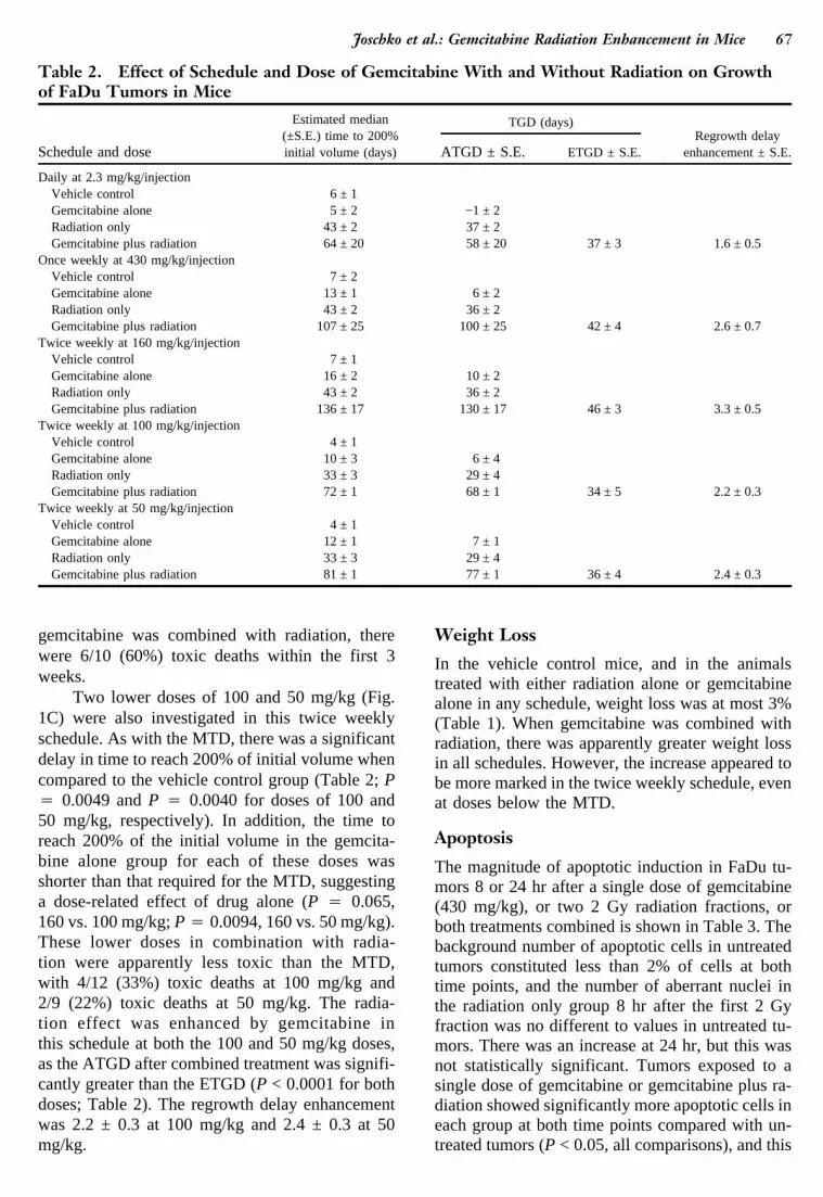
Table 2. Effect of Schedule and Dose of Gemcitabine With and Without Radiation on Growthof FaDu Tumors in Mice
Schedule and dose
Estimated median(±S.E.) time to 200%initial volume (days)
TGD (days)Regrowth delay
enhancement ± S.E.ATGD ± S.E. ETGD ± S.E.
Daily at 2.3 mg/kg/injectionVehicle control 6 ± 1Gemcitabine alone 5 ± 2 −1 ± 2Radiation only 43 ± 2 37 ± 2Gemcitabine plus radiation 64 ± 20 58 ± 20 37 ± 3 1.6 ± 0.5
Once weekly at 430 mg/kg/injectionVehicle control 7 ± 2Gemcitabine alone 13 ± 1 6 ± 2Radiation only 43 ± 2 36 ± 2Gemcitabine plus radiation 107 ± 25 100 ± 25 42 ± 4 2.6 ± 0.7
Twice weekly at 160 mg/kg/injectionVehicle control 7 ± 1Gemcitabine alone 16 ± 2 10 ± 2Radiation only 43 ± 2 36 ± 2Gemcitabine plus radiation 136 ± 17 130 ± 17 46 ± 3 3.3 ± 0.5
Twice weekly at 100 mg/kg/injectionVehicle control 4 ± 1Gemcitabine alone 10 ± 3 6 ± 4Radiation only 33 ± 3 29 ± 4Gemcitabine plus radiation 72 ± 1 68 ± 1 34 ± 5 2.2 ± 0.3
Twice weekly at 50 mg/kg/injectionVehicle control 4 ± 1Gemcitabine alone 12 ± 1 7 ± 1Radiation only 33 ± 3 29 ± 4Gemcitabine plus radiation 81 ± 1 77 ± 1 36 ± 4 2.4 ± 0.3
Joschko et al.: Gemcitabine Radiation Enhancement in Mice 67

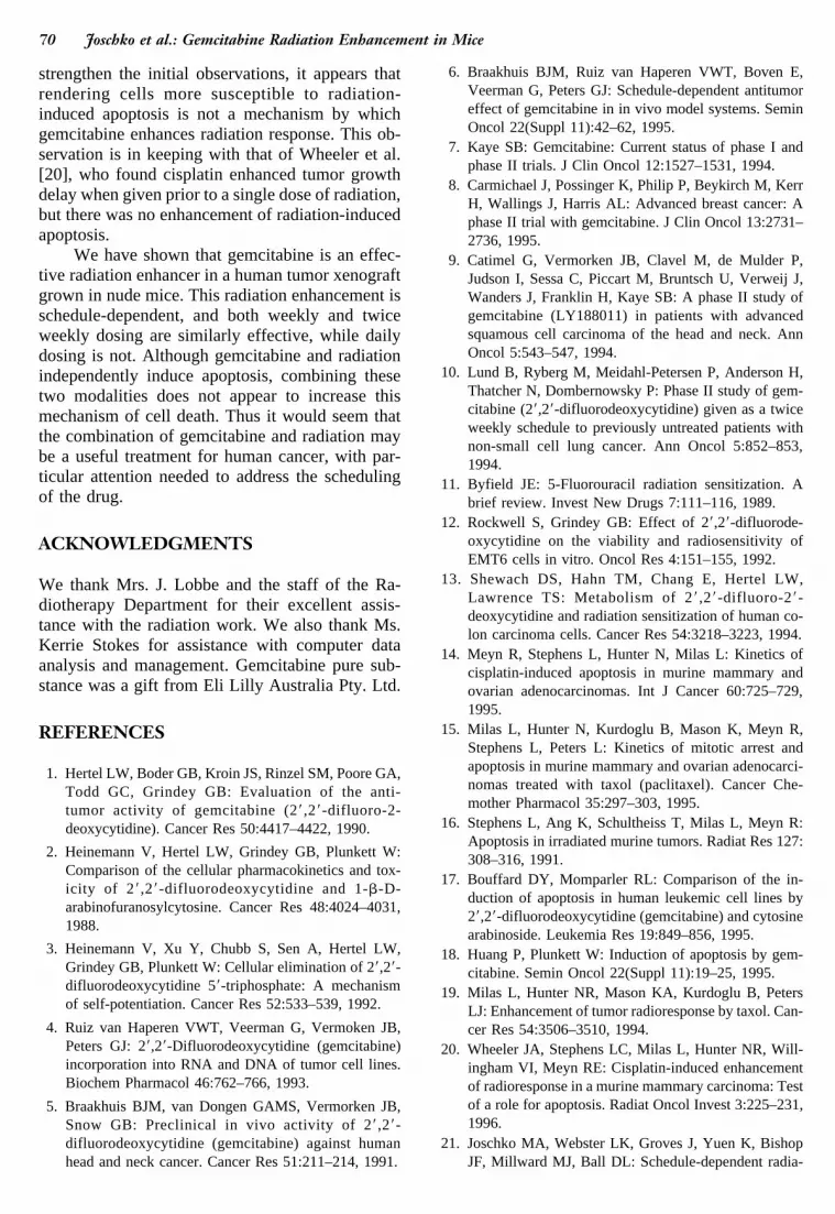
appeared to be greatest at 24 hr. No enhancement inapoptotic cell numbers was observed, however, inthe combined treatment group. While a range ofchanges in cell morphology typical of apoptoticcells was observed, such as cell shrinkage, nuclearcondensation and margination, fragmentation ofnuclei, and rupture of affected cells into debris,most apoptotic cells present at the two time pointsdemonstrated late effects such as vacuolation ofnuclear remnants within neighboring phagocytos-ing cells (Fig. 2).
DISCUSSION
In this study we showed that gemcitabine hasschedule-dependent antitumor activity in a humansquamous cell carcinoma xenograft. Significant an-titumor activity was observed with the twiceweekly and weekly schedule, while daily gemcitab-ine was inactive. These observations confirm pre-vious reports in other tumor models that maximalin vivo activity of gemcitabine occurs with an ev-ery third day (twice weekly) schedule rather thanwith 5 daily doses [5,25]. In our model, the MTDsfor gemcitabine given on the twice weekly anddaily schedule were similar to those reported pre-viously [5], but the MTD for the weekly schedule(430 mg/kg) was higher. This may explain thegreater activity seen with the weekly schedule thanreported by Braakhuis et al. [5] using an MTD of240 mg/kg. Recently, the use of prolonged subcu-taneous or intravenous infusions of gemcitabinehas been evaluated and reported to be superior tobolus gemcitabine schedules against in vivo murinecolon carcinomas [26]. This schedule of gemcita-bine combined with radiation also deserves inves-tigation.
This is the first study that combines gemcita-bine with radiation in an in vivo human tumormodel. There was no local radiation-induced tox-icity, and apart from the twice weekly gemcitabineschedule, the treatment was well tolerated. Never-theless, a therapeutic benefit could not be proved
without some measurement of normal tissue toxic-ity within the radiation field. However, combiningthe two modalities was more effective in delayingtumor regrowth and reducing tumor volume thaneither the drug or radiation alone, when the drugwas given intravenously 30 min prior to the first of2 daily radiation fractions. The degree of potentia-tion was dependent on the drug schedule and doseof gemcitabine, with longer interval schedules lead-ing to greater radiation enhancement. Both weeklyand twice weekly schedules led to an enhancedeffect (at MTD doses) with regrowth delay en-hancement factors of 2.6 and 3.3. The significanttoxicity seen with the twice weekly schedule ofgemcitabine combined with radiation was unex-pected. It appears dose-dependent with respect tomortality, which ranged from 60% at 160 mg/kg to22% at 50 mg/kg. The mechanism of this toxicityhas not been determined, but its occurrence sug-gests caution in performing clinical trials of con-current radiation and gemcitabine, when gemcitab-ine is given at doses near to the MTD.
A number of in vivo studies using othernucleoside analogues and radiation in tumor mod-els have been published. Using single drug andradiation doses, potentiation of the radiation re-sponse in lung metastases of murine osteosarcomaC22LR was observed when ara-C was administeredprior to radiation [27]. Single doses of fludarabineled to a dose modifying factor (DMF) of 1.6 when400 mg/kg was given 1 hr prior to irradiation in amouse fibrosarcoma [28]. Gregoire et al. [22]found significant growth delay enhancement in 3murine tumors when 800 mg/kg of fludarabine wasgiven 1 hr prior to a 25 Gy fraction of radiation,although the optimum effect occurred when thedrug was given 24 hr prior to radiation, with a DMFof 1.82. The DMF was further increased when afractionated schedule of 4 daily doses offludarabine (400 mg/kg) was given 3 hr prior to 4daily 4.5 Gy fractions of radiation [22]. Thusfludarabine enhanced radiation more in a fraction-ated schedule, and the degree of enhancement wasdependent on sequence and timing of administra-tion of fludarabine dose and tumor type.
A single large dose of gemcitabine inducedapoptosis in this solid human tumor model in vivo,in a time-dependent manner, with more apoptosisobserved at 24 hr than at 8 hr after treatment. Lateeffects such as cell fragmentation and phagocytosiswere more pronounced than early evidence of apo-ptosis at both time points. In order to observe earlyapoptotic events following gemcitabine, it is prob-ably necessary to sacrifice mice earlier than 8 hrafter exposure to gemcitabine. The increased
Table 3. Apoptosis in FaDu Tumors in MiceFollowing Gemcitabine, Radiation, or BothTreatments
Treatment
% Apoptotic cells (mean)
8 hr 24 hr
Untreated 1.7 1.7Radiation 2.5 4.8Gemcitabine 430 mg/kg 9.2 17.3Gemcitabine + X-ray 6.3 13.4
68 Joschko et al.: Gemcitabine Radiation Enhancement in Mice

level of apoptosis at 24 hr may be attributed toadditional cytotoxicity at high doses of gemcitabinedue to the reutilization of the drug from dying cells[12]. The present observations confirm previousevidence of apoptotic morphology in in vitro stud-ies using T-lymphoblastoid CEM tumor cells ex-posed to gemcitabine [18]. Radiation also led toapoptosis in tumor cells at 24 hr but not at 8 hrwhen two 2 Gy fractions were given 6 hr apart, andmostly late effects were identified. Previous studiesthat have investigated radiation-induced apoptosisused large single doses ranging from 25 to 45 Gy
and detected aberrant nuclei shortly after treatment[16]. We have shown that lower doses of radiationcan also induce apoptosis in experimental tumors,however, the optimum timing for the identificationof early morphological features of apoptosis needsfurther investigation. Although combining gem-citabine with radiation led to rapid and consider-able tumor shrinkage and greater than additivegrowth delay than in the drug and radiation groupsalone, this was not reflected in enhanced apoptosisin this group. Although additional time points be-tween 8 and 24 hr could be investigated to
Fig. 2. Apoptosis at 24 hr following treatment of FaDu tumors in vivo.a: Control.b: Local irradiation.c: Gemcitabine.d:Local irradiation plus gemcitabine. Thin arrows point to apoptotic cells. Thick arrows point to cells in mitosis. Scale barsrepresent 20mm.
Joschko et al.: Gemcitabine Radiation Enhancement in Mice 69
strengthen the initial observations, it appears thatrendering cells more susceptible to radiation-induced apoptosis is not a mechanism by whichgemcitabine enhances radiation response. This ob-servation is in keeping with that of Wheeler et al.[20], who found cisplatin enhanced tumor growthdelay when given prior to a single dose of radiation,but there was no enhancement of radiation-inducedapoptosis.
We have shown that gemcitabine is an effec-tive radiation enhancer in a human tumor xenograftgrown in nude mice. This radiation enhancement isschedule-dependent, and both weekly and twiceweekly dosing are similarly effective, while dailydosing is not. Although gemcitabine and radiationindependently induce apoptosis, combining thesetwo modalities does not appear to increase thismechanism of cell death. Thus it would seem thatthe combination of gemcitabine and radiation maybe a useful treatment for human cancer, with par-ticular attention needed to address the schedulingof the drug.
ACKNOWLEDGMENTS
We thank Mrs. J. Lobbe and the staff of the Ra-diotherapy Department for their excellent assis-tance with the radiation work. We also thank Ms.Kerrie Stokes for assistance with computer dataanalysis and management. Gemcitabine pure sub-stance was a gift from Eli Lilly Australia Pty. Ltd.
REFERENCES
1. Hertel LW, Boder GB, Kroin JS, Rinzel SM, Poore GA,Todd GC, Grindey GB: Evaluation of the anti-tumor activity of gemcitabine (28,28-difluoro-2-deoxycytidine). Cancer Res 50:4417–4422, 1990.
2. Heinemann V, Hertel LW, Grindey GB, Plunkett W:Comparison of the cellular pharmacokinetics and tox-icity of 28,28-difluorodeoxycytidine and 1-b-D-arabinofuranosylcytosine. Cancer Res 48:4024–4031,1988.
3. Heinemann V, Xu Y, Chubb S, Sen A, Hertel LW,Grindey GB, Plunkett W: Cellular elimination of 28,28-difluorodeoxycytidine 58-triphosphate: A mechanismof self-potentiation. Cancer Res 52:533–539, 1992.
4. Ruiz van Haperen VWT, Veerman G, Vermoken JB,Peters GJ: 28,28-Difluorodeoxycytidine (gemcitabine)incorporation into RNA and DNA of tumor cell lines.Biochem Pharmacol 46:762–766, 1993.
5. Braakhuis BJM, van Dongen GAMS, Vermorken JB,Snow GB: Preclinical in vivo activity of 28,28-difluorodeoxycytidine (gemcitabine) against humanhead and neck cancer. Cancer Res 51:211–214, 1991.
6. Braakhuis BJM, Ruiz van Haperen VWT, Boven E,Veerman G, Peters GJ: Schedule-dependent antitumoreffect of gemcitabine in in vivo model systems. SeminOncol 22(Suppl 11):42–62, 1995.
7. Kaye SB: Gemcitabine: Current status of phase I andphase II trials. J Clin Oncol 12:1527–1531, 1994.
8. Carmichael J, Possinger K, Philip P, Beykirch M, KerrH, Wallings J, Harris AL: Advanced breast cancer: Aphase II trial with gemcitabine. J Clin Oncol 13:2731–2736, 1995.
9. Catimel G, Vermorken JB, Clavel M, de Mulder P,Judson I, Sessa C, Piccart M, Bruntsch U, Verweij J,Wanders J, Franklin H, Kaye SB: A phase II study ofgemcitabine (LY188011) in patients with advancedsquamous cell carcinoma of the head and neck. AnnOncol 5:543–547, 1994.
10. Lund B, Ryberg M, Meidahl-Petersen P, Anderson H,Thatcher N, Dombernowsky P: Phase II study of gem-citabine (28,28-difluorodeoxycytidine) given as a twiceweekly schedule to previously untreated patients withnon-small cell lung cancer. Ann Oncol 5:852–853,1994.
11. Byfield JE: 5-Fluorouracil radiation sensitization. Abrief review. Invest New Drugs 7:111–116, 1989.
12. Rockwell S, Grindey GB: Effect of 28,28-difluorode-oxycytidine on the viability and radiosensitivity ofEMT6 cells in vitro. Oncol Res 4:151–155, 1992.
13. Shewach DS, Hahn TM, Chang E, Hertel LW,Lawrence TS: Metabolism of 28,28-difluoro-28-deoxycytidine and radiation sensitization of human co-lon carcinoma cells. Cancer Res 54:3218–3223, 1994.
14. Meyn R, Stephens L, Hunter N, Milas L: Kinetics ofcisplatin-induced apoptosis in murine mammary andovarian adenocarcinomas. Int J Cancer 60:725–729,1995.
15. Milas L, Hunter N, Kurdoglu B, Mason K, Meyn R,Stephens L, Peters L: Kinetics of mitotic arrest andapoptosis in murine mammary and ovarian adenocarci-nomas treated with taxol (paclitaxel). Cancer Che-mother Pharmacol 35:297–303, 1995.
16. Stephens L, Ang K, Schultheiss T, Milas L, Meyn R:Apoptosis in irradiated murine tumors. Radiat Res 127:308–316, 1991.
17. Bouffard DY, Momparler RL: Comparison of the in-duction of apoptosis in human leukemic cell lines by28,28-difluorodeoxycytidine (gemcitabine) and cytosinearabinoside. Leukemia Res 19:849–856, 1995.
18. Huang P, Plunkett W: Induction of apoptosis by gem-citabine. Semin Oncol 22(Suppl 11):19–25, 1995.
19. Milas L, Hunter NR, Mason KA, Kurdoglu B, PetersLJ: Enhancement of tumor radioresponse by taxol. Can-cer Res 54:3506–3510, 1994.
20. Wheeler JA, Stephens LC, Milas L, Hunter NR, Will-ingham VI, Meyn RE: Cisplatin-induced enhancementof radioresponse in a murine mammary carcinoma: Testof a role for apoptosis. Radiat Oncol Invest 3:225–231,1996.
21. Joschko MA, Webster LK, Groves J, Yuen K, BishopJF, Millward MJ, Ball DL: Schedule-dependent radia-
70 Joschko et al.: Gemcitabine Radiation Enhancement in Mice

tion enhancement by paclitaxel with accelerated frac-tionated radiation in a human squamous carcinoma xe-nograft. Radiat Oncol Invest 4:268–274, 1996.
22. Gregoire V, Hunter N, Milas L, Brock WA, PlunkettW, Hittelman WN: Potentiation of radiation-inducedregrowth delay in murine tumors by fludarabine. Can-cer Res 54:468–474, 1994.
23. Kallman RF, Rapacchietta D, Zaghloul MS: Schedule-dependent therapeutic gain from the combination offractionated irradiation plus c-DDP and 5-FU or plusc-DDP and cyclophosphamide in C3H/Km mousemodel systems. Int J Radiat Oncol Biol Phys 20:227–232, 1991.
24. Dixon WJ, Brown MB, Engelman L, Jenrich RI:BMDP Statistical Software Manual. Berkeley: Univer-sity of California Press, 1988.
25. Boven E, Schipper H, Erkelens CAM, Hatty SA,Pinedo HM: The influence of the schedule and the doseof gemcitabine on the anti-tumour efficacy in experi-mental human cancer. Br J Cancer 68:52–56, 1993.
26. Veerman G, Ruiz van Haperen VWT, Vermorken JB,Noordhuis P, Braakhuis BJM, Pinedo HM, Peters GJ:Antitumor activity of prolonged as compared with bo-lus administration of 28,28-difluorodeoxycytidine invivo against murine colon tumors. Cancer ChemotherPharmacol 38:335–342, 1996.
27. Lelieveld P, Smink T, van Putten L: Experimental stud-ies on the combination of radiation and chemotherapy.Int J Radiat Oncol Biol Phys 4:37–41, 1978.
28. Kim JH, Alfieri AA, Kim SH, Fuks Z: The potentiationof radiation response on murine tumor by fludarabinephosphate. Cancer Lett 31:69–76, 1986.
Joschko et al.: Gemcitabine Radiation Enhancement in Mice 71


