Embed Size (px)
Citation preview
Environmental Factors Affecting Indole Production inEscherichia coli
Thi Hiep Han1, Jin-Hyung Lee1, Moo Hwan Cho1, Thomas K. Wood2, and Jintae Lee1,*
1School of Display & Chemical Engineering, Yeungnam University, Gyeongsan-si,Gyeonsangbuk-do 712-749, Korea2Department of Chemical Engineering, 220 Jack E. Brown Building, Texas A&M University,College Station, TX 77843-3122, USA
AbstractA variety of both Gram-positive and Gram-negative bacteria produce large quantities of indole asan intracellular signal in microbial communities. Biosynthesis of indole is well-studied, and whilecarbon sources and amino acids are important environmental cues for indole production inEscherichia coli, other environmental factors affecting indole production for this strain are lessclear. This study demonstrates that the environmental cue pH is an important factor for indoleproduction that further controls biofilm formation of E. coli. Moreover, E. coli produced a higherlevel of extracellular indole in the presence of the antibiotics ampicillin and kanamycin, and theincreased indole enhanced cell survival during antibiotic stress. Additionally, we found here thattemperature is another important factor for indole production; E. coli produces and accumulates alarge amount of indole at 50°C, even at low cell densities. Overall, our results suggest that indoleis a stable biological compound, and E. coli may utilize indole to protect itself against othermicroorganisms.
Keywordsindole; signal molecule; Escherichia coli; environmental factors; antibiotics
1. INTRODUCTIONIn nature, bacteria are most commonly found in complex communities (Vendeville et al.,2005). Many bacteria have developed their ability to sense the local environment, such asthe nutritional limitation, their population, the presence of toxic chemicals from otherbacteria and host signals. Therefore, it is important to coordinate their behavior in order toadapt and survive in environmental niches. For example, bacteria have developed signalingsystems, such as intercellular signaling systems (Waters and Bassler, 2005; Keller andSurette, 2006) and diverse two-component regulatory systems (Mitrophanov and Groisman,2008) to synchronize cellular behavior.
Among intercellular signal molecules, indole is often produced by a variety of both Gram-positive and Gram-negative bacteria (to date, more than 85 species) (Lee and Lee, 2010).Importantly, recent studies have demonstrated many biological functions of indole, such asdrug resistance in E. coli (Hirakawa et al., 2005), plasmid stability in E. coli (Chant andSummers, 2007), virulence control in pathogenic E. coli (Anyanful et al., 2005; Hirakawa et
*Corresponding author: [email protected] Phone: 82-53-810-2533, Fax: 82-53-810-4631 .
NIH Public AccessAuthor ManuscriptRes Microbiol. Author manuscript; available in PMC 2011 September 13.
Published in final edited form as:Res Microbiol. 2011 ; 162(2): 108–116. doi:10.1016/j.resmic.2010.11.005.
NIH
-PA Author Manuscript
NIH
-PA Author Manuscript
NIH
-PA Author Manuscript
al., 2009), and biofilm formation in E. coli (Di Martino et al., 2003; Lee et al., 2007a) aswell as in Vibrio cholerae (Mueller et al., 2009). Interestingly, indole controls phenotypes ofother bacteria that cannot produce indole. For example, indole decreases the growth offungus Aspergillus niger (Kamath and Vaidyanathan, 1990), increases drug resistance inSalmonella enterica (Nikaido et al., 2008), and attenuates virulence in Pseudomonasaeruginosa (Lee et al., 2009). Moreover, indole significantly affects gene expression forhuman epithelial cells leading to tighter cell-junctions and increases in beneficial cytokines(Bansal et al., 2010).
An early report showed that Bacillus coli (Escherichia coli) and Asiatic cholera (Vibriocholerae) produced indole during a stationary cell growth phase in 1897 (Smith, 1897).Indole biosynthesis in E.coli has been investigated over many decades (Newton and Snell,1965; Botsford and DeMoss, 1971; Yanofsky et al., 1991). In E. coli, indole is produced bytryptophanase (TnaA; EC 4.1.99.1) that can reversibly convert tryptophan into indole,pyruvate, and ammonia (Newton and Snell, 1965) in the tryptophan pathway in E. coli(Pittard, 1996; Lee et al., 2007a). E. coli uses several mechanisms (repression, transcriptionattenuation, and feedback inhibition) to regulate the expression of the tryptophan(trpABCDE) and tna operons (tnaCAB) in tryptophan metabolism (Yanofsky et al., 1991;Gong and Yanofsky, 2002; Lee et al., 2007a). Environmental conditions and composition ofmedia critically influence the level of extracellular indole. For example, the extracellularindole concentration is cell population density-dependent in E. coli (Wang et al., 2001;Kobayashi et al., 2006). Extracellular indole reached 0.5 mM in a rich medium in thestationary phase so that it has been called a stationary-phase signal molecule (Wang et al.,2001; Kobayashi et al., 2006). In addition, glucose repressed indole biosynthesis (John andWyeth, 1919) due to catabolic repression of tnaA (Botsford and DeMoss, 1971). Differentcarbon sources also influence the accumulation of extracellular indole (John and Wyeth,1919; Botsford and DeMoss, 1971). Additionally, temperature and pH affect indolebiosynthesis in E. coli. A low pH inhibited indole production in E. coli (John and Wyeth,1919), and TnaA was one of the most induced proteins at pH 9.0 (Blankenhorn et al., 1999).Gene expression of tnaAB was induced in E. coli by temperature shifting from 30°C to 43°C(Li et al., 2003), and E. coli lost the ability of indole biosynthesis at 44.5°C (Bueschkens andStiles, 1984).
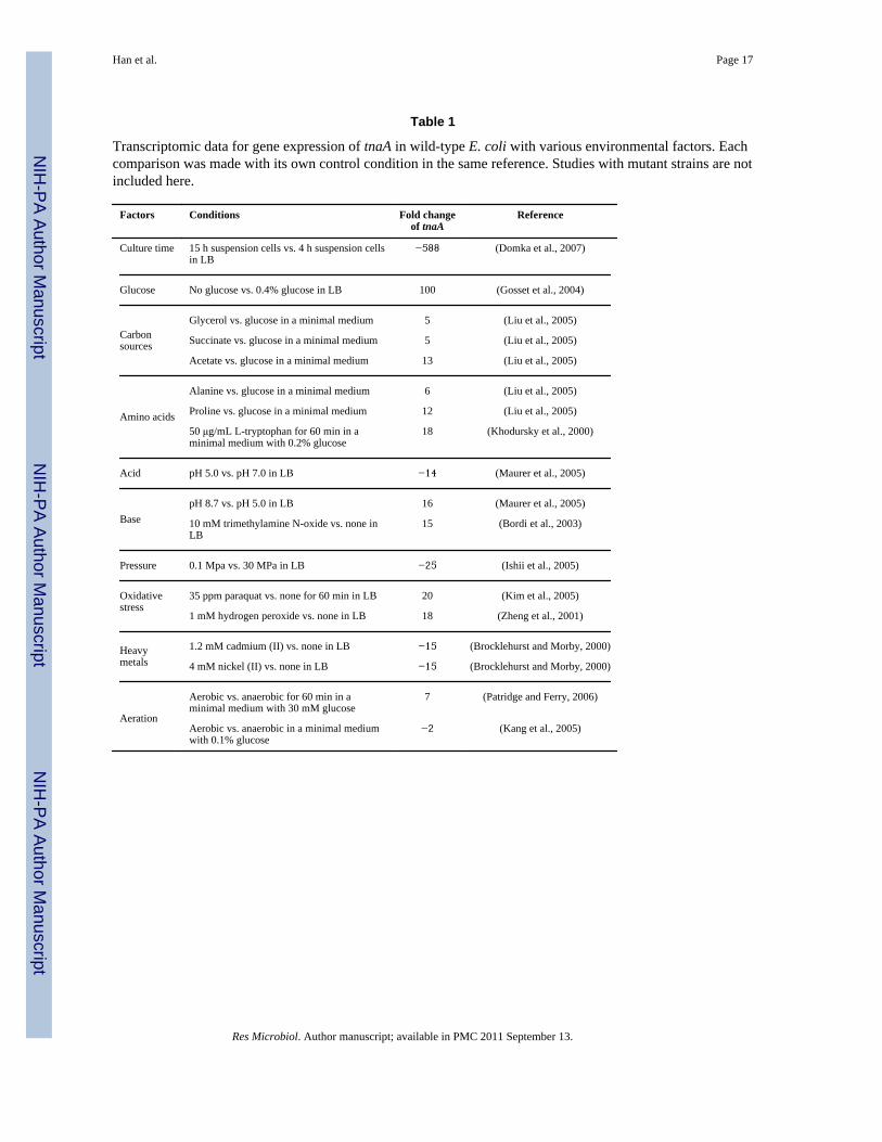
Although the importance of indole signaling in microbial communities (Lee and Lee, 2010)as well as in the human immune system (Wikoff et al., 2009; Bansal et al., 2010) has beenrecently demonstrated, environmental factors that contribute to and regulate indoleproduction remain poorly defined. Recently, several transcriptomic studies provideinformative data on the expression of tnaA in response to various environmental cues in E.coli (Table 1). Hence, the overall objective of this study is to identify the importantenvironmental factors that control indole production as well as cell growth to betterunderstand indole signaling in microbial communities.
2. MATERIALS AND METHODS2.1. Bacterial strains, materials and growth rate measurements
E. coli K-12 BW25113 (lacIq rrnBT14 ΔlacZWJ16 hsdR514 ΔaraBA-DAH33 ΔrhaBADLD78)and its isogenic tnaA mutant (Baba et al., 2006) that does not produce indole (Lee et al.,2008) were used. Luria-Bertani (LB) (Sambrook et al., 1989) was used as a basic mediumfor growth unless indicated. Indole, ampicillin, kanamycin, chloramphenicol, N-hexanoyl-DL-homoserine lactone, N-(3-oxotetradecanoyl)-L-homoserine lactone, N-(3-oxododecanoyl)-L-homoserine, N-(3-oxooctanoyl)-L-homoserine lactone, paraquat,cadmium chloride, sodium dodecyl sulfate (SDS), and 4-(dimethylamino)-benzaldehydewere purchased from Sigma-Aldrich Co. (Missouri, USA). Hydrogen peroxide (H2O2), HCl,
Han et al. Page 2
Res Microbiol. Author manuscript; available in PMC 2011 September 13.
NIH
-PA Author Manuscript
NIH
-PA Author Manuscript
NIH
-PA Author Manuscript
NaOH, NaCl, FeCl3, ethanol, methanol, and dimethyl sulfoxide (DMSO) were purchasedfrom Duksan Pure Chemical Co. (Ansan, Korea). Bacterial strains were initially streakedfrom −80°C glycerol stocks on LB plates, and a fresh single colony was inoculated into LBmedium (25 mL) in 250-mL flasks and routinely cultured at 250 rpm at 37°C unlessindicated. Overnight cultures were diluted 1:100 using LB medium. For cell growthmeasurements, the optical density was measured at 600 nm (OD600) with aspectrophotometer (UV-160, Shimadzu, Japan). When the value of OD600 was above 0.7,culture sample was diluted to fit in a linear range of 0.2 and 0.7. The specific growth rateswere determined by measuring OD600 and calculated by using the linear portion of thenatural logarithm of OD600 versus time. In order to measure cell viability and cell number,diluted cells were enumerated with LB agar plates.
2.2. Indole assaysExtracellular and intracellular indole concentrations were measured as indicated previouslyusing Kovac’s reagent (Kawamura-Sato et al., 1999). Briefly, Kovac’s reagent (0.4 mL) wasmixed with supernatants (1 mL) of bacterial cultures. The reaction mixture was diluted 1:10in HCl-amyl alcohol solution (30 mL of HCl and 90 mL of amyl alcohol), and theabsorbance of the mixture was measured at 540 nm with a spectrophotometer (ShimadzuUV-160). For the intracellular indole concentrations, cells were completely lysed with 1%SDS and 0.2% NaOH by mixing well (Sambrook et al., 1989). In addition, thespectrophotometric indole assay was corroborated with reverse-phase HPLC (Lee et al.,2007a) using a 100 × 4.6 mm Chromolith Performance RP-18e column (Merck KGaA,Darmstadt, Germany) and elution with H2O-0.1% (v/v) trifluoroacetic acid and acetonitrileas the mobile phases at a flow rate of 0.5 mL/min (50:50). Under these conditions, theretention time and the absorbance maximum was 5.1 min/271 nm for indole.
2.3. Crystal-violet biofilm assayA static biofilm formation assay was performed in 96-well polystyrene plates (FisherScientific, Pittsburg, USA) as previously reported (Pratt and Kolter, 1998). Briefly, cellswere inoculated at an initial turbidity at 600 nm of 0.05 for 24 h without shaking at 30°C and37°C. Cell density (turbidity at 620 nm) and total biofilm (absorbance at 540 nm) weremeasured using crystal violet staining. Total biofilm was normalized by bacterial growth foreach condition. Each data point was averaged from at least twelve replicate wells (six wellsfrom each of two independent cultures).
2.4. Scanning electron microscopy (SEM)In order to investigate the impact on cell morphology upon addition of ampicillin and indole,SEM was used by modifying the protocol (Hossain et al., 1996). Briefly, E. coli cells werecultured in LB medium at 37°C for 6.5 h, and cells were directly fixed by addingglutaraldehyde (2.5% final) and formaldehyde (2% final) and incubated at 4°C overnight.Fixed cells were collected by filtering with a 0.45 μm nylon filter (Nalgene, New York,USA) with vacuum. The filter contained cells was cut into 0.5 × 0.5 mm squares and washedwith 0.2 M sodium phosphate buffer before fixating for 90 min with osmium solution(containing 1.5 mL of sodium phosphate buffer 0.2 M, 3 mL of 2% OsO4 and 3mLdeionized water). Then, samples were washed and dehydrated by successive 10 minincubations in 50% ethanol, 70% ethanol, 80% ethanol, 90% ethanol, and 95% ethanolfollowed by two successive incubations for 20 min in 100% ethanol. After dehydrating, thefilters with cells were incubated in isoamyl acetate for 20 min and dried with critical-pointdryer (HCP-2, Hitachi, Japan). The nylon filters were affixed to SEM stubs and coated withwhite gold for 200 seconds using ion sputtering (E-1030, Hitachi). Specimens wereexamined with the SEM S-4100 (Hitachi). The voltage was set at 15 kV and viewed at amagnification from x 2,000 to x15,000.
Han et al. Page 3
Res Microbiol. Author manuscript; available in PMC 2011 September 13.
NIH
-PA Author Manuscript
NIH
-PA Author Manuscript
NIH
-PA Author Manuscript
3. RESULTSThe main purpose of this research was to determine key environmental factors affecting theproduction of extracellular indole in E. coli. Environmental conditions such as pH,antibiotics, temperature, heavy metals, oxidative reagents, osmolarity, solvents, and quorum-sensing molecules (acylhomoserine lactones) from other bacteria were tested. Since indolesignaling shows different responses at different temperatures (Lee et al., 2008), both ahuman body temperature (37°C) and a low temperature (25°C or 30°C) were investigatedwith various conditions.
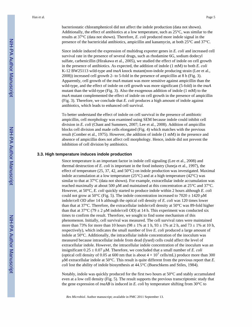
3.1. pH controls indole production and biofilm formationLow pH (pH 4) inhibits indole production (John and Wyeth, 1919) while high pH (pH 9)induces the expression of tnaA significantly in E. coli (Blankenhorn et al., 1999; Stancik etal., 2002; Yohannes et al., 2004). Hence, different pHs (4, 5, 6, 7, 8, 9, and 10 adjusted with37% HCl and 5 N NaOH which caused the total volume change less than 0.2 %) were testedto measure the production of extracellular indole at the same cell turbidity of 1.5 at 30°C and37°C, respectively. Growth rates of E. coli were similar between pH 5 and pH 9 as 0.83 ±0.01 h−1 at pH 5, 1.01 ± 0.01 h−1 at pH 6, 1.04 ± 0.02 h−1 at pH 7, 0.87 ± 0.01 h−1 at pH 8,and 0.70 ± 0.08 h−1 at pH 9, respectively at 30°C and 1.37 ± 0.02 h−1 at pH 5, 1.51 ± 0.04h−1 at pH 6, 1.50 ± 0.02 h−1 at pH 7, 1.35 ± 0.01 at pH 8, and 1.21 ± 0.04 h−1 at pH 9,respectively at 37°C, while cell growth of E. coli was much slow at pH 4 and 10 so that dataat pH 4 and 10 were excluded in the comparison. The initial pH (between pH 5 and pH 9)was not changed until the cell turbidity of 1.5 at which the production of indole wasmeasured.
Fig. 1A clearly shows that low pH inhibits indole production while high pH increases indoleproduction both at 30°C and at 37°C. The results match well with the previoustranscriptomic data (Table 1) in which tnaA was repressed under acidic conditions whiletnaA was induced with a basic condition. It was also observed that indole production washigher at 37°C than at 30°C for all tested pHs.
Since indole inhibited the biofilm formation of E. coli BW25113 strain (Lee et al., 2007a)and pH significantly changes indole production (Fig. 1 A), the effect of pH was investigatedfor biofilm formation in E. coli BW25113 (Fig. 1B). Biofilm formation was clearlydecreased at alkali conditions (pH 8 and 9) due to the high level of indole accumulation. Toconfirm the biofilm reduction by indole, when indole (0.5 mM) was exogenously added inthe medium at pH 5, the biofilm formation/cell was significantly decreased as 0.29 ± 0.06 at30°C and 0.27 ± 0.09 at 37°C. This result supports the previous result that indole decreasesbiofilm formation in nonpathogenic E. coli as well as pathogenic E. coli O157:H7 (Lee etal., 2007b; Lee et al., 2007a; Lee et al., 2008).
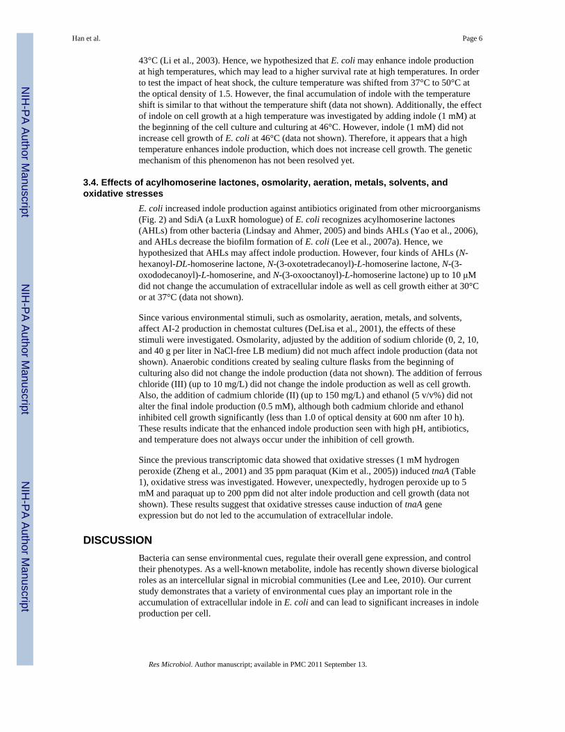
3.2. Antibiotics induce indole productionSince indole increased drug resistance in E. coli by inducing multidrug export genes(Hirakawa et al., 2005), we investigated the effect of antibiotics on indole production.Antibiotics (bactericidal ampicillin of the β-lactam class of antibiotics, bactericidalkanamycin of the aminoglycosides, and bacteriostatic chloramphenicol) at sub-inhibitoryconcentrations were added in the beginning of each cell culture and indole production andcell growth were measured at 25°C and 37°C. E. coli produced significantly moreextracellular indole/cell density (22-fold) in the presence of ampicillin (2 μg/mL) (Fig. 2);1900 ± 260 μM indole/cell OD with ampicillin vs. 88 ± 3 μM indole/cell OD for the controlat 8 h. Similarly, kanamycin (2 μg/mL) also enhanced indole production/cell density by 4-fold with 338 ± 87 μM indole/cell OD compared to the control at 8 h (Fig. 2). However,
Han et al. Page 4
Res Microbiol. Author manuscript; available in PMC 2011 September 13.
NIH
-PA Author Manuscript
NIH
-PA Author Manuscript
NIH
-PA Author Manuscript
bacteriostatic chloramphenicol did not affect the indole production (data not shown).Additionally, the effect of antibiotics at a low temperature, such as 25°C, was similar to theresults at 37°C (data not shown). Therefore, E. coli produced more indole signal in thepresence of the bactericidal antibiotics, ampicillin and kanamycin, at both 25°C and 37°C.
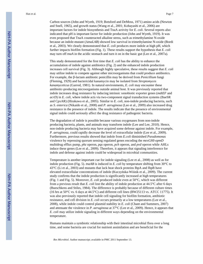
Since indole induced the expression of multidrug exporter genes in E. coli and increased cellsurvival rate in the presence of several drugs, such as rhodamine 6G, sodium dodecylsulfate, carbenicillin (Hirakawa et al., 2005), we studied the effect of indole on cell growthin the presence of antibiotics. As expected, the addition of indole (1 mM) to both E. coliK-12 BW25113 wild-type and tnaA knock mutant(non-indole producing strain (Lee et al.,2008)) increased cell growth 2- to 5-fold in the presence of ampicillin at 8 h (Fig. 3).Apparently, cell growth of the tnaA mutant was more sensitive against ampicillin than thewild-type, and the effect of indole on cell growth was more significant (5-fold) in the tnaAmutant than the wild-type (Fig. 3). Also the exogenous addition of indole (1 mM) to thetnaA mutant complemented the effect of indole on cell growth in the presence of ampicillin(Fig. 3). Therefore, we conclude that E. coli produces a high amount of indole againstantibiotics, which leads to enhanced cell survival.
To better understand the effect of indole on cell survival in the presence of antibioticampicillin, cell morphology was examined using SEM because indole could inhibit celldivision in E. coli (Chant and Summers, 2007; Lee et al., 2008). Addition of ampicillinblocks cell division and made cells elongated (Fig. 4) which matches with the previousresult (Comber et al., 1975). However, the addition of indole (1 mM) in the presence andabsence of ampicillin does not affect cell morphology. Hence, indole did not prevent theinhibition of cell division by antibiotics.
3.3. High temperature induces indole productionSince temperature is an important factor in indole cell signaling (Lee et al., 2008) andthermal destruction of E. coli is important in the food industry (Juneja et al., 1997), theeffect of temperature (25, 37, 42, and 50°C) on indole production was investigated. Maximalindole accumulation at a low temperature (25°C) and at a high temperature (42°C) wassimilar to that at 37°C (data not shown). For example, extracellular indole accumulation wasreached maximally at about 500 μM and maintained at this concentration at 25°C and 37°C.However, at 50°C, E. coli quickly started to produce indole within 2 hours although E. colicould not grow at 50°C (Fig. 5). The indole concentration increased to 7020 ± 1420 μMindole/cell OD after 14 h although the optical cell density of E. coli was 120 times lowerthan that at 37°C. Therefore, the extracellular indole/cell density at 50°C was 89-fold higherthan that at 37°C (79 ± 2 μM indole/cell OD) at 14 h. This experiment was conducted sixtimes to confirm the result. Therefore, we sought to find some mechanism of thisphenomenon. Initially, cell survival was measured. The cell survival rates were maintainedmore than 73% for more than 10 hours (98 ± 1% at 1 h, 93 ± 1% at 2 h, and 73 ± 1% at 10 h,respectively), which indicates the small number of live E. coli produced a large amount ofindole at 50°C. Additionally, the intracellular indole concentration of the inoculum wasmeasured because intracellular indole from dead (lysed) cells could affect the level ofextracellular indole. However, the intracellular indole concentration of the inoculum was aninsignificant 0.25 ± 0.07 μM. Therefore, we concluded that a small number of E. coli(optical cell density of 0.05 at 600 nm that is about 4 × 10⁷ cells/mL) produce more than 300μM extracellular indole at 50°C. This result is quite different from the previous report that E.coli lost the ability of indole biosynthesis at 44.5°C (Bueschkens and Stiles, 1984).
Notably, indole was quickly produced for the first two hours at 50°C and stably accumulatedeven at a low cell density (Fig. 5). The result supports the previous transcriptomic study thatthe gene expression of tnaAB is induced in E. coli by temperature shifting from 30°C to
Han et al. Page 5
Res Microbiol. Author manuscript; available in PMC 2011 September 13.
NIH
-PA Author Manuscript
NIH
-PA Author Manuscript
NIH
-PA Author Manuscript
43°C (Li et al., 2003). Hence, we hypothesized that E. coli may enhance indole productionat high temperatures, which may lead to a higher survival rate at high temperatures. In orderto test the impact of heat shock, the culture temperature was shifted from 37°C to 50°C atthe optical density of 1.5. However, the final accumulation of indole with the temperatureshift is similar to that without the temperature shift (data not shown). Additionally, the effectof indole on cell growth at a high temperature was investigated by adding indole (1 mM) atthe beginning of the cell culture and culturing at 46°C. However, indole (1 mM) did notincrease cell growth of E. coli at 46°C (data not shown). Therefore, it appears that a hightemperature enhances indole production, which does not increase cell growth. The geneticmechanism of this phenomenon has not been resolved yet.
3.4. Effects of acylhomoserine lactones, osmolarity, aeration, metals, solvents, andoxidative stresses
E. coli increased indole production against antibiotics originated from other microorganisms(Fig. 2) and SdiA (a LuxR homologue) of E. coli recognizes acylhomoserine lactones(AHLs) from other bacteria (Lindsay and Ahmer, 2005) and binds AHLs (Yao et al., 2006),and AHLs decrease the biofilm formation of E. coli (Lee et al., 2007a). Hence, wehypothesized that AHLs may affect indole production. However, four kinds of AHLs (N-hexanoyl-DL-homoserine lactone, N-(3-oxotetradecanoyl)-L-homoserine lactone, N-(3-oxododecanoyl)-L-homoserine, and N-(3-oxooctanoyl)-L-homoserine lactone) up to 10 μMdid not change the accumulation of extracellular indole as well as cell growth either at 30°Cor at 37°C (data not shown).
Since various environmental stimuli, such as osmolarity, aeration, metals, and solvents,affect AI-2 production in chemostat cultures (DeLisa et al., 2001), the effects of thesestimuli were investigated. Osmolarity, adjusted by the addition of sodium chloride (0, 2, 10,and 40 g per liter in NaCl-free LB medium) did not much affect indole production (data notshown). Anaerobic conditions created by sealing culture flasks from the beginning ofculturing also did not change the indole production (data not shown). The addition of ferrouschloride (III) (up to 10 mg/L) did not change the indole production as well as cell growth.Also, the addition of cadmium chloride (II) (up to 150 mg/L) and ethanol (5 v/v%) did notalter the final indole production (0.5 mM), although both cadmium chloride and ethanolinhibited cell growth significantly (less than 1.0 of optical density at 600 nm after 10 h).These results indicate that the enhanced indole production seen with high pH, antibiotics,and temperature does not always occur under the inhibition of cell growth.
Since the previous transcriptomic data showed that oxidative stresses (1 mM hydrogenperoxide (Zheng et al., 2001) and 35 ppm paraquat (Kim et al., 2005)) induced tnaA (Table1), oxidative stress was investigated. However, unexpectedly, hydrogen peroxide up to 5mM and paraquat up to 200 ppm did not alter indole production and cell growth (data notshown). These results suggest that oxidative stresses cause induction of tnaA geneexpression but do not led to the accumulation of extracellular indole.
DISCUSSIONBacteria can sense environmental cues, regulate their overall gene expression, and controltheir phenotypes. As a well-known metabolite, indole has recently shown diverse biologicalroles as an intercellular signal in microbial communities (Lee and Lee, 2010). Our currentstudy demonstrates that a variety of environmental cues play an important role in theaccumulation of extracellular indole in E. coli and can lead to significant increases in indoleproduction per cell.
Han et al. Page 6
Res Microbiol. Author manuscript; available in PMC 2011 September 13.
NIH
-PA Author Manuscript
NIH
-PA Author Manuscript
NIH
-PA Author Manuscript
Carbon sources (John and Wyeth, 1919; Botsford and DeMoss, 1971) amino acids (Newtonand Snell, 1965), and growth status (Wang et al., 2001; Kobayashi et al., 2006) areimportant factors for indole biosynthesis and TnaA activity for E. coli. Several reports alsoindicated that pH is important factor for indole production (John and Wyeth, 1919). It waseven proposed that TnaA counteracted alkaline stress, such as trimethylamine N-oxidebecause an indole-mutant (ΔtnaLAB) showed low survival in trimethylamine N-oxide (Bordiet al., 2003). We clearly demonstrated that E. coli produces more indole at high pH, whichfurther impacts biofilm formation (Fig. 1). These results support the hypothesis that E. colimay turn off tnaA in the acidic stomach and turn it on in the basic gut (Lee et al., 2007a).
This study demonstrated for the first time that E. coli has the ability to enhance theaccumulation of indole against antibiotics (Fig. 2) and the enhanced indole productionincreases cell survival (Fig. 3). Although highly speculative, these results suggest that E. colimay utilize indole to compete against other microorganisms that could produce antibiotics.For example, the β-lactam antibiotic penicillin may be derived from Penicillium fungi(Fleming, 1929) and bactericidal kanamycin may be isolated from Streptomyceskanamyceticus (Garrod, 1981). In natural environments, E. coli may encounter theseantibiotic-producing microorganisms outside animal host. It was previously reported thatindole increases drug resistance by inducing intrinsic xenobiotic exporter genes (mdtEF andacrD) in E. coli, where indole acts via two-component signal transduction systems (BaeSRand CpxAR) (Hirakawa et al., 2005). Similar to E. coli, non-indole producing bacteria, suchas S. enterica (Nikaido et al., 2008) and P. aeruginosa (Lee et al., 2009) also increased drugresistance in the presence of indole. The results indicate that the presence of environmentalsignal indole could seriously affect the drug resistance of pathogenic bacteria.
The degradation of indole is possible because various oxygenases from non-indoleproducing bacteria, plants, and animals may transform indole (Lee and Lee, 2010). Hence,non-indole producing bacteria may have acquired some defense against indole. For example,P. aeruginosa, could rapidly decrease the level of extracellular indole (Lee et al., 2009).Furthermore, previous results showed that indole from E.coli diminished Pseudomonasvirulence by repressing quorum sensing regulated genes encoding the mexGHI-opmDmultidrug efflux pump, phz operon, pqs operon, pch operon, and pvd operon while AHLsinduce these genes (Lee et al., 2009). Therefore, it appears that signaling interference forindole and defense against indole could be widespread in microbial communities.
Temperature is another important cue for indole signaling (Lee et al., 2008) as well as forindole production (Fig. 5). tnaAB is induced in E. coli by temperature shifting from 30°C to43°C (Li et al., 2003) and mutants that lack heat shock proteins IbpA and IbpB haveelevated extracellular concentrations of indole (Kuczyńska-Wiśnik et al., 2009). The currentstudy confirms that the indole production is significantly increased at high temperatures(Fig. 1 and Fig. 5). Moreover, E. coli produced indole even at 50°C, which was differentfrom a previous result that E. coli lost the ability of indole production at 44.5°C after 6 days(Bueschkens and Stiles, 1984). The difference is probably because of different culture times(16 hrs at 50°C vs. 6 days at 44.5°C) and different cell lines (BW25113 vs. ATCC 11775). Itwas also previously reported that indole cell signaling for biofilm formation, antibioticresistance, and cell division in E. coli occurs primarily at a low temperatures (Lee et al.,2008), while indole could control plasmid stability in E. coli (Chant and Summers, 2007)and attenuate the virulence in P. aeruginosa at 37°C (Lee et al., 2009). Hence, it appears thatE. coli may utilize indole signaling in different ways depending on the environmentaltemperature.
Humans maintain a symbiotic relationship with their intestinal microbial flora over a longtime, and some bacteria are crucial for nutrient assimilation and are beneficial for the
Han et al. Page 7
Res Microbiol. Author manuscript; available in PMC 2011 September 13.
NIH
-PA Author Manuscript
NIH
-PA Author Manuscript
NIH
-PA Author Manuscript
immune system (Hooper and Gordon, 2001). A variety of intestinal bacteria produce a largequantity of indole in the animal gut (a weak basic condition) including the human intestine(DeMoss and Moser, 1969; Lee and Lee, 2010). A recent study suggested that indole couldbe important in the intestinal epithelial cells response to gastrointestinal tract pathogensbecause indole beneficially affected gene expression of human epithelial cells (Bansal et al.,2010). Moreover, a mass spectrometry-based metabolomics study demonstrated that theproduction of a powerful antioxidant compound indole-3-propionic acid in animal bloodcompletely depended on indole-producing enteric bacteria (Wikoff et al., 2009). The resultsuggested that animals possibly utilize indole derivatives originated from gut microflora fortheir immune systems (Wikoff et al., 2009). The current study further suggests that theintestinal microbial flora may protect itself using indole against pathogenic bacteria.
Recently, the role of indole has been also studied in Vibrio cholerae in which indoledecreases its biofilm formation while increasing its grazing resistance to the phagocyticeukaryote Dictyostelium discoideum, probably by inducing expression of virulence-associated secretion proteins (Mueller et al., 2009). Furthermore, more than 85 species ofboth Gram-positive and Gram-negative bacteria produce large quantities of extracellularindole in microbial communities (Lee and Lee, 2010). Because the habitats of these bacteriaare diverse, the mechanisms of indole production are probably different among bacterialspecies. Hence, it would be interesting to investigate indole production in other indole-producing bacteria. Further studies are essentially required to understand why manybacterial species produce indole, how bacteria regulate indole signaling, and how otherspecies react to environmental indole.
AcknowledgmentsThis research was supported by the Yeungnam University research grant (to J. Lee). T. H. Han was supported bythe Brain Korea 21 Project from the Ministry of Education and Human Resources, Korea. T. Wood is the T.Michael O’Connor endowed chair and is also supported by the NIH (R01 GM089999).
REFERENCESAnyanful A, Dolan-Livengood JM, Lewis T, Sheth S, Dezalia MN, Sherman MA, et al. Paralysis and
killing of Caenorhabditis elegans by enteropathogenic Escherichia coli requires the bacterialtryptophanase gene. Mol Microbiol. 2005; 57:988–1007. [PubMed: 16091039]
Baba T, Ara T, Hasegawa M, Takai Y, Okumura Y, Baba M, et al. Construction of Escherichia coliK-12 in-frame, single-gene knockout mutants: the Keio collection. Mol Syst Biol. 2006; 22006.0008.
Bansal T, Alaniz RC, Wood TK, Jayaraman A. The bacterial signal indole increases epithelial-celltight-junction resistance and attenuates indicators of inflammation. Proc Natl Acad Sci U S A. 2010;107:228–233. [PubMed: 19966295]
Blankenhorn D, Phillips J, Slonczewski JL. Acid- and base-induced proteins during aerobic andanaerobic growth of Escherichia coli revealed by two-dimensional gel electrophoresis. J Bacteriol.1999; 181:2209–2216. [PubMed: 10094700]
Bordi C, Theraulaz L, Mejean V, Jourlin-Castelli C. Anticipating an alkaline stress through the Torphosphorelay system in Escherichia coli. Mol Microbiol. 2003; 48:211–223. [PubMed: 12657056]
Botsford JL, DeMoss RD. Catabolite repression of tryptophanase in Escherichia coli. J Bacteriol.1971; 105:303–312. [PubMed: 4322348]
Brocklehurst KR, Morby AP. Metal-ion tolerance in Escherichia coli: analysis of transcriptionalprofiles by gene-array technology. Microbiology. 2000; 146:2277–2282. [PubMed: 10974115]
Bueschkens DH, Stiles ME. Escherichia coli variants for gas and indole production at elevatedincubation temperatures. Appl Environ Microbiol. 1984; 48:601–605. [PubMed: 6388502]
Chant EL, Summers DK. Indole signalling contributes to the stable maintenance of Escherichia colimulticopy plasmids. Mol Microbiol. 2007; 63:35–43. [PubMed: 17163976]
Han et al. Page 8
Res Microbiol. Author manuscript; available in PMC 2011 September 13.
NIH
-PA Author Manuscript
NIH
-PA Author Manuscript
NIH
-PA Author Manuscript
Comber KR, Osborne CD, Sutherland R. Comparative effects of amoxycillin and ampicillin in thetreatment of experimental mouse infections. Antimicrob Agents Chemother. 1975; 7:179–185.[PubMed: 1094950]
DeLisa MP, Valdes JJ, Bentley WE. Mapping stress-induced changes in autoinducer AI-2 productionin chemostat-cultivated Escherichia coli K-12. J Bacteriol. 2001; 183:2918–2928. [PubMed:11292813]
DeMoss RD, Moser K. Tryptophanase in diverse bacterial species. J Bacteriol. 1969; 98:167–171.[PubMed: 5781572]
Di Martino P, Fursy R, Bret L, Sundararaju B, Phillips RS. Indole can act as an extracellular signal toregulate biofilm formation of Escherichia coli and other indole-producing bacteria. Can JMicrobiol. 2003; 49:443–449. [PubMed: 14569285]
Domka J, Lee J, Bansal T, Wood TK. Temporal gene-expression in Escherichia coli K-12 biofilms.Environ Microbiol. 2007; 9:332–346. [PubMed: 17222132]
Fleming A. On the antibacterial action of cultures of a penicillium, with special reference to their usein the isolation of B. influenzæ. Br J Exp Pathol. 1929; 10:226–236.
Garrod, LP. Churchill Livingstone. 1981. Antibiotic and Chemotherapy; p. 131Gong F, Yanofsky C. Analysis of tryptophanase operon expression in vitro: accumulation of TnaC-
peptidyl-tRNA in a release factor 2-depleted S-30 extract prevents Rho factor action, simulatinginduction. J Biol Chem. 2002; 277:17095–17100. [PubMed: 11880383]
Gosset G, Zhang Z, Nayyar S, Cuevas WA, Saier MH Jr. Transcriptome analysis of Crp-dependentcatabolite control of gene expression in Escherichia coli. J Bacteriol. 2004; 186:3516–3524.[PubMed: 15150239]
Hirakawa H, Inazumi Y, Masaki T, Hirata T, Yamaguchi A. Indole induces the expression ofmultidrug exporter genes in Escherichia coli. Mol Microbiol. 2005; 55:1113–1126. [PubMed:15686558]
Hirakawa H, Kodama T, Takumi-Kobayashi A, Honda T, Yamaguchi A. Secreted indole serves as asignal for expression of type III secretion system translocators in enterohaemorrhagic Escherichiacoli O157:H7. Microbiology. 2009; 155:541–550. [PubMed: 19202102]
Hooper LV, Gordon JI. Commensal host-bacterial relationships in the gut. Science. 2001; 292:1115–1118. [PubMed: 11352068]
Hossain MM, Nakayama H, Goto N. In vitro induction of apoptosis of developing brain cells by 5-azacytidine. Int J Dev Neurosci. 1996; 14:11–17. [PubMed: 8779304]
Ishii A, Oshima T, Sato T, Nakasone K, Mori H, Kato C. Analysis of hydrostatic pressure effects ontranscription in Escherichia coli by DNA microarray procedure. Extremophiles. 2005; 9:65–73.[PubMed: 15340867]
John F, Wyeth S. The Effects of Acids, Alkalies, and Sugars on the Growth and Indole Formation ofBacillus coli. Biochem J. 1919; 13:10–24. [PubMed: 16742836]
Juneja VK, Snyder OP Jr. Marmer BS. Thermal destruction of Escherichia coli O157:H7 in beef andchicken: determination of D- and z-values. Int J Food Microbiol. 1997; 35:231–237. [PubMed:9105932]
Kamath AV, Vaidyanathan CS. New pathway for the biodegradation of indole in Aspergillus niger.Appl Environ Microbiol. 1990; 56:275–280. [PubMed: 2310183]
Kang Y, Weber KD, Qiu Y, Kiley PJ, Blattner FR. Genome-wide expression analysis indicates thatFNR of Escherichia coli K-12 regulates a large number of genes of unknown function. J Bacteriol.2005; 187:1135–1160. [PubMed: 15659690]
Kawamura-Sato K, Shibayama K, Horii T, Iimuma Y, Arakawa Y, Ohta M. Role of multiple effluxpumps in Escherichia coli in indole expulsion. FEMS Microbiol Lett. 1999; 179:345–352.[PubMed: 10518736]
Keller L, Surette MG. Communication in bacteria: an ecological and evolutionary perspective. NatRev Microbiol. 2006; 4:249–258. [PubMed: 16501584]
Khodursky AB, Peter BJ, Cozzarelli NR, Botstein D, Brown PO, Yanofsky C. DNA microarrayanalysis of gene expression in response to physiological and genetic changes that affect tryptophanmetabolism in Escherichia coli. Proc Natl Acad Sci U S A. 2000; 97:12170–12175. [PubMed:11027315]
Han et al. Page 9
Res Microbiol. Author manuscript; available in PMC 2011 September 13.
NIH
-PA Author Manuscript
NIH
-PA Author Manuscript
NIH
-PA Author Manuscript
Kim BC, Youn CH, Ahn JM, Gu MB. Screening of target-specific stress-responsive genes for thedevelopment of cell-based biosensors using a DNA microarray. Anal Chem. 2005; 77:8020–8026.[PubMed: 16351151]
Kobayashi A, Hirakawa H, Hirata T, Nishino K, Yamaguchi A. Growth phase-dependent expression ofdrug exporters in Escherichia coli and its contribution to drug tolerance. J Bacteriol. 2006;188:5693–5703. [PubMed: 16885437]
Kuczyńska-Wiśnik D, Matuszewska E, Laskowska E. E. coli heat shock proteins, IbpA and IbpB,affect biofilm formation by influencing the level of extracellular indole. Microbiology. 2009
Lee J-H, Lee J. Indole as an Intercellular Signal in Microbial Community. FEMS Microbiol rev. 2010;34:426–444. [PubMed: 20070374]
Lee J, Jayaraman A, Wood TK. Indole is an inter-species biofilm signal mediated by SdiA. BMCMicrobiol. 2007a; 7:42. [PubMed: 17511876]
Lee J, Bansal T, Jayaraman A, Bentley WE, Wood TK. Enterohemorrhagic Escherichia coli biofilmsare inhibited by 7-hydroxyindole and stimulated by isatin. Appl Environ Microbiol. 2007b;73:4100–4109. [PubMed: 17483266]
Lee J, Attila C, Cirillo SLG, Cirillo JD, Wood TK. Indole and 7-hydroxyindole diminish Pseudomonasaeruginosa virulence. Microbial Biotech. 2009; 2:75–90.
Lee J, Zhang XS, Hegde M, Bentley WE, Jayaraman A, Wood TK. Indole cell signaling occursprimarily at low temperatures in Escherichia coli. ISME J. 2008; 2:1007–1023. [PubMed:18528414]
Li Y, Cole K, Altman S. The effect of a single, temperature-sensitive mutation on global geneexpression in Escherichia coli. RNA. 2003; 9:518–532. [PubMed: 12702811]
Lindsay A, Ahmer BMM. Effect of sdiA on biosensors of N-acylhomoserine lactones. J Bacteriol.2005; 187:5054–5058. [PubMed: 15995228]
Liu M, Durfee T, Cabrera JE, Zhao K, Jin DJ, Blattner FR. Global transcriptional programs reveal acarbon source foraging strategy by Escherichia coli. J Biol Chem. 2005; 280:15921–15927.[PubMed: 15705577]
Maurer LM, Yohannes E, Bondurant SS, Radmacher M, Slonczewski JL. pH regulates genes forflagellar motility, catabolism, and oxidative stress in Escherichia coli K-12. J Bacteriol. 2005;187:304–319. [PubMed: 15601715]
Mitrophanov AY, Groisman EA. Signal integration in bacterial two-component regulatory systems.Genes Dev. 2008; 22:2601–2611. [PubMed: 18832064]
Mueller RS, Beyhan S, Saini SG, Yildiz FH, Bartlett DH. Indole acts as an Extracellular CueRegulating Gene Expression in Vibrio cholerae. J Bacteriol. 2009; 191:3504–3516. [PubMed:19329638]
Newton WA, Snell EE. Formation and Interrelationships of Tryptophanase and TryptophanSynthetases in Escherichia coli. J Bacteriol. 1965; 89:355–364. [PubMed: 14255701]
Nikaido E, Yamaguchi A, Nishino K. AcrAB multidrug efflux pump regulation in Salmonella entericaserovar Typhimurium by RamA in response to environmental signals. J Biol Chem. 2008;283:24245–24253. [PubMed: 18577510]
Patridge EV, Ferry JG. WrbA from Escherichia coli and Archaeoglobus fulgidus is anNAD(P)H:quinone oxidoreductase. J Bacteriol. 2006; 188:3498–3506. [PubMed: 16672604]
Pittard, AJ. Biosynthesis of the Aromatic Amino Acids: The Tryptophan Pathway. In: Neidhardt, FC.,editor. Escherichia coli and Salmonella: Cellular and Molecular Biology. ASM Press; WashingtonDC, USA: 1996. p. 458-484.
Pratt LA, Kolter R. Genetic analysis of Escherichia coli biofilm formation: roles of flagella, motility,chemotaxis and type I pili. Mol Microbiol. 1998; 30:285–293. [PubMed: 9791174]
Sambrook, J.; Fritsch, EF.; Maniatis, T. Molecular Cloning: A Laboratory Manual. Cold SpringHarbor Laboratory Press; Cold Spring Harbor, NY: 1989.
Smith T. A Modification of the Method for Determining the Production of Indol by Bacteria. J ExpMed. 1897; 2:543–547.
Stancik LM, Stancik DM, Schmidt B, Barnhart DM, Yoncheva YN, Slonczewski JL. pH-dependentexpression of periplasmic proteins and amino acid catabolism in Escherichia coli. J Bacteriol.2002; 184:4246–4258. [PubMed: 12107143]
Han et al. Page 10
Res Microbiol. Author manuscript; available in PMC 2011 September 13.
NIH
-PA Author Manuscript
NIH
-PA Author Manuscript
NIH
-PA Author Manuscript
Vendeville A, Winzer K, Heurlier K, Tang CM, Hardie KR. Making ‘sense’ of metabolism:autoinducer-2, LuxS and pathogenic bacteria. Nat Rev Microbiol. 2005; 3:383–396. [PubMed:15864263]
Wang D, Ding X, Rather PN. Indole can act as an extracellular signal in Escherichia coli. J Bacteriol.2001; 183:4210–4216. [PubMed: 11418561]
Waters CM, Bassler BL. Quorum sensing: cell-to-cell communication in bacteria. Annu Rev Cell DevBiol. 2005; 21:319–346. [PubMed: 16212498]
Wikoff WR, Anfora AT, Liu J, Schultz PG, Lesley SA, Peters EC, Siuzdak G. Metabolomics analysisreveals large effects of gut microflora on mammalian blood metabolites. Proc Natl Acad Sci U SA. 2009; 106:3698–3703. [PubMed: 19234110]
Yanofsky C, Horn V, Gollnick P. Physiological studies of tryptophan transport and tryptophanaseoperon induction in Escherichia coli. J Bacteriol. 1991; 173:6009–6017. [PubMed: 1917834]
Yao Y, Martinez-Yamout MA, Dickerson TJ, Brogan AP, Wright PE, Dyson HJ. Structure of theEscherichia coli quorum sensing protein SdiA: activation of the folding switch by acyl homoserinelactones. J Mol Biol. 2006; 355:262–273. [PubMed: 16307757]
Yohannes E, Barnhart DM, Slonczewski JL. pH-dependent catabolic protein expression duringanaerobic growth of Escherichia coli K-12. J Bacteriol. 2004; 186:192–199. [PubMed: 14679238]
Zheng M, Wang X, Templeton LJ, Smulski DR, LaRossa RA, Storz G. DNA microarray-mediatedtranscriptional profiling of the Escherichia coli response to hydrogen peroxide. J Bacteriol. 2001;183:4562–4570. [PubMed: 11443091]
Han et al. Page 11
Res Microbiol. Author manuscript; available in PMC 2011 September 13.
NIH
-PA Author Manuscript
NIH
-PA Author Manuscript
NIH
-PA Author Manuscript
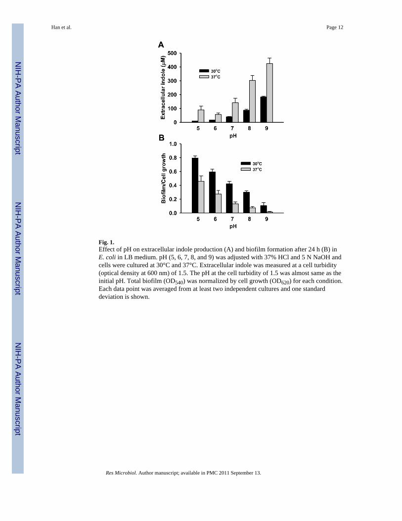
Fig. 1.Effect of pH on extracellular indole production (A) and biofilm formation after 24 h (B) inE. coli in LB medium. pH (5, 6, 7, 8, and 9) was adjusted with 37% HCl and 5 N NaOH andcells were cultured at 30°C and 37°C. Extracellular indole was measured at a cell turbidity(optical density at 600 nm) of 1.5. The pH at the cell turbidity of 1.5 was almost same as theinitial pH. Total biofilm (OD540) was normalized by cell growth (OD620) for each condition.Each data point was averaged from at least two independent cultures and one standarddeviation is shown.
Han et al. Page 12
Res Microbiol. Author manuscript; available in PMC 2011 September 13.
NIH
-PA Author Manuscript
NIH
-PA Author Manuscript
NIH
-PA Author Manuscript
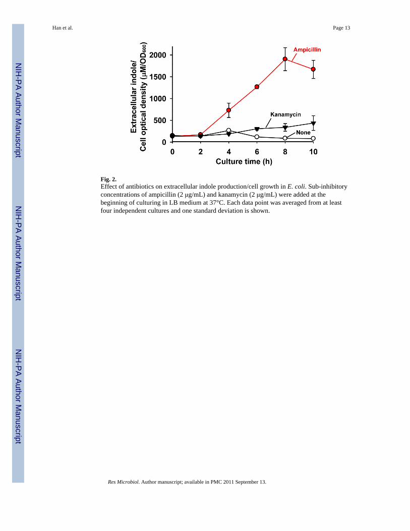
Fig. 2.Effect of antibiotics on extracellular indole production/cell growth in E. coli. Sub-inhibitoryconcentrations of ampicillin (2 μg/mL) and kanamycin (2 μg/mL) were added at thebeginning of culturing in LB medium at 37°C. Each data point was averaged from at leastfour independent cultures and one standard deviation is shown.
Han et al. Page 13
Res Microbiol. Author manuscript; available in PMC 2011 September 13.
NIH
-PA Author Manuscript
NIH
-PA Author Manuscript
NIH
-PA Author Manuscript
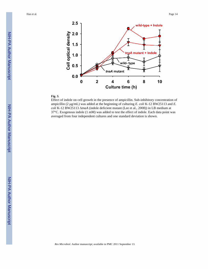
Fig. 3.Effect of indole on cell growth in the presence of ampicillin. Sub-inhibitory concentration ofampicillin (2 μg/mL) was added at the beginning of culturing E. coli K-12 BW25113 and E.coli K-12 BW25113 ΔtnaA (indole deficient mutant (Lee et al., 2008)) in LB medium at37°C. Exogenous indole (1 mM) was added to test the effect of indole. Each data point wasaveraged from four independent cultures and one standard deviation is shown.
Han et al. Page 14
Res Microbiol. Author manuscript; available in PMC 2011 September 13.
NIH
-PA Author Manuscript
NIH
-PA Author Manuscript
NIH
-PA Author Manuscript
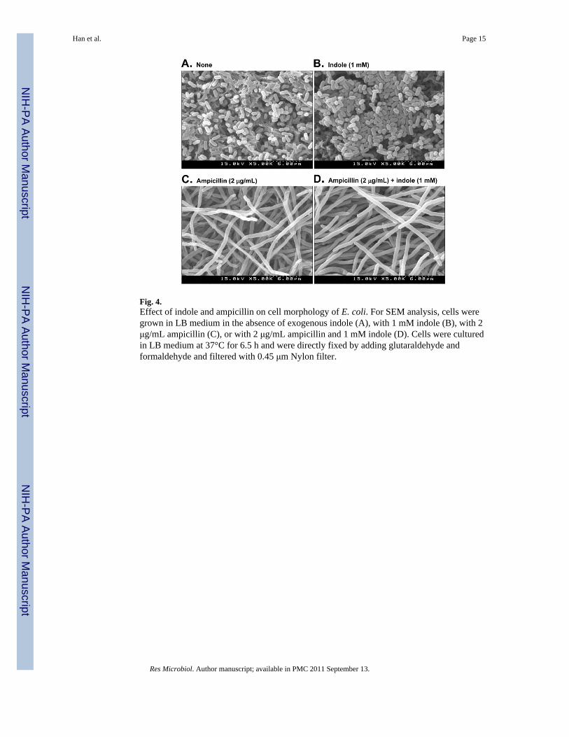
Fig. 4.Effect of indole and ampicillin on cell morphology of E. coli. For SEM analysis, cells weregrown in LB medium in the absence of exogenous indole (A), with 1 mM indole (B), with 2μg/mL ampicillin (C), or with 2 μg/mL ampicillin and 1 mM indole (D). Cells were culturedin LB medium at 37°C for 6.5 h and were directly fixed by adding glutaraldehyde andformaldehyde and filtered with 0.45 μm Nylon filter.
Han et al. Page 15
Res Microbiol. Author manuscript; available in PMC 2011 September 13.
NIH
-PA Author Manuscript
NIH
-PA Author Manuscript
NIH
-PA Author Manuscript
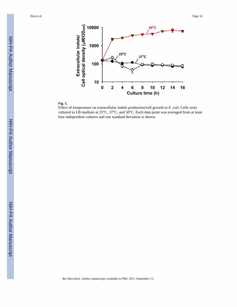
Fig. 5.Effect of temperature on extracellular indole production/cell growth in E. coli. Cells werecultured in LB medium at 25°C, 37°C, and 50°C. Each data point was averaged from at leastfour independent cultures and one standard deviation is shown.
Han et al. Page 16
Res Microbiol. Author manuscript; available in PMC 2011 September 13.
NIH
-PA Author Manuscript
NIH
-PA Author Manuscript
NIH
-PA Author Manuscript
NIH
-PA Author Manuscript
NIH
-PA Author Manuscript
NIH
-PA Author Manuscript
Han et al. Page 17
Table 1
Transcriptomic data for gene expression of tnaA in wild-type E. coli with various environmental factors. Eachcomparison was made with its own control condition in the same reference. Studies with mutant strains are notincluded here.
Factors Conditions Fold changeof tnaA
Reference
Culture time 15 h suspension cells vs. 4 h suspension cellsin LB
−588 (Domka et al., 2007)
Glucose No glucose vs. 0.4% glucose in LB 100 (Gosset et al., 2004)
Carbonsources
Glycerol vs. glucose in a minimal medium 5 (Liu et al., 2005)
Succinate vs. glucose in a minimal medium 5 (Liu et al., 2005)
Acetate vs. glucose in a minimal medium 13 (Liu et al., 2005)
Amino acids
Alanine vs. glucose in a minimal medium 6 (Liu et al., 2005)
Proline vs. glucose in a minimal medium 12 (Liu et al., 2005)
50 μg/mL L-tryptophan for 60 min in aminimal medium with 0.2% glucose
18 (Khodursky et al., 2000)
Acid pH 5.0 vs. pH 7.0 in LB −14 (Maurer et al., 2005)
BasepH 8.7 vs. pH 5.0 in LB 16 (Maurer et al., 2005)
10 mM trimethylamine N-oxide vs. none inLB
15 (Bordi et al., 2003)
Pressure 0.1 Mpa vs. 30 MPa in LB −25 (Ishii et al., 2005)
Oxidativestress
35 ppm paraquat vs. none for 60 min in LB 20 (Kim et al., 2005)
1 mM hydrogen peroxide vs. none in LB 18 (Zheng et al., 2001)
Heavymetals
1.2 mM cadmium (II) vs. none in LB −15 (Brocklehurst and Morby, 2000)
4 mM nickel (II) vs. none in LB −15 (Brocklehurst and Morby, 2000)
Aeration
Aerobic vs. anaerobic for 60 min in aminimal medium with 30 mM glucose
7 (Patridge and Ferry, 2006)
Aerobic vs. anaerobic in a minimal mediumwith 0.1% glucose
−2 (Kang et al., 2005)
Res Microbiol. Author manuscript; available in PMC 2011 September 13.


![3-[1-(4-Methylphenylsulfonyl)-1,4-dihydropyridin-4-yl]-1 H -indole](https://img.pdfslide.net/doc/110x75/6343d55a66152935d001b839/3-1-4-methylphenylsulfonyl-14-dihydropyridin-4-yl-1-h-indole.jpg)


