Embed Size (px)
Citation preview

105 173Rev Esp Quimioter 2013;26(2):173-188
Conclusions. Presence of fungi in respiratory samples from critically ill patients drives to different diagnostic and clinical management approaches. IPA is the most frequent in-fection and with high mortality.
Epidemiología, diagnóstico y tratamiento de las infecciones fúngicas respiratorias en el paciente crítico
RESUMEN
Objetivos. Elaborar unas recomendaciones prácticas ba-sadas en la evidencia científica, cuando esté disponible, o en opiniones de expertos para el diagnóstico, tratamiento y pre-vención de infecciones fúngicas respiratorias en el paciente crítico incluyendo a pacientes trasplantados de órgano sólido.
Metodos. Doce expertos pertenecientes a dos Sociedades Científicas (Sociedad Española de Quimioterapia y Sociedad Española de Medicina Intensiva, Crítica y Unidades Corona-rias) revisaron en una reunión celebrada en Marzo de 2012 los aspectos epidemiológicos y factores de riesgo como base para generar un documento para la prevención, diagnóstico y tratamiento de infecciones fúngicas respiratorias causadas por Candida spp., Aspergillus spp. o Zigomicetos.
Results. A pesar del frecuente aislamiento de Candida spp. del tracto respiratorio, el tratamiento antifúngico no está recomendado debido a que una neumonía por éstas especies es excepcional en pacientes no neutropénicos. En el caso de Aspergillus spp., aproximadamente el 50% de los aislamientos en UCI indican colonización y el otro 50% de los casos están asociados a aspergilosis pulmonar invasora (API), una infección con una alta mortalidad. Los principales factores de riesgo de una infección fúngica invasora en la UCI son el tratamiento previo con esteroides y enfermedad pulmonar obstructiva cró-nica (EPOC). La recogida de muestras mediante lavado bron-coalveolar está recomendada para el cultivo y la determina-
ABSTRACT
Objective. To elaborate practical recommendations based on scientific evidence, when available, or on expert opinions for the diagnosis, treatment and prevention of fungal respira-tory infections in the critically ill patient, including solid organ transplant recipients.
Methods. Twelve experts from two scientific societies (The Spanish Society for Chemotherapy and The Spanish Soci-ety of Intensive Care and Coronary Units) reviewed in a meet-ing held in March 2012 epidemiological issues and risk factors as basis for a document about prevention, diagnosis and treat-ment of respiratory fungal infections caused by Candida spp., Aspergillus spp or Zygomycetes.
Results. Despite the frequent isolation of Candida spp. from respiratory tract samples, antifungal treatment is not recommended since pneumonia by this fungal species is exceptional in non-neu-tropenic patients. In the case of Aspergillus spp., approximately 50% isolates from the ICU represent colonization, and the remaining 50% cases are linked to invasive pulmonary aspergillosis (IPA), an infection of high mortality. Main risk factors for invasive disease in the ICU are previous treatment with steroids and chronic obstructive pulmonary disease (COPD). Collection of BAL sample is recommend-ed for culture and galactomannan determination. Voriconazole and liposomal amphotericin B have the indication as primary therapy while caspofungin has the indication as salvage therapy. Although there is no solid data supporting scientific evidence, the group of experts recommends combination therapy in the critically ill patient with sepsis or severe respiratory failure. Zygomycetes cause respi-ratory infection mainly in neutropenic patients, and liposomal am-photericin B is the elective therapy.
Epidemiology, diagnosis and treatment of fungal respiratory infections in the critically ill patient
1Critical Care and Emergency Clinical Unit, Hospital Universitario Virgen del Rocío, Sevilla.2Intensive Care Department, Hospital de Galdakao-Usansolo, Vizcaya.3Intensive Care Department, Hospital Universitario del Mar, Barcelona.4Intensive Care Department, Complexo Hospitalario Universitario de A’Coruña, A’Coruña.5Intensive Care Department, Hospital Clínico Universitario de Valencia, Valencia.6Intensive Care Department Hospital Universitario La Paz, Madrid.7Intensive Care Department, Hospital Universitari Joan XXIII, Tarragona.8Intensive Care Department, Hospital Universitario Dr. Peset, Valencia.9Infectious Diseases Department, Hospital Universitario 12 de Octubre, Madrid.10Infectious Diseases Department, Hospital Clinic i Provincial, Barcelona.11Pulmonary Transplant and Cystic Fibrosis Unit, Hospital Universitario La Fe, Valencia.12Internal Medicine Department, Hospital Montepríncipe, Universidad San Pablo-CEU, Madrid
José Garnacho-Montero1
Pedro Olaechea2
Francisco Alvarez-Lerma3
Luis Alvarez-Rocha4
José Blanquer5
Beatriz Galván6
Alejandro Rodriguez7
Rafael Zaragoza8
José-María Aguado9
José Mensa10
Amparo Solé11
José Barberán12
Correspondence:José Garnacho-Montero.Critical Care and Emergency Clinical Unit, Hospital Universitario Virgen del Rocío, Avda. Manuel Siurot s/n, 41013 Sevilla. E-mail: [email protected]
State of art

174 106Rev Esp Quimioter 2013;26(2):173-188
EPIDEMIOLOGY OF FUNGAL RESPIRATORY INFECTIONS IN THE CRITICALLY ILL PATIENT
None of the available epidemiological studies on fungal infections in critically ill patients has been specifically focused on respiratory infections. However, valuable information can be obtained from epidemiological studies focused on fungal infections in general. Two multicentre Spanish studies (EPI-FUCI2 and ENVIN-UCI3) and one Italian study4 in critically ill patients showed that while Candida was isolated from multi-ple body sites, nearly all Aspergillus spp. came from respiratory samples. In the last study, the incidence of aspergillosis was 6.3/1,000 admissions4.
The role of other fungi as Zygomycetes, Fusarium spp. or Scedosporium spp. is much less relevant in the critically ill pa-tient, representing less than 1% of all isolates from the ICU1,5. Among them, mucormycosis is the most frequent infection and its management is also addressed in the present document.
Candida spp.
C. albicans is the most frequent species isolated from re-spiratory samples (approx. 50%) followed by C. parapsilosis, C. tropicalis and C. glabrata. Despite the frequent isolation of Candida spp. from respiratory samples, isolation in non-neu-tropenic patients is not considered diagnosis of pneumonia regardless the species isolated6,7.
Aspergillus spp.
In contrast to Candida, the genus Aspergillus acquires relevance in the respiratory infection of the critically ill pa-tient. Aspergillus fumigatus is the most frequent species (80-90% cases) causing invasive pulmonary aspergillosis (IPA), although its frequency seems to be declining in last years with an increase in cases by other no-fumigatus species, es-pecially Aspergillus flavus or Aspergillus terreus5,8. Conidia of Aspergillus are easily aerosolized, being transmission by air nearly universal. A fumigatus presents rapid replication and small size conidia, thus favoring its frequency as etiological agent of IPA. Humans continuously inhale Aspergillus conidia but, in general, they are efficiently eliminated by the immune system9. Isolation of Aspergillus is more frequent when there are renovation works in hospitals10, with reported outbreaks in the ICU linked to isolation of this fungus in air condition-ing systems11.
Principal risk factors for development of IPA are summa-rized in table 1 and can be divided in high-, intermediate- and low- risk factors12. The main risk factor for IPA development is neutropenia; increasing the risk when neutropenia is prolonged and when its magnitude increases. Classically, the highest IPA incidence is observed in patients with hematological malig-nancies, especially in allogeneic hematopoietic progenitor cell transplant recipients13,14. Other immunocompromised patients, especially those under prolonged steroid treatment, have high risk for developing IPA8. Nowadays, severe chronic obstructive
ción de galactomanano. Voriconazol y anfotericina liposomal B presentan la indicación como tratamiento de primera línea mientras que caspofungina está indicada en la terapia de res-cate. Aunque no hay datos sólidos que apoyen la evidencia científica, el grupo de expertos recomiendan la terapia com-binada en el paciente crítico con sepsis o fallo respiratorio se-vero. Los zigomicetos causan infección respiratoria principal-mente en pacientes neutropénicos, y anfotericina liposomal B es la terapia de elección.
Conclusiones. La presencia de hongos en muestras respi-ratorias de pacientes críticos conlleva diferentes enfoques de diagnóstico y manejo clínico. API es la infección más frecuente y presenta una alta mortalidad.
INTRODUCTION
Isolation of Candida spp. and, at a lower rate, Aspergillus spp. from respiratory samples in patients admitted in the In-tensive Care Unit (ICU) is frequent1. Isolation of other filamen-tous fungi as Mucorales, Scedosporium o Fusarium is by far less frequent, but these fungi are associated with high mortal-ity in critically ill patients.
Fungal pulmonary involvement presents particular char-acteristics that complicate the patient’s management. Presence of fungi may represent a true infection although frequently it only implies colonization of the respiratory tract, leading to a very different management and prognosis. Discrimination be-tween colonization and infection is not easy and, frequently, antifungal treatment is initiated associated with an increase in adverse events and costs. On the other hand, fungal infections are associated with high mortality in those cases where treat-ment initiation is delayed. In last years, new antifungals and new routes of administration for some of them have been in-troduced in the clinical practice, thus complicating treatment election by treating clinicians. Lastly, most available informa-tion regarding diagnosis and treatment of fungal respiratory infections (especially in the case of Aspergillus spp.) is referred to neutropenic onco-hematological patients and cannot al-ways be extrapolated to the critically ill patient.
This document summarizes conclusions from a meeting held in Seville (Spain) on March 2-3, 2012 with participanting experts from two scientific societies (The Spanish Society for Chemotherapy and The Spanish Society of Intensive Care and Coronary Units). Approaches to critical issues as epidemiology, diagnosis, discrimination between colonization and infection, treatment and prevention of fungal respiratory infections were addressed. Each participant presented a review of a critical is-sue, which afterwards was discussed by all participants that ap-proved the consensus recommendation. No grades of the qual-ity of the evidence or strength of recommendations were used.
The objective was to elaborate practical recommendations based on scientific evidence, when available, or on expert opin-ions for the management of fungal respiratory infections caused by Candida spp., Aspergillus spp. and Zygomycetes in the criti-cally ill patient, including solid organ transplant recipients.
Epidemiology, diagnosis and treatment of fungal respiratory infections in the critically ill patientJ. Garnacho-Montero, et al.

107 175Rev Esp Quimioter 2013;26(2):173-188
and 6%-16% for invasive/disseminated infection28,29. In the early post-transplantation period, infection mainly occurs in the bronchial suture area. In late post-transplantation periods, invasive and disseminated presentations are the most frequent and severe. Risk factors for early and late IPA presentations in lung transplant recipients are summarized in table 227,30,31.
Order Mucorales
Mucormycosis is an opportunistic acute infection caused by fungi from the order Mucorales of the Zygomycetes class. Within this family, most frequent genera are Rhizopus, Mucor and Lichtheimia (before Absidia)5. Spores of these microorgan-isms enter the organism through inhalation or through open wounds. The most frequent clinical presentation is rhinocere-bral mucormycosis followed by pulmonary infection32.
Risk factors include neutropenia, onco-hematological dis-eases, inadequately controlled diabetes mellitus, severe trau-ma, burns and treatment with deferoxamine in dialyzed pa-tients33. Prolonged treatments with voriconazole, that is not active against these fungi, have been associated with an in-creased incidence of these infections34,35. Among organ solid transplantations, liver and lung transplant recipients are the most affected, with an estimated incidence of 1.5%32.
Conclusions: Aspergillus spp. is the main responsible for fungal respiratory infections in the critically ill patient fol-lowed by far by fungi from the order Mucorales. In the crit-ically ill patient, main risk factors for IPA are COPD and use of steroids. Aspergillus spp. is also the principal fungus caus-ing respiratory infection in solid organ transplant recipients, mainly affecting lung transplant recipients followed by liver transplant recipients. Although risk factors may vary according to the type of transplantation, reintervention, need for renal replacement therapy or CMV infection are risk factors for IPA following solid organ transplantation. Pulmonary infection by Zygomycetes mainly affects neutropenic patients.
pulmonary disease (COPD) treated with steroids is the most fre-quent comorbidity in hospitalized patients with IPA15,16.
In the ICU, only 10-15% patients with IPA present neu-tropenia. Around 50% of IPA cases in the ICU occur in COPD patients, nearly all of them under prolonged steroid treat-ment17,18,19. In a published study including 1,753 patients admitted in 73 Spanish ICUs, the two factors that were sig-nificantly associated with isolation of Aspergillus spp. in the multivariate analysis were steroid treatment (OR: 4.5, 95% CI: 1,73-11), and COPD (OR: 2.9, 95% CI: 1,06-8,08)17. Steroids al-ter distribution and function of neutrophils and macrophages, and directly stimulate the growth of A. fumigatus in vitro20. The immune response is impaired in critically ill patients, with depressed monocyte/macrophage function, especially in the late phase of multiorgan dysfunction that can be considered a low-risk factor for IPA development (table 1)21,22.
Currently, frequency of Aspergillus isolation from lower respiratory tract samples is 16.3 cases per 1,000 hospitalized COPD patients, with an increase from 7 (year 2000) to 13 cases (year 2007) per 1,000 admissions of COPD patients. In these patients IPA was associated with heart insufficiency, antibi-otic treatment within 3 months prior to admission, accumu-lated steroid dose >700 mg (prednisone equivalent) within 3 months prior to admission or from admission to Aspergillus isolation, and ICU admission23,24.
Hepatic transplantations, with an incidence of 1-9%, and mainly pulmonary transplantation, with an incidence of 5-20%, are the solid organ transplantations that present, with the highest frequency, IPA as complication25. Risk factors for IPA in different solid organ transplant recipients are shown in table 2. Specifically, risk factors as retransplantation, renal insufficiency, transplantation due to fulminant hepatic failure and citomegalovirus (CMV) infection have been identified in the case of hepatic transplantation26,27.
Incidence of aspergillosis in lung transplant recipients varies according to presentation: 30%-60% for colonization post-transplantation, 8%-12% for respiratory tract infection
Epidemiology, diagnosis and treatment of fungal respiratory infections in the critically ill patientJ. Garnacho-Montero, et al.
Table 1 Risk factors for invasive fungal infection in critically ill patients
High risk Intermediate risk Low risk
- Neutropenia (< 500 mm3)
- Hematological malignancy
- Allogeneic hematopoietic progenitor cell transplan-tation
- Lung transplantation without prophylaxis
- Prolonged steroid treatment prior to ICU admission
- Autologous hematopoietic progenitor cell transplan-tation
- COPD, especially under inhaled steroid treatment
- Hepatic cirrhosis
- Solid organ malignancy
- HIV infection
- Lung transplantation with prophylaxis
- Systemic treatment with immunosuppressants
- Severe burns
- Solid organ transplantation
- Treatment with steroids for <7 days
- Prolonged ICU stay (>21 days).
- Malnutrition
- Cardiac post-surgery
- Near drowning
- Multiorgan dysfunction syndrome (situation of im-mune paralysis)
- Influenza A (H1N1) infection

IMAGING, MICROBIOLOGICAL AND CLINICAL DIAGNOSIS OF RESPIRATORY INVASIVE FUNGAL INFECTIONS (IFI)
a) Diagnosis of respiratory IFI by Candida
The number or colonies in cultures of respiratory tract samples, even if samples had been collected by fibrobronchos-copy, is not valuable for the diagnosis of pneumonia by Can-dida. To diagnose pneumonia by Candida, it is required biopsy and demonstration of tissue invasion. While colonization of the respiratory tract by Candida is very frequent in critically ill patients with mechanical ventilation, pneumonia by Candida is extremely infrequent since the innate mechanisms of defense of lungs make them relatively resistant to Candida invasion36.
b) Diagnosis of respiratory IFI by Aspergillus
In the critically ill patient, IPA generally presents a nonspecif-ic symptomatology with fever and respiratory insufficiency. Thus, IPA should be suspected in patients with risk factors and respiratory symptoms in the presence of nodules or pulmonary infiltrates25.
Aspergillar tracheobronchitis is a non frequent presen-tation, mostly affecting lung transplant recipients (where it can cause bronchial suture dehiscence) although it can also be found in other immunocompromised patients37. It has also
been described in critically ill patients, and pseudomembra-nous tracheobronchitis can produce airway obstruction38.
Thorax x-ray is nonspecific and usually shows bilateral infiltrates with nodules in some cases. CT scan has also low utility in the critically ill patient since characteristic signs of IPA as the halo sign and the air crescent sign are not frequent, around 5%18,23,39, a very low rate compared with 80% in neu-tropenic patients.
Among respiratory tract samples, BAL is the sample show-ing the highest sensitivity and specificity, which increase if As-pergillus colony count is performed18. In addition, it has been reported the increase in the probability of IPA by the number of positive cultures to Aspergillus: 5.9% (1 culture), 18.4% (2 cultures) and 38.2% (≥3 cultures)15. However, 61% patients with confirmed IPA presented only one positive culture and only 18% patients presented three or more14.
It is important to highlight that 30-50% patients with IPA also present bacterial isolation in respiratory tract cul-tures17,23,40, a fact associated with a worse prognosis40. The presence of Aspergillus in blood culture, perhaps with the exception of A. terreus41, is not considered diagnostic since it means contamination usually.
Cultures of respiratory samples, including those obtained by BAL, are positive in only 50% patients with IPA. Diagnostic meth-
Epidemiology, diagnosis and treatment of fungal respiratory infections in the critically ill patientJ. Garnacho-Montero, et al.
176 108Rev Esp Quimioter 2013;26(2):173-188
Table 2 Risk factors for development of invasive pulmonary aspergillosis (IPA) in solid organ transplant recipients
Early IPA Late IPA (>90 days post-transplantation)
Lung transplantation Respiratory tract ischemia
Recurrent bacterial infections
CMV infection
Previous airway colonization
Renal failure
Renal replacement therapy
Single lung transplantation
Endobronchial prosthesis
Renal insufficiency
Chronic rejection
Liver transplantation Retransplantation
Renal insufficiency, especially if hemodialysis is required post-transplantation
Liver transplantation due to fulminant hepatic failure
CMV infection
Complicated surgery or reintervention
>6 g of prednisone in the 3rd month post-transplantation
Hemodialysis post-transplantation
Renal insufficiency post-transplantation
Post-transplantation leukopenia (<500/mm3)
Heart transplantation Isolation of Aspergillus spp. in respiratory tract cultures
Surgical reintervention
CMV infection
Hemodyalisis post-transplantation
Re-admission to the ICU
Renal insufficiency post-transplantation
Concentrations of tacrolimus >15 ng/mL or of cyclosporine >500 ng/mL in the 3rd month post-transplantation
>2 episodes of acute rejection
Renal transplantation Graft rejection
Hemodialysis
High and prolonged steroid doses

ods different from culture may increase sensitivity, allowing an earlier diagnosis25. Diagnosis of aspergillar tracheobronchitis is performed by fibrobronchoscopy with biopsy and culture42.
Galactomannan (GM)
The galactomannan is a component of the cell wall of As-pergillus that is released during tissue invasion and can be de-tected in serum, BAL, urine or cerebrospinal fluid. The most com-mon technique uses the monoclonal antibody EBA-2 (Platelia Aspergillus®, Bio-Rad). False positives have been described with betalactam treatment, mainly piperacillin-tazobactam, reducing the test specificity43. Positivity is considered when the index is >0.7 in a single sample or >0.5 in two consecutive determina-tions44. Validity as diagnosis depends on the type of patient, be-ing the highest in the neutropenic patient: 85% sensitivity and 95% specificity. In patients with hematological malignancies sensitivity is 70%, in those with bone marrow transplantation it is of 80%, and lower in the case of solid organ transplantation (25-50%)25,45. In ICU patients admitted due to COPD and IPA, positivity of two serum determinations presents a sensitivity of 41.7% and a specificity of 93.5%46.
Quantification of galactomannan in BAL (but not in more ac-cessible respiratory samples) is becoming of great utility, with an adequate diagnostic value in onco-hematological patients with neutropenia47,48 and in critically ill patients. In this sense, in 110 critically ill patients (22% with neutropenia), using a cut-off value of 0.5, sensitivity and specificity in BAL was 88 and 87%, respec-tively, while sensitivity of galactomannan determination in serum was only 42%. In 11 out of the 26 cases with proven IPA, both BAL culture and galactomannan in serum were negative while the galactomannan in BAL was positive49. Similarly, in a Spanish study including 51 critically ill patients with a low number of neutrope-nic patients (11%), the most adequate cut-off value was ≥1, with 100% sensitivity and 89.36% specificity for proven IPA, and of 80% and 87.5%, respectively, for proven and probable IPA cases. In addition, galactomannan positivity anticipated a mean of 4.3 days the positivity of culture to Aspergillus spp50.
Galactomannan determination in BAL has also been as-sessed in two risk populations as critically ill patients with COPD51 or solid organ transplant recipients52, being its diag-nostic value higher than that of the serum determination.
1,3 β-D- glucan (BG)
BG is a component of the cell wall of most fungi (with the exception of Cryptococcus spp. and Zygomycetes). The Fung-itell® test (Associates of Cape Cod Inc., Falmouth, USA) has been approved with cut-off values of <60 pg/ml and >80 pg/ml for negativity and positivity, respectively53. False positives have been described in patients under hemodialysis, in those treated with amoxicillin/clavulanic acid, azithromycin, pentamidin, immuno-globulins, albumin or glucans, with the use of cellulose filters for intravenous administration and in gram-positive bacteremia. In addition, it is also positive in infections by other fungi contain-ing 1,3 β-D- glucan in the cell wall as Candida spp.54,55.
The use of BG detection has provided acceptable diag-nostic values in onco-hematological patients with neutrope-nia55,56,57. However, its utility for the diagnosis of IPA in immu-nocompromised critical patients is limited and lower than that of the galactomannan determination in BAL50,58.
Nucleic acids
Detection of nucleic acids by the polymerase chain re-action (PCR) presents 88% sensitivity and 75% specificity for IPA diagnosis. The lack of a standardized method is the reason for discrepancies in the literature, however, sensitivity of DNA detection in BAL may be higher than in serum, more even if antifungal treatment has been initiated59,60.
Conclusions: Utility for IPA diagnosis of imaging tech-niques, including CT scan, is low in the critically ill patient due to the low frequency of the presence of characteristic signs in non-neutropenic patients. Facing suspicion of IPA, determi-nation of galactomannan in serum should be requested and, if possible, fibrobronchoscopy with BAL for culture and galac-tomannan determination since in this sample the accuracy of the test is higher than in serum.
c) Diagnosis of respiratory IFI by fungi from the order Mu-corales
Pulmonary mucormycosis is characterized by a high degree of necrosis due to invasion of the bronchial wall, peribronchial tissue and blood vessels, producing thrombosis and pulmonary infarction with progressive pneumonia that progresses to cavitations. Clinical presentation does not differ from other bacterial or fungal pneumonias. The CT scan usually shows multiple nodular images and the so-called “reverse halo sign”61. Definitive diagnosis requires demonstration of tissue invasion by the characteristic non septated hyphas. Serologi-cal tests are not useful for diagnosis of mucormycosis, and its identification by PCR is not standardized62.
Conclusions: A high degree of mucormycosis suspicion is required for initiating empirical antifungal treatment in the ab-sence of isolation from respiratory samples, since neither clinical signs nor complementary tests are suggestive for diagnosis.
COLONIZATION VS. INFECTION: GREAT DILEMMA FOR TREATMENT DECISION MAKING IN RESPIRATORY IFI
Differentiation between fungal infection and colonization is one of the major challenges for clinicians.
a) Candida spp.
Pneumonia by Candida spp. is exceptional in non-neutro-penic patients63,64. This was confirmed in a recent study includ-ing 135 ICU patients with evidence of pneumonia in necrop-sies (57% of them presenting BAL or bronchoaspirate cultures
Epidemiology, diagnosis and treatment of fungal respiratory infections in the critically ill patientJ. Garnacho-Montero, et al.
109 177Rev Esp Quimioter 2013;26(2):173-188

positive to Candida spp. in the two previous weeks) where de-finitive diagnosis of pneumonia by Candida spp. was 0%6.
However, Candida spp. colonization of the respiratory tract may have clinical significance since colonized patients present significantly higher length of stay and mortality6, and is a risk factor for development of pneumonia by Pseudomo-nas aeruginosa and, in general, by multiresistant bacteria65,66. It should be taken into account that presence of Candida spp. in respiratory samples can be part of multifocal colonization that, in the presence of risk factors, is associated with a high incidence of invasive candidiasis67.
Conclusions: Isolation of Candida from respiratory sam-ples does not imply diagnosis of pneumonia by this fungus, which is exceptional in the non-neutropenic patient.
b) Aspergillus spp.
Identification of Aspergillus in respiratory samples may represent a simple colonization or be suggestive of IPA. The probability of being a true infection depends on the type of pa-tient: 72% for patients with neutropenia14,68, 55% for solid or-gan transplant recipients14 and 22% for COPD patients23. When prospectively analyzing the significance of Aspergillus isolation from respiratory samples in all patients admitted in a general hospital, only 10% cases corresponded to true IPA69. A recent Spanish series has demonstrated that colonized patients are old-er and present higher number of comorbidities than those pre-senting IPA70. In the case of patients admitted to the ICU, it de-pends on the type of patients, ranging from 25% to 70%17,18,19,71.
In any case, isolation of Aspergillus from respiratory sam-ples in a patient admitted to the ICU is a marker of bad prog-nosis, regardless colonization or infection71. With respect to the implicated species, A. terreus seems to produce true infec-tion more frequently than other species10.
Criteria from the European Organization for Research and Treatment of Cancer/Invasive Fungal Infections Cooperative Group and the National Institute of Allergy and Infectious Dis-eases Mycoses Study Group (EORTC/MSG) continues to be the basis for diagnosis of IPA, classifying it as possible, probable or proven72. However, its utility in the ICU is limited: absence in many cases of classical risk factors, of typical signs in CT scan or frequent negative results in the serum galactomannan test.
For this reason, several scales have been described as tools for IPA diagnosis and subsequent decision making. Bouza et al.15 published an scale, based on relative risk values for sig-nificant variables in a multivariate analysis, but since it was constructed with data from a general hospitalized population, the scale may be not appropriate for critically ill patients.
Vandewoude et al.18 analyzed 172 critically ill patients with isolation of Aspergillus spp. from respiratory samples and proposed a diagnostic algorithm shown in table 3. In 26 cases, the diagnosis based on this clinical algorithm was confirmed by histological data. Applicability and generalization of diag-nostic algorithms requires prospective validation73.
In this sense, a recent multicentre study has validated the clinical algorithm proposed by Vandewoude et al. for IPA di-agnosis. In that study, 524 critically ill patients with at least one endotracheal aspirate culture positive to Aspergillus spp. were included, 115 of them with histological data74. Globally, positive and negative predictive values were 61% and 92%, re-spectively. When only COPD patients receiving prolonged ste-roid therapy were considered, positive and negative predictive values were 45% and 100%, respectively, for an IPA prevalence of 20% among patients with positive endotracheal aspirate culture, and of 77% and 100%, respectively, for a prevalence of 50%. In any case, the diagnostic utility of this algorithm was higher than the EORTC/MSG criteria.
In COPD patients, the high mortality of IPA is due, among others, to the difficulty for diagnosis and to the absence of un-equivocal diagnosis criteria leading to treatment delay75. Bulpa et al. established diagnostic criteria based on a revision of the literature on COPD patients with IPA76 that have not been vali-dated in prospective series.
Conclusions: There are several clinical algorithms that may help clinicians in discriminating patients with IPA from those presenting only colonization by Aspergillus. Up to now, only the one proposed by Vandewoude has been validated in critically ill patients and can be used for decision making when facing Aspergillus spp. isolation from respiratory samples. One of the problems of the algorithm is the requirement of a posi-tive culture of a respiratory sample, because IPA may be pres-ent in the absence of positive culture. Presence of species as A. terreus should be considered indicative of high probability of infection.
Figure 1 summarizes the scheme of actions to follow in the case of isolation of Aspergillus spp. from respiratory sam-ples in patients with respiratory insufficiency that present high-risk or intermediate-risk factors (table 1).
c) Mucorales
Definitive diagnosis of mucormycosis requires histological demonstration of tissue invasion. However, its isolation in the critically patient, especially if there are risk factors and com-patible radiological image, should always lead to initiation of antifungal treatment5.
Conclusions: Isolation of Zygomycetes in a patient with risk factors should be considered an infection and antifungal treatment should be initiated. Considering the high mortali-ty of this infection, colonization should only be considered in the case of isolation in a patient without risk factors, lack of clinical signs/symptoms and with a thorax CT scan showing absence of compatible alterations. In these cases collection of new respiratory samples is recommended. In case of a new positive culture, the possibility of antifungal treatment should be reconsidered.
Epidemiology, diagnosis and treatment of fungal respiratory infections in the critically ill patientJ. Garnacho-Montero, et al.
178 110Rev Esp Quimioter 2013;26(2):173-188

THERAPEUTIC ARSENAL, INDICATIONS FOR TREATMENT, DRUGS OF CHOICE AND POTENTIAL ROUTES OF ADMINISTRATION
Three antifungal classes are available for the treatment of fungal infections: polyenes, azoles and echinocandins. Polyenes, mainly amphotericin B, are fungicidal and present the widest spectrum of activity, with resistance to these agents only reported in Candida lusitaniae and A. terreus77. The tradi-tional formulation of amphotericin B deoxycholate has been replaced by lipid-based formulations: liposomal amphotericin B, amphotericin B lipid complex and amphotericin B colloidal dispersion, being the two first mentioned commercially avail-able in our country. Liposomal amphotericin B improves the pharmacokinetic profile, increasing the Cmax/MIC value, the pharmacodynamic parameter associated with efficacy against Aspergillus78, with a value of the area under the concentra-tion-time curve higher for liposomal amphotericin B than for other lipid-based presentations79.
Lipid formulations of amphotericin B have shown similar efficacy than the conventional formulation, with lower toxici-ty80. Among them, the liposomal formulation is the best toler-ated with a lower incidence of infusion-related adverse reac-tions (fever, chills) and with lower rate of renal failure81. In a double-blind clinical trial comparing liposomal amphotericin B (at 3 and 5 mg/kg) with amphotericin B lipid complex (5 mg/kg) in the treatment of febrile neutropenia, no differences in effi-cacy were found between the two formulations, but the rate of adverse events was significantly higher for amphotericin B lipid complex: fever (23.5% and 19.8% vs. 57.7% on day 1 for liposomal amphotericin B at 3 mg/kg, liposomal amphotericin B at 5 mg/kg vs. amphotericin B lipid complex; p<0.001), chills (18.8% and 23.5% vs. 79.5% on day 1; p<0.001) and espe-cially, nephrotoxicity (14.1% and 14.8% vs. 42.3%; p<0.01)82. Another study comparing these two lipid formulations in the treatment of invasive aspergillosis reported 21.2% nephrotox-icity with amphotericin B lipid complex versus only 2.8% with liposomal amphotericin B (p<0.001)83. Development of renal
Epidemiology, diagnosis and treatment of fungal respiratory infections in the critically ill patientJ. Garnacho-Montero, et al.
111 179Rev Esp Quimioter 2013;26(2):173-188
Table 3 Diagnostic criteria for invasive pulmonary aspergillosis in critically ill patients
Definitive invasive pulmonary aspergillosis
A) Positive result of histological testing and positive result of culture from lung tissue obtained by biopsy or autopsy
B) Positive result of culture of a specimen from a normally sterile site by use of aseptic invasive technique
Probable pulmonary aspergillosis
1. Aspergillus-positive lower respiratory tract specimen culture
2. Compatible signs and symptoms:
Fever refractory to at least 3 days of appropriate antibiotic therapy
Recrudescent fever after a period of defervescence of at least 48 hours while still on antibiotics and without other apparent cause
Pleuritic chest pain
Pleuritic rub
Dyspnoea
Hemoptysis
Worsening respiratory insufficiency in spite of appropriate respiratory therapy and ventilatory support
3. Abnormal medical imaging by portable chest x-ray or CT scan of the lungs
4. Either:
a) Host risk factors: one of the following conditions
Neutropenia (absolute neutrophil count <500 /mm3) preceding or at the time of ICU admission
Underlying haematological or oncological malignancy treated with cytotoxic agents
Glucocorticoid treatment (prednisone or equivalent, >20 mg/day)
Congenital or acquired immunodeficiency
OR
b) Semiquantitative Aspergillus-positive culture of BAL (+ or ++), without bacterial growth together with a positive cytological smear showing branching hyphae
Aspergillus colonization
Not fulfilling the criteria for proven or probable invasive pulmonary aspergillosis
Taken from Vandewoude et al18

dysfunction with liposomal amphotericin B is minimal, even in critically ill patients with previous renal impairment84.
Azoles are fungistatic and despite their similar mecha-nism of action, differences in their chemical structure lead to drug-dependent activity profiles. All are active against yeasts, but while fluconazole is not active against filamentous fungi, itraconazole, voriconazole and posaconazole are active against Aspergillus77. Posaconazole, only available by oral route, is the unique azole active against Mucorales.
Echinocandins are active against Aspergillus and Candida, without activity against Mucorales85. They exhibit fungistatic activity against Aspergillus, being the ratio Cmax/MIC (mini-mum inhibitory concentration) the pharmacodynamic param-eter predicting efficacy with a value of 10-2078. In Europe, only caspofungin has been approved for the treatment of aspergil-losis as salvage therapy.
a) Candida spp.
Expert recommendations and clinical practice guidelines do not recommend antifungal treatment facing isolation of Candida spp in respiratory samples regardless the number of positive samples or the sample type86,87.
b) Aspergillus spp.
Prompt initiation of antifungal therapy has demonstrat-ed benefits in terms of mortality in patients with IPA. In this sense, a secondary analysis of the study Ambiload showed that survival was significantly higher when treatment was initiated in case of possible IPA when compared to probable or prov-en cases88. More recently, a retrospective study that evaluated 412 ICU patients with IPA has demonstrated that a delay in the initiation of antifungal therapy implicates an increment of hospital length of the stay with the corresponding increase of hospital cost89. Thus, it is recommended early initiation of an-tifungal treatment, often empirically90. In critically ill patients, the need for immediate initiation of antifungal treatment in patients with respiratory insufficiency is determined by the presence of high-risk factors (figure 1).
Current recommendations from scientific societies for the treatment of IPA are summarized in table 490,91,92. Regarding treat-ment in critically ill patients and transplant recipients, it should be highlighted that clinical trials carried out to obtain the indication for IPA treatment did not include this type of patients93.
A randomized, open clinical trial showed the superior-ity of voriconazole versus amphotericin B deoxycholate for the treatment of IPA, with a 12-week survival rate signifi-cantly higher for voriconazole (70.8% vs. 57.9%)94. Although important limitations of this clinical trial were evidenced95, voriconazole is indicated as primary therapy in all current guidelines90,91,92. Other observational studies have confirmed the clinical utility of voriconazole in the treatment of IPA96,97. A retrospective study in critically ill hematological patients with IPA requiring mechanical ventilation concluded that treatment with voriconazole was associated with a lower mortality rate40.
However, it should be taken into account that voriconazole presents interactions with an elevated number of drugs. Voriconazole is metabolized by, and inhibits, enzymes of cytochrome P450: CYP2C19, CYP2C9 and CYP3A498. This affects a high number of drugs (table 5), a fact of critical im-portance especially in transplant recipients. In addition, there is a great inter-subject variability in serum voriconazole con-centrations in relation to age, dose, underlying diseases, he-patic function and the genetic polymorphism of CYP2C1999,100, making necessary monitoring of plasmatic concentrations. In critically ill patients there is a great variability of voriconazole serum concentrations, with concentrations ≤1 mg/L associated with therapeutic failure and those ≥5.5 mg/L with toxicity99. In a recent clinical trial randomizing patients with fungal infec-tion to serum levels monitoring from the 4th day on or to fixed standard treatment, the group of patients with monitoring se-rum concentrations showed higher clinical response rate and a significantly lower rate of treatment discontinuation101.
Cyclodextrin sodium is the vehicle used for the intravenous formulation of voriconazole, a compound mainly associated with neurological toxicity that can be accumulated in patients with renal insufficiency, without an adequate clearance by re-nal replacement therapies102. For this reason, in the case of re-nal insufficiency (creatinine clearance <50 ml/min) careful as-sessment of the risk-benefit of its administration is necessary. Voriconazole produces hepatic toxicity in up to 45% of patients with previous hepatic impairment compared with 10.3% pro-duced by liposomal amphotericin B, with a clear correlation be-tween the loading dose and the degree of hepatic impairment103.
With respect to amphotericin B, the liposomal formula-tion has also demonstrated utility in the treatment of IPA. In this sense, in the Ambiload study, 201 patients (93% with he-matological malignancies and 73% with neutropenia) were in-cluded and two liposomal amphotericin B doses (3 mg/kg and 10 mg/kg) compared, with similar efficacy but lower toxicity in the 3 mg/kg arm104. There is no published clinical trial compar-ing lipid amphotericin B formulations in the treatment of IPA. Nevertheless, in a retrospective study no differences in efficacy (clinical cure and mortality) were found, but the frequency of adverse events was significantly lower with liposomal ampho-tericin B105. No information on the utility of nebulized ampho-tericin B in the treatment of IPA is available.
Caspofungin was approved as salvage therapy after intoler-ance or failure of conventional therapy106. Afterwards, one study assessed the use of caspofungin as primary treatment of IPA in high-risk neutropenic patients, obtaining a clinical cure of 33% and 12-week survival of 53%107. Recently, a dose-escalation study using doses up to 200 mg/day in the treatment of IPA showed good tolerance but clinical cure rates were similar to those ob-tained with voriconazole or liposomal amphotericin B108.
Other antifungals with approved indication for the treat-ment of IPA are amphotericin B lipid complex, itraconazole and posaconazole. These drugs receive low strength of recom-mendation in current guidelines and its clinical utility in the treatment of IPA in critically ill patients is limited.
Epidemiology, diagnosis and treatment of fungal respiratory infections in the critically ill patientJ. Garnacho-Montero, et al.
180 112Rev Esp Quimioter 2013;26(2):173-188

Epidemiology, diagnosis and treatment of fungal respiratory infections in the critically ill patientJ. Garnacho-Montero, et al.
113 181Rev Esp Quimioter 2013;26(2):173-188
Figure 1 Management of ICU patients with respiratory insufficiency and isolation of Aspergillus spp. from respiratory samples
Aspergillus spp. identification in a respiratory sample
Consider collection of a second respiratory sample. Estimate the utility of thorax CT scan and galactomannan
determination in blood or BAL1 to confirm diagnosis2
Are there high- or intermediate- risk factors for IPA present (table 1)? 3
Initiate antifungal treatment.
(any) (all)
- Concomitant treatment with drugs metabolized by CYP3A4 or 2C94
- Treatment with drugs that can prolong QT5
- Severe hepatic insufficiency (Child C)- Glomerular filtration rate < 50 mL/min
If microbiological tests with the second sample do not confirm fungal infection, consider treatment withdrawn
IV liposomal amphotericin B6
IV voriconazole6,7
Patient with clinical and/or radiological
imaging compatible with respiratory infection
Probable endobronchial colonizationConsider nebulization of liposomal
amphotericin B8,9
Wait results of microbiological tests
positive negativeEnd of study
YES
YESYES
NO
NONO
1 Thorax CT scan and galactomannan determination in serum and BAL are indicated in all patients with clinical suspicion.2 It is advisable to confirm diagnosis by means of examination of a second respiratory sample, preferably obtained by bronchoalveolar lavage. Visua-
lization of hyphaes with calcofluor staining and/or presence of several colonies in cultures from more than one sample suggests the possibility of infection/colonization rather than accidental contamination.
3 In high- or intermediate- risk patients presenting sepsis or septic shock criteria without other apparent infectious focus, antifungal treatment should be immediately initiated.
4 Carbamazepine, barbiturics, rifamicins, phenytoin, phenobarbital, among others.5 Citalopram, diphenhydramine, fluoxetine, foscarnet, granisetron, metronidazole, nortriptiline, ondansetron, macrolides, among others.6 If the radiological image is bilateral and/or extensive, there is severe respiratory insufficiency, severe sepsis or unfavorable evolution, the use of two
antifungals is recommended (voriconazole or liposomal amphotericin B or caspofungin) and consider addition of nebulized liposomal amphotericin B.7 When using voriconazole, monitor serum concentrations from the 3rd-5th day on.8 Use jet nebulizers with a high flow compressor.9 More published data with liposomal amphotericn B, but a randomized trial on efficacy is lacking.
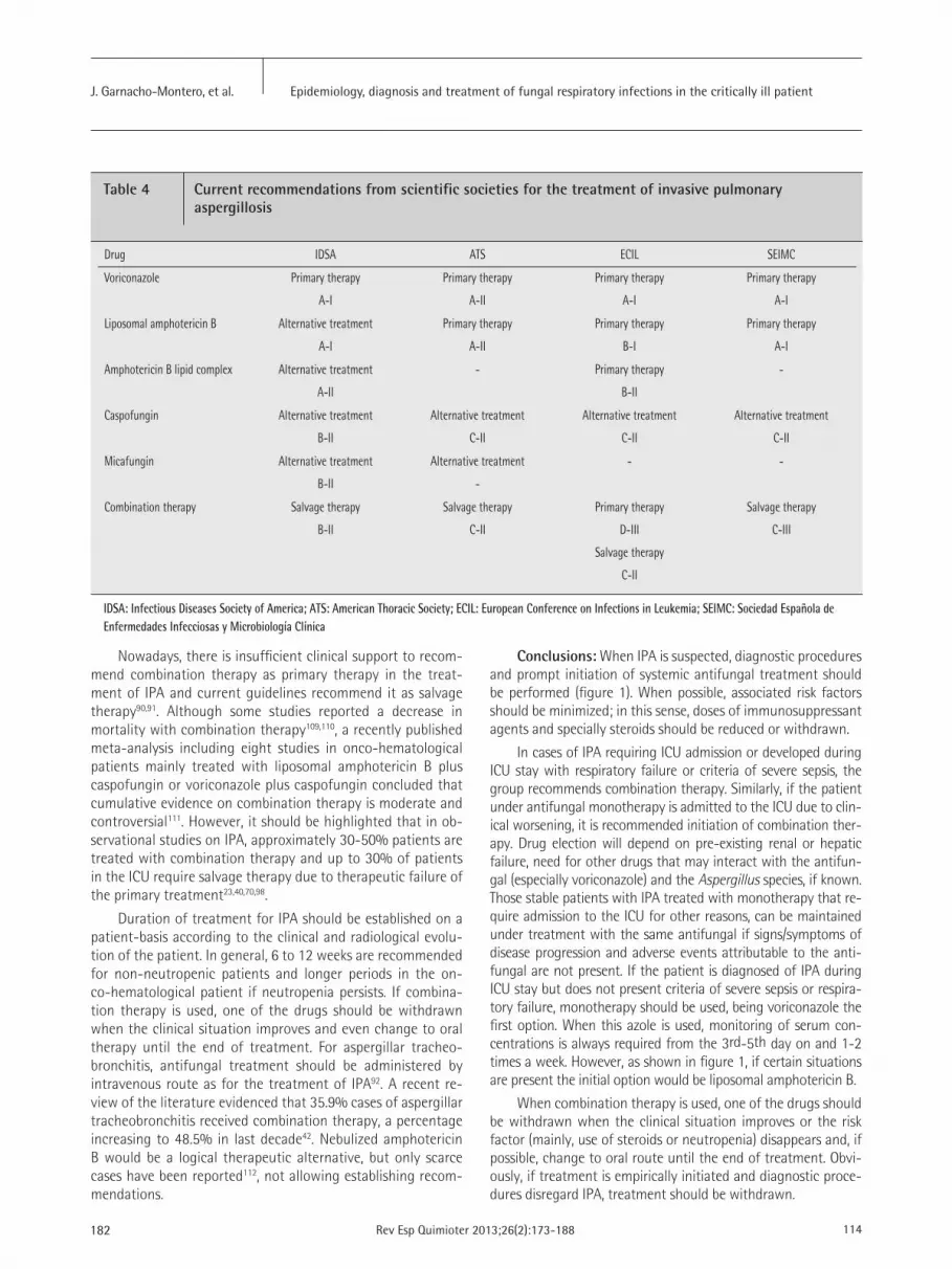
Nowadays, there is insufficient clinical support to recom-mend combination therapy as primary therapy in the treat-ment of IPA and current guidelines recommend it as salvage therapy90,91. Although some studies reported a decrease in mortality with combination therapy109,110, a recently published meta-analysis including eight studies in onco-hematological patients mainly treated with liposomal amphotericin B plus caspofungin or voriconazole plus caspofungin concluded that cumulative evidence on combination therapy is moderate and controversial111. However, it should be highlighted that in ob-servational studies on IPA, approximately 30-50% patients are treated with combination therapy and up to 30% of patients in the ICU require salvage therapy due to therapeutic failure of the primary treatment23,40,70,98.
Duration of treatment for IPA should be established on a patient-basis according to the clinical and radiological evolu-tion of the patient. In general, 6 to 12 weeks are recommended for non-neutropenic patients and longer periods in the on-co-hematological patient if neutropenia persists. If combina-tion therapy is used, one of the drugs should be withdrawn when the clinical situation improves and even change to oral therapy until the end of treatment. For aspergillar tracheo-bronchitis, antifungal treatment should be administered by intravenous route as for the treatment of IPA92. A recent re-view of the literature evidenced that 35.9% cases of aspergillar tracheobronchitis received combination therapy, a percentage increasing to 48.5% in last decade42. Nebulized amphotericin B would be a logical therapeutic alternative, but only scarce cases have been reported112, not allowing establishing recom-mendations.
Conclusions: When IPA is suspected, diagnostic procedures and prompt initiation of systemic antifungal treatment should be performed (figure 1). When possible, associated risk factors should be minimized; in this sense, doses of immunosuppressant agents and specially steroids should be reduced or withdrawn.
In cases of IPA requiring ICU admission or developed during ICU stay with respiratory failure or criteria of severe sepsis, the group recommends combination therapy. Similarly, if the patient under antifungal monotherapy is admitted to the ICU due to clin-ical worsening, it is recommended initiation of combination ther-apy. Drug election will depend on pre-existing renal or hepatic failure, need for other drugs that may interact with the antifun-gal (especially voriconazole) and the Aspergillus species, if known. Those stable patients with IPA treated with monotherapy that re-quire admission to the ICU for other reasons, can be maintained under treatment with the same antifungal if signs/symptoms of disease progression and adverse events attributable to the anti-fungal are not present. If the patient is diagnosed of IPA during ICU stay but does not present criteria of severe sepsis or respira-tory failure, monotherapy should be used, being voriconazole the first option. When this azole is used, monitoring of serum con-centrations is always required from the 3rd-5th day on and 1-2 times a week. However, as shown in figure 1, if certain situations are present the initial option would be liposomal amphotericin B.
When combination therapy is used, one of the drugs should be withdrawn when the clinical situation improves or the risk factor (mainly, use of steroids or neutropenia) disappears and, if possible, change to oral route until the end of treatment. Obvi-ously, if treatment is empirically initiated and diagnostic proce-dures disregard IPA, treatment should be withdrawn.
Epidemiology, diagnosis and treatment of fungal respiratory infections in the critically ill patientJ. Garnacho-Montero, et al.
182 114Rev Esp Quimioter 2013;26(2):173-188
Table 4 Current recommendations from scientific societies for the treatment of invasive pulmonary aspergillosis
Drug IDSA ATS ECIL SEIMC
Voriconazole Primary therapy
A-I
Primary therapy
A-II
Primary therapy
A-I
Primary therapy
A-I
Liposomal amphotericin B Alternative treatment
A-I
Primary therapy
A-II
Primary therapy
B-I
Primary therapy
A-I
Amphotericin B lipid complex Alternative treatment
A-II
- Primary therapy
B-II
-
Caspofungin Alternative treatment
B-II
Alternative treatment
C-II
Alternative treatment
C-II
Alternative treatment
C-II
Micafungin Alternative treatment
B-II
Alternative treatment
-
- -
Combination therapy Salvage therapy
B-II
Salvage therapy
C-II
Primary therapy
D-III
Salvage therapy
C-II
Salvage therapy
C-III
IDSA: Infectious Diseases Society of America; ATS: American Thoracic Society; ECIL: European Conference on Infections in Leukemia; SEIMC: Sociedad Española de Enfermedades Infecciosas y Microbiología Clínica

c) Mucormycosis
As in other fungal infections, it has been demonstrated in patients with mucormycosis that delays in initiation of anti-fungal treatment are associated with higher mortality rates113. Amphotericin B is the drug of choice and due to its lower toxicity, the use of liposomal amphotericin B is recommend-ed90,114,115. Doses to be used should range from 5 to 10 mg/kg/day5,115,116. Treatment duration is not defined and should be individually chosen in any case.
Posaconazole may be an alternative in combination with liposomal amphotericin B in case of therapeutic failure or in-tolerance. Monotherapy with this azole is not recommended, monitoring of serum concentrations is required and existing interactions with a high number of compounds should be tak-en into account117.
Echinocandins do not exhibit in vitro activity against Mu-corales, although some technical reasons and experimental
data suggest that when combined with amphotericin B, they may be of clinical utility118. A retrospective series of patients with rhinocerebral mucormycosis suggested that the combi-nation of caspofungin and a lipid formulation of amphotericin B reduces mortality119. This therapeutic strategy has not been reported for pulmonary mucormycosis.
Conclusions: When pulmonary mucormycosis is suspected, antifungal treatment should be initiated without delay. Liposo-mal amphotericin B at doses of at least 5 mg/kg/day is recom-mended. Its use as empirical treatment has the added value of effectively covering a possible IPA, an infection with very similar clinical and radiological presentations to mucormycosis.
PROPHYLAXIS OF RESPIRATORY FUNGAL INFECTIONS
There are different strategies of demonstrated utility for
Epidemiology, diagnosis and treatment of fungal respiratory infections in the critically ill patientJ. Garnacho-Montero, et al.
115 183Rev Esp Quimioter 2013;26(2):173-188
Table 5 Voriconazole interactions
Type of interaction Recommendation
The drug decreases voriconazole concentrations
Carbamazepine Contraindicated
Rifampicin Contraindicated
Phenobarbital Contraindicated
Ritonavir Contraindicated
Voriconazole increases drug concentrations
Astemizole Contraindicated
Cisapride Contraindicated
Cyclosporine Reduce dose by half and monitor concentrations
Ergotamine alkaloids Contraindicated
Omeprazole Reduce dose by half
Quinidine Contraindicated
Sirolimus Contraindicated
Everolimus Reduce dose
Tacrolimus Reduce dose by two third and monitor concentrations
Terfenadine Contraindicated
Warfarin Monitor prothrombin time
The drug decreases voriconazole concentrations and voriconazole increases drug concentrations
Rifabutin Contraindicated
Phenytoin Double voriconazole dose and monitor phenytoin concentrations
Voriconazole probably increases drug concentrations
Statins Consider reduction
Sulphonylureas
Calcium channel blockers
Benzodiazepines

184 116Rev Esp Quimioter 2013;26(2):173-188
the prevention of invasive fungal infections in high-risk patients with systemic antifungal administration, but its review is out of the scope of this document. On the other hand, review of avail-able data on nebulized antifungal administration, an attractive option for IPA prevention is addressed. Up to now, this route of administration has only been employed for amphotericin B and its lipid formulations, with clinical experience mainly in lung transplant recipients and neutropenic patients. It should be highlighted that although it is common practice and support-ed by extensive scientific literature, none of the amphotericin B formulations is approved for use via nebulization.
In lung transplant recipients, two studies carried out in our country demonstrated the lack of systemic absorption of nebu-lized liposomal amphotericin B and demonstrated persistence of therapeutic concentrations in the epithelial lining fluid (ELF) for 14 days120,121. Recently, therapeutic concentrations in ELF fol-lowing nebulization of amphotericin B lipid complex have been confirmed, but using higher doses and shorter dosing intervals122. With regard to its effect in preventing IPA, a Spanish study ret-rospectively reviewed 60 cases of lung transplant recipients fol-lowed during 6 months. Nebulization of amphotericin B lipid complex was efficacious since only one patient developed a prob-able IPA123. A clinical trial including 100 lung transplant recipients randomized to amphotericin B deoxycholate or amphotericin B lipid complex as aerosolized prophylaxis showed similar efficacy of both compounds in the prevention of infection by Aspergillus124.
Two clinical trials compared nebulized liposomal ampho-tericin B versus placebo in onco-hematological patients with neutropenia present for more than 10 days, and showed a significant decrease in IPA episodes without association with severe adverse events125,126. In fact, the European Conference on Infections in Leukemia (ECIL) recommends (B-II) as prophy-laxis of invasive fungal infections, nebulization of liposomal amphotericin B (together with intravenous fluconazole) in the neutropenia phase of the allogeneic hematopoietic progenitor cell transplantation. In the same circumstance, nebulization of conventional amphotericin B is not recommended (D-I)127.
Nebulized liposomal amphotericin B at the standard 25 mg dose does not alter the pulmonary function nor the com-position of the pulmonary surfactant121,128. It is well known that, in lung transplant recipients, there is a dysfunction of the pulmonary surfactant associated with a dysfunction of the graft, and for this reason, it is important that none exogenous agent alters the composition of the surfactant129.
A jet nebulizer with a high flow compressor should be used to obtain an adequate particle size (from 3 to 5μ)130. When choosing the nebulizer, the compound to be nebulized should be considered since doses of the two available lipid formulations are different and thus, the volume to be nebulized: 25 mg (5 ml) liposomal amphotericin B or 50 mg (10 ml) amphotericin B lipid complex. Some authors recommend loading doses during four consecutive days, doubling doses in intubated patients. Af-terwards, it can be administered 2-3 times/week. An exhaustive bronchial hygiene prior to nebulization is crucial. Contamination of nebulization systems can be the origin of respiratory infec-
tions and thus, strict disinfection protocols should be followed. In addition, obstructions of ventilator filters may occur and this possibility should be closely monitored131.
Conclusions: The use of amphotericin B is a strategy with demonstrated utility for prevention of IPA in high-risk patients, especially in lung transplant recipients and onco-hematological patients with prolonged neutropenia, being the liposomal formu-lation recommended in clinical practice guidelines. Although now-adays there is not a single compound approved for administration via nebulization, its use has become common practice in high-risk patients. Advantages of liposomal amphotericin B nebulization are easy administration, undetectable serum concentrations, excellent tolerance with mild and well tolerated adverse events, low risk of infection by emergent molds and cost-effectiveness.
However, its use as prophylaxis in the critically ill patient has not been established yet. A theoretical possibility would be its use in COPD patients treated with high doses of steroids admitted to the ICU. With current data, no formal recommendation for use can be done and responsible clinicians should examine cases individually.
REFERENCES
1. Vincent JL, Rello J, Marshall J, Silva E, Anzueto A, Martin CD, et al. International study of the prevalence and outcomes of infection in intensive care units. JAMA 2009; 302:2323-9.
2. Alvarez-Lerma F, Palomar M, León C, Olaechea P, Cerdá E, Bermejo B, et al. Colonización y/o infección por hongos en unidades de cui-dados intensivos. Estudio multicéntrico de 1.562 pacientes. Med Clin (Barc) 2003; 121:161-6.
3. Estudio ENVIN. http://hws.vhebron.net/envin-helics/4. Tortorano AM, Dho G, Prigitano A, Breda G, Grancini A, Emmi V,
et al. Invasive fungal infections in the intensive care unit: a mul-ticentre, prospective, observational study in Italy (2006-2008). Mycoses 2012; 55:73-9.
5. Smith JA, Kauffman CA. Pulmonary fungal infections. Respirology 2012; 17:913-26.
6. Meersseman W, Lagrou K, Spriet I, Maertens J, Verbeken E, Pee-termans WE, et al. Significance of the isolation of Candida species from airway samples in critically ill patients: a prospective, autop-sy study. Intensive Care Med 2009; 35:1526-31.
7. Azoulay E, Timsit JF, Tafflet M, de Lassence A, Darmon M, Zahar JR, et al. Candida colonization of the respiratory tract and sub-sequent pseudomonas ventilator-associated pneumonia. Chest 2006;129:110-7.
8. Segal BH. Aspergillosis. N Engl J Med 2009; 360:1870-84.9. Latge JP. Aspergillus fumigatus and aspergillosis, Clin Microbiol
Rev 1999; 12: 310-50.10. Castón JJ, Linares MJ, Gallego C, Rivero A, Font P, Solís F, et al. Risk
factors for pulmonary Aspergillus terreus infection in patients with positive culture for filamentous fungi. Chest 2007; 131:230-6.
11. Peláez T, Muñoz P, Guinea J, Valerio M, Giannella M, Klaassen CH, et al. Outbreak of invasive aspergillosis after major heart surgery caused by spores in the air of the intensive care unit. Clin Infect Dis 2012; 54:e24-31.
Epidemiology, diagnosis and treatment of fungal respiratory infections in the critically ill patientJ. Garnacho-Montero, et al.

117 185Rev Esp Quimioter 2013;26(2):173-188
12. Dutkiiewick R, Hage A. Aspergillus infections in the critically ill. Proc Am Thor Soc 2010;7:204-9.
13. Pemán J, Salavert M. Epidemiología general de la enfermedad fún-gica invasora. Enferm Infecc Microbiol Clin 2012; 30:90-8.
14. Perfect JR, Cox GM, Lee JY, Kauffman CA, de Repentigny L, Chap-man SW, et al. The impact of culture isolation of Aspergillus spe-cies: a hospital-based survey of aspergillosis. Clin Infect Dis 2001; 33:1824-33.
15. Bouza E, Guinea J, Peláez T, Pérez-Molina J, Alcalá L, Muñoz P. Workload due to Aspergillus fumigatus and significance of the or-ganism in the microbiology laboratory of a general hospital. J Clin Microbiol 2005; 43:2075-9.
16. Graf K, Khani SM, Ott E, Mattner F, Gastmeier P, Sohr D, et al. Five-years surveillance of invasive aspergillosis in a university hospital. BMC Infect Dis 2011; 11:163.
17. Garnacho-Montero J, Amaya-Villar R, Ortiz-Leyba C, León C, Alva-rez-Lerma F, Nolla-Salas J et al. Isolation of Aspergillus spp. from the respiratory tract in critically ill patients: risk factors, clinical presentation and outcome. Crit Care 2005; 9:R191-9.
18. Vandewoude KH, Blot SI, Depuydt P, Benoit D, Temmerman W, Co-lardyn F et al. Clinical relevance of Aspergillus isolation from respi-ratory tract samples in critically ill patients. Crit Care 2006; 10:R31
19. Meersseman W, Vandecasteele SJ, Wilmer A, Verbeken E, Peetermans WE, Van Wijngaerden E. Invasive aspergillosis in critically ill patients without malignancy. Am J Respir Crit Care Med 2004; 170:621-5.
20. Ng TT, Robson GD, Denning DW. Hydrocortisone-enhanced growth of Aspergillus spp: implications for pathogenesis. Microbiology 1994; 140: 2475-9.
21. Hartemink KJ, Paul MA, Spijkstra JJ, Girbes AR, Polderman KH. In-munoparalysis as a cause for invasive aspergilosis? Intensive Care Med 2003; 29: 2068-71.
22. Schaffner A. Therapeutic concentrations of glucocorticoids sup-press the antimicrobial activity of human macrophages without impairing their responsiveness to gamma interferon. J Clin Invest 1985; 76:1755–64.
23. Guinea J, Torres-Narbona M, Gijón P, Muñoz P, Pozo F, Peláez T et al. Pulmonary aspergillosis in patients with chronic obstructive pulmonary disease: incidence, risk factors, and outcome. Clin Mi-crobiol Infect 2010; 16:870-7.
24. Xu H, Li L, Huang WJ, Wang LX, Li WF, Yuan WF. Invasive pulmo-nary aspergillosis in patients with chronic obstructive pulmonary disease: a case control study from China. Clin Microbiol Infect 2012; 18:403-8.
25. Fortún J, Meije Y, Fresco G, Moreno S. Aspergilosis. Formas clínicas y tratamiento. Enferm Infecc Microbiol Clin 2012; 30:201-8.
26. Fortún J, Martín-Dávila P, Moreno S, De Vicente E, Nuño J, Cande-las A, et al. Risk factors for invasive aspergillosis in liver transplant recipients. Liver Transpl 2002; 8:1065-70.
27. Gavalda J, Len O, San Juan R, Aguado JM, Fortun J, Lumbreras C et al. Risk factors for invasive aspergillosis in solid-organ transplant recipients: a case-control study. Clin Infect Dis 2005; 41:52-9.
28. Sole A, Morant P, Salavert M, Peman J, Morales P; Valencia Lung Transplant Group. Aspergillus infections in lung transplant re-cipients: risk factors and outcome. Clin Microbiol Infect 2005; 11:359–365.
29. Marik PE. Fungal infections in solid organ transplantation. Expert Opin Pharmacother 2006; 7:297–305.
30. Solé A, Salavert M. Fungal infections after lung transplantation. Curr Opin Pulm Med 2009; 15:243-53.
31. Shahid H. Unique characteristics of fungal infections in lung transplant recipients. Clin Chest Med 2009; 30:307–313.
32. Petrikkos G, Skiada A, Lortholary O, Roilides E, Walsh TJ, Konto-yiannis DP. Epidemiology and clinical manifestations of mucorm-ycosis. Clin Infect Dis 2012; 54 (Suppl 1):S23-34.
33. Skiada A, Pagano L, Groll A, Zimmerli S, Dupont B, Lagrou K et al. Zygomycosis in Europe: analysis of 230 cases accrued by the regis-try of the European Confederation of Medical Mycology (ECMM) Working Group on Zygomycosis between 2005 and 2007. Clin Mi-crobiol Infect 2011; 17:1859-67.
34. Marty FM, Cosimi LA, Baden LR. Breakthrough zygomycosis after voriconazole treatment in recipients of hematopoietic stem-cell transplants. N Engl J Med 2004; 350:950–2.
35. Siwek GT, Dodgson KJ, de Magalhaes-Silverman M, Bartelt LA, Kilborn SB, Hoth PL et al. Invasive zygomycosis in hematopoietic stem cell transplant recipients receiving voriconazole prophylaxis. Clin Infect Dis 2004; 39:584-7.
36. Kontoyiannis DP, Reddy BT, Torres HA, Luna M, Lewis RE, Tarrand J et al. Pulmonary candidiasis in patients with cancer: an autopsy study. Clin Infect Dis 2002; 34:400-3.
37. Garnacho Montero J, Doblas A. Aspergilosis invasiva en el paciente crítico: Diagnóstico, planteamiento terapéutico y nuevas tenden-cias. Med Intensiva 2006; Supl 4: 38-46.
38. Routsi C, Kaltsas P, Bessis E, Rontogianni D, Kollias S, Roussos C. Airway obstruction and acute respiratory failure due to Aspergillus tracheobronchitis. Crit Care Med 2004; 32:580-582.
39. Parrón M, Torres I, Pardo M, Morales C, Navarro M, Martínez-Sch-mizcraft M. The halo sign in computed tomography images: di-fferential diagnosis and correlation with pathology findings. Arch Bronconeumol 2008; 44:386-92.
40. Burghi G, Lemiale V, Seguin A, Lambert J, Lacroix C, Canet E et al. Outcomes of mechanically ventilated hematology patients with invasive pulmonary aspergillosis. Intensive Care Med 2011; 37:1605-12.
41. Kontoyiannis DP, Sumoza D, Tarrand J, Bodey GP, Storey R, Raad II. Significance of aspergillemia in patients with cancer: a 10-year study. Clin Infect Dis 2000; 31:188-9.
42. Fernández-Ruiz M, Silva JT, San-Juan R, de Dios B, García-Luján R, López-Medrano F et al. Aspergillus tracheobronchitis: report of 8 ca-ses and review of the literature. Medicine (Baltimore) 2012; 9:261-73.
43. Viscoli C, Machetti M, Cappellano P, Bucci B, Bruzzi P, Van Lint MT et al. False-positive galactomannan platelia Aspergillus test re-sults for patients receiving piperacillin-tazobactam. Clin Infect Dis 2004; 38:913-6.
44. Cuenca-Estrella M. Diagnóstico de laboratorio de la enfer-medad fúngica invasora. Enferm Infecc Microbiol Clin 2012; 30:257–64.
45. Pfeiffer CD, Fine JP, Safdar N. Diagnosis of invasive aspergillosis using a galactomannan assay: a meta-analysis. Clin Infect Dis 2006; 42:1417-27.
46. He H, Ding L, Li F, Zhan Q. Clinical features of invasive bronchial-pulmonary aspergillosis in critically ill patients with chronic obs-tructive respiratory diseases: a prospective study. Crit Care 2011; 15:R5.
47. Becker MJ, Lugtenburg EJ, Cornelissen JJ, Van Der Schee C, Hoogs-
Epidemiology, diagnosis and treatment of fungal respiratory infections in the critically ill patientJ. Garnacho-Montero, et al.

186 118Rev Esp Quimioter 2013;26(2):173-188
Epidemiology, diagnosis and treatment of fungal respiratory infections in the critically ill patientJ. Garnacho-Montero, et al.
teden HC, De Marie S. Galactomannan detection in computerized tomography-based broncho-alveolar lavage fluid and serum in haematological patients at risk for invasive pulmonary aspergillo-sis. Br J Haematol 2003; 121:448–57.
48. Maertens J, Maertens V, Theunissen K, Meersseman W, Meersse-man P, Meers S et al. Bronchoalveolar lavage fluid galactomannan for the diagnosis of invasive pulmonary aspergillosis in patients with hematologic diseases. Clin Infect Dis 2009; 49:1688–93.
49. Meersseman W, Lagrou K, Maertens J, Wilmer A, Hermans G, Van-derschueren S et al. Galactomannan in bronchoalveolar lavage fluid: a tool for diagnosing aspergillosis in intensive care unit pa-tients. Am J Respir Crit Care Med 2008; 177:27–34.
50. Acosta J, Catalan M, del Palacio-Peréz-Medel A, Lora D, Montejo JC, Cuetara MS et al. A prospective comparison of galactomannan in bronchoalveolar lavage fluid for the diagnosis of pulmonary in-vasive aspergillosis in medical patients under intensive care: com-parison with the diagnostic performance of galactomannan and of (1→ 3)-β-d-glucan chromogenic assay in serum samples. Clin Microbiol Infect 2011; 17:1053-60.
51. He H, Ding L, Sun B, Li F, Zhan Q. Role of galactomannan determi-nations in bronchoalveolar lavage fluid samples from critically ill patients with chronic obstructive pulmonary disease for the diag-nosis of invasive pulmonary aspergillosis: a prospective study. Crit Care 2012; 16:R138.
52. Clancy CJ, Jaber RA, Leather HL, Wingard JR, Staley B, Wheat LJ et al. Bronchoalveolar lavage galactomannan in diagnosis of invasive pulmonary aspergillosis among solid-organ transplant recipients. J Clin Microbiol 2007; 45:1759-65.
53. Lamoth F, Cruciani M, Mengoli C, Castagnola E, Lortholary O, Ri-chardson M et al. β-Glucan antigenemia assay for the diagnosis of invasive fungal infections in patients with hematological malig-nancies: a systematic review and meta-analysis of cohort studies from the Third European Conference on Infections in Leukemia (ECIL-3). Clin Infect Dis 2012; 54:633-43.
54. Hope WW, Walsh TJ, Denning DW. Laboratory diagnosis of invasive aspergillosis. Lancet Infect Dis 2005; 5:609-22.
55. Ayats J, Martín-Mazuelos E, Pemán J, Quindos G, Sanchez F, Gar-cia-Rodriguez J et al. Recomendaciones sobre el diagnóstico de la enfermedad fúngica invasora de la Sociedad Española de Enferme-dades Infecciosas y Microbiología Clínica (SEIMC). Actualización 2010. Enferm Infecc Microbiol Clin 2011; 29:39–9.
56. Ostrosky-Zeichner L, Alexander BD, Kett DH, Vazquez J, Pappas PG, Saeki F et al. Multicenter clinical evaluation of the (1-->3) beta-D-glucan assay as an aid to diagnosis of fungal infections in humans. Clin Infect Dis 2005; 41:654–9.
57. Pazos C, Ponton J, Del Palacio A. Contribution of (1->3)-beta-D-glucan chromogenic assay to diagnosis and therapeutic monito-ring of invasive aspergillosis in neutropenic adult patients: a com-parison with serial screening for circulating galactomannan. J Clin Microbiol 2005; 43:299–305.
58. De Vlieger G, Lagrou K, Maertens J, Verbeken E, Meersseman W, Van Wijngaerden E. Beta-D-glucan detection as a diagnostic test for invasive aspergillosis in immunocompromised critically ill pa-tients with symptoms of respiratory infection: an autopsy-based study. J Clin Microbiol 2011; 49:3783-7.
59. Luong ML, Clancy CJ, Vadnerkar A, Kwak EJ, Silveira FP, Wissel MC et al. Comparison of an Aspergillus real-time polymerase chain
reaction assay with galactomannan testing of bronchoalvelolar la-vage fluid for the diagnosis of invasive pulmonary aspergillosis in lung transplant recipients. Clin Infect Dis 2011; 52:1218-26.
60. Mengoli C, Cruciani M, Barnes RA, Loeffler J, Donnelly JP. Use of PCR for diagnosis of invasive aspergillosis: systematic review and meta-analysis. Lancet Infect Dis 2009; 9:89-96.
61. Georgiadou SP, Sipsas NV,Marom EM, Kontoyiannis DP. The diag-nostic value of halo and reversed halo signs for invasive mold in-fections in compromised hosts. Clin Infect Dis 2011; 52:1144–55.
62. Walsh TJ, Gamaletsou MN, McGinnis MR, Hayden RT, Kontoyiannis DP. Early clinical and laboratory diagnosis of invasive pulmonary, extrapulmonary, and disseminated mucormycosis (zygomycosis). Clin Infect Dis 2012; 54 (Suppl 1):S55-60.
63. Rello J, Esandi ME, Díaz E, Mariscal D, Gallego M, Vallès J. The role of Candida sp isolated from bronchoscopic samples in nonneutro-penic patients. Chest 1998; 114:146-9.
64. el-Ebiary M, Torres A, Fàbregas N, de la Bellacasa JP, González J, Ramirez J et al. Significance of the isolation of Candida species from respiratory samples in critically ill, non- neutropenic pa-tients. An immediate postmortem histologic study. Am J Respir Crit Care Med 1997; 156:583-90.
65. Nseir S, Jozefowicz E, Cavestri B, Sendid B, Di Pompeo C, Dewavrin F et al. Impact of antifungal treatment on Candida-Pseudomonas interaction: a preliminary retrospective case-control study. Inten-sive Care Med 2007; 33:137-42.
66. Hamet M, Pavon A, Dalle F, Pechinot A, Prin S, Quenot JP et al. Candida spp. airway colonization could promote antibiotic-resis-tant bacteria selection in patients with suspected ventilator-asso-ciated pneumonia. Intensive Care Med 2012; 38:1272-9.
67. Garnacho-Montero J, Díaz-Martín A, Cayuela-Dominguez A. Ma-nagement of invasive Candida infections in non-neutropenic cri-tically ill patients: from prophylaxis to early therapy. Int J Antimi-crob Agents 2008; 32 (Suppl 2):S137-41.
68. Horvath JA, Dummer S. The use of respiratory-tract cultures in the diagnosis of invasive pulmonary aspergillosis. Am J Med 1996; 100:171–8.
69. Mortensen KL, Johansen HK, Fuursted K, Knudsen JD, Gahrn-Han-sen B, Jensen RH et al. A prospective survey of Aspergillus spp. in respiratory tract samples: prevalence, clinical impact and antifun-gal susceptibility. Eur J Clin Microbiol Infect Dis 2011; 30:1355-63.
70. Lucena P, Barberán J, Eroles G, Granizo JJ, Giménez MJ, Mir N et al. Significance of lower respiratory tract cultures yielding Aspergillus spp. growth in a hospital without transplant patients. Rev Esp Qui-mioter 2010; 23:190-5.
71. Khasawneh F, Mohamad T, Moughrabieh MK, Lai Z, Ager J, Souba-ni AO. Isolation of Aspergillus in critically ill patients: a potential marker of poor outcome. J Crit Care 2006; 21:322-7.
72. De Pauw B, Walsh TJ, Donnelly JP, Stevens DA, Edwards JE, Ca-landra T et al. Revised definitions of invasive fungal disease from the European Organization for Research and Treatment of Cancer/Invasive Fungal Infections Cooperative Group and the National Institute of Allergy and Infectious Diseases Mycoses Study Group (EORTC/MSG) Consensus Group. Clin Infect Dis 2008; 46:1813-21.
73. Garnacho-Montero J, Amaya-Villar R. A validated clinical approach for the management of aspergillosis in critically ill patients: ready, steady, go! Crit Care 2006; 10:132.
74. Blot SI, Taccone FS, Van den Abeele AM, Bulpa P, Meersseman W,

Epidemiology, diagnosis and treatment of fungal respiratory infections in the critically ill patientJ. Garnacho-Montero, et al.
119 187Rev Esp Quimioter 2013;26(2):173-188
Brusselaers N et al. A clinical algorithm to diagnose invasive pul-monary aspergillosis in critically ill patients. Am J Respir Crit Care Med 2012; 186:56-64.
75. Bulpa PA, Dive AM, Garrino MG, Delos MA, Gonzalez MR, Evrard PA et al. Chronic obstructive pulmonary disease patients with in-vasive pulmonary aspergillosis: benefits of intensive care? Intensi-ve Care Med 2001; 27:59-67.
76. Bulpa P, Dive A, Sibille Y. Invasive pulmonary aspergillosis in pa-tients with chronic obstructive pulmonary disease. Eur Respir J 2007; 30:782-800.
77. Ruiz-Camps I, Cuenca-Estrella M. Antifúngicos para uso sistémico. Enferm Infecc Microbiol Clin 2009; 27:353-62.
78. Lepak AJ, Andes DR. Antifungal PK/PD considerations in fungal pulmonary infections. Semin Respir Crit Care Med 2011; 32: 783-94.
79. Azanza Perea JR, Barberán J. Anfotericina B forma liposómica: un perfil farmacocinético exclusivo. Una historia inacabada. Rev Esp Quimioter 2012; 25:17-24.
80. Ostrosky-Zeichner L, Man KA, Cohen SH. Amphotericin B: Time for a new “gold standard”. Clin Infect Dis 2003; 37:415-25.
81. Moen MD, Lyseng-Williamson KA, Scott LJ. Liposomal amphoteri-cin B: a review of its use as empirical therapy in febrile neutrope-nia and in the treatment of invasive fungal infections. Drugs 2009; 69:361-92.
82. Wingard JR, White MH, Anaissie E, Raffalli J, Goodman J, Arrieta A et al. A randomized, double-blind comparative trial evaluating the sa-fety of liposomal amphotericin B versus amphotericin B lipid com-plex in the empirical treatment of febrile neutropenia. L Amph/ABLC Collaborative Study Group. Clin Infect Dis 2000; 31:1155-63.
83. Hachem RY, Boktour MR, Hanna HA, Husni RN, Torres HA, Afif C et al. Amphotericin B lipid complex versus liposomal amphotericin B monotherapy for invasive aspergillosis in patients with hematolo-gic malignancy. Cancer 2008; 112: 1282-7.
84. Alvarez-Lerma F, Soriano MC, Rodríguez M, Catalán M, Llorente AM, Vidart N et al. Impacto de anfotericina B liposomal en la fun-ción renal en pacientes críticos con la función renal deteriorada. Rev Esp Quimioter 2012; 25:206-15.
85. Sucher AJ, Chahine EB, Balcer HE. Echinocandins: the newest class of antifungals. Ann Pharmacother 2009; 43:1647-57.
86. Garnacho Montero J, León Gil C, Almirante Gragera B, Álvarez Ler-ma F, Cuenca Estrella M, García Rodríguez JA et al. Recomenda-ciones terapéuticas en el paciente crítico no neutropénico. Med Intensiva 2005; Supl. 3: 43-52.
87. Pappas PG, Kauffman CA, Andes D, Benjamin DK Jr, Calandra TF, Edwards JE Jr et al. Clinical practice guidelines for the manage-ment of candidiasis: 2009 update by the Infectious Diseases Socie-ty of America. Clin Infect Dis 2009; 48:503-35.
88. Cornely OA, Maertens J, Bresnik M, Ebrahimi R, Dellow E, Herbre-cht R et al. Efficacy outcomes in a randomised trial of liposomal amphotericin B based on revised EORTC/MSG 2008 definitions of invasive mould disease. Mycoses 2011; 54:e449-55.
89. Baddley JW, Stephens JM, Ji X, Gao X, Schlamm HT, Tarallo M. As-pergillosis in Intensive Care Unit (ICU) patients: epidemiology and economic outcomes. BMC Infect Dis 2013; 13:29.
90. Fortún J, Carratalá J, Gavaldá J, Lizasoain M, Salavert M, de la Cá-mara R et al. Recomendaciones sobre el tratamiento de la enfer-medad fúngica invasiva por Aspergillus spp. y otros hongos fila-
mentosos de la Sociedad Española de Enfermedades Infecciosas y Microbiología Clínica (SEIMC). Actualización 2011. Enferm Infecc Microbiol Clin 2011; 29:435-54.
91. Walsh TJ, Anaissie EJ, Denning DW, Herbrecht R, Kontoyiannis DP, Marr KA et al. Treatment of aspergillosis: clinical practice guideli-nes of the Infectious Diseases Society of America. Clin Infect Dis 2008; 46:327-60.
92. Limper AH, Knox KS, Sarosi GA, Ampel NM, Bennett JE, Catanzaro A et al. An official American Thoracic Society statement: Treatment of fungal infections in adult pulmonary and critical care patients. Am J Respir Crit Care Med 2011; 183:96-128.
93. Meersseman W, Lagrou K, Maertens J, Van Wijngaerden E. Inva-sive aspergillosis in the intensive care unit. Clin Infect Dis 2007; 45:205-16.
94. Herbrecht R, Denning DW, Patterson TF, Bennett JE, Greene RE, Oestmann JW et al. Voriconazole versus amphotericin B for primary therapy of invasive aspergillosis. N Engl J Med 2002; 347:408-15.
95. Jørgensen KJ, Johansen HK, Gøtzsche PC. Flaws in design, analysis and interpretation of Pfizer’s antifungal trials of voriconazole and uncritical subsequent quotations. Trials 2006; 7:3.
96. Denning DW, Ribaud P, Milpied N, Caillot D, Herbrecht R, Thiel E et al. Efficacy and safety of voriconazole in the treatment of acute invasive aspergillosis. Clin Infect Dis 2002; 34:563-71.
97. Jacobs F, Selleslag D, Aoun M, Sonet A, Gadisseur A. An observa-tional efficacy and safety analysis of the treatment of acute inva-sive aspergillosis using voriconazole. Eur J Clin Microbiol Infect Dis 2012; 31:1173-9.
98. Johnson LB, Kauffman CA. Voriconazole: a new triazole antifungal agent. Clin Infect Dis 2003; 36: 630-7.
99. Pascual A, Calandra T, Bolay S, Buclin T, Bille J, Marchetti O. Vo-riconazole therapeutic drug monitoring in patients with invasi-ve mycoses improves efficacy and safety outcomes. Clin Infect Dis 2008; 46:201-11.
100. Jeong S, Nguyen PD, Desta Z. Comprehensive in vitro analysis of voriconazole inhibition of eight cytochrome P450 (CYP) enzy-mes: major effect on CYPs 2B6, 2C9, 2C19, and 3A. Antimicrob Agents Chemother 2009; 53:541-51.
101. Park WB, Kim NH, Kim KH, Lee SH, Nam WS, Yoon SH et al. The effect of therapeutic drug monitoring on safety and efficacy of voriconazole in invasive fungal infections: a randomized contro-lled trial. Clin Infect Dis 2012; 55:1080-7.
102. Hafner V, Czock D, Burhenne J, Riedel KD, Bommer J, Mikus G et al. Pharmacokinetics of sulfobutylether-beta-cyclodextrin and vori-conazole in patients with end-stage renal failure during treatment with two hemodialysis systems and hemodiafiltration. Antimicrob Agents Chemother 2010; 54:2596-602.
103. Solís-Muñoz P, López JC, Bernal W, Willars C, Verma A, Heneghan MA et al. Voriconazole hepatotoxicity in severe liver dysfunction. J Infect 2013; 66:80-6.
104. Cornely OA, Maertens J, Bresnik M, Ebrahimi R, Ullmann AJ, Bouza E. Liposomal amphotericin B as initial therapy for invasive mold infection: a randomized trial comparing a high-loading dose re-gimen with standard dosing (AmBiLoad trial). Clin Infect Dis 2007; 44:1289–97.
105. Hachem RY, Boktour MR, Hanna HA, Husni RN, Torres HA, Afif C et al. Amphotericin B lipid complex versus liposomal amphotericin B

Epidemiology, diagnosis and treatment of fungal respiratory infections in the critically ill patientJ. Garnacho-Montero, et al.
188 120Rev Esp Quimioter 2013;26(2):173-188
monotherapy for invasive aspergillosis in patients with hematolo-gic malignancy. Cancer 2008; 112:1282-7.
106. Maertens J, Raad I, Petrikkos G, Boogaerts M, Selleslag D, Pe-tersenFB et al. Efficacy and safety of caspofungin for treatment of invasive aspergillosis in patients refractory to or intolerant of conventional antifungal therapy. Clin Infect Dis 2004; 39:1563-71.
107. Viscoli C, Herbrecht R, Akan H, Baila L, Sonet A, Gallamini A et al. An EORTC Phase II study of caspofungin as first-line therapy of in-vasive aspergillosis in haematological patients. J Antimicrob Che-mother 2009; 64:1274–81.
108. Cornely OA, Vehreschild JJ, Vehreschild MJ, Würthwein G, Arenz D, Schwartz S et al. Phase II dose escalation study of caspofun-gin for invasive Aspergillosis. Antimicrob Agents Chemother 2011; 55:5798-803.
109. Marr KA, Baeck M, Carter RA, Kim HW, Grey L. Combination an-tifungal therapy for invasive aspergillosis. Clin Infect Dis 2004; 39:797-802.
110. Singh N, Limaye AP, Forrest G, Safdar N, Muñoz P, Pursell K et al. Combination of voriconazole and caspofungin as primary thera-py for invasive aspergillosis in solid organ transplant recipients: A prospective, multicenter, observational study. Transplantation 2006; 81:320-6.
111. Garbati MA, Alasmari FA, Al-Tannir MA, Tleyjeh IM. The role of combination antifungal therapy in the treatment of invasive as-pergillosis: a systematic review. Int J Infect Dis 2012; 16:e76-81.
112. García-Gallo CL, García-Fadul C, Laporta-Hernández R, Ussetti-Gil P. Traqueobronquitis aspergilar en paciente sometido a trasplante pulmonar. Rev Iberoam Micol 2011; 28:129-33.
113. Chamilos G, Lewis RE, Kontoyiannis DP. Delaying amphotericin B–based frontline therapy significantly increases mortality among patients with hematologic malignancy who have zygomycosis. Clin Infect Dis 2008; 47:503–9.
114. Kontoyiannis DP, Lewis R. How I treat mucormycosis. Blood 2011; 118:1216–24.
115. Skiada A, Lanternier F, Groll AH, Pagano L, Zimmerli S, Herbrecht R et al. Diagnosis and treatment of mucormycosis in patients with haematological malignancies: guidelines from the 3rd European Conference on Infections in Leukemia (ECIL 3). Haematologica 2012 Sep 14 [Epub ahead of print]
116. Lewis RE, Albert ND, Liao G, Hou J, Prince RA, Kontoyiannis DP. Comparative pharmacody- namics of amphotericin B lipid com-plex and liposomal ampho- tericin B in a murine model of pul-monary mucormycosis. Antimicrob Agents Chemother 2010; 54:1298–304.
117. Garnacho-Montero J, Jiménez Parrilla F. Interacciones farmacoló-gicas en el paciente crítico. ¿Un factor relevante para usar mica-fungina? Enferm Infecc Microbiol Clin 2011; 29 (Suppl 2):33-7.
118. Spellberg B, Fu Y, Edwards JE, Jr, Ibrahim AS. Combination the-rapy with amphotericin B lipid complex and caspofungin ace-tate of disseminated zygomycosis in diabetic ketoacidotic mice. Antimicrob Agents Chemother 2005; 49:830–2.
119. Reed C, Bryant R, Ibrahim AS, Edwards J Jr, Filler SG, Goldberg R et al. Combination polyene-caspofungin treatment of rhino-orbital-cerebral mucormycosis. Clin Infect Dis 2008; 47:364-71.
120. Monforte V, Ussetti P, López R, Gavaldà J, Bravo C, de Pablo A et al. Nebulized liposomal amphotericin B prophylaxis for Aspergillus
infection in lung transplantation: pharmacokinetics and safety. J Heart Lung Transplant 2009; 28:170-5.
121. Monforte V, Ussetti P, Gavaldà J, Bravo C, Laporta R, Len O et al. Feasibility, tolerability, and outcomes of nebulized liposomal am-photericin B for Aspergillus infection prevention in lung trans-plantation. J Heart Lung Transplant 2010; 29:523-30.
122. Husain S, Capitano B, Corcoran T, Studer SM, Crespo M, Johnson B et al. Intrapulmonary disposition of amphotericin B after aero-solized delivery of amphotericin B lipid complex (Abelcet; ABLC) in lung transplant recipients. Transplantation 2010; 90:1215-9.
123. Borro JM, Solé A, de la Torre M, Pastor A, Fernandez R, Saura A et al. Efficiency and safety of inhaled amphotericin B lipid complex (Abelcet) in the prophylaxis of invasive fungal infections following lung transplantation. Transplant Proc 2008; 40:3090-3.
124. Drew RH, Dodds Ashley E, Benjamin DK Jr, Duane Davis R, Palmer SM, Perfect JR. Comparative safety of amphotericin B lipid complex and amphotericin B deoxycholate as aerosolized antifungal prophylaxis in lung-transplant recipients. Transplantation 2004; 77:232-7.
125. Penack O, Schwartz S, Martus P, Reinwald M, Schmidt-Hieber M, Thiel E et al. Low-dose liposomal amphotericin B in the prevention of invasive fungal infections in patients with prolonged neutro-penia: results from a randomized, single-center trial. Ann Oncol 2006; 17:1306-12.
126. Rijnders BJ, Cornelissen JJ, Slobbe L, Becker MJ, Doorduijn JK, Hop WC et al. Aerosolized liposomal amphotericin B for the prevention of invasive pulmonary aspergillosis during prolonged neutrope-nia: a randomized, placebo-controlled trial. Clin Infect Dis 2008; 46:1401-8.
127. Maertens J, Marchetti O, Herbrecht R, Cornely OA, Flückiger U, Frêre P et al. European guidelines for antifungal management in leukemia and hematopoietic stem cell transplant recipients: sum-mary of the ECIL 3--2009 update. Bone Marrow Transplant 2011; 46:709-18.
128. Monforte V, López-Sánchez A, Zurbano F, Ussetti P, Solé A, Casals C et al. Prophylaxis with nebulized liposomal amphotericin B for Aspergillus infection in lung transplant patients does not cause changes in the lipid content of pulmonary surfactant. J Heart Lung Transplant 2013; 32:313-9.
129. Hohlfeld JM, Tiryaki E, Hamm H, Hoymann HG, Krug N, Haverich A et al. Pulmonary surfactant activity is impaired in lung transplant recipients. Am J Respir Crit Care Med 1998; 158:706-12.
130. Le J, Schiller DS. Aerosolized delivery of antifungal agents. Curr Fungal Infect Rep 2010; 4:96-102.
131. Zaragoza R, Pemán J, Salavert M, Viudes A, Solé A, Jarque I et al. Multidisciplinary approach to the treatment of invasive fungal infections in adult patients. Prophylaxis, empirical, preemptive or targeted therapy, which is the best in the different hosts?. Ther Clin Risk Manag 2008; 4:1261-80.


