Embed Size (px)
Citation preview

Epidurall versus subdural spinal cord cooling: cerebrospinall fluid temperature and pressure
changes s
Svenn A. Meylaert s Corr J. Kalkma n Peterr de Haan
Marjolei nn Porsiu s Michae ll J. Jacob s
AnnalsAnnals of Thoracic Surgery, In Press
59 9

Chapterr 3
Abstract Abstract
Background :: Regional spinal cord cooling can increase the tolerable duration for spinal cord ischemia
resultingg from aortic clamping. We compared the efficacy of epidural and subdural
coolingg and the effect of the resulting CSF-pressure increases on spinal cord motor
neuronn function.
Methods :: In 8 pigs, CSF-temperature and pressure were assessed in the subdural space at L4,
T155 and T7. Saline was infused at 333,666 and 999 ml/h at 4 consecutive locations:
L44 subdural, L4 epidural, T15 subdural and T15 epidural. First, the influence of CSF-
pressuree increases during normothermic infusion on transcranial motor evoked potentials
(tc-MEPs)) was assessed. Then, hypothermic infusion ) was performed to assess
CSF-temperaturee changes.
Results :: During normothermic infusion, baseline CSF-pressures increased uniformly from 6 4
mmHgg to 34 18, 42 17 and 50 18 mmHg with increasing infusion rates (p <
0.001),, and did not differ between epidural or subdural infusion. Tc-MEPs indicated
spinall cord ischemia in 6 animals when CSF-pressures reached 65 11 mmHg.
Duringg hypothermic infusion, CSF-temperatures decreased from C to 35 ,
311 C and 28 , but increasing CSF-temperature gradients were observed
betweenn the infusion location and distant segments. Subdural cooling resulted in lower
CSF-temperaturess (p < 0.001), but caused larger CSF-pressure increases (p < 0.001).
Conclusion :: Subdural and epidural infusion cooling produce localized spinal cord hypothermia in
pigs.. The concurrent pressure increases, however, are uniformly distributed and can
resultt in tc-MEP evidence of ischemia.
60 0

Epidurall versus subdural spinal cord cooling
Introduction Introduction Aorticc crossdamping during thoracoabdominal aortic aneurysm (TAAA) repair can cause
spinall cord ischemia. A prolonged aortic crossdamp time is associated with an increased
incidencee of irreversible spinal cord damage.1 Hypothermia has consistently been shown to
increasee the time that spinal cord ischemia can be sustained without irreversible neurologic
damage.. M
Systemicc hypothermia has been used as a protective measure in TAAA repair and the results
concerningg neurologic outcome are promising.5 However, systemic hypothermia has several
disadvantagess such as increased cardiac excitability, sometimes resulting in dysrythmias6,
coagulationn defects7, and an increased risk for postoperative wound infections.8 Regional
hypothermiahypothermia avoids these systemic complications by confining cooling to the spinal cord
region.. In several animal studies, this adjunct prevented paraplegia following transient aortic
damping.2A99 One clinical study suggested a benefit of epidural infusion cooling in 70 patients
undergoingg TAAA repair.10
Nonetheless,, it remains unclear whether regional spinal cord cooling produces generalized
spinall cord hypothermia or that the effect is confined to several spinal cord segments only.
Thee latter would imply that the protective effect is only localized. In contrast, regional infusion
coolingg can cause considerable cerebrospinal fluid (CSF) pressure increases, which are
thoughtt to be uniformly distributed along the subdural space. These pressure increases can
resultt in spinal cord perfusion pressure decreases.311
Thee efficacy of epidural and subdural infusion for the production of regional spinal cord
hypothermiahypothermia has never been compared. In this experimental study, we compared the
influencee of epidural and subdural cooling on CSF-temperatures and CSF-pressures along
thee spinal cord. Furthermore, we assessed with transcranial motor evoked potentials (tc-
MEPs)) whether CSF-pressure increases were sufficient to impair spinal cord motor function.
Methods Methods Animall care and all procedures were performed in compliance with The National Guidelines
forr Care of Laboratory Animals in the Netherlands. The study protocol was approved by the
Animall Research Committee of the Academic Hospital at the University of Amsterdam, the
Netherlands.. Eight female domestic pigs were studied. Weight of the animals varied between
400 and 50 kg.
Thee anesthetics used in this experiment have no major effect on tc-MEP responses and are
similarr to those used in TAAA patients in our clinic.1215 Ketamine (15 mg/kg, Lm.) was used
ass premedication. Anesthesia was induced with 2.0% isoflurane by mask tn a mixture of
50%% 02 in N20. After induction, one intravenous line (18 G) was introduced in an ear vein.
61 1

Chapterr 3
Thee animals received sufentanil 15 mg/kg. Isoflurane was discontinued and anesthesia
wass maintained with infusion of ketamine 15 mg/kg/h, sufentanil 5 mg/kg/h and N20
(60%).. Animals were intubated and ventilated using intermittent positive pressure ventilation.
Ventilationn was adjusted to maintain an end-tidal C02 (mainstream capnograph [Hewlett-
Packard,, Boebingen, Germany]) within 4.8 to 5.3 kPa (36 to 40 mmHg) throughout the
experiment.. Adequacy of ventilation was confirmed with blood gas analysis at 37 . Arterial
bloodd pressure was measured with a catheter placed into the axillary artery.
Electrocardiogramm (ECG), mean arterial pressure, end-tidal C02 and nasopharyngeal
temperaturee were monitored continuously. Fluids were substituted by Ringers lactate. As
demonstratedd in previous reports, fluid infusion into the perispinal space can cause a
compensatoryy MAP increase.411 We aimed to maintain mean arterial pressure between
70-900 mmHg with fluid administration or sodium-nitroprusside titration as required.
Becausee increases in CSF-pressure, resulting from regional liquid infusion, can impair spinal
cordd blood flow (spinal cord perfusion pressure = MAP - CSF-pressure), this technique
mightt result in spinal cord ischemia. The detection of spina! cord ischemia during regional
infusionn was performed with tc-MEPs, a technique which was described in detail previously.14'5
Tc-MEPss were evoked using a multipulse transcranial electrical stimulator (Digitimer D 185
corticall stimulator, Welwyn Garden City, UK). The stimuli were applied to the scalp with four
needlee electrodes. The anode was placed at the occiput and the cathode consisted of
threee interconnected cathodes placed behind the ears, in the mastoid bone, and in the soft
palate.. The stimulus consisted of a train of 5 pulses with a interstimulus interval of 2.0 ms.
Compoundd muscle action potentials were recorded from the skin over the quadriceps
muscless and foreleg muscles. Data acquisition, processing and analysis were performed on
computerr with a AD-converter and software written in the LabVIEW programming
environmentt (National Instruments, Austin, Texas). The supramaximal stimulus was assessed
(typicallyy 400-500 V, 1.0-1.2 amperes) and tc-MEPs were recorded at a stimulus intensity
off 10% above the level that produced maximal tc-MEP-amplitude. A 25% intra-animal
variationn of tc-MEP-amplitude was accepted as normal. Baseline tc-MEP-amplitudes were
assessedd by averaging the 5 consecutive responses before the first infusion episode. Ischemic
spinall cord dysfunction was defined as an amplitude decrease below 25% of baseline
values.. Because of inter-animal variation, amplitudes are given as percentages of baseline
values.. Tc-MEP-amplitudes of the left leg were used for data analysis.
Thee animals were placed on their right flank. Three laminectomies were performed at the
L4,, T15 and the T7 level. At these levels, the ligamentum flavum and dura were minimally
incised.. Four infusion catheters (4F) were introduced into the epidural and subdural space
att both the L4 and T15 level, and advanced in a cranial direction for approximately 3 cm.
Threee temperature probes (Subcutaneous Temperature Sensor, Monatherm Inc., St. Louis,
62 2

Epidurall versus subdural spinal cord cooling
MO,, USA), connected to a Mon-a-therm, model 6510 (Mallinkrodt Medical, Inc., St. Louis,
MO,, USA)) and three 3F catheters for CSF-pressure measurements were positioned in the
subdurall space at the L4, Th15 and Th7 level. (Fig. 1) The catheters and probes were secured
byy closing the dura and ligamentum flavum with purse-string sutures, and dorsal muscles
T7 7
CSFP P
CSFT T
Epidurall space
Subdurall space
Spinall cord
Figuree 1. Schematic view of the experimental setup. Four infusion catheters are introduced into the epidurall and subdural space at the L4 and T15 level, resulting in 4 infusion locations. Three CSF-temperaturee probes (CSFT) and 3 CSF-pressure catheters (CSFP) are introduced in the subdural space at thee L4, T15 and T7 level. T = thoracic, L = lumbar, SD = subdural, ED = epidural.
weree approximated to prevent CSF leakage. The inflow catheters were connected to an
infusionn pump (IVAC 591, IVAC, San Diego, CA, USA), permitting a infusion rate between
11 and 999 ml/h. From reports in literature, it was established that an infusion rate of 500-
12000 could result in profound CSF-temperature decreases.2,49 We equally divided the
maximumm pump rate into 3 successive infusion rates; 333, 666, and 999 ml/hr. Normal
salinee solution was used for infusion, and cooling of the infusate was performed with a
heatt exchanger (Hyp 10, Gambro, Sweden), placed between the infusion pump and the
subdurall and epidural inflow catheters. The infusate temperature could be varied between
44 C and 40 .
Becausee evoked potential responses can be altered by spinal cord temperature decreases'6,
thee influence of subdural and epidural infusion on CSF-pressure and the resulting effect on
motorr neuron function was first assessed during normothermic infusion. This prevented tc-
MEPP changes by CSF-temperature decreases. Thereafter, hypothermic infusion was
performedd in the same animals to assess the influence of regional infusion cooling on CSF-
temperaturess throughout the subdural space.
63 3

Chapterr 3
Normothermicc infusion Att each of the four infusion locations (L4 subdural, L4 epidural, T15 subdural, T15 epidural),
infusionn was performed at three successive rates; 333, 666 and 999 ml/h. Each rate was
continuedd for 10 min. CSF-pressures and tc-MEPs were assessed at the end of each 10 min
infusionn episode. Every time infusion at the maximum rate was concluded at each of the
fourr infusion locations, spontaneous recovery of CSF-pressure was awaited before infusion
wass recommenced at one of the other infusion locations. In animals 1,3,5 and 7, L4 and
T155 infusion was started in the epidural space and followed by subdural infusion. The
remainingg animals were studied in a reverse infusion sequence.
Hypothermicc infusion Thee infusion speed, location and sequence was identical as during normothermic infusion.
However,, the infusate temperature was decreased to 4 . Every time infusion at the
maximumm rate was concluded at each of the four infusion locations, spontaneous recovery
off the CSF-temperature was awaited before infusion was recommenced at one of the other
infusionn locations. At the end of the experiments, animals were euthanized with pentobarbital
infusion. .
Statisticall analysis Alll data are expressed as mean standard deviation, except for raw tc-MEP-amplitudes,
whichh are presented as medians (+ range). Repeated measures analysis of variance was
performedd with a nested model design to analyze differences between the variables. The
SPSSS 7.5 for windows package was used for statistical analysis.
Results Results Meann arterial pressure was maintained between 70 and 90 mmHg in 5 animals. In the
remainingg 3 animals, pressures increased to values between 91 and 111 mmHg, in spite of
highh doses of Na-nitroprusside. These MAP-increases concurred with CSF-pressure increases
too values between 70 and 88 mmHg. Nasopharyngeal temperatures decreased from 37.8
1.1 C to 37.1 C during the experiment (p = 0.004). Reproducible tc-MEPs could be
recordedd in all animals. Response amplitude was 321 mV (180-4900 u.V) at baseline. Before
thee onset of infusion, CSF-pressures at level L4, T15 and T7 were 5 3, 6 5 and 7 5
mmHg,, respectively and CSF-temperatures were 37.4 1, 37.0 1 and 37.1 1 .
Normothermicc infusion Thee influence of epidural and subdural normothermic infusion on CSF-pressures at the 3
measurementt locations is shown in figure 2. CSF-pressures increased significantly with
64 4
Epidurall versus subdural spinal cord cooling
È È Q. .
u u
Infusionn at L4 Epidurall infusion Subdural infusion
__ 8 0
££ 70
|| 60
TT 50
33 40
££ 3 0 -Q. . H ^ 2 0 -
3333 666 999 333 666 999 Infusionn speed (ml/hr)
Infusionn atT15 Epidurall infusion Subdural infusion
mmlfliTilTllTI I 3333 666 999 333 666
Infusionn speed (ml/hr) 999 9
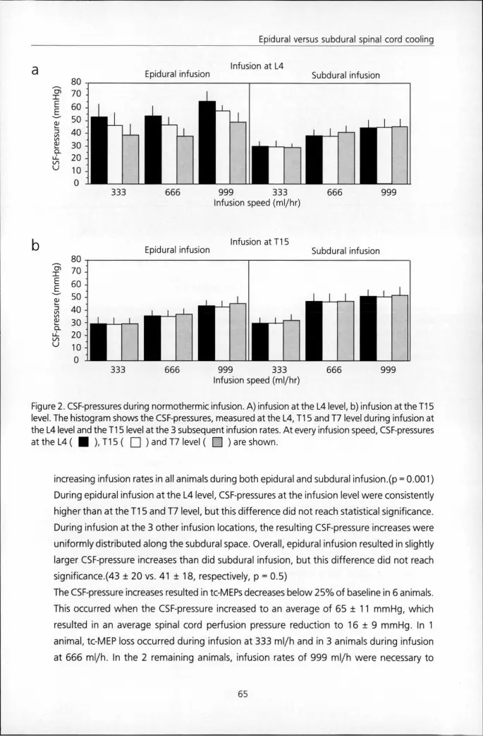
Figuree 2. CSF-pressures during normothermic infusion. A) infusion at the L4 level, b) infusion at the T15 level.. The histogram shows the CSF-pressures, measured at the L4, T15 and T7 level during infusion at thee L4 level and the T15 level at the 3 subsequent infusion rates. At every infusion speed, CSF-pressures a t t heL4 (( ),T15( ) and T7 level ( ) are shown.
increasingg infusion rates in all animals during both epidural and subdural infusion.(p = 0.001)
Duringg epidural infusion at the L4 level, CSF-pressures at the infusion level were consistently
higherr than at the T15 and T7 level, but this difference did not reach statistical significance.
Duringg infusion at the 3 other infusion locations, the resulting CSF-pressure increases were
uniformlyy distributed along the subdural space. Overall, epidural infusion resulted in slightly
largerr CSF-pressure increases than did subdural infusion, but this difference did not reach
significance.(433 20 vs. 41 + 1 8 , respectively, p = 0.5)
Thee CSF-pressure increases resulted in tc-MEPs decreases below 25% of baseline in 6 animals.
Thiss occurred when the CSF-pressure increased to an average of 65 11 mmHg, which
resultedd in an average spinal cord perfusion pressure reduction to 16 9 mmHg. In 1
animal,, tc-MEP loss occurred during infusion at 333 ml/h and in 3 animals during infusion
att 666 ml/h. In the 2 remaining animals, infusion rates of 999 ml/h were necessary to
65 5

Chapterr 3
L44 infusion
epidural l
v -- "
subdural l
T155 infusion
epidural l
f f subdural l
y y 00 333 666 999 333 666 999 333 666 999 333 666 999
^ ^ en n T T fc-fc-£ £ a. . 11 1 U l l
U U
<v <v " O O Z5 5
Q. . F--
LU U
> >
140 0
120 0
100 0
80 0
60 0
40 0
20 0
0 0
Infusionn speed (ml/hr)
Figuree 3. Tc-MEP-amplitude and CSF-pressure measurements in an experimental animal. X-axis: infusion rates,, Y-axis: tc-MEP amplitude (» *-) and CSF-pressure ( ) during infusion at different infusion ratess and locations. Epidural infusion at the L4 level rapidly resulted in CSF-pressures exceeding 60 mmHgg causing tc-MEP amplitude to decrease below 25%. Tc-MEPs recovered when CSF-pressures remainedd below 60 mmHg during subdural infusion at the L4 level and epidural infusion at the T15 level.. When CSF-pressure again increased above 60 mmHg during subdural infusion at the T15 level, tc-MEPss again decreased below 25%.
causee motor neuron dysfunction. Fig. 3 demonstrates the effect of CSF-pressure variations
onn tc-MEP amplitudes in one of the animals. One animal demonstrated a tc-MEP decrease
beloww 25%, immediately following the introduction of the infusion catheters. Tc-MEP
amplitudess remained below 25% for the remaining experiment in this animal.
Hypothermicc experiments Duringg hypothermic infusion, average CSF-temperatures decreased significantly with
increasingg infusion rate, irrespective of the infusion location.(p < 0.001) The average CSF-
temperaturess decreased to 35 + 1.2, 31 2.2 and 28 + 2.8 C at the successive infusion
rates.. CSF-temperature decreases, however, were not uniformly distributed along the
subdurall space, but a temperature gradient was consistently present. As demonstrated in
figuree 4, the CSF-temperature decreases were most pronounced at the infusion location,
duringg infusion at both the L4 and the T15 level. For example, infusion at a rate of 999 ml/
hrr resulted in an average CSF-temperature of 21 + 2 C at the infusion locations, but
remainedd as high as 34 1 C at the location where the effect was least pronounced. In
orderr to decrease CSF-temperatures below 30 C at 2 different measurement locations, an
66 6

Epidurall versus subdural spinal cord cooling
Infusionn at T15
Epidurall infusion Subdural infusion
3333 666 999 333 666 999
Infusionn speed (ml/hr)
Infusionn at T15
Epidurall infusion Subdural infusion
ii mum 3333 666 999 333 666
Infusionn speed (ml/hr)
999 9
Figuree 4. CSF-temperatures during hypothermic infusion, a) infusion at the L4 and b) infusion at the T15 level.. The histogram shows the CSF-temperatures, measured at the L4, T15 and T7 level during infusion att the L4 level andd the T15 level at the 3 subsequent infusion rates. At every infusion speed, CSF-temperaturess at the L 4 ( H ). T 1 5 ( Ü ) and T7 level ) are shown.
infusionn rate of 999 ml/hr was necessary. The maximum CSF-temperature gradient was
analyzed.. During infusion at L4, the maximum gradient was observed between L4 and T7.
Duringg infusion at T15, the maximum gradient was observed between T15 and L4. The
CSF-temperaturee gradient significantly increased wi th increasing infusion rates.(p < 0.001)
Att 333 ml/hr, the maximum gradient was 6 + 3 C and increased to 8 5 CC and 13 6 C
att rates of 666 and 999 ml/hr, respectively. Subdural infusion resulted in an average CSF-
temperaturee gradient of 12 5 C while epidural infusion resulted in a gradient of only 9
5 p < 0.001) Overall, subdural infusion resulted in lower CSF-temperatures than did
epidurall infusion.(p < .0001)
Duringg hypothermic infusion, the average CSF-pressure increases were larger than during
normothermicc infusion.(47 20 vs 41 + 1 9 mmHg, p = 0.02) Furthermore, no CSF-pressure
gradientt was observed during hypothermic infusion at any location. CSF-pressures were
consistentlyy higher during subdural hypothermic infusion as compared to epidural
hypothermicc infusion, (p < 0.002)
67 7

Chapterr 3
Everyy time regional infusion at the maximum infusion rate was terminated, CSF-temperatures
spontaneouslyy returned to 37 C within 12 3 min. CSF-pressures returned to values below
155 mmHg within 2 min in all animals.
Discussion Discussion Thee results of this porcine experiment demonstrated that infusion of cold saline into the
epidurall or subdural space, in order to produce regional spinal cord hypothermia, resulted
inn localized CSF-temperature decreases, only. Subdural cooling was more effective in
decreasingg local CSF-temperature, but it did not extend the cooling over a larger number
off spinal cord segments. The CSF-temperature decreases were obtained at the expense of
significantt CSF-pressure increases, distributed along the entire spinal cord. The resulting
reductionn in spinal cord blood flow was sufficient to produce electrophysiologic evidence
off spinal cord ischemia.
Thee rationale of spinal cord hypothermia as a protective measure for spinal cord ischemia
duringg TAAA surgery is the fact that it increases the tolerable duration of spinal cord ischemia
too the extent that reattachment of segmental arteries and reestablishment of spinal cord
bloodd flow can be completed before irreversible damage has occurred. While systemic
hypothermiaa is accompanied by coagulation abnormalities, cardiac dysrythmias and an
increasedd risk for postoperative infection6"8, regional spinal cord hypothermia avoids these
complicationss at comparable levels of protection. It was demonstrated in animal studies
thatt moderate regional hypothermia (25 C - 27 ) offers sufficient protection during
transientt aortic clamping.34 In a clinical series, comparable CSF-temperatures were obtained
withh epidural cooling to protect the spinal cord in 70 patients undergoing TAAA repair,
resultingg in a low rate of neurologic deficit.10
Nevertheless,, experimental as well as clinical reports emphasize the risk of unavoidable but
potentiallyy hazardous CSF-pressure increases. In rabbits, lethal intracranial hypertension was
described.33 Cambria et al described a patient who developed a cervical spinal cord lesion
duringg epidural infusion cooling, which they ascribedd to the high CSF-pressure.10 During the
normothermicc infusion experiments in our series, CSF-pressures also increased significantly.
Pressuree increases were mostly uniformly distributed throughout the subdural space. Only
duringg epidural normothermic infusion at the L4 level, a pressure gradient was observed,
whichh can possibly be explained by the fact that infusion at the L4 level only allows fluid to
movee in a cranial direction. The contents of the epidural space {fat, veins) possibly act as a
resistancee that induces a pressure gradient. The CSF-pressure increases reduced the spinal
cordd perfusion pressure to values which impaired motor neuron function, as evidenced by
68 8

Epidurall versus subdural spinal cord cooling
tc-MEPs,, in all but two animals. These data imply that regional infusion could impair spinal
cordd perfusion in spinal cord segments at the infusion site, but also at distant spinal cord
segments. .
Thee hypothermic infusion experiments demonstrated that spinal cord hypothermia is limited
too several spinal cord segments only, since CSF-temperatures decreased principally at the
infusionn location. In order to extend moderate hypothermia over a larger spinal cord
segment,, infusion rates had to be increased to 999 ml/h. Nonetheless, CSF-temperatures
att distant levels remained relatively unaffected. The CSF-temperature gradient, which
increasedd with increasing infusion rates, stresses the limitations of this technique. Spinal
cordd segments, not at risk for ischemia resulting from aortic crossclamping, neither protected
byy regional hypothermia, now become at risk for spinal cord ischemia as a result of infusion
cooling.. Lower CSF-temperatures at distant levels could possibly be accomplished when
thee infusion episodes would have been prolonged. Berguer et al reached CSF-temperatures
off 15 C at the cistema magna after 37 min of subdural infusion at the L6 level in dogs, but
ann inflow rate of 1500 ml/h was used and outflow was obtained by a durotomy.9 In this
series,, the CSF-temperature decrease reached a plateau within the 10 min infusion period.
Furthermore,, durotomies are not clinically feasible, and our model may therefore represent
aa better approximation of the clinical situation.
Interestingly,, CSF-temperatures at the L4 level hardly decreased when infusion cooling was
performedd at the T15 level in this experiment. One explanation for this observation is that
thee tips of the infusion catheters were directed cranially, and saline inflow was thereby
directedd to the T7 measurement location.
Wee observed higher CSF-pressures during hypothermic infusion compared to normothermic
infusion.. This could possibly be explained by the fact that perispinal vasoconstriction, caused
byy the CSF-temperature decrease, decreased the resorption of infused saline, resulting in
higherr CSF-pressures during hypothermic infusion.
Inn the present study we were unable to find a distinct advantage of either infusion method
forr the purpose of regional spinal cord cooling. Subdural cooling seemed to be more efficient
inn decreasing local CSF-temperature, but larger CSF-temperature gradients annulled this
effectt at distant spinal cord segments. Furthermore, subdural catheter introduction was a
possiblee cause for the persistent motor neuron function decrease in one of the animals
causedd by local spinal cord compression. However, clinical application of this method would
nott imply multicatheter introduction, so the risk for mechanical damage would be smaller.
Introductionn of infusion catheters into the epidural space might be safer and therefore turn
thee scale towards epidural infusion.
Thee difference in the recovery of CSF-pressures and temperatures after the termination of
infusionn suggests a possible solution for the problem of CSF-pressure increases during regional
69 9

Chapterr 3
infusionn cooling. If spontaneous CSF-pressure normalization occurs within minutes while
CSF-temperaturess remain within protective values for a longer period of time, efficient spinal
cordd cooling might be obtained with intermittent infusion. This requires confirmation in
furtherr investigations.
Thee experimental setup of this study has a possible limitation. Actual spinal cord tissue
temperaturess were not assessed. As Berguer demonstrated during regional cooling in dogs,
aa difference of several degrees Celsius occurs between the infusate and the spinal cord
temperature.99 However, introduction of a temperature needle probe into the spinal cord
wouldd have prevented motor neuron function assessment with tc-MEPs, because of
mechanicall damage to the spinal cord.
Inn conclusion, both epidural and subdural infusion cooling produce relatively localized areas
off spinal cord hypothermia, dependent on the speed of infusion. The concurrent CSF-
pressuree increases, however, are uniformly distributed along the subdural space and result
inn reductions of spinal cord perfusion pressure sufficient to produce spinal cord ischemia.
70 0

Epidurall versus subdural spinal cord cooling
References References 1.. Crawford ES, Crawford JL, Safi HJ, et al. Thoracoabdominal aortic aneurysms: preoperative and
intraoperativee factors determining immediate and long-term results of operations in 605 patients. JJ Vase Surg 1986;3:389-04.
2.. Wisselink W, Becker MO, Nguyen JH, Money SR, Hollier LH. Protecting the ischemic spinal cord duringg aortic clamping: the influence of selective hypothermia and spinal cord perfusion pressure. JJ Vase Surg 1994;19:788-95.
3.. Gonzalez-Fajardo J, Beatriz A, Perez-Burkhardt JL, et al. Epidural regional hypothermia for preventionn of paraplegia after aortic occlusion: experimental evaluation in a rabbit model. J Vase Surgg 1996;23:446-52.
4.. Marsala M, Vanicky I, Galik J, Radonak J, Kundrat I, Marsala J. Panmyelic epidural cooling protects againstt ischemic spinal cord damage. J Surg Res 1993;55:21-31.
5.. Kouchoukos NT, Rokkas CK. Hypothermic cardiopulmonary bypass for spinal cord protection: rationalee and clinical results. Ann Thor Surg 1999;67:1940-2.
6.. Okada M. The cardiac rhythm in accidental hypothermia. Electrocardiol 1984;17:123-8.
7.. Rohrer MJ, Natale AM. Effect of hypothermia on the coagulation cascade. Crit Care Med 1992;20:1402-5. .
8.. Kurz A, Sessler Dl, Lenhardt R. Perioperative normothermia to reduce the incidence of surgical-woundd infection and shorten hospitalization. Study of Wound Infection and Temperature Group. Neww Engl J Med 1996;334:1209-15.
9.. Berguer R, Porto J, Fedoronko B, Dragovic L. Selective deep hypothermia of the spinal cord preventss paraplegia after aortic cross-clamping in the dog model. J Vase Surg 1992;15:62-71.
10.. Cambria RP, Davison JK, Zannetti S, et al. Clinical experience with epidural cooling for spinal cord protectionn during thoracic and thoracoabdominal aneurysm repair. J Vase Surg 1997;25:234-41.
11.. de Haan P, Kalkman CJ, Meylaerts SA, Lips J, Jacobs MJ. Development of spinal cord ischemia afterr clamping of non-critical segmental arteries in the pig. Ann Thor Surg, 1999;68; 1278-84.
12.. Ubags LH, Kalkman CJ, Been HD, Porsius M, Drummond JC. The use of ketamine or etomidate too supplement sufentanil/N20 anesthesia does not disrupt monitoring of myogenic transcranial motorr evoked responses. J Neurosurg Anesth 1997;9:228-33.
13.. de Haan P, Kalkman CJ, de Mol BA, Ubags LH, Veldman DJ, Jacobs MJ. Efficacy of transcranial motor-evokedd myogenic potentials to detect spinal cord ischemia during operations for thoracoabdominall aneurysms. J Thorac Cardiovasc Surg 1997;113:87-100.
14.. Jacobs,MJ; Meylaerts,SA; Haan de,P; Mol de,BA; Kalkman,CJ. Strategies to prevent neurologic deficitt based on motor evoked potentials in type I and II thoracoabdominal aortic aneurysm repair.. J Vase Surg 1999;29:48-59.
15.. Meylaerts SA, Jacobs MJ, de Haan P, Kalkman CJ. Comparison of transcranial motor evoked potentialss and somatosensory evoked potentials during thoracoabdominal aortic aneurysm repair. Annn Surg 1999;230:742-9.
71 1

Chapterr 3
16.. Browning JL, Heizer ML, Baskin DS. Variations in corticomotor and somatosensory evoked potentials:: effects of temperature, halothane anesthesia, and arterial partial pressure of C02. Anesthh Analg 1992;74:643-8.
72 2


