Embed Size (px)
Citation preview

Phytomedicine 22 (2015) 213–222
Contents lists available at ScienceDirect
Phytomedicine
journal homepage: www.elsevier.com/locate/phymed
Epigallocatechin gallate, ellagic acid, and rosmarinic acid perturbdNTP pools and inhibit de novo DNA synthesis and proliferationof human HL-60 promyelocytic leukemia cells: Synergismwith arabinofuranosylcytosine
Philipp Saiko a, Marie-Thérèse Steinmann a, Heike Schuster a, Geraldine Graser a,Sabine Bressler a, Benedikt Giessriglb, Andreas Lackner c, Michael Grusch c, Georg Krupitzab,Zsuzsanna Bago-Horvathb, Walter Jaegerd, Monika Fritzer-Szekeres a, Thomas Szekeres a,∗
aDepartment of Medical and Chemical Laboratory Diagnostics, Medical University of Vienna, Waehringer Guertel 18-20, A-1090 Vienna, AustriabDepartment of Pathology, Medical University of Vienna, Waehringer Guertel 18-20, A-1090 Vienna, AustriacDepartment of Medicine I, Division of Cancer Research, Medical University of Vienna, Borschkegasse 8a, A-1090 Vienna, AustriadDepartment of Clinical Pharmacy and Diagnostics, University of Vienna, Althanstrasse 14, A-1090 Vienna, Austria
a r t i c l e i n f o
Article history:
Received 14 July 2014
Revised 13 November 2014
Accepted 14 November 2014
Keywords:
Epigallocatechin gallate
Ellagic acid
Rosmarinic acid
Ribonucleotide reductase
Arabinofuranosylcytosine
HL-60 cells
a b s t r a c t
Epigallocatechin gallate (EGCG), ellagic acid (EA) and rosmarinic acid (RA) are natural polyphenols exerting
cancer chemopreventive effects. Ribonucleotide reductase (RR; EC 1.17.4.1) converts ribonucleoside diphos-
phates into deoxyribonucleoside diphosphates being essential for DNA replication, which is why the enzyme
is considered an excellent target for anticancer therapy.
EGCG, EA, and RA dose-dependently inhibited the growth of human HL-60 promyelocytic leukemia cells,
exerted strong free radical scavenging potential, and significantly imbalanced nuclear deoxyribonucleoside
triphosphate (dNTP) concentrations without distinctly affecting the protein levels of RR subunits (R1, R2,
p53R2). Incorporation of 14C-cytidine into nascent DNA of tumor cells was also significantly lowered, being
equivalent to an inhibition of DNA synthesis. Consequently, treatment with EGCG and RA attenuated cells in
the G0/G1 phase of the cell cycle, finally resulting in a pronounced induction of apoptosis. Sequential com-
bination of EA and RA with the first-line antileukemic agent arabinofuranosylcytosine (AraC) synergistically
potentiated the antiproliferative effect of AraC, whereas EGCG plus AraC yielded additive effects.
Taken together, we show for the first time that EGCG, EA, and RA perturbed dNTP levels and inhibited
cell proliferation in human HL-60 promyelocytic leukemia cells, with EGCG and RA causing a pronounced
induction of apoptosis. Due to these effects and synergism with AraC, these food ingredients deserve further
preclinical and in vivo testing as inhibitors of leukemic cell proliferation.
© 2014 Elsevier GmbH. All rights reserved.
Introduction
Epigallocatechin gallate (EGCG) is the major catechin found in
green tea (Camellia sinensis (L.) Kuntze) (Lambert and Elias 2010).
Accompanied by (−)-epicatechin-3-gallate, (−)-epigallocatechin, and
(−)-epicatechin, EGCG accounts for 50–80% of the polyphenols in a
brewed cup of green tea (Singh et al. 2011). These catechins show
a pronounced radical scavenging activity and inhibit cell prolifera-
tion (Brown 1999). The potential health benefits ascribed to EGCG in-
clude cancer chemoprevention, amelioration of cardiovascular health
∗ Corresponding author. Tel.: +43 1 40400 53650; fax: +43 1 40400 53900.
E-mail address: [email protected], [email protected]
(T. Szekeres).
and neurodegenerative maladies, preventive effects against diabetes,
and protection of the skin from damage caused by ionizing radiation
(Singh et al. 2011). Regarding its cancer chemopreventive abilities,
numerous mechanisms have been proposed including antioxidant
effects, inhibition of growth factor signaling, and induction of tumor
suppressor genes (Lambert and Elias 2010).
Ellagic acid (EA) is present in fruits, nuts, and berries and belongs
to a family of bioactive ellagitannins (ETs). All ETs share the ability
to release EA upon hydrolysis, resulting in a prolonged release of EA
into the bloodstream (Heber 2008). Pomegranate (Punica granatum
(Horan.) S.A. Graham, Thorne & Reveal), an ancient fruit-bearing de-
ciduous shrub represents the richest source of ETs and, subsequently,
EA among fruits (Heber 2008; Viladomiu et al. 2013). Pomegranate
juice has been reported to exert antioxidant, anticancer and
http://dx.doi.org/10.1016/j.phymed.2014.11.017
0944-7113/© 2014 Elsevier GmbH. All rights reserved.
214 P. Saiko et al. / Phytomedicine 22 (2015) 213–222
chemopreventive effects by inhibition of cancer cell proliferation
and modulation of inflammatory signaling pathways (Hagiwara et al.
2010). ETs and their hydrolysis product, EA, attenuate prostate can-
cer cell growth through cell cycle arrest and subsequent induction of
apoptosis, including the inhibition of angiogenesis, the latter having
been demonstrated in vitro and in vivo (Heber 2008; Lansky et al.
2005).
Rosmarinic acid (RA) is an ester of caffeic acid and 3,4-
dihydroxyphenyllactic acid belonging to the main active compounds
of rosemary (Rosmarinus officinalis (L.)), together with caffeic acid,
ursolic acid, carnosic acid, and carnosol. The phenolic constituents
of rosemary have also been found to exert preventive effects on sev-
eral types of cancer through induction of cancer protective factors like
apoptosis, or inhibitionof tumorpromoting events such as free radical
generation (Ngo et al. 2011; Petersen and Simmonds 2003). In addi-
tion, RA possesses antioxidant as well as anti-inflammatory activities
through which it has been shown to reduce the risk of myocardial
infarction and to inhibit lipid peroxidation (Wu andWang 2012). Due
to its radical scavenging properties, RA was also able to exert pro-
tective effects against memory impairments induced by neurotoxins
(Wu and Wang 2012).
Ribonucleotide reductase (RR; EC 1.17.4.1) is responsible for the
reduction of ribonucleoside diphosphates into deoxyribonucleoside
diphosphates, which are the building blocks for DNA synthesis and
repair in all living organisms (Guarino et al. 2014; Shao et al. 2013).
RR is highly active in malignant tissues due to an increased
need of deoxynucleoside triphosphates (dNTPs; the products of RR
metabolism) of rapidly growing cells while showing low enzyme
activity in slowly growing normal cells (Takeda and Weber 1981).
RRs are divided into three classes depending on the mode of free
radical generation. Class I RRs are aerobic enzymes being further
subdivided depending on the metallocofactor employed (Guarino
et al. 2014). Eukaryotes mainly use class Ia RRs, which comprise
an α2β2 complex made up of two subunits α (termed R1) and β(termed R2) that conglomerate to build the holoenzyme with the
active form supposed to adopt α2β2 quaternary state (Aye et al.
2014). Effector-binding R1 harbors substrate and allosteric effective
sites for controlling enzyme activity and substrate specificity. Non-
heme R2 contains two oxo-bridged dinuclear iron centers each sta-
bilizing a protein tyrosyl radical. An inhibition of the nonheme iron
subunit can thus be caused by chelation of the iron centers or by scav-
enging of the tyrosyl radical (Shao et al. 2013). Class II enzymes oper-
ate independently of oxygen and require 5′-deoxyadenosylcobalamin
for radical generation, whereas class III RRs only work under strictly
anaerobic conditions, thereby using a stable glycine radical for catal-
ysis (Guarino et al. 2014). Hydroxyurea has been the first RR inhibitor
being introduced in clinical practice and was used in chemotherapy
regimens against chronic myeloid leukemia (CML) and a number of
other malignant diseases (Saban and Bujak 2009; Tennant 2001). An-
other RR inhibitor, gemcitabine (difluordeoxycytidine; dFdC), is com-
monly used in the treatment of pancreatic cancer and non-small cell
lung cancer (Noble and Goa 1997; Toschi et al. 2005).
EGCG, EA, and RA have never been tested for their RR inhibiting
capabilities but all of them have a number of free hydroxyl groups
being located at their aromatic rings which might be able to scav-
enge the tyrosyl radical harbored in the R2 subunit of the enzyme
thus attenuating its activity. Following this strategy, we now inves-
tigated the antiproliferative and biochemical effects of these natural
compounds in the human HL-60 promyelocytic leukemia cell line,
which already served as a cellularmodel for testing extracts of ethno-
pharmacological healing plants (Gridling et al. 2009). The free radical
scavenging potential of EGCG, EA, and RA was measured by DPPH•
assay, the DNA synthesis activity was determined by incorporation
of radio-labeled 14C-cytidine into nascent DNA of tumor cells, and
the steady state of dNTPs was analyzed by HPLC. The protein lev-
els of RR subunits R1, R2, and p53R2 were examined by western
blotting. Cell cycle perturbations were evaluated by FACS, and the
induction of apoptosis was quantified employing a specific Hoechst
33258/propidium iodide double staining method developed by our
group.
Acute promyelocytic leukemia (APL) is a distinct subtype of
acute myeloid leukemia (AML) mainly characterized by specific mor-
phology, leucopenia, and a life-threatening coagulopathy being the
most prominent manifestation of the disease (Tallman and Altman
2009). Anthracycline plus arabinofuranosylcytosine (AraC) combina-
tion chemotherapy has been the only treatment option for APL until
the late 1980s (Ades et al. 2006), with AraC still being in charge of
reducing relapses in APL (Kelaidi et al. 2009). Hence, we combined all
drugs with AraC in order to elucidate potential additive or synergistic
behavior.
Materials and methods
Chemicals and supplies
EGCG, EA, RA, solvent DMSO, and all other chemicals and reagents
were obtained from Sigma–Aldrich GmbH (Vienna, Austria) and of
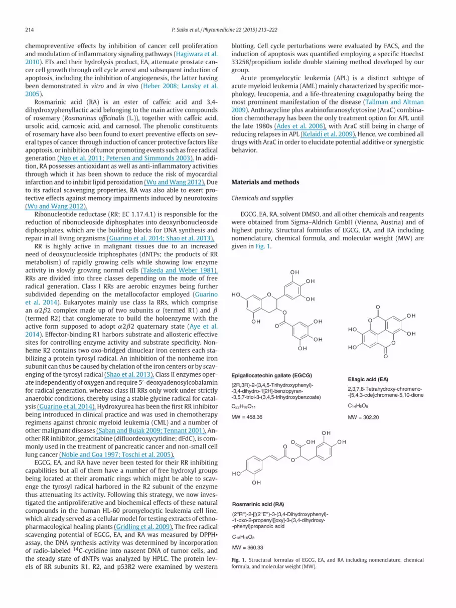
highest purity. Structural formulas of EGCG, EA, and RA including
nomenclature, chemical formula, and molecular weight (MW) are
given in Fig. 1.
Epigallocatechin gallate (EGCG)
(2R,3R)-2-(3,4,5-Trihydroxyphenyl)--3,4-dihydro-1[2H]-benzopyran--3,5,7-triol-3-(3,4,5-trihydroxybenzoate)
C22H18O11
MW = 458.36
Ellagic acid (EA)
C14H6O8
MW = 302.20
2,3,7,8-Tetrahydroxy-chromeno--[5,4,3-cde]chromene-5,10-dione
Rosmarinic acid (RA)
(2''R'')-2-[[(2''E'')-3-(3,4-Dihydroxyphenyl)--1-oxo-2-propenyl]]oxy]-3-(3,4-dihydroxy--phenyl)propanoic acid
C18H16O8
MW = 360.33
Fig. 1. Structural formulas of EGCG, EA, and RA including nomenclature, chemical
formula, and molecular weight (MW).

P. Saiko et al. / Phytomedicine 22 (2015) 213–222 215
DPPH• radical scavenging activity assay
The radical scavenging activity of drugs was determined using the
free radical 2,2-diphenyl-1-picrylhydrazyl (DPPH•). DPPH• absorbs at
515 nm but when reduced by an antioxidant or radical species, its ab-
sorptiondecreases. The reactionwas initiatedbyaddingEGCG, ES, and
RS (10 µl; 1–100 µM final concentration) to 3.0 ml of 0.1 mM DPPH•
in methanol. The bleaching of DPPH• was followed using an HP 8453
diode array spectrometer equippedwith amagnetically stirred quartz
cell. Absorbancewas recorded for up to 15min, although steady states
of reaction were reached within 5 min in most cases. The reference
cuvette contained up to 0.1 mM DPPH• in 3.0 ml of methanol. The
DPPH• radical scavenging activity obtained for each compound was
compared with that of α-tocopherol or ascorbic acid, both of which
served as reference compounds. Data are means ± standard errors of
three determinations.
Cell culture
The humanHL-60 promyelocytic leukemia cell linewas purchased
from ATCC (American Type Culture Collection, Manassas, VA, USA).
Cells were grown in RPMI 1640medium supplementedwith 10% heat
inactivated fetal calf serum (FCS), 1% l-glutamine, and 1% penicillin–
streptomycin at 37 °C in a humidified atmosphere containing 5% CO2
using a Heraeus cytoperm 2 incubator (Heraeus, Vienna, Austria).
All media and supplements were purchased from Gibco Life Tech-
nologies (Paisley, Scotland, UK). Cell counts were determined using a
microcellcounter CC-110 (SYSMEX, Kobe, Japan). Cells growing in the
logarithmic phase of growth were used for all experiments described
below.
Growth inhibition assay
HL-60 cells (0.1× 10⁶ cells/ml) were seeded in 25 cm2 Nunc tissue
culture flasks and incubated with increasing concentrations of EGCG,
EA, or RA at 37 °C under cell culture conditions. Cell counts and IC50
values (IC50 = 50% growth inhibition of tumor cells) were determined
after 24, 48, and 72 h using a microcellcounter CC-110. Viability of
cells was determined by staining with trypan blue. Results were cal-
culated as number of viable cells. Data are means ± standard errors
of three determinations.
Determination of deoxyribonucleoside triphosphates (dNTPs)
HL-60 cells were seeded in 175 cm2 tissue culture flasks (1 × 10⁸
per flask) and then incubatedwith increasing concentrations of EGCG,
EA, or RA for 24 h. The cells were then centrifuged at 1800 g for
5 min, resuspended in 100 µl of PBS, and extracted with 10 µl of
trichloracetic acid (90%). The lysate was allowed to rest on ice for
30 min and neutralized by the addition of 1.5 volumes of freon con-
taining 0.5 M tri-n-octylamine. Concentrations of dNTPs were then
determined using the method initially described by Garrett and Santi
(1979), which has been slightly modified by our group (Saiko et al.
2007a). In brief, aliquots (100 µl) of the samples were analyzed us-
ing a Merck "La Chrom" high-performance liquid chromatography
(HPLC) system (Merck, Darmstadt, Germany). Detection time was set
at 80min, with the detector operating on 280 nm for 40min and then
switched to 260 nm for another 40 min. Samples were eluted with a
3.2 M ammonium phosphate buffer (pH 3.6, adjusted by the addition
of 0.32 M H3PO4) containing 2% acetonitrile using a 4.6 × 250 mm
PARTISIL 10 SAX column (Whatman Ltd., Kent, UK). Separation was
performed at a flow rate of 2 ml/min. The concentration of each dNTP
was calculated as percentage of the total area under the curve for each
sample. Data are means ± standard errors of three determinations.
Incorporation of 14C-labeled cytidine into DNA
To determine the effect of drug treatment on DNA synthesis, we
performed an assay as described previously (Saiko et al. 2007b).
Radio-labeled 14C-cytidine has to be reduced by RR in order to be
incorporated into the DNA of tumor cells following incubation with
a given compound. Cells (0.4 × 10⁶ cells/ml) were incubated with
effective concentrations of EGCG, EA, or RA for 24 h. Subsequently,
cells were counted and pulsed with 14C-cytidine (0.3125 µCi, 5 nM)
for 30 min at 37 °C. Cells were then collected by centrifugation
and washed with PBS. Total DNA from 5 × 10⁶ cells was purified
by phenol–chloroform–isoamyl alcohol extraction and specific ra-
dioactivity of the samples was measured using a Wallac 1414 liquid
scintillation counter (PerkinElmer, Boston, MA), whose read out was
normalized by a Hitachi U-2000 Double Beam Spectrophotometer to
ensure equal amounts and purity of DNA. Data are means ± standard
errors of three determinations.
Cell cycle distribution analysis
Cells (0.4 × 10⁶ cells/ml) were seeded in 25 cm2 Nunc tissue cul-
ture flasks and incubatedwith increasing concentrations of EGCG, EA,
or RA at 37 °C under cell culture conditions. After 48 h, cells were
harvested and suspended in 5 ml cold PBS, centrifuged, resuspended
and fixed in 3 ml cold ethanol (70%) for 30 min at 4 °C. After two
washing steps in cold PBS RNase A and propidium iodide were added
to a final concentration of 50 µg/ml each and incubated at 4 °C for
60 min before measurement. Cells were analyzed on a FACSCalibur
flow cytometer (BD Biosciences, San Jose, CA, USA) and cell cycle
distribution was calculated withModFit LT software (Verity Software
House, Topsham,ME, USA). Data aremeans± standard errors of three
determinations.
Hoechst dye 33258 and propidium iodide double staining
The Hoechst staining was performed according to the method de-
scribed by our group (Grusch et al. 2002). Cells (0.2 × 10⁶ cells/ml)
were seeded in 25 cm2 Nunc tissue culture flasks and exposed to in-
creasing concentrations of EGCG, EA, or RA for 24 or 48 h. Hoechst
33258 (HO, Sigma, St. Louis, MO, USA) and propidium iodide (PI,
Sigma, St. Louis, MO, USA) were added directly to the cells to final
concentrations of 5 µg/ml and 2 µg/ml, respectively, followed by
90min of incubation at 37 °C. Cells were examined on a Nikon Eclipse
TE-300 Inverted Epi-Fluorescence Microscope (Nikon, Tokyo, Japan)
equippedwith aNikonDS-5M-L1Digital Sight Camera System includ-
ing appropriate filters for Hoechst 33258 and PI. This method allows
distinguishing between early apoptosis, late apoptosis, and necrosis
and is therefore superior to TUNEL assay that fails to discriminate
among apoptosis and necrosis (Grasl-Kraupp et al. 1995) and does
not provide any morphological information. In addition, the HO/PI
staining is more sensitive than a customary FACS based Annexin V
binding assay (Grusch et al. 2001). Cells were judged according to
their morphology and the integrity of their cell membranes, counted
under the microscope and the number of apoptotic cells was given as
percentage value. Experiments were repeated twice.
Western blotting
After incubation with 30 µM EGCG, HL-60 cells (2 × 10⁶ cells/ml)
were harvested, washed twice with ice-cold PBS (pH 7.2) and lysed
in a buffer containing 150mMNaCl, 50 mM Tris–buffered saline (Tris
pH 8.0), 1% Triton X-100, 2.5% 100mMphenylmethylsulfonylfluoride
(PMSF) and 2.5% protease inhibitor cocktail (PIC; from a 100× stock).
The lysate was centrifuged at 12,000 rpm for 20 min at 4 °C, and the
supernatant was stored at −20 °C until further analysis as reported
previously (Saiko et al. 2011). Equal numbers of cells were lysed

216 P. Saiko et al. / Phytomedicine 22 (2015) 213–222
for each sample and PVDF membranes were stained with Ponceau
S to ensure equal sample loading. The latter was also controlled by
β-actin expression, which appeared to be stable when inspected in
short term exposures to X-ray films. Antibodies directed against R1
(T-16), R2 (I-15), p53R2 (N-16), and donkey anti-goat IgG were from
Santa Cruz (Santa Cruz, CA, USA), and the antibody directed againstβ-
actinwas fromSigma (St. Louis,MO, USA). The primary and secondary
antibodies were used at dilutions of 1:500 and 1:2000, respectively.
Sequential growth inhibition assay
HL-60 cells (0.1 × 10⁶ cells/ml) were seeded in 25 cm2 Nunc tis-
sue culture flasks and first incubated with effective concentrations of
EGCG, EA, or RA for 24 h. Concentrations were considered effective
when significantly depleting at least one dNTP pool. Then the respec-
tive compound was washed out and cells were further exposed to
various concentrations of AraC for another 48 h. Since AraC is still
being in charge of reducing relapses in APL (Kelaidi et al. 2009), we
decided to combine all natural compounds with this clinically estab-
lished antileukemic drug. Concentrations were chosen such that they
were strictly in the IC10–IC30 range to avoid exaggerated growth in-
hibition after 72 h when being applied in combination. Cells were
counted every 24 h using a microcellcounter CC-110. Experiments
were repeated twice.
Calculations of combination effects
The calculations of combination effects were performed using the
Calcusyn 2.0 software designed by Chou and Talalay (Biosoft, Fergu-
son, MO) (Chou and Talalay 1984). The analytical method of Chou and
Talalay (1981, 1984) yields two parameters that describe the inter-
actions among drugs in a given combination: the combination index
(CI) and the dose reduction index (DRI). DRI measures by what factor
the dose of each drug in a combination may be reduced at a given
effect level compared with the dose when each drug is used alone.
DRI may be influenced by the combination ratio and the number of
drugs. Toxicity towards the host may be avoided or reduced when
the dose is reduced. The advantage of this method is that it takes
into account not only the potency (median effect dose values [Dm] or
drug concentration at 50% neutralization [EC50]), but also the shape
(sigmoidicity) of the dose–effect curve, based on the median effect
equation of Chou. The latter correlates drug dose and cytostatic effect
using the following form:
fa/fu =(
D/Dm
)mor D = Dm
[
fa/(
1 − fa)]1/m
D represents the dose of the drug; Dm is the median effect dose
meaning the potency, determined from the x-intercept of themedian
effect plot; fa is the fraction affected by the dose; fu is the fraction
unaffected (fu = 1 − fa); andm is an exponent that signifies the shape
(sigmoidicity) of the dose–effect curve, which is given by the slope of
themedian effect plot. Themedian effect equation is utilized to calcu-
late Dx, which is the dose of a drug that inhibits x% of cells. For drugs
with mutually nonexclusive mechanisms of action (i.e. drugs that
have a differentmode of action, thus being not competitive inhibitors
of each other), the CI is then calculated by the following equation:
CI =(
D)
1/(
Dx
)
1+
(
D)
2/(
Dx
)
2+
[(
D)
1
(
D)
2
]
/[(
D)
1
(
D)
2
]
The CI equation determines the additive effect of drug combi-
nations, such that a CI of <0.9 indicates synergism, a CI of 0.9–1.1
indicates additive effects, and a CI of >1.1 indicates antagonism.
Experiments were repeated twice.
Statistical calculations
Dose–response curves were calculated using the Prism 5.01 soft-
ware (GraphPad, San Diego, CA, USA) and significant differences be-
tween controls and eachdrug concentration appliedweredetermined
by unpaired t-test. All p-values below 0.05 were considered signifi-
cant and marked with an asterisk (∗).
Results
Reduction of HL-60 cell numbers
HL-60 cells (0.1 × 10⁶ cells/ml) were seeded in 25 cm2 Falcon
tissue culture flasks and treated with increasing concentrations of
drugs. After the incubation period, the cell number of viable leukemia
cells was determined using a microcellcounter. Exposure to EGCG
for 24, 48, and 72 h resulted in IC50 values of 30, 18, and 16 µM,
respectively (Fig. 2a). Treatment with EA for 72 h led to an IC50 value
of 35 µM (Fig. 2b). Incubation with RA for 24, 48, and 72 h yielded
IC50 values of 147, 74, and 69 µM, respectively (Fig. 2c). IC50 values
after 72 h are also listed in Table 1.
Antioxidant activity of EGCG, EA, and RA
The in vitro free radical-scavenging activity of EGCG, EA, and RA
was determined by trapping the DPPH• radical. After incubation for
15 min EGCG, EA, and RA reduced DPPH• absorption with IC50 values
of 5.8, 6.5, and22.2µM, respectively. Therefore, thepotential of EGCG,
EA, and RA to inhibit HL-60 cell expansion correlated directly with
their potential to scavenge free radicals. Tocopherol and ascorbic acid
were used as reference compounds resulting in IC50 values of 21.3
and 16.2 µM, respectively (Table 1).
Alterations of intracellular dNTP pools
Under physiological conditions, RRmaintains stable deoxyribonu-
cleotide production, which is essential for DNA synthesis; accord-
ingly, inhibition of RR activity eliminates this balance. EGCG, EA, or
RA treatment for 24 h each caused a significant imbalance of dNTPs in
HL-60 cells, which was reminiscent of the imbalances caused by the
RR inhibitors hydroxyurea and gemcitabine (Hakansson et al. 2006;
Heinemann et al. 1995; Ostruszka and Shewach 2003; Smid et al.
2001). Incubation with 20 and 40 µM EGCG resulted in a depletion
of intracellular dATP pools to 51 and 37% of controls, respectively,
whereas dTTP pools were increased to 141% of untreated cells upon
40 µM EGCG (Fig. 3a). Treatment with 20, 30, and 60 µM EA reduced
dCTP pools to 77, 57, and 49% of untreated controls, respectively, and
20 and 30 µM EA depleted dTTP pools to 72 and 65%, respectively
(Fig. 3b). Exposure to 50 µM RA led to a depletion of dTTP pools to
71%, and 50, 100, and 150 µM RA reduced dATP pools to 68, 48, and
45%of controls, respectively (Fig. 3c). All dGTPpools remainedbeyond
the detectability limit of the method.
Inhibition of incorporation of 14C-cytidine into DNA of HL-60 cells
To quantify DNA synthesis, the incorporation of 14C-cytidine into
nascent DNA of tumor cells was measured after incubation with ef-
fective concentrations of EGCG, EA, or RA (i.e. doses that have been
shown to significantly reduce dNTP pools). Exposure of cells to 20µM
EGCG, 60 µM EA, and 100 µM RA significantly lowered the incorpo-
ration of radiolabeled cytidine to 45%, 62%, and 46% of control values,
respectively (Fig. 4).
Expression levels of RR subunits R1, R2, and p53R2 after treatment with
EGCG
Tomonitor the effect of EGCG on the expression of RR subunits R1,
R2, andp53R2,HL-60 cellswere incubatedwith30µMEGCG for0.5, 2,
4, 8, and24h and subjected towestern blot analysis. R1 andR2protein
levels remained almost unchanged throughout the time course being
in line with the fact that enzyme activity can be attenuated without

P. Saiko et al. / Phytomedicine 22 (2015) 213–222 217
Fig. 2. Reduction of cell numbers, dose–effect curves, and Fa–CI plots after incubation of HL-60 cells with (a) EGCG, (b) EA, and (c) RA. HL-60 cells (0.1 × 10⁶ cells/ml) were
seeded in 25 cm2 Nunc tissue culture flasks and incubated with increasing concentrations of EGCG, EA, or RA at 37 °C under cell culture conditions. Cell counts and IC50 values
(IC50 = 50% growth inhibition of tumor cells) were determined after 24, 48, and 72 h using a microcellcounter CC-110. Viability of cells was determined by staining with trypan
blue. Results were calculated as number of viable cells. In another set of experiments, HL-60 cells were first incubated with effective concentrations of EGCG, EA, or RA for 24 h.
Concentrations were considered effective when significantly depleting at least one dNTP pool. Then the respective compound was washed out and cells were further exposed to
various concentrations of AraC for another 48 h. Concentrations were chosen such that they were strictly in the IC10–IC30 range to avoid exaggerated growth inhibition after 72 h
when being applied in combination. The calculations of dose–effect curves and Fa–CI plots were performed using the Calcusyn 2.0 software designed by Chou and Talalay (Biosoft,
Ferguson, MO) (Chou and Talalay, 1984).
influencing the protein levels of its subunits (Saiko et al. 2011, 2013),
whereas p53R2 expression was slightly decreased (Fig. 5).
Cell cycle distribution in HL-60 cells after treatment with EGCG, EA,
and RA
Treatment with EGCG, EA, or RA led to significant alterations of
the cell cycle distribution. Incubation with 50 µM EGCG attenuated
cells in the G0-G1 phase of the cell cycle, increasing this cell popula-
tion from 34.6% to 48.2%, whereas S phase cells decreased from 48.5%
to 40.1%. In contrast, exposure to 60 µM EA resulted in an accumu-
lation of cells in the S phase, thereby augmenting these cells from
21.8% to 38.8% while reducing cells in the G0–G1 phase from 62.8% to
43.9%. RA acted similarly to EGCG; 150 µMRA attenuated cells in the
G0-G1 phase, increasing this population from 33.1% to 47.4% while
decreasing cells mainly in the G2-M phase from 17.3% to 9.0% (Fig. 6).
Table 1
DPPH• radical scavenging activity after treatment for 15 min and growth
inhibition of HL-60 cells after incubation for 72 h.
Compound DPPH• activity HL-60 cells
IC50 (µM) IC50 (µM)
EGCG 5.8 ± 0.18 16.0 ± 0.23
EA 6.5 ± 0.31 35.0 ± 1.53
RA 22.2 ± 0.65 69.0 ± 2.28
Ascorbic acid 21.3 ± 1.51 –
Tocopherol 16.2 ± 0.90 –
Induction of apoptosis in HL-60 cells after exposure to EGCG, EA, and RA
Tumor cells were incubated with increasing concentrations of
drugs for 24 or 48 h and then double stained with Hoechst 33258
and propidium iodide to quantify apoptotic cells death. Treatment
with 30, 40, and 50 µM EGCG led to 16.7, 56.3, and 84.3% apoptotic
cells, respectively, but after incubationwith EA, we could not observe
a prominent induction of apoptosis at any concentration employed.
Exposure to 50 and 100 µM RA did not result in apoptosis as well,
whereas 150 µM RA led to 60.7% apoptotic cells (Table 2).
Combination effects with AraC on the growth of HL-60 cells
To examine the impact of EGCG, EA, or RA in combination with
AraC, HL-60 cells were seeded at a density of 0.1 × 10⁶ cells/ml and
incubated with increasing concentrations of the respective drug for
24 h, followed by AraC treatment for 48 h as described in the "Ma-
terials and methods" section. Dose–effect curves and Fa–CI plots for
EGCG, EA, and RA are given in Fig. 2a, b, and c, respectively. Regarding
EGCG, six out of nine combinations applied yielded additive effects
according to the equation of Chou and Talalay (1984) (Table 3). In
contrast, four out of nine EA combinations and eight out of nine RA
combinations applied showed synergism according to the equation
of Chou and Talalay (1984) (Tables 4 and 5).
Discussion
EGCG, EA, and RA are polyphenolic plant ingredients that have
been shown to possess antioxidant capabilities (Lambert and Elias

218 P. Saiko et al. / Phytomedicine 22 (2015) 213–222
(a)
dCTP dTTP dATP0
50
100
150
200Co
10 µM
20 µM
40 µM
*
*
*
dNTPs
AUC(% of control)
(b)
dCTP dTTP dATP0
50
100
150
200Co
20 µM
30 µM
60 µM
*** *
*
dNTPs
AUC(% of control)
(c)
dCTP dTTP dATP0
50
100
150Co
50µM
100µM
150µM* *
**
dNTPs
AUC(% of control)
Fig. 3. Concentration of dNTP pools in HL-60 cells upon treatment with (a) EGCG, (b)
EA, and (c) RA. Cells (0.8× 10⁶ cells/ml)were incubatedwith EGCG (10, 20, and 40µM),
EA (20, 30, and 60 µM) or RA (50, 100, and 150 mM) for 24 h. Afterwards, 1 × 10⁸ cells
were separated for the extraction of dNTPs. The concentration of dNTPs was calculated
as percent of total area under the curve for each sample. Values significantly (p < 0.05)
different from control are marked with an asterisk (∗).
2010), to induce cell cycle arrest and apoptosis, and to act as can-
cer chemopreventive agents in a multitude of preclinical and animal
studies (Hagiwara et al. 2010; Heber 2008; Ngo et al. 2011; Petersen
and Simmonds 2003; Singh et al. 2011; Viladomiu et al. 2013; Wu
and Wang 2012).
In this study,we report for the first time the effects of these natural
compounds on ribonucleotide reductase (RR) metabolism in human
HL-60 promyelocytic leukemia cells.
Acute promyelocytic leukemia is a distinct subtype of acute
myeloid leukemia and represents a relatively rare hematologi-
cal disease, accounting for approximately 5–8% of all AML cases
(Mi 2011). The balanced reciprocal translocation t(15;17) generates
a PML (promyelocytic leukemia)-RARα (retinoic acid receptor alpha)
fusion (onco)gene (Mi et al. 2012). This PML–RARα transcript initiates
APLbyblockingmyeloiddifferentiation andby increased self-renewal
of leukemic progenitor cells (Ades et al. 2006; de The and Chen 2010).
(a)
Control 20 µM0
25
50
75
100
125
*Specific activity(% of control)
(b)
Control 60 µM0
25
50
75
100
125
*Specific activity(% of control)
(c)
Control 100 µM0
25
50
75
100
125
*Specific activity(% of control)
Fig. 4. Inhibition of 14C-cytidine incorporation into DNA of HL-60 cells after exposure
to (a) EGCG, (b) EA, and (c) RA. Cells (0.4 × 10⁶ cells/ml) were incubated with EGCG
(20 µM), EA (60 µM), or RA (100 µM) for 24 h. After the incubation period, cells were
counted and pulsed with 14C-cytidine (0.3125 µCi, 5 nM) for 30 min at 37 °C. Then
cells were collected by centrifugation and washed with PBS. Total DNA was extracted
from 5 × 10⁶ cells and specific radioactivity of the samples was determined using a
Wallac 1414 liquid scintillation counter (PerkinElmer, Boston,MA). Values significantly
(p < 0.05) different from control are marked with an asterisk (∗).
Fig. 5. Expression levels ofRR subunitsR1, R2, andp53R2 inHL-60cells upon treatment
with EGCG. After incubation with 30 µM EGCG for 0.5, 2, 4, 8, and 24 h, HL-60 cells
(2× 10⁶ cells/ml)were harvested, washed twicewith ice-cold PBS (pH 7.2) and lysed in
a buffer containing 150 mM NaCl, 50 mM Tris–buffered saline (Tris pH 8.0), 1% Triton
X-100, 1 mM phenylmethylsulfonylfluoride (PMSF) and protease inhibitor cocktail
(PIC; from a 100× stock). The lysate was centrifuged at 12,000 rpm for 20 min at 4 °C,
and the supernatant was subjected to western blot analysis.

P. Saiko et al. / Phytomedicine 22 (2015) 213–222 219
(a)
0 10 20 30 40 500
25
50
75
100
125G0 - G1
S
G2 - M
*
Concentration (µM)
% of cells
(b)
0 60 90 1200
25
50
75
100
125G0 - G1
S
G2 - M
* * *
Concentration (µM)
% of cells
(c)
0 50 100 1500
25
50
75
100
125G0 - G1
S
G2 - M
*
Concentration (µM)
% of cells
Fig. 6. Cell cycle distribution in HL-60 cells after incubation with (a) EGCG, (b) EA, and
(c) RA. Cells (0.4 × 10⁶ cells/ml) were seeded in 25 cm2 Nunc tissue culture flasks and
incubated with increasing concentrations of EGCG, EA, or RA for 48 h under cell culture
conditions. Cells were analyzed on a FACSCalibur flow cytometer (BD Biosciences,
San Jose, CA, USA) and cell cycle distribution was calculated with ModFit LT software
(Verity Software House, Topsham, ME, USA). Values significantly (p < 0.05) different
from control are marked with an asterisk (∗).
Table 2
Induction of apoptosis in HL-60 cells after exposure to EGCG, EA, and RA.
EGCG Control 10 µM 20 µM 30 µM 40 µM 50 µM
24 h 2% 3% 3% 17% 41% 84%
48 h 3% 6% 3% 17% 56% 84%
EA Control 30 µM 45 µM 60 µM 90 µM 120 µM
24 h 3% – – 5% 7% 7%
48 h 1% 2% 2% 2% – –
RA Control 50 µM 100 µM 150 µM
24 h 2% 2% 7% 5%
48 h 1% 1% 3% 61%
Table 3
Combination effects of EGCG and AraC in HL-60 cells employing a sequential growth
inhibition assay.
Compound Concentration Cell number Combination indexa
(µM/nM) (% of control)
EGCG (µM) 15 93.2
20 75.6
25 23.1
AraC (nM) 5 87.3
10 83.0
20 71.6
EGCG + AraC 15
5 81.3 1.328
EGCG + AraC 15
10 73.6 1.319
EGCG + AraC 15
20 58.7 1.136
EGCG + AraC 20
5 55.9 1.030b
EGCG + AraC 20
10 53.0 1.078b
EGCG + AraC 20
20 39.7 1.004b
EGCG + AraC 25
5 15.8 0.914b
EGCG + AraC 25
10 17.8 0.941b
EGCG + AraC 25
20 14.4 0.916b
Cells were sequentially incubated with (1) EGCG for 24 h and (2) AraC for 48 h, and
then the cell number was determined. Data are means of two determinations.a Combination indices according to the equation of Chou and Talalay (1984).b Additive combination effect.
Table 4
Combination effects of EA and AraC in HL-60 cells employing a sequential growth
inhibition assay.
Compound Concentration Cell number Combination indexa
(µM/nM) (% of control)
EA (µM) 10 96.5
20 94.2
30 87.7
AraC (nM) 5 86.9
10 83.6
20 76.3
EA + AraC 10
5 87.3 1.285
EA + AraC 10
10 81.7 1.079
EA + AraC 10
20 74.4 0.916
EA + AraC 20
5 84.0 1.055
EA + AraC 20
10 78.5 0.939
EA + AraC 20
20 71.0 0.797b
EA + AraC 30
5 78.0 0.777b
EA + AraC 30
10 69.5 0.580b
EA + AraC 30
20 63.4 0.555b
Cells were sequentially incubated with (1) EA for 24 h and (2) AraC for 48 h, and then
the cell number was determined. Data are means of two determinations.a Combination indices according to the equation of Chou and Talalay (1984).b Synergistic combination effect.
220 P. Saiko et al. / Phytomedicine 22 (2015) 213–222
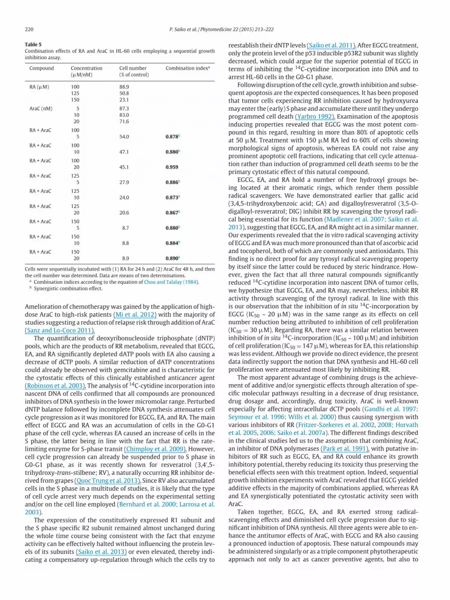
Table 5
Combination effects of RA and AraC in HL-60 cells employing a sequential growth
inhibition assay.
Compound Concentration Cell number Combination indexa
(µM/nM) (% of control)
RA (µM) 100 88.9
125 50.8
150 23.1
AraC (nM) 5 87.3
10 83.0
20 71.6
RA + AraC 100
5 54.0 0.878b
RA + AraC 100
10 47.1 0.880b
RA + AraC 100
20 45.1 0.959
RA + AraC 125
5 27.9 0.886b
RA + AraC 125
10 24.0 0.873b
RA + AraC 125
20 20.6 0.867b
RA + AraC 150
5 8.7 0.880b
RA + AraC 150
10 8.8 0.884b
RA + AraC 150
20 8.9 0.890b
Cells were sequentially incubated with (1) RA for 24 h and (2) AraC for 48 h, and then
the cell number was determined. Data are means of two determinations.a Combination indices according to the equation of Chou and Talalay (1984).b Synergistic combination effect.
Amelioration of chemotherapywas gained by the application of high-
dose AraC to high-risk patients (Mi et al. 2012) with the majority of
studies suggesting a reduction of relapse risk through addition of AraC
(Sanz and Lo-Coco 2011).
The quantification of deoxyribonucleoside triphosphate (dNTP)
pools, which are the products of RR metabolism, revealed that EGCG,
EA, and RA significantly depleted dATP pools with EA also causing a
decrease of dCTP pools. A similar reduction of dATP concentrations
could already be observed with gemcitabine and is characteristic for
the cytostatic effects of this clinically established anticancer agent
(Robinson et al. 2003). The analysis of 14C-cytidine incorporation into
nascent DNA of cells confirmed that all compounds are pronounced
inhibitors of DNA synthesis in the lowermicromolar range. Perturbed
dNTP balance followed by incomplete DNA synthesis attenuates cell
cycle progression as it wasmonitored for EGCG, EA, and RA. Themain
effect of EGCG and RA was an accumulation of cells in the G0-G1
phase of the cell cycle, whereas EA caused an increase of cells in the
S phase, the latter being in line with the fact that RR is the rate-
limiting enzyme for S-phase transit (Chimploy et al. 2009). However,
cell cycle progression can already be suspended prior to S phase in
G0-G1 phase, as it was recently shown for resveratrol (3,4′,5-
trihydroxy-trans-stilbene; RV), a naturally occurring RR inhibitor de-
rived fromgrapes (Quoc Trung et al. 2013). Since RV also accumulated
cells in the S phase in a multitude of studies, it is likely that the type
of cell cycle arrest very much depends on the experimental setting
and/or on the cell line employed (Bernhard et al. 2000; Larrosa et al.
2003).
The expression of the constitutively expressed R1 subunit and
the S phase specific R2 subunit remained almost unchanged during
the whole time course being consistent with the fact that enzyme
activity can be effectively halted without influencing the protein lev-
els of its subunits (Saiko et al. 2013) or even elevated, thereby indi-
cating a compensatory up-regulation through which the cells try to
reestablish their dNTP levels (Saiko et al. 2011). After EGCG treatment,
only the protein level of the p53 inducible p53R2 subunit was slightly
decreased, which could argue for the superior potential of EGCG in
terms of inhibiting the 14C-cytidine incorporation into DNA and to
arrest HL-60 cells in the G0-G1 phase.
Followingdisruptionof the cell cycle, growth inhibition and subse-
quent apoptosis are the expected consequences. It has been proposed
that tumor cells experiencing RR inhibition caused by hydroxyurea
mayenter the (early) Sphase andaccumulate thereuntil theyundergo
programmed cell death (Yarbro 1992). Examination of the apoptosis
inducing properties revealed that EGCG was the most potent com-
pound in this regard, resulting in more than 80% of apoptotic cells
at 50 µM. Treatment with 150 µM RA led to 60% of cells showing
morphological signs of apoptosis, whereas EA could not raise any
prominent apoptotic cell fractions, indicating that cell cycle attenua-
tion rather than induction of programmed cell death seems to be the
primary cytostatic effect of this natural compound.
EGCG, EA, and RA hold a number of free hydroxyl groups be-
ing located at their aromatic rings, which render them possible
radical scavengers. We have demonstrated earlier that gallic acid
(3,4,5-trihydroxybenzoic acid; GA) and digalloylresveratrol (3,5-O-
digalloyl-resveratrol; DIG) inhibit RR by scavenging the tyrosyl radi-
cal being essential for its function (Madlener et al. 2007; Saiko et al.
2013), suggesting that EGCG, EA, andRAmight act in a similarmanner.
Our experiments revealed that the in vitro radical scavenging activity
of EGCG and EAwasmuchmore pronounced than that of ascorbic acid
and tocopherol, both of which are commonly used antioxidants. This
finding is no direct proof for any tyrosyl radical scavenging property
by itself since the latter could be reduced by steric hindrance. How-
ever, given the fact that all three natural compounds significantly
reduced 14C-cytidine incorporation into nascent DNA of tumor cells,
we hypothesize that EGCG, EA, and RA may, nevertheless, inhibit RR
activity through scavenging of the tyrosyl radical. In line with this
is our observation that the inhibition of in situ 14C-incorporation by
EGCG (IC50 ~ 20 µM) was in the same range as its effects on cell
number reduction being attributed to inhibition of cell proliferation
(IC50 = 30 µM). Regarding RA, there was a similar relation between
inhibition of in situ 14C-incorporation (IC50 ~ 100 µM) and inhibition
of cell proliferation (IC50 = 147µM), whereas for EA, this relationship
was less evident. Althoughwe provide no direct evidence, the present
data indirectly support the notion that DNA synthesis and HL-60 cell
proliferation were attenuated most likely by inhibiting RR.
The most apparent advantage of combining drugs is the achieve-
ment of additive and/or synergistic effects through alteration of spe-
cific molecular pathways resulting in a decrease of drug resistance,
drug dosage and, accordingly, drug toxicity. AraC is well-known
especially for affecting intracellular dCTP pools (Gandhi et al. 1997;
Seymour et al. 1996; Wills et al. 2000) thus causing synergism with
various inhibitors of RR (Fritzer-Szekeres et al. 2002, 2008; Horvath
et al. 2005, 2006; Saiko et al. 2007a). The different findings described
in the clinical studies led us to the assumption that combining AraC,
an inhibitor of DNA polymerases (Park et al. 1991), with putative in-
hibitors of RR such as EGCG, EA, and RA could enhance its growth
inhibitory potential, thereby reducing its toxicity thus preserving the
beneficial effects seen with this treatment option. Indeed, sequential
growth inhibition experiments with AraC revealed that EGCG yielded
additive effects in the majority of combinations applied, whereas RA
and EA synergistically potentiated the cytostatic activity seen with
AraC.
Taken together, EGCG, EA, and RA exerted strong radical-
scavenging effects and diminished cell cycle progression due to sig-
nificant inhibition of DNA synthesis. All three agents were able to en-
hance the antitumor effects of AraC, with EGCG and RA also causing
a pronounced induction of apoptosis. These natural compounds may
be administered singularly or as a triple component phytotherapeutic
approach not only to act as cancer preventive agents, but also to

P. Saiko et al. / Phytomedicine 22 (2015) 213–222 221
support conventional chemotherapy regimens and therefore deserve
further preclinical and in vivo testing.
Conflict of interest
The authors declare that they have no conflict to disclose.
Acknowledgments
This investigation was supported by the Medical-Scientific Fund
of the Mayor of Vienna, grant #11006 to Z.B.-H., the "Hochschuljubi-
laeumsstiftung der Stadt Wien", grant #H-2498/2011 to P.S., and by
the Herzfelder Family Foundation with grants to T.S., P.S., and G.K.
References
Ades, L., Chevret, S., Raffoux, E., de Botton, S., Guerci, A., Pigneux, A., Stoppa, A.M.,Lamy, T., Rigal-Huguet, F., Vekhoff, A., Meyer-Monard, S., Maloisel, F., Deconinck, E.,Ferrant, A., Thomas, X., Fegueux, N., Chomienne, C., Dombret, H., Degos, L.,Fenaux, P., 2006. Is cytarabine useful in the treatment of acute promyelocyticleukemia? Results of a randomized trial from the European Acute PromyelocyticLeukemia Group. J. Clin. Oncol. 24, 5703–5710.
Aye, Y., Li, M., Long, M.J., Weiss, R.S., 2014. Ribonucleotide reductase and cancer: bio-logical mechanisms and targeted therapies. Oncogene doi:10.1038/onc.2014.155.
Bernhard, D., Tinhofer, I., Tonko, M., Hubl, H., Ausserlechner, M.J., Greil, R., Kofler, R.,Csordas, A., 2000. Resveratrol causes arrest in the S-phase prior to Fas-independentapoptosis in CEM-C7H2 acute leukemia cells. Cell Death Differ. 7, 834–842.
Brown, M.D., 1999. Green tea (Camellia sinensis) extract and its possible role in theprevention of cancer. Altern. Med. Rev. 4, 360–370.
Chimploy, K., Diaz, G.D., Li, Q., Carter, O., Dashwood,W.M.,Mathews, C.K.,Williams,D.E.,Bailey, G.S., Dashwood, R.H., 2009. E2F4 and ribonucleotide reductase mediate S-phase arrest in colon cancer cells treated with chlorophyllin. Int. J. Cancer 125,2086–2094.
Chou, T.C., Talalay, P., 1981. Generalized equations for the analysis of inhibitions ofMichaelis–Menten and higher-order kinetic systems with two or more mutuallyexclusive and nonexclusive inhibitors. Eur. J. Biochem. 115, 207–216.
Chou, T.C., Talalay, P., 1984. Quantitative analysis of dose–effect relationships: thecombined effects of multiple drugs or enzyme inhibitors. Adv. Enzyme Regul. 22,27–55.
de The, H., Chen, Z., 2010. Acute promyelocytic leukaemia: novel insights into themechanisms of cure. Nat. Rev. Cancer 10, 775–783.
Fritzer-Szekeres, M., Salamon, A., Grusch, M., Horvath, Z., Hochtl, T., Steinbrugger, R.,Jager, W., Krupitza, G., Elford, H.L., Szekeres, T., 2002. Trimidox, an inhibitor ofribonucleotide reductase, synergistically enhances the inhibition of colony forma-tion by Ara-C in HL-60 human promyelocytic leukemia cells. Biochem. Pharmacol.64, 481–485.
Fritzer-Szekeres, M., Savinc, I., Horvath, Z., Saiko, P., Pemberger, M., Graser, G.,Bernhaus, A., Ozsvar-Kozma, M., Grusch, M., Jaeger, W., Szekeres, T., 2008. Bio-chemical effects of piceatannol in human HL-60 promyelocytic leukemia cells–synergism with Ara-C. Int. J. Oncol. 33, 887–892.
Gandhi, V., Huang, P., Chapman, A.J., Chen, F., Plunkett, W., 1997. Incorporationof fludarabine and 1-beta-d-arabinofuranosylcytosine 5′-triphosphates by DNApolymerase alpha: affinity, interaction, and consequences. Clin. Cancer Res. 3,1347–1355.
Garrett, C., Santi, D.V., 1979. A rapid and sensitive high pressure liquid chromatographyassay for deoxyribonucleoside triphosphates in cell extracts. Anal. Biochem. 99,268–273.
Grasl-Kraupp, B., Ruttkay-Nedecky, B., Koudelka, H., Bukowska, K., Bursch,W., Schulte-Hermann, R., 1995. In situ detection of fragmented DNA (TUNEL assay) fails todiscriminate among apoptosis, necrosis, and autolytic cell death: a cautionary note.Hepatology 21, 1465–1468.
Gridling, M., Stark, N., Madlener, S., Lackner, A., Popescu, R., Benedek, B., Diaz, R.,Tut, F.M., Nha Vo, T.P., Huber, D., Gollinger, M., Saiko, P., Ozmen, A., Mosgoeller, W.,DeMartin, R., Eytner, R.,Wagner, K.H., Grusch,M., Fritzer-Szekeres,M., Szekeres, T.,Kopp, B., Frisch, R., Krupitza, G., 2009. In vitro anti-cancer activity of two ethno-pharmacological healing plants from Guatemala Pluchea odorata and Phlebodiumdecumanum. Int. J. Oncol. 34, 1117–1128.
Grusch, M., Fritzer-Szekeres, M., Fuhrmann, G., Rosenberger, G., Luxbacher, C.,Elford, H.L., Smid, K., Peters, G.J., Szekeres, T., Krupitza, G., 2001. Activation ofcaspases and induction of apoptosis by novel ribonucleotide reductase inhibitorsamidox and didox. Exp. Hematol. 29, 623–632.
Grusch, M., Polgar, D., Gfatter, S., Leuhuber, K., Huettenbrenner, S., Leisser, C.,Fuhrmann, G., Kassie, F., Steinkellner, H., Smid, K., Peters, G.J., Jayaram, H.N.,Klepal, W., Szekeres, T., Knasmuller, S., Krupitza, G., 2002. Maintenance of ATPfavours apoptosis over necrosis triggered by benzamide riboside. Cell Death Differ.9, 169–178.
Guarino, E., Salguero, I., Kearsey, S.E., 2014. Cellular regulation of ribonucleotide reduc-tase in eukaryotes. Semin. Cell Dev. Biol. 30, 97–103.
Hagiwara, Y., Kasukabe, T., Kaneko, Y., Niitsu, N., Okabe-Kado, J., 2010. Ellagic acid, anatural polyphenolic compound, induces apoptosis and potentiates retinoic acid-induceddifferentiationofhuman leukemiaHL-60cells. Int. J.Hematol. 92, 136–143.
Hakansson, P., Hofer, A., Thelander, L., 2006. Regulation of mammalian ribonucleotidereduction and dNTP pools after DNA damage and in resting cells. J. Biol. Chem. 281,7834–7841.
Heber, D., 2008. Multitargeted therapy of cancer by ellagitannins. Cancer Lett. 269,262–268.
Heinemann, V., Schulz, L., Issels, R.D., Plunkett, W., 1995. Gemcitabine: a modula-tor of intracellular nucleotide and deoxynucleotide metabolism. Semin. Oncol. 22,11–18.
Horvath, Z., Murias, M., Saiko, P., Erker, T., Handler, N., Madlener, S., Jaeger, W.,Grusch, M., Fritzer-Szekeres, M., Krupitza, G., Szekeres, T., 2006. Cytotoxic and bio-chemical effects of 3,3′ ,4,4′ ,5,5′-hexahydroxystilbene, a novel resveratrol analog inHL-60 human promyelocytic leukemia cells. Exp. Hematol. 34, 1377–1384.
Horvath, Z., Saiko, P., Illmer, C., Madlener, S., Hoechtl, T., Bauer, W., Erker, T., Jaeger, W.,Fritzer-Szekeres, M., Szekeres, T., 2005. Synergistic action of resveratrol, an ingre-dient of wine, with Ara-C and tiazofurin in HL-60 human promyelocytic leukemiacells. Exp. Hematol. 33, 329–335.
Kelaidi, C., Chevret, S., De Botton, S., Raffoux, E., Guerci, A., Thomas, X., Pigneux, A.,Lamy, T., Rigal-Huguet, F., Meyer-Monard, S., Chevallier, P., Maloisel, F.,Deconinck, E., Ferrant, A., Fegueux, N., Ifrah, N., Sanz, M., Dombret, H., Fenaux, P.,Ades, L., 2009. Improved outcome of acute promyelocytic leukemiawith highWBCcounts over the last 15 years: the European APL Group experience. J. Clin. Oncol. 27,2668–2676.
Lambert, J.D., Elias, R.J., 2010. The antioxidant and pro-oxidant activities of green teapolyphenols: a role in cancer prevention. Arch. Biochem. Biophys. 501, 65–72.
Lansky, E.P., Jiang, W., Mo, H., Bravo, L., Froom, P., Yu, W., Harris, N.M., Neeman, I.,Campbell, M.J., 2005. Possible synergistic prostate cancer suppression by anatom-ically discrete pomegranate fractions. Invest. New Drugs 23, 11–20.
Larrosa, M., Tomas-Barberan, F.A., Espin, J.C., 2003. Grape polyphenol resveratroland the related molecule 4-hydroxystilbene induce growth inhibition, apopto-sis, S-phase arrest, and upregulation of cyclins A, E, and B1 in human SK-Mel-28melanoma cells. J. Agric. Food Chem. 51, 4576–4584.
Madlener, S., Illmer, C., Horvath, Z., Saiko, P., Losert, A., Herbacek, I., Grusch, M.,Elford, H.L., Krupitza, G., Bernhaus, A., Fritzer-Szekeres, M., Szekeres, T., 2007. Gal-lic acid inhibits ribonucleotide reductase and cyclooxygenases in human HL-60promyelocytic leukemia cells. Cancer Lett. 245, 156–162.
Mi, J., 2011. Current treatment strategy of acute promyelocytic leukemia. Front Med. 5,341–347.
Mi, J.Q., Li, J.M., Shen, Z.X., Chen, S.J., Chen, Z., 2012. How tomanage acute promyelocyticleukemia. Leukemia 26, 1743–1751.
Ngo, S.N., Williams, D.B., Head, R.J., 2011. Rosemary and cancer prevention: preclinicalperspectives. Crit. Rev. Food Sci. Nutr. 51, 946–954.
Noble, S., Goa, K.L., 1997. Gemcitabine. A review of its pharmacology and clinical po-tential in non-small cell lung cancer and pancreatic cancer. Drugs 54, 447–472.
Ostruszka, L.J., Shewach, D.S., 2003. The role of DNA synthesis inhibition in the cy-totoxicity of 2′ ,2′-difluoro-2′-deoxycytidine. Cancer Chemother. Pharmacol. 52,325–332.
Park, J.K., Lee, J.S., Lee, H.H., Choi, I.S., Park, S.D., 1991. Accumulation of polycyclic aro-matic hydrocarbon-induced single strand breaks is attributed to slower rejoiningprocesses by DNA polymerase inhibitor, cytosine arabinoside in CHO-K1 cells. LifeSci. 48, 1255–1261.
Petersen, M., Simmonds, M.S., 2003. Rosmarinic acid. Phytochemistry 62, 121–125.Quoc Trung, L., Espinoza, J.L., Takami, A., Nakao, S., 2013. Resveratrol induces cell cycle
arrest and apoptosis inmalignantNK cells via JAK2/STAT3pathway inhibition. PLoSOne 8, e55183.
Robinson, B.W., Im, M.M., Ljungman, M., Praz, F., Shewach, D.S., 2003. Enhanced ra-diosensitizationwith gemcitabine inmismatch repair-deficient HCT116 cells. Can-cer Res. 63, 6935–6941.
Saban, N., Bujak, M., 2009. Hydroxyurea and hydroxamic acid derivatives as antitumordrugs. Cancer Chemother. Pharmacol. 64, 213–221.
Saiko, P., Graser, G., Giessrigl, B., Lackner, A., Grusch,M., Krupitza,G., Basu, A., Sinha, B.N.,Jayaprakash, V., Jaeger, W., Fritzer-Szekeres, M., Szekeres, T., 2011. A novel N-hydroxy-N′-aminoguanidine derivative inhibits ribonucleotide reductase activity:effects in human HL-60 promyelocytic leukemia cells and synergism with arabi-nofuranosylcytosine (Ara-C). Biochem. Pharmacol. 81, 50–59.
Saiko, P., Graser, G., Giessrigl, B., Steinmann, M.T., Schuster, H., Lackner, A., Grusch, M.,Krupitza, G., Jaeger,W., Somepalli, V., Golakoti, T., Fritzer-Szekeres,M., Szekeres, T.,2013. Digalloylresveratrol, a novel resveratrol analog inhibits the growth of humanpancreatic cancer cells. Invest. New Drugs 31, 1115–1124.
Saiko, P., Ozsvar-Kozma,M., Bernhaus, A., Jaschke,M., Graser, G., Lackner, A., Grusch,M.,Horvath, Z., Madlener, S., Krupitza, G., Handler, N., Erker, T., Jaeger, W., Fritzer-Szekeres, M., Szekeres, T., 2007. N-Hydroxy-N′-(3,4,5-trimethoxyphenyl)-3,4,5-trimethoxy-benzamidine, a novel resveratrol analog, inhibits ribonucleotide re-ductase in HL-60 human promyelocytic leukemia cells: synergistic antitumor ac-tivity with arabinofuranosylcytosine. Int. J. Oncol. 31, 1261–1266.
Saiko, P., Ozsvar-Kozma, M., Madlener, S., Bernhaus, A., Lackner, A., Grusch, M.,Horvath, Z., Krupitza, G., Jaeger, W., Ammer, K., Fritzer-Szekeres, M., Szekeres, T.,2007. Avemar, a nontoxic fermented wheat germ extract, induces apoptosis andinhibits ribonucleotide reductase in human HL-60 promyelocytic leukemia cells.Cancer Lett. 250, 323–328.
Sanz, M.A., Lo-Coco, F., 2011. Modern approaches to treating acute promyelocyticleukemia. J. Clin. Oncol. 29, 495–503.
Seymour, J.F., Huang, P., Plunkett,W., Gandhi, V., 1996. Influence of fludarabine onphar-macokinetics and pharmacodynamics of cytarabine: implications for a continuousinfusion schedule. Clin. Cancer Res. 2, 653–658.
Shao, J., Liu, X., Zhu, L., Yen, Y., 2013. Targeting ribonucleotide reductase for cancertherapy. Expert Opin. Ther. Targets 17, 1423–1437.

222 P. Saiko et al. / Phytomedicine 22 (2015) 213–222
Singh, B.N., Shankar, S., Srivastava, R.K., 2011. Green tea catechin, epigallocatechin-3-gallate (EGCG): mechanisms, perspectives and clinical applications. Biochem.Pharmacol. 82, 1807–1821.
Smid, K., Van Moorsel, C.J., Noordhuis, P., Voorn, D.A., Peters, G.J., 2001. Interference ofgemcitabine triphosphate with the measurements of deoxynucleotides using anoptimized DNA polymerase elongation assay. Int. J. Oncol. 19, 157–162.
Takeda, E., Weber, G., 1981. Role of ribonucleotide reductase in expression in theneoplastic program. Life Sci. 28, 1007–1014.
Tallman, M.S., Altman, J.K., 2009. How I treat acute promyelocytic leukemia. Blood 114,5126–5135.
Tennant, L., 2001. Chronic myelogenous leukemia: an overview. Clin. J. Oncol. Nurs. 5,218–219 .
Toschi, L., Finocchiaro, G., Bartolini, S., Gioia, V., Cappuzzo, F., 2005. Role of gemcitabinein cancer therapy. Future Oncol. 1, 7–17.
Viladomiu, M., Hontecillas, R., Lu, P., Bassaganya-Riera, J., 2013. Preventive and pro-phylacticmechanisms of action of pomegranate bioactive constituents. Evid. BasedComplement. Alternat. Med. 2013, 789764.
Wills, P.W., Hickey, R., Malkas, L., 2000. Ara-C differentially affects multiproteinforms of human cell DNA polymerase. Cancer Chemother. Pharmacol. 46,193–203.
Wu, W.Y., Wang, Y.P., 2012. Pharmacological actions and therapeutic applications ofSalvia miltiorrhiza depside salt and its active components. Acta Pharmacol. Sin. 33,1119–1130.
Yarbro, J.W., 1992. Mechanism of action of hydroxyurea. Semin. Oncol. 19, 1–10.


