Embed Size (px)
Citation preview

4
Estrogens Involvement in the Physiopathology of Articular Cartilage
Safa Moslemi, Magali Demoor, Karim Boumediene, Philippe Galera and Laure Maneix
Laboratory of Extracellular Matrix and Pathology, University of Caen/Lower-Normandy,
Faculty of Medicine, Caen France
1. Introduction Life expectancy increases in developed countries and results in a high prevalence of age-related diseases namely in women. Elderly subjects generate a huge demand on the social and health services linked to their disability and dependence. Besides the own effects of aging, the estrogenic deficiency, especially during menopause, is the major cause of degenerative diseases such as osteoporosis, skin aging, Alzheimer’s disease and osteoarthritis (OA) (Antonicelli et al., 2008; Candore et al., 2006; Elders, 2000; Felson & Nevitt, 1998; Pietschmann et al., 2008; Stovall & Pinkerton, 2008). OA is a worldwide public health problem which may affect different sites of the skeleton. In the Western world, at least 10% of the population has OA symptoms and 80% of the population will be potentially affected after the age of 70 years. Knee OA represents one of the major causes of morbidity and disability in relation to the worsening of quality of life (Elders, 2000). Pathogenesis of knee OA involves multiple factors including gender, weight, and genetics. Although age-dependent degenerative pathologies, especially OA, involve complex mechanisms in which the imbalance of the cytokines/growth factors plays a crucial role, it is more and more obvious that estrogens participate, with their receptors and modulators, to all of these diseases affecting connective tissues (Calleja-Agius & Brincat, 2009). Estrogens are steroidal hormones irreversibly synthesized from androgens via a crucial enzyme of steroidogenesis called aromatase, a member of cytochrome P450 superfamily, encoded by the CYP19 gene and being expressed in both sexes of many species (Simpson, 2003). Although non genomic action of estrogens is now recognized but these sexual hormones act often as signaling molecules that exert their effects by binding to estrogen receptors (ER) within the cells. The estrogen-receptor complex interacts then with DNA to change the expression of estrogen-responsive genes. The two known estrogen receptors, ER and ER, are present in numerous tissues other than those associated with reproduction, including bone, liver, heart, brain and cartilage and being selectively expressed in some targets; for instance ER is selectively expressed in lung and intestine. The presence of ER and ER was detected in articular chondrocytes of different species, especially in human, rat, pig, cow and rabbit
Challenges in Rheumatology 42
(Classen et al., 2001; Dayani et al., 1988; Oshima et al., 2007; Ushiyama et al., 1999). The action of 17-estradiol (17-E2) on the cartilage appears to be intimately linked to the sex of individuals since it was shown that the binding capacity of 17-E2 to its receptor was significantly higher in chondrocytes derived from male rather than female individuals (Nasatzky et al., 1994). In addition, 17-E2 can stimulate the nuclear expression of its own receptors in chondrocytes and create therefore a loop that reinforces the activation effects of the hormone in this cell type (Richmond et al., 2000). Moreover, a strong correlation was found between the polymorphism of ESR1 gene encoding ER and increased risk of OA in several populations (Bergink et al. 2003; Valdes et al., 2006). All these observations tend to prove that the cartilage is an estrogen-sensitive tissue, in which 17-E2 would be likely to play a preponderant role in regulating chondrocyte homeostasis in joint diseases such as OA. Even thought hormone replacement therapy (HRT) is the most efficient treatment against, at least, some of the degenerative pathologies mentioned above but studies confirm that risks linked to HRT can exceed the discounted benefits. As a consequence, it becomes important to address the question of the age-associated disorders and to find new targets to treat them. Selective Estrogen Receptor Modulators (SERM) like tamoxifen, raloxifen, fulvestrant and genistein have shown to be useful and attracted the attention of researchers (Cotter & Cashman, 2003; Khalil, 2010; Riggs & Hartmann 2003). A characteristic that distinguishes these substances from pure receptor agonists and antagonists is that their action is different in various tissues, thereby giving the possibility to selectively inhibit or stimulate estrogen-like action in various tissues. For instance tamoxifen (a first generation SERM) and raloxifen have been clinically used as antagonists of ER against breast cancer while they could be potentially agonists in bone and growth plate (Chagin et al, 2007; Nilsson et al, 2003). It has been proposed that these SERM could be used to replace natural estrogens to induce growth plate fusion reducing thereby the final height in girls expected to achieve extreme tall stature. Moreover, phytoestrogens (flavones, isoflavones, lignans) have aroused much interest as natural SERM and potential substitutes in the hormonal treatment of post-menopausal women, but they require much further investigation regarding their mechanism(s) of action and their safety (Dodin et al, 2003).
2. Estrogen synthesis and action 2.1 Estrogen synthesis via aromatase Estrogens are steroidal hormones composed from 18 atoms of carbon and produced essentially in gonads, ovary and testis, but also in non reproductive tissues such as bone and adipose tissue. These lipophilic compounds irreversibly synthesized from androgens via a crucial enzyme of steroidogenesis called aromatase, a member of cytochrome P450 superfamily, encoded by the CYP19 gene and being expressed in both sexes of many species. In pre-menopoausal women, estrogens such as 17-E2 (the more estrogenic) and estrone are synthesized in ovary during follicular phase whereas estriol being produced essentially by placenta during pregnancy. After menopause, when ovary activity disappears, production of 17-E2 from testosterone via aromatase is assumed by peripheral tissues like liver, adipose tissue, bone, vascular endothelium, chondrocyte and synovial cells (Simpson, 2003; Takeuchi et al., 2007; Tanko et al., 2008). Consequently, circulating 17-E2 concentrations decrease drastically to 0.04-0.21 nM to reach those found in man (Chambliss & Shaul, 2002). Thus, estrogen action being localized and switch from endocrine to auto-,

Estrogens Involvement in the Physiopathology of Articular Cartilage 43
intra- and/or paracrine actions. Due to their lipophilic feature, estrogen can diffuse into the cell through plasma membrane and induce estrogen-dependent intracellular signaling pathways and/or bind the intracellular receptors especially ER stimulating an estrogen-dependent response in target cells.
2.2 ER structure In mammals, estrogens produced locally in different tissues, may exert various biological effects but are also responsible of the development of some pathological process such as hormone-dependent cancers. Estrogens exert their physiological and pathological effects through specific receptors considered as nuclear factors by binding target genes at a specific cis sequence called Estrogen Responsive Element (ERE). The first study on estrogen receptor, published by Toft & Groski in 1966, demonstrated that a specific protein of rat uterus was able to bind specifically estrogens. Later, the gene of this protein has been cloned (Walter et al., 1985) and sequenced (Green et al., 1986) from mammal cancer epithelium (MCF-7) of a patient suffering from breast cancer. This protein is now recognized as ER. A second gene (ER) has been also cloned from a cDNA library of the rat prostatic cells (Kuiper et al., 1996), being expressed selectively in some tissues and presenting more affinity for phytoestrogens. It is now established that the majority of the normal and pathological action of estrogens is generally mediated through these two estrogen receptors ERand ER which are members of nuclear receptor super family including progesterone, glucocorticoids, androgens and vitamin D receptors (Nuclear Receptor Nomenclature Committee, 1999; Germain et al., 2006; Mangelsdorf et al., 1995). In human, ERis encoded by ESR1 gene located on chromosome 6 (6q25.1) whereas ER is encoded by ESR2 gene located on chromosome 14 (14q23.2). Both ESR1 and ESR2 genes encoding the estrogen receptor may undergo alternative splicing of mRNA. Most of these transcripts differ only in their 5'UTR (Untranslated Region) and will be mainly translated into a long form of ER, recognized as ER66 (Flouriot et al., 1998). However, a second form of ER protein, derived from alternative splicing of exon 1A mRNA or a site of alternative translation initiation (AUG codon 174) was discovered and named ER46 (Barrailler et al. 1999; Flouriot et al., 2000). After translation, the 46 kDa isoform is truncated of the 173 first amino acids of the long form of 66 kDa. ER46 protein is thus composed of 422 amino acids, whereas ER66 has 595 amino acids. Both ER isoforms are capable of inducing a physiological response after ligand binding. The 46 kDa isoform can heterodimerize with ER66 and competitively inhibit the functions of ligand-independent trans activation of the long form of the receptor (Flouriot et al., 2000). ERand ER530 amino acids) are structurally organized in six distinct functional domains (A to F) (Fig. 1). There are structural and functional similarities more or less strong between ER and ER whose homology percentages vary significantly depending on the area considered. DNA binding domain (DBD) is conserved at about 97%, which means that both receptors can bind the same cis nucleotide sequences and thus activate transcription ofidentical target genes. However, there is only 55% homology at the level of ligand binding domain (LBD), indicating that ER and ER have different ligand binding specificity. Finally, ER has only a truncated form of ligand-independent trans activation domain (AF-1), thus limiting its trans activation ability (Hall & McDonnell, 1999; Pearce & Jordan, 2004).

Challenges in Rheumatology 44
Fig. 1. Structures and functional domains of estrogen receptors (ER)
Structures, functional domains and sequence homology percentage (italic) of and isoforms. AF-1, ligand-independent trans activation domain; AF-2, ligand-dependent trans activation domain; DBD, DNA Binding Domain; hsp90, heat-shock protein 90; LBD, Ligand Binding Domain; NLS, Nuclear Localization Signal. These 3 isoforms, ERα46, ERα66 and ERβ are expressed in articular chondrocytes.
2.3 Genomic and non genomic action of estrogen It is well established that estrogens act at target genes level to modulate their transcription via ER binding, so playing a transcriptional factor role recognized as ligand-dependent mechanism (Jensen & DeSombre, 1973). However, estrogen action becomes more complicated following the discovery of ER, or different ER and ER isoforms but also with the identification of new plasma-associated membrane receptors. ER and ER regulate thus estrogen-dependent genes expression through two distinct mechanisms: ERE-dependent genomic pathway and ERE-independent genomic pathway.
2.3.1 ERE-dependent genomic pathway In this mechanism ER and ER, following ligand fixation (estrogen, phytoestrogen, SERM), are subjected to homo- or hetero-dimerization, then ligand/ER complex move to the nucleus to bind directly ERE cis element (AGGTCAxxxTGACCT) (Hall et al., 2001) present in the promoter of target genes (Fig. 2). The determination of consensus sequences ERE and the fixation of ER will help to recruit transcriptional factors such as FOXA1 (Forkhead Box A1) or GATA4 (GATA binding protein 4), which will first ensure the chromatin remodeling necessary for ER binding. Ligand binding also allows the recruitment of transcription cofactors such as SRC-1 (Steroid Receptor Coactivator-1), GRIP-1 (Glucocorticoid Receptor Interacting Protein-1), CBP (CREB Binding Protein)/p300, or TRAP220 (Thyroid Hormone Receptor Activating Protein 220) (McKenna et al., 1999). The classic genomic pathway requires the activity of the two trans activation domains AF-1 and AF-2 that will allow the sequential and cyclic recruitment of different co-factors of transcription (Metivier et al., 2001).
Estrogens Involvement in the Physiopathology of Articular Cartilage 45
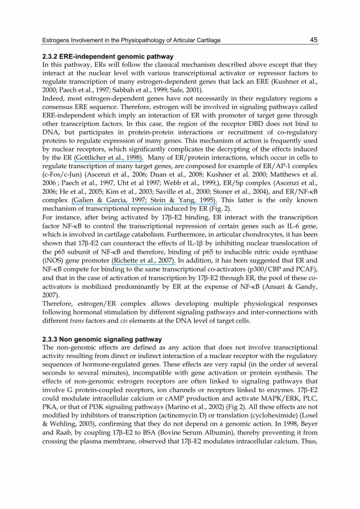
2.3.2 ERE-independent genomic pathway In this pathway, ERs will follow the classical mechanism described above except that they interact at the nuclear level with various transcriptional activator or repressor factors to regulate transcription of many estrogen-dependent genes that lack an ERE (Kushner et al., 2000; Paech et al., 1997; Sabbah et al., 1999; Safe, 2001). Indeed, most estrogen-dependent genes have not necessarily in their regulatory regions a consensus ERE sequence. Therefore, estrogen will be involved in signaling pathways called ERE-independent which imply an interaction of ER with promoter of target gene through other transcription factors. In this case, the region of the receptor DBD does not bind to DNA, but participates in protein-protein interactions or recruitment of co-regulatory proteins to regulate expression of many genes. This mechanism of action is frequently used by nuclear receptors, which significantly complicates the decrypting of the effects induced by the ER (Gottlicher et al., 1998). Many of ER/protein interactions, which occur in cells to regulate transcription of many target genes, are composed for example of ER/AP-1 complex (c-Fos/c-Jun) (Ascenzi et al., 2006; Duan et al., 2008; Kushner et al. 2000; Matthews et al. 2006 ; Paech et al., 1997, Uht et al 1997; Webb et al., 1999,), ER/Sp complex (Ascenzi et al., 2006; He et al., 2005; Kim et al., 2003; Saville et al., 2000; Stoner et al., 2004), and ER/NF-B complex (Galien & Garcia, 1997; Stein & Yang, 1995). This latter is the only known mechanism of transcriptional repression induced by ER (Fig. 2). For instance, after being activated by 17-E2 binding, ER interact with the transcription factor NF-B to control the transcriptional repression of certain genes such as IL-6 gene, which is involved in cartilage catabolism. Furthermore, in articular chondrocytes, it has been shown that 17-E2 can counteract the effects of IL-1 by inhibiting nuclear translocation of the p65 subunit of NF-B and therefore, binding of p65 to inducible nitric oxide synthase (iNOS) gene promoter (Richette et al., 2007). In addition, it has been suggested that ER and NF-B compete for binding to the same transcriptional co-activators (p300/CBP and PCAF), and that in the case of activation of transcription by 17-E2 through ER, the pool of these co-activators is mobilized predominantly by ER at the expense of NF-B (Ansari & Gandy, 2007). Therefore, estrogen/ER complex allows developing multiple physiological responses following hormonal stimulation by different signaling pathways and inter-connections with different trans factors and cis elements at the DNA level of target cells.
2.3.3 Non genomic signaling pathway The non-genomic effects are defined as any action that does not involve transcriptional activity resulting from direct or indirect interaction of a nuclear receptor with the regulatory sequences of hormone-regulated genes. These effects are very rapid (in the order of several seconds to several minutes), incompatible with gene activation or protein synthesis. The effects of non-genomic estrogen receptors are often linked to signaling pathways that involve G protein-coupled receptors, ion channels or receptors linked to enzymes. 17-E2 could modulate intracellular calcium or cAMP production and activate MAPK/ERK, PLC, PKA, or that of PI3K signaling pathways (Marino et al., 2002) (Fig 2). All these effects are not modified by inhibitors of transcription (actinomycin D) or translation (cycloheximide) (Losel & Wehling, 2003), confirming that they do not depend on a genomic action. In 1998, Beyer and Raab, by coupling 17-E2 to BSA (Bovine Serum Albumin), thereby preventing it from crossing the plasma membrane, observed that 17-E2 modulates intracellular calcium. Thus,

Challenges in Rheumatology 46
Fig. 2. Different pathways involved in estrogen receptors (ER) signaling.
These different pathways occur in estrogen sensitive cells including ERE-dependent genomic, ERE-independent genomic (via Sp1, AP-1 and NF-B) and membrane associated ER/G protein mechanisms. NF-B pathway is the only inhibitory mechanism. In articular chondrocytes ER66 homodimer complex binds predominantly Sp1 proteins to activate GC-box mediated trans activation of target genes such as Uridine diphospho-glucose deshydrogenase (UDPGD) and type II collagen (type II Col). Other mechanisms mediated by ER46 , ER, ER and ER homo- and/or heterodimer and finally membrane associated ER /protein G need to be elucidated in articular chondrocytes. Note that 17-E2 production from androgens like testosterone takes place via aromatase (CYP19) locally (intracrine effect) or in other cells (paracrine or endocrine effects). Adc, Adenylate cyclase; AP-1, Activating Protein-1; ERK, Extracellular signal Regulated Kinase; ERE, Estrogen Responsive Element; G, G protein; Hsp-90; Heat-shock protein 90; MAPK, Mitogen-Activated Protein Kinase; NF-B, Nuclear Factor-B; PI3K, Phosphatidyl Inositol 3-Kinase; PKA, Protein Kinase A; PLC, Phospholipase C; SERM, Selective Estrogen Receptor Modulator; Sp1, Specific Protein 1.
they confirm that this action is induced by a membrane ER. The existence of membrane estrogen receptor was discovered in the late 1970s in endometrial cells (Pietras & Szego, 1977). Various studies tempted to show that these membrane receptors are structurally similar to the classical cytoplasmic ER and that the and forms are represented (Chambliss & Shaul, 2002; Pappas et al., 1995; Razandi et al., 1999). More recently, a novel isoform of ER was highlighted: ER36. This 36 kDa protein lacks the trans activation domains AF-1 and AF-2 and has a DBD and a partial LBD, suggesting a membrane localization for this isoform (Wang et al., 2006). Also, estrogen binding sites localized at the membrane and the cytoplasm were detected in MCF-7 (Harrington et al., 2006). Since ER does not have a transmembrane domain,

Estrogens Involvement in the Physiopathology of Articular Cartilage 47
it appears that palmitoylation of the classical form of the receptor is necessary for its membrane addressing (Acconcia et al., 2005; Ellmann et al., 2009). In addition, it has been demonstrated that ER could be anchored to the plasma membrane through interactions with many membrane proteins such as caveolin 1 and 2, striatine or with adapter proteins like Shc and p130 Cas (Crk -associated substrate) (Cheskis et al., 2007).
2.3.4 Ligand-independent signaling pathway In absence of 17-E2, ER can be activated by phosphorylation via protein kinases A or C by extracellular signals like growth factors or cytokines, neurotransmitters, or by cell cycle regulators (Le Goff et al., 1994). Epidermal growth factor (EGF) mimics the effects of 17-E2 in the mouse uterus. Similarly, insulin, insulin-like growth factor-I (IGF-I), dopamine or transforming growth factor- (TGF-) may activate ER. The main targets of these growth factors are the many serine residues present in the AF-1 domain of ER particularly Ser118 and Ser167 (Nilsson et al., 2001). In summary, estrogens are involved in many signaling pathways, allowing fine control of cell and tissue functions.
3. Estrogen involvement in osteoarthritis 3.1 Epidemiological observations and clinical data OA is a worldwide public health problem which may affect different sites of the skeleton. In the Western world, at least 10% of the population present OA symptoms and 80% of the population will be potentially affected after the age of 70 years. Knee OA represents one of the major causes of morbidity and disability in relation to the worsening of quality of life. Pathogenesis of knee OA involves multiple factors including gender, weight, and genetics. Association between OA and estrogen deficiency during menopause has been firstly evoked by Cecil & Archer in 1925 following clinical observation. The hypothesis that estrogen deficiency may promote the development of OA has then been relied on the results of observational epidemiological studies. It is known that the frequency of knee OA is higher in women than in men; it is worsened after menopause and is lower in women receiving hormone replacement therapy (HRT) (Oliveria et al., 1995; Wilson et al., 1990). Although contradictions exist, some studies have shown that hysterectomy may also be associated with OA suggesting the potential role of estrogens to prevent these age-related diseases (Inoue et al., 1995; Spector et al., 1988; Spector et al., 1991). In addition, women with metastatic breast cancer when treated with aromatase inhibitors develop joint pains and cessation of aromatase inhibitors therapy resolved this joint pain (Burstein & Winer, 2007; Crew et al., 2007); aromatase inhibitors are widely used as adjuvant therapy in postmenopausal women with ER positive breast cancer (for review see Moslemi & Seralini, 2005). Moreover, an association of estrogen receptor (ER and ER) polymorphisms has been found in patients with OA compared to unaffected subjects. All these data suggest a protective role of estrogen and HRT on OA through functional isoforms of ER. Such a protective effect of estrogen on the development of OA may suggest two potential mechanisms: a direct effect on cartilage and an indirect effect through modifications of sub-chondral bone remodelling. Nevertheless, some clinical studies based on symptomatic parameters failed to report any effect of HRT on cartilage metabolism, that’s why further studies are required to clearly demonstrate beneficial effects of estrogens on molecular regulations of articular cartilage homeostasis.

Challenges in Rheumatology 48
3.2 Biological in vivo and in vitro studies In vivo animal studies indicate that estrogens may have a protective effect against OA, reversing or reducing the cartilage degradation in ovariectomized mice, rats, sheeps and monkeys (Cuzzocrea et al., 2003; Ham et al., 2002; Høegh-Andersen et al., 2004; Oestergaard et al., 2006). However, the way by which estrogens act to prevent the pathogenesis of OA in these models remains unclear. It is supposed that estrogens prevent cartilage degradation by increasing production of growth factors such as insulin growth factor-I (IGF-I) and suppressing pro-inflammatory cytokines expression such as interleukin-6 (IL-6). A few data exist about the role of estrogen in regulating the synthesis of matrix compounds. In vivo, 17-E2 prevents the degradation of collagen type II in the women treated with HRT (Mouritzen et al., 2003) but in vitro, it does not seem capable of modulating neosynthesis nor secretion of type II collagen in articular chondrocytes in primary culture (Ab-Rahim et al., 2008; Claassen et al., 2006). The measurement of serum C-terminal telopeptide of type II collagen (CTX-II) in bovine articular cartilage explants has determined that 17-E2 significantly protected the cartilage degradation induced by tumor necrosis factor- (TNF-) and oncostatin M (Oestergaard et al., 2006). In similar ex vivo experiments, 17-E2 can increase glycosaminoglycan (GAG) content of articular explants (Englert et al., 2006). Finally, the double invalidation of ER and ER gene receptors (double knock-out ER -/-, ER -/-) in mice aged of 6 months increases the number and size of osteophytes as well as a thinning of the sub-chondral plate, without changing the cartilage degradation (Sniekers et al., 2009). We demonstrated recently that 17-E2 (the most potent among all estrogens) at physiologic doses, and ER66 (wild type receptor) but not ER46 (AF-1 deleted receptor) could up-regulate at both mRNA and protein levels UDP-glucose dehydrogenase (UDPGD) in primary cultured articular chondrocytes via specific protein 1 (Sp1) binding sites (Maneix et al., 2008). This enzyme is responsible of UDP-glucuronate synthesis which is the main component of GAG chains polymerization in cartilage: it plays an essential role in the elongation of GAG chains and their attachment to the axial protein of proteoglycans (PGs). Its decarboxylation provides UDP xylose, which serves to anchor chains of chondroitin sulfate (CS), dermatan sulfate (DS) and heparan sulphate (HS). Moreover, we also established that 17-E2 increases the gene expression of type II collagen (COL2A1), the main collagen of hyaline cartilage, via trans activation domains AF-1 of ER66 in coordination with the transcription factors Sp1, Sp3, p300 and Soxs (Maneix et al, 2010). It would thus be a genomic mechanism ERE-independent. Finally, our preliminary results also showed that phytoestrogens such as apigenin and genistein could up-regulate the expression of UDPGD (unpublished data). Overall, these data indicate that estrogen/phytoestrogens and their receptors can be considered as potent regulators of chondrocyte homeostasis and are pro-anabolic for extracellular matrix synthesis. This may have potential applications in the tissue engineering of articular cartilage and offer new perspectives to prevent and/or to treat OA.
4. Mechanisms of estrogen action in OA 4.1 Effects on growth factors and cytokines 17-E2 can firstly interact with pathways affecting the synthesis and secretion of the key growth factors involved in the regulation of cartilage metabolism. So, TGF- expression in the iliac crest chondrocytes is influenced by 17-E2 in a biphasic manner: low concentrations of

Estrogens Involvement in the Physiopathology of Articular Cartilage 49
17-E2 increase TGF- expression whereas supra-physiologic doses decrease strongly its expression (Saggese et al., 1993). The concept of dose-dependence is confirmed by the majority of the studies on 17-E in the articular cartilage where physiological doses of the hormone show to be protective when considering the structural integrity of tissue while supra-physiologic concentrations are often deleterious (Richette et al., 2003) (Fig. 3). Experiments on chondrocytes from ovariectomized monkeys treated with 17-E showed that this hormone increases concomitantly the expression of the binding protein of IGF-I (IGFBP-2) and synthesis of PG (Richmond et al., 2000). In addition, the synovial fluid of these animals contains IGF-I twice more than untreated animals. Thereafter, it was shown that estrogen deficiency increases the sensitivity of cell response to certain pro-inflammatory cytokines through an increase in the number of receptors or cytokine cofactors amplifying consequently the effects of catabolic cytokines in these cells. Estrogen treatment in ovariectomized animals significantly reduced the production and secretion of pro-inflammatory cytokines in articular chondrocytes. This anti-inflammatory effect of 17-E2 was highlighted by Le Bail et al. (2001) who showed that the localized production of 17-E2 by synovial cells has inhibited the IL-6 secretion by articular chondrocytes. In addition, 17-E2 can also reduce production of pro-inflammatory prostaglandins through a decrease in the mRNA steady-state levels of cyclooxygenase-2 (COX-2) in bovine articular chondrocytes (Morisset et al., 1998).
4.2 Metalloproteinases (MMPs) expression and activity The majority of the beneficial effects of 17-E2 in articular chondrocytes is focused on the inhibition of catabolic pathways. Thus, Lee et al. (2003) found that 17-E2 decreases the secretion of the metalloproteinase MMP-1 in human osteoarthritic articular chondrocytes. In addition, 17-E2 can antagonize the degradation of PGs and the expression and activity of MMP-1, MMP-3 and MMP-13 enzymes induced by IL-1 in rabbit articular chondrocytes (Richette et al., 2004). Especially, the effects of 17-E2 on the expression of MMP-13 seem to be transmitted through the indirect binding of ER66 at an AP-1 site in the promoter of MMP-13 gene (Lu et al., 2006). Like the mode of action of 17-E2 on the expression levels of TGF-, the effects of the hormone on the expression of MMPs and their inhibitors TIMPs (tissue inhibitors of MMPs) appear to be dose-dependent. Low doses of 17-E2 decreased MMP-1/TIMP-1 and MMP-3/TIMP-1 ratios while higher concentrations have the opposite effect (Song et al., 2003).
4.3 Anti-oxidant effects The generation of reactive oxygen species (ROS) contributes actively to the cartilage ECM degradation in OA, in particular by inducing a decrease in the synthesis of PGs. However, due to the structure of their phenol nuclei, 17-E2 and its metabolites have anti-oxidant features (Liehr & Roy, 1998) that protect the cartilage degradation induced by ROS (Claassen et al., 2005). It is now established that 17-E2 is a modulator of the redox status of chondrocytes. During the arthritic process, overproduction of nitric oxide (NO) is a consequence of the action of proinflammatory cytokines such as IL-1 or TNF- that promote the activation of inducible nitric oxide synthase (iNOS) in OA chondrocytes. The iNOS gene is under the control of an estrogen-dependent promoter. Indeed, estrogen deficiency activates transcription of this promoter while a replacement therapy by 17-E2 inhibits it in ovariectomized mice (Cuzzocrea et al., 2003). 17-E2 can counteract the deleterious effects induced by IL-1 in rabbit articular chondrocytes by reducing the binding capacity of NF-B

Challenges in Rheumatology 50
Fig. 3. Role of 17-E2 and estrogen receptor ER /ER on joint cartilage homeostasis.
Col II, type II collagen; IGF-I, Insulin-like Growth Factor-I; IL, Interleukin; iNOS, Inducible Nitrogen Oxide Synthase; MMPs, Metalloproteinases; PGs, Proteoglycans; ROS, Reactive Oxygen Species; SERM, Selective Estrogen Receptor Modulators; TIMPs, Tissue Inhibitors MMPs; TGF-, Transforming Growth Factor-; TNF-, Tumor Necrosis Factor-; UDPGD, Uridine Diphospho-Glucose Dehydrogenase. The roles of SERM and phytoestrogens need to be clarified.
in the promoter of iNOS gene resulting in inhibition of nuclear translocation of this factor (Richette et al., 2007). This results in an inhibition of iNOS gene transcription and a subsequent reduction in NO production by chondrocytes.
4.4 Matrix components turn-over To better understand the physiopathology of articular cartilage in osteoarthritis, Høegh-Andersen et al. (2004) have validated an experimental model of ovariectomized rats with modification in cartilage structure representative of in vivo pathological changes observed in early human osteoarthritis. In animals aged from 5 to 7 months, estrogen deficiency increases the erosion of the articular surface and accelerates the renewal of matrix molecules. This animal model also showed that the effectiveness of HRT in the prevention of cartilage loss is increased when estrogen is administered to the animal from its operation and not after a period of 3 weeks (Oestergaard et al. 2006). These works need to be compared with the many epidemiological data which showed that the benefits of HRT are strengthened when patients are treated at the first signs of menopause and continued their treatment over a period exceeding 5 years. The importance of prevention in the treatment of damaged cartilage appears essential.
4.5 Sub-chondral bone regulation Within the joint, cartilage is not the only target tissue for estrogens. 17-E2 also controls the renewal of the sub-chondral bone by maintaining a balance between the two players in bone

Estrogens Involvement in the Physiopathology of Articular Cartilage 51
remodeling: osteoblasts that synthesize bone matrix and osteoclasts that are responsible for the resorption (bone loss) of existing bone. Estrogens inhibit osteoclasts activation and are therefore considered as inhibitors of bone resorption. The predominant mechanism that appears to be involved is the inhibition of IL-6 synthesis, the main cytokine involved in the activation of resorption. This inhibition of IL-6 synthesis by estrogens is mediated through the modification of NF-B binding on IL-6 gene promoter; there is no direct binding of ER to DNA (Ray et al., 1994). Consequently, an increase of bone resorption, generally favoring the occurrence of osteoporosis, is frequently seen in women after menopause which is related to the decrease of circulating estrogen levels during this period (Reginster et al., 2003). In this context, HRT could effectively prevent postmenopausal osteoporosis. Paradoxically, the frequency of OA and osteoporosis in postmenopausal women are most often inversely correlated, as reflected in the levels of osteocalcin, a marker of bone turnover, which are generally lower among women with OA than women without OA (for review: Dequeker et al., 2003). Patients suffering from OA of the knee are generally less subjected to bone loss. The increase in bone mass is a risk factor for incidence and development of knee and hip osteoarthritis. It was suggested that an increase in bone density in the area of the sub-chondral bone may induce bone stiffness and accelerate cartilage destruction. Thus, high bone density is associated with an increase of OA prevalence of the hip, hand and knee in postmenopausal women. However, high bone density increases risk of knee OA but protects against the progression of the disease once it is established. Therefore, bone loss in people with OA which is already established accelerates the progression of the disease. Indeed, Jacobsen et al. (2007) showed that the reduction of intra-articular space in the hip was correlated with the decrease in mineral density of sub-chondral bone in postmenopausal women. It was suggested that the alteration of bone structure can cause changes in the load distribution within the joint and promote the development of OA (Sniekers et al. 2008). In this case, the favorable effect of estrogen on cartilage may be due in part to the anti-resorption effect of 17-E2 on bone.
5. Phytoestrogen and Selective Estrogen Receptor Modulators (SERM) During the second half of the 20th century, HRT has been repeatedly promoted as the only pharmacological approach allowing a global prevention of all disorders related to or potentiated by estrogen deprivation. Recently, the risk/benefit profile of HRT has been severely challenged because of apparent increased risks of invasive breast cancer, coronary heart disease events, stroke and pulmonary embolism among treated women. These new findings imply a careful reassessment of the current evidence justifying the prescription of HRT for the prevention of the management of chronic disorders. The many contradictions recorded concerning benefit/risk effects of HRT are often related to differences in methodology and criteria used in measuring the effects of estrogen therapy in clinical studies. Indeed, analysis based strictly on symptomatic parameters showed that HRT had no effect or even adverse effects on the progression of OA (Von Muhlen et al., 2002). Conversely, more advanced techniques of magnetic resonance imaging showed that recipients of long term HRT (5 years or more) had a volume of articular cartilage largest in the knee than women having never taken any treatments (Wluka et al., 2001). As to the evolution of medical imaging techniques and advances in the diagnosis of OA, it appears that estrogens have a strong potential for preventing disease and preserving the structural integrity of the articulation among postmenopausal women. Thus, recent clinical trials of

Challenges in Rheumatology 52
consortium “Women's Health Initiative” showed that women treated with equine estrogens over a period of 7 years had 17% lower risk to undergo a hip replacement compared to control group (Cirillo et al., 2006). Finally, an in vivo study performed on 180 ovariectomized monkeys for 3 years found that HRT reduced the severity of arthritic lesions, through attenuation of the PGs loss and reducing the number of osteophytes in these animals (Ham et al., 2002). Given its chondroprotective potential, it would seem that HRT is capable of modulating chondrocyte metabolism. Studies have also shown that SERM and phytoestrogens may have beneficial effect on cartilage metabolism and to alleviate OA symptoms (Arjmandi et al, 2004; Bassleer et al., 1996; Guiducci et al., 2005; Tsai et al., 1992). It has been shown that when administered at a clinically relevant dose in young male rats, tamoxifen causes persistent retardation of longitudinal and cortical radial bone growth through systemic suppression of IGF-I production and local effects on the growth plate cartilage; it increases in chondrocytes proliferation/apoptosis and decreases the number of hypertophic chondrocytes (Karimian et al., 2008). Similarly, raloxifen could act as estrogen agonist on the growth plate of ovariectomized immature rabbits, accelerating growth plate senescence and thus hastening epiphyseal fusion (Nilsson et al, 2003). Besides estrogens, natural molecules such as phytoestrogens (estrogen-like compounds in plants) sharing structural and functional homologies with endogenous estrogens, could also act (even thought at micromolar concentrations) as agonists and/or antagonists in hormone-sensitive target cells. Scientists are now interested in the tissue-selective activities of phytoestrogens considering anti-estrogenic effects in reproductive tissue that could help to reduce the risk of hormone-associated cancers (breast, uterine, ovarian, and prostate), while estrogenic effects on bone and cardiovascular system for instance could favor the maintenance of bone density and protect against atherosclerosis respectively. Moreover, it has been suggested that the consumption of dietary phytoestrogens (soja isoflavones, lignans, etc.) may have beneficial effects on bone health at all stages of life. That’s why phytoestrogens have aroused much interest as potential substitutes in HRT of postmenopausal women, but they require much further investigation regarding their mechanism of action and their safety. Like estrogens, phytoestrogens may act via genomic and non genomic pathways. The most relevant molecular actions of phytoestrogens are those mediated by ERs. They may act on protein tyrosine kinase, MAP kinase, topoisomerase II and at all stages of cell cycle and apoptosis. They can also change the response to growth factors and cytokines. Genistein up to 100 M reduces the production of lipopolysaccharide (LPS)-stimulated pro-inflammatory molecules (COX-2, NO) but not that of COX-1 responsible of releasing prostaglandins in normal human chondrocytes (Hooshmand et al., 2007). When tested on human chondrocytes and chondrocytic cell line CHON-002, bavachin, a flavonid phytoestrogen isolated from Psoralea corylifolia, potentially protected cartilage from inflammation-mediated damage in joints of OA through decreasing IL-1beta-induced activation of IKK-IB alpha-NF-B signaling pathway (Cheng et al., 2010). Formononetin, a phytoestrogen isolated from Astragalus membranaceus showed to have biphasic positive effects on human normal osteoblasts and OA sub-chondral osteoblasts by modifying their biological synthetic capacities (Huh et al., 2010). Using female bovine articular chondrocytes, it has been demonstrated that the stimulating effect of insulin on GAGs sulfate incorporation was enhanced significantly after preincubation of cells with 10-11 -10-5 M daidzein or 10-9 -10-5 M genistein but not by 17-E2 (Claassen et al., 2008). More recently, xanthohumol, a prenylflavonoid extracted from hop, showed to prevent hyaluronan overproduction as well as PG and collagen loss in bovine chondrocytes;

Estrogens Involvement in the Physiopathology of Articular Cartilage 53
hyluronan overproduction is considered as an early reaction in OA followed by PG loss and collagen degradation (Stracke et al., 2001). However, additional research is critical to determine how phytoestrogens act on cartilage cells to obtain a more complete understanding of the effects.
6. Conclusion From epidemiological, clinical, in vivo and in vitro studies, a huge amount of data consistent with the fact that estrogens and their receptors can now be considered among the main players involved in the chondrocyte homeostasis and participate in cartilage protection from degradation and erosion occurring during menopause. Indeed, once ligand/ER complex is formed, estrogens such as 17-E2 could act with ER to induce or to inhibit trans activation of target genes of chondrocytes, the predominant cells of articular cartilage allowing expression of the specific chondrogenic markers such as UDPGD, PGs, type II collagen or inhibition of catabolic markers such as metalloproteinases and interleukins. There are different pathways by which estrogen and ER may interact with target genes in chondrocytes but it seems that Sp1 mediated trans activation being preferentially used in this cell type. In this mechanism ER needs AF-1 sequence (ligand-independent trans activation domain) to exert its action on GC boxes via Sp1 in target genes. In addition, some molecules sharing structural and functional features with estrogens like SERM and phytoestrogens can mimic estrogenic action and are therefore useful to repair cartilage erosion or might contribute at least to protect premenopausal joint cartilage and to maintain its homeostasis in a prevention strategy but these molecules need further investigation and deserves more attention from the scientific community to prove their safety and efficacy.
7. Key points Osteoarthritis, cartilage, chondrocytes, estrogens, phytoestrogens, selective estrogen receptor modulators, hormone replacement therapy, menopause.
8. References Ab-Rahim, S., Selvaratnam, L. & Kamarul, T. (2008). The effect of TGF-beta1 and beta-
estradiol on glycosaminoglycan and type II collagen distribution in articular chondrocyte cultures. Cell Biol. Int., Vol. 32, No. 7, (Jul 2008), pp. (841-847)
Acconcia, F., Ascenzi, P., Bocedi, A., Spisni, E., Tomasi, V., Trentalance, A., Visca, P. & Marino, M. (2005). Palmitoylation-dependent estrogen receptor alpha membrane localization: regulation by 17beta-estradiol. Mol. Biol. Cell, Vol. 16, No. 1, (Jan 2005), pp. (231-237)
Ansari, R.A. & Gandy, J. (2007). Determining the transrepression activity of xenoestrogen on nuclear factor-kappa B in Cos-1 cells by estrogen receptor-alpha. Int. J. Toxicol., Vol. 26, No. 5, (Sep-Oct 2007), pp. (441-449)
Antonicelli R, Olivieri F, Morichi V, Urbani E, Mais V. (2008). Prevention of cardiovascular events in early menopause: a possible role for hormone replacement therapy. Int J Cardiol., Vol. 130, No. 2, (Nov 2008), pp. (140-146), Review
Arjmandi, B.H., Khalil, D.A., Lucas, E.A., Smith, B.J., Sinichi, N., Hodges, S.B., Juma, S., Munson, M.E., Payton, M.E., Tivis, R.D. & Svanborg, A. (2004). Soy protein may

Challenges in Rheumatology 54
alleviate osteoarthritis sympthoms. Phytomedicine, Vol. 11, No. 7-8, (Nov 2004), pp. (567-575)
Ascenzi, P., Bocedi, A. & Marino, M. (2006). Structure-function relationship of estrogen receptor alpha and beta: impact on human health. Mol. Aspects Med., Vol. 27, No. 4, (Aug 2006), pp. (299-402), Review
Barrailler, P., Chinestra, P., Bayard, F. & Faye, J.C. (1999). Alternative initiation of translation accounts for a 67/45 kDa dimorphism of the human estrogen receptor ERalpha. Biochem. Biophys. Res. Commun., Vol. 257, No. 1, (Apr 1999), pp. (84-88)
Bassleer, C.T., Franchimont, P.P., Henrotin, Y.E., Franshimount, N.M., Geenen, V.G. & Reginster, J.Y. (1996). Effects of ipriflavone and its matabolites on human articular chondrocytes cultivated in clusters. Osteoarthritis Cartilage, Vol. 4, No. 1, (Mar 1996), pp. (1-8)
Bergink, A.P., Van Meurs, J.B., Loughlin, J., Arp, P.P., Fang, Y., Hofman, A., Van Leeuwen, J.P., Van Duijn, C.M., Uitterlinden, A.G. & Pols, H.A. (2003). Estrogen receptor alpha gene haplotype is associated with radiographic osteoarthritis of the knee in elderly men and women. Arthritis Rheum., Vol.48, No. 7, (Jul 2003), pp. (1913-1922)
Beyer, C. & Raab, H. (1998). Nongenomic effects of oestrogen: embryonic mouse midbrain neurones respond with a rapid release of calcium from intracellular stores. Eur. J. Neurosci., Vol. 10, No. 1 (Jan 1998), pp. (255-262)
Calleja-Agius, J. & Brincat, M.P. (2009). Effects of hormone replacement therapy on connective tissue: why is this important? Best Practice & Research Clinical Obstetrics and Gynaecology, Vol. 23, No. 1, (Feb 2009), pp. (121–27)
Candore, G., Balistreri, CR., Grimaldi, M.P., Vasto, S., Listì, F., Chiappelli, M., Licastro, F., Lio, D. & Caruso, C. (2006). Age-related inflammatory diseases: role of genetics and gender in the pathophysiology of Alzheimer's disease. Ann N Y Acad Sci, Vol. 1089, (Nov 2006), pp. (472-486), Review
Cecil, R.L. & Archer, B.H. (1925). Arthritis of the menopause, JAMA, Vol. 84 pp. (75–79) Chagin, A.S., Karimian, E., Zaman, F., Takigawa, M., Chrysis, D., Sävendahl, L. (2007).
Tamoxifen induces permanent growth arrest through selective induction of apoptosis in growth plate chondrocytes in cultured rat metatarsal bone. Bone, Vol. 40, No. 5, (May 2007), pp. (1415-1424)
Cheng, C.C., Chen, Y.H., Chang, W.L., Yang, S.P., Chang, D.M., Lai, J.H. & Ho, L.J. (2010). Phytoestrogen bavachin mediates anti-inflammation targeting Ikappa B kinase-I kappaB alpha-NF-kappaB signaling pathway in chondrocytes in vitro. Eur J Pharmacol. Vol. 636, No. 1-3, (Jun 2010), pp. (181-188)
Chambliss, K.L. & Shaul, P.W. (2002). Estrogen modulation of endothelial nitric oxide synthase. Endocr. Rev., Vol.23, No. 5, (Oct 2002), pp. (665-686), Review
Cheskis, B.J., Greger, J.G., Nagpal, S. & Freedman, L.P. (2007). Signaling by estrogens. J. Cell Physiol., Vol. 213, No. 3, (Dec 2007), pp. (610-617), Review
Cirillo, D.J., Wallace; R.B., Wu; L. & Yood; R.A. (2006). Effect of hormone therapy on risk of hip and knee joint replacement in the Women's Health Initiative. Arthritis Rheum., Vol. 54, No. 10, (Oct 2006), pp. (3194-3204)
Claassen, H., Briese, V., Manapov, F., Nebe, B., Schünke, M. & Kurz, B. (2008). The phytoestrogens daidzein and genistein enhance the insulin-stimulated sulfate

Estrogens Involvement in the Physiopathology of Articular Cartilage 55
uptake in articular chondrocytes. Cell Tissue Res., Vol. 333, No. 1, (Jul 2008), pp. (71-79)
Claassen, H., Schunke, M. & Kurz, B. (2005). Estradiol protects cultured articular chondrocytes from oxygen-radical-induced damage. Cell Tissue Res., Vol. 319, No. 3, (Mar 2005), pp. (439-445)
Claassen, H., Schluter, M., Schunke, M. & Kurz, B. (2006). Influence of 17beta-estradiol and insulin on type II collagen and protein synthesis of articular chondrocytes. Bone, Vol. 39, No. 2, (Aug 2006), pp. (310-317)
Cotter, A. & Cashman, K.D. (2003). Genistein appears to prevent postmenopausal bone loss as effectively as hormone replacement therapy. Nutr Rev., Vol. 61, No. 10, (Oct 2003), pp. (346-351)
Cuzzocrea, S., Mazzon, E., Dugo, L., Genovese, T., Di Paola, R., Ruggeri, Z., Vegeto, E., Caputi, A.P., Van De Loo, F.A., Puzzolo, D. & Maggi, A. (2003). Inducible nitric oxide synthase mediates bone loss in ovariectomized mice. Endocrinology, Vol. 144, No. 3, (Mar 2003), pp. (1098-1107)
Dequeker, J., Aerssens, J. & Luyten, F.P. (2003). Osteoarthritis and osteoporosis: clinical and research evidence of inverse relationship. Aging Clin. Exp. Res., Vol. 15, No. 5, (Oct 2003), pp. (426-439), Review
Dodin, S., Blanchet, C. & Marc, I. (2003). Phytoestrogens in menopausal women: a review of recent findings. Med Sci (Paris), Vol. 19, No. 10, (Oct 2003), pp. (1030-1037), Review (French)
Duan, R., Ginsburg, E. & Vonderhaar, B.K. (2008). Estrogen stimulates transcription from the human prolactin distal promoter through AP1 and estrogen responsive elements in T47D human breast cancer cells. Mol. Cell Endocrinol., Vol. 281, No. 1-2, (Jan 2008), pp. (9-18)
Elders, M.J. (2000). The increasing impact of arthritis on public health. J Rheumatol Suppl, Vol. 6, (Oct 2000), pp. (6-8)
Ellmann, S., Sticht, H., Thiel, F., Beckmann, M.W., Strick, R. & Strissel, P.L. (2009). Estrogen and progesterone receptors: from molecular structures to clinical targets. Cell Mol. Life Sci., Vol. 66, No. 15, (Aug 2009), pp. (2405-2426)
Englert, C., Blunk, T., Fierlbeck, J., Kaiser, J., Stosiek W., Angele, P., Hammer, J. & Straub, R.H. (2006). Steroid hormones strongly support bovine articular cartilage integration in the absence of interleukin-1beta. Arthritis Rheum., Vol. 54, No. 12, (Dec 2006), pp. ( 3890-3897)
Felson, D.T. & Nevitt, M.C. (1998). The effect of estrogen on osteoarthritis. Curr Opin Rheumatol, Vol. 10, No. 3, (May 1998), pp. (269-272)
Flouriot, G., Brand, H., Denger, S., Métivier, R., Kos, M., Reid, G., Sonntag-Buck, V. & Gannon, F. (2000). Identification of a new isoform of the human estrogen receptor-alpha (hER-alpha) that is encoded by distinct transcripts and that is able to repress hER-alpha activation function 1. EMBO J., Vol. 19, No. 17, (Sep 2000), pp. (4688-4700)
Flouriot, G., Griffin, C., Kenealy, M., Sonntag-Buck, V. & Gannon, F. (1998). Differentially expressed messenger RNA isoforms of the human estrogen receptor-alpha gene are generated by alternative splicing and promoter usage. Mol. Endocrinol., Vol. 12, No. 12, (Dec 1998), pp. (1939-1954)

Challenges in Rheumatology 56
Galien, R. & Garcia, T. (1997). Estrogen receptor impairs interleukin-6 expression by preventing protein binding on the NF-kappaB site. Nucleic Acids Res, Vol. 25, No. 12, (Jun 1997), pp. (2424-2429)
Germain, P., Staels, B., Dacquet, C., Spedding, M. & Laudet, V. (2006). Overview of nomenclature of nuclear receptors. Pharmacol. Rev., Vol. 58, No. 4, (Dec 2006), pp. (685-704)
Gottlicher, M., Heck, S. & Herrlich, P. (1998). Transcriptional cross-talk, the second mode of steroid hormone receptor action. J. Mol. Med., Vol. 76, No. 7, (Jun 1998), pp. (480-489)
Green, S., Walter, P., Kumar, V., Krust, A., Bornert, J.M., Argos, P. & Chambon, P. (1986). Human oestrogen receptor cDNA: sequence, expression and homology to v-erb-A. Nature, Vol. 320, No. 6058, (Mar 1986), pp. (134-139)
Guiducci, S., Del Rosso, A., Cinelli, M., Margheri, F., D'Alessio, S., Fibbi, G., Matucci Cerinic, M., Del Rosso, M. (2005). Rheumatoid synovial fibroblasts constitutively express the fibronolytic pattern of invasive tumor-like cells. Clin Exp Rheumatol., Vol. 23, No. 3, (May 2005), pp. (364-372)
Hall, J.M., Couse, J.F. & Korach, K.S. (2001). The multifaceted mechanisms of estradiol and estrogen receptor signaling. J. Biol. Chem., Vol. 276, No. 40, (Oct 2001), pp. (36869-36872)
Hall, J.M. & McDonnell, D.P. (1999). The estrogen receptor beta-isoform (ERbeta) of the human estrogen receptor modulates ERalpha transcriptional activity and is a key regulator of the cellular response to estrogens and antiestrogens. Endocrinology, Vol. 140, No. 12, (Dec 1999), pp. (5566-5578)
Ham, K.D., Loeser, R.F., Lindgren, B.R. & Carlson, C.S. (2002). Effects of long-term estrogen replacement therapy on osteoarthritis severity in cynomolgus monkeys. Arthritis Rheum., Vol. 46, No. 7, (Jul 2002), pp. (1956-1964)
Harrington, W.R., Kim, S.H., Funk, C.C., Madak-Erdogan, Z., Schiff, R., Katzenellenbogen, J.A. & Katzenellenbogen, B.S. (2006). Estrogen dendrimer conjugates that preferentially activate extranuclear, nongenomic versus genomic pathways of estrogen action. Mol. Endocrinol., Vol. 20, No. 3, (Mar 2006), pp. (491-502)
He, S., Sun, J.M., Li, L. & Davie, J.R. (2005). Differential intranuclear organization of transcription factors Sp1 and Sp3. Mol. Biol. Cell, Vol. 16, No. 9, (Sep 2005), pp. (4073-4083)
Høegh-Andersen, P., Tanko, L.B., Andersen, T.L., Lundberg, C.V., Mo, J.A., Heegaard, A.M., Delaisse, J.M. & Christgau, S. (2004). Ovariectomized rats as a model of postmenopausal osteoarthritis: validation and application. Arthritis Res. Ther., Vol. 6, No. 2, (Feb 2004), pp. (R169-R180)
Hooshmand, S., Soung do, Y., Lucas, E.A., Madihally, SV., Levenson, C.W. & Arjmandi, B.H. (2007). Genistein reduces the production of proinflammatory molecules in human chondrocyes. J Nut Biochem., Vol. 18, No. 9, (Sep 2007), pp. (609-614)
Huh, J.E., Seo, D.M., Baek, Y.H., Choi, D.Y., Park, D.S. & Lee, J.D. (2010). Biphasic positive effect of formononetin on metabolic activity of human normal and osteoarthritic subchondral osteoblasts. Int Immunopharmacol., Vol. 10, No. 4, (Apr 2010), pp. (500-507)

Estrogens Involvement in the Physiopathology of Articular Cartilage 57
Inoue, K., Ushiyama, T., Kim, Y., Shichikawa, K., Nishioka, J., Hukuda, S. (1995). Increased rate of hysterectomy in women undergoing surgery for osteoarthritis of the knee. Osteoarthritis Cartilage, Vol. 3, No. 3, (Sep 1995), pp. (205–209)
Jacobsen, S., Jensen, T.W., Bach-Mortensen, P., Hyldstrup, L. & Sonne-Holm, S. (2007). Low bone mineral density is associated with reduced hip joint space width in women: results from the Copenhagen Osteoarthritis Study. Menopause, Vol. 14, No. 6, (Nov-Dec 2007), pp. (1025-1030)
Jensen, E.V. & DeSombre, E.R. (1973). Estrogen-receptor interaction. Science, Vol. 182, No. 108, (Oct 1973), pp. (126-134), Review
Karimian, E, Chagin, AS., Gjerde, J., Heino, T., Lien, EA., Ohlsson, C. & Sävendahl, L. (2008). Tamoxifen impairs both longitudinal and cortical bone growth on young male rats. J bone Miner Res., Vol. 23, No. 8, (Aug 2008), pp. (1267-1277)
Khalil, R.A. (2010). Potential approaches to enhance the effects of estrogen on senescent blood vessels and postmenopausal cardiovascular disease. Cardiovasc Hematol Agents Med Chem., Vol. 8, No. 1, (Jan 2010), pp. (29-46)
Kim, K., Thu, N., Saville, B. & Safe, S. (2003). Domains of estrogen receptor alpha (ERalpha) required for ERalpha/Sp1-mediated activation of GC-rich promoters by estrogens and antiestrogens in breast cancer cells. Mol. Endocrinol., Vol. 17, No. 5, (May 2003), pp. (804-817)
Kuiper, G.G., Enmark, E., Pelto-Huikko, M., Nilsson, S. & Gustafsson, J.A. (1996). Cloning of a novel receptor expressed in rat prostate and ovary. Proc. Natl. Acad. Sci. U. S. A., Vol. 93, No. 12, (Jun 1996), pp. (5925-5930)
Kushner, P.J., Agard, D.A., Greene, G.L., Scanlan, T.S., Shiau, A.K., Uht, R.M. & Webb, P. (2000). Estrogen receptor pathways to AP-1. J. Steroid Biochem. Mol. Biol., Vol. 74, No. 5, (Nov 2000), pp. (311-317)
Le Bail, J., Liagre, B., Vergne, P., Bertin, P., Beneytout, J. & Habrioux, G. (2001). Aromatase in synovial cells from postmenopausal women. Steroids, Vol. 66, No. 10, (Oct 2001), pp. (749-757)
Lee, Y.J., Lee, E.B., Kwon, Y.E., Lee, J.J., Cho, W.S., Kim, H.A. & Song, Y.W. (2003). Effect of estrogen on the expression of matrix metalloproteinase (MMP)-1, MMP-3, and MMP-13 and tissue inhibitor of metalloproteinase-1 in osteoarthritis chondrocytes. Rheumatol. Int., Vol. 23, No. 6, (Nov 2003), pp. (282-288)
Le Goff, P., Montano, M.M., Schodin, D.J. & Katzenellenbogen, B.S. (1994). Phosphorylation of the human estrogen receptor. Identification of hormone-regulated sites and examination of their influence on transcriptional activity. J. Biol. Chem., Vol. 269, No. 6, (Feb 1994), pp. (4458-4466)
Liehr, J.G. & Roy, D. (1998). Pro-oxidant and antioxidant effects of estrogens. Methods Mol. Biol., Vol. 108, pp. (425-435)
Losel, R. & Wehling, M. (2003). Nongenomic actions of steroid hormones. Nat. Rev. Mol. Cell Biol., Vol. 4, No. 1, (Jan 2003), pp. (46-56), Review
Lu, T., Achari, Y., Sciore, P. & Hart, D.A. (2006). Estrogen receptor alpha regulates matrix metalloproteinase-13 promoter activity primarily through the AP-1 transcriptional regulatory site. Biochim. Biophys. Acta, Vol. 1762, No. 8, (Aug 2006), pp. (719-731)
Maneix, L., Beauchef, G., Servent, A., Wegrowski, Y., Maquart, F.X., Boujrad, N., Flouriot, G., Pujol, J.P., Boumediene, K., Galéra, P. & Moslemi, S. (2008) 17-Estradiol up-

Challenges in Rheumatology 58
regulates the expression of a functional UDP-glucose dehydrogenase in articular chondrocytes: Comparison with effects of cytokines and growth factors. Rheumatology (Oxford), Vol. 47, No. 3, (Mar 2008), pp. (281-288)
Maneix, L., Boujrad, N., Flouriot, G., Demoor, M., Boumediene, K., Moslemi, S. & Galéra, P. (2010). 17β-estradiol-induced up-regulation of type II collagen expression is mediated by ERα/Sp/SOX9/p300 complex through COL2A1 promoter/first intron interactions in differentiated and dedifferentiated articular chondrocytes. World Congress on osteoarthritis, OARSI, Brussels, Belgium, September 2010
Mangelsdorf, D.J., Thummel, C., Beato, M., Herrlich, P., Schütz, G., Umesono, K., Blumberg, B., Kastner, P., Mark, M., Chambon, P. & Evans R.M. (1995). The nuclear receptor superfamily: the second decade. Cell, Vol. 83, No. 6, (Dec 1995), pp. (835-839), Review
Marino, M., Acconcia, F., Bresciani, F., Weisz, A. & Trentalance, A. (2002). Distinct nongenomic signal transduction pathways controlled by 17beta-estradiol regulate DNA synthesis and cyclin D1 gene transcription in HepG2 cells. Mol. Biol. Cell, Vol. 13, No. 10, (Oct 2002), pp. (3720-3729)
Matthews, J., Wihlen, B., Tujague, M., Wan, J., Strom, A. & Gustafsson, J.A. (2006). Estrogen receptor (ER) beta modulates ERalpha-mediated transcriptional activation by altering the recruitment of c-Fos and c-Jun to estrogen-responsive promoters. Mol. Endocrinol., Vol. 20, No. 3, (Mar 2006), pp. (534-543)
McKenna, N.J., Lanz, R.B. & O'Malley, B.W. (1999). Nuclear receptor coregulators: cellular and molecular biology. Endocr. Rev., Vol. 20, No. 3, (Jun 1999), pp. (321-344), Review
Metivier, R., Penot, G., Flouriot, G. & Pakdel, F. (2001). Synergism between ERalpha transactivation function 1 (AF-1) and AF-2 mediated by steroid receptor coactivator protein-1: requirement for the AF-1 alpha-helical core and for a direct interaction between the N- and C-terminal domains. Mol. Endocrinol., Vol. 15, No. 11, (Nov 2001), pp. (1953-1970)
Morisset, S., Patry, C., Lora, M. & De Brum-Fernandes, A.J. (1998). Regulation of cyclooxygenase-2 expression in bovine chondrocytes in culture by interleukin 1alpha, tumor necrosis factor-alpha, glucocorticoids, and 17beta-estradiol. J. Rheumatol., Vol. 25, No. 6, (Jun 1998), pp. (1146-1153)
Moslemi, S. & Seralini, S. (2005). Estrogens and breast cancer: Aromatase activity disruption. In: Trends in Breast Cancer Research, Editor: Andrew, P. Yao, pp. (101-127), ISBN, 1-59454-134-5, Nova Science Publishers, Inc., New York
Mouritzen, U., Christgau, S., Lehmann, H.J., Tanko, L.B. & Christiansen, C. (2003). Cartilage turnover assessed with a newly developed assay measuring collagen type II degradation products: influence of age, sex, menopause, hormone replacement therapy, and body mass index. Ann. Rheum. Dis., Vol. 62, No. 4, (Apr 2003), pp. (332-336)
Nasatzky, E., Schwartz, Z., Soskolne, W.A., Brooks B.P., Dean D.D., Boyan B.D. & Ornoy A. (1994). Evidence for receptors specific for 17 beta-estradiol and testosterone in chondrocyte cultures. Connect. Tissue Res., Vol. 30, No. 4, pp. (277-294)
Estrogens Involvement in the Physiopathology of Articular Cartilage 59
Nilsson, O., Falk, J., Ritzén, E.M., Baron, J. & Sävendahl, L. (2003). Raloxifene acts as an estrogen agonist on the rabbit growth plate. Endocrinology, Vol. 144, No. 4, (Apr 2003), pp. (1481-1485)
Nilsson, S., Makela, S., Treuter, E., Tujague, M., Thomsen, J., Andersson, G., Enmark, E., Pettersson, K., Warner, M. & Gustafsson, J.A. (2001). Mechanisms of estrogen action. Physiol Rev., Vol. 81, No. 4, (Oct 2001), pp. (1535-1565)
Nuclear Receptor Nomenclature Committee (1999). A unified nomenclature system for the nuclear receptor superfamily, Cell, Vol. 97, No. 2, (Apr 1999), pp. (161-163)
Oestergaard, S., Sondergaard, B.C., Hoegh-Andersen, P., Henriksen, K, Qvist, P., Christiansen, C., Tanko, L.B. & Karsdal, M.A. (2006). Effects of ovariectomy and estrogen therapy on type II collagen degradation and structural integrity of articular cartilage in rats: implications of the time of initiation. Arthritis Rheum., Vol. 54, No. 8, (Aug 2006), pp. (2441-2451)
Oliveria, SA., Felson, D.T., Reed, J.I., Cirillo, P.A. & Walker, AM. (1995). Incidence of symptomatic hand, hip, and knee osteoarthritis among patients in a health maintenance organization. Arthritis Rheum., Vol. 38, No. 8, (Aug 1995), pp. (1134-1141)
Paech, K., Webb, P., Kuiper, G.G., Nilsson, S., Gustafsson, J., Kushner, P.J. & Scanlan, T.S. (1997). Differential ligand activation of estrogen receptors ERalpha and ERbeta at AP1 sites. Science, Vol. 277, No. 5331, (Sep 1997), pp. (1508-1510)
Pappas, T.C., Gametchu, B. & Watson, C.S. (1995). Membrane estrogen receptors identified by multiple antibody labeling and impeded-ligand binding. FASEB J., Vol. 9, No. 5, (Mar 1995), pp. (404-410)
Pearce, S.T. & Jordan, V.C. (2004). The biological role of estrogen receptors alpha and beta in cancer. Crit. Rev. Oncol. Hematol., Vol. 50, No. 1, (Apr 2004), pp. (3-22), Review
Pietras, R.J. & Szego, C.M. (1977). Specific binding sites for oestrogen at the outer surfaces of isolated endometrial cells. Nature, Vol. 265, No. 5589, (Jan 1977), pp. (69-72)
Pietschmann, P., Rauner, M., Sipos, W. & Kerschan-Schindl K. (2008). Osteoporosis: an age-related and gender-specific disease - a mini-review. Gerontology, Vol. 55, No. 1, pp. (3-12)
Ray, A., Préfontaine, K.E. & Ray, P. (1994). Down-modulation of interleukin-6 gene expression by 17beta-estradiol in the absence of high affinity DNA binding by the estrogen receptor. J. Biol. Chem., Vol. 269, No. 17, (Apr 1994), pp. (12940-12946)
Razandi, M., Pedram, A., Greene, G.L. & Levin, E.R. (1999). Cell membrane and nuclear estrogen receptors (ERs) originate from a single transcript: studies of ERalpha and ERbeta expressed in Chinese hamster ovary cells. Mol. Endocrinol., Vol. 13, No. 2, (Feb 1999), pp. (307-319)
Reginster, J.Y., Kvasz, A., Bruyère, O. & Henrotin, Y. (2003). Is there any rationale for prescribing hormone replacement therapy (HRT) to prevent or to treat osteoarthritis? Osteoarthritis Cartilage, Vol.,11, No. 2, (Feb 2003), pp. (87-91)
Richette, P., Corvol, M. & Bardin, T. (2003). Estrogens, cartilage, and osteoarthritis. Joint Bone Spine, Vol. 70, No. 4, (Aug 2003), pp. (257-262)
Richette, P. Dumontier, M.F., Francois, M., Tsagris, L., Korwin-Zmijowska, C., Rannou, F. & Corvol, M.T. (2004). Dual effects of 17beta-oestradiol on interleukin 1beta-induced

Challenges in Rheumatology 60
proteoglycan degradation in chondrocytes. Ann. Rheum. Dis., Vol. 63, No. 2, (Feb 2004), pp. (191-199)
Richette, P., Dumontier, M.F., Tahiri, K., Widerak, M., Torre, A., Benallaoua, M., Rannou, F., Corvol, M.T. & Savouret, J.F. (2007). Oestrogens inhibit interleukin 1beta-mediated nitric oxide synthase expression in articular chondrocytes through nuclear factor-kappa B impairment. Ann. Rheum. Dis., Vol. 66, No. 3, (Mar 2007), pp. (345-350)
Richmond, R.S., Carlson, C.S., Register, T.C., Shanker, G. & Loeser, R.F. (2000). Functional estrogen receptors in adult articular cartilage: estrogen replacement therapy increases chondrocyte synthesis of proteoglycans and insulin-like growth factor binding protein 2. Arthritis Rheum., Vol. 43, No. 9, (Sep 2000), pp. (2081-2090)
Riggs, B.L. & Hartmann L.C. (2003). Selective estrogen-receptor modulators - mechanisms of action and application to clinical practice. N Engl J Med, Vol.,348, No.7, (Feb 2003), pp. (618–629)
Sabbah, M., Courilleau, D., Mester, J. & Redeuilh, G. (1999). Estrogen induction of the cyclin D1 promoter: involvement of a cAMP response-like element. Proc. Natl. Acad. Sci. U. S. A., Vol. 96, No. 20, (Sep 1999), pp. (11217-11222)
Safe, S. (2001). Transcriptional activation of genes by 17 beta-estradiol through estrogen receptor-Sp1 interactions. Vitam. Horm., Vol. 62, pp. (231-252)
Saggese, G., Federico, G. & Cinquanta, L. (1993). In vitro effects of growth hormone and other hormones on chondrocytes and osteoblast-like cells. Acta Paediatr. Suppl, Vol. 82, Suppl. 391, (Sep 1993), pp. (54-59)
Saville, B., Wormke, M., Wang, F., Nguyen, T., Enmark, E., Kuiper, G., Gustafsson, J.A. & Safe, S. (2000). Ligand-, cell-, and estrogen receptor subtype (alpha/beta)-dependent activation at GC-rich (Sp1) promoter elements. J. Biol. Chem., Vol. 275, No. 8, (Feb 2000), pp. (5379-5387)
Simpson, E.R. (2003). Sources of estrogen and their importance. J. Steroid Biochem., Mol. Biol., Vol. 86, No. 3-5, (Sep 2003), pp. (225-230)
Sniekers, Y.H., Intema, F., Lafeber, F.P., Van Osch, G.J., Van Leeuwen, J.P., Weinans, H. & Mastbergen, S.C. (2008). A role for subchondral bone changes in the process of osteoarthritis; a micro-CT study of two canine models. BMC Musculoskelet. Disord., (Feb 2008), pp. (9-20)
Sniekers, Y.H., Van Osch, G.J., Ederveen, A.G., Inzunza, J., Gustafsson, J.A., Van Leeuwen, J.P. & Weinans, H. (2009). Development of osteoarthritic features in estrogen receptor knockout mice. Osteoarthritis Cartilage, Vol. 17, No. 10, (Oct 2009), pp. (1356-1361)
Stein, B. & Yang, M.X. (1995). Repression of the interleukin-6 promoter by estrogen receptor is mediated by NF-kappa B and C/EBP beta. Mol. Cell Biol., Vol. 15, No. 9, (Sep 1995), pp. (4971-4979)
Song, Y.J., Wu, Z.H., Lin, S.Q., Weng, X.S. & Qiu, G.X. (2003). The effect of estrogen and progestin on the expression of matrix metalloproteinases, tissue inhibitor of metalloproteinase and interleukin-1beta mRNA in synovia of OA rabbit model. Zhonghua Yi. Xue. Za Zhi., Vol. 83, No. 6, (Mar 2003), pp. (498-503)
Spector, T.D., Brown, G.C. & Silman, A.J. (1988). Increased rates of previous hysterectomy and gynecological operations in women with osteoarthritis. BMJ, Vol. 297, No. 6653, (Oct 1988), pp. (899–900)

Estrogens Involvement in the Physiopathology of Articular Cartilage 61
Spector, T.D., Hart, D.J., Brown, P., Almeyda, J., Dacre, J.E., Doyle, D.V. & Silman, A.J. (1991). Frequency of osteoarthritis in hysterectomized women. J Rheumatol., Vol. 18, No. 12, (Dec 1991), pp. (1877–1883)
Stoner, M., Wormke, M., Saville, B., Samudio, I., Qin, C., Abdelrahim, M. & Safe, S. (2004). Estrogen regulation of vascular endothelial growth factor gene expression in ZR-75 breast cancer cells through interaction of estrogen receptor alpha and SP proteins. Oncogene, Vol. 23, No. 5, (Feb 2004), pp. (1052-1063)
Stovall, D.W. & Pinkerton, J.V. (2008). Estrogen agonists/antagonists in combination with estrogen for prevention and treatment of menopause-associated signs and symptoms. Womens Health (Lond Engl), Vol. 4, No. 3, (May 2008), pp. (257-268)
Stracke, D., Schulz, T. & Prehm, P. (2011). Inhibitors of hyaluronan export from hops prevent osteoarthritic reactions. Mol Nutr Food Res. Vol. 55, No. 3, (Mar 2011), pp. (485-494)
Takeuchi, S., Mukai, N., Tateishi, T. & Miyakawa, S. (2007). Production of sex steroid hormones from DHEA in articular chondrocyte of rats. Am. J. Physiol. Endocrinol. Metab., Vol. 293, No. 1, (Jul 2007), pp. (E410-E415)
Tanko, L.B., Sondergaard, B.C., Oestergaard, S., Karsdal, M.A. & Christiansen, C. (2008). An update review of cellular mechanisms conferring the indirect and direct effects of estrogen on articular cartilage. Climacteric, Vol. 11, No. 1, (Feb 2008), pp. (4-16)
Toft, D. & Gorski, J. (1966). A receptor molecule for estrogens: isolation from the rat uterus and preliminary characterization. Proc. Natl. Acad. Sci. U. S. A., Vol. 55, No. 6, (Jun 1966), pp. (1574-1581)
Tsai, C.L., Liu, T.K. & Chen, T.J. (1992). Estrogen and osteoarthritis: a study of synovial estradiol and estradiol receptor binding in human osteoarthritic knees. Biochem Biophys Res Commun., Vol. 183, No. 3, (Mar 1992), pp. (1287-1291)
Uht, R.M., Anderson, C.M., Webb, P. & Kushner, P.J. (1997). Transcriptional activities of estrogen and glucocorticoid receptors are functionally integrated at the AP-1 response element. Endocrinology, Vol. 138, No. 7, (Jul 1997), pp. (2900-2908)
Valdes, A.M., Van Oene, M., Hart, D.J., Surdulescu, G.L., Loughlin, J., Doherty, M. & Spector, T.D. (2006). Reproducible genetic associations between candidate genes and clinical knee osteoarthritis in men and women. Arthritis Rheum., Vol. 54, No. 2, (Feb 2006), pp. (533-539)
Von Muhlen, D., Morton, D., Von Muhlen, C.A. & Barrett-Connor, E. (2002). Postmenopausal estrogen and increased risk of clinical osteoarthritis at the hip, hand, and knee in older women. J. Womens Health Gend. Based. Med., Vol. 11, No. 6, (Jul-Aug 2002), pp. (511-518)
Walter, P., Green, S., Greene, G., Krust, A., Bornert, J.M., Jeltsch, J.M., Staub, A., Jensen, E., Scrace, G., Waterfield, M. & Chambon P. (1985). Cloning of the human estrogen receptor cDNA. Proc. Natl. Acad. Sci. U. S. A., Vol. 82, No. 23, (Dec 1985), pp. (7889-7893)
Wang, Z., Zhang, X., Shen, P., Loggie, B.W., Chang, Y. & Deuel, T.F. (2006). A variant of estrogen receptor-{alpha}, hER-{alpha}36: transduction of estrogen- and antiestrogen-dependent membrane-initiated mitogenic signaling. Proc. Natl. Acad. Sci. U. S. A., Vol. 103, No. 24, (Jun 2006), pp. (9063-9068)

Challenges in Rheumatology 62
Webb, P., Nguyen, P., Valentine, C, Lopez, G.N., Kwok, G.R., McInerney, E., Katzenellenbogen, B.S., Enmark, E., Gustafsson, J.A., Nilsson, S. & Kushner, P.J. (1999). The estrogen receptor enhances AP-1 activity by two distinct mechanisms with different requirements for receptor transactivation functions. Mol. Endocrinol., Vol. 13, No. 10, (Oct 1999), pp. (1672-1685)
Wilson, M.G., Michet, C.J. Jr., Ilstrup, D.M. & Melton, L.J. (1990). Idiopathic symptomatic osteoarthritis of the hip and knee: A population-based incidence study. Mayo Clin Proc., Vol. 65, No. 9, (Sep 1990), pp. (1214-1221)
Wluka, A.E., Davis, S.R., Bailey, M., Stuckey, S.L. & Cicuttini, F.M. (2001). Users of oestrogen replacement therapy have more knee cartilage than non-users. Ann. Rheum. Dis., Vol. 60, No. 4, (Apr 2001), pp. (332-336)


![[Tumoral angiogenesis: physiopathology, prognostic value and therapeutic perspectives]](https://img.pdfslide.net/doc/110x75/634a777d6bb2dc8f25052f94/tumoral-angiogenesis-physiopathology-prognostic-value-and-therapeutic-perspectives.jpg)


