Embed Size (px)
Citation preview

Breast cancer risk and the combined effect of environmental estrogens
Jesus M. Ibarluzea1, Mariana F. Fernandez2, Loreto Santa-Marina1, Maria F. Olea-Serrano2, Ana M. Rivas2, JuanJ. Aurrekoetxea1, Jose Exposito3, Miguel Lorenzo4, Pablo Torne5, Mercedes Villalobos6, Vicente Pedraza6, AnnieJ. Sasco7 & Nicolas Olea2,*1Department of Health Guipuzkoa, San Sebastian, Basque Country, Spain; 2Laboratory of Medical Investigations,Hospital Clınico University of Granada, Granada, Spain; 3Department of Oncology, Virgen de las Nieves UniversityHospital, Granada, Spain; 4Department of Surgery, Torrecardenas Hospital, Almerıa, Spain; 5Department of Surgery,Hospital Clınico University of Granada, Granada, Spain; 6Department of Radiotherapy, Hospital Clınico University ofGranada, Granada, Spain; 7International Agency for Research on Cancer and Institut National de la Sante et de laRecherche Medicale, Lyon, France
Received 14 October 2003; accepted in revised form 9 March 2004
Key words: breast cancer, environmental estrogens, epidemiology, organochlorine pesticides, risk factors.
Abstract
Objective: The present study aimed to determine whether the combined effects of environmental estrogens measuredas the total effective xenoestrogen burden (TEXB-alpha) are a risk factor for breast cancer over and above the riskpotentially linked to specific pesticides.Methods: We measured the levels of 16 organochlorine pesticides as well as TEXB in adipose tissue of 198 womenat the time of breast cancer diagnosis. These were compared with findings in 260 age and hospital matched controlwomen without breast cancer.Results: The median levels of p,p¢-DDE (1,1-dichloro-2,2-bis(p-chlorophenyl)ethylene), aldrin, endosulfan etherand lindane (the pesticides detected in >40% of the study population) were higher in cases than controls, althoughthe differences did not reach statistical significance. After adjusting for potential confounders, the odds ratio (OR)for breast cancer in women with detectable levels of aldrin was 1.55 (95% confidence interval (CI) 1.00–2.40).Among the postmenopausal women, the OR for aldrin and lindane was 1.84 (95% CI 1.06–3.18) and 1.76 (95% CI1.04–2.98), respectively. Among cases with body mass index (BMI) below the median (28.6 kg/m2), the OR was 3.42(95% CI 1.22–9.58) for women in the highest quartile of TEXB-alpha versus those in the lowest. The subgroup ofleaner postmenopausal women showed an increased risk (OR: 5.67; 95% CI 1.59–20.21) for those in the highesttertile versus those in the lowest.Conclusions: We found an increased risk for breast cancer in the leaner women, especially in the leanerpostmenopausal subgroup, related to the TEXB-alpha. The pesticides aldrin and lindane are also individuallyassociated with risk.
Introduction
The disturbing possibility that the bioaccumulation ofenvironmental estrogens (xenoestrogens) may causebreast cancer was raised by some past epidemiologicalstudies on environmental and occupational exposure[1–3]. Certain organochlorine compounds such as
2,2-bis-(p-chlorophenyl)-1,1,1-trichloroethane (DDT)and related metabolites, dieldrin, hexachlorocyclohex-ane isomers (HCH), and some polychlorinated biphe-nyls (PCBs) have been described as candidates for thiseffect [4, 5]. Associations have been reported betweenbreast cancer risk and serum or fat tissue levels of1,1-dichloro-2,2-bis(p-chlorophenyl)ethylene (DDE) orDDT [4, 6–10], sometimes linked to women withestrogen positive tumors [8]. Serum levels of dieldrinhave been associated with a significance increase inbreast cancer risk and mortality [11–13]. PCBs have also
*Address correspondence to: Laboratory of Medical Investigations,
Hospital Clınico, University of Granada, 18071 Granada, Spain.
Ph.: +34-958-24-2864; fax: +34-958-24-9953; E-mail: [email protected]
Cancer Causes and Control 15: 591–600, 2004. 591� 2004 Kluwer Academic Publishers. Printed in the Netherlands.

been related to breast cancer risk, either individuallyor collectively, mainly in subgroups of study popula-tions [13–15]. Some authors have also associatedexposure to PCBs with the aggressiveness of thetumor [16]. Some weakly positive results have beendescribed for PBB [17], OCDD [18] and hexachloroben-zene [15]. However, the evidence is contradictory andmany other studies found no association between thesechemicals and breast cancer risk [19–33]. More recentstudies have focused on the relationship between expo-sure to xenoestrogens and polymorphism in the genesencoding biotransformation enzymes [34], pointing tothe need for a better definition of susceptible populationgroups.Investigation of this issue faces difficult challenges,
which may explain the lack of consistency in the results.The association may vary among population or ethnicgroups [23], as well as among subgroups defined bygenetic predisposition, thereby limiting the replication ofthe results. Xenobiotics may also interact with otherenvironmental, dietary, lifestyle and reproductive fac-tors, which are not systematically measured acrossstudies [35]. More importantly, a hypothetical associa-tion between organochlorines and breast cancer riskcannot be tested on the basis of individual compoundlevels, and account must also be taken of possiblesynergetic, additive, or antagonistic interactionsbetween the chemicals.There has been scant research on interactions between
xenoestrogens and natural estrogens or between chem-icals with hormonal activity, and only a few compoundshave been studied [36–39]. Different methods have beenproposed to overcome the unpredictability of xenoes-trogen interactions, which derives from possible addi-tive, synergistic, or antagonistic effects. According toPayne et al. [39], mixture effects can be predicted fromthe potency of individual agents if the effects ofindividual agents and mixtures are analyzed within thesame system in relation to identical endpoints, regard-less of the complexity of the system. The majordrawback of this approach is that enormous resourceswould be required to test all the compounds known tohave anti-estrogenic or estrogenic activity. Moreover, anunknown number of such compounds have yet to beidentified.In order to facilitate the rigorous testing of this
putative link between exposure to xenoestrogens anddisease, we developed and standardized a method toassess the total effective xenoestrogen burden (TEXB) inhuman adipose tissue and serum [40–43]. High perfor-mance liquid chromatography (HPLC) is used toseparate environmental estrogens (alpha-fraction) fromsex-steroids (beta-fraction), and the combined estro-
genic effect of the extracts is then determined from itsproliferative effect on MCF-7 human breast cancer cells[36]. Extensive testing [41–43] demonstrated that thepesticides DDT and metabolites, dieldrin, aldrin andlindane, among other organochlorines, as well as otherchlorinated and/or brominated organohalogenatedchemicals, all elute in the HPLC alpha-fraction. Thebeta-fraction eluted by HPLC contains endogenous sex-steroids and more polar xenoestrogens, distinct fromthose eluted in the alpha-fraction, such as sex-steroids,nonylphenol, octylphenol, and bisphenol-A. The estro-genicity of the alpha-fraction, which contains no endog-enous sex-hormones, can be considered a marker of theTEXB of environmental organohalogenated estrogens[43].The present study aimed to determine whether the
combined estrogenic effects of environmental estrogensare a risk factor for breast cancer and to establish thepotential role of specific pesticides. Our measurementwas performed on adipose tissue samples collected in ahospital-based, case–control study on breast cancer. Thecombined effect of chemical residues was assessed in abiological assay for estrogenicity, and patients wereclassified according to their TEXB.
Materials and methods
Participants
A hospital-based case–control study was conductedfrom April 1996 through June 1998 in the three largestpublic hospitals serving Granada and Almeria provincesin Southern Spain. Cases were recruited from womenaged between 35 and 70 years undergoing surgery fornewly diagnosed malignant breast carcinoma (77.2%infiltrating ductal carcinoma, 9.8% lobular carcinomaand 13% others), either invasive (95.5%) or in situ(4.5%), and without previous history of cancer. Con-trols were matched for age (±3 yrs) and hospital.Because adipose tissue was needed for the studypurpose, controls were recruited from women undergo-ing non-cancer-related surgery (65% gall bladder sur-gery; 20% inguinal hernia or abdominal surgery; 5%varicose vein surgery; and 10% other surgery). Exclu-sion criteria for controls were the presence of gyneco-logical or endocrine disease, including diabetes, andhistory of cancer. All the women participating in thestudy were of Caucasian origin.We identified 260 cases and 352 controls; 10 (4%)
cases and 12 (3%) controls declined to participate. Allparticipants signed informed consent. Adequate adiposetissue samples and interview reports were obtained for
592 J.M. Ibarluzea et al.

219 (84%) cases and 307 (87%) controls. Breast orabdominal adipose tissue from cases and controls,respectively, were obtained from participants in thecourse of surgery and always before the initiation ofchemotherapy or radiotherapy. Structured face-to-faceinterviews before surgery were conducted at the hospi-tals by trained interviewers to gather data on sociode-mographic characteristics, reproductive history andfertility, menopausal status, use of exogenous hormones,diet, tobacco and alcohol consumption, and familyhistory of breast cancer. The questionnaire, chemicalanalysis, and estrogenicity assay were carried out in 198(76%) cases and 260 (73%) controls.
Laboratory analyses
The laboratory methods have been described in detailelsewhere [43]. Briefly, 200 mg of adipose tissue wasextracted in hexane, and pooled fractions were separatedby HPLC. Fractions alpha and beta (eluted from 0 to11, and 13 to 30 min, respectively) underwent parallelchemical and biological analyses.The presence of aldrin, dieldrin, endrin, lindane,
methoxychlor, endosulfan I and II, mirex, p-p¢-DDT,o-p¢-DDT, o,p¢-DDD, p,p¢-DDE, endosulfan 1-1 diol,sulfate, lactone and ether was analyzed by gas chroma-tography with electron-capture detection, using p,p¢-dichlorobenzofenone as internal standard. The identityof all chemicals was confirmed by gas chromatographyand mass spectrometry (GC/MS), as described else-where [44, 45]. The reproducibility of the process wasestablished by running 10 fat samples 10 times. Spikedfat samples were run in parallel to assess the recovery ofpesticides from adipose tissue. Recoveries of the or-ganohalogenated compounds ranged from 83.45% forlindane to 102.12% for dieldrin. Coefficients of variationranged from 3.63 (p,p¢DDT) to 10.95 (endosulfan-ether)[43]. Operational quality control procedures also in-cluded daily calibrations. We previously reported thelimits of detection (LD), which ranged from 0.1 ng/mlfor endosulfan ether to 3 ng/ml for endrin [43]. Lipidcontent was quantified gravimetrically and xenoestrogenconcentrations were expressed in nanograms per gramof lipid [43].To assess the biological effect of the tissue extracts, we
used a slightly modified version [46] of the MCF7 cell-estrogenicity test [36]. Each sample was assayed intriplicate with a negative (vehicle) and positive (estra-diol) control in each plate. The proliferative effect of thefractions was referred to the maximal effect obtainedwith estradiol and expressed as TEXB (TEXB-alpha andTEXB-beta) in estradiol equivalent units (Eeq) per gramof lipid [43].
Statistical analysis
The organochlorine content and TEXB-alpha and -betavalues were converted to natural logarithms. A concen-tration equal to half the LD was assumed for sampleswith organochlorine levels below the LD. Descriptivestatistics are reported as geometric mean and standarddeviation. The Student’s t test was used to compare log-transformed adipose concentrations of target chemicalsbetween cases and controls. Associations among con-tinuous variables were assessed with Spearman correla-tion coefficient. A two-sided p less than 0.05 wasconsidered statistically significant. All statistical analy-ses were performed using SPSS statistical software [47].Odds ratios (ORs) for breast cancer and 95% confi-
dence interval (CI) were computed by unconditionallogistic regression. The OR was only calculated forDDE, aldrin, lindane, and endosulfan-ether, i.e., thechemicals detected above the assay LD in at least 40%of the samples. For aldrin, lindane and endosulfan-ether, the analysis compared the proportion of womenabove and below the LD. For DDE, TEXB-alpha andTEXB-beta, quartiles based on the frequency distribu-tion of the controls were used in the statistical analysis.Adjustment was made for potential confounders and
the matching variables, age and hospital. Potentialconfounders included marital status, education level,social class, occupation, number of full-term pregnan-cies, age at first full-term pregnancy, months of lacta-tion, natural logarithm of the body mass index (BMI),first-degree family history of breast cancer, use of oralcontraceptives, use of hormone replacement therapy,menopausal status, age at menarche, age at menopause,and tobacco and alcohol consumption. We investigatedthe modifying effect of these variables and the associ-ation with organochlorine levels and TEXB values. Wealso conducted stratified analyses for menopausal status,BMI (above/below median value) and parity (nullipa-rous/parous).
Results
Mean age of the cases was slightly lower than controls(54.8 versus. 56.8; p¼ 0.06), despite the matching on age.BMI was also lower in cases (27.3 kg/m2 versus. 29.6 kg/m2; p < 0.01). Differences in other risk factors forbreast cancer between case and control subjects werestatistically significant for marital status, educationlevel, occupation, number of full-term pregnancies, ageat first full-term pregnancy, months of lactation, BMI,family history of breast cancer, menopausal status, andsmoking and alcohol consumption (Table 1).
Environmental estrogens and breast cancer 593

Table 1. Characteristics of the study population
Cases (n = 198) Controls (n = 260) ORa (95% CI) p for trend
Marital status
Single 17 7 1
Married 152 205 0.31 (0.11–0.81)
Separated 29 48 0.25 (0.08–0.74)
Education level
Illiterate 30 68 1
Write and read 80 107 1.69 (0.98–2.94)
Secondary 68 78 1.98 (1.11–3.51)
University 20 7 6.48 (2.28–19.07) <0.01
Occupationb
Homemaker 50 91 1
NCO groups 4–9 120 153 1.43 (0.92–2.22)
NCO groups 1–3 28 16 3.19 (1.49–6.85)
No. of full-term pregnancies
0–1 35 24 1
2–3 110 130 0.58 (0.31–1.07)
4–5 40 68 0.40 (0.20–0.81)
‡6 13 38 0.23 (0.10–0.57) <0.01
Age at 1st full-term pregnancy
£19 17 32 1
20–25 80 138 1.09 (0.54–2.20)
‡26 76 73 1.96 (0.95–4.05) <0.01
Lactation (months)
0 53 59 1
1–10 59 55 1.19 (0.69–2.08)
11–33 54 63 0.95 (0.55–1.66)
‡34 32 83 0.43 (0.24–0.77) <0.01
BMI (kg/m2)
£25.2 70 44 1
25.3–28.5 49 63 0.49 (0.28–0.86)
28.6–32.0 42 75 0.35 (0.20–0.62)
‡32.1 37 78 0.30 (0.17–0.53) <0.01
Family history of breast cancer
Yes 21 6 5.02 (1.99–12.70)
No 177 254 1
Contraceptives
Yes 60 64 1.33 (0.88–2.02)
No 138 196 1
Menopausal status
Premenopausal 80 77 1
Postmenopausal 118 183 0.62 (0.41–0.93)
Age at menopause
£44 20 30 1
45–49 37 62 0.90 (0.42–1.91)
‡50 61 91 1.01 (0.50–2.03) 0.87
Age at menarche
£11 32 62 1
12–13 103 106 1.88 (1.10–3.22)
‡14 63 92 1.33 (0.75–2.34) 0.54
Tobacco
Never 150 223 1
Former 15 15 1.49 (0.67–3.32)
Current 33 22 2.23 (1.21–4.14) <0.01
Alcohol
Never 144 217 1
Former 13 12 1.63 (0.68–3.95)
Current 41 31 1.99 (1.16–3.43) <0.01
a Unadjusted. An OR of 1 denotes the reference category.b The occupations of the women were codified according to the Spanish adaptation of the International Standard Classification of Occupations
(NCO), International Labour Office: groups 1–3, directors, managers, technicians and professionals; groups 4–9, administrative and general
workers.
594 J.M. Ibarluzea et al.
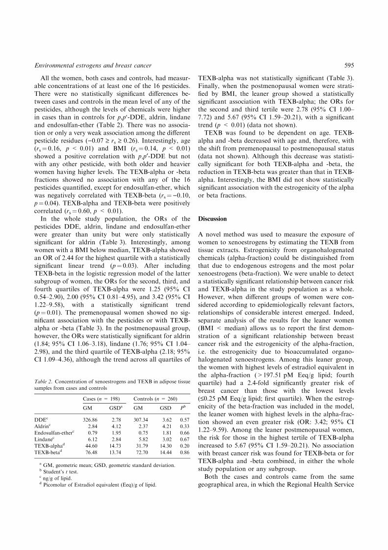
All the women, both cases and controls, had measur-able concentrations of at least one of the 16 pesticides.There were no statistically significant differences be-tween cases and controls in the mean level of any of thepesticides, although the levels of chemicals were higherin cases than in controls for p,p‘-DDE, aldrin, lindaneand endosulfan-ether (Table 2). There was no associa-tion or only a very weak association among the differentpesticide residues ()0.07 ‡ rs ‡ 0.26). Interestingly, age(rs¼ 0.16, p < 0.01) and BMI (rs¼ 0.14, p < 0.01)showed a positive correlation with p,p¢-DDE but notwith any other pesticide, with both older and heavierwomen having higher levels. The TEXB-alpha or -betafractions showed no association with any of the 16pesticides quantified, except for endosulfan-ether, whichwas negatively correlated with TEXB-beta (rs¼)0.10,p¼ 0.04). TEXB-alpha and TEXB-beta were positivelycorrelated (rs¼ 0.60, p < 0.01).In the whole study population, the ORs of the
pesticides DDE, aldrin, lindane and endosulfan-etherwere greater than unity but were only statisticallysignificant for aldrin (Table 3). Interestingly, amongwomen with a BMI below median, TEXB-alpha showedan OR of 2.44 for the highest quartile with a statisticallysignificant linear trend (p¼ 0.03). After includingTEXB-beta in the logistic regression model of the lattersubgroup of women, the ORs for the second, third, andfourth quartiles of TEXB-alpha were 1.25 (95% CI0.54–2.90), 2.00 (95% CI 0.81–4.95), and 3.42 (95% CI1.22–9.58), with a statistically significant trend(p¼ 0.01). The premenopausal women showed no sig-nificant association with the pesticides or with TEXB-alpha or -beta (Table 3). In the postmenopausal group,however, the ORs were statistically significant for aldrin(1.84; 95% CI 1.06–3.18), lindane (1.76; 95% CI 1.04–2.98), and the third quartile of TEXB-alpha (2.18; 95%CI 1.09–4.36), although the trend across all quartiles of
TEXB-alpha was not statistically significant (Table 3).Finally, when the postmenopausal women were strati-fied by BMI, the leaner group showed a statisticallysignificant association with TEXB-alpha; the ORs forthe second and third tertile were 2.78 (95% CI 1.00–7.72) and 5.67 (95% CI 1.59–20.21), with a significanttrend (p < 0.01) (data not shown).TEXB was found to be dependent on age. TEXB-
alpha and -beta decreased with age and, therefore, withthe shift from premenopausal to postmenopausal status(data not shown). Although this decrease was statisti-cally significant for both TEXB-alpha and -beta, thereduction in TEXB-beta was greater than that in TEXB-alpha. Interestingly, the BMI did not show statisticallysignificant association with the estrogenicity of the alphaor beta fractions.
Discussion
A novel method was used to measure the exposure ofwomen to xenoestrogens by estimating the TEXB fromtissue extracts. Estrogenicity from organohalogenatedchemicals (alpha-fraction) could be distinguished fromthat due to endogenous estrogens and the most polarxenoestrogens (beta-fraction). We were unable to detecta statistically significant relationship between cancer riskand TEXB-alpha in the study population as a whole.However, when different groups of women were con-sidered according to epidemiologically relevant factors,relationships of considerable interest emerged. Indeed,separate analysis of the results for the leaner women(BMI < median) allows us to report the first demon-stration of a significant relationship between breastcancer risk and the estrogenicity of the alpha-fraction,i.e. the estrogenicity due to bioaccumulated organo-halogenated xenoestrogens. Among this leaner group,the women with highest levels of estradiol equivalent inthe alpha-fraction (>197.51 pM Eeq/g lipid; fourthquartile) had a 2.4-fold significantly greater risk ofbreast cancer than those with the lowest levels(£0.25 pM Eeq/g lipid; first quartile). When the estrog-enicity of the beta-fraction was included in the model,the leaner women with highest levels in the alpha-frac-tion showed an even greater risk (OR: 3.42; 95% CI1.22–9.59). Among the leaner postmenopausal women,the risk for those in the highest tertile of TEXB-alphaincreased to 5.67 (95% CI 1.59–20.21). No associationwith breast cancer risk was found for TEXB-beta or forTEXB-alpha and -beta combined, in either the wholestudy population or any subgroup.Both the cases and controls came from the same
geographical area, in which the Regional Health Service
Table 2. Concentration of xenoestrogens and TEXB in adipose tissue
samples from cases and controls
Cases (n = 198) Controls (n = 260)
GM GSDa GM GSD Pb
DDEc 326.86 2.78 307.34 3.62 0.57
Aldrinc 2.84 4.12 2.37 4.21 0.33
Endosulfan-etherc 0.79 1.95 0.75 1.81 0.66
Lindanec 6.12 2.84 5.82 3.02 0.67
TEXB-alphad 44.60 14.73 31.79 14.30 0.20
TEXB-betad 76.48 13.74 72.70 14.44 0.86
a GM, geometric mean; GSD, geometric standard deviation.b Student’s t test.c ng/g of lipid.d Picomolar of Estradiol equivalent (Eeq)/g of lipid.
Environmental estrogens and breast cancer 595

Table
3.Concentrationofxenoestrogensandtotaleff
ectivexenoestrogen
burden
(TEXB)in
adipose
tissueandtherisk
ofbreast
cancer
Alla
BMI<
Median
BMI>
Median
Premenopausal
Postmenopausal
Cases
Controls
Cases
Controls
Cases
Controls
Cases
Controls
OR
b(95%
CI)
n=
121
n=109
OR
c(95%
CI)
n=
77
n=151
OR
c(95%
CI)
n=80
n=
76
OR
c(95%
CI)
n=118
n=
184
OR
c(95%
CI)
DDEd
£201.72
133
32
117
33
130
23
120
42
1
201.73–397.67
1.04(0.59–1.84)
40
29
1.29(0.59–2.83)
14
36
0.74(0.30–1.82)
22
17
1.07(0.40–2.87)
32
48
1.14(0.54–2.43)
397.68–675.97
1.23(0.69–2.17)
29
19
1.94(0.83–4.52)
21
46
0.80(0.35–1.87)
17
20
0.85(0.30–2.40)
33
45
1.41(0.66–3.00)
‡675.98
1.22(0.68–2.21)
19
29
0.91(0.38–2.14)
25
36
1.46(0.62–3.43)
11
16
0.64(0.20–1.99)
33
49
1.58(0.74–3.37)
p-trend=
0.40
p-trend=
0.85
p-trend=
0.33
p-trend=
0.48
p-trend=
0.18
Aldrind
<LD
180
78
151
110
153
50
178
138
1
>LD
1.55(1.00–2.40)
41
31
1.74(0.92–3.30)
26
41
1.50(0.79–2.84)
27
26
1.07(0.47–2.42)
40
46
1.84(1.06–3.18)
Endosulfan-
ether
d
<LD
159
51
134
83
140
39
153
95
1
>LD
1.35(0.90–2.02)
62
58
1.09(0.61–1.94)
43
68
1.63(0.91–2.93)
40
37
0.83(0.37–1.84)
65
89
1.47(0.88–2.45)
Lindaned
<LD
167
67
145
99
147
46
165
120
1
>LD
1.40(0.92–2.13)
54
42
1.51(0.84–2.74)
32
52
1.39(0.76–2.56)
33
30
1.10(0.50–2.37)
53
64
1.76(1.04–2.98)
TEXB-alphae
£0.25
125
34
118
37
115
12
128
59
1
0.26–41.00
1.15(0.64–2.05)
25
30
1.12(0.50–2.52)
17
29
1.24(0.52–2.95)
16
23
0.57(0.17–1.87)
26
36
1.76(0.85–3.67)
41.01–197.50
1.33(0.76–2.33)
35
25
1.58(0.70–3.58)
22
40
1.08(0.48–2.42)
22
21
0.63(0.20–2.02)
35
44
2.18(1.09–4.36)
‡197.51
1.31(0.74–2.31)
36
20
2.44(1.03–5.78)
20
45
0.80(0.35–1.84)
27
20
1.27(0.39–4.14)
29
45
1.39(0.68–2.84)
p-trend=
0.30
p-trend=
0.03
p-trend=
0.55
p-trend=
0.53
p-trend=
0.22
TEXB-betae
£9.95
123
26
122
37
114
11
131
52
1
9.96–100.00
1.08(0.61–1.90)
37
35
1.19(0.53–2.66)
15
31
1.01(0.42–2.45)
16
19
0.34(0.09–1.23)
36
47
1.52(0.77–3.00)
100.01–550.00
1.05(0.59–1.86)
35
24
1.48(0.63–3.48)
18
41
0.80(0.35–1.83)
25
21
0.69(0.19–2.49)
28
44
1.18(0.58–2.38)
‡550.01
0.99(0.55–1.79)
26
24
0.96(0.39–2.38)
22
42
0.91(0.42–2.02)
25
25
0.69(0.20–2.39)
23
41
0.99(0.47–2.10)
p-trend=
0.99
p-trend=
0.91
p-trend=
0.72
p-trend=
0.82
p-trend=
0.88
aAllwomen.
bOR
adjusted
forage,
reference
hospital,ln
BMI,number
ofchildren,ageatfirstfull-term
pregnancy,familyhistory
ofbreast
cancer,andalcoholandtobaccoconsumption.
cOR
adjusted
forage,
reference
hospital,number
ofchildren,ageatfirstfull-term
pregnancy,familyhistory
ofbreast
cancer,andalcoholandtobaccoconsumption.
dng/g
oflipid.
epicomolarofEstradiolequivalent(Eeq)/goflipid.
596 J.M. Ibarluzea et al.

provides universal medical cover. There are no privatehospitals with Oncology Departments in the area understudy. One possible shortcoming of the present work isthat most of the controls underwent surgery for diseasesof the gall bladder and hernia, which are associated withobesity as a risk factor. The high mean BMI of thecontrols in our study is consistent with a recent reportby the European Prospective Investigation into Cancerand Nutrition (EPIC), which stated that 92% ofGranada women in the same age range were overweight(BMI > 25) [48]. Wolff and Anderson [49] proposed apharmacokinetic model by which heavier women show alower concentration of organochlorine compounds dur-ing the uptake period and subsequently for around15 years, after which time they show higher concentra-tions of these compounds in comparison with leanerwomen. According to their model, our study mightunderestimate the risk, because the greater weight of ourcontrols would imply higher xenoestrogen levels than ifthey had been leaner. To control for this variable, thewhole study population was stratified by BMI. It is notclear why the significant effect of TEXB-alpha in theleaner women was not found in the more obese group.One possible explanation may be a greater relativeimpact of exogenous estrogens in leaner women, inwhom there is no counterbalance due to endogenoushormones accumulated in fat.A further potential limitation is that breast fat was
obtained for cases and abdominal fat for controls.However, several studies have shown a good correlationbetween measurements at these two sites [50]. Studiesthat used fat from sites other than the breast as controls,such as those by Van’t Veer et al. [25] and Stellmanet al. [26] did not report that the different origin of thetissue modified the results. In fact, Stellman et al. [26]found that levels of DDE and other organochlorines didnot differ between adipose breast tissue samples fromcontrols with benign breast disease and samples derivedfrom surgery for gall bladder disease or abdominalhernia, very similar to the present study.When the results were studied according to meno-
pausal status, a further significant relationship wasdisclosed between breast cancer risk and the estrogenic-ity of the alpha-fraction. The third quartile of thepostmenopausal women had a 2.18-fold increased risk incomparison to those with lowest estrogenic levels in thealpha-fraction. Moreover, when postmenopausal statuswas stratified by BMI, the leaner postmenopausalwomen in the highest tertile of TEXB-alpha showed a5-fold increased risk for breast cancer. Interestingly, thisrelationship was not found among the premenopausalwomen, and may be related to the decline in ovarianestrogen production with menopause, and to the
increased relative importance of the xenoestrogensbioaccumulated in their adipose tissue. Indeed, theTEXB of both alpha and beta fractions varied accordingto menopausal status, with a 2.6-fold decline in the totalburden of the beta-fraction and a lower fall (1.6-fold) inthat of the alpha-fraction after menopause. The age hada similar effect on TEXB values, with a 3.5- and 5-folddecline in the estrogenicity of the alpha and betafractions, respectively, between the first and last quar-tiles.Endogenous estrogens, responsible for the hormonal
activity of the beta-fraction, may arise from the localproduction and depot of circulating precursors. Adiposetissue is known to be the primary source of endogenousestrogens after menopause. These estrogens are pro-duced in the fat of both pre- and postmenopausalwomen through the conversion of precursors by aro-matase cytochrome P450, product of the CYP19 gene.Interestingly, a similar mechanism has been suggestedfor the transcriptional regulation and expression of theCYP19 gene in adipose tissue, regardless of its localiza-tion (breast or abdomen) [51]. This expression isstrongly dependent on the age of the subject, with anincreasing expression of aromatase activity in adiposetissue and, consequently, a higher estrone production.This appears to be inconsistent with the observed declinein the estrogenicity of the beta-fraction with the onset ofmenopause. However, adipose tissue also plays animportant role in the storage and regulation of estrogenin premenopausal women. For example, concentrationsof estradiol esters were reported to be 3-fold higher inthe adipose tissue of premenopausal versus postmeno-pausal women [52], in agreement with the decrease inestrogenicity observed in our patients with the onset ofmenopause.Older women could be expected to have longer
exposure to xenoestrogens, especially to bioaccumula-tive, fat-soluble xenobiotics, and to show higher levels ofxenoestrogen-derived estrogenicity (alpha-fraction).However, the TEXB of the alpha-fraction significantlydecreased with the age of the patients and at the onset ofmenopause, as occurred with the estrogenicity of thebeta-fraction. The fall in estrogenic activity with age wasnot accompanied by a decline in organohalogenatedchemical levels, which would account for this finding. Infact, DDE levels increased with age, confirming previousobservations of the age-dependent bioaccumulation oflipophilic compounds in adipose tissue [53]. The otherorganochlorine residues showed no variation with age.All women had measurable concentrations of at least
one of the sixteen organochlorines quantified, clearlyreflecting the ubiquity of exposure to pesticides in thepopulation, which hampers the demonstration of an
Environmental estrogens and breast cancer 597

etiologic role. No single chemical could be positivelyand statistically significantly associated with the biolog-ical effect measured by TEXB-alpha. There may beseveral reasons for this lack of concordance: (i) theestrogenic effects depicted in the E-Screen bioassay are aconsequence of the combined effect of several organo-halogens; and (ii) the proliferative effect is due to otherchemicals not measured, either other organochlorinepesticides or other lipophilic compounds. However, wefound that aldrin and lindane may increase the risk ofbreast cancer. This relationship is biologically supportedby the estrogenic properties of both pesticides [54, 55].Our finding for aldrin may corroborate previous resultsfor dieldrin [11], because aldrin can readily degrade todieldrin, as reported in soils and multiple species. Bothaldrin and dieldrin tend to accumulate in adipose tissue.The use of lindane has been prohibited or restricted inmany countries; in Spain and several other Europeancountries, it is allowed for certain specific agriculturalpurposes and as a medication for head and body lice.We found no differences in DDE levels between cases
and their matched controls. DDE levels were lower inour series compared with other studies, which may bebecause this pesticide has not been in use since 1980. Ithas been recommended [33] to explore the associationbetween DDE and breast cancer in populations withmore recent exposure, such as Colombia or MexicoCity, where epidemiological studies have found amoderately high risk of breast cancer in women withhigher levels of DDE [8, 9]. However, negative resultswere also observed in these areas [20, 22, 56].The estrogenicity of adipose tissue extracts due to
bioaccumulated xenoestrogens, measured as theTEXB-alpha, was associated with a higher risk of breastcancer in the leaner women, especially in the postmeno-pausal leaner group. Complex interactions betweenchemicals, endogenous or exogenous hormones andtheir natural ligands and receptors may alter the internalhomeostasis of the estrogenic environment of mammarytissue, leading to malignant transformation and cancer.Thus, future studies of the association between environ-mental estrogens and breast cancer or other adversehuman health effects should analyze the combined effectof these compounds and the interactions with endoge-nous hormones and other substances that affect endo-crine function.
Acknowledgements
We are indebted to all participants and staff of thisstudy, without whom this work would not have beenpossible; and to Richard Davies for editorial assistance.
Supported by grants from the Spanish Ministry ofHealth (FIS 00/543 and 02/1314) and the EuropeanUnion Commission (QLK4-1999-01422 and QLK4-2002-00603).
References
1. Wassermann M, Nogueira DP, Tomatis L, et al. (1976) Organo-
chlorine compounds in neoplastic and adjacent apparently normal
breast tissue. Bull Environ Contam Toxicol 15: 478–484.
2. Unger M, Olsen J (1980) Organochlorine compounds in the
adipose tissue of deceased people with and without cancer. Environ
Res 23: 257–263.
3. Unger M, Kiaer H, Blichert-Toft M, Olsen J, Clausen J (1984)
Organochlorine compounds in human breast fat from deceased
with and without breast cancer and in a biopsy material from
newly diagnosed patients undergoing breast surgery. Environ Res
34: 24–28.
4. Djordjevic MV, Hoffmann D, Fan J, Prokopczyk B, Citron ML,
Stellman SD (1994) Assessment of chlorinated pesticides and
polychlorinated biphenyls in adipose breast tissue using a super-
critical fluid extraction method. Carcinogenesis 15: 2581–2585.
5. Davis DL, Bradlow HL, Wolff M, Woodruff T, Hoel DG,
Anton-Culver H (1993) Medical hypothesis: xenoestrogens as
preventable causes of breast cancer. Environ Health Perspect 101:
372–377.
6. Wolff MS, Toniolo PG, Lee EW, Rivera LM, Dubin N (1993)
Blood levels of organochlorine residues and risk of breast cancer.
J Natl Cancer Inst 85: 648–652.
7. Dewailly E, Dodin S, Verreault R, et al. (1994) High organochlo-
rine body burden in women with estrogen receptor-positive breast
cancer. J Natl Cancer Inst 86: 232–234.
8. Olaya-Contreras P, Rodriguez-Villamil J, Posso-Valencia HJ,
Olaya-Contreras P, Cortez JE (1998) Organochlorine exposure
and breast cancer risk in Colombian women. Cad Saude Publica
14: 125–132.
9. Romieu I, Hernandez-Avila M, Lazcano-Ponce E, Weber JP,
Dewailly E (2000) Breast cancer, lactation history, and serum
organochlorines. Am J Epidemiol 152: 363–370.
10. Falck F, Ricci A, Wolff MS, Godbold J, Deckers P (1992)
Pesticides and polychlorinated biphenyl residues in human breast
lipids and their relation to breast cancer. Arch Environ Health 47:
143–146.
11. Hoyer AP, Grandjean P, Jorgersen T, Brock JW, Hartvig HB
(1998) Organochlorine exposure and risk of breast cancer. Lancet
352: 1816–1820.
12. Hoyer AP, Jorgensen T, Brock JW, Grandjean P (2000) Organo-
chlorine exposure and breast cancer survival. J Clin Epidemiol 53:
323–330.
13. Hoyer AP, Jorgensen T, Grandjean P, Hartvig HB (2000)
Repeated measurements of organochlorine exposure and breast
cancer risk (Denmark). Cancer Causes Control 11: 177–184.
14. Aronson KJ, Miller AB, Woolcott ChG, Sterns E, McCredy DR,
Lickley LA (2000) Breast adipose tissue concentrations of poly-
chlorinated biphenyls and other organochlorines and breast cancer
risk. Cancer Epidemiol Biomarkers Prev 9: 55–63.
15. Dorgan JF, Brock JW, Rothman N, et al. (1999) Serum organo-
chlorine pesticides and PCBs and breast cancer risk: results from a
prospective analysis (USA). Cancer Causes Control 10: 1–11.
16. Demers A, Ayotte P, Brisson J, Dodin S, Robert J, Dewailly E
(2000) Risk and aggressiveness of breast cancer in relation to
598 J.M. Ibarluzea et al.

plasma organochlorine concentrations. Cancer Epidemiol Biomar-
kers Prev 9: 161–166.
17. Henderson AK, Rosen D, Miller GL, et al. (1995) Breast cancer
among women exposed to polybrominated biphenyls. Epidemiol-
ogy 6: 544–546.
18. Hardell L, Lindstrom G, Liljegren G, Dahl P, Magnuson A (1996)
Increased concentrations of octachlorodibenzo-p-dioxin in cases
with breast cancer-results from a case–control study. Eur J Cancer
Prev 5: 351–357.
19. Hunter D, Hankinson S, Laden F, et al. (1997) Plasma organo-
chlorine levels and the risk of breast cancer. N Engl J Med 337:
1253–1258.
20. Lopez-Carrillo L, Blair A, Lopez-Cervantes M, et al. (1997)
Dichlordyltrichloroethane serum levels and breast cancer risk: a
case–control study from Mexico. Cancer Res 57: 3728–3732.
21. Helzlsouer KJ, Alberg AJ, Huang HY, et al. (1999) Serum
concentrations of organochlorine compounds and subsequent
development of breast cancer. Cancer Epidemiol Biomarkers Prev
8: 525–532.
22. Mendoca GA, Eluf-Neto J, Andrada-Serpa MJ, et al. (1999)
Organochlorines and breast cancer: a case–control study in Brazil.
Int J Cancer 83: 596–600.
23. Krieger N, Wolff MS, Hiatt RA, Rivera M, Vogelman J,
Orentreich N (1994) Breast cancer and serum organochlorines: a
prospective study among white, black, and Asian women. J Natl
Cancer Inst 86: 589–599.
24. Laden F, Collman G, Iwamoto K, et al. (2001) 1,1-Dichloro-2,2-
bis(p-chlorophenyl)ethylene and polychlorinated biphenyls and
breast cancer: combined analysis of five US studies. J Natl Cancer
Inst 93: 768–776.
25. van’t Veer P, Lobbezoo IE, Martin-Moreno JM, et al. (1997) DDT
(dicophane) and postmenopausal breast cancer in Europe: case–
control study. BMJ 315: 81–85.
26. Stellman SD, Djordjevic MV, Britton JA, et al. (2000) Breast
cancer risk in relation to adipose concentrations of organochlorine
pesticides and polychlorinated biphenyls in Long Island, New
York. Cancer Epidemiol Biomarkers Prev 9: 1241–1249.
27. Wolff MS, Zeleniuch-Jacquotte A, Dubin N, Toniolo P (2000)
Risk of breast cancer and organochlorine exposure. Cancer
Epidemiol Biomarkers Prev 9: 271–277.
28. Zheng T, Holford TR, Taylor Mayne S, et al. (2000) Risk of
female breast cancer associated with serum polychlorinated biphe-
nyls and 1,1-dichloro-2,2’-bis(p-chlorophenyl)ethylene. Cancer Ep-
idemiol Biomarkers Prev 9: 167–174.
29. Laden F, Hankinson SE, Wolff MS, et al. (2001) Plasma organo-
chlorine levels and the risk of breast cancer: an extended follow-up
in the Nurses’ Health Study. Int J Cancer 91: 568–574.
30. Gammon MD, Wolff MS, Neugut AI, et al. (2002) Environmental
toxins and breast cancer on Long Island.II. Organochlorine
compound levels in blood. Cancer Epidemiol Biomarkers Prev 11:
686–697.
31. Safe SH (1997) Xenoestrogens and breast cancer. N Engl J Med
337: 1303–1304.
32. Laden F, Hunter DJ (1998) Environmental risk factors and female
breast cancer. Annu Rev Public Health 19: 101–123.
33. Snedeker S (2001) Pesticides and breast cancer risk: a review of
DDT, DDE, and Dieldrin. Environ Health Perspect 109: 35–47.
34. Laden F, Ishibe N, Hankinson SE, et al. (2002) Polychlorinated
biphenyls, cytochrome P450 1A1, and breast cancer risk in the
nurses’ health study. Cancer Epidemiol Biomarkers Prev 11:
1560–1565.
35. Sasco AJ (2001) Epidemiology of breast cancer: an environmental
disease? APMIS 109: 321–332.
36. Soto A, Sonnenschein C, Chung KL, Fernandez MF, Olea N,
Olea-Serrano MF (1995) The E-SCREEN Assay as a tool to
identify estrogens: an update on estrogenic environmental pollu-
tants. Environ Health Perspect 103: 113–122.
37. Soto A, Fernandez MF, Luizzi M, Oles Karasko AS, Sonnensch-
ein C (1997) Developing a marker of exposure to xenoestrogen
mixtures in human serum. Environ Health Pespect 105: 647–663.
38. Shekhar PVM, Werdell J, Basrur VS (1997) Environmental
estrogen stimulation of growth and estrogen receptor function in
preneoplastic and cancerous human cells lines. J Natl Cancer Inst
89: 1774–1782.
39. Payne J, Scholze M, Kortenkamp A (2001) Mixtures of four
organochlorines enhance human breast cancer cell proliferation.
Environ Health Perspect 104: 391–397.
40. Sonnenschein C, Soto A, Fernandez MF, Olea N, Olea-Serrano
MF (1995) Development of a marker of estrogenic exposure in
human serum. Clin Chem 41: 1888–1895.
41. Pazos P, Perez P, Rivas A, et al. (1998) Development of a marker
of estrogenic exposure in breast cancer patients. Adv Exp Med Biol
444: 29–40.
42. Rivas A, Olea N, Olea-Serrano MF (1997) Human exposure to
endocrine-disrupting chemicals: assessing the total estrogenic
xenobiotic burden. TRAC 16: 613–619.
43. Rivas A, Fernandez MF, Cerrillo I, et al. (2001) Human exposure
to endocrine disrupters: standardisation of a marker of estrogenic
exposure in adipose tissue. APMIS 109: 185–197.
44. Martınez Vidal JL, Moreno M, Garrido A, Olea-Serrano MF,
Olea N (2000) Trace determination of alpha and beta endosulfan
and three metabolites in human serum by GC-MS. Rapid Comm
Mass Spectrometry 14: 939–946.
45. Hernandez F, Pitarch E, Serrano R, Gaspar JV, Olea N (2002)
Multiresidue determination of endosulfan and metabolic deriva-
tives in human adipose tissue using automated liquid chromato-
graphic cleanup and gas chromatographic analysis. J Anal Toxicol
26: 94–103.
46. Villalobos M, Olea N, Brotons JA, Olea-Serrano MF, Ruiz de
Almodovar JM, Pedraza V (1995) The E-SCREEN assay: com-
parison among different MCF7 cell stocks. Environ Health
Perspect 103: 844–850.
47. SPPS Inc. (1999) SPSS 9.0 Base Sintax Reference Guide. Chicago:
SPSS Inc.
48. Haftenberger M, Lahmann PH, Panico S, et al. (2002) Overweight,
obesity and fat distribution in 50- to 64-year-old participants in the
European Prospective Investigation into Cancer and Nutrition
(EPIC). Public Health Nutr 5: 1147–1162.
49. Wolf MS, Anderson HA (1999) Correspondence re: J.M. Schil-
dkrauut et al., Environmental contaminants and body fat distri-
bution. Cancer Epidemiol. Biomark. Prev. 8: 179–183, 1999.
Cancer Epidemiol Biomarkers Prev 8: 951–952.
50. Kohlmeier L, Kohlmeier M (1995) Adipose tissue as a medium for
epidemiologic exposure assessment. Environ Health Perspect 103:
99–106.
51. Agarwal VR, Takayama K, Van Wyk JJ, et al. (1998) Molecular
basis of severe gynecomastia associated with aromatase expression
in a fibrolamellar hepatocellular carcinoma. J Clin Endocrinol
Metab 83: 1797–1800.
52. Larner JM, Shackleton CHL, Roitman E, Schwartz PE, Hochberg
RB (1992) Measurement of estradiol-17 fatty acid-esters in human
tissues. J Clin Endocrinol Metab 75: 195–200.
53. Moysich KB, Ambrosone CB, Mendola P, et al. (2002) Exposures
associated with serum organochlorine levels among postmeno-
pausal women from Westren New York State. Am J Ind Med 41:
102–110.
Environmental estrogens and breast cancer 599

54. Bigsby RM, Caperell-Grant A, Madhukar BV (1997) Xenobiotics
released from fat during fasting produce estrogenic effects in
ovariectomized mice. Cancer Res 57: 865–869.
55. Traina ME, Rescia M, Urbani E, et al. (2003) Long-lasting effects
of lindane on mouse spermatogenesis induced by in utero
exposure. Reprod Toxicol 17: 25–35.
56. Schecter A, Toniolo P, Dai LC, Thuy LT, Wolff MS (1997) Blood
levels of DDT and breast cancer risk among women living in the
north of Vietnam. Arch Environ Contam Toxicol 33: 453–456.
600 J.M. Ibarluzea et al.