Embed Size (px)
Citation preview

PHYSIOLOGICAL RESEARCH • ISSN 0862-8408 (print) • ISSN 1802-9973 (online) © 2008 Institute of Physiology v.v.i., Academy of Sciences of the Czech Republic, Prague, Czech Republic Fax +420 241 062 164, e-mail: [email protected], www.biomed.cas.cz/physiolres
Physiol. Res. 57: 601-611, 2008 Evidences of Apoptosis during the Early Phases of Soleus Muscle Atrophy in Hindlimb Suspended Mice R. FERREIRA1,2, M. J. NEUPARTH1,3, R. VITORINO1,2, H. J. APPELL1,4, F. AMADO2, J. A. DUARTE1,3
1CIAFEL, Faculty of Sport, University of Porto, 2Department of Chemistry, University of Aveiro, 3ESSVA Health School, IPSN, CESPU CRL, Portugal and 4 Department of Physiology and Anatomy, German Sports University, Germany
Received April 26, 2007 Accepted June 28, 2007 On-line July 26, 2007 Summary
The purpose of this study was to investigate the occurrence and
time-course of apoptosis in soleus skeletal muscle during the first
48 hours of unloading. Fifty Charles River mice were randomly
divided into five groups (n=10 each) according to the time of
hindlimb suspension (HS). Mice were suspended for 0 (Control), 6
(6HS), 12 (12HS), 24 (24HS), and 48 hours (48HS). Soleus muscle
atrophy was confirmed by a significant decrease of 20 % in
muscle-wet weight and of 5 % in the ratio protein
concentration/muscle wet-weight observed after 48 hours of
unloading. The apoptotic index, the AIF (apoptosis-inducing factor)
and p53 expression presented their uppermost value (304 %, 241
% and 246 %, respectively) at 24HS, and were preceded by the
highest activity of caspase-3 and -8 at 12HS (170 % and 218 %,
respectively) and of Bax/Bcl-2 content at 6HS (160 %). There were
no marked ultrastructural alterations until 24 hours of simulated
weightlessness. Lysosomal autophagic activity and infiltration of
phagocytic cells were observed at 24HS and 48HS and might have
contributed to the degenerative changes noticed in both groups.
Though not consistently supported by morphological evidences, the
biochemical parameters sustain the concept that the occurrence of
apoptosis parallels the soleus atrophic response in its early phase.
Key words
Unloading • Muscle wasting • Mitochondria • Cell death • Muscle
ultrastructure
Corresponding author
R. M. Ferreira, CIAFEL, Faculty of Sport, University of Porto, Rua
Dr. Plácido Costa, 91, 4200-450 Porto, Portugal. Fax:
+351225500689. E-mail: [email protected]
Introduction
Skeletal muscle atrophy is a morphological adaptation that normally occurs in response to conditions of disuse (e.g. immobilization, denervation, muscle unloading), aging, starvation, and several other pathological conditions (Appell 1990, Jackman and Kandarian 2004). It is characterized by a decrease in muscle mass, protein content, fiber diameter, force production, fatigue resistance, and also by fiber type changes (Asmussen and Soukup 1991, Fitts et al. 2000, Jackman and Kandarian 2004). Understanding the molecular mechanisms behind muscle atrophy is important to develop countermeasures in order to prevent this muscle wasting and to preserve its physiological function (Powers et al. 2005). In fact, weak muscles compromise the activities of daily living and consequently lead to a reduced quality of life (Kandarian and Jackman 2006).
Although muscle atrophy has received increasing attention, the cellular and molecular mechanisms are not completely understood, and several possibilities can be envisioned (Allen et al. 1999, Dirks and Leeuwenburgh 2002). A decrease in myonuclear number in atrophying muscles has been reported under various experimental conditions including spinal cord isolation, microgravity, hindlimb suspension, and denervation (for review see Allen et al. 1999). The mechanisms of myonuclear loss from muscle fibers are currently not well comprehended, nor is it clear how individual myonuclei can be selectively eliminated

602 Ferreira et al. Vol. 57 without the destruction of either all nuclei or of the entire myofiber (Allen et al. 1999, Edgerton et al. 2002). Apoptosis has been pointed out as the principal mechanism behind this selective loss of myonuclei and of related sarcoplasmic domains (Hikida et al. 1997, Adams et al. 2001, Adhihetty and Hood 2003, Siu et al. 2005, Dupont-Versteegden et al. 2006). This internally encoded suicide program has been widely accepted to be crucial in coordinating the balance between cell survival and cell death in various tissues (Siu et al. 2005). One of the hallmarks of this energy-dependent, asynchronous, and genetically controlled process is the regulated destruction of the nucleus (Allen et al. 1999, Adhihetty and Hood 2003). The nuclear fragments and surrounding organelles become condensed and are, ultimately packaged in membrane-bound vesicles, exocytosed and ingested by interstitial phagocytes. Regarding muscle atrophy induced by disuse, few experimental studies have reported evidence for the occurrence of apoptosis in hindlimb suspended (Dupont-Versteegden et al. 2006) or immobilized animals (Smith et al. 2000) in its earlier phases. Nevertheless, the role of apoptosis in regulating the atrophic process remains questionable since the analysis of DNA fragmentation cannot be seen by itself as an universal indicator of the presence and intensity of apoptosis (Borisov and Carlson 2000) since DNA lyses can also be induced by lytic enzymes released from neighbor dead cells or even by secondary necrosis. Moreover, even when complemented with other apoptotic markers such as caspases or endonucleases activities, the histological studies using TUNEL or immunohisto-chemistry assay (Smith et al. 2000, Dupont-Versteegden et al. 2006) can only give a limited qualitative view on the entire process (Dirks and Leeuwenburgh 2002) considering the heterogeneity of skeletal muscle and the number of slices needed to analyse the whole muscle. The results of Appell et al. (2004) exacerbate the debate around this issue since they suggest that during immobilization, necrosis rather than apoptosis seems to play a major role in muscle tissue wasting, and that the occurrence of an autophagic process in the surviving areas of fibers may exacerbate skeletal muscle wasting. Additionally, some other studies dealing with neuromuscular disorders also reinforced the major role of necrosis and the inflammatory response in such muscle wasting conditions (Tews 2002, 2003).
Considering the controversy regarding the role of apoptosis in the development of skeletal muscle atrophy, the purpose of this study was to investigate the
occurrence of biochemical [nuclear DNA fragmentation, caspase-3 and -8 activity, Bax to Bcl-2 ratio, AIF (apoptosis-inducing factor) and p53 protein content] and morphological (structural and ultrastructural) evidences of apoptosis activation in muscle when submitted to a short period of simulated weightlessness (up to 48 hours), since the most important mechanisms leading to muscle atrophy seem to be triggered during this period of time (Booth and Seider 1979, Appell 1990, Smith et al. 2000, Dupont-Versteegden et al. 2006). It was also our goal to establish the main pathways by which the apoptotic signal is transduced in atrophic muscle cells. The experimental model chosen for this study was the hindlimb suspension, once it is generally accepted as the animal model of choice to mimic reduced mechanical load that occurs in spaceflight and prolonged bed rest in humans (Fitts et al. 2000, Morey-Holton and Globus 2002). We selected the soleus muscle for analysis since it is well established that the atrophic process is more pronounced in postural antigravity muscles, which intrinsically express a slow phenotype (Appell 1990, Hikida et al. 1997, Fitts et al. 2000, 2001, Haddad et al. 2003a,b). We hypothesized that the biochemical parameters chosen will quantitatively support the morphological qualitative evidences of apoptosis described by others (Smith et al. 2000, Dupont-Versteegden et al. 2006), clarifying the respective cellular signaling pathways. Materials and Methods Experimental Design
The experiments were performed after approval by the local Ethics Committee. Following the Guidelines for Care and Use of Laboratory Animals in research, 50 Charles River CD1 male mice (aged 6-8 weeks, weighing 30-35 g) were used. During the experimental period, the animals were housed in collective cages (two mice per cage) and were maintained at normal atmosphere (21-22 ºC, 50-60 % humidity) receiving commercial food for rodents and water ad libitum during inverted 12-h light/dark cycles. The animals were randomly divided into five groups of 10 animals each, according to their periods of hindlimb suspension (HS). Except for the control group (Cont), the hindlimbs of all mice were risen with a tail harness, as previously described (McDonald and Fitts 1995), to avoid ground support and loading for 6 hours (group 6HS), 12 hours (group 12HS), 24 hours (group 24HS), and 48 hours

2008 Muscular Apoptosis in Hindlimb Suspended Mice 603 (group 48HS), respectively. The forelimbs maintained contact with the cage floor, which permitted the mice a full range of mobility. After the experimental period, the animals of each group were sacrificed by cervical dislocation. Both soleus muscles were completely excised and their wet weight was determined. One of the muscles was immediately divided transversely into two pieces. One piece, together with the complete contralateral soleus, was used for biochemical analysis, while the remaining sample was processed for light (LM) and transmission electron microscopy (TEM) evaluation.
Tissue preparation for LM and TEM evaluation
Small blocks of tissue (~1 mm3) from the mid-portion of the soleus were immersed for two hours in a solution containing 4 % paraformaldehyde and 2.5 % sucrose diluted in phosphate buffer at pH 7.2. The samples were rinsed with 0.1 M and 0.2 M cacodylate buffer and subsequently dehydrated with graded ethanol. The muscles were embedded in LR White and placed at 60 ºC during 24 hours to promote resin polymerization. Semi-thin (1 μm) sections for light microscopy were cut from the blocks and stained with toluidine blue for analysis in a photomicroscope (Zeiss Phomi 3). Photographs of soleus cross sections were digitalized and analyzed with the NIH ImageJ (Image Processing and Analysis in Java, USA) software. About 100 muscle fibers from each muscle were analyzed for area quantification. The ultrathin (100 nm) sections prepared from the tissue blocks were contrasted with uranylacetate and lead citrate for subsequent examination in a transmission electron microscope (Zeiss EM10A) at an accelerating voltage of 60kV. All used reagents were of analytical grade and purchased from acknowledged companies.
Preparation of muscle extract for evaluation of caspase-3 and caspase-8 activity and determination of Bax, Bcl-2, AIF and p53 content
A portion of soleus muscle was homogenized in lyses buffer containing 1 mM Na-EDTA, 1 mM Na-EGTA, 1 mM MgCl2, 5 mM KCl and 25 mM HEPES, pH 7.5 supplemented in the day of the experiment with 100 μM PMSF, 2 mM DTT, 1:100 anti-proteases cocktail and 0.05 % Triton X-100. After 30 min of incubation on ice, the homogenate was centrifuged at 14,000g for 10 min. The supernatant was then taken and protein content was assayed spectrophotometrically using bovine serum albumin as standard according to Sedmak et al. (1977).
Determination of caspase-3 and caspase-8 activity 100 μg of protein in the muscle homogenate was
used for the analysis of caspase-3 and caspase-8 activity. After adding reaction buffer (10 % sucrose, 0.1 % CHAPS, 25 mM HEPES pH 7.4) and 40 μM DEVD-pNA (acetyl-Asp-Glu-Val-Asp-p-nitroaniline, Calbiochem, catalogue number 235400) or IEPD-pNA (acetyl-Ile-Glu-Pro-Asp-p-nitroaniline, product number A-6470, Sigma) substrate for caspase-3 and -8 respectively, the samples were incubated at 37 ºC during 2 hours. A405 nm was read in a microtiter plate reader (Labsystem iEMS Reader MF). Percentage of increase in caspase activity was determined by comparing the data with those obtained for simultaneously incubated control samples.
Evaluation of Bax, Bcl-2, AIF and p53 content
The Bax, Bcl-2, AIF and p53 content were assessed using Western blotting analysis. From each group, equal amounts of protein were loaded on the same gel to avoid any potential intergel variations. Proteins were separated using a 15 % SDS-PAGE gel, followed by blotting on a nitrocellulose membrane (Hybond-ECL, Amersham Pharmacia Biotech). After blotting, non-specific binding was blocked with 5 % nonfat dry milk in TTBS (Tris-buffered saline (TBS) with Tween 20) and the membrane was incubated with anti-Bcl-2 (1:1000, sc-7382 mouse monoclonal IgG, Santa Cruz Biotechnology), anti-Bax (1:1000, sc-493 rabbit polyclonal IgG, Santa Cruz Biotechnology), anti-AIF (1:2000, sc-13116 mouse monoclonal IgG2b, Santa Cruz Biotechnology) or anti-p53 (1:2000, sc-99 mouse monoclonal IgG1, Santa Cruz Biotechnology) antibodies during 2 hours at room temperature, washed and incubated with secondary horseradish peroxidase-conjugated anti-mouse or anti-rabbit IgG antibodies (Amersham Pharmacia Biotech) for 2 hours. The membrane was then washed and developed with Western blotting chemiluminescence reagents (Amersham Pharmacia Biotech) according to manufacturer’s instructions, followed by exposure on X-ray films (Sigma, Kodak Biomax Light Film, St. Louis, USA). The films were analyzed with QuantityOne Software (BioRad). Optical density results were expressed per milligram of protein and results of all groups were presented as percentage of controls.
Determination of cytosolic mono- and oligonucleosomes
The soleus muscle was homogenized in isolation buffer (225 mM mannitol, 75 mM sucrose, 0.2 % bovine

604 Ferreira et al. Vol. 57
serum albumin (BSA), 1 mM EDTA, pH 7.4) with a dilution of 1:25 using a Potter-Elvehjem glass homogenizer. The sample was then centrifuged at 1,000 g for 10 min. The supernatant was centrifuged at 14,000 g for 10 min. The resulting supernatant was used for analysis of cytosolic mono- and oligonucleosomes after determination of protein content according to Sedmak et al. (1977). Endogenous endonucleases activated during apoptosis cleave double-stranded DNA in the linker region between nucleosomes to generate mono- and oligonucleosomes of 180 bp or multiples. The apoptotic DNA fragmentation was quantified according to Dirks and Leeuwenburgh (2004) by measuring the amount of cytosolic mono- and oligonucleosomes using a Cell Death ELISA kit (Catalogue number: 1544675, Roche Molecular Biochemicals) following the manufacturer instructions. Results were expressed as percentage of controls.
Statistical analysis
Means and standard deviations were calculated for all variables in each of the experimental groups. The ANOVA for repeated measures was used to detect any differences within the experimental protocol. The Statistical Package for the Social Sciences (SPSS Inc. version 12.0) was used for all analysis. The significance level was set at 5 %.
Results Muscle response to hindlimb suspension
Soleus cross-sectional area decreased by 9 % after 48 hours of hindlimb suspension, although without statistical significance (p=0.06) (data not presented). The wet weight, protein concentration, and the ratio protein
concentration/muscle wet weight taken during this period of weightlessness followed the same time-course (Table 1). A 20 % reduction in muscle wet weight was observed after 48 hours of hindlimb suspension, with the greatest initial soleus weight loss, though not being statistically significant, during the first 6 hours (7 %). Similarly, total protein content decreased by 23 % after 48 hours of hindlimb suspension. However, the major decline occurred between hour 6 and 12 of hindlimb suspension (11 %). Regarding the ratio between protein concentration and muscle weight, the above-referred tendency was attenuated, which suggest that the fall in protein concentration overwhelmed the wet weight variation. The obviously most relevant differences in this ratio appeared to be the change in protein concentration between hour 6 and 12 of hindlimb suspension. Indicators of apoptosis
As illustrated in Figure 1, there were significant variations in the apoptotic parameters evaluated during the studied simulated weightlessness period. The general apoptotic index assessed by the cytosolic content of mono- and oligonucleosomes, exhibited its highest increment in group 24HS (304 % above control) returning to basal levels in 48HS (118 %). The maximal activities of caspase-3 and caspase-8, 170 % and 218 % of control, respectively, were observed in group 12HS.
The pattern of Bax content paralleled the activities of caspases, increasing until hour 12 of hindlimb suspension (Fig. 2). In contrast, Bcl-2 content decreased until 24 hours of simulated weightlessness, with the lowest value observed in group 6HS (~ 27 % below control). These effects analyzed together for both markers, resulting in the Bax to Bcl-2 ratio, presented its utmost significance at 6 hours of hindlimb suspension.
Table 1. Effect of time (in hours) of hindlimb suspension (HS) on soleus muscle wet weight, on protein concentration and on protein concentration/wet weight.
Control 6HS 12HS 24HS 48HS
Soleus wet weight (mg)
7.26 ± 0.77 6.76 ± 0.69 6.42 ± 1.26 6.25 ± 0.82* 5.79 ± 0.47*
Soleus protein concentration (mg/ml)
0.86 ± 0.04 0.81 ± 0.08 0.72 ± 0.08* 0.68 ± 0.03* 0.66 ± 0.07*
Protein concentration/muscle weight (mg/mg)
0.117 ± 0.002 0.119 ± 0.006 0.112 ± 0.006 0.106 ± 0.006* 0.111 ± 0.004*
Values are mean ± standard deviation. * Control vs all other groups (p<0.05)

2008 Muscular Apoptosis in Hindlimb Suspended Mice 605
The AIF (apoptosis-inducing factor) and p53 expression profile followed the pattern observed for the apoptotic index, with utmost values of 241 % and 246 %, respectively, at 24HS (Fig. 3). The higher increment of p53 protein content (108 %) was noticed in the first 6 hours whereas the maximum percentage increase (75 %) of AIF content was observed between groups 12HS and 24HS.
Qualitative morphological changes
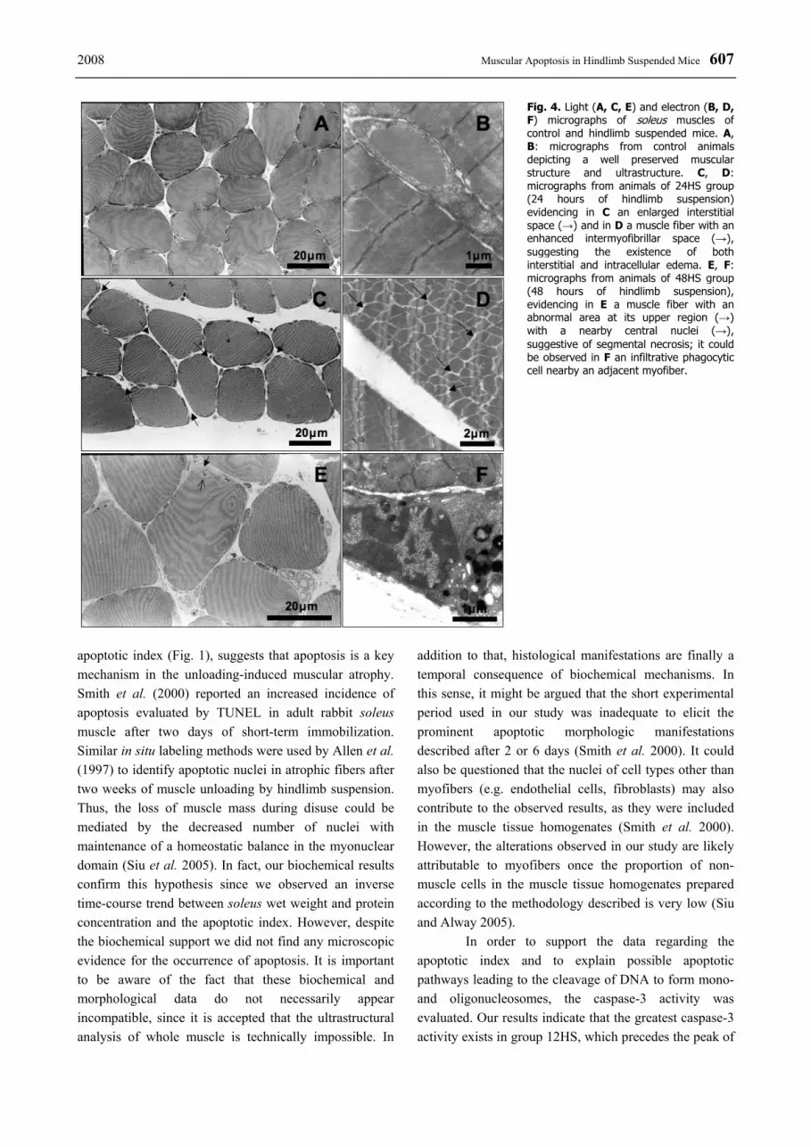
The morphologic analysis obtained by LM and EM revealed a normal structure of soleus muscle in the controls (Figs 4A, 4B). The hindlimb-suspended animals showed a sarcoplasmic vacuolization, which became more evident over time. Beyond of some slight widening of the interstitial space, no additional structural alterations were detected by LM (Fig. 4C). The ultrastructural analysis revealed that the sarcoplasmic
vacuolization noticed by LM consisted of mitochondrial swelling. This morphological abnormality affected the ultrastructure of the majority of the observed muscles fibres. An enlargement of the intermyofibrillar space in some fibers was obvious, suggesting an intrafiber edema, particularly after 12-24 hours of hindlimb suspension (Fig. 4D). Signs of an activation of the lysosomal system, evidenced by the presence of secondary lysosomes, were observed in skeletal muscle fibers and also in endothelial cells, particularly at 24 and 48 hours of simulated weightlessness. A diffuse endothelial vacuolization was evident even before the recognition of ultrastuctural alterations in the muscle fibers. From all the analyzed experimental groups, despite the existence of several endothelial nuclei highlighting peripheral chromatin condensation, we did not find any unequivocal morphologic signs for apoptosis neither in muscle fibers nor in interstitial cells. As typical sign of muscle fiber
Fig. 1. Effect of time-course (hours) hindlimb suspension (HS) on caspase-3 and caspase-8 activity and on apoptotic index (cytosolic mono-and oligonucleosomes content) in soleus muscle. Values (mean and standard deviation) are expressed as percentage of control (Cont). * p<0.05 Cont vs all other groups; # p<0.05 12HS vs 6HS, 24HS and 48HS; † p<0.05 24HS vs 6HS and 48HS. Fig. 2. Effect of time-course (hours) hindlimb suspension (HS) on Bax and Bcl-2 protein content, as well as on Bax/Bcl-2 ratio in soleus muscle. Immediately below the histogram, the panel shows a representative western blotting of Bax and of Bcl-2, respectively, for each group. Values are mean and standard deviation are expressed as percentage of control. Bax values for all groups are significant distinct (p<0.05); * Cont vs all other groups (p<0.05); # 48HS vs 6HS, 12HS and 24HS (p<0.05).

606 Ferreira et al. Vol. 57
atrophy, the scalloped appearance of the sarcolemma became noticeable in groups 24HS and 48HS. Some infiltrated phagocytes were also observed in these groups (Fig. 4F). Discussion
Despite the short experimental period of hindlimb suspension (48 hours) evaluated, our results are suggestive of muscle atrophy characterized particularly by a noticeable decline in muscle wet weight (~ 20 %), which is in agreement with prior unloading studies (Appell 1990, Borisov and Carlson 2000, Fitts et al. 2000, 2001, Haddad et al. 2003a,b, Picquet and Falempin 2003, Appell et al. 2004). According to previous reports (Picquet and Falempin 2003) this atrophy correlates well with a decline in muscle force, which has usually been attributed to a proportional decrease in total protein content. In fact, our results presented in table 1 substantiate this relationship. Interestingly, the vast majority of declining muscle mass was observed during the first 6 hours of simulated weightlessness. The soleus protein content mirrored this tendency, although the most noteworthy protein loss occurred between 6 and 12 hours of unloading. At the end of the experimental protocol a 23 % total protein loss was recorded, similar like in other suspension studies (Thomason and Booth 1990, Machida and Booth 2004). It has been suggested that this protein loss, mostly of myofibrillar proteins (Thomason and
Booth 1990, Fitts et al. 2000, Haddad et al. 2003a,b), is due to both, a decrease in muscle protein synthesis and an increase in the rate of proteolysis (Thomason and Booth 1990, Fitts et al. 2000). It is assumed that the earliest event after the onset of muscle unloading is a rapid decline in the rate of protein synthesis, followed by an increase in the proteolytic enzymes activity (Fitts et al. 2000, Glass 2003, Powers et al. 2005), which may involve the deactivation of the phosphatidylinositol-3-kinase – Akt – forkhead box subgroup O (FOXO) signaling pathway (Kandarian and Jackman 2006). Regarding proteolysis, the lysosomal autophagic activity, probably triggered by intracellular calcium accumulation (Appell et al. 2004), can partially explain the increased protein breakdown that occurs in muscles of suspended hindlimb. Furthermore, the presence of phagocytic cells as components of the inflammatory reaction may aggravate this phenomenon (Fig. 4F). After 24 hours of simulated weightlessness, the decreased protein concentration was more accentuated than the decline in muscle wet weight. Thus, considering the intracellular and interstitial edema observed in suspended soleus muscle (Fig. 4D), we might speculate that the decrease of muscle wet weight was somewhat masked by the increased water content, thereby underestimating the magnitude of atrophy.
Aside from the degenerative response described above, the significant increase in soleus muscle internucleosomal DNA fragmentation, given by the
Fig. 3. Effect of time-course (hours) hindlimb suspension (HS) on AIF and p53 content in soleus muscle. Immediately below the histogram, the panel shows a representative western blotting of AIF and p53, respectively, for each group. Values (mean and standard deviation) are expressed as percentage of control (Cont). * Cont vs all other groups (p<0.05); # 48HS vs 6HS, 12HS and 24HS (p<0.05).
2008 Muscular Apoptosis in Hindlimb Suspended Mice 607
apoptotic index (Fig. 1), suggests that apoptosis is a key mechanism in the unloading-induced muscular atrophy. Smith et al. (2000) reported an increased incidence of apoptosis evaluated by TUNEL in adult rabbit soleus muscle after two days of short-term immobilization. Similar in situ labeling methods were used by Allen et al. (1997) to identify apoptotic nuclei in atrophic fibers after two weeks of muscle unloading by hindlimb suspension. Thus, the loss of muscle mass during disuse could be mediated by the decreased number of nuclei with maintenance of a homeostatic balance in the myonuclear domain (Siu et al. 2005). In fact, our biochemical results confirm this hypothesis since we observed an inverse time-course trend between soleus wet weight and protein concentration and the apoptotic index. However, despite the biochemical support we did not find any microscopic evidence for the occurrence of apoptosis. It is important to be aware of the fact that these biochemical and morphological data do not necessarily appear incompatible, since it is accepted that the ultrastructural analysis of whole muscle is technically impossible. In
addition to that, histological manifestations are finally a temporal consequence of biochemical mechanisms. In this sense, it might be argued that the short experimental period used in our study was inadequate to elicit the prominent apoptotic morphologic manifestations described after 2 or 6 days (Smith et al. 2000). It could also be questioned that the nuclei of cell types other than myofibers (e.g. endothelial cells, fibroblasts) may also contribute to the observed results, as they were included in the muscle tissue homogenates (Smith et al. 2000). However, the alterations observed in our study are likely attributable to myofibers once the proportion of non-muscle cells in the muscle tissue homogenates prepared according to the methodology described is very low (Siu and Alway 2005).
In order to support the data regarding the apoptotic index and to explain possible apoptotic pathways leading to the cleavage of DNA to form mono- and oligonucleosomes, the caspase-3 activity was evaluated. Our results indicate that the greatest caspase-3 activity exists in group 12HS, which precedes the peak of
Fig. 4. Light (A, C, E) and electron (B, D, F) micrographs of soleus muscles of control and hindlimb suspended mice. A, B: micrographs from control animals depicting a well preserved muscular structure and ultrastructure. C, D: micrographs from animals of 24HS group (24 hours of hindlimb suspension) evidencing in C an enlarged interstitial space (→) and in D a muscle fiber with an enhanced intermyofibrillar space (→), suggesting the existence of both interstitial and intracellular edema. E, F: micrographs from animals of 48HS group (48 hours of hindlimb suspension), evidencing in E a muscle fiber with an abnormal area at its upper region (→) with a nearby central nuclei (→), suggestive of segmental necrosis; it could be observed in F an infiltrative phagocytic cell nearby an adjacent myofiber.

608 Ferreira et al. Vol. 57 cytoplasmatic mono- and oligonucleosomes observed at 24 hours of simulated weightlessness. These results are in accordance with the well established concept that caspase-3 is able to cleave endonuclease inhibitors and therefore activate CAD (caspase activated DNase), which is in turn responsible for the internuclesomal DNA fragmentation (Adams et al. 2001, Dirks and Leeuwenburgh 2002, Adams 2003, Adhihetty and Hood 2003). Therefore, the data obtained in this study suggests that activation of this proteolytic caspase may be partly involved in muscle protein degradation and muscle wet weight reduction observed in the earlier phases of suspension. On the other hand, our results do not support the observations of Ruest et al. (2002) which sustain the hypothesis that caspase-3 activity comes exclusively from embryonic and regenerating muscle. The premise of satellite cell activation is not excluded in atrophic conditions (Jejurikar et al. 2002), but considering the time-course of caspase-3 activity, it seems unlikely that this proteolytic activity can be attributed to a regenerative process.
Alternative pathways can activate this “executioner” caspase. One pathway is triggered by engagement of “death receptors” on the cell surface, which in turn activates a cascade of events that use the caspase-8 as the “initiator” protease. The other, much longer assumed pathway involves the mitochondria and is initiated by the release of cytochrome c into the cytosol (Adams et al. 2001, Tews 2002, Adams 2003). So, we chose caspase-8 as a marker of the extrinsic apoptotic pathway and Bax to Bcl-2 ratio as an indicator of the intrinsic pathway. Overall, the results (Figs 1 and 2) clearly suggest that both pathways contribute to the occurrence of apoptosis in the experimental conditions imposed. Alway et al. (2003) also attributed denervation-induced soleus wasting to apoptosis, based on the significant increase of caspase-8 activity and Bax levels. Indeed, our data suggests that the studied unloading condition may have activated the death domain receptor, because caspase-8 activity paralleled caspase-3 activity with an uppermost value in group 12HS (Fig. 1). Thus, caspase-8 may contribute to soleus myonuclear death, probably activated by the enhanced production of tumor necrosis factor α (Adhihetty and Hood 2003). This hypothesis was also raised by Phillips and Leeuwenburgh (2005) based on the increased levels of TNF-α observed in muscle atrophy of aged animals. Additionally, caspase-8 can amplify the proteolytic cascade by cleaving the cytoplasmic factor Bid, a pro-apoptotic member of the
Bcl-2 family, into a small 15kDa pro-apoptotic fragment called truncated Bid (tBid). In the mitochondria, tBid interacts with Bax and induces the release of cytochrome c (Benchoua et al. 2002, Adhihetty and Hood 2003). Benchoua et al. (2002) also suggested that caspase-8 can act as an executioner caspase at nuclear level once it cleaves PARP-2, a member of the poly (ADP-ribose) polymerase family involved in DNA repair. Thus, caspase-8 can be seen not only as an initiator protease but also as an amplifier and effector caspase.
The mechanism by which the Bcl-2 family of proteins regulates the release of cytochrome c from the mitochondria is not yet clear. However, it is thought that the ratio of Bax to Bcl-2 may be one determining factor influencing cytochrome c release (Adams et al. 2001, Adams 2003). It is assumed that regulation of apoptosis by this family of proteins occurs primarily at the mitochondrial outer membrane and involves mitochondrial permeabilization or its prevention (Mikhailov et al. 2001). Under the experimental conditions considered in the present study, the Bax to Bcl-2 ratio increased and presented its uppermost value (~1,6 times above control) after 6 hours of simulated weightlessness. This was particularly due to the enhanced Bax levels since there were minor changes in the Bcl-2 protein content (Fig. 2). This observation is in accordance with Adams et al. (2001) who reported that in apoptotic skeletal myocytes the expression of Bcl-2 was unchanged or even reduced, whereas the Bax expression increased, thereby shifting the balance between pro- and antiapoptotic factors towards the proapoptotic side, with a consequent efflux of mitochondria-resided apoptogenic factors (e.g. cytochrome c, AIF and endonuclease G) (Mikhailov et al. 2001, Dupont-Versteegden et al. 2006). This ratio of prodeath and prosurvival members of the Bcl-2 family may be modulated by FOXO factors (Greer and Brunet 2005). Once in the cytosol, cytochrome c activates a cascade of events that culminates in caspase-3 activation (Adams et al. 2001, Adams 2003). While our data show that the caspase cascade unquestionably plays an important role in the apoptotic signal transduction in hindlimb suspended muscle, it has also been suggested that apoptosis can be executed without the participation of caspase (Adhihetty and Hood 2003, Siu et al. 2005). Since caspase-independent apoptotic signalling is chiefly driven by specific mitochondria-housed apoptotic factors (e.g. AIF and endonuclease G) that are released during apoptosis (Chung and Ng 2005, Siu et al. 2005, Dupont-Versteegden et al. 2006), the prominent increase of AIF

2008 Muscular Apoptosis in Hindlimb Suspended Mice 609 content in soleus unloaded muscle (Fig. 3) suggested that the caspase-independent machinery may have contributed to the activation of apoptosis under the experimental condition studied. AIF released from the intermembrane space provides a direct molecular conduit between the mitochondria and nuclear breakdown/DNA fragmentation. Indeed, the results of comparative analysis of apoptotic index (Fig. 1) and AIF content (Fig. 3) support this notion. Beside large-scale DNA fragmentation and peripheral chromatin condensation, the overexpression of AIF induces other characteristics of apoptotic cell death such as the exposure to phophatidylserine on the plasma membrane (van Loo et al. 2002). This action seems to be complemented by the DNA laddering action of endonuclease G, which tranlocates to myonucleus during apoptosis induced by hindlimb suspension (Dupont-Versteegden et al. 2006).
Tumor suppressor p53 has been implicated as one of the regulators in apoptotic signal transduction by either increasing the expression of Bax or decreasing the expression of Bcl-2, also by directly translocating to the mitochondria (Chung and Ng 2006, Siu and Alway 2005). However, information regarding the role of p53 in the apoptotic cascade in skeletal muscle is very limited. We found that p53 protein content is markedly elevated in parallel with the upregulation of Bax in soleus muscle under simulated weightlessness conditions (Figs 2 and 3).
These findings suggest that p53 may also be involved in the apoptotic signaling transduction in skeletal muscle under such conditions.
In conclusion, we observed apoptotic DNA fragmentation, an increase in Bax-to-Bcl-2 ratio, mitochondrial release of AIF, an increase in caspase-3 and -8 activity and upregulation of p53 in soleus muscle during 48 hours of hindlimb suspension. Altogether, our data extend the previous indications that mitochondria-associated apoptosis is activated in skeletal muscle by simulated weightlessness conditions with contributions of the extrinsic pathway. Further experimental work will be crucial to clarify the contribution and the importance of the cellular and molecular mechanisms responsible for muscle wasting in order to develop effective countermeasures. Conflict of Interest There is no conflict of interest. Acknowledgements The authors want to thank Celeste Resende for her skilled technical assistance. This research was supported by the Fundação para a Ciência e a Tecnologia (FCT) POCI/DES/58772/2004, SFRH/BPD/14968/2004 and SFRH/BPD/24158/2005.
References ADAMS JM: Ways of dying: multiple pathways to apoptosis. Genes Dev 17: 2481-2495, 2003. ADAMS V, GIELEN S, HAMBRECHT R, SCHULER G: Apoptosis in skeletal muscle. Front Biosci 6: D1-D11, 2001. ADHIHETTY PJ, HOOD DA: Mechanisms of apoptosis in skeletal muscle. Basic Applied Myology 13: 171-179, 2003. ALLEN DL, LINDERMAN JK, ROY RR, BIGBEE AJ, GRINDELAND RE, MUKKU V, EDGERTON VR:
Apoptosis: a mechanism contributing to remodeling of skeletal muscle in response to hindlimb unweighting. Am J Physiol 273: C579-C587, 1997.
ALLEN DL, ROY RR, EDGERTON VR: Myonuclear domains in muscle adaptation and disease. Muscle Nerve 22: 1350-1360, 1999.
ALWAY SE, DEGENS H, KRISHNAMURTHY G, CHAUDHRAI A: Denervation stimulates apoptosis but not Id2 expression in hindlimb muscles of aged rats. J Gerontol A Biol Sci Med Sci 58: 687-697, 2003.
APPELL HJ, ASCENSÃO A, NATSIS K, MICHAEL J, DUARTE JA: Signs of necrosis and inflammation do not support the concept of apoptosis as the predominant mechanism during early atrophy in immobilized muscle. Basic Applied Myology 14: 191-196, 2004.
APPELL HJ: Muscular atrophy following immobilisation. A review. Sports Med 10: 42-58, 1990. ASMUSSEN G, SOUKUP T: Arrest of developmental conversion of type II to type I fibres after suspension
hypokinesia. Histochem J 23: 312-322, 1991. BENCHOUA A, COURIAUD C, GUEGAN C, TARTIER L, COUVERT P, FRIOCOURT G, CHELLY J,
MENISSIER-DE MURCIA J, ONTENIENTE B: Active caspase-8 translocates into the nucleus of apoptotic cells to inactivate poly(ADP-ribose) polymerase-2. J Biol Chem 277: 34217-34222, 2002.

610 Ferreira et al. Vol. 57 BOOTH FW, SEIDER MJ: Early change in skeletal muscle protein synthesis after limb immobilization of rats. J Appl
Physiol 47: 974-977, 1979. BORISOV AB, CARLSON BM: Cell death in denervated skeletal muscle is distinct from classical apoptosis. Anat Rec
258: 305-318, 2000. CHUNG L, NG YC: Age-related alterations in expression of apoptosis regulatory proteins and heat shock proteins in rat
skeletal muscle. Biochim Biophys Acta 1762: 103-109, 2006. DIRKS A, LEEUWENBURGH C: Apoptosis in skeletal muscle with aging. Am J Physiol 282: R519-R527, 2002. DIRKS AJ, LEEUWENBURGH C: Aging and lifelong calorie restriction result in adaptations of skeletal muscle
apoptosis repressor, apoptosis-inducing factor, X-linked inhibitor of apoptosis, caspase-3, and caspase-12. Free Radic Biol Med 36: 27-39, 2004.
DUPONT-VERSTEEGDEN EE, STROTMAN BA, GURLEY CM, GADDY D, KNOX M, FLUCKEY JD, PETERSON CA: Nuclear translocation of EndoG at the initiation of disuse muscle atrophy and apoptosis is specific to myonuclei. Am J Physiol 291: R1730-R1740, 2006.
EDGERTON VR, ROY RR, ALLEN DL, MONTI RJ: Adaptations in skeletal muscle disuse or decreased-use atrophy. Am J Phys Med Rehabil 81: S127-147, 2002.
FITTS RH, RILEY DR, WIDRICK JJ: Physiology of a microgravity environment invited review: microgravity and skeletal muscle. J Appl Physiol 89: 823-839, 2000.
FITTS RH, RILEY DR, WIDRICK JJ: Functional and structural adaptations of skeletal muscle to microgravity. J Exp Biol 204: 3201-3208, 2001.
GLASS DJ: Molecular mechanisms modulating muscle mass. Trends Mol Med 9: 344-350, 2003. GREER EL, BRUNET A: FOXO transcription factors at the interface between longevity and tumor suppression.
Oncogene 24: 7410-7425, 2005. HADDAD F, ROY RR, ZHONG H, EDGERTON VR, BALDWIN KM: Atrophy responses to muscle inactivity.
I. Cellular markers of protein deficits. J Appl Physiol 95: 781-790, 2003a. HADDAD F, ROY RR, ZHONG H, EDGERTON VR, BALDWIN KM: Atrophy responses to muscle inactivity. II.
Molecular markers of protein deficits. J Appl Physiol 95: 791-802, 2003b. HIKIDA RS, VAN NOSTRAN S, MURRAY JD, STARON RS, GORDON SE, KRAEMER WJ: Myonuclear loss in
atrophied soleus muscle fibers. Anat Rec 247: 350-354, 1997. JACKMAN RW, KANDARIAN SC: The molecular basis of skeletal muscle atrophy. Am J Physiol 287: C834-C843,
2004. JEJURIKAR SS, MARCELO CL, KUZON WM, JR.: Skeletal muscle denervation increases satellite cell susceptibility
to apoptosis. Plast Reconstr Surg 110: 160-168, 2002. KANDARIAN SC, JACKMAN RW: Intracellular signaling during skeletal muscle atrophy. Muscle Nerve 33: 155-165,
2006. MACHIDA S, BOOTH FW: Regrowth of skeletal muscle atrophied from inactivity. Med Sci Sports Exerc 36: 52-59,
2004. MCDONALD KS, FITTS RH: Effect of hindlimb unloading on rat soleus fiber force, stiffness, and calcium sensitivity.
J Appl Physiol 79: 1796-1802, 1995. MIKHAILOV V, MIKHAILOVA M, PULKRABEK DJ, DONG Z, VENKATACHALAM MA, SAIKUMAR P: Bcl-2
prevents Bax oligomerization in the mitochondrial outer membrane. J Biol Chem 276: 18361-18374, 2001. MOREY-HOLTON ER, GLOBUS RK: Hindlimb unloading rodent model: technical aspects. J Appl Physiol 92: 1367-
1377, 2002. PHILLIPS T, LEEUWENBURGH C: Muscle fiber specific apoptosis and TNF-alpha signaling in sarcopenia are
attenuated by life-long calorie restriction. FASEB J 19: 668-670, 2005. PICQUET F, FALEMPIN M: Compared effects of hindlimb unloading versus terrestrial deafferentation on muscular
properties of the rat soleus. Exp Neurol 182: 186-194, 2003. POWERS SK, KAVAZIS AN, DERUISSEAU KC: Mechanisms of disuse muscle atrophy: role of oxidative stress.
Am J Physiol 288: R337-R344, 2005. RUEST LB, KHALYFA A, WANG E: Development-dependent disappearance of caspase-3 in skeletal muscle is post-
transcriptionally regulated. J Cell Biochem 86: 21-28, 2002.

2008 Muscular Apoptosis in Hindlimb Suspended Mice 611 SEDMAK JJ, GROSSBERG SE: A rapid, sensitive, and versatile assay for protein using Coomassie brilliant blue
G250. Anal Biochem 79: 544-552, 1977. SIU PM, ALWAY SE: Mitochondria-associated apoptotic signalling in denervated rat skeletal muscle. J Physiol Lond
565: 309-323, 2005. SIU PM, PISTILLI EE, BUTLER DC, ALWAY SE: Aging influences cellular and molecular responses of apoptosis to
skeletal muscle unloading. Am J Physiol 288: C338-C349, 2005. SMITH HK, MAXWELL L, MARTYN JA, BASS JJ: Nuclear DNA fragmentation and morphological alterations in
adult rabbit skeletal muscle after short-term immobilization. Cell Tissue Res 302: 235-241, 2000. TEWS DS: Apoptosis and muscle fibre loss in neuromuscular disorders. Neuromuscul Disord 12: 613-622, 2002. TEWS DS: Role of apoptosis in myopathies. Basic Applied Myology 13: 181-190, 2003. THOMASON DB, BOOTH FW: Atrophy of the soleus muscle by hindlimb unweighting. J Appl Physiol 68: 1-12,
1990. VAN LOO G, SAELENS X, VAN GURP M, MACFARLANE M, MARTIN SJ, VANDENABEELE P: The role of
mitochondrial factors in apoptosis: a Russian roulette with more than one bullet. Cell Death Differ 9: 1031-1042, 2002.