Embed Size (px)
Citation preview
Transactions on Machine Learningand Data MiningVol. 1, No. 2 (2008) 49 - 65c©ISSN:1865-6781 (Journal),IBaI Publishing ISSN 1864-9734
Experiences Using Clustering andGeneralizations for Knowledge Discovery in
Melanomas Domain
A. Fornells1, E. Armengol2, E. Golobardes1, S. Puig3, and J. Malvehy3
1 Grup de Recerca en Sistemes Intel·ligentsEnginyeria i Arquitectura La Salle, Universitat Ramon Llull
Quatre Camins 2, 08022 Barcelona (Spain)email:{afornells,elisabet}@salle.url.edu
2 IIIA - Artificial Intelligence Research Institute,CSIC - Spanish Council for Scientific Research,
Campus UAB, 08193 Bellaterra, Catalonia (Spain)email: [email protected]
3 Melanoma Unit, Dermatology DepartmentIDIBAPS, U726 CIBERER, ISCIII
Hospital Clinic i Provincial de Barcelona (Spain)email: {spuig,jmalvehy}@clinic.ub.es
Abstract. One of the main goals in prevention of cutaneous melanomais early diagnosis and surgical excision. Dermatologists work in order todefine the different skin lesion types based on dermatoscopic features toimprove early detection. We propose a method called SOMEX with theaim of helping experts to improve the characterization of dermatoscopicmelanoma types. SOMEX combines clustering and generalization to per-form knowledge discovery. First, SOMEX uses Self-Organizing Maps toidentify groups of similar melanoma. Second, SOMEX builds generaldescriptions of clusters applying the anti-unification concept. These de-scriptions can be interpreted as explanations of groups of melanomas.Experiments prove that explanations are very useful for experts to re-consider the characterization of melanoma classes.
Keywords: Melanoma, Skin Tumour, Dermoscopy, Medicine, Knowl-edge Discovery, Clustering, Self-Organizing Maps, Explanations.
50 A. Fornells et al.
1 Introduction
Early diagnosis and surgical excision are the main goals in the secondary pre-vention of cutaneous melanoma. Nowadays, the diagnosis of melanoma is basedon the ABCD rule [1] which considers four clinical features commonly observedin this kind of tumour: asymmetry, border irregularity, colour variegation, anda diameter larger than 5 mm. Although most of melanomas are correctly diag-nosed following this rule, a variable proportion of melanomas does not complywith these criteria. The current procedure when a suspicious skin lesion appearsis to excise and to analyse it by means of biopsy. Commonly, the result of thebiopsy allows to determine the accurate malignity of the lesion.
Dermoscopy is a non-invasive technique for a more accurate evaluation ofskin lesions introduced by dermatologists two decades ago. Dermoscopy pro-vides the opportunity to avoid the excision of benign skin lesions. However,dermatologists need to achieve a good dermoscopic classification of lesions pre-viously to extraction [2]. Hofmann-Wellenhof et al [3] suggested a classificationof benign melanocytic lesions. Recently, Argenziano et al [4] hypothesized thatdermoscopic classification may be better than the classical clinico pathologicalclassification of benign melenocytic lesions (nevi). Currently, there is no der-moscopic classification of melanoma located in trunk and extremitis. In the eraof genetic profiling, molecular studies including microarrays suggest that thereis more than one type of melanoma in these locations. The aim of the presentwork is to help dermatologists in the classification of early melanoma (in situmelanoma) based on dermoscopy characteristics. For this reason, dermatologistsdefine several dermoscopic classes of in situ melanoma based on their dermo-scopic features. Dermatopathologies also suggest another classification based onhistological features.
The goal of this work is twofold: on one hand we want to confirm that thedermoscopic classes are well defined and, on the other hand, we want to relatethese classes to the histological classes of melanomas from the histopathologicalanalysis of biopsies. The present paper describes a method called SOMEX to helpdermatologists in their research. SOMEX is a combination of two machine learn-ing approaches: clustering and generalization. In a first step, a Self-OrganizingMap [5] clusters a set of skin lesions in patterns according to their similar char-acteristics. In a second step, a generalization method based on the notion ofanti-unification [6] is used to explain clustering results. Results should help der-matologists to discover what fails in defining classes and why lesions that theyconsider belong to different classes have been clustered together.
The paper is organized as follows. The next section describes the combina-tion of clustering and generalization in SOMEX. Section 3 explains briefly themelanoma domain and it also describes some particular results achieved withSOMEX application. Section 4 describes some related work. Finally, section 5summarizes the article with conclusions and future work.
Experiences Using Clustering and Generalizations 51
2 SOMEX
Let us suppose the following scenario: there is a set of objects belonging to severalclasses and we want to test whether or not these classes are correctly defined.The first idea is to apply some clustering technique in order to achieve naturalgroups of similar objects. By testing these groups taking into account the classeswe can determine their commonalties. This is exactly what SOMEX achieves bymeans of generalization of the clusters defined by the clustering technique calledSelf-Organizing Maps. Next sections explain in detail how SOMEX works.
2.1 Self-Organizing Maps
Self-Organizing Map (SOM) [5] is one of the major unsupervised learning parad-igms in the family of artificial neural networks. It has many important propertieswhich make it useful for clustering [7]: (1) It preserves the original topology; (2)It works well even though the original space has a high number of dimensions;(3) It incorporates the selection feature approach; (4) Although one class hasfew examples they are not lost; (5) It provides an easy way to show data; (6)It is organized in an autonomous way to be better adjusted to data. Moreover,SOM is a soft-computing technique that allows the management of uncertain,approximate, partial truth and complex knowledge. These capabilities are usefulin order to manage real domains, which are often complex and uncertain.
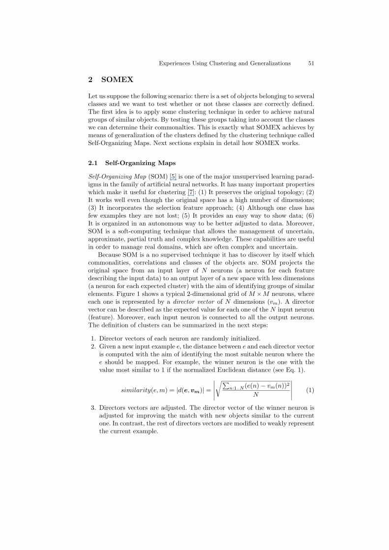
Because SOM is a no supervised technique it has to discover by itself whichcommonalities, correlations and classes of the objects are. SOM projects theoriginal space from an input layer of N neurons (a neuron for each featuredescribing the input data) to an output layer of a new space with less dimensions(a neuron for each expected cluster) with the aim of identifying groups of similarelements. Figure 1 shows a typical 2-dimensional grid of M ×M neurons, whereeach one is represented by a director vector of N dimensions (vm). A directorvector can be described as the expected value for each one of the N input neuron(feature). Moreover, each input neuron is connected to all the output neurons.The definition of clusters can be summarized in the next steps:
1. Director vectors of each neuron are randomly initialized.2. Given a new input example e, the distance between e and each director vector
is computed with the aim of identifying the most suitable neuron where thee should be mapped. For example, the winner neuron is the one with thevalue most similar to 1 if the normalized Euclidean distance (see Eq. 1).
similarity(e,m) = |d(e,vm)| =
∣∣∣∣∣√∑
n:1..N (e(n)− vm(n))2
N
∣∣∣∣∣ (1)
3. Directors vectors are adjusted. The director vector of the winner neuron isadjusted for improving the match with new objects similar to the currentone. In contrast, the rest of directors vectors are modified to weakly representthe current example.
52 A. Fornells et al.
Fig. 1. SOM groups similar elements according to their data features.
4. Steps 2 and 3 are repeated for all training examples until director vectorsare representative enough. Usually their representativeness is determined byestablishing a minimal error value computed as the global sum of distancebetween the set of cases of each cluster and its respectively director vector.Nevertheless, other common criteria is to establish a maximum number ofalgorithm iterations.
5. When the training process ends, step 2 is the procedure used to map thenew input example in the most suitable clusters.
The main drawback of the method is the definition of the training parameters.First aspect is to determine the map size, which is related to the final number ofclusters. Thus, a big size of maps will produce a high number of clusters, whereeach cluster will contain few objects. Conversely, small maps will produce fewclusters containing a lot of objects and, consequently director vectors of clusterswill be overgeneralized. A second aspect to take into account is neighborhoodfactor, which is the influence of each cluster over others. The third aspect isthe learning factor, which determines the convergence of algorithm. High valuesof this factor could produce a random behavior of learning procedure and lowvalues could produce slow ratio of convergence. Finally, the last aspect is thedistance measure used to make comparisons.
To conclude, SOM is a smart technique to identify hidden and complex rela-tionships between elements and also to identify the most relevant features thanksto its knowledge discovery and soft-computing capabilities. This is exactly whatexperts need: to discover relationships between elements to improve the precisionof the classes proposed by them.
2.2 How to Explain a Cluster
Director vectors can be described as the expected values that each attribute hasto satisfy to be classified as belonging to a cluster. However, from the user’spoint of view these tuples do not give an easy intuition of why some objectshave been clustered together. Because of this in [8] we propose to build symbolicexplanations of the clusters with the purpose of justifying why a set of caseshave been clustered together (this is a concept similar to characterization used
Experiences Using Clustering and Generalizations 53
in data mining terminology [9]). Experts found symbolic explanations more un-derstandable than director vectors since the former are constructed using thesame representation language than they used to describe the domain objects.
Thus, we propose to explain a cluster using a symbolic description that isa generalization of all objects contained in the cluster. This generalization isbased on the anti-unification concept [6] although with some differences. Theanti-unification (AU) of a set of objects is a description defined as their mostspecific generalization. The AU contains attributes shared by the set of objectsand where each attribute takes as value the most specific of all the values holdingin the original set. In this paper we only work with the idea of shared attributesamong a set of objects.
Let Mi be a cluster and let c1, ..., cn be the set of objects that belong tothat cluster after the application of SOM to a set of objects. Each object cj isdescribed by a set of attributes A. The explanation Di of why a subset of objectshave been clustered in Mi is built in the following way:
– Di contains attributes which are common to all the objects in Mi. Attributeswith unknown value in some object cj ∈ Mi are not in Di.
– Let ak be an attribute common to all objects in Mi such that ak takessymbolic values on a set Vk. The attribute ak will not be in Di when theunion of the values that ak takes in Mi is exactly Vk.
– An attribute ai takes in Di the union of all values that ai holds in the objectsin Mi.
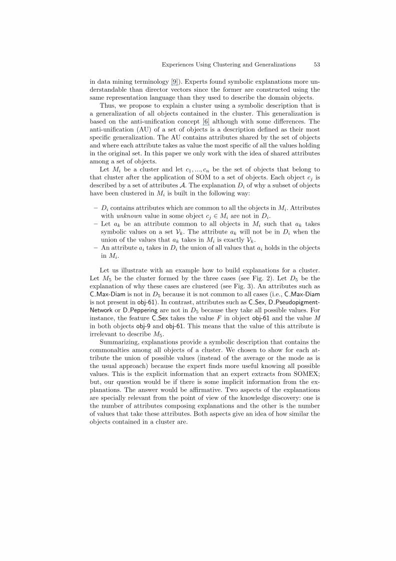
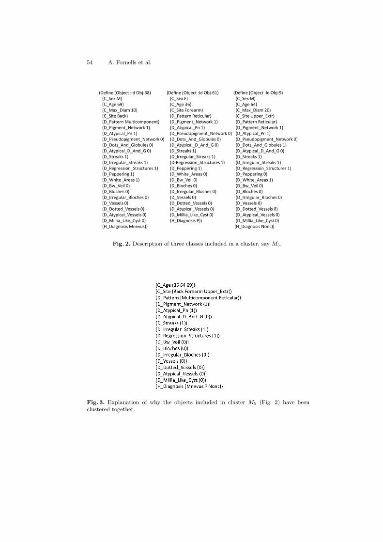
Let us illustrate with an example how to build explanations for a cluster.Let M5 be the cluster formed by the three cases (see Fig. 2). Let D5 be theexplanation of why these cases are clustered (see Fig. 3). An attributes such asC Max-Diam is not in D5 because it is not common to all cases (i.e., C Max-Diamis not present in obj-61). In contrast, attributes such as C Sex, D Pseudopigment-Network or D Peppering are not in D5 because they take all possible values. Forinstance, the feature C Sex takes the value F in object obj-61 and the value Min both objects obj-9 and obj-61. This means that the value of this attribute isirrelevant to describe M5.
Summarizing, explanations provide a symbolic description that contains thecommonalties among all objects of a cluster. We chosen to show for each at-tribute the union of possible values (instead of the average or the mode as isthe usual approach) because the expert finds more useful knowing all possiblevalues. This is the explicit information that an expert extracts from SOMEX;but, our question would be if there is some implicit information from the ex-planations. The answer would be affirmative. Two aspects of the explanationsare specially relevant from the point of view of the knowledge discovery: one isthe number of attributes composing explanations and the other is the numberof values that take these attributes. Both aspects give an idea of how similar theobjects contained in a cluster are.
54 A. Fornells et al.
(Define (Object :Id Obj-68)
(C_Sex M)
(C_Age 69)
(C_Max_Diam 10)
(C_Site Back)
(D_Pattern Multicomponent)
(D_Pigment_Network 1)
(D_Atypical_Pn 1)
(D_Pseudopigment_Network 0)
(D_Dots_And_Globules 0)
(D_Atypical_D_And_G 0)
(D_Streaks 1)
(D_Irregular_Streaks 1)
(D_Regression_Structures 1)
(Define (Object :Id Obj-61)
(C_Sex F)
(C_Age 36)
(C_Site Forearm)
(D_Pattern Reticular)
(D_Pigment_Network 1)
(D_Atypical_Pn 1)
(D_Pseudopigment_Network 0)
(D_Dots_And_Globules 0)
(D_Atypical_D_And_G 0)
(D_Streaks 1)
(D_Irregular_Streaks 1)
(D-Regression_Structures 1)
(D_Peppering 1)
(Define (Object :Id Obj-9)
(C_Sex M)
(C_Age 64)
(C_Max_Diam 20)
(C_Site Upper_Extr)
(D_Pattern Reticular)
(D_Pigment_Network 1)
(D_Atypical_Pn 1)
(D_Pseudopigment_Network 0)
(D_Dots_And_Globules 1)
(D_Atypical_D_And_G 0)
(D_Streaks 1)
(D_Irregular_Streaks 1)
(D_Regression_Structures 1)(D_Regression_Structures 1)
(D_Peppering 1)
(D_White_Areas 1)
(D_Bw_Veil 0)
(D_Bloches 0)
(D_Irregular_Bloches 0)
(D_Vessels 0)
(D_Dotted_Vessels 0)
(D_Atypical_Vessels 0)
(D_Millia_Like_Cyst 0)
(H_Diagnosis Mnevus))
(D_Peppering 1)
(D_White_Areas 0)
(D_Bw_Veil 0)
(D_Bloches 0)
(D_Irregular_Bloches 0)
(D_Vessels 0)
(D_Dotted_Vessels 0)
(D_Atypical_Vessels 0)
(D_Millia_Like_Cyst 0)
(H_Diagnosis P))
(D_Regression_Structures 1)
(D_Peppering 0)
(D_White_Areas 1)
(D_Bw_Veil 0)
(D_Bloches 0)
(D_Irregular_Bloches 0)
(D_Vessels 0)
(D_Dotted_Vessels 0)
(D_Atypical_Vessels 0)
(D_Millia_Like_Cyst 0)
(H_Diagnosis Nonc))
Fig. 2. Description of three classes included in a cluster, say M5.
Fig. 3. Explanation of why the objects included in cluster M5 (Fig. 2) have beenclustered together.
Experiences Using Clustering and Generalizations 55
Concerning the number of attributes, explanations with a high number of at-tributes represent very similar objects whereas explanations with few attributesmean that these objects have few aspects in common. Nevertheless, the numberof values holding the attributes of an explanation also plays a crucial role. Thus,the more values an attribute holds the more irrelevant this attribute is. Noticethat the explanation is built using common attributes and taking as values forthese attributes the union of all values hold by the objects of a cluster. Thus,a common attribute that takes several values, means that has a high variabilityand this attribute is probably not too relevant. Conversely, attributes holdingonly one value represent aspects of the objects that could be taken as candidatesto characterize a cluster.
In short, clusters explained by means of descriptions composed of a highnumber of attributes where each attribute holds one value, can be interpreted asgood clusters in the sense that all the objects included in them are very similar.On the other hand, if the object class is known two situations can happen: 1) allobjects of the cluster belong to the same class, or 2) objects belong to severalclasses. This second situation is the interesting one from the point of view ofknowledge discovery since it means that objects that, in principle, belong todifferent classes are highly similar. For instance, in the melanomas domain suchsituation means that melanomas that dermatologists classified as belonging todifferent clusters, the clustering process of SOMEX has put them together in thesame cluster. The explanation of the corresponding cluster allows the expertsto assess the actual relevance of the commonalties of these a priori differentmelanomas. This should be a starting point from the expert to reconsider thedefinition of classes (for instance, by merging classes to which objects belong).
Similarly, clusters explained by means of descriptions with a lot of attributesholding almost all possible values, can be interpreted as imprecise clusters in thesense that objects in the cluster have not many similarities. From the knowledgediscovery point of view, this situation is interesting when all objects of suchclusters belong to the same class, since it means that although they have beenclassified as belonging to the same class, these objects are not actually similar.
These situations will be illustrated in more detail in the next section whereSOMEX is applied to support dermatologists in the definition and validation ofsome classes of malignant skin lesions.
3 Using SOMEX for Knowledge Discovery
Dermatologists take into account dermoscopic aspects of skin lesions with theaim of determining whether or not it will become a melanoma (malignant skinlesions) prior to lesion excision. The aim of SOMEX is to support dermatologiststo obtain patterns of different kinds of melanomas in situ. First, SOM clusterstogether objects (descriptions of skin lesions) that are similar independently ofthe class. Then, symbolic explanations show dermatologists the common featuresof objects clustered together, allowing them to consider some modifications in
56 A. Fornells et al.
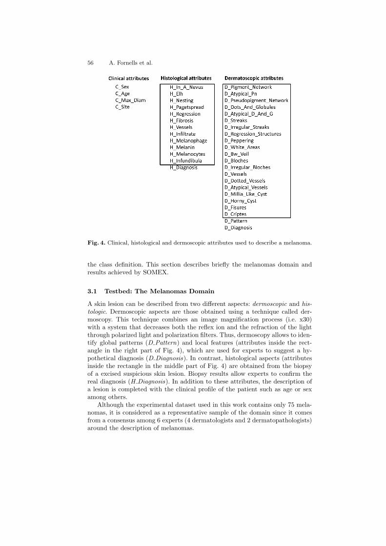
Fig. 4. Clinical, histological and dermoscopic attributes used to describe a melanoma.
the class definition. This section describes briefly the melanomas domain andresults achieved by SOMEX.
3.1 Testbed: The Melanomas Domain
A skin lesion can be described from two different aspects: dermoscopic and his-tologic. Dermoscopic aspects are those obtained using a technique called der-moscopy. This technique combines an image magnification process (i.e. x30)with a system that decreases both the reflex ion and the refraction of the lightthrough polarized light and polarization filters. Thus, dermoscopy allows to iden-tify global patterns (D Pattern) and local features (attributes inside the rect-angle in the right part of Fig. 4), which are used for experts to suggest a hy-pothetical diagnosis (D Diagnosis). In contrast, histological aspects (attributesinside the rectangle in the middle part of Fig. 4) are obtained from the biopsyof a excised suspicious skin lesion. Biopsy results allow experts to confirm thereal diagnosis (H Diagnosis). In addition to these attributes, the description ofa lesion is completed with the clinical profile of the patient such as age or sexamong others.
Although the experimental dataset used in this work contains only 75 mela-nomas, it is considered as a representative sample of the domain since it comesfrom a consensus among 6 experts (4 dermatologists and 2 dermatopathologists)around the description of melanomas.
Experiences Using Clustering and Generalizations 57
With our experiments we want to support dermatologists in finding 1) howto dermoscopically describe histologic classes, and 2) to test whether or not his-tologic classes have been correctly defined. The next section describes the con-ditions under which experiments have been performed and also some interestingresults obtained from SOMEX.
3.2 Experiments
Since our purpose was to support dermatologists in determining the dermoscopicfeatures that describe the histologic classes, we only focused on the clinical anddermoscopic attributes (see Fig. 4). We also included the histological class rep-resented by the H Diagnosis attribute, i.e. the histologic class considered by theexperts. Dermatologists defined the following histologic classes: LTG M, LMM,nonc, PL M, P, P LTG, Mnevus, and SKlMM.
Bearing in mind this information, we performed several SOM configurationsin order to find out interesting results. SOM was tested using several map sizes of2-dimensions (3×3, 4×4 and 5×5 to analyze several data dispersions), two differ-ent distance measures (normalized Euclidean distance and normalized Hammingdistance) and 10 random seeds (to minimize the random effects of initialization).The learning factor, the neighbor factor and maximum iterations were set re-spectively to values from 0.6 to 0.01, from M to 1 and 500 iterations by neuron.
3.3 Discussion of the Results
Independently of the map size and of the distance measure used to build theclusters, SOMEX results showed that the definition of histological classes shouldbe adjusted. The reason is that most clusters include objects belonging to severalhistologic classes and explanations show that these objects have a lot of commonaspects. This is reflected in the fact that most of explanations are very specific,i.e. they have a lot of common attributes holding a unique value. Notice thatthe high number of common features with only one value, the more similar theobjects are. Conversely, explanations with features holding more than one valuemean that, although objects are described by similar features, they have a lotof variability and they are not so similar.
The use of clustering techniques allowed a natural group of similar objects.Then by means of generalizations SOMEX explains why a subset of objects havebeen clustered together. Results show that some melanomas that dermatologistsconsidered as belonging to different classes actually are not so different sincethey belong to the same cluster. Moreover, the explanation supports the user indiscovering the common aspects and also characteristics that are different amongobjects of a same cluster. In fact, this provides them a clue to reconsider thedefinition of histological classes. Prior to SOMEX experiments, dermatologistshad the hypothesis that the pattern (feature D Pattern) of a skin lesion could bean important aspect to determine the classification of a lesion. As we will detaillater, from SOMEX experiments we point out that the pattern, at least taken itisolated from other characteristics, is not enough for classification.
58 A. Fornells et al.
A conclusion from the experiments is that criteria used by dermatologistswhen defining histologic classes (H Diagnosis) do not take into account all as-pects describing a melanoma. In fact, clusters almost always contain melanomasof several histological classes. thus, from the predictivity point of view, clustersare not appropriate. However, when experts analyze the explanations of clustersthey find them interesting despite their entropy. Experts noted that attributesshared by melanomas into a cluster usually are those considered as importantfor experts (for instance, D dots and globules or D Pigment network). For thisreason we prefer to show the analysis that experts performed of the SOMEXexplanations instead of giving predictivity measures.
Experiments produced three types of clusters: 1) clusters with a reasonablenumber of objects belonging to different classes, 2) clusters with few objectsbelonging all of them to the same class, and 3) clusters with few objects ofseveral classes. SOMEX results show that there are not clusters with a highnumber of objects belonging all of them to the same class nor clusters with fewobjects with a general explanation. Let us to analyze in more detail the SOMEXresults.
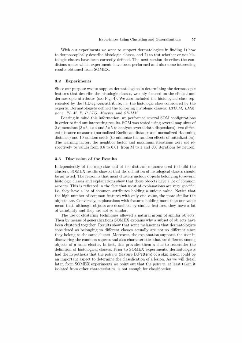
Example 1 Let us suppose the cluster M15 containing 10 objects. The expla-nation of this cluster can be seen in Fig. 5. Concerning the number of attributesof the explanation, we see that there is a subset of 15 attributes (from the 28composing a complete description of an object) shared by all the objects of thecluster. Focusing on values of these common attributes, we seen that most ofthem have an unique value, meaning that the explanation is specific enough.
In the explanation of the cluster M15 there are five attributes with morethan one value: C Age, C Max-Diam, C Site, D Pattern and H Diagnosis. Two ofthese attributes, C age and C Max-Diam are numerical and currently we cannotextract any conclusion from them. This is because explanations are not able tohandle with continuous attributes. Dermatologists plan to establish some kind ofdiscretization to establish ranges of equivalent values for these attributes. Con-cerning the values of attributes C Site and D Pattern, SOMEX shown that theyhold a lot of values (almost all the possible values in the case of D Pattern). Inparticular, the role of a lesion pattern as potential relevant aspect of a melanomaseems to be compromised according to the explanation of this cluster.
Finally, an interesting analysis can be carried out from values of H Diagnosis.This is, in fact, the classification proposed by dermatopathologists; therefore, ac-cording to their criterion objects of M15 belong to five different classes (LTG M,nonc, PL M, P LTG and Mnevus). However SOMEX show that these objectshave a high similarity and the explanation suggests to dermatologists a possibleanalysis of the relevance of object commonalties so as that they should reconsiderthe criteria used to classify objects in different histological classes. An analysisof the differences among the objects in M15 could also clarify the class definition.
Experiences Using Clustering and Generalizations 59
Fig. 5. Explanations justifying the clusters M15 and M24.
Example 2 The explanation of cluster M24 is composed of three objects withthe same histological class. There are 4 multi-valued attributes: C Age, C Max-Diam (both numerical), C Site, and D Pattern. Globally this explanation seemsa good partial characterization for the class PL M since a further analysis of thenumerical values could produce a more specific explanation. An important as-pect to take into account is that the attribute D Pattern has two possible values,unspecific and reticular. The importance of this fact is that according to SOMEXresults, dermatologists should consider the possibility to reject D Pattern as rel-evant for classifying a melanoma, since this feature holds different values inobjects of the same histological class. In our current experiments we do not con-sider neither the relationship among attributes nor the weight of some attributesin order to bias the clustering. A possibility is that the pattern of a melanomacould be relevant in relation to the value of any other attribute.
Example 3 The cluster M5 shown in Fig. 2 is an example of a small one withelements of several histological classes (in fact, each object belongs to a differentclass). The explanation of this cluster is shown in Fig. 3. This explanation isspecific since it is composed by 17 common attributes and all of them except4 hold an unique value. Notice that as in previous examples, attributes withmultiple values are C Age, C Site, D Pattern and H Diagnosis. Once again the
60 A. Fornells et al.
conclusion should be to reconsider the definition of histological classes and toanalyse the relevance of attributes that dermatopathologists used to define them.
An important point from the application of SOMEX is that symbolic ex-planations obtained from clusters give to dermatopathologists descriptions ofgroups of melanomas that they commonly recognize as different. For instance,the explanation for cluster M15 (see Fig. 6) describes lesions that under der-moscopy presents both dots and globules and typical pigment network (noticethat all other features have as value 0, meaning absence). This description isclearly recognized from the dermatological point of view since they provided usthe picture shown in Fig. 5 left, that corresponds to a lesion belonging to clus-ter M15 (in particular, it is the object <Obj-11>. Similarly, the explanation ofcluster M24 describes lesions, completely different that those of cluster M15. Inparticular, lesions in cluster M24 have as unique feature the presence of typi-cal vessels, dermatologists recognized lesions such as the shown in Fig. 5 right(corresponds to <Obj-13>) of cluster M24). Summarizing, SOMEX provides anatural clustering of objects and the use of symbolic explanations supports der-matologists in analysing the correctness of the clusters and also in redefiningsome of the histological classes they propose.
4 Related Work
Clustering techniques are a smart way to extract relationships from huge datasets. Consequently, this useful property has been widely used in medical do-mains such as the one in which this work addresses. The focus of works foundin the literature mainly depend on the data topology and the usage of extractedrelations from analysis. There are melanoma studies focused in the identifica-tion of relationships between malignant melanoma and familiar or hereditarytumors (i.e. breast cancer, ovarian cancer, colon cancer, pancreatic cancer) suchas in [10]. Other works analyze thousands of genes with the aim of extractingthe ’guilty’ genes [11, 12] related to the cancer. Anyway, both approaches helpexperts to be aware and detect melanoma formation in early stages. The maindifference between our work and others is that we use SOMEX to help expertsto improve their melanoma definition and classification. This improvement willhas as a consequence an increment of the precision in melanoma diagnosis.
The idea of using symbolic descriptions for characterizing clusters can beinterpreted as a memory organization. In this sense, our approach is similar toPerner’s [13] and Abidi’s [14] works. Perner proposes to organize the cases fol-lowing a hierarchy similar to a decision tree where each node ci is described by asymbolic description (prototype). Each symbolic description subsumes descrip-tions of all nodes included in the subtree rooted by ci until reach the leaves thatcontain the individual cases. Somehow, nodes of that hierarchy could be inter-preted as explanations, i.e. why a subset of domain objects (cases) have beengrouped under a node. This work relies on the context of case-based reasoningwhere the main aim is to classify a new problem, therefore prototypes are used
Experiences Using Clustering and Generalizations 61
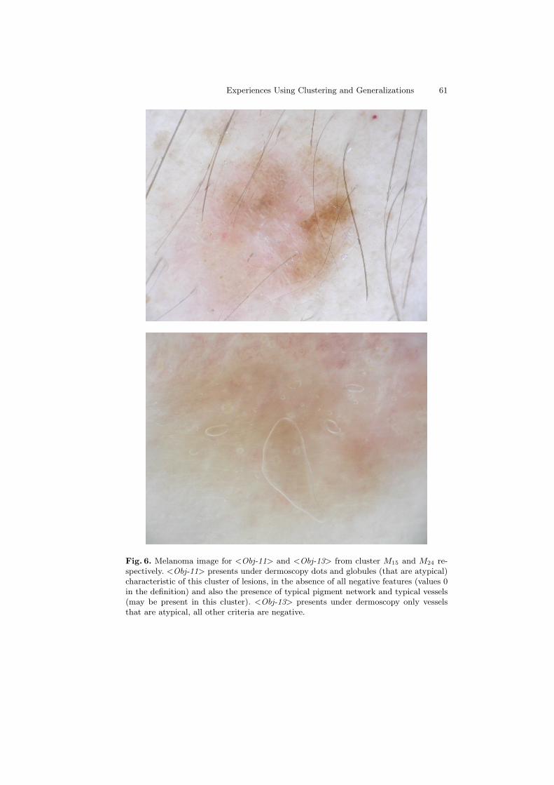
Fig. 6. Melanoma image for <Obj-11> and <Obj-13> from cluster M15 and M24 re-spectively. <Obj-11> presents under dermoscopy dots and globules (that are atypical)characteristic of this cluster of lesions, in the absence of all negative features (values 0in the definition) and also the presence of typical pigment network and typical vessels(may be present in this cluster). <Obj-13> presents under dermoscopy only vesselsthat are atypical, all other criteria are negative.
62 A. Fornells et al.
to select a subset of cases to solve a new problem. In previous works such as [15],we also proposed the use of explanations during the retrieval phase of the case-based reasoning, nevertheless in SOMEX the use of explanations is different.SOMEX does not take into account the class class of cases (i.e. the H Diagnosisattribute in the melanoma domain), since we assume that these classes could nobe accurately defined.
The procedure proposed by Abidi et al [14] is similar to SOMEX because theyproduce rules that describe objects included in a cluster without using the classinformation. Firstly, domain objects are clustered according to their similarity,secondly continuous values are discretized, and finally they use rough sets togenerate symbolic rules for each cluster. In fact, the explanation generated bySOMEX could also be interpreted as a domain rule (as we suggested in [16]).
The basic difference among SOMEX and the works above is the use of ex-planations. Perner uses symbolic descriptions to organize the memory of caseswith the purpose of achieving a more efficient retrieval. Abidi et al. propose aprocedure to obtain symbolic rules from clusters. In SOMEX, explanations areused as a basis for knowledge discovery. By analyzing the explanation of clus-ters, experts can compare the classification they proposed with the classificationobtained by the clustering method. Because of the clustering method do no takeinto account the class label, differences among both expert and explanationsclassification give some clues for redefining the classes.
5 Conclusions and Further Work
This paper describes SOMEX and how it can be used for knowledge discovery.SOMEX is a combination of clustering and generalizations, to support derma-tologists in discovering knowledge about in situ melanomas. The purpose ofdermatologists was to define several classes of melanomas and finding dermo-scopic features characterizing these classes. SOMEX supported dermatologistsin focusing on groups of similar objects and commonalties among them. In par-ticular, dermatologists can analyze the entropy of clusters, i.e. why melanomasthat they consider as belonging to different histological classes are actually sosimilar. Dermatologists can also analyze the relevance of attributes for classifi-cation. A particular example is the melanoma pattern, considered as a relevantaspect prior to SOMEX application and that results proved that taken in isola-tion is not a good classifier due to its variability on the objects of a cluster.
As future work we plan to modify some parameters of the clustering in twoways. Firstly we want to confirm the relevance of pattern and we plan to weightsome of these features in order to highlight the relationship of this feature withothers. A second kind of experiments could be focused on enforcing the numberof clusters and experimentally determining the best group of melanomas in orderto empirically define their histological classes. Finally, from the point of view ofthe explanations, we could analyze relations between them.
Experiences Using Clustering and Generalizations 63
Acknowledgments
We would like to thank the Spanish Government for the support in MID-CBRproject under grant TIN2006-15140-C03 and the Generalitat de Catalunya forthe support under grants 2005SGR-302 and 2007FIC-0976. We would like tothank Enginyeria i Arquitectura La Salle of Ramon Llull University for thesupport to our research group as well.
On the other hand, we also would like to thank the clinicians involved inthe confection of the dataset: Dr Paolo Carli (Dermatologist), Dr Vinzenzo diGiorgi (dermatologist), Dr Daniela Massi (dermatopathologist) from the Uni-versity of Firenze; Dr Josep Malvehy (dermatologist and co-author), Dr SusanaPuig (dermatologist and co-author) and Dr Josep Palou (dermatopathologist)from Melanoma Unit in Hospital Clinic i Provincial de Barcelona. Part of thework performed by S. Puig and J. Malvehy is partially supported by: Fondode Investigaciones Sanitarias (FIS), grant 0019/03 and 06/0265; Network ofExcellence, 018702 GenoMel from the CE.
References
1. Friedman, R.J., Rigel, D.S., Kopf, A.W.: Early detection of malignant melanoma:The role of physician examination and self-examination of the skin. Ca-A CancerJ Clinicians 35 (1985) 130–151
2. Puig, S., Argenziano, G., Zalaudek, I., Ferrara, G., Palou, J., Massi, D., Hofmann-Wellenhof, R., Soyer, H., Malvehy, J.: Melanomas that failed dermoscopic detec-tion: a combined clinicodermoscopic approach for not missing melanoma. DermatolSurg 33(10) (2007) 1262–1273
3. Hofmann-Wellenhof, R., Blum, A., Wolf, I., Zalaudek, I., Piccolo, D., Kerl,H., Garbe, C., Soyer, H.: Dermoscopic classification of clark’s nevi (atypicalmelanocytic nevi). Clin Dermatol 20(3) (2002) 255–258
4. Argenziano, G., Zalaudek, I., Ferrara, G., Hofmann-Wellenhof, R., Soyer, H.: Pro-posal of a new classification system for melanocytic naevi. Br J Dermatol 157(2)(2007) 217–227
5. Kohonen, T.: Self-Organizing Maps. 3rd edn. Springer (2000)
6. Armengol, E., Plaza, E.: Bottom-up induction of feature terms. Machine Learning41(1) (2000) 259–294
7. Haykin, S.: Neural Networks: A Comprehensive Foundation. 2nd edn. PrenticeHall (1999)
8. Corral, G., Armengol, E., Fornells, A., Golobardes, E.: Data security analysis usingunsupervised learning and explanations. In Corchado, E., Corchado, J., Abraham,A., eds.: Innovations in Hybrid Intelligent Systems. Volume 44., Springer-Verlag(2007) 112–119
9. Witten, I.H., Frank, E.: Data Mining: Practical Machine Learning Tools and Tech-niques. Second edn. Morgan Kaufmann Series in Data Management Systems. Mor-gan Kaufmann (2005)
10. Stefano, P., Fabbrocini, G., Scalvenzi, M., Emanuela, B., Pensabene, M.: Malignantmelanoma clustering with some familiar/hereditary tumors. Annals of Oncology3 (2002) 100
64 A. Fornells et al.
11. Eisen, M., Spellman, P., Brown, P., Botstein, D.: Cluster analysis and displayof genome-wide expression patterns. National Academy Scientific 25(95) (1998)1486314868
12. Dang, H., T. Le, T.S., Levy, J.: Integrating database information in microarrayexpression analyses: Application to melanoma cell lines profiled in the nci60 dataset. Journal of Biomolecular Techniques 13 (2002) 199–204
13. Perner, P.: Case-base maintenance by conceptual clustering graphs. EngineeringApplications of Artificial Intelligence 9 (2006) 381 – 393
14. Abidi, S.S.R., Hoe, K.M., Goh, A.: Analyzing data clusters: A rough sets approachto extract cluster-defining symbolic rules. In: IDA ’01: Proceedings of the 4thInternational Conference on Advances in Intelligent Data Analysis, London, UK,Springer-Verlag (2001) 248–257
15. Fornells, A., Armengol, E., Golobardes, E.: Explanation of a clustered case memoryorganization. In: Artificial Intelligence Research and Development. Volume 160.,IOS Press (2007) 153–160
16. Armengol, E.: Usages of generalization in cbr. In Weber, R., Richter, M.M.,eds.: Case-based Reasoning and Development. Number 4626 in Lecture Notes inArtificial Intelligence, Springer-Verlag (2007) 31–45
Vitae
Dr. Albert Fornells received his Ph.D. degree in Computer Science by Uni-versitat Ramon Llull, Spain, in 2007. He is a member of the Research Groupin Intelligent Systems from the university since 2000, and an associate professorsince 2003. His research interests include data mining and knowledge discovery,self-organizing maps, case-based reasoning, soft computing, soft-case based rea-soning and Artificial Intelligence applied in Health Care (Medicine).
Dr. Eva Armengol received her Ph.D. in Computer Science by the Universi-tat Politecnica de Catalunya in 1997. She is now a permanent scientist of theArtificial Intelligence Institute (IIIA). Her research has been mainly focused onknowledge representation, machine learning and case-based reasoning. Currentlyshe focuses her research on how to explain the results of learning methods.
Dr. Elisabet Golobardes received her Ph.D. in Computer Science by Univer-sitat Ramon Llull in 1998. She is a member of the Research Group in IntelligentSystems (GRSI) of Enginyeria i Arquitectura La Salle - Universitat Ramon Llullsince 1994. Her research interests are mainly focused on Case-Based Reasoning,Soft-Computing, Clustering, Machine Learning, Computer Aided Systems, Ar-tificial Intelligence applied in Health Care (Medicine) and applied in NetworkSecurity.
Dr. S. Puig graduated in 1988 at the Faculty of Medicine of Barcelona (Spain)and obtained her specialization diploma in Dermatology and Venereology in1992 and the doctoral degree in 2000. She is a dermatologist, the director ofthe research program of the melanoma Unit at the University Hospital Clinicof Barcelona. She is co-director of the Oncology and Dermoscopy groups at the

Experiences Using Clustering and Generalizations 65
CILAD. Special research areas are dermoscopy, digital follow-up of melanocytictumors and genetics of melanoma. She is member of the board of the Interna-tional Dermoscopy Society, Genomel and EORTIC.
Dr. Josep Malvehy graduated in 1992 at the Faculty of Medicine of Barcelonain Spain and obtained his specialization diploma in Dermatology and Venereol-ogy in 1996 and the doctoral degree in 2006. He is the director of the melanomaUnit at the University Hospital Clinic of Barcelona (Spain). His main researchfield is the melanocytic tumors and melanoma and particularly the study of newdiagnostic tools for the evaluation in vivo of skin tumors based in dermoscopy,confocal microscopy and other techniques.


